Embed Size (px)
Citation preview

17
Metals for Biomedical Applications
Hendra Hermawan, Dadan Ramdan and Joy R. P. Djuansjah Faculty of Biomedical Engineering and Health Science, Universiti Teknologi Malaysia
Malaysia
1. Introduction
In modern history, metals have been used as implants since more than 100 years ago when Lane first introduced metal plate for bone fracture fixation in 1895 (Lane, 1895). In the early development, metal implants faced corrosion and insufficient strength problems (Lambotte, 1909, Sherman, 1912). Shortly after the introduction of the 18-8 stainless steel in 1920s, which has had far-superior corrosion resistance to anything in that time, it immediately attracted the interest of the clinicians. Thereafter, metal implants experienced vast development and clinical use. Type of metal used in biomedical depends on specific implant applications. 316L type stainless steel (316L SS) is still the most used alloy in all implants division ranging from cardiovascular to otorhinology. However, when the implant requires high wear resistance such as artificial joints, CoCrMo alloys is better served. Table 1 summarized the type of metals generally used for different implants division.
Division Example of implants Type of metal
Cardiovascular Stent Artificial valve
316L SS; CoCrMo; Ti Ti6Al4V
Orthopaedic Bone fixation (plate, screw, pin) Artificial joints
316L SS; Ti; Ti6Al4V CoCrMo; Ti6Al4V; Ti6Al7Nb
Dentistry Orthodontic wire Filling
316L SS; CoCrMo; TiNi; TiMo AgSn(Cu) amalgam, Au
Craniofacial Plate and screw 316L SS; CoCrMo; Ti; Ti6Al4V Otorhinology Artificial eardrum 316L SS
Table 1. Implants division and type of metals used
Metallic biomaterials are exploited due to their inertness and structural functions; they do not possess biofunctionalities like blood compatibility, bone conductivity and bioactivity. Hence, surface modifications are required. Improving their bone conductivity has been done by coating with bioactive ceramics like hydroxyapatite (Habibovic, 2002), or blood compatibility by coating with biopolymers (Lahann, 1999). Nowadays, large number of metallic biomaterials composed of nontoxic and allergy-free elements are being developed. Even more, a new type of biodegradable metals has been proposed as temporary implants (Hermawan, 2009). Generally, all metal implants are non-magnetic and high in density. These are important for the implants to be compatible with magnetic resonance imaging (MRI) techniques and to be
www.intechopen.com
Biomedical Engineering – From Theory to Applications
412
visible under X-ray imaging. Most of artificial implants are subjected to loads, either static or repetitive, and this condition requires an excellent combination of strength and ductility. This is the superior characteristic of metals over polymers and ceramics. Specific requirements of metals depend on the specific implant applications. Stents and stent grafts are implanted to open stenotic blood vessels; therefore, it requires plasticity for expansion and rigidity to maintain dilatation. For orthopaedic implants, metals are required to have excellent toughness, elasticity, rigidity, strength and resistance to fracture. For total joint replacement, metals are needed to be wear resistance; therefore debris formation from friction can be avoided. Dental restoration requires strong and rigid metals and even the shape memory effect for better results. In overall, the use of biomaterials in clinical practice should be approved by an authoritative body such as the FDA (United States Food and Drug Administration). The proposed biomaterial will be either granted Premarket Approval (PMA) if substantially equivalent to one used before FDA legislation of 1976, or has to go through a series of guided biocompatibility assessment.
2. Common metals used for biomedical devices
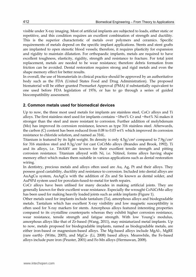
Up to now, the three most used metals for implants are stainless steel, CoCr alloys and Ti alloys. The first stainless steel used for implants contains ~18wt% Cr and ~8wt% Ni makes it stronger than the steel and more resistant to corrosion. Further addition of molybdenum (Mo) has improved its corrosion resistance, known as type 316 stainless steel. Afterwards, the carbon (C) content has been reduced from 0.08 to 0.03 wt% which improved its corrosion resistance to chloride solution, and named as 316L. Titanium is featured by its light weight. Its density is only 4.5g/cm3 compared to 7.9g/cm3 for 316 stainless steel and 8.3g/cm3 for cast CoCrMo alloys (Brandes and Brook, 1992). Ti and its alloys, i.e. Ti6Al4V are known for their excellent tensile strength and pitting corrosion resistance. Titanium alloyed with Ni, i.e. Nitinol, forms alloys having shape memory effect which makes them suitable in various applications such as dental restoration wiring. In dentistry, precious metals and alloys often used are Au, Ag, Pt and their alloys. They possess good castability, ductility and resistance to corrosion. Included into dental alloys are AuAgCu system, AuAgCu with the addition of Zn and Sn known as dental solder, and AuPtPd system used for porcelain-fused-to-metal for teeth repairs. CoCr alloys have been utilised for many decades in making artificial joints. They are generally known for their excellent wear resistance. Especially the wrought CoNiCrMo alloy has been used for making heavily loaded joints such as ankle implants (Figure 1). Other metals used for implants include tantalum (Ta), amorphous alloys and biodegradable metals. Tantalum which has excellent X-ray visibility and low magnetic susceptibility is often used for X-ray markers for stents. Amorphous alloys featured interesting properties compared to its crystalline counterparts whereas they exhibit higher corrosion resistance, wear resistance, tensile strength and fatigue strength. With low Young’s modulus, amorphous alloys like that of Zr-based (Wang, 2011), may miniaturized metal implants. Up to now, metals proposed for biodegradable implants, named as biodegradable metals, are either iron-based or magnesium-based alloys. The Mg-based alloys include MgAl-, MgRE (rare earth)- (Witte, 2005), and MgCa- (Li, 2008) based alloys. Meanwhile, the Fe-based alloys include pure iron (Peuster, 2001) and Fe-Mn alloys (Hermawan, 2008).
www.intechopen.com
Metals for Biomedical Applications
413
Fig. 1. A set of ankle implants (Courtesy of MediTeg, UTM).
3. Structure and property of metals
3.1 Microstructure of metal and its alloys
When molten metals are cooled into a solid state, the atoms rearrange themselves into a crystal structure. There are three basic crystal structures for most metals: (1) body-centered cubic, (2) face-centered cubic, and (3) hexagonal close-packed. Each structure has different properties and shows distinct behaviour when subjected under loading in the application. Under external force a crystal undergoes elastic deformation. When the force is removed, it returns to its original shape. However, if the force is increased beyond its elastic limit, the crystal undergoes plastic or permanent deformation, and it does not return to its original shape even after the removal of the applied force. Imperfections usually exist in metals include interstitial atom, impurity, dislocations, grain boundaries, and pores. Dislocation is a defect which could explain the discrepancy between the actual strength of metals and the theoretical calculations based on molecular dynamics. After the invention of electron microscope, many scientists have directly observed the existence of dislocation. Since then, dislocation theory has evolved and explains many of the physical and mechanical phenomena in metals. Another important type of defect is the grain boundary. The mechanical properties of metals are significantly influenced by the size of their grain. At ambient temperature, metals with large grain size generally have a low strength and hardness, and also low in ductility. Since grain boundaries hinder the dislocations movement, they also influence the strain hardening process to increase the strength and ductility of metals. Pure metals have relatively limited properties; however these properties can be enhanced by alloying the metals. Most of the metals used in engineering applications are in the form of their alloy. Most alloys consist of two or more solid phase in the form of either solid
www.intechopen.com
Biomedical Engineering – From Theory to Applications
414
solutions or intermetallic compounds that depend on the alloying composition and temperature. A phase is defined as a homogenous portion in a material that has its own physical and chemical characteristics and properties. Every pure metal is considered as a phase, as also is every solid solution and intermetallic compound. Alloying a metal with finely dispersed particles as a second-phase is one of the important method of strengthening alloys and enhancing their properties. The second-phase particles present as obstacles to the movement of dislocations thus increase the overall strength and hardness of the alloys.
3.2 Physical and mechanical properties of metals
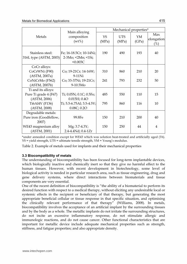
One important criterion in metals selection is the consideration of their physical properties, such as density, melting point, specific heat, thermal conductivity, thermal expansion and corrosion. Density of a metal plays a significant role on the specific strength and specific stiffness, which are the ratio of strength-to-weight and stiffness-to-weight, respectively. For many applications, one of the most important considerations is their deterioration by corrosion. Corrosion of metal depends on the metals composition and the corrosive media in the surrounding environment. The most common and easiest way of preventing corrosion is the careful selection of metals once the corrosion environment has been characterized. Nonferrous metals, stainless steel, and non-metallic materials generally have high corrosion resistance due to the presence of protective passive layer. Titanium develops a film of titanium oxide, TiO2. A similar phenomenon also occurs in stainless steels due to the presence of chromium in the alloy that develops chromium oxide layer on the surfaces. If the protective film is broken and exposes the metal underneath, a new oxide film begins to form for further protection. Unlike the physical properties, mechanical properties of metal are the behaviour of metals that measured under the effect of external forces. Tension test is the most common method to determine the mechanical properties of materials, such as strength, ductility, toughness, elastic modulus, and strain hardening capability. The specimen used in this test usually is prepared according to ASTM specifications. Another important mechanical property of metal is the hardness which gives a general indication of its resistance to localize plastic deformation. Several test methods which use different indenter materials and shapes have been developed to measure the hardness of metals. Table 2 shows mechanical properties of some alloys used for implant. It also shows chemical composition of the alloy which is a determined factor for the formation of microstructure and phases, thus their properties, i.e. mechanical properties. For example, the addition of Al and V into pure Ti greatly increase its tensile strength. Beside composition, metallurgical state and synthesis process of the metals change their mechanical properties, i.e. annealed condition has better ductility than that of cold worked and cast metal implants usually possess lower strength than those made by forging. Different from the breakdown in the tension test where the specimen is subjected under gradual increase of loading until fractures, failure of a component practically occurs after a lengthy period of repeated stress or strain cycling. This phenomenon is called fatigue failure and responsible for the majority of failures in many mechanical components. To avoid this kind of failure, the stress level should be reduced to a level which the material can be subjected without fatigue failure, regardless of the number of cycle. The maximum level of loading stress is known as the endurance limit or fatigue limit.
www.intechopen.com
Metals for Biomedical Applications
415
Metals Main alloying composition
(wt%)
Mechanical properties*
YS (MPa)
UTS (MPa)
YM (GPa)
Max elongation
(%)
Stainless steel: 316L type (ASTM, 2003)
Fe; 16-18.5Cr; 10-14Ni;
2-3Mo; <2Mn; <1Si; <0.003C
190
490
193
40
CoCr alloys: CoCrWNi (F90) (ASTM, 2007a)
CoNiCrMo (F562) (ASTM, 2007b)
Co; 19-21Cr; 14-16W;
9-11Ni Co; 33-37Ni; 19-21Cr;
9-10.5Mo
310
241
860
793
210
232
20
50
Ti and its alloys: Pure Ti grade 4 (F67)
(ASTM, 2006) Ti6Al4V (F136) (ASTM, 2008)
Ti; 0.05N; 0.1C; 0.5Fe;
0.015H; 0.4O Ti; 5.5-6.75Al; 3.5-4.5V;
0.08C; 0.2O
485
795
550
860
110
116
15
10
Degradable metals: Pure iron (Goodfellow,
2007) WE43 magnesium alloy
(ASTM, 2001)
99.8Fe
Mg; 3.7-4.3Y;
2.4-4.4Nd; 0.4-1Zr
150
150
210
250
200
44
40
4
*under annealed condition except for WE43 which was solution heat-treated and artificially aged (T6). YS = yield strength, UTS = ultimate tensile strength, YM = Young’s modulus.
Table 2. Example of metals used for implants and their mechanical properties
3.3 Biocompatibility of metals
The understanding of biocompatibility has been focused for long-term implantable devices, which biologically inactive and chemically inert so that they give no harmful effect to the human tissues. However, with recent development in biotechnology, some level of biological activity is needed in particular research area, such as tissue engineering, drug and gene delivery systems, where direct interactions between biomaterials and tissue components are very essential. One of the recent definition of biocompatibility is “the ability of a biomaterial to perform its desired function with respect to a medical therapy, without eliciting any undesirable local or systemic effects in the recipient or beneficiary of that therapy, but generating the most appropriate beneficial cellular or tissue response in that specific situation, and optimising the clinically relevant performance of that therapy” (Williams, 2008). In metals, biocompatibility involves the acceptance of an artificial implant by the surrounding tissues and by the body as a whole. The metallic implants do not irritate the surrounding structures, do not incite an excessive inflammatory response, do not stimulate allergic and immunologic reactions, and do not cause cancer. Other functional characteristics that are important for metallic device include adequate mechanical properties such as strength, stiffness, and fatigue properties; and also appropriate density.
www.intechopen.com
Biomedical Engineering – From Theory to Applications
416
Since many applications of metallic devices are for the structural implant, metal biocompatibility is of considerable concern since metals can corrode in an in vivo environment. The corrosion of metallic implant gives adverse effects to the surrounding tissues and to the implant itself. It produces chemical substances that harmful for human organs and deteriorates the mechanical properties of the implant. Therefore, corrosion resistance of a metallic implant is an important aspect of its biocompatibility. Even though, in special cases, metals which can degrade are proposed for temporary implants but certainly without ignoring the biocompatibility requirement (Hermawan, 2010, Witte, 2009).
4. Processing of metals
4.1 Primary processes
In general, primary process of metals include the processing ingot to mill products in the case of wrought alloys, and casting process in the case of cast alloy. In addition, the primary products of metals can also be produced by powder metallurgy. Processing of implant alloys is thought to be a very expensive process, which involves complex process of production, especially for the case of Ti alloy. The main reason for this condition is due to the high reactivity of the alloys, therefore special handlings are required to perform their process of production. These conditions also induce the necessity in developing new material which is easier to be processed (Zhuka, 2007). Thermo-mechanical process (TMP) is the most implemented primary fabrication process to convert ingot into mill products. It serves two functions; to produce certain shape of product such as slab, bloom and billet, and to improve the mechanical properties of the initial ingot materials by grain size refinement and the production of more uniform microstructure (Weiss, 1999). TMP of Ti alloys and similarly with other alloys involves several stages of processes. Forging is typically become the first process prior to other TMP. The selection of the type of TMP after forging depends on the mill product which is going to be produced, whether it is a billet, bar, plate or sheet (Campbel, 2006). Determination of temperature is a crucial step for TMP (Liu, 1995, Germain, 2008, Ming-Wei, 2007), which determine the properties of the alloys and therefore different alloy will be treated at different temperature. As example, during 304 stainless steels‘s TMP, instability bands were found when the process temperature are below 1100°C, 1000°C, 800°C after hammer forging, rolling and press forging, respectively. It was suggested that the ideal process can be performed at 1200°C to obtain a defect-free microstructure (Venugopal, 1995). Other important parameters are degree of deformation and phase composition. It was reported that increase in degree of plastic deformation during forging of dual phase Ti alloys results in lower fatigue strength (Kubiak, 1998). However the decrease in the fatigue strength is smaller for the case of forging process in the beta range as compared to (┙+┚) range. In the case of phase composition, it has long been recognized that different phase has different ability to be deformed, therefore different phase composition will have different performance during and after TMP. As an example, it was reported that the flow-ability of dual phase Ti alloys depends on the beta phase grain size and volume fraction (Hu, 1999). As mentioned earlier, casting process of implant alloy is relatively more difficult than TMP of wrought alloys. The reason is due to the high reactivity of the alloys, especially for the case of Ti alloys, which easily reacts with both the atmosphere and the cast mold (Campbel, 2006). However recent progress shows the used of investment casting has increased which is followed with hot iso-static pressing (HIP) process as the primary process for the production
www.intechopen.com
Metals for Biomedical Applications
417
of implant alloys. In addition, different with other alloys, cast products of Ti alloys are generally comparable with the wrought products and in certain case can be superior (ASM, 1998a). On the other hand, Co base alloys are considered to have a better cast-ability than both Ti and stainless steel alloys. These alloys shows several important characteristics for casting process such as good fluidity, low melting points, freedom from dissolved-gas defects and low alloy losses due to oxidation (ASM, 1998c). Improvement of cast-ability and cast product of this alloy can be achieved by additional alloying element such as carbon and vacuum melting process. It was reported that additional carbon content up to 0.5wt% lower the melting temperature (increase cast-ability) and in turn produce finer grain size as compared to binary CoCr alloys (Black, 1998). Another primary fabrication process of metal implant is powder metallurgy. The process includes blending and mixing of ingredient materials, compaction, sintering and in most cases followed with HIP process. This process is relatively expensive; therefore it is suitable for the production of highly loaded implants like femoral stems of total hip prostheses (Black, 1998). One of the important requirements for implant materials is porosity which is expected in the range between 20-50% volume fractions (Dewidar, 2007). This condition can be achieved by controlling the sintering process parameters. In addition, improvement in the mechanical properties of implant by this method can be achieved by conducting HIP process after sintering by decreasing defects like gas or shrinkage pores.
4.2 Advanced processes
There are several processes which can be considered as advanced processes in the manufacturing of implant materials, such as superplastic deformation, isothermal forging and directed metal deposition. They offer an improvement in the process of production as well as achieveing a better quality of product. Superplastic deformation (SPD) is an advanced forming process where higher degree of deformation applied to form complex shape of product whereas low rate forming process is required (Krishna, 1997). Dual phase materials have potential to be treated by SPD process with the additional requirement that the materials have ultra-fine grain structure. This superplasticity, i.e. in duplex stainless steel, is due to dynamic recrystallization assisted grain boundary sliding (Han, 1999) where different rate of sliding for the different type of grain boundary is required in order to achieve an optimum superplasticity (Miyamoto, 2001). Ultra-fine grain structure can be produced by several methods of severe plastic deformation processes, such as equal channel angular pressing (ECAP), accumulative roll-bonding (ARB), high pressure torsion (HPT) and others similar processes (Azushima, 2008). At the present time superplasticity is used for superplastic deformation and diffusion bonding processes (Huang, 1999). Another advanced process is isothermal forging where the dies is maintained at higher temperature and therefore reduces die chill and increases metal flow (Campbel, 2006). Relatively low strain rate condition is preferable in order to provide superplasticity condition and therefore high degree uniform deformation can be achieved after the process. This process offers a more uniform microstructure, longer lifetime of dies, and reduces the step of process to obtain near net shape of product. However initial cost of the process is high due to the usage of high temperature dies materials which is more expensive than dies for conventional forging process. Another near net shape process is directed metal deposition which use focus laser beam that melt metal powder on metal substrate plate. This process reduces the cost of production of
www.intechopen.com
Biomedical Engineering – From Theory to Applications
418
Ti parts especially by the saving in the material utilized through the process. The material saving is higher in the case of production complex shape of product.
4.3 Surface treatment
Surface treatment or surface modification is considered as one major concern on recent developments in metallic biomaterials (Kohn, 1998). The treatment includes surface morphological modification and chemical modification. Surface morphology such as roughness, texture and porosity are important characteristics of implant since it influences the ability of cells to adhere to solid substrate (Peckner, 1977). For the case of chemical modification, the objective of the modification is to provide specific biological response on the metallic surface and increase the stability of bio-molecules. An appropriate surface roughness can be achieved by applying electro-polishing where an improvement in the corrosion resistance of stainless steels can be achieved. Surface grain refinement, by a process similar to SPD but only employed on the surface, improves fatigue life of stainless steel alloys since ultra fine grain boundary of surface can impede the dislocation movement, whereas the compressive residual stress on the surface can delay the crack initiation (Roland, 2006). In addition, improvement on the corrosion resistance is also observed since more grain boundaries results in the more active site for diffusion of chromium (Mordyuk, 2007). The surface of material can also be modified by using laser where an improvement in the corrosion resistance of stainless steel was reported (Kwok, 2003). This improvement is believed due to the dissolution or refinement of carbide particles and the presence of retained austenite after the process. Chemical modification on the stainless steel alloys by hybrid plasma surface alloying process using nitrogen and methane gas mixtures below 450°C was reported (Sun, 2008). The formed dual layer of hard nitrogen-enriched on the hard carbon enrich-layer improves the corrosion resistance of the alloy. Another chemical modification was also reported by nitrogen ion beam processing on stainless steel alloy (Williamson, 1998). A relatively low-energy beam of nitrogen ions was used with the substrate temperature was held at 400°C during a 15 minutes treatment to introduce nitrogen onto the surface of the alloy and form N-rich layer that improve the surface hardness of the alloy. By applying cyclic potentiodynamic polarisation to a 316LVM stainless steel between the potential of hydrogen and oxygen evolution, it was found that the passive surface film formed will possess very good resistance to general corrosion and pitting (Bou-Saleh, 2007). Cyclic potentiodynamic polarisation in sodium nitrate or phosphate also significantly beefs up the pitting corrosion resistance of the same steel, because the density of oxygen vacancies, which may act as initiation sites for pits, in the passive film formed in this way is lowered (Shahryari, 2008).
4.4 Coating
Ti6Al4V offers excellent corrosion resistance and ability to be deformed superplastically that make it preferable to substitute complex shape hard tissue. However, Ti6Al4V alone does not fully satisfy biocompatibility requirements as implant product. Therefore ceramic bio-apatite such as hydroxyapatite (HAP) or carbonated apatite (CAP), normally are coated on this alloy. Bio-apatite deposited on Ti implants shows good fixation to the host bone and increases bone ingrowth to the implant (Adell, 1981). This improved biocompatibility is due to the chemical and biological similarity of bio-apatite to hard tissues (Ratner, 1993). Beside
www.intechopen.com
Metals for Biomedical Applications
419
biocompatibility, coated implant also shows improvement on the mechanical properties due to the combination of hard surface and ductile substrate. Numerous coating methods have been employed to improve bio-compatibility of metal implant including plasma spray (Schrooten, 2000) and sol-gel (Nguyen, 2004). Among the processes, plasma spray has been the most popular method for the coating process of bio-apatite on Ti substrate. Process parameters such as temperature and pressure play important role on the bonding strength of the coating. Composition of the alloy was also reported to play important role on the bonding strength of ceramic dental on CoCr alloys (Chan, 2010). Pre-treatment such as sand blasting process on the alloy substrate is also required to enhance the bonding strength (Kern, 1993). A combination of deformation in superplastic condition and coating process was reported in (Ramdan, 2008). Here, carbonated apatite was deposited using continuous pressing at elevated temperature, which can be considered as superplastic deformation-like method. Beside diffusion process from thermal energy at elevated temperature, continuous pressing is expected to give additional energy that forces the bio-apatite to move inside the substrate and in turn enhance good bonding properties of bioapatite on the substrate.
4.5 Sterilization and cleaning
In order to avoid bacteria contamination which could be transferred to patients, sterilization and cleaning are important requirements on metal implant. Descaling is a method to clean metal implant surface which can be done mechanically, chemically or by combination of both of the methods. Mechanically it can be done with sand blasting process and chemical cleaning can be done by pickling using strong acid such as NaOH and H2SO4. On the other hand sterilization can be done by several processes such as autoclaving, glow discharge Ar plasma treatment and -irradiation (Serro, 2003). Beside serves as a method to clean any contaminant from the surface, sterilization methods are also considered to play an important role in the bio-mineralization of Ti alloys.
5. Failure of metals for biomedical devices
5.1 Corrosion
Metal implant is prone to corrosion during its services due to corrosive medium of implantation site and in most cases subjected to cyclic loading. Types of corrosion that frequently found in implant applications are fretting, pitting and fatigue. Fretting corrosion most frequently happens in hip joint prostheses due to small movement in corrosive aqueous medium (Geringera, 2005). Fretting corrosion refers to corrosion damage at the small area of contact surface due to repeated load, the mechanism of which frequently refers to corrosion which is activated by friction (Tritschler, 1999). Corrosive medium, chemical composition of alloy and level of stress at the contact surfaces are among important parameters that determine fretting corrosion behavior of metallic implant (Aparicioa, 2003). It was reported that the presence of chlorides influences the degradation acceleration of the stainless steel surface (Tritschler, 1999). On the other hand it was observed that corrosion resistance of Ti15Mo alloy is strongly depend on the concentration of fluoride ions for dental application (Kumar, 2008). Prevention of corrosion will be greatly assisted by evaluation of corrosion behavior using methods which resemble the services condition of the metal implants. Since stress and corrosive medium play an important role, special devices that combine these two factors
www.intechopen.com
Biomedical Engineering – From Theory to Applications
420
should be developed. Ultrasonic frequency was used in corrosive medium in order to evaluate the fatigue corrosion of metallic implant which enables the application of very-high stress cycle within reasonable testing period (Papakyriacou, 2000). On the other hand the fretting corrosion behavior of metallic implant can be evaluated by a typical pin-on disc method in an artificial physiological medium (Tritschler, 1999, Kumar, 2010). Parameters that are needed to be set include concentration of corrosive medium, load or friction forces, frequency and number of fretting cycles. In the case of pitting corrosion, it can be evaluated with the absences of applied forces. It was reported that a good example of pitting corrosion evaluation was obtained in a buffered saline solution using anodic polarisation and electrochemical impedance measurements (Aziz-Kerrzo, 2001). Titanium nitride coating on the metallic implant has been a popular method to improve corrosion resistance of metallic implant such as Ti alloy and Co based alloy by physical vapor deposition, plasma spray process, etc. Modification of metallic implant surface by electropolishing, sand blasting or shot peening method were also reported to improve the corrosion resistance of the implant (Aparicioa, 2003). It is known that a significant improvement of corrosion resistance can be achieved for the electropolished surfaces and sand blasted surfaces, where the former surfaces are corroded most slowly. The modification of corrosion resistance properties by the two methods are considered due to the increasing surface area and the introduction of compressive stress on the surface. In addition, chemical composition modification is also possible by sand blasting process with the introduction of sand particle that form certain layer on the surface being blasted.
5.2 Fatigue and fracture
During its service most of metallic implants are subjected to cyclic loading inside the human body which leads to the possibility for fatigue fracture. Factors determine the fatigue behavior of implant materials include microstructure of the implant materials. It was reported that Ti6Al4V with equiaxed structure has a better fatigue strength properties than the elongated structure (Akahori, 1998). Another important parameter is the frequency of the cyclic loading or the cycling rate (Karla, 2009, Lee, 2009) whereas a different fatigue behavior was found for the sample subjected to cyclic loading at 2 Hz than 38 Hz. Design of the implants also plays an important role on the fatigue failure characteristics. It was reported that fatigue failure of femoral screw had initiated near a keyway, and suggestion on design improvement has been proposed by the lengthening the barrel around the lag screw (Amis, 1987). In addition, beside the type of fluid medium of the implant, the existence of other substances such as protein was also reported to have significant influence on the surface reaction and fatigue resistance of Ti implant (Fleck, 2010). Since fatigue failure is generally accompanied with corrosion process, thus in addition to cyclic loading, corrosive medium is needed to be introduced in order to evaluate the fatigue properties of implant materials. One of the methods to conduct implant fatigue test was reported by (Leinenbach, 2004) which used rotating bending in physiological media. This method gives a reliable detection on the initial crack growth for the fatigue failure. In most cases, fatigue failure is indicated by the appearance of beach marks and fatigue striation on the failed surfaces as observed by scanning electron microscope (Triantafylldis, 2007). Depend on the stress concentration factor, in certain case such as in the cast of CoCrMo alloys, fracture was observed locally at the (111) faceted fractures (Zhuang, 1988). Similar with the corrosion failure, various surface modification methods give beneficial influences in improving the fatigue resistance of implant materials. These surface
www.intechopen.com
Metals for Biomedical Applications
421
modifications include shot blasting and shot peening (Papakyriacou, 2000) that were observed to work well in any medium or environment. Beside improvement in the fatigue resistance, this method was also observed to improve the osseointegration on the implant materials.
5.3 Wear
Together with corrosion process, wear is among the surface degradation that limits the use of metallic implant such as Ti alloy (Dearnley, 2004). Removal of dense oxide film which naturally formed on the surface of this metallic implant in turn caused wear process (Komotori, 2007). In fact, the major factor that causing premature failure of hip prostheses is due to the wear process with multiple variables interact and thus increase the resultant wear rates (Buford, 2004). A common method to measure the wear behavior of metallic implant is by pin on disc method which enables lubrication with artificial human body fluid. There are several variables which determine the performance of wear test such as contact stresses, lubricants and clearance, surface hardness and roughness, type of articulation due to motion, number of cycles, oxidation of materials, and surface abrasions (Buford, 2004). Volume of material removed was measured to characterize the wear rate as a function of the contact loads and surface stress state (Mitchell, 2007). It was reported that a critical level of contact stresses is required to initiate wear of the CoCr surface and increase this parameter value will increase the wear rate process. On the other hand the formation of thick oxide layer on Ti alloy after thermal-treatment for 36 h at 625C was reported to significantly reduce the corrosion and wear of Ti alloy due to the significant increasing of hardness over 1000 HV (Dearnley, 2004). Since wear is type of failure due to surface contact, thus surface modification is an appropriate method to improve wear resistance. An improvement on the wear properties of Ti alloy was reported due to titanium nitride coating on hip implant (Harman, 1997). Another way to avoid the catastrophic wear failure can be done by proper material selection. For the case of joint materials in knee replacement, it was reported that changing implant material from UHMWPE (ultra-high molecular weight polyethylene) to CoCrMo implant alloys significantly reduces the wear debris process (Harman, 1997). Similar condition was also reported that metal-on-metal arthroprostheses show better wear performance than metal-on-UHMPWE (Spriano, 2005).
5.4 Metal ions release
It is realized that high strength alloys possess good mechanical strength but has relatively poor corrosion resistance properties. In most situations it is worst if metal ion release follow corrosion process which can be a toxic contaminant inside human body. As an example, the vanadium ions release on Ti alloys that is preceded with corrosion process (Morais, 2007, Ferrari, 1993). Similar condition was found on CoCrMo alloys which are used as orthopedic implant materials, that these alloys release Co, Cr, Mo ions to host tissues (Öztürk, 2006). There are several factors which play important role on the metal ion release. First, the existence of passive oxide films where once it is broken, metal ions release will be easier to occur (Hanawa, 2004). Second, pH factor where ion release in both stainless steel and Co are affected by pH of the body fluid at a degree that higher for stainless steel (Okazaki, 2008). Similar situation was reported in (Brune, 1986) that Co based alloys show less ion released during the test using natural and synthetic saliva for dental alloys. In order to reduce the metal ion release from metallic implant, coating is the appropriate method to reduce this process. Nitrogen ion implantation on the CoCrMo alloys enables
www.intechopen.com
Biomedical Engineering – From Theory to Applications
422
modification of near surface region of this alloy by forming protective layer on the surface (Öztürk, 2006). Titanium nitride layer was found to have an excellent biocompatibility and the formation of hard nitride layer showed a lower ion release on the metallic implant (Ferrari, 1993). Therefore coating of titanium nitride has been implemented on the Ti alloy and Co based alloy (Ferrari, 1993). Hydroxyapatite coating was also reported to decrease the metal ion release (Browne, 2000). On the other hand significant improvement was also reported by Ti coating on Co base alloy using plasma spraying method (Reclarua, 2005). One point to be noted here is the morphology and surface roughness of the coating layer also determine the corrosion resistance and in turn the metal ion release behavior. Therefore proper coating process as well as substrate preparation is required to obtain optimum results.
6. Recent developments in metals for biomedical devices
Along with the advances in biomedical technology and tissue engineering, biomaterials are desired to exhibit low elastic modulus, shape memory effect or superelasticity, wear resistance, superplasticity and workability. In addition, they are required to eliminate all possibility of toxic effects from leaching, wear and corrosion. One of the concerns is avoiding the use of Ni in fabricating metal alloys. This demand leads to the development of new generation of metallic biomaterials and their novel processing
6.1 New generation of metallic biomaterials
Stainless steels for metal implants have been further developed to be Ni-free. Replacing Ni with other alloying elements while maintaining the stability of austenitic phase, corrosion resistance, magnetism and workability, has lead to the use of nitrogen creating FeCrN, FeCrMoN and FeCrMnMoN systems. The high strength which has been achieved opens the possibility for reduction of implant sizes where limited anatomical space is often an issue, for example, coronary stents with finer meshes (Yang, 2010). In CoCr alloys system, maximizing C content to its upper limit and addition of Zr and N with optimal precipitation hardening permit the formation of fine and distributed carbides and the suppression of -phase which in turn improves the wear resistance of cast CoCr alloy (Lee, 2008). Contrary, for wrought CoCr alloys, addition of N and suppression of carbides and intermetallics results into the desired better workability (Chiba, 2009). -type Ti alloy exhibits a lower elastic modulus than type and + type which makes it considered to be the first candidate for low elastic modulus metallic biomaterials (Narushima, 2010). In Ti-Nb systems such as Ti29Nb13Ta4.6Zr (Kuroda, 1998) and Ti35Nb4Sn (Matsumoto, 2005), the elastic moduli can be reduced to 50-60 GPa which are closer to that of cortical bone (10-30 GPa). Metallic glasses are novel class of metals which currently gets attention from biomaterialist (Schroers, 2009). As represented by some Ni-free Zr based bulk metallic glasses, they show interesting properties in term of higher tensile strength, lower elastic modulus and higher corrosion resistant compared to those of crystalline alloys (Chen, 2010). Besides, the development in alloy’s composition and microstructure, the processing technology for metallic biomaterials is also progressed. Porous structure further reduces elastic modulus to get closer to that of cortical bone. This structure can be obtained through powder sintering, space holder methods, decomposition of foaming agents and rapid prototyping (Ryan, 2006). A combination of rapid prototyping with investment casting (Lopez-Heredia, 2008), with powder sintering (Ryan, 2008), with 3D fibre deposition (Li,
www.intechopen.com
Metals for Biomedical Applications
423
2007) and with selective laser melting (Hollander, 2006) are some of promising process for the development of porous metal structure for biomedical implants.
6.2 Biodegradable metals
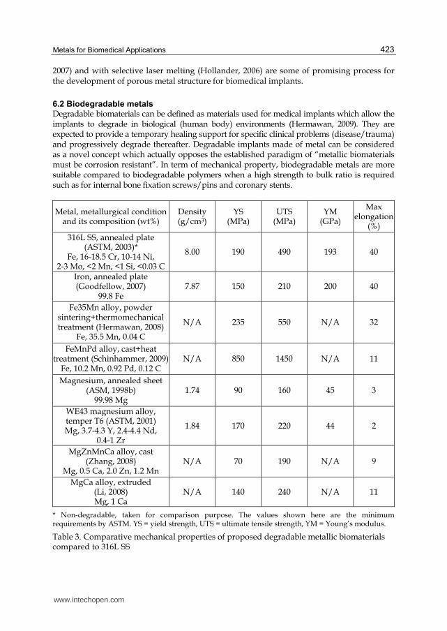
Degradable biomaterials can be defined as materials used for medical implants which allow the implants to degrade in biological (human body) environments (Hermawan, 2009). They are expected to provide a temporary healing support for specific clinical problems (disease/trauma) and progressively degrade thereafter. Degradable implants made of metal can be considered as a novel concept which actually opposes the established paradigm of “metallic biomaterials must be corrosion resistant”. In term of mechanical property, biodegradable metals are more suitable compared to biodegradable polymers when a high strength to bulk ratio is required such as for internal bone fixation screws/pins and coronary stents.
Metal, metallurgical condition and its composition (wt%)
Density (g/cm3)
YS (MPa)
UTS (MPa)
YM (GPa)
Max elongation
(%) 316L SS, annealed plate
(ASTM, 2003)* Fe, 16-18.5 Cr, 10-14 Ni,
2-3 Mo, <2 Mn, <1 Si, <0.03 C
8.00 190 490 193 40
Iron, annealed plate (Goodfellow, 2007)
99.8 Fe7.87 150 210 200 40
Fe35Mn alloy, powder sintering+thermomechanical treatment (Hermawan, 2008)
Fe, 35.5 Mn, 0.04 C
N/A 235 550 N/A 32
FeMnPd alloy, cast+heat treatment (Schinhammer, 2009)
Fe, 10.2 Mn, 0.92 Pd, 0.12 C N/A 850 1450 N/A 11
Magnesium, annealed sheet (ASM, 1998b)
99.98 Mg 1.74 90 160 45 3
WE43 magnesium alloy, temper T6 (ASTM, 2001) Mg, 3.7-4.3 Y, 2.4-4.4 Nd,
0.4-1 Zr
1.84 170 220 44 2
MgZnMnCa alloy, cast (Zhang, 2008)
Mg, 0.5 Ca, 2.0 Zn, 1.2 Mn N/A 70 190 N/A 9
MgCa alloy, extruded (Li, 2008) Mg, 1 Ca
N/A 140 240 N/A 11
* Non-degradable, taken for comparison purpose. The values shown here are the minimum requirements by ASTM. YS = yield strength, UTS = ultimate tensile strength, YM = Young’s modulus.
Table 3. Comparative mechanical properties of proposed degradable metallic biomaterials compared to 316L SS
www.intechopen.com
Biomedical Engineering – From Theory to Applications
424
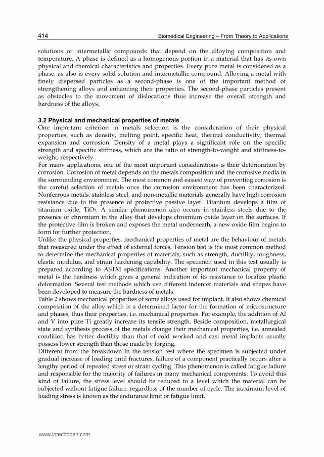
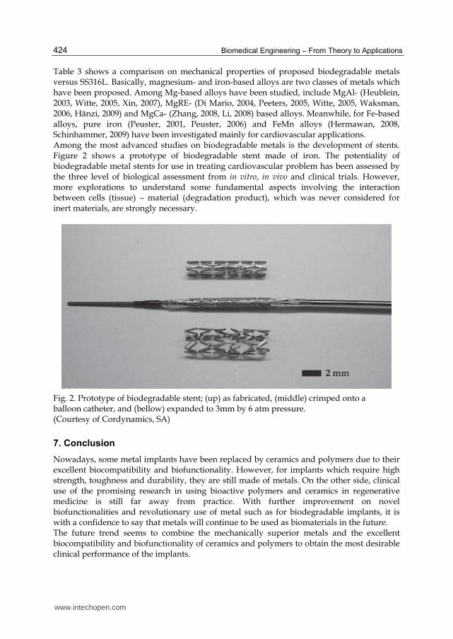
Table 3 shows a comparison on mechanical properties of proposed biodegradable metals versus SS316L. Basically, magnesium- and iron-based alloys are two classes of metals which have been proposed. Among Mg-based alloys have been studied, include MgAl- (Heublein, 2003, Witte, 2005, Xin, 2007), MgRE- (Di Mario, 2004, Peeters, 2005, Witte, 2005, Waksman, 2006, Hänzi, 2009) and MgCa- (Zhang, 2008, Li, 2008) based alloys. Meanwhile, for Fe-based alloys, pure iron (Peuster, 2001, Peuster, 2006) and FeMn alloys (Hermawan, 2008, Schinhammer, 2009) have been investigated mainly for cardiovascular applications. Among the most advanced studies on biodegradable metals is the development of stents. Figure 2 shows a prototype of biodegradable stent made of iron. The potentiality of biodegradable metal stents for use in treating cardiovascular problem has been assessed by the three level of biological assessment from in vitro, in vivo and clinical trials. However, more explorations to understand some fundamental aspects involving the interaction between cells (tissue) – material (degradation product), which was never considered for inert materials, are strongly necessary.
Fig. 2. Prototype of biodegradable stent; (up) as fabricated, (middle) crimped onto a balloon catheter, and (bellow) expanded to 3mm by 6 atm pressure. (Courtesy of Cordynamics, SA)
7. Conclusion
Nowadays, some metal implants have been replaced by ceramics and polymers due to their excellent biocompatibility and biofunctionality. However, for implants which require high strength, toughness and durability, they are still made of metals. On the other side, clinical use of the promising research in using bioactive polymers and ceramics in regenerative medicine is still far away from practice. With further improvement on novel biofunctionalities and revolutionary use of metal such as for biodegradable implants, it is with a confidence to say that metals will continue to be used as biomaterials in the future. The future trend seems to combine the mechanically superior metals and the excellent biocompatibility and biofunctionality of ceramics and polymers to obtain the most desirable clinical performance of the implants.
www.intechopen.com
Metals for Biomedical Applications
425
8. Acknowledgment
Authors would like to acknowledge the kind assistantship from Medical Implant Technology Research Group (MediTeg), Universiti Teknologi Malaysia (UTM). This work was supported by the UTM’s Research Grant Schemes.
9. References
Adell, R., Lekholm, U., Rockler, B., & Branemark, P.I. (1981). A 15-year study of osseointegrated implants in the treatment of the edentulous jaw. Intl J Oral Surg, 10, 387.
Akahori, T., & Niinomi, M. (1998). Fracture characteristics of fatigued Ti6Al4V ELI as an implant material. Mater Sci Eng A, 243, 237.
Amis, A.A., Bromagen, J.D., & Lazvin, M. (1987). Fatigue fracture of a femoral sliding compression screw-plate device bone union. Biomaterials, 8, 153.
Aparicioa, C., Gil, F.J., Fonseca, C., Barbosa, M., & Planell, J.A. (2003). Corrosion behaviour of commercially pure Ti shot blasted with different materials and sizes of shot particles for dental implant applications. Biomaterials, 24, 263.
ASM (1998a). ASM Handbook Vol. 2, Properties and Selection Nonferrous Alloys and Special-Purpose Materials, Materials Park, ASM Internaional.
ASM (1998b), ASM Handbook Vol. 7, Powder Metals Technologies and Applications, Materials Park, ASM International.
ASM (1998c), ASM Handbook Vol. 15, Casting, Materials Park, ASM International. ASTM (2001). ASTM B 80: Standard Specification for Magnesium-Alloy Sand Castings, West
Conshohocken, ASTM International. ASTM (2003). ASTM F 138: Standard Specification for Wrought 18chromium-14nickel-
2.5molybdenum Stainless Steel Bar and Wire for Surgical Implants (UNS S31673), West Conshohocken, ASTM International.
ASTM (2006). ASTM F 67: Standard Specification for Unalloyed Titanium, for Surgical Implant Applications (UNS R50250, UNS R50400, UNS R50550, UNS R50700), West Conshohocken, ASTM International.
ASTM (2007a). ASTM F 90: Standard Specification for Wrought Cobalt-20Chromium-15Tungsten-10Nickel Alloy for Surgical Implant Applications (UNS R30605), West Conshohocken, ASTM International.
ASTM (2007b). ASTM F 562: Standard Specification for Wrought 35Cobalt-35Nickel-20Chromium-10Molybdenum Alloy for Surgical Implant Applications (UNS R30035), West Conshohocken, ASTM International.
ASTM (2008). ASTM F 136: Standard Specification for Wrought Titanium-6 Aluminum-4 Vanadium ELI (Extra Low Interstitial) Alloy for Surgical Implant Applications (UNS R56401), West Conshohocken, ASTM International.
Aziz-Kerrzo, M., Conroy, K.G., Fenelon, A.M., Farrell, S.T., & Breslin, C.B. (2001). Electrochemical studies on the stability and corrosion resistance of Ti-based implant materials, Biomaterials, 22, 1531-1539.
Azushima, A., Kopp, R., Korhonen, A., Yang, D.Y., Micari, F., Lahoti, G.D., Groche, P., Yanagimoto, J., Tsuji, N., Rosochowski, A., & Yanagida, A. (2008). Severe plastic deformation (SPD) processes for metals, CIRP Annal Manuf Technol, 57, 716.
Black, J., & Hastings, G. (1998). Handbook of Biomaterial Properties, Chapman Hall.
www.intechopen.com
Biomedical Engineering – From Theory to Applications
426
Bou-Saleh, Z., Shahryari, A., & Omanovic, S. (2007). Thin Sol Film, 515, 4727. Brandes, E.A. & Brook, G.B. (1992). Smithells Metals Reference Book. 7th ed. Oxford,
Butterworth-Heinemann. Browne, M., & Gregson, P.J. (2000). Effect of mechanical surface pretreatment on metal ion
release. Biomaterials, 21, 385. Brune, D. (1986). Metal release from dental biomaterialsm, Biomaterials, 7, 163. Buford, A., & Goswami, T. (2004). Review of wear mechanisms in hip implants: Paper I –
General. Mater Des, 25, 385. Campbel, F.C. (2006). Manufacturing Technology for Aerospace Structural, Elsevier. Chan S.Y., Park, C.H., & Moriwaki. T. (2010). Mirror finishing of CoCrMo alloy using
elliptical vibration cutting. Prec Eng, 34, 784. Chen, Q., Liu, L. & Zhang, S.-M. (2010). The potential of Zr-based bulk metallic glasses as
biomaterials. Front Mater Sci China, 4, 34. Chiba, A., Lee, S.-H., Matsumoto, H. & Nakamura, M. (2009). Construction of processing
map for biomedical Co28Cr6Mo0.16N alloy by studying its hot deformation behavior using compression tests. Mater Sci Eng A, 513-514, 286.
Dearnley, P.A., Dahma, K.L., & Çimenoglu, H. (2004). The corrosion–wear behaviour of thermally oxidised CP-Ti and Ti6Al4V. Wear, 256, 469.
Dewidar, M.M., Khalil, K.A, & Lim J.K. (2007). Processing and mechanical properties of porous 316L stainless steel for biomedical applications, Trans Nonferrous Met Soc China, 17, 468.
Di Mario, C., Griffiths, H., Goktekin, O., Peeters, N., Verbist, J., Bosiers, M., Deloose, K., Heublein, B., Rohde, R., Kasese, V., Ilsley, C. & Erbel, R. (2004). Drug-eluting bioabsorbable magnesium stent. J Interv Cardiol, 17, 391.
Ferrari, F., Miotello, A., Pavloski, L., Galvanetto, E., Moschini, G., Galassini, S., Passi, P., Bogdanovie, S., Fazini, S., Jaksi, M., & Valkovi, V. (1993). Metal-ion release. from Ti and TiN coated implants in rat bone. Nuclear Instr Meth Phys Res B, 79, 421.
Fleck, C., & Eifler, D. (2010). Corrosion, fatigue and corrosion fatigue behaviour of metal implant materials, especially Ti alloys. Intl J Fatigue, 32, 929.
Geringera, J., Foresta, B., & Combrade, P. (2005). Fretting-corrosion of materials used as orthopaedic implants, Wear, 259, 943.
Germain, L., Gey, N., Humbert, M., Vob, P., Jahazi, M., & Bocher, P. (2008). Texture heterogeneities induced by subtransus processing of near a Ti alloys. Acta Mater, 56, 4298.
Goodfellow (2007). Iron (Fe) - Material information. Goodfellow Corporation. Habibovic, P., Barrère, F., Blitterswijk, C.A.V., Groot, K.D. & Layrolle, P. (2002). Biomimetic
hydroxyapatite coating on metal implants. J Am Ceram Soc, 83, 517. Han, Y.S., & Hong, S.H. (1999). Microstructural changes during superplastic deformation of
Fe24Cr7Ni3Mo0.14N duplex stainless steel. Mater Sci Eng A, 266, 276. Hanawa, T. (2004). Metal ion release from metal implants, Mater Sci Eng C, 24, 745. Hänzi, A.C., Sologubenko, A.S. & Uggowitzer, P.J. (2009). Design strategy for microalloyed
ultra-ductile magnesium alloys for medical applications. Mater Sci Forum, 618-619, 75.
Harman, M.K., Banks, S.A., & Hodge, W.A. (1997). Wear analysis of a retrieved hip implant with titanium nitride coating. J Arthroplast, 12, 938.
www.intechopen.com
Metals for Biomedical Applications
427
Hermawan, H. & Mantovani, D. (2009). Degradable metallic biomaterials: The concept, current developments and future directions. Minerv Biotecnol, 21, 207.
Hermawan, H., Alamdari, H., Mantovani, D. & Dubé, D. (2008). Iron-manganese: New class of degradable metallic biomaterials prepared by powder metallurgy. Powder Metall, 51, 38.
Hermawan, H., Dubé, D. & Mantovani, D. (2010). Developments in metallic biodegradable stents. Acta Biomater, 6, 1693.
Heublein, B., Rohde, R., Kaese, V., Niemeyer, M., Hartung, W. & Haverich, A. (2003). Biocorrosion of magnesium alloys: A new principle in cardiovascular implant technology? Heart, 89, 651.
Hollander, D.A., Von Walter, M., Wirtz, T., Sellei, R., Schmidt-Rohlfing, B., Paar, O. & Erli, H.-J. (2006). Structural, mechanical and in vitro characterization of individually structured Ti6Al4V produced by direct laser forming. Biomaterials, 27, 955.
Hu, Z.M., Brooks, J.W., & Dean, T.A. (1999). Experimental and theoretical analysis of deformation and microstructural evolution in the hot-die forging of Ti alloy aerofoil sections. J Mater Proc Technol, 88, 251.
Huang, J.C., & Chuang, T.H. (1999). Progress on superplasticity and superplastic forming in Taiwan during 1987-1997. Mater Chem Phys, 57, 195.
Karla, M., & Kelly, J.R. (2009). Influence of loading frequency on implant failure under cyclic fatigue conditions. Dent Mater, 25, 1426.
Kern, M., & Thompson, V.P. (1993). Sandblasting and silica-coating of dental alloys: volume loss, morphology and changes in the surface composition. Dent Mater, 9, 155.
Kohn, D.H. (1998). Metals in medical applications, Curr Op Solid State Mater Sci, 3, 309. Komotori, L.J., Hisamori, N., & Ohmoric, Y. (2007). The corrosion/wear mechanisms of
Ti6Al4V alloy for different scratching rates. Wear, 263, 412. Krishna, V.G., Prasad, Y.V.R.K., Birla, N.C., & Rao, G.S. (1997). Processing map for the hot
working near-alpha Ti alloy 685. J Mater Proc Technol, 71, 377. Kubiak, K., & Sieniaski, J. (1998). Development of the microstructure and fatigue strength of
two phase Ti alloys in the processes of forging and heat treatment. J Mater Proc Technol, 78, 117.
Kumar, S., & Narayanan, T.S.N.S. (2008). Corrosion behaviour of Ti15Mo alloy for dental implant applications. J Dentist, 36, 500.
Kumar, S., Narayanan, T.S.N.S., Raman, S.G.S., & Seshadri, S.K. (2010). Evaluation of fretting corrosion behaviour of CP-Ti for orthopaedic implant applications, Tribol Intl, 43, 1245.
Kuroda, D., Niinomi, M., Morinaga, M., Kato, Y. & Yashiro, T. (1998). Design and mechanical properties of new [beta] type Ti alloys for implant materials. Mater Sci Eng A, 243, 244.
Kwok, C.T., Cheng, F.T., & Man, H.C. (2003). Effect of processing conditions on the corrosion performance of laser surface-melted AISI 440C martensitic stainless steel. Surf Coat Technol, 166, 221.
Lahann, J., Klee, D., Thelen, H., Bienert, H., Vorwerk, D. & Hocker, H. (1999). Improvement of haemocompatibility of metallic stents by polymer coating. J Mater Sci Mater Med, 10, 443.
Lambotte, A. (1909). Technique et indication des prothèses dans le traitement des fractures. Presse Med, 17, 321.
www.intechopen.com
Biomedical Engineering – From Theory to Applications
428
Lane, W.A. (1895). Some remarks on the treatment of fractures. Brith Med J, 1, 861. Lee, C.K., Karl, M., & Kelly, J.R. (2009). Evaluation of test protocol variables for dental
implant fatigue research. Dent Mater, 25, 1419. Lee, S.H., Nomura, N. & Chiba, A. (2008). Significant improvement in mechanical properties
of biomedical CoCrMo alloys with combination of N addition and Cr-enrichment. Mater Trans, 49, 260.
Leinenbach, C., Fleck, C., & Eifler, D. (2004). The cyclic deformation behaviour and fatigue induced damage of the implant alloy TiAl6Nb7 in simulated physiological media. Intl J Fatigue, 26, 857.
Li, J.P., Habibovic, P., Van Den Doel, M., Wilson, C.E., De Wijn, J.R., Van Blitterswijk, C.A. & De Groot, K. (2007). Bone ingrowth in porous Ti implants produced by 3D fiber deposition. Biomaterials, 28, 2810.
Li, Z., Gu, X., Lou, S. & Zheng, Y. (2008). The development of binary Mg-Ca alloys for use as biodegradable materials within bones. Biomaterials, 29, 1329.
Liu, Y., & Baker, T.N. (1995). Deformation characteristics of IMI685 Ti alloy under ┚ isothermal forging conditions. Mater Sci Eng A, 197, 125.
Lopez-Heredia, M.A., Sohier, J., Gaillard, C., Quillard, S., Dorget, M. & Layrolle, P. (2008). Rapid prototyped porous Ti coated with calcium phosphate as a scaffold for bone tissue engineering. Biomaterials, 29, 2608.
Matsumoto, H., Watanabe, S. & Hanada, S. (2005). Beta TiNbSn alloys with low Young’s modulus and high strength. Mater Trans, 46, 1070.
Ming-Wei, W., Li-Wen, Z., Ji-Bin, P., Li Chen, P., & Fan, H. (2007). Effect of temperature on vacuum hot bulge forming of BT20 Ti alloy cylindrical work piece. Trans Nonferrous Met Soc China, 17, 957.
Mitchell, A., & Shrotriya, P. (2007). Onset of nanoscale wear of metallic implant materials:Influence of surface residual stresses and contact loads. Wear, 263, 1117.
Miyamoto, H., Mimaki, T., & Hashimoto, S. (2001). Mater Sci Eng A, 319–321, 779. Morais, L.S., Serra, G.G., Muller, C.A., Andrade, L.R., Palermo, E.F.A., Elias, C.N., & Meyers,
M. (2007). Titanium alloy mini-implants for orthodontic anchorage: Immediate loading and metal ion release. Acta Biomater, 3, 331.
Mordyuk, B.N., Prokopenko, G.I. Vasylyev, M.A., & Iefimov, M.O. (2007). Effect of structure evolution induced by ultrasonic peening on the corrosion behavior of AISI-321 stainless steel. Mater Sci Eng A, 458, 253.
Narushima, T. (2010). New-generation metallic biomaterials. IN NIINOMI, M. (Ed.) Metals for Biomedical Devices. Cambrigde, Woodhead Publishing.
Nguyen, H.Q., Deportera, D.A., Pilliara, R.M., Valiquettea, N., & Yakubovich, R. (2004). The effect of sol–gel-formed calcium phosphate coatings on bone ingrowth and osteoconductivity of porous-surfaced Ti alloy implants. Biomaterials, 25, 865.
Okazaki, Y., & Gotoh, E. (2008). Metal release from stainless steel, CoCrMoNiFe and NiTi alloys in vascular implants. Corr Sci, 50, 3429.
Öztürk, O., Türkan, U., & Ahmet, E.E. (2006). Metal ion release from nitrogen ion implanted CoCrMo orthopedic implant material. Surf Coat Technol, 200, 5687.
Papakyriacou, M., Mayer, H. Pypen, C., Plenk Jr, H., & Stanzl-Tschegg, S. (2000). Effects of surface treatments on high cycle corrosion fatigue of metallic implant materials. Intl J Fatigue, 22, 873.
Peckner, D., & Bernstein, I.M. (1977). Handbook of Stainless Steels, McGraw-Hill Inc.
www.intechopen.com
Metals for Biomedical Applications
429
Peeters, P., Bosiers, M., Verbist, J., Deloose, K. & Heublein, B. (2005). Preliminary results after application of absorbable metal stents in patients with critical limb ischemia. J Endovasc Ther, 12, 1.
Peuster, M., Hesse, C., Schloo, T., Fink, C., Beerbaum, P. & Von Schnakenburg, C. (2006). Long-term biocompatibility of a corrodible peripheral iron stent in the porcine descending aorta. Biomaterials, 27, 4955.
Peuster, M., Wohlsein, P., Brugmann, M., Ehlerding, M., Seidler, K., Fink, C., Brauer, H., Fischer, A. & Hausdorf, G. (2001). A novel approach to temporary stenting: Degradable cardiovascular stents produced from corrodible metal-results 6-18 months after implantation into New Zealand white rabbits. Heart, 86, 563.
Ramdan, R.D., Jauhari, I., Hasan, R., & Nik Masdek, N.R. (2008). The role of strain rate during deposition of CAP on Ti6Al4V by superplastic deformation-like method using high-temperature compression test machine. Mater Sci Eng A, 477, 300.
Ratner, B.D. (1993). New ideas in biomaterials science-A path to engineered biomaterials. J Biomed Mater Res, 27, 837.
Reclarua, L., Eschlera, P., Lerf, R., & Blatter, A. (2005) Electrochemical corrosion and metal ion release from CoCrMo prosthesis with Ti plasma spraycoating, Biomaterials, 26, 4747.
Roland, T., Retraint, D., Lu, K., & Liu, J. (2006) Fatigue life improvement through surface nanostructuring of stainless steel by means of surface mechanical attrition treatment. Script Mater, 54, 1949.
Ryan, G.E., Pandit, A.S. & Apatsidis, D.P. (2008). Porous Ti scaffolds fabricated using a rapid prototyping and powder metallurgy technique. Biomaterials, 29, 3625.
Ryan, G., Pandit, A. & Apatsidis, D.P. (2006). Fabrication methods of porous metals for use in orthopaedic applications. Biomaterials, 27, 2651.
Schinhammer, M., Hänzi, A.C., Löffler, J.F. & Uggowitzer, P.J. (2010). Design strategy for biodegradable Fe-based alloys for medical applications. Acta Biomater, 6, 1705.
Schroers, J., Kumar, G., Hodges, T., Chan, S. & Kyriakides, T. (2009). Bulk metallic glasses for biomedical applications. J Mater, 61, 21.
Schrooten, J., & Helsen, J.A. (2000). Adhesion of bioactive glass coating to Ti6Al4V oral implant. Biomaterials, 21, 1461.
Serro, A.P., & Saramago, B. (2003). Influence of sterilization on the mineralization of Ti implants induced by incubation in various biological model fluids. Biomaterials, 24, 4749.
Shahryari, A., Omanovic, S., & Szpunar, J.A. (2008). Electrochemical formation of highly pitting-resistant passive films on biomedical grade 316LVM stainless steel surface. Mater Sci Eng C, 28, 94.
Sherman, W.O. (1912). Vanadium steel bone plates and screws. Surg Gynecol Obstet, 14, 629. Spriano, S., Vernè, E., Faga, M.G., Bugliosi, S., & Maina, G., Surface treatment on an
implant cobalt alloy for high biocompatibility and wear resistance. Wear, 259, 919. Sun, Y., & Haruman, E. (2008) Influence of processing conditions on structural
characteristics of hybrid plasma surface alloyed austenitic stainless steel. Surf Coat Technol, 202, 4069.
Triantafylldis, G.K., Kazantzis, A.V., & Karageorgiou, K.T. (2007). Premature fracture of a stainless steel 316L orthopaedic plate implant by alternative episodes of fatigue and cleavage decoherence. Eng Failure Anal, 14, 1346.
www.intechopen.com
Biomedical Engineering – From Theory to Applications
430
Tritschler, B., Forest, B., & Rieu, J. (1999). Fretting corrosion of materials for orthopaedic implants: a study of a metal/polymer contact in an artificial physiological medium. Tribol Intl, 32, 587.
Venugopal, S., Sivaprasad, P.V., Vasudevan, M., Mannan, S.L., Jha, S.K., Pandey, P., & Prasad, Y.V.R.K. (1995). Validation of processing maps for 304L stainless steel using hot forging, rolling and extrusion. J Mater Proc Technol, 59, 343.
Waksman, R., Pakala, R., Kuchulakanti, P.K., Baffour, R., Hellinga, D., Seabron, R., Tio, F.O., Wittchow, E., Hartwig, S., Harder, C., Rohde, R., Heublein, B., Andreae, A., Waldmann, K.-H. & Haverich, A. (2006). Safety and efficacy of bioabsorbable Mg alloy stents in porcine coronary arteries. Catheter Cardiovasc Interv, 68, 606.
Wang, Y.B., Zheng, Y.F., Wei, S.C. & Li, M. (2011). In vitro study on Zr-based bulk metallic glasses as potential biomaterials. J Biomed Mater Res, 96B, 34.
Weiss, I., & Semiatin, S.L. (1999). Thermomechanical processing of alpha Ti alloys—An overview. Mater Sci Eng A, 263, 243.
Williams, D.F. (2008). On the mechanisms of biocompatibility. Biomaterials, 29, 2941. Williamson, D.L., Davis, J.A., & Wilbur, P.J. (1998). Effect of austenitic stainless steel
composition on low-energy, high-flux, nitrogen ion beam processing. Surf Coat Technol, 103-104, 178.
Witte, F., Hort, N., Vogt, C., Cohen, S., Kainer, K.U., Willumeit, R. & Feyerabend, F. (2009). Degradable biomaterials based on magnesium corrosion. Curr Op Solid State Mater Sci, 12, 63.
Witte, F., Kaese, V., Haferkamp, H., Switzer, E., Linderberg, A.M., Wirth, C.J. & Windhagen, H. (2005). In vivo corrosion of four magnesium alloys and the associated bone response. Biomaterials, 26, 3557.
Xin, Y., Liu, C., Zhang, X., Tang, G., Tian, X. & Chu, P.K. (2007). Corrosion behavior of biomedical AZ91 magnesium alloy in simulated body fluids. J Mater Res, 22, 2004.
Yang, K. & Ren, Y. (2010). Nickel-free austenitic stainless steels for medical applications. Sci Technol Adv Mater, 11, 1.
Zhang, E. & Yang, L. (2008). Microstructure, mechanical properties and biocorrosion properties of MgZnMnCa alloy for biomedical application. Mater Sci Eng A, 497, 111.
Zhuang, L.Z., & Langer, E.W. (1988). Effects of the range of the stress intensity factor on the appearance of localized fatigue fracture in cast CoCrMo alloy used for surgical implants. Mater Sci Eng A, 102, L9.
Zhuka, H.V., Kobryn, P.A., & Semiatin, S.L. (2007). Influence of heating and solidification conditions on the structure and surface quality of electron-beam melted Ti6Al4V ingots. J Mater Proc Technol, 190, 387.
www.intechopen.com
Biomedical Engineering - From Theory to ApplicationsEdited by Prof. Reza Fazel
ISBN 978-953-307-637-9Hard cover, 486 pagesPublisher InTechPublished online 29, August, 2011Published in print edition August, 2011
InTech EuropeUniversity Campus STeP Ri Slavka Krautzeka 83/A 51000 Rijeka, Croatia Phone: +385 (51) 770 447 Fax: +385 (51) 686 166www.intechopen.com
InTech ChinaUnit 405, Office Block, Hotel Equatorial Shanghai No.65, Yan An Road (West), Shanghai, 200040, China
Phone: +86-21-62489820 Fax: +86-21-62489821
In all different areas in biomedical engineering, the ultimate objectives in research and education are toimprove the quality life, reduce the impact of disease on the everyday life of individuals, and provide anappropriate infrastructure to promote and enhance the interaction of biomedical engineering researchers. Thisbook is prepared in two volumes to introduce a recent advances in different areas of biomedical engineeringsuch as biomaterials, cellular engineering, biomedical devices, nanotechnology, and biomechanics. It is hopedthat both of the volumes will bring more awareness about the biomedical engineering field and help incompleting or establishing new research areas in biomedical engineering.
How to referenceIn order to correctly reference this scholarly work, feel free to copy and paste the following:
Hendra Hermawan, Dadan Ramdan and Joy R. P. Djuansjah (2011). Metals for Biomedical Applications,Biomedical Engineering - From Theory to Applications, Prof. Reza Fazel (Ed.), ISBN: 978-953-307-637-9,InTech, Available from: http://www.intechopen.com/books/biomedical-engineering-from-theory-to-applications/metals-for-biomedical-applications