Embed Size (px)
Citation preview
BNL-79479-2007-BC
Metal Oxide Nanoparticles
Marcos Fernández-Garcia, José A. Rodriguez
To be published in “Nanomaterials: Inorganic and Bioinorganic Perspectives”
October 2007
Chemistry Department
Brookhaven National Laboratory P.O. Box 5000
Upton, NY 11973-5000 www.bnl.gov
Notice: This manuscript has been authored by employees of Brookhaven Science Associates, LLC under Contract No. DE-AC02-98CH10886 with the U.S. Department of Energy. The publisher by accepting the manuscript for publication acknowledges that the United States Government retains a non-exclusive, paid-up, irrevocable, world-wide license to publish or reproduce the published form of this manuscript, or allow others to do so, for United States Government purposes. This preprint is intended for publication in a journal or proceedings. Since changes may be made before publication, it may not be cited or reproduced without the author’s permission.

DISCLAIMER
This report was prepared as an account of work sponsored by an agency of the United States Government. Neither the United States Government nor any agency thereof, nor any of their employees, nor any of their contractors, subcontractors, or their employees, makes any warranty, express or implied, or assumes any legal liability or responsibility for the accuracy, completeness, or any third party’s use or the results of such use of any information, apparatus, product, or process disclosed, or represents that its use would not infringe privately owned rights. Reference herein to any specific commercial product, process, or service by trade name, trademark, manufacturer, or otherwise, does not necessarily constitute or imply its endorsement, recommendation, or favoring by the United States Government or any agency thereof or its contractors or subcontractors. The views and opinions of authors expressed herein do not necessarily state or reflect those of the United States Government or any agency thereof.
METAL OXIDE NANOPARTICLES
Marcos Fernández-Garcíaa and José A. Rodriguezb
a Instituto de Catálisis y Petroleoquímica, CSIC, C/Marie Curie 2, Cantoblanco, 28049-
Madrid, Spain
b Department of Chemistry, Brookhaven National Laboratory, Upton, NY 11973, USA
Emails: [email protected]; [email protected] Abstract This chapter covers the fundamental science, synthesis, characterization, physico-chemical properties and applications of oxide nanomaterials. Explains fundamental aspects that determine the growth and behavior of these systems, briefly examines synthetic procedures using bottom-up and top-down fabrication technologies, discusses the sophisticated experimental techniques and state of the art theory results used to characterize the physico-chemical properties of oxide solids and describe the current knowledge concerning key oxide materials with important technological applications. Keywords Oxides; Al2O3, MgO; ZrO2; CeO2, TiO2; ZnO; Fe2O3; SnO; structural and electronic properties; physico-chemical properties; nanosize; nanostructure; confinement and quantum-size effects
BNL-79479-2007-BC

INTRODUCTION: THE WORLD OF OXIDE NANOMATERIALS Metal oxides play a very important role in many areas of chemistry, physics and
materials science.1, , , , ,2 3 4 5 6 The metal elements are able to form a large diversity of oxide
compounds.7 These can adopt a vast number of structural geometries with an electronic
structure that can exhibit metallic, semiconductor or insulator character. In technological
applications, oxides are used in the fabrication of microelectronic circuits, sensors,
piezoelectric devices, fuel cells, coatings for the passivation of surfaces against corrosion,
and as catalysts. In the emerging field of nanotechnology, a goal is to make
nanostructures or nanoarrays with special properties with respect to those of bulk or
single particle species.8, , , ,9 10 11 12 Oxide nanoparticles can exhibit unique physical and
chemical properties due to their limited size and a high density of corner or edge surface
sites. Particle size is expected to influence three important groups of basic properties in
any material. The first one comprises the structural characteristics, namely the lattice
symmetry and cell parameters13. Bulk oxides are usually robust and stable systems with
well-defined crystallographic structures. However, the growing importance of surface
free energy and stress with decreasing particle size must be considered: changes in
thermodynamic stability associate with size can induce modification of cell parameters
and/or structural transformations14, ,15 16 and in extreme cases the nanoparticle can
disappear due to interactions with its surrounding environment and a high surface free
energy.17 In order to display mechanical or structural stability, a nanoparticle must have a
low surface free energy. As a consequence of this requirement, phases that have a low
stability in bulk materials can become very stable in nanostructures. This structural
phenomenon has been detected in TiO2, VOx, Al2O3 or MoOx oxides.16,17, 18
Size-induced structural distortions associated with changes in cell parameters
have been observed, for example, in nanoparticles of Al2O3,17 NiO,19 Fe2O3,20 ZrO2,21
MoO3,23 CeO2,22 and Y2O3.23 As the particle size decreases, the increasing number of
surface and interface atoms generates stress/strain and concomitant structural
perturbations.24 Beyond this “intrinsic” strain, there may be also “extrinsic” strain
associated with a particular synthesis method which may be partially relieved by
annealing or calcination.25 Also, non-stoichiometry is a common phenomenon.25 On the
other hand, interactions with the substrate on which the nanoparticles are supported can
complicate the situation and induce structural perturbations or phases not seen for the
bulk state of the oxide.18,26
The second important effect of size is related to the electronic properties of the
oxide. In any material, the nanostruture produces the so-called quantum size or
confinement effects which essentially arise from the presence of discrete, atom-like
electronic states. From a solid-state point of view, these states can be considered as being
a superposition of bulk-like states with a concomitant increase in oscillator strength.27
Additional general electronic effects of quantum confinement experimentally probed on
oxides are related to the energy shift of exciton levels and optical bandgap.28,29 An
important factor to consider when dealing with the electronic properties of a bulk oxide
surface are the long-range effects of the Madelung field, which are not present or limited
in a nanostructured oxide.30, ,31 32 Theoretical studies for oxides show a redistribution of
charge when going from large periodic structures to small clusters or aggregates which
must be roughly considered to be relatively small for ionic solids while significantly
larger for covalent ones.33, , , , ,34 35 36 37 38 The degree of ionicity or covalency in a metal-
oxygen bond can however strongly depend on size in systems with partial ionic or
covalent character; an increase in the ionic component to the metal-oxygen bond in
parallel to the size decreasing has been proposed.15
Structural and electronic properties obviously drive the physical and chemical
properties of the solid, the third group of properties influenced by size in a simple
classification. In their bulk state, many oxides have wide band gaps and a low
reactivity.39 A decrease in the average size of an oxide particle do in fact change the
magnitude of the band gap,32,40 with strong influence in the conductivity and chemical
reactivity.41,42 Surface properties are a somewhat particular group included in this subject
due to their importance in chemistry. Solid-gas or solid-liquid chemical reactions can be
mostly confined to the surface and/or sub-surface regions of the solid. As above
mentioned, the two dimensional (2D) nature of surfaces has notable structural, typically a
rearrangement or reconstruction of bulk geometries,3,11,43 and electronic, e.g. presence of
mid-gap states,42,44 consequences. In the case of nanostructured oxides, surface properties
are strongly modified with respect to 2D-infinite surfaces, producing solids with
unprecedent sorption or acid/base characteristics.45 Furthermore, the presence of under-
coordinated atoms (like corners or edges) or O vacancies in an oxide nanoparticle should
produce specific geometrical arrangements as well as occupied electronic states located
above the valence band of the corresponding bulk material,46, ,47 48 enhancing in this way
the chemical activity of the system.43,45, ,49 50
In this Chapter we will analyze how nanoparticulated oxides are synthesized, their
most significant physico-chemical properties, and will focus the ending part on several,
well known oxides. Particular attention will be paid to the effect of the primary
nanostructure, e.g. primary particle size, on structural/electronic properties and how these
alter other industrial-related properties.
SYNTHESIS OF NANOPARTICULATED OXIDES
The first requirement of any novel study of nanoparticulated oxides is the
synthesis of the material. The development of systematic studies for the synthesis of
oxide nanoparticles is a current challenge and, essentially, the corresponding preparation
methods may be grouped in two main streams based upon the liquid-solid 51 and gas-
solid52 nature of the transformations.
Liquid-solid transformations are possibly the most broadly used in order to
control morphological characterstics with certain “chemical” versatility and usually
follow a “bottom-up” approach. A number of specific methods have been developed,
among which those broadly in use are: 1) Co-precipitation methods. This involves
dissolving a salt precursor (chloride, nitrate, etc.) in water (or other solvent) to precipitate
the oxo-hydroxide form with the help of a base. Very often, control of size and chemical
homogeneity in the case of mixed-metal oxides are difficult to achieve. However, the use
of surfactants, sonochemical methods, and high-gravity reactive precipitation appear as
novel and viable alternatives to optimize the resulting solid morphological
characteristics.51, ,53 54 2) Sol-gel processing. The method prepares metal oxides via
hydrolysis of precursors, usually alcoxides in alcoholic solution, resulting in the
corresponding oxo-hydroxide. Condensation of molecules by giving off water leads to the
formation of a network of the metal hydroxide: Hydroxyl-species undergo polymerization
by condensation and form a dense porous gel. Appropriate drying and calcinations lead to
ultrafine porous oxides.55 3) Microemulsion technique. Microemusion or direct/inverse
micelles represent an approach based on the formation of micro/nano-reaction vessels
under a ternary mixture containing water, a surfactant and oil. Metal precursors on water
will proceed precipitation as oxo-hydroxides within the aqueous droplets, typically
leading to monodispersed materials with size limited by the surfactant-hydroxide
contact.56 4) Solvothermal methods. In this case, metal complexes are decomposed
thermically either by boiling in an inert atmosphere or using an autoclave with the help of
pressure. A suitable surfactant agent is usually added to the reaction media to control
particle size growth and limit agglomeration. 5) Template/Surface derivatized methods.
Template techniques are common to some of the previous mentioned methods and use
two types of tools; soft-templates (surfactants) and hard-templates (porous solids as
carbon or silica). Template- and surface-mediated nanoparticles precursors have been
used to synthesize self-assembly systems. 51
Gas-solid transformation methods with broad use in the context of ultrafine oxide
powder synthesis are restricted to chemical vapor deposition (CVD) and pulsed laser
deposition (PLD). 6) There are a number of CVD processes used for the formation of
nanoparticles among which we can highlight the classical (thermally
activated/pydrolytic), metalorganic, plasma-assisted, and photo CVD methodologies.57
The advantages of this methodology consist of producing uniform, pure and reproduce
nanoparticles and films although requires a careful initial setting up of the experimental
parameters. 7) Multiple-pulsed laser deposition heats a target sample (4000 K) and leads
to instantaneous evaporation, ionization, and decomposition, with subsequent mixing of
desired atoms. The gaseous entities formed absorb radiation energy from subsequent
pulses and acquire kinetic energy perpendicularly to the target to be deposited in a
substrate generally heated to allow crystalline growth.58
Irrespective of the preparation method use to obtain ultrafine nano-oxides, the
studies of nanoparticle preparation yielded compelling evidence concerning the fact that
crystallization does not follow a traditional nucleation and growth mechanism. Although
subjected to further assessment, it appears that the simple idea that a small primary size
would prime nucleation as the key step of crystallization seems essentially correct and
holds certain general validity, at least in solid-solid crystallization mechanisms (e.g.
heating of oxo-hydroxides to form oxides). When additional liquid/gas phase
crystallization steps are involved in the final formation of the nanoparticle (e.g. as in
solvothermal methods), other steps like Ostwald ripening may be also of prime
importance. In any case, a lot of novel insights have been recently uncover in solid-solid
transformations and two main theories describe crystallization to proceed either by
surface (single particle) and/or interface (two or multiple particle) nucleation.59,60 The
primacy of one of them has been postulated to be a function of the oxide chemical nature
and temperature, being presumably surface effects always predominant at higher
temperatures. Both theories mostly received support from kinetic approaches but very
recent analyses sensitive to structural order in the amorphous precursor materials have
demonstrated the key role of intraparticle local order (below 1 nm) in driving the
nucleation temperature onset in a broad interval of ca. 200 K, showing that the whole
crystallization mechanism of oxide nanoparticles appears only compatible with some
kind of intraparticle, dimensional-restricted (“surface”) mechanism.61 This invokes for
the crucial structural characterization of the initial, XRD-amorphous materials in order to
further progress in the understanding the nanostructure influence in morphological
properties of oxides.
PROPERTIES OF NANOPARTICULATED OXIDES
The current knowledge on oxide materials allows to affirm that most of their
physico-chemical properties display an acute size dependence. Physico-chemical
properties of special relevance in Chemistry are mostly related to the industrial use of
oxides as sensors, ceramics, absorbents and/or catalysts. A bunch of novel application
within these fields rely on the size-dependence of the optical, (electronic and/or ionic)
transport, mechanical and, obviously, surface/chemical (redox, acid/base) properties of
oxide nanomaterials. We should stress that size effects in oxide chemistry have
frequently two interrelated faces, structural/electronic quantum-size and size-defect or
non-stoichiometry effects. Hence, here we will describe the influence of these two
phenomena in the main physico-chemical properties of oxides.
Optical properties. The optical conductivity is one of the fundamental properties
of metal oxides and can be experimentally obtained from reflectivity and absorption
measurements. While reflectivity is clearly size-dependent as scattering can display
drastic changes when the oxide characteristic size (primary/secondary particle size) is
in/out the range of photon wavelength,62 absorption features typically command main
absorption behavior of solids. Due to quantum-size confinement, absorption of light
becomes both discrete-like and size-dependent. For nano-crystalline semiconductors,
both linear (one exciton per particle) and non-linear optical (multiple excitons) properties
arise as a result of transitions between electron and hole discrete or quantized electronic
levels. In the first case, depending on the relationship between the radius of the nano-
particle (R) and the Bohr radius of the bulk exciton (RB = ε ħ2/µe2; µ exciton reduced
mass and ε dielectric constant of the semiconductor), the quantum confinement effect can
be divided into three regimes; weak, intermediate and strong confinement regimes, which
correspond to R >> RB, R ≈ RB, and R << RB, respectively.63 The effective mass theory
(EMA) is the most elegant and general theory to explain the size dependence of the
optical properties of nano-meter semiconductors, although other theories as the free-
exciton collision model (FECM)64 or those based in the bond length - strength
correlation65 have been developed to account for several deficiencies of the EMA theory.
For the onset of light absorption, e.g. the optical band gap, as well as for all other
electronic transitions present in the optical absorption spectrum, the EMA theory predicts
a r-2 dependence, with a main r-1 correction term in the strong confinement regime, while
FECM gives a exp(1/r) behavior. It can be thus concluded that metal oxide
semiconductors would present, as a first rough approximation, an optical band gap energy
with an inverse squared dependence of the primary particle size if quantum confinement
dominates the energy behavior of the band gap. Figure 1 shows that this happens to be
the case for (direct band gap) Fe2O366 or (indirect band gap) CdO67 but not for Cu2O,68
CeO2,,69 ZnO,70,71 and TiO2.72,73 Limited deviations from the R-2 behavior, as observed for
ZnO70 in Figure 1 or SnO2,74 can be based in the known fact that the theory overestimates
the blue shift and can be justified with a proper calculation of electronic states by using
simple quantum mechanical methods, while marked deviations are usually based in
several chemical/physical phenomena not accounted for in the previous discussion. In the
case of Cu2O68 or CeO2,75 it appears to be directly related with the presence of Cu2+
(remarkably for very low particle size) and Ce3+ ions at the surface of the nanostructured
materials. At the moment it is not clear if the presence of these oxidation states are
intrinsic to the nanostructure or result from the specific procedure of preparation. The
case of WO3 share also some of the difficulties pointed out above. Kubo et al. were able
to show that the band gap of this oxide decrease with size from ca. 3.0 to 2.8 eV as a
function of R-1,76 but the presence of a variable number of oxygen defects, reduced W
redox states and mid-gap electronic states with size makes this an open question.77 TiO2
is the other example included in Fig. 1 having a band gap energy behavior with marked
differences from that expected r-2 behavior. While bulk TiO2 is an indirect
semiconductor, nanostructured TiO2 materials are likely direct ones.72,78 This may be a
general result. As discussed in ref. 79, the confinement of charge carriers in a limited
space causes their wavefunctions to spread out in momentum space, in turn increasing the
likehood of radiative transitions for bulk indirect semiconductors. This may also be the
case of NiO.80 The indirect nature of the absorption onset would complicate the analysis
of the optical band gap energy due to the above mentioned step structure of the
absorption onset (which includes phonon-related absorption/emission features).81 In spite
of this, the steady behavior shown in Fig. 1 can not be accounted for by small variations
in the absorption onset and should be grounded in other physical phenomena.
Other optical excitations which showed quantum-size confinement effects
concerns the excitation of optical phonons of oxides. The effects of size on the phonon
spectra of oxide materials have been well established by using Raman scattering
experiments on nanocrystals, in combination with the theoretical phonon confinement
models.-83 Essentially, the theoretical background for the study of nanocrystalline
materials is provided by the phonon confinement model. This factor is the main
responsible for the changes observed in the Raman spectrum which are caused by the size
effect. Nevertheless, other factors have been described which can contribute to Raman
spectrum modification as the non-stoichiometry or the internal stress/surface tension. The
phonon confinement model links q vector selection rule for the excitation of Raman
active optical phonons with long-range order and crystallite size82,83 In an amorphous
material, owing to the lack of long-range order, the q-vector selection rule breaks down
and the Raman spectrum resembles the phonon density of states. Nanocrystals represent
an intermediate behavior. For a nanocrystal of average diameter L, the strict “infinite”
crystal selection rule is replaced by a relaxed version, with the result that a range of q
vectors is accessible due to the uncertainty principle,84 The q vector relaxation model can
be used for the purpose of comparing experimental data with theoretically predicted
phonon confinement. According to this model, for finite sized crystals, the Raman
intensity can be expressed using the equation 1:82,85
( ) ( )[ ( ) ] [ ]
(1) 2 / q -
q d 8 / q- exp 2
0
3
BZ
22
Γ+≅ ∫ ωω
ω LI
The ( )Lρ represents the particle size distribution, q is expressed in units of a Lπ (being
aL the unit cell parameter), ( ) q ω is the phonon dispersion, and 0Γ is the intrinsic
linewidth of the bulk crystal. A spherically symmetric phonon dispersion curve is
assumed and approximated by a simple linear chain model.84 For a given phonon mode,
the slope of dispersion away from the BZ center determines the nature of the
modification in the Raman line shape as a function of crystallite size: a negative slope,
towards lower frequency, would produce a downshifted (red-shifted) Raman peak, while
a positive slope would result in an up-shifted (blue-shifted) Raman peak, in addition to an
asymmetric peak broadening, as the crystallite size reduces. Usually is chosen for this
kind of analysis the most intense Raman mode for the solid studied. Some examples of
application of the confinement model for qualitative interpretation of Raman results in
series of nanostructured oxides like anatase ZnO, TiO2, CuO, Cr2O3, ZrTiO4, CeO2 or
manganese oxides can be found elsewhere.25, , , , , , , ,85 86 87 88 89 90 91 92
In all cases, optical absorption features of nanosized oxides are additionally
influenced by “non-stoichiometry” size-dependent defect effects. Typical point defects in
nanostructured oxides concern oxygen or cation vacancies and/or the presence of aliens
species, like Cu2+ and Ce3+. Vacancy defects introduce gap states in proportion to the
defect number; in fact, a random distribution of (equal) vacancy defects introduce a
gaussian-like density of states which may produce mid-gap states and/or be localized near
the valence and conduction bands depending on the electronic nature (donor/acceptor) of
the defect and giving characteristic “localized” features in the UV-visible spectrum. Such
point defects mainly contribute to the Raman spectra by producing a broadening of the
peaks. Alien cations display specific features, like the localized d-d or f-f transitions of
Cu/Ce. Besides electronic modifications, point defects but particularly alien ions, like
Cu2+ and Ce3+ above, induce strain effects and concomitant structural differences in
atomic positions with respect to bulk positions. The influence of strain in the optical
absorption spectrum has been nicely demonstrated in the work of Ong et al. for ZnO,93
showing the splitting of the first exciton peak for large values of compressive strain.
Strain effects (including parameter variations measured in optical phonons with the help
of the Gruneisen parameter) are inherent to nanostructured materials25,71 and may be
comprised in the general, ambiguous term of “surface” effects usually claimed to account
for significant deviations the confinement theories. Surface effects and, particularly, non-
stoichiometry related to the preparation method are critically important for very low
particle size and produce characteristic features in the UV-visible spectrum for certain
oxides, as SnO294 or ZrO2.
95
Transport properties. Oxide materials can present ionic or mixed
ionic/electronic conductivity and it is experimentally well established than both can be
influenced by the nanostructure of the solid. The number of electronic charge carriers in a
metal oxide is a function of the band gap energy according to the Boltzmann statistics.
The electronic conduction is referred to as n- or p-hopping-type depending on whether
the principal charge carrier are, respectively, electrons or holes. The number of “free”
electron/holes of an oxide can be enhanced by introducing non-stoichiometry and, in such
case, are balanced by the much less mobile oxygen/cation vacancies. In an analogous
manner to hoping-type conduction, ionic conduction takes place when ions can hop from
site to site within a crystal lattice as a result of thermal activation, and is typically
interpreted on the basis of a modified Fick´s second law. Four mechanism types have
been observed for ionic conduction: direct interstitial, interstitialcy, vacancy, and
grotthus. As charge species (defects; impurities) in polycrystalline oxides typically
segregate to particle boundaries to minimize strain and electrostatic potential
contributions to the total energy, there is a contribution to the conductivity parallel to the
surface which becomes important at the nanoscale regime. The charge carrier (defect)
distribution also suffers strong modification from bulk materials as there is presence of
charge carries through the whole material as a consequence of the shielded electrostatic
potential depletion at surface layers of nanosized materials.96 As a result of these
nanoscale derived effects, it is well known that CeO2 exhibits an improved n-type
conductivity which may be four order of magnitude greater than the corresponding to
bulk/micro-crystalline ceria, and is ascribed to a significant enhancement of the electronic
contribution.97 Alteration of the transport properties is also observed in ZrO2 but the
physical ground is still far from being understood.96 The strong size-dependence observed
for the electrical conductance in the context of gas-sensing devices has been recently
reviewed for the SnO2, WO3, and In2O3 oxides.98 In proton conductors, like
SrCe0.95Yb0.05O3-d, enhanced conduction and faster kinetics under H-atmospheres are
observed in nanosized samples as these phenomena are largely determined by
boundary/interfacial effects.99 Interesting to stress here is that some of the most dramatic
effects of he nanostructure on ionic transport in oxides are observed in the field of Li+-ion
batteries. An outstanding enhancement of Li+-ion vacancy conductivity have been
achieved using Li-infiltrated nanoporous Al2O3.100
Mechanical properties. Main mechanical properties concerns low (yield stress
and hardness) and high (superpasticity) temperature observables. Information on oxide
nanomaterials is scarce and mainly devoted to analyze sinterability, ductibility, and
superpasticity. In particular, an important number of works have showed significant
improvement in sintering with up to 600 K lower temperatures with respect to bluk
counterparts. In conventional/bulk materials the yield stress (σ) and harness (H) follow
the Hall-Petch (H-P) equation:
(2) dk H/ H/ -1/200 += σσ
where the initial constants describe friction stress and hardness, d is the primary
particle/grain size and k the corresponding slope. The H-P effect in bulk materials is
attributed to the particle/grain boundaries acting as efficient obstacles for slip transfer
(stress) or dislocations (hardness). Typically by decrease the particle/grain size down to
the order of a few tens nanometers the H-P slope, which is positive, gets smaller values.
However, below such critical point it appears that conventional dislocation mechamisn(s)
cease(s) to operate and a d-n (│n│> ½) behavior or a “reversal” H-P mechanism would
become progressively dominant.101, ,102 103 On top of this, these mechanical properties are
also found to be strain-rate dependent; an enhanced strain rate sensitivity at room
temperature is observed for TiO2 and ZrO2 with decreasing primary particle/grain size. In
spite of such facts, it is clear that oxide materials (like Al2O3, ZrO2, CeO2, and TiO2)
sintered under vacuum or using the spark plasma technique display enhanced yield
strength and hardness with respect to conventional/bulk ceramic materials and have the
additional properties of being transparent (films), being potential materials for the
aerospatial industry.102,103,104
Superplasticity refers to the capacity of oxide materials to undergo extensive
tensile deformation without necking or fracture. The phenomenological relationship for
superplasticity is defined as:
(3) G
db
Tk DGbA
np
⎟⎠⎞
⎜⎝⎛
⎟⎠⎞
⎜⎝⎛=
σε
where ε is the strain rate, D is the adequate diffusion coefficient, G is the shear modulus,
b is the Burger´s vector, σ is the applied yield strength, and p/n the particle size and yield
strength exponents. Equation 3 implies that reduction of the particle size leads to an
increase of the superplasticity strain rate at constant temperature, or to a reduction in the
superplasticity temperature as a constant strain rate, but very studies have been reported
having oxides as the subject of the work. Essentially, policrystalline tetragonal ZrO2
appears as the most celebrated example of a superpacticity ceramic, and together with
TiO2 are the only nano-oxides subjected to studies. At room temperature, nanocrystalline
oxides may have a small amount of ductibility beyond that exhibited by bulk materials
but they are not superplastics. Al high temperatures, they seem to exhibit significant
compressive ductility and strain rate sensitivities that are indicate of superplasticity.102
Chemical properties. Metal oxides are used for both their redox and acid/base
properties in the context of Absorption and Catalysis. The three key features essential for
their application as absorbents or catalysts are (i) the coordination environment of surface
atoms, (ii) the redox properties, and (iii) the oxidation state at surface layers. Both redox
and acid/base properties are interrelated and may attempts can be found in the literature
to establish correlations of both properties.105,106 In a simple classification, oxides having
only s or p electrons in their valence orbitals tend to be more effective for acid/base
catalysis, while those having d or f outer electrons find a wider range of uses.
The solid in a given reaction conditions that undergoes reduction and reoxidation
simultaneously by giving out surface lattice oxyen anions and taking oxygen from the gas
phase is called a redox catalyst. This process necessarily demands microscopy
reversibility and implies dynamic operation. The commonly accepted mechanism was
developed by Mars van Krevelen and essentially implies that redox systems require high-
electronic conduction cations to manage electrons and high oxygen-lattice mobility.
Based on modern isotopic exchange experiments, the redox mechanism of chemical
reactions can be more specifically divided in (i) extrafacial oxygen in which adsorbed
(oxygen) species react (electrophilic reaction), and (ii) interfacial oxygen where lattice
oxygen vacancies are created (nucleophilic reaction). There are enormous evidence that
nucleophilic oxygen is capable of carrying out selective oxidations while it seems that
electrophilic species seems to exclusively work on non-selective ones. Latter, it was
shown that hydrocarbon selective oxidation starts with H-abstraction steps and that the
filling of oxygen vacancies require the cooperation of a significant number of cations.105
So, typically, an oxidation reaction demands to optimize three important steps: the
activation of the C-H bond and molecular oxygen, and the desorption of products (to
limit over-oxidation). The effect of size on these key steps is unknown but can be
speculated to be related to the oxidation state of surface cations and their ability to
manage electrons and the influence of non-stoichiometry on the gas-phase oxygen
species handling and activation.
Many oxides also display acid/base properties. Oxide materials can contain
Bronsted and Lewis acid/base sites. Bronsted acid (A) and base (B) interactions consist of
an the exchange of protons as HA + B = A- + HB+. Lewis proposed a different approach
to measure acid-base interaction as depicted by (B:) + A = d-B Bd+. Latter, Petterson
introduced the concepts of hard and soft acid and base but, usually, acid/base properties
of solid are rationalized in terms of Bronsted and Lewis definitions. In any solid, two
independent variables, the acid/base strength and amount (density per surface unit) need
to be addressed to give a complete picture of its acid/base characteristics. Such
characteristics are basically linked to the nature (valence/cation size) of the element
present in the oxide and general views of the behavior of Bronsted/Lewis acidity as a
function of solid state variables have been published.106 Essentially, Lewis acidity is
characeristic of ionic oxides and practically absent (unless very aggressive outgassing
treatments) in covalent oxides. The strongest Lewis acid oxides are Al2O3 and Ga2O3. As
a general rule, the stronger the Lewis acid, the few available sites (amount) due to the
higher level of surface hydroxylation. As mentioned, because Lewis acidity is mostly
associated to oxides with ionic character, Lewis basicity is mostly associated with them.
This means that the stronger the Lewis acid sites, the weaker the basic sites and vice
versa. On the contrary, most of the ionic metal oxides do not carry sufficiently strong
Bronsted acidity to protonate pyridine or ammonia at room temperature although the
more acid of them can do it at higher temperatures. In spite of this, the surface OH groups
of most ionic oxides have a basic more than acid character. Covalent low-valent nonmetal
oxides (SiO2, GeOx, BOx) also show quite weak Bronsted acid properties. Finally, strong
Bronsted acidity appears in oxides of elements with formal valence five or higher (WO3,
MoO3, N2O5, V2O5, and S-containing oxides).
CASE STUDIES
Nanostructures have been prepared for many oxides but only in a few cases there
are systematic reports concerning the nanostructure effect on the physical and/or
chemical properties and behavior of the oxide materials. As most relevant case studies we
will detail here the examples given by the Al2O3, MgO, ZrO2, CeO2, and TiO2 oxide
systems. In addition, we will briefly describe some sparse work devoted to other single
oxide systems containing Zn Fe, and Sn. Much less is known for nanostructured mixed
oxides although a recent review on the catalytic use of solid solutions has been
published.107
1.- Aluminium oxides. Attention in the Al-O system is centered in the Al2O3
stoichiometry due to its importance as a catalyst component or absorbent and ceramic
material in a multitude of industrial processes. Novel nanostructured aluminas are
currently used as a support of active phases in the field of catalysis or are coated with
other materials, like YAG or nano-Ni/-W, to produce materials with unprecedent
mechanical properties related to a strong resistance to deformation at moderates
temperatures (YAG) or with hardness above 30 GPa (Ni,W).5,96 There are seven Al2O3
polymorphs, although only four, called α, δ, θ, and γ, are typically involved in most of the
indutrial processes.108 Theoretical studies of (Al2O3)n (n ≤ 15) small clusters yielded
certain structural, electronic, and chemical (behavior against adsorbates) resemblances
with some α-Al2O3 surfaces.109 The γ-Al2O3 is the nanostructured phase commonly
obtained by most synthetic methods but also the α-Al2O3 polymorph is synthesized
having high surface area.15 The corundum, α-Al2O3 structure is the bulk
thermodymanically stable phase but the calorimetry work of McHale et al.15 gives
conclusive evidence that γ-Al2O3 has a lower surface energy and becomes energetically
stable at size below a point close to 10 nm (surface BET area ca. 75 m2 g-1). They also
showed the importance of surface hydroxyls or water molecules in the energetics of
surfaces.15,110 The surface characteristics of the gamma phase were extensively studied by
Knozinger and Ratnasamy,111 and Busca complied the most recent work concerning the
surface properties of the important (gamma, alpha, and others) alumina polymporhs.106 In
addition, modern studies showed that surface chemistry of nanostructured alumina not
only depends on primary particle size but also on nanoporosity characteristics.112
The gamma polymorph suffers a complex phase excursion in reaching the alpha
phase by increasing the temperature; the delta alumina is typically observed between 973-
1273 K, evolving in the theta phase which finally yields the alpha polymorph at
temperatures between 1273-1373 K. The γ-δ-θ transformation occurs topotactically with
extensive sintering and loss of surface area but maintaining the fcc cubic packing of the
oxygen sub-lattice. This complex transformation has been theoretically adressed,
confirming the simple idea that occurs through aluminium atom migration while oxygen
atoms remains essentially fixed.113 The number and nature of the intermediates as well as
the temperature(s) of the γ to θ transformation depends on a wide number of parameters
as the nature of the precursors or preparation pH114 and, certainly, the initial size of the
gamma phase.115 The α phase is obtained by a nucleation growth mechanism by which
the key step is the anion packing reordering from the cubic to a hexagonal
structure.15,114,115 The analysis of the θ α phase transformation mechanism, whether is
a shear mechanism with the shear in an oxygen lattice direction or is a diffusional one as
a part of the typical “nucleation and growth” model mechanism, has been subjected to
many studies.116,117 Temperature of phase transformation to end into the alpha phase and
presence of some of the mentioned intermediates phases not only depend on size but also
on the presence of impurities as Si118 or presence of surface stabilizers.96 Doping is an
obvious route to modify all the phase transformations mentioned, presumably by the
initial occupation of some interstitial positions of the polymorphs, altering in this way the
above mentioned cation movement paths466 and being progressively at the surface layer
of grain boundaries of the material as the temperature increases. Typically, Ti and Mg do
not alter the temperature of the phase transformation119 while Y, Zr,119 Er,120 Ba, La and
Pr121 delay it and Fe,122 Y and Cr119 decrease it. As already noted for other oxides, the K,
Ba, or La presence at the surface is also known to retard the phase transformation to
alpha; the exact mechanism depends primarily on the hetero-atom concentration as above
a certain limit the occurrence of binary phases is detected.119,123 An alternative method to
control the phase transformation temperature and alpha primary particle size involves the
seeding of the starting material with well-controlled nanosized germs of the alpha
polymorph.119 Unfortunately, full details of the influence of all these variables in the
phase behavior have not been addressed to date, although some attempts are
reported.115,116,117,119
2.- MgO and other alkaline-earth oxides. Magnesium oxide (MgO) is widely used in
the chemical industry as a scrubber for air pollutant gases (CO2, NOx, SOx) and as a
catalyst support.124 It exhibits a rock salt structure like oxides of other alkaline earth
metals. The non-polar (100) face is by far the most stable surface,125, ,126 127 and particles
of MgO usually display a cubic shape. For example, when Mg metal is burned in air or
oxygen, the MgO smoke particles that are formed are almost perfect cubes having (100)
faces.128 Special procedures to prepare MgO nanoparticles exhibiting (110) and (111)
faces have been partially successful,129 but in general they tend to facet to surfaces
containing (100) planes.130 The rocksalt (110) surface is also non-polar, but its surface
energy is twice that of a (100) surface. In the case of a (111) surface, the situation is more
complex because it will contain either a layer of Mg cations or a layer of O anions.
Neither of these planes is charge neutral (a net dipole moment exists).124 Thus, MgO
nanoparticles exhibiting (111) faces are intrinsically unstable and should undergo a
structural transformation.
Highly porous (~ 90%), high-surface area (~ 1000 m2/g), thermally stable (1200
K) crystalline films of magnesium oxide nanostructures were prepared using a novel
ballistic deposition technique (a collimated atomic beam of Mg was deposited on a silica
support under a background pressure of O2).131,132 The films consisted of a tilted array of
porous nanoscale crystalline filaments. Surprisingly, the individual filaments exhibited a
high degree of crystallographic order with respect to each other.131 The films had
chemical binding sites analogous to those of MgO(100) surfaces.131,132 However, the
fraction of chemically active, high energy binding sites was greatly enhanced on the
nanoporous film. Such properties make these materials attractive candidates for
applications as sensors and heterogeneous catalysts.131 For example, they display
interesting chemical properties towards CO and n-alkanes131,133
In its bulk state, MgO is a highly ionic compound and a wide bandgap (~ 7 eV)
insulator. For small nanoparticles of MgO, a reduction in the bandgap could be measured
by using optical absorption techniques.6,124 and the effects of the electrostatic Madelung
potential could not be as strong as those in bulk MgO.127 The Mg cations in a (100) face
are pentacoordinated and have a charge that is close to that found for the cations in the
bulk.34 These atoms are expected to have a low activity34 and may not be of interest in
chemical applications of MgO nanoparticles. On the other hand, Mg atoms located at
corner or edge sites of MgO nanoparticles have a relatively low coordination number and
a positive charge that is substantially smaller than that in the bulk.34,35 These cations are
expected to be the chemically active sites of the nanoparticles.134,135
An important aspect to consider when dealing with MgO nanoparticles is the
possible presence of O vacancies.136 These can have a tremendous influence on the
electronic and chemical properties of the nanoparticles. The anionic vacancies in MgO
are known as F centers; depending on the charge one can have F, F+, and F2+ centers
which correspond to the removal of a neutral O atom, of an O- or of an O2- anion,
respectively.136 The F centers can be described as an electron pair trapped in the cavity
left by the missing oxygen. They can produce electronic states localized well above the
valence band of MgO.137 The F+ centers consist of a single electron associated with the
vacancy and give rise to a typical signal in EPR.138 Finally, F2+ centers are strongly
electron deficient and have a tendency to ionize bonded molecules.139 Theoretical studies
have shown that the presence of O vacancies is essential for having MgO nanoparticles
with high chemical activity.34,136,139 In fact, the O vacancies are so reactive that they may
not be stable under the chemical environment of most catalytic reactions.
For several industrial applications MgO is doped with small amounts of a
transition metal.124,125 Such doping can induce structural transformations and be used to
stabilize MgO nanoparticles that expose (110) or (111) faces. The doping also can lead to
perturbations in the electronic properties of the nanoparticles by favoring the formation of
O vacancies or by introducing new occupied states above the valence band of MgO as
shown in Figure 2.140 The position of the new occupied states depends on the nature of
the dopant element. This phenomenon is particularly important when the doping is done
with metals like Fe or Cr that induce states 2-3 eV above the MgO valence band. In
general, the TMxMg1-xO systems (TM= Ni, Fe, Mn, Cr) exhibit electronic and chemical
properties different from those of pure MgO.141, , , , ,142 143 144 145 146
Experimental and theoretical studies have been performed that allow to compare
the chemical reactivity and surface properties of MgO nanoparticles, MgO bulk powders,
and extended MgO(100) surfaces.6,146,147 Thermal desorption spectra for CO on a
MgO(100) single-crystal surface cleaved in vacuum exhibit a peak at ~ 60 K.148 The
adsorption energy on flat terraces of MgO is ~ 3.2 kcal/mol.148 On defects or
imperfections of MgO(100), the desorption temperature of CO can increase to ~ 120
K.149 Cubic nanoparticles of MgO with a size larger than 150 nm (bulk-like systems)
have a CO desorption temperature of ~ 135 K, while a CO desorption temperature of ~
190 K is seen for MgO nanoparticles in the range of 4-6 nm.6,147 This last temperature
implies an increase of ~ 5 kcal/mol with respect to the CO adsorption energy on a perfect
MgO(100) surface. Thus, the reactivity of the oxide systems increases following the
sequence: MgO(100) surface < bulk-like MgO particles < MgO nanoparticles. An
identical trend is found when comparing the corresponding NO desorption
temperatures.147 In the MgO nanoparticles one can expect a substantial percentage of Mg
cations that have only four or three oxygen neighbors.127 Theoretical studies have shown
that these sites interact better with CO, NO, H2O and SO2 than the pentacoordinated Mg
cations present in a perfect MgO(100) surface.35, ,150 151 Water adsorbs and dissociates
readily on small nanoparticles of MgO,152,153 while no dissociation is seen on extended
surfaces of bulk MgO. Bulk MgO is not useful for removing chlorine from
chloroethylethyl sulfide (a mimic of mustard gas), whereas nanocrystals of MgO are
highly reactive:153
2CH3CH2SCH2CH2Cl + MgO → 2CH3CH2SCH=CH2 + MgCl2 + H2O
These nanoparticles were generated by specially designed sol-gel or aerogel processes. 153
Depending on the exact procedure followed for the preparation, nanoparticles of MgO
with polyhedral or hexagonal shapes can be synthesized, but they also contain OH
groups. In these morphological shapes, the nanoparticles posses more defects than
expected for the typical cubic shape of MgO.153
Cr-doped MgO systems adsorb CO stronger than pure MgO. Doping with Cr
introduces occupied electronic states above the valence band of MgO (see Figure 2)
which are very efficient for bonding interactions with CO.145,146 The adsorption energies
of CO are ~ 15 and 19 kcal/mol on the Mg0.93Cr0.07O(100) surface and Mg0.95Cr0.05O
nanoparticles (3-6 nm in size), respectively.6 Again the nanoparticles bond CO better than
the surface of the bulk oxide, probably due to the presence of corner or edge sites.6,130
Recently the preparation of nanoparticles of CaO and BaO is receiving a lot of
attention due to their potential use in the control of NOx emissions from automotive
engines and the cleaning of other environmental pollutants. 153, ,154 155 As in the case of
MgO, nanoparticles of CaO and BaO usually prefer to adopt a nearly perfect or
somewhat distorted cubic shape, exposing the (100) face of a rocksalt crystal
structure.156,157 Nanoparticles exhibiting (110) and (111) faces are much less common and
are not stable at high temperatures. In nanoparticles of CaO and BaO, the chemical
activity is mainly associated with Ca, Ba or O atoms located at corner or edge positions
in the cubic structure.158,159
3.- Ziconium oxides. Zirconium dioxide (ZrO2) is very interesting from a technological
point of view, since it can be used as a structural ceramic, a solid electrolyte, a gas
sensor, and as a catalyst.160, , , ,161 162 163 164 Decreasing the size of zirconia-based particles to
nanometric levels provides significant changes in their physical and chemical properties
due to modifications produced at structural or electronic levels.6 Pure bulk ZrO2 exhibits
three structures in different ranges of temperature at atmospheric pressure (other different
orthogonal-type structures can be stabilized at high pressure).165 The most stable
thermodynamic form is monoclinic and transforms to unquenchable tetragonal and cubic
(fluorite) structures at ca. 1400 and 2700 K (up to the melting point of ca. 2950 K),
respectively.165,166 A significant consequence of decreasing the size of pure zirconia is the
possibility of stabilizing the tetragonal phase for particles of less than ca. 30 nm.167 The
characteristics of the tetragonal-monoclinic transition in the nanoparticles are affected by
a number of intrinsic or extrinsic factors like the particle size, the pressure, potential
mismatch between local and log-range order, or the presence of phase stabilizers either in
the bulk (dopants) or at the surface (like water-derived or sulphate
groups).168, , , , , , ,169 170 171 172 173 174 175 In general, it is agreed that the tetragonal-monoclinic
transformation in nanosized pure zirconia is favored upon increasing the particle size or
decreasing the pressure.6
As mentioned, stabilization of the tetragonal phase of zirconia can be achieved
upon introduction of cationic dopants.6, , ,176 177 178 The amount of dopant required for the
tetragonal stabilization in the nanoparticles generally depends on the nature of the dopant;
a comparative study employing different rare-earth M3+ dopants shows that it decreases
with increasing the ionic size of the dopant.6,178 The nature of the dopant also affects the
oxide ion conductivity in the nanoparticles, which, as observed for the extended
systems,177 increases with decreasing the ionic radius of the rare-earth dopant.178
The use of zirconia nanoparticles as starting material in the preparation of dense
oxygen permeation membranes presents advantages since they exhibit improved sintering
behavior6,177,179 In addition, as pointed out for other oxide materials,180 unique transport
properties have been shown for membranes constituted by nanograins of stabilized
zirconia.181,182 Thus, in nanosized ytria-stabilized zirconia (YSZ), the activation energies
of intragrain and grain boundary oxide ion conductivities were found to be slightly lower
than in comparable microsize samples.181
Modification of the optical absorption properties upon decreasing particle sizes to
sub-micrometric scale has also been observed.6,95, ,183 184 An increase in the band gap
energy observed for particles lower than 100 nm (0.25 eV shift upon decreasing the size
to 1 nm) is well explained by quantum confinement effects except for particles lower than
ca. 10 nm; deviations in such small size range are likely attributable to a crystalline →
amorphous transition occurring for such very low size particles.11,183 Another approach
for modification of the optical properties of zirconia consists in surface modification of
the nanocrystalline metal oxide particles with enediol ligands, resulting in red shifts of
the optical absorption with respect to the unmodified nanocrystallite.185 Such shifts are
found to be proportional to the density of delocalised π electrons and the dipole moment
of the surface oxide-ligand complex, decreasing with the ligand size. Coupling of this
ligand-dependent (ligand to metal oxide) charge transfer interaction with the adjustable
quantum size effects in the nanoparticles is interesting for tuning of electronic properties
in frequency- selective photochemical applications.
4.- Cerium oxides. Ceria (CeO2) is an oxide with important applications in areas of
catalysis, electrochemistry, photochemistry and materials science.186,187 In its most stable
phase, bulk CeO2 adopts a fluorite-type Fm3m crystal structure in which each metal
cation is surrounded by eight oxygen atoms.187 The band gap of pure ceria is ~ 5 eV,6 but
crystal defects or impurities can transform the material in a good n-type semiconductor.
187 Experimental and theoretical studies indicate that bulk CeO2 is not a fully ionic
oxide.188, , , ,189 190 191 192 CeO2 is best described as an ionocovalent compound or covalent
insulator.190-192 One of the most interesting properties of ceria is its ability to undergo a
facile conversion between “+4” and “+3” formal oxidation states.166 Because of this,
ceria is a key component in catalysts commonly used to reduce the emissions of CO,
NOx, and hydrocarbons from automobile exhaust,193 or is used as base material of
electrolytes and electrodes in solid oxide fuel cells.186,187,194
In the area of catalysis, nanoparticles of ceria have been studied since the early
1970s but they were poorly characterized.194 In recent years, substantial progress has
been made thanks the use of better synthetic methods and sophisticated techniques for
characterizing structural and electronic properties.195, , , , ,196 197 198 199 200 It is not easy to find
synthetic methods that allow the preparation of ceria nanoparticles that are small (< 3 nm)
and have a narrow distribution of sizes.6,103,194 However, it is known that very small
particles of ceria may deviate from the fluorite structure of the bulk oxide.6,103,201 For
particles that are a little bit larger (4-7 nm), measurements of XAS, Raman and XRD
would suggest the existence of local distortions on the cubic fluorite structure as a
consequence of defects in the oxide lattice.6,202,203 Depending on the method of
preparation, and, particularly, of the Ce oxidation state of the precursor salt, the content
of O vacancies and concomitant presence of Ce3+ in a ceria nanoparticle can change; this
has been shown by using Raman and XRD.6 Since “Ce3+” is significantly bigger than
“Ce4+” (atomic sizes 1.14 and 0.97 Å, respectively),204 the presence of O vacancies
increases the size of the unit cell and can distort it.6,200 In addition to O vacancies, other
structural imperfections as well as surface effects can be present in a ceria nanoparticle
introducing strain in the lattice.25,196,2001 Part of this strain can be removed by annealing at
high temperatures, but sintering may concomitantly occur. Defects like dislocations,
edges or cuts are probably removed during the sintering process. 6,200 The O vacancies
and defects present in ceria nanoparticles can lead to special electronic properties,
introducing electronic states within the band gap of the oxide. 6,194 Ceria particles with
diameters of less than 10 nm have a substantially higher electronic conductivity than bulk
ceria.205
Bulk ceria is able to absorb and store hydrogen.206,207 Ceria nanoparticles have the
same property.200 The absorption of hydrogen causes an expansion in the lattice constant
of the oxide detected by using XRD. Theoretical calculations indicate that the H atoms do
not remain at a high-symmetry position in the center of the cavities of the ceria lattice,
instead move towards the O sites forming hydroxyl species.208 These species can be seen
as the precursors for the removal of oxygen during a reduction process that generates
Ce3+ cations. Results of temperature-programmed reduction and time-resolved XRD
indicate that ceria nanoparticles reduce at temperatures that are lower than those seen for
the reduction of bulk powders of ceria or well-defined CeO2(111) surfaces.208 During the
reduction process, before the appearance of Ce2O3, there is a substantial expansion in the
unit cell of the CeO2 nanoparticles as a consequence of the embedding of hydrogen and
the formation of O vacancies. The CeO2-x nanoparticles adsorb CO and decompose NO
and SO2 at room temperature.
Ce-containing mixed-metal oxides. The performance of ceria in automotive
catalysts and fuel cells can be enhanced by doping this oxide with a second metal (M=
Zr, Ca, Cu, Au, Pt, Tb, La, Mn, etc).209, ,210 211 Mixed-oxides maintain fluorite-type
structures, particularly the cation sub-lattice, up to a high level of doping. The doping
element in many cases enhances the thermal stability of the support system or favors the
transport of oxygen (conversion between “Ce3+” and “Ce4+” oxidation states). In some
situations, the doped-ceria nanoparticles become very active catalysts for reactions such
as the water-gas shift or the destruction of SO2.194,208 This effect is achieved by doping
with noble metals like Cu, Au or Pt, and the phenomenon is not fully understood.212, ,213 214
However, as described below, these properties of ceria-based systems are mainly driven
by two physico-chemical phenomena; the local M-O ordering and distance and the way
the systems achieve charge neutrality, which in the case of ceria is mainly through
presence of oxygen vacancies.
The CeO2-ZrO2 system is one of the most studied mixed-metal oxides in the
literature due to its important role in the operation of automotive catalysts.194 To enhance
the redox properties and thermal stability of pure ceria, zirconia (ZrO2) is often mixed as
an additive to form solid solutions of the Ce1-xZrxO2 type (x ≈ 0.5).215, ,216 217 Typically,
cations are randomly distributed in a cubic-type subcell, whereas the total symmetry is
governed by the anion subcell. For these materials, several tetragonal (called t, t´, t´´) and
cubic (c) structures are possible.194 The presence of t´/t´´ metastable phases has been
observed for Ce:Zr atomic ratios near to unity,218 which are presumed to be stabilized by
particle size (surface) effects in the nanometer regime (below ca. 15 nm).219 In some
cases, the mixed oxide lacks complete homogeneity, having microregions where the
content of Ce or Zr varies from the overall average composition, while in other it seems
to have reasonable homogeneity.220 Homogeneity at a local scale seems a crucial
(although not unique) parameter to get improved thermal stability.200,208 In general, the
results of time-resolved XRD indicate that doping with Zr can enhance the thermal
stability of ceria nanoparticles.200
In a recent study Ce1-xZrxO2 nanoparticles were found to be more reactive towards
H2 and SO2 than Ce1-xZrxO2(111) surfaces.50 The Ce1-xZrxO2(111) surfaces did not reduce
in hydrogen at 300 oC. At temperatures above 250 oC, the Ce1-xZrxO2 nanoparticles
reacted with H2 and water evolved into gas phase with the generation of Ce3+ cations in
the oxide matrix. 50 S K-edge XANES spectra pointed to SO4 as the main product of the
adsorption of SO2 on the Ce1-xZrxO2 nanoparticles and Ce1-xZrxO2(111) surfaces, see
Figure 3. Full dissociation of SO2 was seen on the nanoparticles but not on the Ce1-
xZrxO2(111) surfaces.50 The defects introduced by Zr into the ceria lattice favored the
cleavage of the S-O bonds. 50
One problem of Zr as a doping agent is the fact that it induces a very limited
concentration of O vacancies in the ceria nanoparticles208 and, thus, has a moderate effect
on the redox properties of the system under oxidizing environment. To improve this
aspect, one can try doping using Ca.200,221 The introduction of Ca into a CeO2-ZrO2
oxidic network strongly modifies surface and bulk oxygen handling properties,25,210,221
and CaO-CeO2 catalysts are used for the destruction of SO2.221 The radii of atomic Ca
(1.97 Å) and the “Ca+2” cation (1.06 Å) are somewhat bigger than the corresponding radii
of atomic Ce (1.83 Å) and the “Ce+4” cation (0.97 Å).222 According to formal oxidation
states, a charge imbalance is produced in Ce1-xCaxO2 after replacing “Ce4+” ions with
“Ca2+” ions. In principle, this should induce the formation of O vacancies. However the
situation is more complex, because the Ce1-xCaxO2 systems are far from being fully
ionic.200 Cation charges derived from DFT calculations indicate that these systems obey
the Barr model223,224 for charge redistribution in mixed-metal oxides. The Ca atoms in
Ce1-xCaxO2 are more electropositive than the cations in CaO, while the Ce cations of Ce1-
xCaxO2 are less electropositive than those of CeO2.200 These trends are consistent with
XANES measurements at the Ca K- and Ce LIII-edges.200 When taking a metal cation
from a CaO matrix into a CeO2 matrix, a gain in lattice strain occurs due to differences in
metal-oxygen bond distances and in the number of oxygen neighbors per metal cation.200
All these electronic and structural perturbations favor the creation of O vacancies.
DFcalculations and molecular dynamics simulations indicate that the formation of O
vacancies in Ce0.75Ca0.25O2 is a much more exothermic reaction than the formation of O
vacancies in CeO2. Consistent with this prediction, the analysis of XRD data for
Ce0.8Ca0.2O2 and Ce0.66Ca0.33O2 nanoparticles proves that their real compositions are
Ce0.8Ca0.2O1.82 and Ce0.66Ca0.33O1.72, respectively.200
In a ceria lattice the presence of Ca (associated with O vacancies) introduces a
larger strain than the presence of Zr. This phenomenon can be problematic at high
temperatures.25,200 For Ce1-xCaxO2-x nanoparticles which contain a substantial amount of
Ca (x > 0.2), experiments of time-resolved XRD show the segregation of CaO.200 In
technological applications at high temperature, one wants a dopant agent that introduces a
reasonable amount of O vacancies in the lattice of ceria and produces a mixed-metal
oxide that has a high thermal stability.194 The compromise between these two properties
depends on a significant number of variables. In any case, doping with Zr or Ca fully
satisfies only one of these two requirements. Doping simultaneously with Zr/Ca25 or
more simply with Tb and other lanthanides as Pr maybe a solution to this complex
problem.225 In the case of Tb, the relative stabilities of the “Tb3+” and “Tb4+” states favor
the formation of O vacancies in Ce1-xTbxO2.226 Thus, for a Ce0.5Tb0.5O2 sample, the real
composition is Ce0.5Tb0.5O1.76. The O vacancies in the mixed-metal oxide make it
chemically active towards H2, NO, SO2 and hydrocarbons.50 A Rietveld analysis of XRD
data for Ce1-xTbxO2 nanoparticles shows that terbium produces a small decrease in the
lattice constant of the ceria host ( ≤ 0.06 Å) and a relatively minor strain.226 The Ce1-
xTbxO2 nanoparticles do not exhibit phase segregation at elevated temperatures as the
Ce1-xCaxO2 nanoparticles do. Studies comparing the thermal stability of CeO2,
Ce0.66Zr0.33O2, Ce0.66Ca0.33O2, and Ce0.66Tb0.33O2 nanoparticles show sintering at
temperatures above 600 oC.6,226 The agglomeration rate increased following the order:
Ce0.66Zr0.33O2 < Ce0.66Tb0.33O2 < Ce0.66Ca0.33O2 ≈ nano ceria. This indicates that both Tb
and Zr cations improve the thermal stability of the ceria nanoparticles, while Ca cations
did not. For these samples, the magnitude of the strain at room temperature decreased
according to the sequence: Ce0.66Ca0.33O2 > Ce0.66Zr0.33O2 > Ce0.66Tb0.33O2 > nano
ceria.226 Among the three types of doped ceria nanoparticles, the Tb-doped system had an
intermediate thermal stability and concentration of O vacancies.226 The combination of
these two properties makes Tb-doped ceria nanoparticles special for applications in
automotive catalysts.
Ce1-xCuxO2 nanoparticles are excellent precursors for water-gas shift catalysts.227
The Cu atoms embedded in ceria had an oxidation state higher than those of the cations in
Cu2O or CuO.228 The lattice of the Ce1-xCuxO2 systems still adopts a fluorite-type
structure, but has highly distorted with multiple cation-oxygen distances with respect to
the single cation-oxygen bond distance seen in pure ceria.227 The doping of CeO2 with
copper introduces a large strain into the oxide lattice and favors the formation of O
vacancies. The Ce1-xCuxO2 nanoparticles are not stable under H2 or CO
atmospheres,227,228 forming metallic Cu and partially reduced ceria. Interestingly, the
reduction of the Ce1-xCuxO2 nanoparticles was completely reversible without the
generation of CuO or Cu2O phases during reoxidation.228 This reversible process
probably reflects the unusual structural and chemical properties of the Ce1-xCuxO2
nanomaterials.
5.- Titanium oxides. The Ti-O bond appears to have an increasing covalent character
with the oxygen content of the oxide, so the departure of Tin+ from formal oxidation state
grows from +2 to +4.229 Titanium dioxide (TiO2) is one of the most prominent oxide
materials for performing various kinds of industrial applications related to catalysis
(among which the selective reduction of NOx in stationary sources,230,231 and
photocatalysis for pollutant elimination232 or organic synthesis,233 appear as rather
important), its use as a white pigment in paintings,234 as part of photovoltaic devices,235
or electrochromic devices,236 sensors,237 as a food additive,238 in cosmetics239 and as a
potential tool in cancer treatment.240 In TiO2 materials, the so-called “quantum-
confinement” or “quantum-size effect” is restricted to very low sizes, below 10 nm, due
to their rather low exciton Bohr radii. This would mean that a significant part of the
potential novel chemical or physical applications needs to be carefully explored in the
range of a few nanometers,.6, ,241 242
TiO2 occurs in nature in three different polymorphs which, in order of abundance, are
rutile, anatase, and brookite. Additional synthetic phases are called TiO2(B), TiO2(H) and
TiO2(R)243 while several high pressure polymorphs have been also reported.244
Mesoporuous amorphous materials have been additionally prepared having a Ti local
structure similar to that present in surface/bulk nanostructured anatase samples.245 As an
extended (bulk) system, rutile is though to be the thermodynamic stable phase. When
primary particle size is scaled down, a thermodynamic analysis of phase stability
indicates that surface free energy and stress contributions stabilize anatase below a
certain size close to 15 nm.16 Above such limit, brookite and rutile appears to have very
close free energy values up to a size close to 35 nm, above which rutile seems the stable
phase.16,246 First principles analysis of surface energy also suggests that the average
surface energy of an anatase crystal may be lower than that of a rutile phase.246 In
contrast, experimental measurements of the surface stress contribution give, for a similar
particle size, a larger value for the anatase than the rutile matrix.247 In these nano-TiO2
materials, surface energy appears to be related with the presence of under-coordinated Ti
cations; the surfaces with fourfold-coordinated centers having larger energy than those
with fivefold coordination, and the surface energy approximately increasing with the
number of under-coordinated positions.248 XAS measurements seem in agreement with
this as only fivefold-coordinated Ti centers are observed at the surface of nanostructured
materials,249, , ,250 251 252 although very small amorphous-like clusters (below 2 nm) may also
present four-fold surface species.253
Although nanostructured anatase254, , , ,255 256 257 258 rutile255,256,257,,258,,259 and brookite260,261
materials have been prepared, the above mentioned ideas suggest that a monphasic
nanoparticle with an average size in the 2-10 nm range is only possible with the anatase
structure in absence of impurities (like Cl which may stabilize rutile, for example). Thus,
upon heating, amorphous Ti-containing materials would transform on nanoparticulated
anatase.6,262 Exarhos et al. were the first to study the kinetics of the corresponding
transition of amorphous films supported on silica substrates.262 Under hydrothermal
conditions, several groups gave evidence of the media influence (pH, presence of ions)
on the crystallization mechanism and pointed out that the rate determining step can be
related to the incorporation of new building units at the surface of the growing anatase
crystal (solid-type step) and/or the dissolution of small anatase particles (Ostwald
ripening; liquid-type step).263, , ,264 265 266 In other studies, using sol-gel267,268 or
microemulsion59 procedures, details of the solid-state transformation mechanism leading
to the anatase phase have been reported. Quantitative analysis of the key kinetic
parameters controlling the amorphous titania to anatase transformation has been
attempted in liquid media under hydrothermal conditions262 and for solid-solid
transformations concerning titania films,262 powders,59,268 or mesostructured269 systems.
The broad range of temperatures (623-873 K) where amorphous titania solids transform
into anatase tells of a wide range of situations within the air-assisted transformation. In
fact, crystallization has been considered to be controlled either by surface59,262,267 or
interface268 nucleation processes. As a first approach, one may expect that interface
nucleation can work at low temperature, starting from the lowest onset published (ca. 350
oC), while the surface dominated mechanism may get primacy above certain temperature,
ca. 600 oC.60 However, a point to stress is that all the above analyses are mainly of kinetic
nature and always content several assumptions. So, recent approaches aimed to get rid of
such limitations and tried to establish the crystallization mechanism exclusively on
structural basis.61,270 From these studies and irrespective of the liquid- or gas-solid nature
of the crystallization process, it was shown that precursor materials with may evolve
either in anatase, rutile, and brookite display key differences in the amorphous intra-
particle order, in particular, in the staking of sixfold-coordinated (TiO6) and fivefold-
coordinated (TiO5) units. Although there are still unresolved issues, this interprets the
physical basis of the crystallization and is only compatible with the dimensional-
restricted (surface-type) nucleation mechanism.
The nanostructure of the TiO2 material strongly affects the phase behavior, tuning
the thermal stability and corresponding phase transformation of the polymorphs.
Concerning anatase samples, the anatase rutile phase transformation occurs in a broad
temperature range, from around 723 to 1273 K.59,60, , , ,271 272 273 274 As it is obvious,
grain/particle size growth and phase transformation are parallel phenomena during a
thermal treatment of a nanostructured solid but in TiO2 the size-dependent relative
stability of the polymorphs16 interrelates these two variables, in turn difficulting the
identification of themodynamic and kinetic parameters present in the phase transition. In
ref. 271, it was shown that a smaller average primary particle size decreases the onset and
the rate of the phase transformation, displaying thus a broader range of coexistence
between anatase and rutile with decreasing particle size. Further analyses272 indicate
however that not only the primary but also the secondary particle size (e.g. the porosity of
the sample) are key properties to modulate the anatase to rutile phase transformation. The
exact influence of these variables is still a matter of debate. An even more complicated
behavior is observed for brookite; the presence of anatase as an intermediate phase to a
final conversion into rutile also appears an up to date unveil function of the above
mentioned variables.16,271,272
The nanostructure also affects other important properties of the TiO2 material, of
importance in its technological applications. As a semiconductor used in photochemical
and photophysical applications, one critical parameter is the bangap energy and
characteristics. Measurements of the optical bandgap give a variety of results; papers
dedicated to optical measurements6,80 give evidence of a steady behavior of the optical
band gap energy as a function of primary particle size. In contrast, other works display
the expected (based in a R-2 dependence of the optical band gap energy) blue shift of the
exciton energy with decreasing particle size.250 This apparent contradiction could be
connected with the presence of impurities like carbon275 and/or amorphous phases in the
latter case, and thus be a consequence of the preparation method.
Electrical/ionic conductivity is the other type of property of the TiO2 materials
which can be modulate by nanostructure and finds current technological applications in
the field of sensors or electronic devices. The metal/TiO2 contact is used at low
temperatures in sensor devices. At high temperatures, TiO2 can be easily reduced and this
decisively influences conductivity. The Titanium-oxygen phase diagram is very rich with
many stable phases with a variety of crytal structures. As an example, the region TiO2-
Ti2O3 contents Ti2O3, Ti3O5, seven discrete phases of the homologous series TinO2n-1
(Magnelli phases) and TiO2.276 Bulk defects result in n-type doping and high
conductivity,277 and are of various types like doubly charged oxygen vacancies, Ti3+/Ti4+
interstitials, and planar defects like crystallographic shear planes,277,278 while surface
defects are mostly ascribed to under-coordinated Ti anions and (doubly charged) oxygen
vacancies.16 Their presence, characteristics and development under reductive
atmospheres as a function of temperature is less defined for nanostructured materials.
Grain boundaries, on the other hand, strongly influence electrical conductivity as
measured by impedance spectroscopy.279 The onset temperature of the material reduction
is expected to depend on defect nature and concentration and thus on primary particle
size,271,280 however this has not been fully analyzed in the literature.
Ti-containing mixed-metal oxides. The doping of TiO2 structures constitutes an
extensive field of research and requires a word apart. Surface and bulk doping have been
used to stabilize the anatase or rutile phases, influence the temperature of the anatase
rutile phase transformation, modulate the optical band gap or alter the ionic/electrical
conductivity by the presence of intrinsic vacancies. The properties of the mixed oxide
depend primarily of the doping process nature; substitutional mixed oxides have been
shown to be formed in the case of Ca, Sr, and Ba,280 V,249, , , ,281 282 283 284 Fe,285,286 Cr,282
Zr,287,288 Ta,285,289 Nb,280,285,290 Mo,291 W,292, ,293 294 and Sn,295,296 but in certain cases, like
Cr or V, presence of interstitial cations are also observed as a function of the doping level
and/or preparation method. Analysis of XRD/XAS/Neutron data indicate that the
presence of anion vacancies for substitutional doping with trivalent/divalent ions and
cation vacancies for W/Nb/Mo substitutional or V/Fe interstitial doping seems to be
formed together with, for example, Ti lattice defects related to the presence of hydroxyls
in the case of nanostructured Fe-doped TiO2 calcined at T < 673 K,285 or strong structural
modifications in the oxygen sub-lattice in the case of nanostructured W-doped TiO2.293
Thus, charge neutrality appears to be a rather complex phenomena with elaborated
structural/electronic implications both at local and long range order (forming thus
punctual and non-punctual defects), and with a more complex structural basis than, for
example, the case of cerium oxide above detailed (previous subsection) On the other
hand, the doping process typically decreases primary particle size when comparing with
TiO2 samples prepared in a similar way while the presence of hetero-atoms at the surface
usually favors coalescence of grains/particles, with a concomitant increase of the
secondary particle size and loss of surface area.249,286,292 Concerning the influence in the
phase stability and transformation, summaries of literature reports can be found in refs.
284, 289, and 297. These papers indicate that doping with Al, Ni, Ga, Nb, Ta, and W
retards grain/particle growth of anatase and rutile phases and delays the anatase to rutile
phase transformation while Mn, Fe, Cu and Zn generally promote the phase
transformation. The theoretical basis of the different cation effect is however not known.
It must be here recalled that such effect strongly depends, among other things, on the
surface characteristics of the material and, therefore, of several phenomena like surface
segregation or enrichment which are largely influenced by the preparation procedure; this
limits the usefulness of the above generalization. Apart form that, the stable bulk phase is
expected to have a lower solubility limit for dopants as it is “cation-rich” with respect to
the stable nanostructured polymorph. This means that, for a total level of dopant, the
degree of segregation at surface/grain boundaries will presumably increase as the primary
size increases, limiting again the mentioned generalization. Other dopand atoms like Ca,
Sr, and Ba,281 or Sn296 produce blue shifts of the optical band gap energy which may, at
least, partially be due to a decrease in primary particle size and concomitant quantum
confinement effect, likely associated to the presence of the hetero-atom in the TiO2
structure. In the case of V, Cr, Nb, Mo, and W,298 or rare earth atoms299 a red shift of the
optical band gap appears to be observed. Appearance of hetero-metal states at the bottom
of the conduction band seems a possible explanation for the band gap decrease. Some of
these cations can additionally introduce detectable d-d transitions in the UV-vis
spectra.298,299
6.- Other oxides. Nnostructured have been prepared for may oxides, but they have not
been studied in a systematic way as described in previous sections. Some interesting
cases will be discussed in this subsection. Here we will briefly describe some work
devoted to other single oxide sysmtes containing Zn, Fe, and Sn.
Zinc oxide presents the wurtzite structure and dislays a high covalent Zn-O
bond.300 ZnO is a wide band gap semiconductor extensively studied due to its intrinsic
properties but with a limited industrial use as a UV-blocker in sun lotions,301 as a
component in mixed oxide varistors,302 as a catalyt/photcatalyst.303 Additionaly,
forthcoming applications are envisaged as a gas sensor,98 solar cell and or/non-linear
optical systems.300,304 Of particular interest is the fact that ZnO can display novel
nanostructures (nanorings, nanosprings, nanohelices, and nanobows), not typically
observed in other oxides, due to the polar characteristics of their surfaces.305 Surface and
quantum size effects have been described as responsible of compressive strain and band
gap blue shift in nanostructured ZnO nanoparticles.70,93 However, surface effects and,
particularly, non-stoichiomety and the presence of hydrogen (forming part ort no of
hydroxyls) seem persistent phenomena with larger influence in the oxide properties when
comparing with quantum-alone. Although acoustic phonons are dominated by quantum
confinement,306 optical phonons25 and visible (yellow/green) luminescence71 display
properties mostly related to the presence of defects and/or hydrogen impurities without
significant chemical/physical sensibility to confinement. Chemical properties seems also
enhanced by the nanostructure but again a critical role of oxygen vacancies and hydroxyl
radicals is noticed.303,307 There also been also a lot of work concerning ZnO-based mixed
oxides mostly by doping with Mg, Mn, and Cd in order to modulate the band gap of the
oxide.308
Fe and O form a number of phases, e.g., FeO (wustite); Fe3O4 (magnetite), α-
Fe2O3 (hematite), γ-Fe2O3 (maghemite), and ε-Fe2O3.309 The latter two phases are
synthetic while remaining oxides occur in nature. The Fe-O phase diagram shows the
predominance of the Fe2O3 stoichiometry for most temperature and pressure preparation
conditions.310 The magnetic properties of the Fe oxides have been extensively studied; in
particular, the enhancing magnetic recording properties of magnetite311 and maghemite312
for nanostructured materials, or the use of the latter in Fe2O3-SiO2 composite materials
having magneto-optical properties.313,314 Most physico-chemical studies are centered in
the alpha (corundum structure with a distorted hexagonal anion closed-packed) and
gamma (cubic inverse spinel) phases.20,311,313, , ,315 316 317 Size stability of the polymorphs
has been studied but there is still a lack of consensus in a significant number of issues;
particularly related to the existence of nano-particles with alpha structure.20,317 As occurs
with Ti-containing oxides, it appears that the presence of some anions, like Cl-, strongly
favors the production of a specific polymorph, α-Fe2O3,316 while nitrate precursors
yielded γ-Fe2O3313,314,316 at low concentration of the precursor in the preparation solution
but a mixture of these two phases at higher concentrations.20 A significant difference
between these two polymorphs comes from their surface structure as measured by XAS
techniques. In the case of the alpha phase, Fe surface ions have a lowered local symmetry
(possibly fivefold coordinated) from the C3v of the bulk,315 while the gamma phase
appears to have minimal modifications at the surface, in spite that two different local
symmetries (Oh; Td) can be encountered in such polymorph,316 one of which would be
likely favored at the surface. The γ-Fe2O3 converts to the alpha phase at rather low
temperatures, above 623 K,20, ,318 319 by a shear mechanism,119 and the incorporation of
gamma nanoparticles in polymeric, ceramic, or glassy matrices have been shown to
stabilize the thermal evolution of the maghemite polymorph.314,315 The influence of
dopants has been also addressed in a few cases.320 However, the influence of size, which
is certainly presumed in the above mentioned stabilization mechanism, has not been fully
addressed to date.
Tin (IV) oxide adopts the tetragonal rutile structure (cassiterite in its mineral
form) with the (110) surface being the most stable one.321, , ,322 323 324 SnO2 is a
semiconductor with a direct but forbidden wide bandgap of ca. 3.6 eV.325 The presence of
oxygen deficiency in the nominally pure material induces n-type conductivity attributable
to the appearance of shallow donor levels at ca. 0.03 and 0.15 eV below the conduction
band.326 Tin oxide is widely applied (in association with other metals in order to improve
gas selectivity) as a sensor component, which is a consequence of the high sensitivity of
its conductivity to the surrounding atmosphere. 98,327 Another application of SnO2-based
materials is in selective oxidation catalysis, in particular when combined with Sb.328
Achievement of nanostructured configurations in the tin oxide particles is of high
relevancy for both types of applications, in which the surface properties play a major
role.322,327,329 Thus, higher sensitivities and lower operation temperatures have been
shown to be achieved upon lowering the particle size to the nanoscale in SnO2-based
sensors.98,326, ,330 Such properties are closely related to strong variations of the surface
electrical conductivity with deviation from stoichiometry.326 In fact, non-stoichiometric
surface layers of about 1 nm thick (with crystalline stoichiometric cores) are proposed to
be present in SnO2 nanoparticles from Raman results.321 On the other hand, the variation
observed in the electrical conductivity with oxygen pressure (P-1/4 dependence) is
compatible with singly ionized oxygen vacancies as the main structural defect in SnO2
nanoparticles.327
Acknowledgements
Financial support from CICYT-SPAIN (projects CT2004-03409/BQU and CTQ2006-
60480/BQU) and US Department of Energy (Division of Chemical Sciences; DE-AC02-
98CH10886) is fully appreciated.
References (1) Noguera, C. Physics and Chemistry at Oxide Surfaces; Cambridge University Press: Cambridge, UK, 1996. (2) Kung, H.H. Transition Metal Oxides: Surface Chemistry and Catalysis; Elsevier: Amsterdam, 1989. (3) Henrich, V.E.; Cox, P.A. The Surface Chemistry of Metal Oxides; Cambridge University Press: Cambridge, UK, 1994. (4) Wells, A.F. Structural Inorganic Chemistry, 6th ed; Oxford University Press: New York, 1987. (5) Rodríguez, J.A., Fernández-García, M; (Eds.) Synthesis, Properties and Applications of Oxide Nanoparticles. Whiley: New Jersey, 2007. (6) Fernández-García, M.; Martínez-Arias, A.; Hanson, J.C.; Rodríguez, J.A. Chem. Rev. 2004, 104, 4063. (7) Wyckoff, R.W.G. Crystal Structures, 2nd ed; Wiley: New York, 1964. (8) Gleiter, H. Nanostruct. Mater. 1995, 6, 3. (9) Valden, M.; Lai, X.; Goodman, D.W. Science, 1998, 281, 1647. (10) Rodriguez, J.A.; Liu, G.; Jirsak, T.; Hrbek, Chang, Z.; Dvorak, J.; Maiti, A. J. Am. Chem. Soc. 2002, 124, 5247.
(11) Baumer, M.; Freund, H.-J. Progress in Surf. Sci. 1999, 61, 127. (12) Trudeau, M.L.; Ying, J.Y Nanostruct. Mater. 1996, 7, 245. (13) Ayyub, P.; Palkar, V.R.; Chattopadhyay, S.; Multani, M.; Phys. Rev. B. 1995, 51, 6135. (14) (a) Millot, N.; Aymes, D.; Bernard, F.; Niepce, J.C.; Traverse, A.; Bouree, F.; Cheng, B.L.; Perriat, P. J. Phys. Chem. B, 2003, 107, 5740.(b) Schoiswohl, J.; Kresse, G.; Surnev, S.; Sock, M.; Ramsey, M.G.; Netzer, F.P. Phys. Rev. Lett. 2004, 92, 206103. (15) McHale, J.M.; Auroux, A.; Perrota, A.J.; Navrotsky, A.; Science 1997, 277, 788. (16) Zhang, H.; Bandfield, J.F.; J. Mater. Chem. 1998, 8, 2073. (17) Samsonov, V.M.; Sdobnyakov, N.Yu.; Bazulev, A.N. Surf. Sci. 2003, 532-535, 526. (18) Song, Z.; Cai, T.; Chang, Z.; Liu, G.; Rodriguez, J.A.; Hrbek, J. J. Am. Chem. Soc. 2003, 125, 8060. (19) Rodriguez, J.A.; Rodriguez, L.; Ruette, F.; Gonzalez, L. in preparation. (20) Ayyub, P.; Multani, M.; Barma, M.; Palkar, V.R.; Vijayaraghavan, R. J. Phys. C: Solid State Phys. 1988, 21, 229. (21) Garvie, R.C.; Goss, M.F. J. Mater. Sci. 1986, 21, 1253. (22) Hernández-Alonso, M.D.; Hungría, A.B.; Coronado, J.M.; Martínez-Arias, A.; Conesa, J.C.; Soria, J.; Fernández-García, M.; Phys. Chem. Chem. Phys. 2004, 6, 3524. (23) Skandan, G.; et al. Nanostruct. Mater. 1992, 1, 313. (24) Cammarata, R.C.; Sieradki, K.; Phys. Rev. Lett. 1989, 62, 2005. (25) (a) Fernandez-Garcia, M., Wang, X.; Belver, C; Hanson, Iglesias-Juez, A.; J.C.; Rodriguez, J.A.; Chem. Mater. 2005, 17, 4181; (b) Alim, K.A.; Fonobevor, V.A.; Balandin, A.A.; Appl. Phys. Lett. 2005, 86, 053103. (26) Surnev, S.; Kresse, G.; Ramsey, M.G.; Netzer, F.P. Phys. Rev. Lett. 2001, 87, 86102. (27) Moriarty; P.; Rep. Prog. Phys. 2001, 64, 297. (28) Yoffre, A.D.; Advances in Physics 1993, 42, 173. (29) Brus, L.; J. Phys. Chem. 1986, 90, 2555. (30) Pacchioni, G.; Ferrari, A.M.; Bagus, P.S. Surf. Sci. 1996, 350, 159. (31) Mejias, J.A.; Marquez, A.M.; Fernandez-Sanz, J.; Fernandez-Garcia, M.; Ricart, J.M.; Sousa, C.; Illas, F. Surf. Sci. 1995, 327, 59. (32) Fernández-García, M.; Conesa, J.C.; Illas, F.; Surf. Sci. 1996, 349, 207. (33) Albaret, T.; Finocchi, F.; Noguera, C. Faraday Discuss. 2000, 114, 285. (34) Rodriguez, J.A.; Maiti, A. J. Phys. Chem. B, 2000, 104, 3630. (35) Rodriguez, J.A.; Jirsak, T.; Chaturvedi, S. J. Chem. Phys. 1999, 111, 8077. (36) Bredow, T.; Apra, E.; Catti, M.; Pacchioni, G. Surf. Sci. 1998, 418, 150. (37) Casarin, M.; Maccato, C.; Vittadini, A. Surf. Sci. 1997, 377-379, 587. (38) Scamehorn, C.A.; Harrison, N.M.; McCarthy, M.I. J. Chem. Phys. 1994, 101, 1547. (39) Rodriguez, J.A. Theor. Chem. Acc. 2002, 107, 117. (40) Rodriguez, J.A.; Chaturvedi, S.; Kuhn, M.; Hrbek, J. J. Phys. Chem. B, 1998, 102, 5511. (41) Hoffmann, R. Solids and Surfaces: A Chemist’s View of Bonding in Extended Structures; VCH: New York, 1988. (42) Albright, T.A.; Burdett, J.K.; Whangbo, M.H. Orbital Interactions in Chemistry; Wiley-Interscience: New York, 1985. (43) Luth, H.; Surface and interface of solid materials; Springer, Berlin, 1997.
(44) Bardeen, J.; Phys. Rev. 1947, 71, 717. (45) Jeevanadam, J.; Klabunde, K.J.; Adsorbents in “Synthesis, Properties and Applications of Oxide Nanoparticles” (Rodríguez, J.A., Fernández-García, M; Eds.). Whiley: N.J., 2007. Chpt. 14. (46) Anchell, J.L.; Hess, A.C. J. Phys. Chem. 1996, 100, 18317. (47) Rodriguez, J.A.; Hrbek, J.; Dvorak, J.; Jirsak, T.; Maiti, A. Chem. Phys. Lett. 2001, 336, 377. (48) Ferrari, A.M.; Pacchioni, G. J. Phys. Chem. 1995, 99, 17010. (49) Richards, R., Li, W.; Decker, S.; Davidson, C.; Koper, O.; Zaikovski, V.; Volodin, A.; Rieker, T.; J. Am. Chem. Soc. 2000, 122, 4921. (50) Rodriguez, J.A.; Wang, X.; Liu, G.; Hanson, J.C.; Hrbek, J.; Peden, C.H.F.; Iglesias-Juez, A.; Fernández-García, M. J. Molec. Catal. A: Chemical 2005, 228, 11. (51) D´Souza; L.; Richards, R.; Synthesis of Metal-Oxide Nanoparticles: Liquid-Solid transformations in “Synthesis, Properties and Applications of Oxide Nanoparticles” (Rodríguez, J.A., Fernández-García, M; Eds.). Whiley: N. J., 2007. Chpt. 3. (52) Buzby; S.; Franklin, Rs.; Shah, S.I.; Synthesis of Metal-Oxide Nanoparticles: Liquid-Solid transformations in “Synthesis, Properties and Applications of Oxide Nanoparticles” (Rodríguez, J.A., Fernández-García, M; Eds.). Whiley: N.J., 2007. Chpt. 4. (53) Suslick, K.S.; Choe, S.B.; Cichowlas, A.A.; Geenstaff, M.W.; Nature 1991, 353, 414. (54) Chen, J.-F.; Wang, Y.-H.; Gou, F.; Wang, X.-M.; Zheng, C.; Ind. Eng. Chem. Res. 2002, 39, 948. (55) Interrante, L.V.; Hampen-Smith, M.J.; Chemistry of Advanced Materials: An Overview, While-VCH: New York, 1998. (56) Uskokovick, V.; Drofenik, M.; Surf. Rev. Letter 2005, 12, 239. (57) Ohring, J. The Material Science of Thin Films. Academic-Press: San Diego, 1992. (58) Hubler, G.K.; Mater. Res. Bull. 1992, 17, 25. (59) Fernandez-Garcia, M., Wang, X.; Belver, C; Hanson, J.C.; Rodriguez, J.A.; J. Phys. Chem. C 2007 111, 674. (60) Zhang, H.; Bandfield, J.F.; J. Phys. Chem. C 2007, 111, 6621. (61) Fernandez-Garcia, M., Belver, C; Wang, X.; Hanson, J.C.; Rodriguez, J.A.; J. Am. Chem. Soc. 2007 (in press). (62) Scott, B.J.; Wirnsberger, G.; Stocky, G.D.; Chem. Mater. 2001, 13, 3140. (63) Yoffre I, Adv. Phys. 50, 1 (2001). (64) Glinka YD, Lin SH, Hwang LP, Chen YT, Tolk NH, Phys. Rev. B 2001, 64, 085421. (65) Pan LK, Sun CQ, J. Appl. Phys. 2004, 95, 3819. (66) Iwamoto, M.; Abe, T.; Tachibana, Y.; J. Mol. Catal. A 2000, 55, 143. (67) Vigil, O.; Cruz, F.; Morales-Acabedo, A.; Contreras-Puente, G.; Vaillant, L.; Santana, G.; Mat. Chem. Phys. 2001, 68, 249. (68) Borgohain, K.; Morase, N.; Mahumani, S.; J. Appl. Phys. 2002, 92, 1292. (69) Suzuki, T.; Kosacki, I.; Petrovsky, V.; Anderson, H.U.; J. Appl. Phys. 2001, 91, 2308. (70) Viswanaha, R.; Sapra, S.; Satyani, B.; Der, B.N.; Sarma, D.D.; J. Mat. Sci. 2004, 14, 661.
(71) Li, L.; Qui ,X.; Li, G.; J. Appl. Phys. 2005, 87, 124101. (72) Serpone, N.; Lawless, D.; Khairutdinov, R.; J. Phys. Chem. 1995, 98, 16646. (73) Monticone, S.; Tufeu, R.; Kanaev, A.V.; Scolan, E.; Sánchez, C.; Appl. Surf. Sci. 2000, 162-163, 565. (74) Deng H, Hossenlopp JM, J. Phys. Chem. B 2005, 109, 66. (75) Tsunekawa, S.; Fukuda, T.; Kasuya, A.; J. Appl. Phys. 2000, 87, 1318. (76) Kubo T, Nishikitani Y, J. Electrochem. Soc. 1998, 145, 1729. (77) Karazhanov SZh, Zhang Y, Wang L-W, Masacarenas A, Deb S, Phys. Rev. B 2003, 68, 233204. (78) Reddy, K.M.; Manorama, S.V.; Reddy, A.R.; Mater. Chem. Phys. 2002, 78, 239. (79) Iyer, S.S.; Xie, Y.-H.; Science 1993, 260, 40. (80) Mahmond, S.A.; Akl, A.A.; Kand, H.; Adbel-Had, K.; Physica B 2002, 111, 366. (81) Matsumoto, T.; Suzuki, J.-I.; Ohmura, M.; Kanemitsu, Y.; Masumoto Y.; Phys. Rev. B 2001, 63, 195322. (82) Richter H, Wang ZP, Ley L, Solid State Commun. 1981, 39, 625. (83) Doss CJ, Zallen R, Phys. Rev. B 1993, 21, 15626. (84) Nelly S, Pollak FH, Tomkiewicz M, J. Phys. Chem. B 1997, 101, 2730. (85) Zhang, W.F.; He, Y.L.; Zhang, M.S.; Yin, Z.; Chen, Q. J. Phys. D: Appl. Phys. 2000, 33, 912. (86) Swamy, V.; Muddle, B.C.; Dai, Q.; Appl. Phys. Lett. 2006, 89, 163118. (87) Xu, J.F.; Ji, W.; Shen, Z.X.; Li, W.S.; Tang, S.H.; Ye, X.R.; Jia, D.Z.; Xin, X.Q. J. Raman Spect. 1999, 30, 413. (88) Zuo, J.; Xu, C.; Hou, B.; Wang, C.; Xie, Y.; Qian, Y. J. Raman. Spectrosc. 1996, 27, 921. (89) Zuo, J.; Xu, C.; Liu, Y.; Qian, Y. Nanost. Mater. 1998, 10, 1331. (90) Kim, Y.K.; Jang, H.M. Solid. State Commun. 2003, 127, 433. (91) Zhen, Z.; Tan, S.; Zhang, S.; Wang, J.; Jin, S.; Zhang, Y.; Sekine, H. Jpn. J. Appl. Phys. 2000, 39, 6293. 92 Popovic, Z.V.; Dohcevic-Mitrovic, Z.; Konstantinovic, M.J.; Scepanovic, M.; J. Raman. Specrosct. 2007, 38, 750. (93) Ong, H.C.; Zhu, A.X.E.; Du, G.T.; Appl. Phys. Lett. 2002 80, 941. (94) Kang, J.; Tsunekawa, S.; Kasuya, A.; Appl. Surf. Sci. 2001, 174, 306. (95) Chang, S-M.; Doong, R-A.; Chem. Mater. 2007, 19, 4108.. (96) Mather, G.C.; Martinez-Arias; A.; Transport properties and Oxygen Handling in “Synthesis, Properties and Applications of Oxide Nanoparticles” (Rodríguez, J.A., Fernández-García, M; Eds.). Whiley: N.J., 2007. Chpt. 13. (97) Chiang, Y.-M.; Lavik, E.B.; Kosacki, I.; Tuller, H.L.; Ying, J.H.; J. Electroceram. 1997, 1, 7. (98) Franke M.E.; Koplin, T.J.; Simon, U.; Small 2006, 2, 36. (99) Kosacki, I, Anderson, H.U.; Solid State Ionics 1997, 97, 429. (100) Maekawa, H.; Tanaka, R.; Sato, T.; Fujimaki, Y.; Yamamura, T.; Solid State Ionics 2004, 175, 281. (101) Weertman, J.R.; Averback, R.S. Mechanical properties in “Nanomaterials: synthesis, properties and applications” (Edelstein, A.S.; Cammarata, R.C.; Eds.). Inst. Physics Publishing: London, 1996.
(102) Tjong, S.C.; Chen, H.; Mat. Sci. Eng. 2004, 45, 1. (103) Zhang, Z.; Seal, S.; Patil, S.; Zha, C.; Xue, Q.; J. Phys. Chem. C. 2007, 111, 11756. (104) Lu, L.; Li, S.X.; Lu, K.; Scr. Mater. 2001, 45, 1163. (105) Reddy, B.M.; Redox properties of oxides in “Metal Oxides” (Fierro, J.L.G:; Ed.). CRC, Boca Ratón, 2006. (106)Busca, G.; The surface acidity and basicity of solid oxides and zeolites in “Metal Oxides” (Fierro, J.L.G:; Ed.). CRC, Boca Ratón, 2006. (107) Cimino, A.; Stone, F.S.; Adv. Catal., 2002, 47, 141. (108) Stumpf, H.C.; Russel, A.S.; Newsome, J.W.; Tucker, C.M.; Ind. Eng. Chem., 1950, 42, 1398. (109) Fernández, E.M.; Eglitis, R.; Borstel, G.; Balbás, L.C.; Phys. Stat. Sol. B 2005, 242, 807; and references therein. (110) McHale, J.M.; Novrotsky, A.; Perrota, A.J.; J. Phys. Chem. B, 1997, 102, 603. (111) Knozinger, H.; Ratnasamy, P.; Catl. Rev. Sic. Eng., 1978, 17, 31. (112) Wang, Y.; Bryan, C.; Xu, H.; Pohl, P.; Yang, Y.; Brinker, C.J.; J. Colloid Interf. Sci. 2002, 254, 23. (113) Cai, S.-H.; Rashkeev, S.N.; Pantelides, S.T.; Sohlberg, K.; Phys. Rev. B 2003, 67, 224104. (114) Sharma, P.K.; Varadan, V.V.; Varadan, V.K.; J. European Ceramic Soc. 2003, 23, 659. (115) Noda, H.; Muramoto, H.K.; J. Mater. Sci. 2003, 38, 2043. (116) Bagwell, R.B.; Messing, G.L.; Howell, P.R.; J. Mater. Sci. 2001, 36, 1833. (117) Yen, F.S.; Wen, H.L.; How, Y.T.; J. Crys. Growth 2001, 233, 761. (118) McHale, J.M.; Yureki, K.; Dabbs, D.M.; Novrotsky, A.; Sunderesan, S.; Aksay, I.A.; Chem. Mater., 1997, 9, 3096. (119) Bowen, P.; Carry, C.; Powder Technol. 2002, 128, 248. (120) Ragan, D.D.; Mates, T.; David, R.; J. American Ceramics Soc. 2003, 86, 541. (121) Rossignol, S.; Kappenstein, C.; Int. J. Inorg. Mat. 2001, 3, 51. (122) Appel, S.; Clausen, R.; Chouvankov, A.; Natter, H.; Hempelman, R.; Sabine, X.; Vollath, D.; Ceramic Eng. Sci. Proceedings 2002, 23, 585 (123) Zou, H.; Ge, X.; Shen, J.; Thermochim. Acta 2003, 397, 81. (124) Föller, A.M. Magnesium Oxide and its Applications; Vollhardt: Berlin, 1978. (125) Tasker, P.W. Adv. in Ceramics, 1984, 10, 176. (126) Gibson, A.; Haydock, R.; LaFemina, J.P. J. Vac. Sci. Technol. A 1992, 10, 2361. (127) Stener, M.; Fronzoni, G.; De Francesco, R. Chem. Phys. 2005, 309, 49. (128) Moodie, A.F.; Warble, C.E. J. Crystal Growth, 1971, 10, 26. (129) Mackrodt, W.C.; Tasker, P.W. Chem. Britain, 1985, 21, 13. (130) DeSantis, E.; Ferrari, J. private communication. (131) Dohnálek, Z.; Kimmel, G.A.; McCready, D.E.; Young, J.S.; Dohnálková, A.; Smith, R.S.; Kay, B.D. J. Phys. Chem. B, 2002, 106, 3526. (132) Kim, J.; Dohnalek, Z.; White, J.M.; Kay, B.D. J. Phys. Chem. B, 2004, 108, 11666. (133) Tait, S.L.; Dohnalek, Z.; Campbell, C.T.; Kay, B.D. J. Chem. Phys. 2005, 122, 164708. (134) Rodriguez, J.A.; Jirsak, T.; Kim, J.-Y.; Larese, J.Z.; Maiti, A. Chem. Phys. Lett. 2000, 330, 475.
(135) Soave, R.; Pacchioni, G. Chem. Phys. Lett. 2000, 320, 345. (136) Pacchioni, G.; Pescarmona, P. Surf. Sci. 1998, 412/413, 657. (137) Gibson, A.; Haydock, R.; LaFemina, J.P. Appl. Surf. Sci. 1993, 72, 285. (138) Giamello, E.; Paganini, M.C.; Murphy, D.M.; Ferrari, A.M.; Pacchioni, G. J. Phys. Chem. 1997, 101, 971. (139) Ferrari, A.M.; Pacchioni, G. J. Phys. Chem. 1996, 100, 9032. (140) Rodriguez, J.A. Catal. Today 2003, 85, 177. (141) Rodriguez, J.A.; Jirsak, T.; Pérez, M.; Chaturvedi, S.; Kuhn, M.; González, L.; Maiti, A. J. Am. Chem. Soc. 2000, 122, 12362. (142) Nagaoka, K.; Karasuda, T.; Aika, K. J. Catal. 1999, 181, 160. (143) Rodriguez, J.A.; Jirsak, T.; Pérez, M.; González, L.; Maiti, A. J. Chem. Phys. 2001, 114, 4186. (144) Tomishige, K.; Chen, Y.; Fujimoto, K. J. Catal. 1999, 181, 91. (145) Rodriguez, J.A.; Jirsak, T.; Freitag, A.; Larese, J.Z.; Maiti, A. J. Phys. Chem. B, 2001, 104, 7439. (146) Rodriguez, J.A.; Jirsak, T.; González, L.; Evans, J.; Perez, M.; Maiti, A. J. Chem. Phys. 2001, 115, 10914. (147) DeSantis, E.; Ferrari, J.; González, L.; Pérez, M.; Evans, J.; Rodriguez, J.A. to be published. (148) Wichtendahl, R.; Rodriguez-Rodrigo, M.; Härtel, U.; Kuhlenbeck, H.; Freund, H.-J. Phys. Status Solidi A, 1999, 173, 93. (149) He, J.-W.; Estrada, C.A.; Corneille, J.S.; Wu, M.-C.; Goodman, D.W. Surf. Sci. 1992, 261, 164. (150) Pacchioni, G. Surf. Rev. Lett. 2000, 7, 2777. (151) Pacchioni, G.; Clotet, A.; Ricart, J.M. Surf. Sci. 1994, 315, 337. (152) McGraw, L.T.; Yu, C.-M.; Thomas, J. Langmuir, 2000, 23, 352. (153) Klabunde, K.J.; Stark, J.; Koper, O.; Mobs, C.; Park, D.G.; Decker, S.; Jiang, Y.; Lagadic, I.; Zhang, D. J. Phys. Chem. 1996, 100, 12142. (154) Schmitz, P.J.; Baird, R.J. J. Phys. Chem. B, 2002, 106, 4172. (155) Wang, X.; Hanson, J.; Rodriguez, J.A.; Szanyi, J., in preparation. (156) Carnes, C.L.; Klabunde, K.J.; Chem. Mater. 2002, 14, 1806. (157) Hackey, V.V.; Stoimenov, P.K.; Ho, D.L.; Song, L.P.; Klabunde, K.J.; J. Appl. Cryst. 2005, 38, 619. (158) Schneider, W.F. J. Phys. Chem. B, 2004, 108, 273. (159) Pacchioni, G.; Ricart, J.M.; Illas, F. J. Am. Chem. Soc. 1994, 116, 10152. (160) Science and Technology of Zirconia, Advances in Ceramics, Vol. 3. A.H. Heuer and L.W. Hobbs (eds.) (The American Ceramics Society, Westerville, OH, 1981). (161) Steele, B.C.H.; Heinzel, A. Nature 2001, 414, 345. (162) Brailsford, A.D.; Logothetis, E.M. Sensors and Actuators B 1998, 52, 195. (163) Somov, S.I.; Reinhardt, G.; Guth, U.; Gopel, W. Solid State Ionics 2000, 136-137, 543. (164) Stichert, W.; Shüth, F. Chem. Mater. 1998; 10, 2020. (165) Howard, C.J.; Kisi, E.; Ohtaka, O. J. Am. Ceram. Soc. 1991, 74, 2321. (166) (a) McCullough, J.D.; Trueblood, K.N. Acta Crystallogr. 1959, 15, 1187. (b) Smith, D.K.; Newkirk, H.W. Acta Crytallogr. 1965, 18, 982.
(167) Finnis, M.W.; Paxton, A.T.; Methfessel, M.; van Schilfgaarde, M. Phys. Rev. Lett. 1998, 81, 5149. (168) Bouvier, P.; Godlewski, J.; Lucazeau, G. J. Nucl. Mater. 2002, 300, 118. (169) Gateshki, M.; Petkov, V.; Williams, G.; Pradhan, S.K.; Ren, Y.; Phys. Rev. B 2005, 71, 224107. (170) Pacheco, G.; Fripiat, J.J. J. Phys. Chem. B 2000, 104, 11906. (171) Mayo, M.J.; Suresh, A.; Porter, W.D. Rev. Adv. Mater. Sci. 2003, 5, 100. (172) Xie, S.; Iglesia, E.; Bell, A.T. Chem. Mater. 2000, 12, 2442. (173) Srinivasan, R.; Watkins, T.R.; Hubbard, C.R.; Davis, B.H. Chem. Mater. 1995, 7, 725. (174)Chang, S.; Doong, R. Chem. Mater. 2005, 17, 4837. (175) Chadwick, A.V.; Mountjoy, G.; Nield, V.M.; Poplett, I.J.F.; Smith, M.E.; Strange, J.H.; Tucker, M.G. Chem. Mater. 2001, 13, 1219. (176) Boulch, F.; Djurado, E. Solid State Ionics 2003, 157, 335. (177) Adachi, G.; Imanaka, N.; Tamura, S. Chem. Rev. 2002, 102, 2405. (178) Boulch, F.; Dessemond, L.; Djurado, E. Solid State Ionics 2002, 154-155, 143. (179) D. Liu J. Mater. Sci. Lett. 1998, 17, 467. (180) Schoonman, J. Solid State Ionics, 2000, 135, 5. (181) Mondal, P.; Klein, A.; Jaegermann, W.; Hahn, H. Solid State Ionics 1999, 118, 331. (182) Knöner, G.; Reimann, K.; Röwer, R.; Södervall, U.; Schaefer, H. Proc. Nat. Acad. Sci. U.S.A. 2003, 100, 3870. (183) Kosacki, I.; Petrovsky, V.; Anderson, H.U. Appl. Phys. Lett. 1999, 74, 341. (184) Chen, J.P.; Zhang, X.B.; Tao, X.Y.; Lu, H.M.; Luo, Z.Q.; Liu, F.; J. Phys. Chem. B 2006, 110, 10348. (185) Rajh, T.; Chen, L.X.; Lukas, K.; Liu, T.; Thurnauer, M.C.; Tiede, D.M. J. Phys. Chem. B 2002, 106, 10543. (186) Schermanz, K. in: Catalysis by Ceria and Related Materials; Trovarelli, A. Editor; World Scientific: London, 2002; Chapter 1 (187) Thompson, A.M. Oxides of the Rare Earths; Wiley: New York, 1978. (188) Fujimori, A. Phys. Rev. B, 1983, 28, 2281. (189) Wuillioud, E.; Delley, B.; Schneider, W.-D.; Baer, Y. Phys. Rev. Lett. 1984, 53, 202. (190) Marabelli, F.; Wachter, P.; Phys. Rev. B, 1987, 36, 1987. (191) Koelling, D.D.; Boring, A.M.; Wood, J.H. Solid State Commun. 1983, 47, 227. (192) Liu, G.; Rodriguez, J.A.; Chang, Z.; Hrbek, J.; Peden, C.H.F. J. Phys. Chem. B, 2004, 108, 2931. (193) Shelef, M.; Graham, G.W. Catal. Rev. –Sci. Eng. 1994, 36, 433. (194) Trovarelli, A. Catal. Rev. Sci.- Eng. 1996, 38, 439. (195) Martínez-Arias, A.; Fernández-García, M.; Ballesteros, V.; Salamanca, L.N.; Conesa, J.C.; Otero, C.; Soria, J. Langmuir, 1999, 15, 4797. (196) Fernández-García, M.; Martínez-Arias, A.; Hungría, A.B.; Iglesias-Juez, A.; Conesa, J.C.; Soria, Phys. Chem. Chem. Phys. 2002, 4, 2473. (197) Robinson, R.D.; Spanier, J.E.; Zhang, F.; Chan, S.-W.; Herman, I.P. J. Applied Physics, 2002, 92, 1936.
(198) Zhang, F.; Chan, S.-W.; Spanier, J.E.; Apak, E.; Jin, Q.; Robinson, R.D.; Herman, I.P. Appl. Phys. Lett. 2002, 80, 127. (199) Adachi, G.; Masui, T. In Catalysis by Ceria and Related Materials; Trovarelli, A. (Editor); Imperial College Press: London, 2002; Chapter 3. (200) Rodriguez, J.A.; Wang, X.; Hanson, J.C.; Liu, G.; Iglesias-Juez, A.; Fernández-García, M. J. Chem. Phys. 2003, 119, 5659. (201) Kossoy, a.; Feldman, Y.; Wachtel, E.; Gatsman, K.; Lubomirsky, L.; Fleig, J.; Meier, J.; Phys. Chem. Chem. Phys. 2006, 8, 111. (202) Di Monte, R.; Kašpar, J.; Catal. Today, 2005, 100, 27. (203) Lorschen, C.; Brombley, S.T.; Neyman, K.M.; Illas, F.; J. Phys. Chem. C 2007, 111, 10142. (204) Nagai, Y.; Yamamoto, T.; Tanaka, T.; Yoshida, S.; Nonaka, T.; Okamoto, T.; Suda, A.; Sugiura, M. Catal. Today, 2002, 74, 225. (205) Chiang, Y.M.; Lavik, E.B.; Kosacki, I.; Tuller, H.L.; Ying, J.Y. J. Electroceramics, 1997, 1, 7. (206) Fierro, J.L.G.; Soria, J.; Sanz, J.; Rojo, J.M. J. Solid State Chem. 1987, 66, 154. (207) Sohlberg, K.; Pantelides, S.K.; Pennycook, S.J. J. Am. Chem. Soc. 2001, 123, 6609. (208) Rodriguez, J.A.; Hanson, J.C.; Kim, J.-Y.; Liu, G.; Iglesias-Juez, A.; Fernández-García, M. J. Phys. Chem. B, 2003, 107, 3535. (209) Fu, Q.; Saltsburg, H.; Flytzani-Stephanopoulos, M. Science, 2003, 301, 935. (210) Fernández-García, M.; Martínez-Arias, A.; Guerrero-Ruiz, A.; Conesa, J.C.; Soria, J. J. Catal. 2002, 211, 326. (211) Vlaic, G.; Di Monte, R.; Fornasiero, P.; Fonda, E.; Kašpar, J.; Graziani, M. J. Catal. 1999, 182, 378. (212) Liu, W.; Wadia, C.; Flytzani-Stephanopoulos, M. Catal. Today, 1996, 28, 391. (213) Rodriguez, J.A.; Jirsak, T.; Freitag, A.; Hanson, J.C.; Larese, J.Z.; Chaturvedi, S. Catal. Lett. 1999, 62, 113. (214) Fu, Q.; Weber, A.; Flytzani-Stephanopoulos, M. Catal. Lett. 2001, 77, 87. (215) Shelef, M.; McCabe, R.W. Catal. Today, 2000, 62, 35. (216) Colón, G.; Valdivieso, F.; Pijolat, M.; Baker, R.T.; Calvino, J.J.; Bernal, S.; Catal. Today 1999, 50, 271. (217) Putna, E.S.; Bunluesin, T.; Fan, X.L.; Gorte, R.J.; Vohs, J.M.; Lakis, R.E.; Egami, T. Catal. Today, 1999, 50, 343. (218) Yashima, M.; Arashi, H.; Kakihana, M.; Yoshimura, M.; J. Am. Ceram. Soc., 1994, 77, 1067. (219) Kaspar, J.; Fornasiero, P.; Balducci, G.; Di Monte, R.; Hickey, N.; Sergo, V.; Inorg. Chim. Acta, 2003, 349, 217. (220) A. Martínez-Arias, M. Fernández-García, A.B. Hungría, J.C. Conesa, G. Munuera, J. Phys. Chem. B, 2003, 107, 2667. (221) de Carolis, S.; Pascual, J.L.; Petterson, L.G.M.; Baudin, M.; Wojcik, M.; Hermansson, K.; Palmqvist, A.E.C.; Muhammed, M. J. Phys. Chem. B, 1999, 103, 7627. (222) Emsley, J. The Elements; Clarendon, Oxford, 1989. (223) Barr, T.L.; Brundle, C.R. Phys. Rev. B, 1992, 46, 9199. (224) Guittet, M.J.; Crocombette, J.P.; Gautier-Soyer, M. Phys. Rev. B, 2001, 63, 125117.
(225) Bernal, S.; Blanco, G.; Cifredo, G.A.; Delgado, J.J.; Finol, D.; Gatica, J.M.; Rodríguez-Izquierdo, J.M.; Vidal, H.; Chem. Mater., 2002, 14, 844. (226) Wang, X.; Hanson, J.C.; Liu, G.; Rodríguez, J.A. Iglesias-Juez, A.; Fernández-García, M. J. Chem. Phys. 2004, 121, 5434. (227) Wang, X.; Rodriguez, J.A.; Hanson, J.C.; Gamarra, D.; Martínez-Arias, A.; Fernández-García, M. J. Phys. Chem. B, 2006, 110, 428. (228) Wang, X.; Rodriguez, J.A.; Hanson, J.C.; Gamarra, D.; Martínez-Arias, A.; Fernández-García, M. J. Phys. Chem. B, 2005, 109, 19595. (229) Sousa, C.; Illas, F.; Phys. Rev. B 1994, 50, 13974. (230) Bosh, H.; Janssen, F.; Catal. Today 1988, 2, 369. (231) Forzatti, P.; Catal. Today 2000, 62, 51. (232) Hoffman, M.R.; Martin, S.T.; Choi, W.; Bahneman, D.W.; Chem. Rev. 1995, 95, 69. (233) Maldoti, A.; Molinari, A.; Amadeni,R.; Chem. Rev. 2002, 102, 3811. (234) Johnson, R.W.; Thieles, E.S.; French, R.H.; Tappi. J. 1997, 80, 233. (235) Kalyanasendevan, K.; Gratzel, M.; in Optoelectronics Properties of Inorganic Compounds; p. 169-194; Roundhill, D.M.; Fackler, J.P. (Editors); Plenum, New York, 1999. (236) Bonhole, P.; Gogniat, E.; Gratzel, M.; Ashrit, P.V.; Thin Solid Films 1999, 350, 269. (237) Sheveglieri, G. (Editor); Gas sensors; Kluwer, Dordrecht, 1992. (238) Phillips, L.G.; Barbeno, D.M.; J. Dairy Sci. 1997, 80, 2726. (239) Selhofer, H.; Vacuum Thin Films (August, 1999) 15. (240) Fujishima, A.; Rao, T.N.; Tryk, D.A.; J. Photochem. Photobiol. C: Photochem. Rev. 2000, 1, 1. (241) Zhang, H.-J.; Wang, L.-S.; J. Am. Chem. Soc. 2007, 129, 3022. (242) Hu, Z-W.; Kroes, G.-J.; J. Phys. Chem. B 2006, 110, 8998. (243) Marchand, R.; Broham, L.; Tournoux, M.; Mater. Res. Bull. 1980, 15, 1129. (244) Muscat, J.; Swamy, V.; Harrison, N.M.; Phys. Rev. B. 2002, 65, 224112. (245) Yoshitake, H.; Sugahara, H.; Tatsumi, T.; Phys. Chem. Chem. Phys. 2003, 5, 707. (246) Zhang, H.; Banfield, J.F.; J. Phys. Chem. B 2000, 104, 3481. (247) Depero, L.E.; Sangetti, L.; Allieri, B.; Bontempi, E.; Marino, A.; Zocchi, M.; J. Cryst. Growth 1999, 198/199, 516. (248) Lazzeri, M.; Vittadini, A.; Selloni, A.; Phys. Rev. B 2001, 63, 155409. (249)Chen, L.X.; Rajh, T.; Wang, Z.; Thurnauer, M.C.; J. Phys. Chem. B 1997, 101, 10688. (250)Luca, V.; Djajanti, S.; Howe, R.F.; J. Phys. Chem. B 1998, 102, 10650. (251) Yeung, K.L.; Maira, A.J.; Stolz, J.; Hung, E.; Hu, N.K.-C.; Wei, A.C.; Soria, J.; Chao, K.-J.; J. Phys. Chem. B 2002, 106, 4608. (252) Steward, S.; Fernández-García, M.; Belver, C.; Mun, B.S.; Requejo, F.G.; J. Phys. Chem. B 2006, 110, 16482. (253) Hamad, H.; Catlow, C.R.A.; Woodly, M; Lago, S.; Mejías, L.A.; J. Phys. Chem. B 2005, 109, 15741. (254) Chemseddine, A.; Moritz, T.; Eur. J. Inorg. Chem. 1999, 235. (255) Wang, C.-C.; Ying, J.Y.; Chem. Mater. 1999, 11, 3133.
(256) Wu, M.; Long, J.; Huang, A.; Luo, Y.; Langmuir 1999, 15, 8822. (257) Huang, W.; Tang, X.; Wang, Y.; Kolypin, Y.; Gedankan, A.; Chem. Comm. 2000, 1415. (258) Yin, H.; Wada, Y.; Kitamura, T.; Kambe, S.; Murasawa, A.; Mori, H.; Sakata, T.; Yangida, S.; J. Mater. Chem. 2001, 11, 1694. (259) Aruna, S.T.; Tirosh, S.; Zaban, A.; J. Mater. Chem. 2000, 10, 2388. (260) Kominami,H.; Kohno, M.; Kera, Y.; J. Mater. Chem. 2000, 10, 1151. (261) Tang, J.; Redl, F.; Zhu, Y.; Siegrist, T.; Brus, L.E.; Steigerwald, M.L.; Nano Letters 2005, 5, 543. (262) Exarhos, G.J.; Aloi M.; Thin Solid Films 1990, 193, 42. (263) Yanagisawa, K; Ovenstone, J; J. Phys. Chem. B 1999, 103, 7781. (264) Inoue, Y.; Yin, S.; Uchida, S.; Fujishiro, Y.; Ishitsura, M.; Min, E.; Sato, T.; Br. Ceram. Trans. 1998, 97, 222. (265) Yin, H.; Wadax, X.; Kitamura, T.; Kanbe, S.; Murasawa, S.; Mori H.; Sakata, T.; Yamagida, T.; J. Mater. Chem. 2001, 11, 1694. (266) Testino, A.; Bellobono, I.R.; Buscaglia, V.; Canevali, C.; D´Arienzo, M.; Polizzi, S.; Scotti, R.; Morazzoni, F.; J. Am. Chem. Soc. 2007, 129, 3564. (267) Ohtani, B.; Ogawa, Y.; Wishimoto, S.; J. Phys. Chem. B 1997, 101, 3746 (268) Zhang, H.; Banfield, J.F.; Chem. Mater. 2002, 14, 4145. (269) Kirsch, B.L.; Richman, E.K.; Riley, A.E.; Tolbert, S.H.; J. Phys. Chem. B 2004, 108, 12698. (270) Gateshki, M.; Yin, S.; Ren, Y.; Petkov, V.; Chem. Mater. 2007, 19, 2512. (271) Ding, X.Z.; Liu, X.-H.; J. Mater. Res. 1998, 13, 2556. (272) Hu, Y.; Tsai, H.-L.; Huang, C.-L.; Mat. Res. Eng. A. 2003, 344, 209. (273) Gouma, P.I.; Millis, M.J.; J. Am. Ceram. Soc. 2001, 84, 619. (274) Penn, R.L.; Banfield, J.F.; Am. Mineral. 1999, 84, 1231. (275) Sakthivel, S.; Kirsch, H.; Angew. Chem. Int. Ed. 2003, 42, 4908. (276) Sansonov, G.V.; The oxide handbook; IFI/plenum Press, New York, 1982. (277) Yagi, E.; Hasiguti, R.; Phys. Rev. B 1996, 54, 7945. (278) Smith, D.J.; Bursill, L.A.; Blanchin, M.G.; Philisophical Mag. A 1984, 50, 473. (279) Knauth, P.; Tuller, H.L.; J. Appl. Phys. 1999, 85, 897. (280) Al-Salim, N.I.; Bagshaw, S.A.; Bittar, A.; Kemmit, T.; McQuillan, A.J.; Mills, A.M.; Ryan, M.J.; J. Mater. Chem. 2000, 10, 2358. (281) Anpo, M; Takeuchi, M; J. Catal. 2003, 216, 505. (282) Shyre, J.J., DeGrure, M.R. J. Am. Chem. Soc. 2005, 127, 12736. (283) Kubacka, A.; Fuerte, A.; Martínez-Arias, A.; Fernández-García, M.; Appl. Catal. B 2007, 74, 26. (284) Depero, Sagaletti, L., Allieri, B., Bontempi, E., L.E., Marino, A., Zachii, M., J. Cryst. Growth 1999, 198, 516. (285) Wang, J.A.; Limas-Ballesteros, R.; López, T.; Moreno, A.; Gómez, R.; Novaro, O.; Bokhim, X.; J. Phys. Chem. B 2001, 105, 9692. (286) Adán, C.; Bahamonde-Santos, A.; Fernández-García, M.; Martínez-Arias, A.; Appl. Catal. B 2007, 72, 11. (287) Yu, J.C.; Lin, J.; Kwok, R.W.M.; J. Phys. Chem. B 1998, 102, 5094.
(288) Hernández-Alonso, Coronado, J.M.; Bachiller-Baeza, B.; Fernández-García, M.; Soria, J.; Chem. Mater. 2007, 19, 4283. (289) Guidi, V.; Carotta, M.C.; Ferroni, M.; Martinelli, G.; Sacerdoti, M.; J. Phys. Chem. B 2003, 107, 120. (290) Mattson, A.; Leideburg, M.; Larsson, K.; Westin, G.; Osterlund, L.; J. Phys. Chem. B 2006, 110, 1210. (291) Jeon, M.S.; Yoon, W.S.; Joo, H.; Lee, T.K.; Lee, H.; Appl. Surf. Sci. 2000, 165, 209. (292) Fuerte, A.; Hernández-Alonso, M.D.; Maira, A.J.; Martínez-Arias, A.; Fernández-García, M.; Conesa, J.C.; Soria, J.; Munuera, G.; J. Catal. 2002, 212, 1. (293) Fernández-García, M.; Martínez-Arias, A.; Fuerte, A.; Conesa, J.C.; J. Phys. Chem. B. 2005, 109, 6075. (294) Dai, X.C.; Xiao, H.Y.; Li, W.S.; Na, Y.Q.; Appl. Catal. A 2005, 290, 25. (295) Sensato, F.R.; Custodio, R.; Longo, E.; Beltrán, A.; Andrés, J.; Catal. Today 2003, 85, 145. (296) Fresno, F.; Tudela, D.; Coronado, J.M.; Fernández-García, M.; Hungría, A.; Soria, J.; Phys. Chem. Chem. Phys. 2006, 8, 2421. (297) Depero, L.E; Marino, A.; Allieri, B.; Bontempi, E.; Sangaletti, L.; Casale, C.; Notaro, M.; J. Mater. Res. 2000, 15, 2080. (298) Fuerte, A.; Hernández-Alonso, M.D.; Maira, A.J.; Martínez-Arias, A.; Fernández-García, M.; Conesa, J.C.; Soria, J.; Chem. Comm. 2001, 2178. (299) Xu, A.-W.; Gao, Y.; Liu, H.-Q.; J. Catal. 2002, 207, 151. (300) Klingshirn, C.; Phys. Stat. Sol. 2007, 244, 3027. (301) Dransfield, G.P.; Rad. Protection Dosimetry 2000, 91, 271. (302) Fan, J.; Freer, R.; J. Appl. Phys. 1995, 77, 4795. (303) Colón-Ibañez, G.; Belver, C.; Fernández-García, M.; Nanostrcuctured oxides in Photo-catalysis in “Synthesis, Properties and Applications of Oxide Nanoparticles” (Rodríguez, J.A., Fernández-García, M; Eds.). Whiley: N. J., 2007. Chpt. 14. (304) Wang, Z.L.; J. Phys. Condens. Matter 2004, 16, R829. (305) Wu, X.; Jiang, P.; Ding, Y.; Cai, W.; Xie, S.S.; Wang, Z.L.; Adv. Mater. 2007, 19, 2319. (306) Yadar, H.K.; Gupta, V.; Sreeniva, K.; Singh, S.P.; Sundarakannan, B.; Katiyar, R.S.; Phys. Rev. Lett. 2006, 97, 085502. (307) Jin, L.; Xu, Z.; Shang, J.; Sun, X.; Guo, H.; Mater. Sci. Eng. A 2002, 332, 356. (308) Wang, Y.S.; Thomas, P.J.; O´Brien, P.; J. Phys. Chem. B 2006, 110, 21412. (309) Cornell, R.M.; Schwertmann, U. “The iron oxides” (VCH; Weinheim, 1996). (310) Weiss, W.; Ranke, W.; Prog. Surf. Sci., 2002, 70, 151. (311) Feltin, N.; Pileni, M.P.; Langmuir, 1997, 13, 3927. (312) Morales, M.P.; Pecharroman, C.; Conzales Carreño, T.; Serna, C.J.; J. Sol. State Chem., 1994, 108, 158. (313) Moreno, E.M.; Zayat, M.; Morales, M.P.; Serna, C.J.; Roig, A.; Levi, D.; Langmuir, 2002, 18, 4972. (314) Jolivet, J.J.; Chaneac, C.; Tronc, E.; Chem. Comm., 2004, 472 (315) Chen, L.X.; Liu, T.; Thurnauer, M.; Csenesits, R.; Rajh, T.; J. Phys. Chem. B 2002, 106, 8539.
(316) Corrias, A.; Ennas, G.; Montjoy, G.; Paschina, G.; Phys. Chem. Chem. Phys., 2000, 2, 1045. (317) Lu, L.; Li, L.; Wang, X.; Li, G.; J. Phys. Chem. B 2005, 109, 17151. (318) Ennas, G.; Marongini, G.; Musini, A.; Falqui, A.; Belliarano, P.; Camini, R.; J. Mater. Res., 1999, 14, 1570. (319) Chanéac, C.; Tronc, E.; Jolivet, J.P.; Nanostruct. Mater., 1995, 6, 715. (320) Larcher, D.; Masquelier, C.; Bonnin, D.; Chabre, Y.; Masson, V.; Leriche, J.B.; Tarasxob, J.M.; J. Electrchem. Soc. 2003, 150, A133. (321) Diéguez, A.; Romano-Rodríguez, A.; Vilá, A.; Morante, J.R.; J. Appl. Phys. 2001, 90, 1550. (322) Jones, F.H.; Dixon, R.; Foord, J.S.; Egdell, R.G.; Pethica, J.B. Surf. Sci. 1997, 376, 367. (323) Cox, D.F.; Fryberger, T.B.; Semancik, S. Phys. Rev. B 1988, 38, 2072. (324) Manassidis, I.; Goniakovski, J.; Kantorovich, L.N.; Gillan, M.J. Surf. Sci. 1995, 339, 258. (325) Fröhlich, D.; Klenklies, R.; Helbig, R. Phys. Rev. Lett. 1978, 41, 1750. (326) Samson, S.; Fonstad, C.G. J. Appl. Phys. 1973, 44, 4618. (327) Bläser, G.; Rühl, Th.; Diehl, C.; Ulrich, M.; Kohl, D. Phys. A 1999, 266, 218. (328) Centi, G.; Trifiro, F. Catal. Rev. 1986, 28, 165. (329) Popescu, D.A.; Herrmann, J.-M.; Ensuque, A.; Bozon-Verduraz, F. Phys. Chem. Chem. Phys. 2001, 3, 2522. (330) Kennedy, M.K.; Kruis, F.E.; Fissan, H.; Mehta, B.R.; Stappart, S.; Dumpich, G.; J. Appl. Phys. 2003, 93, 551.
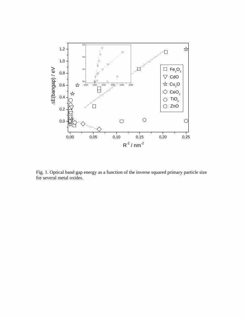
0,00 0,05 0,10 0,15 0,20 0,25
0,0
0,2
0,4
0,6
0,8
1,0
1,2
-0,001 0,000 0,001 0,002 0,003 0,004
0,0
0,1
0,2
0,3
ZnO
Cu2O
TiO2
CeO2
CdO
∆E(b
anga
p) /
eV
R-2 / nm-2
Fe2O3
Fig. 1. Optical band gap energy as a function of the inverse squared primary particle size for several metal oxides.
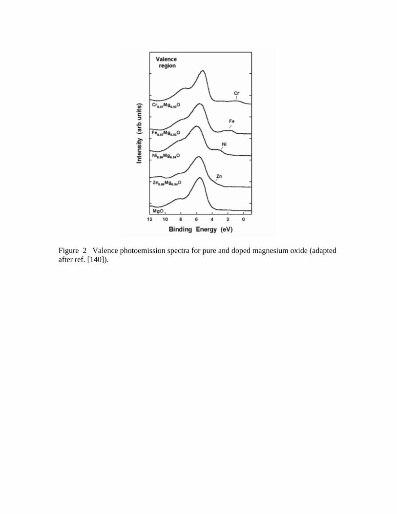
Figure 2 Valence photoemission spectra for pure and doped magnesium oxide (adapted after ref. [140]).
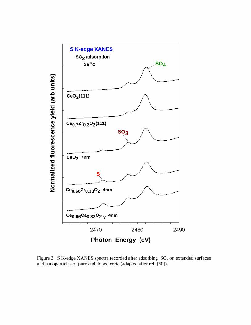
Photon Energy (eV)2470 2480 2490
Nor
mal
ized
fluo
resc
ence
yie
ld (a
rb u
nits
)S K-edge XANES
SO2 adsorption25 oC
CeO2 7nm
CeO2(111)
SO4
SO3
Ce0.7Zr0.3O2(111)
Ce0.66Ca0.33O2-y 4nm
Ce0.66Zr0.33O2 4nm
S
Figure 3 S K-edge XANES spectra recorded after adsorbing SO2 on extended surfaces and nanoparticles of pure and doped ceria (adapted after ref. [50]).


![Antioxidant Cerium Oxide Nanoparticles in Biology and … · Antioxidant Cerium Oxide Nanoparticles in Biology ... dermal burn cream (Flammacerium) [5] ... Antioxidant Cerium Oxide](https://img.pdfslide.us/doc/110x75/5ade477c7f8b9ae1408e286b/antioxidant-cerium-oxide-nanoparticles-in-biology-and-cerium-oxide-nanoparticles.jpg)