Embed Size (px)
Citation preview

International Journal of Ebola, AIDS, HIV and Infectious Diseases and Immunity
Vol.2, No.2, pp.1-14, April 2016
__Published by European Centre for Research Training and Development UK (www.eajournals.org)
1
METAL DYSHOMEOSTASIS IN THE ETIOLOGY FF BILIRUBIN-INDUCED
NEUROLOGIC DYSFUNCTION (D-PENICILLAMINE AS AN
ANTIRETROVIRAL DRUG IN HIV OR EBOLA INFECTION DUE TO VERTICAL
TRANSMISSION)
György Balla1, Lajos Lakatos*2, Zsuzsanna Vekerdy-Nagy3
1Department of Pediatrics, Clinical Centre, University of Debrecen, Debrecen, Hungary. 2Kenézy Teaching Hospital, Debrecen, Hungary.
3Department of Physical and Rehabilitation Medicine, Clinical Centre, University of
ABSTRACT AIM: The aim of this review was to demonstrate a new concept in the etiology
of bilirubin-induced neurologic dysfunction (BIND) and highlight the role of D-Penicillamine
(D-PA) in the treatment of HIV or EBOLA infection due to vertical transmission. METHOD:
We conducted a review searching the literature of bilirubin metabolism and of metal-
homeostasis, furthermore of neonatal HIV and EBOLA infection. RESULTS: Over the past
two decades there have been significant advances in our understanding of copper homeostasis
and of neurodegenerative and neurodevelopmental diseases (NDs), and the pathological
consequences of copper dysregulation. Thus, comprehension of metal homeostasis, details of
transport and interactions with biomolecules, such as unconjugated bilirubin (UCB) or
albumin, is essential for understanding the normal and pathological processes occurring in
the neonatal period. UCB has a special affinity for the globus pallidus, the hippocampus, and
the subthalamic nucleus. Furthermore, immaturity of the blood-brain barrier (BBB) also
plays a role in kernicterus. Homeostasis of metal ions usually involves a huge set of proteins
which regulate the proper metal biology. Metal ions, especially copper and iron play very
important roles in NDs including BIND, having impact on both protein structure (misfolding)
and oxidative stress. INTERPRETATION: Free copper ion in itself or binding to UCB and
forming metal-bilirubin complex(es) involved in neurologic dysfunction; therefore they are
important factors for central nervous system (CNS) damage processes in BIND by the
production of free radicals. Our present research article address the medical necessity of the
use of a chelating agent (D-PA) in the treatment of neonatal hyperbilirubinemia (NHBI).
Finally, the authors highlight that D-PA may have a huge impact on HIV or EBOLA infection
caused by vertical transmission where NHBI is a very common symptom.
KEYWORDS: Bilirubin-Induced Neurologic Dysfunction; Reactive Oxygen Species;
Copper Dyshomeostasis; HIV or EBOLA infection; Neurodegeneration; D-Penicillamine in
the Neonatal Period
INTRODUCTION
The classic form of chronic bilirubin encephalopathy (kernicterus) is a well-described clinical
tetrad of abnormal movements and muscle tone, an auditory processing disturbance with
or without hearing loss, oculomotor impairments, especially impairment of upward vertical
gaze, and dysplasia of the enamel of deciduous teeth. Hervieux first described the condition
in 1847, and Schmorl first used the term kernicterus as early as 1903. 1, 2 NHBI is a common
condition in the first week of postnatal life. Although generally harmless, some neonates may
develop very high levels of UCB. Subtle encephalopathy or BIND refers to individuals with
International Journal of Ebola, AIDS, HIV and Infectious Diseases and Immunity
Vol.2, No.2, pp.1-14, April 2016
__Published by European Centre for Research Training and Development UK (www.eajournals.org)
2
subtle NDs. 3-5 In the past decades, interest in bilirubin damage of the brain has been
reawakened by an increase in its prevalence, owing to failure to closely observe infants
discharged from the hospital well before the peak of NHBI. 6 There is a tremendous variability
in babies' vulnerability toward UCB for reasons not yet explained, but preterm birth, sepsis,
hypoxia, hypoperfusion, hyperosmolality, acidosis, hypalbuminemia and hemolytic disease et
cet. (underlying diseases or comorbidities) are comprised as risk factors, so, the UCB levels
and neurological abnormalities are not strictly correlated. Kernicterus may only be the “tip of
the iceberg.” Subtle UCB damage may account for many more cases of: learning disabilities,
central auditory processing disorders, dyslexia, oculomotor dyspraxia, movement disorders,
and autism spectrum disease (ASD), and may even predispose to Parkinson’s disease (PD) or
schizophrenia in adulthood. 7 The pathomechanisms of BIND have not been fully understood
yet. The mechanisms of UCB neurotoxicity are still also unclear, and little is known about
lasting sequelae attributable to NHBI. Our hypothesis addresses the medical necessity of
chelation therapy (with D-Penicillamine – D-PA) in the neonatal period 8, 9 as it is feasible
that UCB molecule reviels particular affinity to copper stored in basal ganglia (BG) of the
neonatal brain, where copper-bilirubin complex can be formed together with the production
of hydroxyl radical (OH–). In addition, various amount of free metal ions can be found in the
intravascular space and in the tissues (especially in BG) during hemolytic processes.
Pathological basal ganglia activity 10
The BG is a collection of large subcortical nuclear masses. It is agreed that core components
comprise the caudate nucleus, the nucleus accumbens, the putamen, and the globus pallidus.
The caudate nucleus and putamen together are sometimes called the striatum, and the putamen
and globus pallidus are together sometimes described as the lentiform nucleus. 11, 12
Functionally, the BG has considerable connections to the cerebral cortex, thalamus, and brain
stem; so, anatomists consider portions of the thalamus as components of the BG. 13 A
literature review was aimed at assisting us (as pediatricians) to provide further understanding
with bilateral symmetrical BG and thalamic lesions on magnetic resonance imaging (MRI).
The high-signal-intesity lesions on T2-weighted images can be caused by edema, gliosis,
demyelinization, neuronal necrosis, or cystic degeneration both in Wilson disease (WD) and
BIND. 14
Role of metals and oxidative stress in the human neurodegenerative and
neurodevelopmental disorders
The brain (mostly the BG) accumulates among the highest levels of transition metals in the
body for normal function, including redox-active copper. This high-redox metal load, in
combination with the brain disproportionately active oxygen metabolism, makes this organ
particularly susceptible to oxidative stress. 15-18 Metal ions such as calcium, zinc, iron and
copper are key players in brain neurobiology; their homeostasis is altered in most ND
conditions. The metal dyshomeostasis (MD) in the brain and related organs, and loss of the
strict regulation is implicated in neurotoxic stress 19-21 and in a variety of NDs including
BIND and prion-mediated encephalopathies and other diseases 22-24 which can be seen in the
Table 1. 25-47 Parts emphasized with bold-faces show associations between the ND illnesses
and BIND. Pathologic changes to the CNS in these disorders are always associated with a
significant dyshomeostasis of tissue metals (particularly copper). Excess copper may combine
with sulfhydryl, carboxyl, or amine groups, resulting in improper enzymatic activity or
damage to cellular structure. Despite the ubiquitous presence of toxic copper within the brain,
pathologic findings are limited primarily to the BG, thalamus, and brain stem. Histopathologic

International Journal of Ebola, AIDS, HIV and Infectious Diseases and Immunity
Vol.2, No.2, pp.1-14, April 2016
__Published by European Centre for Research Training and Development UK (www.eajournals.org)
3
studies have shown abnormalities throughout this system in patients suffering from MD.
These abnormalities include atrophy, spongy softening, cavitation, a general reduction of
neurons, increased cellularity, and the presence of characteristic cells (Opalski, Lewy bodies).
The pathologic changes are presumed to result from an increased amount of extracellular
copper, which causes oxidative stress and results in cell destruction. 48 Many diseases of the
BG have some disorder of movement as their primary symptom, ranging from an excess of
(abnormal) involuntary movements such as in chorea to a poverty and slowness of movement
as in PD, Alzheimer disease (AD) and WD as illustrated in several clinical cases 49 and UCB
encephalopathy 50 where a characteristic yellow staining can be observed in fresh or frozen
sections of the brain obtained within 7-10 days after the initial bilirubin insult. If the affected
infant survives the neonatal period and subsequently dies, the yellow staining may no longer
be present, but the BG will display microscopic evidence of cell injury, neuronal loss, and
glial replacement. Newborns, especially preterm infants, are particularly vulnerable to
reactive oxygen species (ROS) because they exhibit accelerated production of free radical and
limited antioxidant protection, which increases the susceptibility of rapidly growing tissues to
damage. „Free radical-related diseases” of neonates promote cellular, tissue, and organ
impairments. In 1988, Saugstad coined the phrase „oxygen radical disease in neonatology” to
highlight the crucial role of ROS in a wide range of neonatal disorders. 51 There is now a large
body of literature demonstrating that free or weakly bound iron and copper ions may exert
their toxic action on BG. In a way, metals may provide the link between protein misfold and
aggregation, oxidative stress and the cascade of biochemical alterations, eventually leading to
neuronal cell death. Predominantly the cellular content of copper determines copper-induced
toxicity in brain astrocytes. 52
Potential molecular mechanisms of bilirubin-induced neurologic dysfunction
The “classic” interpretation of bilirubin neurotoxicity does not give sufficient answers to the
following questions: (1) How to call bilirubin: friend or foe? (2) If the bilirubin is really an
„enemy”, how does it induce its dangerous effects?
Ad (1)
The exact UCB concentration associated with kernicterus in the healthy term infant is
unpredictable. In a Danish popolation-based study, the neonates with total serum bilirubin
levels of ≥ 25 mg/dL didn’t show any neurologic dysfunctions at 5 years of follow up. 53
Toxicity levels may vary among ethnic groups, with maturation of an infant, and mainly in
the presence of hemolytic disease. Bilirubin, which is derived from its metabolic precursor
biliverdin, is the end product of heme catabolism. It has been proposed that UCB is an
excellent endogen antioxidant present in human extracellular fluids 54. Bilirubin can suppress
oxidation of lysosomes at oxygen concentrations that are physiologically relevant. It can act
as an important cytoprotector of tissues that are poorly equipped with antioxidant defense
systems, including myocardium and nervous tissue. 55, 56 Serum bilirubin in jaundiced and
non-jaundiced pups exposed to 95% O2 shows a negative correlation with lipid
hydroperoxides at 3 days of exposure. Higher UCB concentrations resulted in lower lipid
hydroperoxide levels. 57 Therefore, we think that UCB in itself is actually our friend, that is:
Bilirubin, The Gold Within.58 (BILIRUBIN- FRIEND OR FOE?? :
Function as natural antioxidants in newborns. Attenuates graft rejection in cardiac transplant
models. Inverse relation between bilirubin and coronary artery disease. Inverse relation

International Journal of Ebola, AIDS, HIV and Infectious Diseases and Immunity
Vol.2, No.2, pp.1-14, April 2016
__Published by European Centre for Research Training and Development UK (www.eajournals.org)
4
between bilirubin and colorectal cancer. – 2005 Powerpoint Presentation
www.sfrbm.org/frs/FrielBilirubin.pdf )
Ad (2) Toxic Side of Bilirubin
Erythrocyte morphological changes have been seen with incubation of cells with different
molar ratios of UCB. These changes occur as the bilirubin/human serum albumin molar ratio
increases. This indicates that bilirubin can illicit toxicity in the erythrocyte membrane in a
concentration and temperature-dependent manner, causing hemolysis. 59 Several studies have
found that NHBI is associated with higher risk of movement disorder and, even more,
developmental delay. The management dilemma for a clinician is that UCB is a beneficial
antioxidant at low (and may be at moderatly higher) levels, but a neurotoxin at >20 mg/dL
levels (“vigintophobia” 60), where it can impair the normal developmental maturation of the
neonatal brain.
Among the 23 elements with known physiological functions, 12 are metals (sodium,
magnesium, potassium, calcium, vanadium, chromium, manganese, iron, cobalt, copper, zinc,
and molybdenum). 61, 62 Copper is essential for the normal growth and development of human
fetuses, infants, and children and it is crucial for the normal development of the brain 63 which
has among the highest levels of copper, as well as iron and zinc, in the body. Copper is an
interesting essential micronutrient. Deficiency and excess intake both induce a variety of
clinical manifestations affecting mainly the hematopoietic system, the skeleton, the liver, and
the brain. Although copper transport to the fetus is high and liver storage is efficient, copper
export from the hepatocytes to the bile and to blood ceruloplasmin (Cp) are reduced during
this stage of life because of liver function immaturity. This leads to a high copper
accumulation in the liver and brain, in a magnitude similar to that observed in Wilson disease.
In fact, an obvious analogy can be observed between the newborns and patients with Wilson
disease in the field of the copper „(dys)homeostasis” (see: Table 1). The increased liver and
brain copper storage of the fetus may have a selective evolutionary advantage since it may
prevent copper deficiency during the first months of life when the child receives a relatively
low copper supply from breast milk.64
Metal regulatory proteins in the neonatal period
A variety of proteins are involved in the regulation of metal metabolism and the oxidative
response and many are involved in iron or copper metabolism due to the redox activity of
those metals. Protein misfolding and conformational changes are also a cornerstone of NDs
All metals with known physiological functions are bound by albumin. 65, 66 A decrease in
metal binding of albumin means more free metal available to produce oxidative stress and
other physiological effects such as influence of calcium (Ca++) homeostasis by altering the
conformational structure of the pumps, enzymes, binding proteins, and channels that regulate
Ca++ flow. Often, this results in elevating free intracellular Ca++ levels which may produce
depletion of glutathione/GSH with a downstream induction of DNA damage and eventual cell
death. 67 Therefore, the bilirubin-mediated neurotoxicity is partly due to increased rate of cell
apoptosis and higher levels of intracellular free Ca++ ion level (as analogy, see Table 1.:
Fahr’s disease). 42
Ceruloplasmin (Cp) is a large blood protein synthesized by the liver with the primary role
of transporting copper. If a disease process (e.g. hepatic failure) or insufficient synthesis in the
neonatal period lowers the production of Cp, the free copper would increase and copper

International Journal of Ebola, AIDS, HIV and Infectious Diseases and Immunity
Vol.2, No.2, pp.1-14, April 2016
__Published by European Centre for Research Training and Development UK (www.eajournals.org)
5
mediated oxidative stress would be enhanced. In addition, there is some evidence that under
oxidative stress conditions, Cp may induce further oxidative stress in a manner akin to a
positive feedback mechanism. Also, when this protein is exposed to ROS, its ability to bind
copper is reduced, releasing free copper, producing further oxidative stress. 68
Copper transporter 1 (Ctr1) has a high affinity for copper and serves to transport copper
into the interior of the cell. It is not highly expressed in the brain, where the choroid plexus
may contain the greater proportion. The lower levels of expression in the brain, however,
should not be taken as a sign that Cu metabolism is not important in the brain as several neural
pathologies (Alzheimer’s disease, spongiform encephalopathies) have been linked to
disordered copper metabolism. 69
Metallothionein (MT) is a cysteine rich protein involved in the regulation of zinc and other
metals (mainly copper, and selenium). This protein is found in a variety of forms (I-IV) in
mammals, and MTII are the most abundant in the CNS where MT is found mostly in
astrocytes. MT plays an important role in cell signaling. Neonatal brain has lower MT
concentrations than adult brain, increasing to adult levels by Day 21. 70 Elucidating the role
of the metallochaperone Atox1, 71, 72 it is obvious that Atox1-deficient cells accumulates
high levels of intracellular copper, and metabolic studies indicate: this defect originated from
the impaired cellular copper efflux. These data reveal a direct role for Atox1 in trafficking of
intracellular copper to the secretory pathway of mammalian cells and demonstrate that
metallochaperone plays a critical role in perinatal copper homeostasis.To sum up, the number
of proteins involved in metal oxidative stress is large and fall into two groups: those involved
in iron or copper metabolism, and those involved with the rest of the metals. Bilirubin-metal
complexes have been made in vitro producing hydroxyl radical (OH͞― ) and there are no
reasons not to believe that such complexes can also exist in vivo, especially since UCB can
take on a ring-like configuration with one of the end pyrrol-rings in a lactim form and the
other in a lactam form. 73, 74 To clarify how hyperbilirubinemia influences
neurodevelopmental outcome, more sophisticated consideration is needed both of how to
assess bilirubin exposure leading to neurotoxicity and of those comorbid conditions which
may lower the threshold for braininjury.46 A decrease in metal binding means of course more
free metal available to produce oxidative stress and other physiological effects. Six substances
are transported with albumin: long chain fatty acids bilirubin, steroids, thyroid hormones,
drugs and copper (also other metals eg. Zn, Pb…).75 Albumin interacts with a broad spectrum
of compounds. Most strongly bound are hydrophobic organic anions of medium size, 100 to
600 Da–long-chain fatty acids, hematin, and bilirubin. The equilibrium constant of UCB is
about 3.8 ± 2.0 x 10⁷ M-1 and the calculated Cu ion binding constant is 1.50 x 10⁶ M-1. In
comparison, albumin is interacting selectively and non-covalently with Cu ions. 76-79 Neonatal
blood has low content of glutathione peroxidase, superoxide dismutase, β-carotene,
riboflavin, α-proteinase, vitamin E, selenium, copper, zinc, ceruloplasmin and other plasma
factors. The premature brain is rich in polyunsaturated fatty acids, and easily oxidized
compared to monounsaturated fatty acids. 80
New concept for development of bilirubin-induced neurologic dysfunction
Table 1. illustrates well that a remarkable similarity exists between Wilson disease and BIND.
Very wide-ranging studies have long been made on the possible biochemical transformations
of UCB, which is formed during the decomposition of haemoglobin. Particular attention has
been paid to its photochemical and redox reactions 81 but the relevant publications comprise
only a very small proportion of those dealing with the molecular biochemistry of UCB and

International Journal of Ebola, AIDS, HIV and Infectious Diseases and Immunity
Vol.2, No.2, pp.1-14, April 2016
__Published by European Centre for Research Training and Development UK (www.eajournals.org)
6
metals interactions. Bilirubin has a special affinity for the globus pallidus, the hippocampus,
and the subthalamic nucleus because they are also target brain regions for divalent metal (Cu,
Fe, Zn et cet.) accumulation.82
Neurodegeneration: a return to immaturity? 83 This question certainly arouses the attention
of neonatologists as the immature and strikingly vulnerable neurons play important role in
the pathogenesis of BIND. The increased vulnerability of premature infants to brain damage
may be due to a proneness of immature nerve cells to toxic stimulus. The developing neurons
undergo programmed cell death, a necessary phenomenon for proper nervous system
development. Following the developmental period, neurons mature and restrict the apoptotic
pathway to permit long-term survival. On the basis of above described abundant research data
and hypotheses, according to our concept, the BIND is an ND of immature brain caused by
accumulation of free metals and UCB-Cu complex (as prooxidant) in the BG and other parts
of CNS relevant to BIND. The rate of formation of UCB-Cu complex when bilirubin extracts
copper from copper–albumin complex, as obtained in a very exciting experiment, is
34.98 l mol−1 s−1 84. The main comorbidity is the hemolysis of neonatal blood red cells. 85
During this process a great amount of heavy metals (mainly iron and copper) may circulate in
free form in the bloodstream, and can pass through the BBB, finding entrance into the CNS
as well. Understanding the differences between neonatal and adult erythrocytes is critical in
the evaluation of perinatal erythrocyte disorders. The reason for the reduced RBC survival
observed in newborns is not known, although there are many biochemical differences between
adult and neonatal RBCs.86 Increased oxidant sensitivity of newborn red cells and relative
instability of fetal hemoglobin have been considered as possible causes for this shortened
lifespan. In a chinese study, 87 the erythrocyte’s copper content was significantly lower in the
maternal blood than in the newborn cord blood. The compounds to be bound and transported
by albumin are quite diverse and include bilirubin, fatty acids, metal ions and therapeutic
agents. Bilirubin itself can displace metals (copper) from the albumin binding because UCB
binds stronger to albumin than copper, in other words, copper loosely bound to albumin. 88
Free or loosely bound, redox-active transition metal ions are potentially extremely pro-
oxidant, having the ability to catalyze the formation of damaging and aggressive ROS from
much more innocuous organic and inorganic species. In strictly biological terms the two most
important such metals are iron and copper.89 In fact, oxidative stress has been demonstrated
to be a common link between several conditions such as PD, AD, stroke, prion diseases and
UCB encephalopathy, where it is involved in neuronal injury.
D-penicillamine as neonatal neuroprotectant 90, 91
Our recently published case reports 92 and other healthy and highly educated patients of the
long-term (28-42 years) follow-up suggest that D-PA therapy of newborn infants may have
significant neuroprotective effects in cases jeopardized by BIND or ROP. The first patient (42
ys) is now a member of a famous operahouse in Germany as an opera singer, the second one
(16 ys) is excellent in music and matematics. These cases are all the more remarkable as the
most common sequelae of NHBI is the sensorineural hearing impairment. These unexpected
effects may be related to DPA capability to alter the most important gasotransmitters (nitric
oxide /NO/ system, carbon monoxide /CO/, hydrogen sulfide /H2S/ biosynthesis, and Cu++
homeostasis in the brain). According to our hypothesis DPA can modulate the function of
these neurotransmitters and can protect the brain (especially the BG and retina) from injury,
such as BIND and ROP. D-PA not only chelates copper from tissue, but also detoxifies tissue
copper by promoting the synthesis of metallothionein, which forms a non-toxic combination
with copper. 36 There are very important age-related effects of D-PA in the neonatal period.

International Journal of Ebola, AIDS, HIV and Infectious Diseases and Immunity
Vol.2, No.2, pp.1-14, April 2016
__Published by European Centre for Research Training and Development UK (www.eajournals.org)
7
In the Table 2. we demonstrate the results of our animal experiments regarding the age related
differences in effects of D-PA. 93 The high activity of heme oxygenase in the newborn could
reflect the enzyme-inducing action of metals: Cu and Fe derived from the breakdown of fetal
erythrocytes. Chelation therapy in neonates restores the normal activity of enzymes
participating in heme metabolism. Briefly, chelating agents facilitate heme synthesis and
inhibit heme degradation. In other words, DPA as a chelating agent, boost or inhibit the
immature enzyme systems to the adult levels The main comorbidity is the hemolysis of
neonatal blood red cells. 85 During this process a great amount of heavy metals (mainly iron
and copper) may circulate in free form in the bloodstream, and can pass through the BBB,
finding entrance into the CNS as well.94
CONCLUSION
The basic role of metal ions in neurological pathologies is generally accepted, ― except for
the case of BIND. Free copper ion in itself or binding to UCB and forming metal-bilirubin
complex(es) involved in neurologic dysfunction, therefore they are important factors for
whole brain damage processes in BIND. Figure 1. demonstrates our concept about the chronic
bilirubin encephalopathy based on the above described hypothesis. We hope that our theory
will help answer some of the unsolved questions and concerns ocurred in the etiology and
pathomechanisms of BIND. The beneficial neuropharmacological actions of metal-targeted
(chelating) agents most likely arise from local metal redistribution rather than from massive
metal removal. 95 The chelation therapy for non-metal overload indications continues to be
investigated. Our present research article address the medical necessity of the use of a
chelating agent (D-PA) in the treatment of NHBI. 96-106
Possible beneficial effects of D-PA on the lethality of HIV orEBOLA infection due to
vertical transmission
West Africa is currently in the midst of the largest EBOLA outbreak in the history and HIV
prevalence in sub saharan Africa is also very high.
The structural and functional properties of DPA make it suitable for exerting antiviral activity.
This drug caused a marked inhibition of polyo-virus-specific RNA and the protein
synthesis.108 Searching the pertinent literature, several publications relating to the beneficial
effects of D-PA-therapy in the treatment of AIDS-patients were found. The high doses
resulted in good outcomes, but adult patients did not tolerate this therapy. In addition to this,
it has been determined that the selective inhibition of replication of HIV type 1 (further: HIV)
by this drug was concentration dependent, that is, at 40 microgram/ml concentration D-PA
completely inhibited HIV replication in H9 cells in vitro 109 (a single 100 mg/kg bw.
IVadministered DPA resulted in more multiple plasma concentration in premature infants
110). This study has a promising idea wondering whether or not it is true that DPA has possible
beneficial effects on the AIDS or EBOLA associated infant mortality rates because of its
prolonged antiviral activity. Abundant experimental evidence and clinical observations exist
to suggest that early viremia and immune responses in vertical HIV infection are different
from those of adults. The developing immune system might allow for more efficient viral
replication and less efficient immune containment of viral replication. In this respect, D-PA-
therapy may be a potent early regime to control HIV (or EBOLA) replication and offers the
golden opportunity to prevent or reverse the rapid progression of these diseases. The potential

International Journal of Ebola, AIDS, HIV and Infectious Diseases and Immunity
Vol.2, No.2, pp.1-14, April 2016
__Published by European Centre for Research Training and Development UK (www.eajournals.org)
8
mechanism of antiretroviral actions of D-PA in infections caused by vertical transmission are
as follows:
-PA is a well-known direct antioxidant) may
provide a promising and cost-effective therapeutic aproach in treating neonatal HIV or
EBOLA infection. The newborn infants, especially the prematures, are suffering in an
oxidative stress condition 111
112.
newborn infants is strikingly similar:
they both have large quantities of copper in the liver and low ceruloplasmine in the
blood. It was previously found that cupric chloride, in the presence of a chelating agent,
could inhibit the HIV-1 protease 113.
β-β-dimethylcysteine) can cause an increase in
intracellular cysteine and glutation content which play an important role as HIV
inhibitors, at least in part because they facilitate the intracellular transport of Zn and
Cuions 114.
-1 nucleocapsid p7 protein contains two retrovirus-type zinc finger domains that
are required for multiple phases of viral replication. Considering the chelating properties
of DPA and its disulfide reaction with cysteine, one can conclude that HIVorEBOLA-
replication could be inhibited by this drug 115-120.
It would be very exciting to be involved in this work, especially that D-PA will have a huge
impact on HIV or EBOLA infection caused by vertical transmission where NHBI is a very
common symptom.
Conflict of Interest Statement
The authors declare that the research was conducted in the absence of any commercial or
financial relationships that could be construed as a potential conflict of interest.
Abbreviations
AD - Alzheimer disease; ASD - autism spectrum disease; BBB - Blood brain barrier; BG
- basal ganglia; BIND - Bilirubin-induced neurologic dysfunction; CNS- central nervous
system; Cp - ceruloplasmin; CuFr - Free copper ion; D-PA - D-Penicillamine; MD -
Metal dyshomeostasis; MRI - Magnetic resonance imaging; MT - Metallothionein; NHBI
- Neonatal hyperbilirubinemia; OH– - Hydroxyl radical; NDs - Neurodegenerative and
neurodevelopmental diseases; PD - Parkinson disease; ROS - Reactive oxygen species;
UCB - Unconjugated bilirubin; UCBFr - free UCB; WD - Wilson disease
REFERENCES
Hansen TWR: Pioneers in the scientific study of neonatal jaundice and kernicterus.
Pediatrics 2000; 106: e15 http://www.pediatrics.org/cgi/content/full/106/2/e15;
Shapiro SM. Chronic bilirubin encephalopathy: diagnosis and outcome.
Semin Fetal Neonatal Med 2010; 15: 157–163.

International Journal of Ebola, AIDS, HIV and Infectious Diseases and Immunity
Vol.2, No.2, pp.1-14, April 2016
__Published by European Centre for Research Training and Development UK (www.eajournals.org)
9
Stevenson DK, Vreman HJ, Wong RJ. Bilirubin production and the risk of
bilirubin neurotoxicity. Semin Perinatol 2011; 35: 121-126.
Brites D. The evolving landscape of neurotoxicity by unconjugated bilirubin: role
of glial cells and inflammation. Front Pharmacol 2012; 3: 136.doi:10.3389/fphar.2012
Lunsing RJ. Subtle bilirubin-induced neurodevelopmental dysfunction (BIND) in the
term and late preterm infant: Does it exist? Semin Perinatol 2014 38: 465–471.
Shapiro SM. Kernicterus; Neonatal Hyperbilirubinemia, Neurotoxicity and
Bilirubin-Induced Neurological Disorders (BIND). Clinical Advences in Pediatrics
November 16, 2012 Children’s Mercy HOSPITALS & CLINICS. Kansas City
Maisels MJ, Newman TB. Jaundice in full-term and near-term babies who leave the
hospital within 36 hours. The pediatrician' nemesis. Clin Perinatol 1998; 25: 295–302.
Lakatos L, Kövér B, Oroszlán G, et al. D-Penicillamine Therapy in AB0
Hemolytic Disease of the Newborn Infant. Europ J Pediat 1976; 123: 133-137.
Balla G, Lakatos L, Vekerdy-Nagy Z. Chelation therapy in the neonatal period:
D-Penicillamine can exert neuroprotective effects in kernicterus and retinopathy of
prematurity. IJPSR 2015; 6: 4269-4276.
Wichmann T, Dostrovsky JO. Pathological basal ganglia activity in movement
disorders. Neuroscience 2011; 198: 232–244.
Russmann H, Vingerhoets F, Ghika J, et al. Acute Infarction Limited to the Lenticular
Nucleus, Clinical, Etiologic, and Topographic Features. Arch Neurol 2003; 60: 351-355.
Abbruzzese G, Berardelli A. Sensorimotor integration in movement disorders.
Mov Disord 2003; 18: 231-240.
Zuccoli G, Yannes MP, Nardone R et al. Bilateral symmetrical basal ganglia
and thalamic lesions in children: an update. Neuroradiology 2015; sDOI
10.1007/s00234-015-1568-7.
Bekiesinska-Figatowska M, Mierzewska H, Jurkiewicz E: Basal ganglia lesions in
children and adults. Europ J Radiol 2013; 82: 837–849.
Dodani SC, Firl A, Chan J, et al. Copper is an endogenous modulator of neural circuit
spontaneous activity. PNAS 2014; 111: 16280–16285.
Dickinson BC, Chang CJ. Chemistry and biology of reactive oxygen species in
signaling or stress responses. Nat Chem Biol 2011; 7: 504–511.
Barnham KJ, Masters CL, Bush AI.Neurodegenerative diseases and oxidative stress.
Nat Rev Drug Discov 2004; 3: 205–214.
Savelieff MG, Lee S, Liu Y et al. Untangling amyloid-β, tau, and metals in
Alzheimer’s disease. ACS Chem Biol 2013; 8: 856–865.
Schlief ML, Craigand AM, Gitlin JD. NMDA receptor activation mediates
copper homeostasis in hippocampal neurons. J Neurosci 2005; 25: 239–246.
Schlief ML, Wes T, Craig AM, et al. Role of the Menkes copper-transporting ATPase
in NMDA receptor-mediated neuronal toxicity. PNAS 2006; 10: 14919–14924.
Sparks DL, Schreurs BG. Trace amounts of copper in water induce beta-amyloid
plaques and learning deficits in a rabbit model of Alzheimer’s disease.
PNAS 2003; 100: 11065–11069.
Singh I, Sagare AP, Coma M, et al. Low levels of copper disrupt brain amyloid-β
homeostasis by altering its production and clearance. PNAS 2013; 110: 14771–14776.
You H, Tsutsui S, Hameed S, et al. A beta neurotoxicity depends on interaction
between copper ions, prion protein, and N-methyl-d-asparate receptors.
PNAS 2012; 109: 1737–1742.
Barnham KJ, Bush AI. Biological metals and metal-targeting compounds
in major neurodegenerative diseases. Chem Soc Rev 2014; 43: 6727-6749.

International Journal of Ebola, AIDS, HIV and Infectious Diseases and Immunity
Vol.2, No.2, pp.1-14, April 2016
__Published by European Centre for Research Training and Development UK (www.eajournals.org)
10
Gonzalez-Cuyar LF, Perry G, Miyajima H, et al. Redox active iron accumulation in
aceruloplasminemia. Neuropathology 2008; 28: 466–471.
López OL, DeKosky ST. Clinical symptoms in Alzheimer's disease.
Handbook of Clinical Neurology 2008; 89: 207–216.
Henneman WJ, Sluimer JD, Barnes J, et al. Hippocampal atrophy rates in
Alzheimer disease: added value over whole brain volume measures.
Neurology 2009; 72: 999-1007.
Squitti R, Polimanti R. Copper phenotype in Alzheimer's disease: dissecting the
pathway. Am J Neurodegener Dis 2013; 21: 46-56.
Kiernan MC, Vucic S, Cheah BC, et al. Amyotrophic lateral sclerosis.
The Lancet 2011; 377: 942–955.
Kaler SG, Holmes CS, Goldstein DS, et al. Neonatal Diagnosis and
Watchko JF, Oski FA: Bilirubin 20 mg/dL = Vigintiphobia. Pediatrics 1983; 71: 660-663.

International Journal of Ebola, AIDS, HIV and Infectious Diseases and Immunity
Vol.2, No.2, pp.1-14, April 2016
__Published by European Centre for Research Training and Development UK (www.eajournals.org)
11
Table 1. Neurodegenerative and neurodevelopment diseases involving metal dyshomeostasis (interpretation can be found in the text)
Disease Clinical symptoms Histopathology
MRI
Metal dyshomeostasis
Aceruloplasminemia 25 Ataxia, chorea,
blepharospasmus,
dementia
Nerve cell loss,
abnormal mastocytes, glubular structure;
Bilateral low signal on T2 at the basal ganglia
Redox active iron
accumulation
Alzheimer disease 26-28 Dementia and movement
disorders
Neuronal and synaptic loss, amyloid plaques,
neurofibrillar
degeneration;
Hippocampal and whole brain atrophy
Abnormal accumulation
and
distribution of reactive iron
and copper
Amyotrophic lateral sclerosis
29
Slowly progressive, painless
weakness, with limb
involvement or bulbar
symptoms
Cerebral atrophy, upper motor neuron dysfunction
Hyperintense corticospinal tracts
Copper mediated
neurological disorder
Menkes’s disease 30 Brittle, kinky hair Feeding
difficulties Irritability
Lack of muscle tone,
floppiness (hypotonia)
Extensive neurodegeneration int he gray matter and
arteries of the brain.
Cerebral and cerebellar atrophy, delayed myelination,
bilateral basal ganglia changes (thalamus)
Copper mediated neurological disorder
Huntington disease 31 Impairments in voluntary
movements and cognitive
function, behavioral and
pszichiatric disorders
The striatum is the area most severely impacted; the
outer cortical regions also shows damage T2
ypointensities in basal ganglia
Mitochondrial metal
dyshomeostasis and
dysfunction
Copper and iron
concentrations are increased
in the striata

International Journal of Ebola, AIDS, HIV and Infectious Diseases and Immunity
Vol.2, No.2, pp.1-14, April 2016
__Published by European Centre for Research Training and Development UK (www.eajournals.org)
12
Occipital horn syndrome 32 Soft skin, weak muscle tone,
stroke, internal bleeding,
kyphoscoliosis
A rare disorder of elastic tissue resulting in loose,
redundant, hypoelastic skin. Irregularities consistent
with periventricular leukomalacia
Inborn errors of metal
(copper) metabolism
Disease Clinical symptoms Histopathology
MRI
Metal dyshomeostasis
Parkinson disease 33-35 Tremor (shaking)
Slowness of movement
Rigidity (stiffness) Sleep
problems Anxiety, Dementia
Depression
Loss of dopaminergic neurons from the substantia nigra
associated with the presence of intraneuronal inclusions
called Lewy bodies.
With new imaging techniques: diffusional changes in the
orbital-frontal region in the pre-motor phase of PD
Iron accumulation and
dyshomeostasis
Wilson disease 36, 37 Deterioration in school
performance or handwriting,
mild tremors, dystonia,
ataxia, muscular rigidity,
dysarthria
The lenticular nuclei macroscopically appear brown in
color, degeneration occurs with disease progression,
leading to necrosis, gliosis and cystic changes, and
lesions can be seen in the brainstem, thalamus,
cerebellum and cerebral cortex, presence of Opalski
cells. Hyperintense signals in putamen and globus
pallidus
Copper accumulation in
the basal ganglia
Distonias 38 Dystonia is an undesirable,
involuntary muscular
movement of a part of the
body.
Related to a problem in the basal ganglia.
In most cases, no abnormalities are visible using MRI or
other diagnostic imaging.
Detrimental effects of
transition metal (copper)
dysregulation
Tourette syndrome 39 Different types of tics: arm
thrusting, eye blinking,
jumping, kicking, repeated
throat clearing or sniffing,
shoulder shrugging
Structural changes in frontal cortex and striatum
Structural changes in the basal ganglia, limbic structures
and prefrontal cortex, T2 relaxation time asymmetries
Genetic heavy metal
toxicity, Free copper
may be elevated
Autism spectrum
disorders (ASD) 40, 41
Delayed speech
development,
The cerebellum have documented cellular and
neurochemical
The Zn/Cu ratio could be
considered as biomarker of

International Journal of Ebola, AIDS, HIV and Infectious Diseases and Immunity
Vol.2, No.2, pp.1-14, April 2016
__Published by European Centre for Research Training and Development UK (www.eajournals.org)
13
Disease Clinical symptoms Histopathology
MRI
Metal dyshomeostasis
Fahr’s disease 42
IBSPDC.
Hallervorden-Spatz
(HSD) disease 43
Gait and speech
disturbance,
limb and truncal ataxia,
dementia, bradykinesia,
mild parkisonism
Progressive dementia,
spasticity, rigidity,
dystonia, and
choreoathetosis
Idiopathic bilateral striatopallidodentate calcinosis
(IBSPDC)
A striking high density
area in the basal ganglia and the dentate nuclei of the
cerebellum.
Juvenile-onset generalized neuroaxonal dystrophy
Bilaterally symmetrical, hyperintense signal changes in the
anterior medial globus pallidus, with surrounding
hypointensity in the globus pallidus, on T2-weighted images.
The increased levels of Cu, Zn, Fe
and Mg reflect the involvement of
metabolism of several metals and/or
metal-binding proteins during the
progression of IBSPDC.
Neurodegeneration with brain
iron accumulation
BIND 44- 47 Neurological, learning and
movement disorders,
isolated hearing loss, and
auditory dysfunction
The abnormalities result from damage to the basal ganglia.
High T2 signal in globus pallidus
„Abnormal” accumulation
and distribution of
reactive iron, copper and
zinc in the basal ganglia
reacting unusally negatively,
having repetitive
movements.
Developmental disorder
affecting
people interact with the
world.
alterations, cellular and gross anatomic changes of the
vermis
cerebelli
Abnormally increased total brain volume.
Decreases in cortical thickness in the frontal, temporal,
and occipital lobes.
ASD. Free copper may be
elevated
International Journal of Ebola, AIDS, HIV and Infectious Diseases and Immunity
Vol.2, No.2, pp.1-14, April 2016
__Published by European Centre for Research Training and Development UK (www.eajournals.org)
14
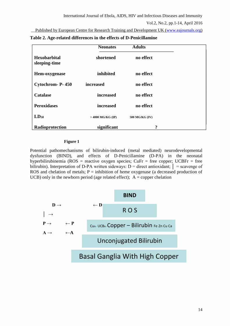
Table 2. Age-related differences in the effects of D-Penicillamine
Neonates Adults
Hexobarbital shortened no effect
sleeping-time
Hem-oxygenase inhibited no effect
Cytochrom- P- 450 increased no effect
Catalase increased no effect
Peroxidases increased no effect
LD50 > 4000 MG/KG (IP) 500 MG/KG (IV)
Radioprotection significant ?
Figure 1
Potential pathomechanisms of bilirubin-induced (metal mediated) neurodevelopmental
dysfunction (BIND), and effects of D-Penicillamine (D-PA) in the neonatal
hyperbilirubinemia (ROS = reactive oxygen species; CuFr = free copper; UCBFr = free
bilirubin). Interpretation of D-PA written sideways: D = direct antioxidant; │ = scavenge of
ROS and chelation of metals; P = inhibition of heme oxygenase (a decreased production of
UCB) only in the newborn period (age related effect); A = copper chelation
D → ← D
│ → ← │
P → ← P
A → ←A
Basal Ganglia With High Copper
Unconjugated Bilirubin
CuFr UCBFr Copper – Bilirubin Fe Zn Cu Ca
│
R O S
BIND