Embed Size (px)
Citation preview
Metabolism in Fungal Pathogenesis
Iuliana V. Ene1,5,6, Sascha Brunke2,6, Alistair J.P. Brown1, and Bernhard Hube2,3,4
1Aberdeen Fungal Group, School of Medical Sciences, Institute of Medical Sciences, Universityof Aberdeen, Foresterhill, Aberdeen AB25 2ZD, United Kingdom
2Department of Microbial Pathogenicity Mechanisms, Hans Knoell Institute, 07745 Jena, Germany3Friedrich Schiller University, 07743 Jena, Germany4Center for Sepsis Control and Care, Universitatsklinikum Jena, 07747 Jena, Germany
Correspondence: [email protected]; [email protected]
Fungal pathogens must assimilate local nutrients to establish an infection in their mammalianhost. We focus on carbon, nitrogen, and micronutrient assimilation mechanisms, discussinghow these influence host–fungus interactions during infection. We highlight several emerg-ing trends based on the available data. First, the perturbation of carbon, nitrogen, or micro-nutrient assimilation attenuates fungal pathogenicity. Second, the contrasting evolutionarypressures exerted on facultative versus obligatory pathogens have led to contemporary path-ogenic fungal species that display differing degrees of metabolic flexibility. The evolution-arily ancient metabolic pathways are conserved in most fungal pathogen, but interestinggaps exist in some species (e.g., Candida glabrata). Third, metabolic flexibility is generallyessential for fungal pathogenicity, and in particular, for the adaptation to contrasting hostmicroenvironments such as the gastrointestinal tract, mucosal surfaces, bloodstream, andinternal organs. Fourth, this metabolic flexibility relies on complex regulatory networks,some of which are conserved across lineages, whereas others have undergone significantevolutionary rewiring. Fifth, metabolic adaptation affects fungal susceptibility to antifungaldrugs and also presents exciting opportunities for the development of novel therapies.
Nutrient assimilation is a central and funda-mental prerequisite for the growth and sur-
vival of all living organisms. Pathogenic fungiinhabit dynamic and contrasting niches andmust display rapid and effective adaptation tochanges in nutrient availability in these mi-croenvironments. To achieve this, they regulatespecific nutrient uptake mechanisms and mod-ulate their metabolism, displaying an impres-
sive degree of metabolic flexibility. This meta-bolic flexibility, which enhances the fitness ofthe fungus, is often as essential for pathogenic-ity as virulence factors, thereby representing anattractive target for potential therapeutic inter-vention.
The major fungal pathogens of humans haveevolved in a polyphyletic manner (i.e., pathoge-nicity has emerged independently in different
5Present address: Department of Molecular Microbiology and Immunology, Brown University, Providence, RI 02912.6These authors contributed equally to this work.
Editors: Arturo Casadevall, Aaron P. Mitchell, Judith Berman, Kyung J. Kwon-Chung, John R. Perfect, and Joseph Heitman
Additional Perspectives on Human Fungal Pathogens available at www.perspectivesinmedicine.org
Copyright # 2014 Cold Spring Harbor Laboratory Press; all rights reserved
Advanced Online Article. Cite this article as Cold Spring Harb Perspect Med doi: 10.1101/cshperspect.a019695
1
ww
w.p
ersp
ecti
vesi
nm
edic
ine.
org
on March 27, 2019 - Published by Cold Spring Harbor Laboratory Press http://perspectivesinmedicine.cshlp.org/Downloaded from
phylogenetic branches of the fungal kingdom).Furthermore, major fungal pathogens displaydifferent lifestyles, and, consequently, their met-abolic flexibility has been shaped by differentevolutionary pressures. For example, the asco-mycete Candida albicans is a facultative patho-gen that exists primarily as a commensal of theoral cavity, gastrointestinal tract, and urogenitaltract but can also persist within other extracel-lular microenvironments (blood, tissues) andwithin intracellular microenvironments (insidedamaged epithelial and endothelial cells, andeven the phagolysosome of macrophages). Dur-ing its commensal stage, C. albicans must adaptto the nutrients present on mucosal surfacesand compete with cohabitating microflora,whereas during infection, the fungus exploitsan alternative array of nutrients available inhost tissues. The apparent lack of a significantenvironmental niche for C. albicans (Odds1988) means that, in recent evolutionary time,the metabolic flexibility of this fungus has beentuned to these host niches. In contrast, fungisuch as the basidiomycete Cryptococcus neo-formans and the ascomycete Histoplasma cap-sulatum, are opportunistic pathogens that areassociated with environmental niches such aspigeon guano, soil, and trees but can causechronic pulmonary infections and devastatingsystemic infections in immunocompromisedindividuals (Sil 2006; Heitman 2011). The met-abolic flexibility of these fungal pathogens en-sures survival during the saprophytic phases oftheir life cycle as well as promoting their abilityto cause pulmonary and disseminated infec-tions (Kronstad et al. 2012). Therefore, differentevolutionary pressures have been imposed onthe processes that mediate metabolic adaptationin major fungal pathogens.
In addition, there are significant differencesbetween fungal pathogens regarding the degreeof evolutionary adaptation to their mammalianhosts. For example, the filamentous ascomycete,Aspergillus fumigatus is probably an accidentalpathogen. This “grass eater” (Tekaia and Latge2005) lives mainly as a saprophyte, degradingplant and other organic material in the environ-ment but can cause severe pulmonary infec-tions in immunocompromised patients. At the
other end of the spectrum, Pneumocystis speciesare obligate fungal pathogens that appear tohave coevolved with their mammalian hosts tosuch an extent that they have shed several met-abolic pathways and, so far, it has not been pos-sible to culture them in vitro (Cushion 2004;Cushion et al. 2007; Hauser et al. 2010). C. al-bicans and Candida glabrata lie between theseextremes, apparently being obligate parasites ofwarm-blooded animals (Odds 1988) and yetretaining a high degree of metabolic flexibilityin vitro and in vivo (Brown et al. 2007; Wilsonet al. 2009). Clearly the metabolic flexibility offungal pathogens has been influenced by theevolutionary time scales over which environ-mental selection pressures have been exerted,as well as by the nature of these pressures.
In this review, we focus on the metabolicflexibility of major fungal pathogens duringthe infection process: commensalism, coloniza-tion, and disease progression. We address majorfungal pathogens that are highly significant inclinical settings, such as C. albicans, A. fumiga-tus, and C. neoformans, mentioning other path-ogens where appropriate. We concentrate onnitrogen and carbon metabolism, and micro-nutrient assimilation, which are both criticalfor pathogenesis and well characterized in path-ogenic fungi.
NITROGEN ASSIMILATION
Nitrogen is required for almost all biosyntheticprocesses and, like carbon, must be assimilatedin large quantities. Therefore, the acquisition ofboth nitrogen and carbon compounds from thehost is essential for pathogenic fungi to survive,grow, and persist within a host. The high degreeof metabolic flexibility is reflected in the differ-ent types of nitrogen sources exploited by fun-gal pathogens. In the absence of preferred nitro-gen sources, such as ammonia or certain aminoacids, these fungi can use compounds like pro-teins or polyamines. Some pathogenic fungi(e.g., pathogenic Aspergillus species) can alsoreduce nitrate to ammonia (Zhou et al. 2002).However, the pathogenic Candida and Crypto-coccus species are unable to use nitrate as a ni-trogen source.
I.V. Ene et al.
2 Advanced Online Article. Cite this article as Cold Spring Harb Perspect Med doi: 10.1101/cshperspect.a019695
ww
w.p
ersp
ecti
vesi
nm
edic
ine.
org
on March 27, 2019 - Published by Cold Spring Harbor Laboratory Press http://perspectivesinmedicine.cshlp.org/Downloaded from
Although fungal nitrogen utilization hasbeen extensively studied in vitro, it is less clearwhich nitrogen sources are used by humanpathogenic fungi in vivo. However, transcrip-tional profiling studies using microarrays or se-rial analysis of gene expression (SAGE) in exvivo or in vivo infection models have providedinsights into the nitrogen metabolism of path-ogenic fungi such as C. albicans (Fradin et al.2005; Thewes et al. 2007; Zakikhany et al. 2007;Walker et al. 2009; Wilson et al. 2009; Chenget al. 2013), C. glabrata (Kaur et al. 2007), A.fumigatus (McDonagh et al. 2008), C. neofor-mans (Fan et al. 2005; Steen et al. 2003), anddermatophytes (Staib et al. 2010) during infec-tion (reviewed by Cairns et al. 2010).
Proteases, Oligopeptide, and Amino AcidTransporters
During infection, C. albicans expresses varioussecreted aspartic proteases (Saps), one of thebest-investigated virulence attributes of thisfungus (Naglik et al. 2003). Liberated oligopep-tides and amino acids are then taken up bydedicated oligopeptide transporters (Opt1–8)(Reuss and Morschhauser 2006) and a family of22 predicted amino acid permeases (Sychrovaand Souciet 1994; Kraidlova et al. 2011). Whenexpressed in host tissues, these factors enableC. albicans to use a broad range of nitrogensources over the course of an infection, drivinghost tissue damage and invasion (Villar et al.2007; Naglik et al. 2008; Dalle et al. 2010; Chenget al. 2013). This proteolytic activity might alsopromote immune evasion, as factors of the im-mune system, antimicrobial proteins, and pep-tides may be degraded (Naglik et al. 2003;Gropp et al. 2009; Meiller et al. 2009). Similarly,the aspartic proteases of C. glabrata and Candi-da parapsilosis have been implicated in survivalfollowing phagocytosis by macrophages (Kauret al. 2007; Horvath et al. 2012).
Extracellular proteases have also been re-ported to be virulence attributes for the mostcommon airborne pathogenic fungus, A. fumi-gatus (Monod et al. 1999), which contains mul-tiple protease genes (Nierman et al. 2005). Al-though an oligopeptide transporter and a key
regulator for extracellular proteolysis are re-quired for in vitro growth of A. fumigatus oncomplex substrates, these proteins are not cru-cial for survival in vivo (Hartmann et al. 2011).Similarly, C. albicans Sap2, a protease essentialfor the growth on protein as the sole nitrogensource, is not essential for survival in vivo (Hubeet al. 1994, 1997). Furthermore, similar obser-vations have been made for dermatophytes suchas Arthroderma benhamiae and Trichophyton ru-brum. Proteases have always been considered defacto to be a key virulence attribute of dermato-phytes, as these fungi infect keratin-containingtissue and are necessarily keratinolytic. Indeed,their genomes display a clear enrichment in pro-tease genes (Burmester et al. 2011). However,those protease genes most highly expressed dur-ing growth on keratin in vitro, and hence forwhich an important role in virulence would beexpected, differ from those genes that are highlyup-regulated during infection (Zaugg et al.2009; Staib et al. 2010). This highlights the factthat in vitro growth conditions often differ sig-nificantly from in vivo microenvironments.
Nitrogen Acquisition in Host Niches
Little is known about the nitrogen sources thatpathogenic fungi assimilate in host niches. Pre-sumably the types and concentrations of nitro-gen source differ significantly between certainniches, such as mucosal surfaces, the gastroin-testinal (GI) tract, and the bloodstream. Mostinformation about fungal nitrogen assimilationhas been gleaned through fungal transcriptom-ics using ex vivo infection models combinedwith limited molecular dissection via targetedgene mutation.
The environment inside the phagosome ofphagocytes is thought to be poor in nitrogen.This is reflected in the transcriptional responseof C. albicans and C. glabrata after internaliza-tion by macrophages (Lorenz et al. 2004; Seideret al. 2010; Roetzer et al. 2011; Brunke and Hube2013; Mayer et al. 2013; Miramon et al. 2013).For example, C. albicans up-regulates aminoacid biosynthetic pathways, suggesting that thefungus at least transiently faces amino acid dep-rivation. The arginine biosynthetic pathway is
Metabolism in Fungal Pathogenesis
Advanced Online Article. Cite this article as Cold Spring Harb Perspect Med doi: 10.1101/cshperspect.a019695 3
ww
w.p
ersp
ecti
vesi
nm
edic
ine.
org
on March 27, 2019 - Published by Cold Spring Harbor Laboratory Press http://perspectivesinmedicine.cshlp.org/Downloaded from
the only amino acid biosynthesis pathway sig-nificantly up-regulated in C. albicans cells afterphagocytosis by macrophages (Lorenz et al.2004). Engulfment by neutrophils induces theup-regulation of arginine, leucine, lysine, andmethionine anabolic pathways as well as GCN4(Rubin-Bejerano et al. 2003; Fradin et al. 2005),the gene encoding the master regulator of aminoacid synthesis (Tripathi et al. 2002). C. glabrataalso up-regulates the synthesis of arginine andlysine on internalization by macrophages (Kauret al. 2007).
The specific contributions of these aminoacids to fungal survival within the phagocyteremain to be tested. However, arginine biosyn-thetic gene induction might play a critical rolein the fungal response to macrophage-derivedreactive oxygen species (Jimenez-Lopez et al.2013). Arginase can convert arginine to urea,which in turn is degraded by urea amidolyaseto produce ammonia and CO2. These break-down products can contribute to neutralizationof the acidic pH of the phagolysosome, promot-ing the intracellular induction of hyphal forma-tion and allowing fungal escape and killing ofthe host cell (Ghosh et al. 2009). Similarly, theextracellular ureases of C. neoformans and Cryp-tococcus gattii (Ure1) degrade urea, which isabundant in the cerebrospinal fluid. The liber-ated CO2 acts as a signal to induce capsule for-mation, a well-studied virulence factor in thisfungus (Frazzitta et al. 2013).
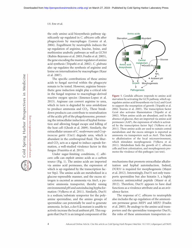
Under sugar-limiting conditions, C. albi-cans cells can exploit amino acids as a carbonsource (Fig. 1). The amino acids are importedvia amino acid permeases, the expression ofwhich is up-regulated by the transcription fac-tor Stp2. The amino acids are metabolized in aglucose-repressible manner, and the excess ni-trogen is excreted as ammonia via Ato5, a pu-tative ammonia transporter, thereby raisingenvironmental pH and autoinducing hypha for-mation (Vylkova et al. 2011). Similarly, Dur31is a sodium/substrate symporter for the poly-amine spermidine, and the amino groups ofspermidine can potentially be used to generateammonia. In fact, a dur31D mutant is unable toactively increase the local ambient pH. This sug-gests that Dur31 is an integral component of the
mechanisms that promote extracellular alkalin-ization and hyphal autoinduction. Indeed,DUR31 is required for morphogenesis (Mayeret al. 2012). Interestingly, Dur31 not only trans-ports spermidine but also histatin 5, a highlycytotoxic antimicrobial peptide (Mayer et al.2012). Therefore, Dur31 appears to have dualfunctions as a virulence attribute and as an avir-ulence factor.
The response of C. albicans to neutrophilsalso includes the up-regulation of the ammoni-um permease genes MEP1 and MEP2 (Fradinet al. 2005). By analogy to the amino acid trans-porters and the spermidine transporter Dur31,the roles of these ammonium transporters ex-
Starvation Abundant
Stp2
NH3
Gcn2
Gcn4
Ambient pH
Morphogenesis
Pathogenesis
Growth
Amino acidbiosynthesis
Centralmetabolism
Amino acids
Glucose
Excess NAto5
AAP
Figure 1. Candida albicans responds to amino acidstarvation by activating the GCN pathway, which up-regulates amino acid biosynthesis via Gcn2 and Gcn4to support the resumption of growth (Tripathi et al.2002; Tournu et al. 2005). The transcription factorGcn4 also activates filamentation (Tripathi et al.2002). When amino acids are abundant, and in theabsence of glucose, they are imported via amino acidpermeases (AAP), the expression of which is activat-ed by the transcription factor Stp2 (Vylkova et al.2011). These amino acids are used to sustain centralmetabolism and the excess nitrogen is exported asammonia via transporters such as Ato5. This leadsto alkalinization of the local microenvironment,which stimulates morphogenesis (Vylkova et al.2011). Metabolism fuels the growth of C. albicanscells and host colonization, and morphogenesis pro-motes the virulence of this pathogen (see text).
I.V. Ene et al.
4 Advanced Online Article. Cite this article as Cold Spring Harb Perspect Med doi: 10.1101/cshperspect.a019695
ww
w.p
ersp
ecti
vesi
nm
edic
ine.
org
on March 27, 2019 - Published by Cold Spring Harbor Laboratory Press http://perspectivesinmedicine.cshlp.org/Downloaded from
tend beyond the uptake of nitrogen, becauseMep2 can activate hyphal formation in responseto ammonium limitation. This signaling roleappears to be independent of the role in theextracellular alkalinization hyphal autoinduc-tion pathway (Biswas and Morschhauser 2005).
Interestingly, C. glabrata can form pseudo-hyphae-like structures under certain nitro-gen starvation conditions (Csank and Haynes2000). However, unlike C. albicans, this filamen-tous morphology is not used as an escape mech-anism from macrophages. Instead, C. glabratacontinues to replicate as yeasts inside thephagosome until the host cell bursts by an un-known mechanism (Seider et al. 2011). This in-tracellular survival and replication depends on aspecialized form of autophagy (pexophagy) tosurvive internalization by macrophages (Roet-zer et al. 2010).
In general, the recycling of cellular proteinsis a common strategy of fungi to overcome ni-trogen starvation. For example, C. albicans vac-uolar protease genes associated with intracellu-lar protein degradation such as APR1, PRB1,PRB2, or PRC1 are up-regulated in responseto phagocytosis by either macrophages (Lorenzet al. 2004) or neutrophils (Fradin et al. 2005). Asecond, ubiquitin-dependent pathway contrib-utes to protein recycling under nutrient-limit-ing conditions in C. albicans (Leach et al. 2011).This might explain why autophagy is not essen-tial for this fungus to survive and to form hy-phae within macrophages. C. albicans cells withdefects in autophagy and cytoplasm-to-vacuoletrafficking (atg9D) can survive macrophagesand retain the ability to kill host cells (Palmeret al. 2007). Meanwhile, C. albicans polyubiqui-tin (ubi4) mutants display attenuated virulence(Leach et al. 2011). Similarly, A. fumigatus doesnot require autophagy for full virulence in aneutropenic mouse model (Richie et al. 2007),probably because its hyphae are not taken up bymacrophages (Palmer et al. 2008). In contrast,fungi that can reside inside the phagosome for along time, such as C. glabrata and C. neofor-mans, seem to require autophagy for full viru-lence, and the inactivation of the autophagicsystem reduces the virulence of C. neoformansin mice (Hu et al. 2008).
Regulation of Nitrogen Metabolism
Flexibility in nitrogen assimilation requiressensing of the available nitrogen sources andappropriate regulation of nitrogen assimilationgenes. There are strong similarities betweenC. albicans and the model yeast, Saccharomycescerevisiae, with respect to the regulation of keynitrogen acquisition pathways. In C. albicans,this occurs mainly via the SPS sensor mecha-nism, comprising the amino acid receptor Csy1(a Ssy1 homolog), the scaffold protein Ptr3, andthe signaling endopeptidase Ssy5 (Ljungdahl2009). Similar to S. cerevisiae, the presence ofextracellular amino acids is detected by theSPS sensor, leading to the proteolytic process-ing, activation, and nuclear translocation of thetranscription factors Stp1 and Stp2 (Andreas-son and Ljungdahl 2002; Martinez and Ljung-dahl 2005). In an interesting twist, C. albicansStp1 and Stp2 each activate a specific subset ofnitrogen acquisition-related genes. Stp1 acti-vates the transcription of genes involved inprotein utilization, such as the genes encodingthe secreted protease Sap2 and the oligopeptidetransporter Opt1. On the other hand, Stp2 ac-tivates transcription of amino acid permeasegenes, for example, GAP1, GAP2, and CAN1(Martinez and Ljungdahl 2005). As Stp1 levelsare also strongly reduced in the presence of ami-no acids, this system allows C. albicans to usefree amino acids when they are available, andto obtain nitrogen from extracellular proteinsunder amino acid-limiting conditions. In thehost, this allows optimal utilization of availablenitrogen sources, and hence contributes to thepathogenicity potential of C. albicans (Ljung-dahl 2009).
Microarray data suggest a key role for theGpa1-cAMP–PKA pathway in the survival ofC. neoformans within macrophages, which in-cludes the up-regulation of amino acid trans-porters (Fan et al. 2005). This pathway notonly regulates the transcription of genes encod-ing the major virulence attributes of C. neofor-mans, capsule, and melanin production butalso responses to nutrient limitation withinphagosomes. Consequently, gpa1D and pka1Dmutants display reduced growth within macro-
Metabolism in Fungal Pathogenesis
Advanced Online Article. Cite this article as Cold Spring Harb Perspect Med doi: 10.1101/cshperspect.a019695 5
ww
w.p
ersp
ecti
vesi
nm
edic
ine.
org
on March 27, 2019 - Published by Cold Spring Harbor Laboratory Press http://perspectivesinmedicine.cshlp.org/Downloaded from

phages (Fan et al. 2005). Microarray experi-ments also indicate that Paracoccidioides brasi-liensis regulates amino acid metabolism duringinteractions with macrophages (Tavares et al.2007), and that there is a close link betweenamino acid assimilation and the infectious andpathogenic states of H. capsulatum as aminoacid transporters are differentially regulated inconidia, yeast, and mycelia (Inglis et al. 2013).
Amino acid starvation in C. albicans triggersthe induction of genes on essentially all aminoacid biosynthetic pathways via general aminoacid control (the GCN response) (Fig. 1) (Tri-pathi et al. 2002; Yin et al. 2004; Tournu et al.2005). This response is analogous to the GCNresponse in S. cerevisiae (Hinnebusch 1988; Na-tarajan et al. 2001; Hinnebusch and Natarajan2002) and cross-pathway control in Aspergillusand Neurospora species (Paluh et al. 1988; Hoff-mann et al. 2001; Krappmann et al. 2004).Briefly, amino acid starvation activates Gcn2,which phosphorylates eIF2a thereby decreasingthe activity of this essential translation initiationfactor. This reduces the overall rate of mRNAtranslation while enhancing the translationof GCN4 via upstream open reading frames(uORFs) in the unusually long 50-leader se-quence of this mRNA. Hence, Gcn4 levels in-crease, leading to the transcriptional activationof amino acid biosynthetic genes via GCN re-sponse elements in their promoters. In S. cere-visiae, GCN4 expression is regulated primarilyat the translational level (Hinnebusch 1988),whereas in C. albicans Gcn4 synthesis is primar-ily regulated at the transcriptional level (Tripa-thi et al. 2002; Tournu et al. 2005). Also, in bak-ers’ yeast purine biosynthesis is induced by theGCN response, unlike in C. albicans (Yin et al.2004). Furthermore, in C. albicans Gcn4 inter-acts with the Ras-cAMP pathway to inducemorphogenesis in an Efg1-dependent fashion(Fig. 1) (Tripathi et al. 2002). The GCN re-sponse contributes to biofilm formation inC. albicans but is not required for the virulenceof this pathogen in the mouse model of systemicinfection (Brand et al. 2004; Garcia-Sanchezet al. 2004). In contrast, the correspondingCPC response is required for A. fumigatus viru-lence in the murine model of pulmonary asper-
gillosis (Krappmann et al. 2004). These con-trasting observations might reflect differentialtranscriptional outputs of the C. albicans Gcn4and A. fumigatus CpcA transcription factors,and/or differential amino acid availabilities inthe mouse lung and kidney.
CARBON ASSIMILATION
Carbon assimilation is essential for the genera-tion of new biomass, and rapid fungal growth inthe host relies on the efficient uptake and me-tabolism of available carbon sources. These caninclude fermentable sugars (such as glucose,fructose, and galactose) and nonfermentablecarbon sources (such as amino acids and organ-ic acids) (Lorenz and Fink 2001; Lorenz et al.2004; Piekarska et al. 2006; Vieira et al. 2010;Ueno et al. 2011). Fungal pathogens haveevolved different carbon assimilation profilesthat presumably reflect their different niches.For example, saprobes that are opportunisticpathogens (such as A. fumigatus) have retainedthe ability to use a broad range of carbonsources. C. albicans does not display any knownauxotrophies, can metabolize a broad range ofsugars, and can use all amino acids as sole ni-trogen sources (Odds 1988; Kaur et al. 2005).However, C. glabrata lacks certain metabolicpathways that exist in other pathogenic yeastsas a result of gene losses that have occurred dur-ing its evolution (Dujon et al. 2004). C. glabratacannot catabolize galactose (loss of GAL1, 7, 10)or allantoin (DAL1–4, 7), and is auxotrophicfor pyridoxine (SNO1, 2, 3), thiamine, and nic-otinic acid (BNA1–6) (Dujon et al. 2004; Wongand Wolfe 2005). Presumably, these nutritionalrestrictions are overcome in the specific hostniches that are colonized by C. glabrata. Moredramatic gene loss has occurred during the evo-lution of Pneumocystis species, which appears tohave shed significant numbers of metabolicgenes, retaining only two of 20 amino acidbiosynthetic pathways. Based on gene content,glycolysis, the tricarboxylic acid (TCA) cycle,mitochondrial function, and energy metabo-lism appear to have remained intact (Cushion2004; Cushion et al. 2007; Hauser et al. 2010).However, the inability to culture pathogenic
I.V. Ene et al.
6 Advanced Online Article. Cite this article as Cold Spring Harb Perspect Med doi: 10.1101/cshperspect.a019695
ww
w.p
ersp
ecti
vesi
nm
edic
ine.
org
on March 27, 2019 - Published by Cold Spring Harbor Laboratory Press http://perspectivesinmedicine.cshlp.org/Downloaded from
Pneumocystis species in vitro has prevented thedirect exploration of their metabolic capacity.
Most pathogenic fungi prefer to assimilatesugars because their catabolism via glycolysisand respiration is energetically favorable. Dur-ing the assimilation of alternative carbonsources, energetically demanding pathwayssuch as gluconeogenesis and the glyoxylate cyclemust be invoked to generate the hexose andpentose sugars required for cell wall and nucle-otide synthesis, for example. Fungi can be clas-sified into Crabtree-negative and Crabtree-pos-itive species based on their carbon utilizationpatterns. Crabtree-negative species up-regulatethe pyruvate dehydrogenase complex in thepresence of glucose, such that most of this car-bon flows into the TCA cycle to generate bio-mass and CO2 (Chambergo et al. 2002; Maedaet al. 2004; Xie et al. 2004). Crabtree-positivefungi ferment most of the glucose to generateethanol (Klein et al. 1998), which is thought topromote the competitive ability of yeasts withinpolymicrobial microenvironments owing to theantiseptic nature of ethanol (Crabtree 1928).
Regulatory Rewiring of Central CarbonMetabolism
In general, the central metabolic pathways areconserved across the fungal kingdom. However,S. cerevisiae and C. albicans do display somesignificant differences in their carbon metabo-lism gene sets. Indeed, genome-wide compari-sons have revealed striking differences, with C.albicans harboring additional genes with rolesin respiration and oxidative metabolism (Joneset al. 2004). In addition, there are significantdifferences between C. albicans and S. cerevisiaewith respect to the regulatory networks thatcontrol central metabolism. S. cerevisiae is aCrabtree-positive yeast, the balance between fer-mentation and respiratory metabolism beingmodulated by glucose concentration, oxygenavailability, and growth rate (Gancedo 1998).In contrast, C. albicans has been classified as aCrabtree-negative yeast because it retains respi-ratory activity even in the presence of glucose(Niimi et al. 1988). Bioinformatic and tran-scriptomic analyses have revealed significant
regulatory rewiring between these yeasts, andclose links between pathogenicity and metabo-lism in C. albicans (Ihmels et al. 2005; Mart-chenko et al. 2007; Askew et al. 2009; Lavoie etal. 2009). For instance, although galactose utili-zation genes display a similar syntenic organi-zation in C. albicans and S. cerevisiae, theirupstream regulatory regions are completely dis-tinct (Martchenko et al. 2007). In S. cerevisiaethe transcription factor Gal4 activates the galac-tose utilization genes GAL1, GAL7, and GAL10,whereas in C. albicans, Gal4 regulates the bal-ance between respiration and fermentation in acarbon-source-dependent fashion (Askew et al.2009). This presumably reflects the importanceof galactose as a carbon source for the patho-genic fungus (Sabina and Brown 2009), par-ticularly in lactating mothers and their infants.The glycolytic transcriptional circuit has alsoundergone significant transcriptional rewiring.C. albicans lacks homologs of the S. cerevisiaeregulators, Gcr1 and Gcr2, which induce glycol-ysis regulators in this benign yeast (Askew et al.2009). Instead Tye7 acts as the key regulator ofglycolytic genes in C. albicans with gal4D tye7Dmutants displaying altered glycolytic regulationunder hypoxia and attenuated virulence inmouse models of infection (Askew et al. 2009).This highlights the importance of balancing car-bon flux between respiration and fermentationin host niches during disease progression.
Recent work has revealed that this regulato-ry rewiring extends to the posttranscriptionalcircuitry. Many yeast species, including S. cere-visiae, repress pathways involved in the utiliza-tion of alternative carbon sources in the pres-ence of glucose with a view to prioritizing sugarutilization over less favorable carbon sources(Flores et al. 2000). This repression operatesat multiple levels. In S. cerevisiae, the transcrip-tion of genes involved in alternative carbonsource utilization (for example, gluconeogenicand glyoxylate cycle genes) is down-regulatedvia AMP kinase and cAMP signaling, and thetranscriptional repressor Mig1 (Gancedo 1998;Carlson 1999; Johnston 1999; Turcotte et al.2010). In addition, S. cerevisiae enzymes in-volved in the assimilation of alternative carbonsources, such as Fbp1, Icl1, and Pck1, are target-
Metabolism in Fungal Pathogenesis
Advanced Online Article. Cite this article as Cold Spring Harb Perspect Med doi: 10.1101/cshperspect.a019695 7
ww
w.p
ersp
ecti
vesi
nm
edic
ine.
org
on March 27, 2019 - Published by Cold Spring Harbor Laboratory Press http://perspectivesinmedicine.cshlp.org/Downloaded from

ed for ubiquitin-mediated degradation (Schorket al. 1995; Gancedo and Gancedo 1997; Schuleet al. 2000; Horak et al. 2002; Regelmann et al.2003). However, in C. albicans the evolution-ary rewiring of ubiquitination targets allowsthese enzymes to persist longer in the cell follow-ing glucose exposure (Sandai et al. 2012) eventhough their genes are subject to strong tran-scriptional repression in response to glucose(Lorenz et al. 2004; Rodaki et al. 2009). There-fore, unlike S. cerevisiae, C. albicans is able toexpress glycolytic, gluconeogenic, and glyoxy-late cycle enzymes at the same time, allowingit to assimilate glucose and alternative carbonsources simultaneously (Sandai et al. 2012).This has presumably evolved to promote theefficient assimilation of complex mixtures ofcarbon sources, and hence to enhance the fit-ness of this pathogen in the host (Lorenz 2013).
Regulation of Central Carbon Metabolismin Host Niches
In some host niches, the assimilation of certainalternative carbon sources is essential for fungalproliferation. This is the case for C. glabrata,which relies on lactate assimilation to survivein the mouse intestine (Ueno et al. 2011). Theimportance of central carbon metabolism infungal pathogenesis has been reinforced, for ex-ample, by the targeted mutational disruption ofspecific metabolic functions in C. albicans (Lo-renz and Fink 2001; Barelle et al. 2006). Glyox-ylate cycle (Icl1), glycolytic (Pyk1), and gluco-neogenic enzymes (Pck1) are all required for thefull virulence of C. albicans in the murine modelof systemic candidiasis (Lorenz and Fink 2001;Barelle et al. 2006). These requirements are par-alleled in C. neoformans to a reasonable extent,as mutants with glycolytic defects ( pyk1D andhxk1D hxk2D) are severely attenuated in a mu-rine inhalation model of cryptococcosis anddisplay decreased persistence in the central ner-vous system (Price et al. 2011).
Additional evidence for the significant im-pact of central carbon metabolism on fungalpathogenicity has been generated by transcriptprofiling of fungal cells exposed to macrophag-es. For example, C. albicans glyoxylate cycle and
fatty acid b-oxidation genes are induced follow-ing phagocytosis by macrophages (Lorenz et al.2004). Following phagocytosis, C. albicans dis-plays a starvation response, reprogrammingits metabolism to generate hexose sugars vialipid catabolism, the glyoxylate cycle, and glu-coneogenesis (Lorenz et al. 2004). This repro-gramming is absent in the benign yeast S. cer-evisiae, suggesting that C. albicans has evolvedtranscriptional programs to match its patho-genic lifestyle (Lorenz and Fink 2001; Lorenzet al. 2004).
Intriguingly, both glycolytic genes (e.g.,PFK2, ENO1, PYK1) and glyoxylate cycle genes(e.g., ICL1, MLS1, MDH1) were shown to beinduced in C. albicans cells within 20 min ofexposure to human blood (Fradin and Hube2003). At the time, this was surprising becauseC. albicans was thought to follow the S. cerevi-siae paradigm in which cells do not transcribeglycolytic and gluconeogenic genes at the sametime (Yin et al. 2003; Lorenz et al. 2004; Rodakiet al. 2009). Indeed, both species stronglydown-regulate gluconeogenic and glyoxylatecycle genes in response to glucose concentra-tions lower than those found in the bloodstream(about 0.07%) (Yin et al. 2003; Rodaki et al.2009). Yet the C. albicans glyoxylate cycle is re-quired for virulence during systemic infection(Lorenz and Fink 2001). The paradoxical acti-vation of competing carbon metabolism path-ways was further complicated by analyses of fun-gal cells from different infection models. Bothmucosal and intraperitoneal infection modelswere characterized by the simultaneous induc-tion of glycolytic, gluconeogenic, and TCA cyclegenes (Thewes et al. 2007; Wilson et al. 2007;Zakikhany et al. 2007). These observations werethought to reflect the heterogeneous nature offungal populations in complex host microenvi-ronments. For example, phagocytosed cells thatare exposed to carbon starvation activate theglyoxylate cycle, whereas nonphagocytosed cellsretain access to glucose and activate glycolysis(Brown et al. 2007; Wilson et al. 2007).
To address this paradox regarding the con-comitant roles of opposing central metabolicpathways during C. albicans pathogenesis, sin-gle cell profiling was performed with specific
I.V. Ene et al.
8 Advanced Online Article. Cite this article as Cold Spring Harb Perspect Med doi: 10.1101/cshperspect.a019695
ww
w.p
ersp
ecti
vesi
nm
edic
ine.
org
on March 27, 2019 - Published by Cold Spring Harbor Laboratory Press http://perspectivesinmedicine.cshlp.org/Downloaded from
GFP (green fluorescent protein) fusions to mon-itor the activation of the relevant metabolicpathways (PCK1, a gluconeogenic-specific en-zyme; ICL1, a glyoxylate cycle enzyme; PFK2and PYK1, two glycolysis-specific enzymes)(Barelle et al. 2006; Miramon et al. 2012). Thesestudies confirmed that gluconeogenesis andglyoxylate cycle were up-regulated in C. albicansfollowing phagocytosis by macrophages or neu-trophils, and that these pathways are indeed re-pressed by physiologically relevant concentra-tions of glucose in the majority of fungal cellsinfecting the kidney. These studies also con-firmed the heterogeneity of C. albicans cell pop-ulations in the kidneyand, hence, the complexityof these microenvironments. However, many ofthese C. albicans cells expressed both glycolyticand gluconeogenic genes. Subsequently, this ap-parent paradox was resolved with the discoverythat the rewiring of ubiquitin targets in centralmetabolism permits the relaxation of cataboliterepression in C. albicans (Sandai et al. 2012).
In A. fumigatus, the isocitrate lyase Icl1,which is part of the glyoxylate cycle, is not re-quired for full virulence (Schobel et al. 2007;Olivas et al. 2008), although this enzyme is con-stitutively expressed in conidia during germina-tion within macrophages and not in restingconidia (Ebel et al. 2006). C. neoformans alsoup-regulates Icl1 after phagocytosis by macro-phages, although the ICL1 gene is not necessaryfor full virulence or growth within macrophages(Rude et al. 2002). In these species, Icl1 has beenregarded as a marker for lipid utilization, and,hence, these observations suggest that lipid uti-lization is not required for infection. Similarly,the C. neoformans malate synthase (Mls1) isdispensable for pathogenicity despite its up-regulation during infection (Idnurm et al.2007; Kronstad et al. 2012). Nevertheless, per-oxisomal and mitochondrial fatty acid b-oxida-tion affects capsule production and is requiredfor the virulence of C. neoformans (Kretschmeret al. 2012). In contrast, fatty acid catabolismdoes not appear to be required for the virulenceof C. albicans as a fox2D mutant, which lacks akey enzyme of fatty acid b-oxidation, displaysonly a minor defect in virulence in the murinemodel of systemic candidiasis (Piekarska et al.
2006; Ramirez and Lorenz 2007). Although theglyoxylate cycle and fatty acid catabolism mightnot be virulence determinants for some species,the gluconeogenic pathway might be crucialduring the latter stages of infection and for per-sistence within infected tissues. Indeed, C. neo-formans cells recovered from mouse lungs inthe later stages of infection displayed increasedlevels of enzymes involved in gluconeogenesis,glyoxylate cycle, and b-oxidation, indicative ofa glucose-limited environment (Hu et al. 2008).Moreover, a C. neoformans pck1D mutant,which has a block in gluconeogenesis, displaysa virulence defect (Panepinto et al. 2005).
Impact of Carbon Metabolism on FungalVirulence Factors
As well as being essential for proliferation inthe host, the assimilation of alternative carbonsources can profoundly influence the fitness,physiology, and pathogenicity of fungal patho-gens. For example, glucose represses C. neofor-mans melanization (Zhu and Williamson 2004),and growth on mannitol increases capsule sizeboth in vitro and in vivo, relative to growth onglucose (Guimaraes et al. 2010). Both the cap-sule and melanization represent important vir-ulence factors in C. neoformans. For C. albicans,growth on lactate rather than glucose modifiesthe composition and architecture of the cell wall,and, hence, the interaction of C. albicans cellswith innate immune cells and host recognition(Ene et al. 2012a,c, 2013). This cell wall remod-eling also impacts on adherence, biofilm forma-tion, stress, and drug resistance (Ene et al.2012a,c). On the other hand, for C. albicans cellsgrown on lactate, transient exposure to glucoseleads to the activation of stress-responsive path-ways, increasing their resistance to reactive oxy-gen species and to an azole antifungal agent (Ro-daki et al. 2009). Therefore, changes in carbonsource affect many aspects of host–pathogen in-teractions, and can dramatically impact the vir-ulence of C. albicans and its susceptibility totherapeutic intervention (Fig. 2) (Ene et al.2012c).
An early indication of the intimate links be-tween the regulation of carbon metabolism and
Metabolism in Fungal Pathogenesis
Advanced Online Article. Cite this article as Cold Spring Harb Perspect Med doi: 10.1101/cshperspect.a019695 9
ww
w.p
ersp
ecti
vesi
nm
edic
ine.
org
on March 27, 2019 - Published by Cold Spring Harbor Laboratory Press http://perspectivesinmedicine.cshlp.org/Downloaded from

virulence was provided by the observation thatglycolytic transcript levels are modulated inC. albicans cells undergoing the yeast-hyphaltransition, a morphogenetic program that iscrucial for the virulence of C. albicans (Swobodaet al. 1994). Also, this morphogenetic transitionis influenced by nutritional cues such as glucoseand amino acids (Hudson et al. 2004; Maidanet al. 2005; Vylkova et al. 2011). Furthermore,transcript-profiling studies have revealed that,in addition to blocking morphogenesis, the in-activation of the morphogenetic regulator Efg1has a significant impact on the expression ofmetabolic genes (Nantel et al. 2002; Doedtet al. 2004). Efg1 induces glycolytic genes whilerepressing gluconeogenic, TCA cycle, and respi-ratory functions (Doedt et al. 2004), therebystimulating fermentative metabolism and re-pressing respiratory metabolism. Furthermore,a recent report has highlighted the importance
of Efg1 during gastrointestinal colonization(Pierce et al. 2013). C. albicans cells that lackEfg1 induce metabolic pathways (carnitine andfatty acid metabolism) that promote hypercolo-nization of the GI tract in mice (Pierce et al.2013). Indeed, there is now considerable evi-dence to suggest that metabolic reprogrammingand virulence attributes are integrated by keyregulators such as Efg1, Tup1, and Gcn4, whichare at the intersections of these regulatory net-works (Braun and Johnson 1997; Murad et al.2001a,b; Tripathi et al. 2002; Doedt et al. 2004).
The white-opaque switch, a developmentalprogram that regulates sexual reproduction inC. albicans, is also accompanied by extensivemetabolic rewiring. Glycolytic genes are down-regulated in opaque cells, the mating-competentform of C. albicans, whereas TCA cycle and fattyacid b-oxidation genes are up-regulated (Lanet al. 2002; Tuch et al. 2010). White cells ap-pear to favor fermentative metabolism, whereasopaque cells favor respiratory metabolism, andthese programs are presumably associated withtheir preference for different anatomical nichesin the host.
MICRONUTRIENT ASSIMILATION
Microbial survival, persistence, and growthwithin the host are not only dependent onmacronutrients, such as nitrogen and carbon,but also on micronutrients such as trace metals.Among the most important metals are iron,zinc, manganese, and copper, all of which arerequired for the functionality of many proteinsand enzymes. Fungal pathogens must have suf-ficient access to these essential metals to achievehost colonization, and yet all sterile extracellularniches in humans are essentially devoid of un-bound metals. Fungal infections disturb glob-al metal homeostasis in the mammalian host(Potrykus et al. 2013). However, the host hasevolved sophisticated mechanisms that restrictmicrobial access to these metals via a processcalled “nutritional immunity” (Ganz 2009;Weinberg 2009; Hood and Skaar 2012). Mean-while, fungal pathogens have evolved elaboratestrategies to circumvent this immunity. For ex-ample, all studied fungal pathogens can scav-
AlternativeC sourcesSugars
(phagocytes,tissues)
Stressadaptation
Cell wallMorphogenesis
Metabolicadaptation
Pathogenesis
Antifungal drugresistance
(blood,GI tract)
Carbon source
Figure 2. Candida albicans adapts to changes in car-bon sources (C sources) via a complex regulatorynetwork involving sugar signaling and other regula-tory modules to tune its metabolism, cell wall archi-tecture, morphology, and stress resistance to theavailable nutrients (Nantel et al. 2002; Doedt et al.2004; Hudson et al. 2004; Maidan et al. 2005; Rodakiet al. 2009; Sabina and Brown 2009; Ene et al. 2012c).These changes affect the antifungal drug susceptibil-ity of C. albicans cells and strongly influence theirvirulence (Ene et al. 2012c) (see text).
I.V. Ene et al.
10 Advanced Online Article. Cite this article as Cold Spring Harb Perspect Med doi: 10.1101/cshperspect.a019695
ww
w.p
ersp
ecti
vesi
nm
edic
ine.
org
on March 27, 2019 - Published by Cold Spring Harbor Laboratory Press http://perspectivesinmedicine.cshlp.org/Downloaded from

enge iron from the host, and this process is es-sential for their virulence (Almeida et al. 2009;Schrettl and Haas 2011; Kronstad et al. 2012).
The battle over iron during systemic C. albi-cans infections provides an excellent example ofthis interplay between pathogen and host (Po-trykus et al. 2013). Red blood cell recycling inthe spleen is inhibited during systemic candidi-asis, leading to the accumulation of heme-asso-ciated iron in the kidney. Meanwhile, at a locallevel, the immune infiltrates surrounding thefungal lesions in the renal cortex impose nutri-tional immunity thereby limiting the availabil-ity of iron to the invading fungus. C. albicansthen responds to this host restriction by up-reg-ulating its heme-iron-acquisition mechanisms(Potrykus et al. 2013).
Iron Assimilation
Free iron in human blood is limited to low con-centrations (10224
M), thereby restricting itsavailability to invading microbes. C. albicanshas evolved three main iron-acquisition systemsto counteract this restriction (Fig. 3) (Almeidaet al. 2009). First, C. albicans does not synthesizesiderophores (low molecular mass ferric iron-specific chelators) but it can exploit sidero-phores synthesized by other microorganisms(xenosiderophores) via the Sit1 transporter—amicrobial strategy also described as “iron para-sitism”(Heymannet al.2002). Sit-mediated ironuptake appears universally conserved in the fun-gal kingdom, having been described in S. cerevi-siae, Candida species, Aspergillus species, andC. neoformans (Schrettl and Haas 2011). Second,C. albicans can bind two of the major iron trans-port and storage proteins of humans: transferrinand ferritin. Transferrin is bound via an un-known receptor (Knight et al. 2005), whereasferritin is bound via the hypha-associated adhe-sin and invasin Als3 during invasion into hostcells (Almeida et al. 2009). Fungal iron acquisi-tion from host transferrin and ferritin requiresthe reductive pathway, which is also the mainpathway for the uptake of free iron, if available,for example, following host-cell lysis. The reduc-tive pathway requires reductases, oxidases, andiron permeases, each encoded by large gene
families (Almeida et al. 2009). These includethe high-affinity iron permease gene Ftr1, andthe oxidase Fet3. Third, C. albicans can exploitthe iron in hemoglobin and other heme proteins.The fungus expresses hemolysins that disruptred blood cells (Watanabe et al. 1999) and thenbinds and uses the resultant hemoglobin andheme proteins via the Rbt5/Hmx1 system (Pen-drak et al. 2004; Weissman and Kornitzer 2004).
C. glabrata expresses a reductive iron-up-take pathway but is not known to use host fer-ritin or transferrin as iron sources. Unlike C.albicans, C. glabrata cannot use bacterial side-rophores as an iron source (Nevitt and Thiele2011). However, C. glabrata can use hydroxa-mate-type xenosiderophores of fungal origin(ferrichrome, ferrirubin, or coprogen) via aSit1 homolog, and this significantly increasesfungal fitness and survival after phagocytosisby macrophages (Nevitt and Thiele 2011).
The macrophage phagosome is character-ized by low iron concentrations, and therefore,phagocytosed microbes must rely on internal
Iron limitation(blood)
Iron sufficiency(GI tract)
Sfu1
Sef1
Hap43
Ironutilization
Ironassimilation
Reductivepathway
(Fet3, Ftr1)
Siderophoreuptake(Sit1)
Heme proteinpathway
(Rbt5, Hmx1)
Iron–sulfurproteins
(Isa1, Isu1)
Respiration(Cyc1)
Hemesynthesis(Heme3)
Nutritionalimmunity
Figure 3. Candida albicans regulates iron assimilationand iron utilization pathways in response to ironavailability within host niches via a regulatory net-work involving Sfu1, Sef1, and Hap43 (Chen et al.2011). Host niches differ markedly with respect toiron availability, the local concentrations of freeiron within host tissues being reduced by nutritionalimmunity (Potrykus et al. 2013). The fungus differ-entially exploits three main iron assimilation path-ways depending on the nature of the available iron(Almeida et al. 2009; Potrykus et al. 2013) (see text).
Metabolism in Fungal Pathogenesis
Advanced Online Article. Cite this article as Cold Spring Harb Perspect Med doi: 10.1101/cshperspect.a019695 11
ww
w.p
ersp
ecti
vesi
nm
edic
ine.
org
on March 27, 2019 - Published by Cold Spring Harbor Laboratory Press http://perspectivesinmedicine.cshlp.org/Downloaded from
iron reserves or exploit alternative strategies toscavenge iron and survive within macrophages.H. capsulatum, for example, gains iron via atleast three different strategies. First, the fungusinhibits phagosomal acidification thereby pro-moting the gradual release of iron from trans-ferrin (Eissenberg et al. 1993). Second, H. cap-sulatum synthesizes and secretes siderophoreswithin macrophages, in contrast to the iron par-asitism displayed by C. albicans and C. glabrata(Hwang et al. 2008). Third, H. capsulatum pro-duces surface-bound and secreted enzymes withiron-reducing activity (for example, a secretedg-glutamyltransferase, Ggt1), which generatesan efficient ferric reductant (Zarnowski et al.2008).
In contrast to the species discussed above,A. fumigatus seems unable to scavenge ironfrom host proteins. Instead, to acquire iron,this pathogen depends on a low-affinity ferrousiron acquisition system (which exists in otherfungi and can also transport zinc) as well astwo high-affinity iron uptake systems that in-volve reductive iron assimilation and sidero-phore-assisted iron uptake (reviewed in Schrettland Haas 2011). A. fumigatus excretes two dif-ferent siderophores, fusarinine C (FsC) and tri-acetylfusarinine C (TAFC), to mobilize extracel-lular iron. This pathogen also synthesizes twointracellular siderophores, hyphal ferricrocin(FC) and conidial hydroxyferricrocin (HFC),for the distribution and storage of iron insidethe cell (Schrettl et al. 2007; Wallner et al.2009). In addition, like other fungi, A. fumigatusprobably uses vacuolar iron storage, as suggest-ed by the iron-inducible expression of CccA(Schrettl et al. 2008), which is an ortholog ofthe vacuolar iron importer Ccc1p in S. cerevisiae(Kaplan and Kaplan 2009).
C. neoformans also possesses several com-plementary iron acquisition strategies. C. neo-formans expresses cell surface reductases that re-duce ferric iron to its ferrous state, and alsoexports reductants such as 3-hydroxyanthranilicacid (Nyhus et al. 1997; Jacobson et al. 1998;Nyhus and Jacobson 1999; Kronstad et al.2012). Cfo1 and Cft1, which are orthologs ofFet3 and Ftr1, are essential for ferric iron uptakeand iron acquisition from transferrin. However,
they are not required for iron acquisition fromheme or the siderophore ferrioxamine. LikeC. albicans and C. glabrata, C. neoformans doesnot produce its own siderophores but expressesa Sit1 transporter that facilitates ferrioxamineB uptake (Tangen et al. 2007). Inactivation ofSit1 does not attenuate the virulence of C. neo-formans but inhibits growth under iron-limitingconditions.
Regulation of Fungal Iron Homeostasis
Clearly, evolutionarily diverse fungal patho-gens exploit common strategies to scavengeiron from their hosts via parallel pathways thatinclude high-affinity ferroxidase/permease com-plexes, heme protein utilization, and sidero-phore uptake systems. However, iron uptakeand storage must be tightly regulated becauseiron is an essential element but excess iron istoxic, generating reactive oxygen species. Thisis beautifully illustrated by the Sef1-Sfu1 tran-scriptional circuitry in C. albicans, which differ-entially controls iron sequestration and irontoxicity in host niches with contrasting ironavailabilities. The levels of available iron in thegastrointestinal tract are much higher than inthe bloodstream, in which free iron is limitedby the host (Martin et al. 1987; Miret et al.2003). Under iron-limiting conditions (e.g.,in the bloodstream), the zinc finger transcrip-tion factor Sef1 activates iron assimilation func-tions and down-regulates the transcriptionalrepressor Sfu1 (Fig. 3) (Chen et al. 2011).When iron is abundant (e.g., in the gastrointes-tinal tract), Sfu1 strongly down-regulates Sef1activity. Sfu1 represses SEF1 expression and in-hibits Sef1 functionality by regulating its cellu-lar localization, as well as by promoting Sef1phosphorylation and destabilization (Chen andNoble 2012). Consequently, Sef1 promotes sys-temic candidiasis, whereas Sfu1 enhances gas-trointestinal commensalism (Chen et al. 2011;Chen and Noble 2012).
An analogous negative-feedback loop regu-lates iron homeostasis in A. fumigatus. Iron star-vation triggers a transcriptional response that isregulated by two interconnected transcriptionfactors, SreA and HapX (Schrettl and Haas
I.V. Ene et al.
12 Advanced Online Article. Cite this article as Cold Spring Harb Perspect Med doi: 10.1101/cshperspect.a019695
ww
w.p
ersp
ecti
vesi
nm
edic
ine.
org
on March 27, 2019 - Published by Cold Spring Harbor Laboratory Press http://perspectivesinmedicine.cshlp.org/Downloaded from

2011). Under conditions of iron sufficiency,SreA represses iron acquisition pathways to lim-it iron toxicity, and represses the expression ofHapX to derepress iron-consuming pathwayssuch as heme biosynthesis, the TCA cycle, andrespiration. During iron starvation, HapX re-presses SreA to up-regulate iron acquisition,and down-regulates iron-consuming pathwayssuch that the available iron is used sparingly. Adeficiency in HapX (but not SreA) attenuatesthe virulence of A. fumigatus, reinforcing thecrucial role of iron adaptation mechanisms inthe host (Schrettl et al. 2010).
Hap proteins (HapX, Hap3, and Hap5)combine with the Cir1 transcription factor toregulate the response to changing iron levels inC. neoformans (Jung et al. 2006, 2010; Jung andKronstad 2011). Cir1 is related to C. albicansSfu1, H. capsulatum Sre1, and Blastomyces der-matitidis SREB (Lan et al. 2004; Chao et al.2008; Gauthier et al. 2010; Kronstad et al.2012). Cir1 up-regulates siderophore transportgenes via HapX, and down-regulates reductiveiron uptake genes. C. neoformans hapX mutantsdisplay a minor virulence defect, whereas cir1Dcells are avirulent (Jung et al. 2006, 2010). Thestrong virulence defect of C. neoformans cir1Dmutants could also reflect the roles of Cir1 inthe expression of key virulence factors that in-clude growth at 37˚C, melanin production,and capsule formation. Cir1 represents a keynode in the complex regulatory network, in-volving HapX, Sre1, Cir1, Tup1, and Gat1,which integrate iron acquisition with criticalvirulence factors in C. neoformans (Jacobsonand Hong 1997; Kronstad et al. 2012). This isfurther complicated by the influence of ambientpH, which modulates iron homeostasis via thetranscriptional regulator Rim101 (Kronstadet al. 2012).
Zinc Assimilation
Zinc is the second most abundant metal in mostliving organisms and a critical cofactor formany proteins. The mammalian host activelylimits zinc availability during bacterial infec-tions via nutritional immunity (Corbin et al.2008), and the same is probably true during
systemic fungal infections. To successfully colo-nize the host, the fungus must counter this zincrestriction.
C. albicans sequesters host zinc by a “zinco-phore” system (Citiulo et al. 2012). Analogousto siderophore-mediated iron acquisition, thefungus secretes the zinc-binding protein Pra1(the “zincophore”), which sequesters the metalfrom host cells. Pra1 then binds to Zrt1 at thefungal cell surface. Zrt1 is a plasma membranezinc transporter that mediates the uptake ofzinc into the fungal cell. The PRA1 and ZRT1genes are coregulated in response to zinc avail-ability through their divergent transcriptionfrom a common promoter region (Citiulo etal. 2012). The components of this zincophoresystem, which are essential for C. albicans zincscavenging during host-cell invasion, are con-served across the fungal kingdom (Wilson et al.2012). For example, PRA1 and ZRT1 orthologsexist in A. fumigatus (Aspf2 and ZrfC, re-spectively), and these are organized in an anal-ogous syntenic arrangement, are regulated byenvironmental zinc levels, and are required forgrowth under zinc starvation (Amich et al.2010). However, some fungal pathogens appearto lack a Pra1 orthlog. These species mustrely on alternative zinc acquisition systems orrely solely on transporters for zinc uptake (Wil-son et al. 2012). For example, C. glabrata appar-ently lacks both Pra1 and Zrt1. Instead,C. glabrata could acquire zinc via two homologsof the low-affinity S. cerevisiae zinc transporterZrt2 (Brunke and Hube 2013). Alternatively,other convergently evolved, secreted zinc-bind-ing protein(s) may be deployed. Some fungimay secrete small molecule zinc chelators tosequester this essential metal (Wilson et al.2012).
Other essential metals such as manganese(Kehl-Fie and Skaar 2010) and copper (Hodg-kinson and Petris 2012) may play importantroles in host–pathogen interactions. However,their uptake systems in pathogenic fungi remainlargely uncharacterized. A putative manganesetransporter (Ccc1) and a copper transporter(Ctr1) have been identified in C. albicans (Ingliset al. 2012) but their roles in virulence havenot yet been experimentally defined.
Metabolism in Fungal Pathogenesis
Advanced Online Article. Cite this article as Cold Spring Harb Perspect Med doi: 10.1101/cshperspect.a019695 13
ww
w.p
ersp
ecti
vesi
nm
edic
ine.
org
on March 27, 2019 - Published by Cold Spring Harbor Laboratory Press http://perspectivesinmedicine.cshlp.org/Downloaded from
CONCLUDING REMARKS
In conclusion, our review has highlighted thefundamental importance of nitrogen, carbon,and micronutrient assimilation for fungal path-ogenicity. Clearly fungal pathogens must assim-ilate the available nutrients within host nichesto colonize them. These niches are dynamic,complex, and varied, including the skin, muco-sal surfaces, the urogenital and gastrointestinaltracts, the bloodstream, and internal organs.These niches differ with respect to the typesand concentrations of available carbon and ni-trogen sources, and with regard to micronutri-ent levels. Fungal pathogens respond by tuningtheir nutrient acquisition and assimilationmechanisms accordingly (Steen et al. 2003; Lo-renz et al. 2004; Fan et al. 2005; Fradin et al.2005; Kaur et al. 2007; Thewes et al. 2007; Za-kikhany et al. 2007; McDonagh et al. 2008;Walker et al. 2009; Wilson et al. 2009; Cairnset al. 2010; Staib et al. 2010; Cheng et al. 2013).In some instances, the host actively reduces theavailability of essential nutrients in an attemptto limit fungal colonization. For example, nu-tritional immunity limits iron availability in therenal cortex during systemic candidiasis, and C.albicans responds by up-regulating its heme-as-similation mechanisms (Potrykus et al. 2013).
Fungal pathogens have evolved to addressthese metabolic challenges but the evolutionaryoutcomes often differ for several reasons. First,pathogenicity has evolved independently indifferent phylogenetic branches of the fungalkingdom. Second, fungal pathogens have beensubject to differing evolutionary pressures, pos-sibly over different periods of evolutionary time.Some pathogens, such as A. fumigatus, C. neo-formans, and H. capsulatum, have retained sig-nificant environmental niches, whereas others,such as C. albicans and Pneumocystis jirovecii,appear to be obligately associated with thehost. Third, pathogens differ with respect totheir routes of infection. Environmental patho-gens such as A. fumigatus, C. neoformans, andH. capsulatum are often inhaled and initiallyestablish pulmonary infections. In contrast,commensal organisms such as C. albicans causesystemic infections when mucosal or gastroin-
testinal barriers and host immune defenses arecompromised. Despite these differences, fungalpathogens display common, evolutionarily an-cient strategies to address certain metabolicchallenges. These include the tight coordina-tion of iron acquisition and iron-consumingpathways via transcriptional circuitry involvingmultiple negative-feedback loops, and the glob-al activation of amino acid biosynthesis inresponse to amino acid starvation via a centraltranscriptional regulator (Gcn4/CpcA). On theother hand, some conserved metabolic path-ways have undergone significant evolutionaryrewiring at both transcriptional and posttran-scriptional levels during the evolution of fungalpathogens (Ihmels et al. 2005; Martchenko et al.2007; Askew et al. 2009; Lavoie et al. 2009; San-dai et al. 2012).
The essentiality of nutrient assimilation forfungal pathogenicity represents an opportunityfor the development of novel antifungal thera-pies. Large-scale screens have revealed metabol-ic targets that might be amenable to pharma-ceutical intervention (Roemer et al. 2003), andin some cases, significant differences in assimi-lation pathways or the catalytic mechanisms ofessential mammalian and fungal enzymes en-hance their attractiveness as potential antifun-gal targets (Rodaki et al. 2006; Schrettl and Haas2011). Recent data have revealed that the sus-ceptibility of fungal pathogens to the currentlyavailable antifungal drugs is strongly influencedby nutrient availability. In particular, changes incarbon source affected the resistance of C. albi-cans cells to azole, polyene, and echinocandinantifungals (Ene et al. 2012c), suggesting thatfungal adaptation to different nutrients in hostniches affects their susceptibility to therapeuticintervention. Changes in carbon source also af-fect the stress resistance of C. albicans and exertmajor effects on the architecture and content ofthe cell wall, thereby affecting the recognitionand killing of C. albicans cells by innate immunecells and the virulence of this pathogen (Rodakiet al. 2009; Ene et al. 2012b,c, 2013).
Although the study of metabolic flexibilityand reprogramming has set new paradigms forhow microbes adapt to their environment, theseprocesses are fundamental for fungi as they
I.V. Ene et al.
14 Advanced Online Article. Cite this article as Cold Spring Harb Perspect Med doi: 10.1101/cshperspect.a019695
ww
w.p
ersp
ecti
vesi
nm
edic
ine.
org
on March 27, 2019 - Published by Cold Spring Harbor Laboratory Press http://perspectivesinmedicine.cshlp.org/Downloaded from
drive both commensalism and infection. Theseobservations highlight the need to investigatethe impact of fungal nutrient adaptation on im-mune recognition and pathogenicity and mighthelp the design of more effective therapeuticstrategies.
ACKNOWLEDGMENTS
We are grateful to many colleagues for stimulat-ing discussions, and, in particular, to ourfriends and colleagues in the European FINSysBConsortium. We apologize to all in the fieldwhose work we were unable to cite because ofspace limitations. The work of S.B. and B.H. issupported by the European Commission (FIN-SysB, PITN-GA-2008-214004), the Internation-al Leibniz Research School for Microbial andBiomolecular Interactions (ILRS), the excel-lence graduate school Jena School for MicrobialCommunication (JSMC), the ERA-NET Patho-GenoMics Program (Candicol; BMBF 0315901 B), the Center for Sepsis Control and Care(CSCC; BMBF 01EO1002), and the DeutscheForschungsgemeinschaft (DFG Hu 528/15, 16,and 17) including the SPP1580 and the SFB/TR FungiNet. I.V.E. and A.J.P.B. are generouslysupported by the European Commission (FIN-SysB, PITN-GA-2008-214004; STRIFE, ERC-2009-AdG-249793), by the UK Biotechnologyand Biological Research Council (BBS/B/06679; BB/C510391/1; BB/D009308/1; BB/F000111/1; BB/F010826/1; BB/F00513X/1;BB/K017365/1), and by the Wellcome Trust(080088, 097377).
REFERENCES
Almeida RS, Wilson D, Hube B. 2009. Candida albicans ironacquisition within the host. FEMS Yeast Res 9: 1000–1012.
Amich J, Vicentefranqueira R, Leal F, Calera JA. 2010. As-pergillus fumigatus survival in alkaline and extreme zinc-limiting environments relies on the induction of a zinchomeostasis system encoded by the zrfC and aspf2 genes.Eukaryot Cell 9: 424–437.
Andreasson C, Ljungdahl PO. 2002. Receptor-mediated en-doproteolytic activation of two transcription factors inyeast. Genes Dev 16: 3158–3172.
Askew C, Sellam A, Epp E, Hogues H, Mullick A, Nantel A,Whiteway M. 2009. Transcriptional regulation of carbo-
hydrate metabolism in the human pathogen Candidaalbicans. PLoS Pathog 5: e1000612.
Barelle CJ, Priest CL, Maccallum DM, Gow NA, Odds FC,Brown AJP. 2006. Niche-specific regulation of centralmetabolic pathways in a fungal pathogen. Cell Microbiol8: 961–971.
Biswas K, Morschhauser J. 2005. The Mep2p ammoniumpermease controls nitrogen starvation-induced filamen-tous growth in Candida albicans. Mol Microbiol 56: 649–669.
Brand A, MacCallum DM, Brown AJP, Gow NA, OddsFC. 2004. Ectopic expression of URA3 can influencethe virulence phenotypes and proteome of Candidaalbicans but can be overcome by targeted reintegrationof URA3 at the RPS10 locus. Eukaryot Cell 3: 900–909.
Braun BR, Johnson AD. 1997. Control of filament formationin Candida albicans by the transcriptional repressorTUP1. Science 277: 105–109.
Brown AJP, Odds FC, Gow NA. 2007. Infection-relatedgene expression in Candida albicans. Curr Opin Microbiol10: 307–313.
Brunke S, Hube B. 2013. Two unlike cousins: Candida albi-cans and C. glabrata infection strategies. Cell Microbiol15: 701–708.
Burmester A, Shelest E, Glockner G, Heddergott C, Schind-ler S, Staib P, Heidel A, Felder M, Petzold A, Szafranski K,et al. 2011. Comparative and functional genomics pro-vide insights into the pathogenicity of dermatophyticfungi. Genome Biol 12: R7.
Cairns T, Minuzzi F, Bignell E. 2010. The host-infectingfungal transcriptome. FEMS Microbiol Lett 307: 1–11.
Carlson M. 1999. Glucose repression in yeast. Curr OpinMicrobiol 2: 202–207.
Chambergo FS, Bonaccorsi ED, Ferreira AJ, Ramos AS, Fer-reira Junior JR, Abrahao-Neto J, Farah JP, El-Dorry H.2002. Elucidation of the metabolic fate of glucose in thefilamentous fungus Trichoderma reesei using expressedsequence tag (EST) analysis and cDNA microarrays. JBiol Chem 277: 13983–13988.
Chao LY, Marletta MA, Rine J. 2008. Sre1, an iron-modu-lated GATA DNA-binding protein of iron-uptake genes inthe fungal pathogen Histoplasma capsulatum. Biochemis-try 47: 7274–7283.
Chen C, Noble SM. 2012. Post-transcriptional regulation ofthe Sef1 transcription factor controls the virulence ofCandida albicans in its mammalian host. PLoS Pathog8: e1002956.
Chen C, Pande K, French SD, Tuch BB, Noble SM. 2011. Aniron homeostasis regulatory circuit with reciprocal rolesin Candida albicans commensalism and pathogenesis.Cell Host Microbe 10: 118–135.
Cheng S, Clancy CJ, Xu W, Schneider F, Hao B, Mitchell AP,Nguyen MH. 2013. Profiling of Candida albicans geneexpression during intra-abdominal candidiasis identifiesbiologic processes involved in pathogenesis. J Infect Dis208: 1529–1537.
Citiulo F, Jacobsen ID, Miramon P, Schild L, Brunke S, ZipfelP, Brock M, Hube B, Wilson D. 2012. Candida albicansscavenges host zinc via Pra1 during endothelial invasion.PLoS Pathog 8: e1002777.
Metabolism in Fungal Pathogenesis
Advanced Online Article. Cite this article as Cold Spring Harb Perspect Med doi: 10.1101/cshperspect.a019695 15
ww
w.p
ersp
ecti
vesi
nm
edic
ine.
org
on March 27, 2019 - Published by Cold Spring Harbor Laboratory Press http://perspectivesinmedicine.cshlp.org/Downloaded from
Corbin BD, Seeley EH, Raab A, Feldmann J, Miller MR,Torres VJ, Anderson KL, Dattilo BM, Dunman PM, Ger-ads R, et al. 2008. Metal chelation and inhibition of bac-terial growth in tissue abscesses. Science 319: 962–965.
Crabtree HG. 1928. The carbohydrate metabolism of certainpathological overgrowths. Biochem J 22: 1289–1298.
Csank C, Haynes K. 2000. Candida glabrata displays pseu-dohyphal growth. FEMS Microbiol Lett 189: 115–120.
Cushion MT. 2004. Comparative genomics of Pneumocystiscarinii with other protists: Implications for life style. JEukaryot Microbiol 51: 30–37.
Cushion MT, Smulian AG, Slaven BE, Sesterhenn T, ArnoldJ, Staben C, Porollo A, Adamczak R, Meller J. 2007. Tran-scriptome of Pneumocystis carinii during fulminate infec-tion: Carbohydrate metabolism and the concept of acompatible parasite. PLoS ONE 2: e423.
Dalle F, Wachtler B, L’Ollivier C, Holland G, Bannert N,Wilson D, Labruere C, Bonnin A, Hube B. 2010. Cellularinteractions of Candida albicans with human oral epithe-lial cells and enterocytes. Cell Microbiol 12: 248–271.
Doedt T, Krishnamurthy S, Bockmuhl DP, Tebarth B, Stem-pel C, Russell CL, Brown AJP, Ernst JF. 2004. APSES pro-teins regulate morphogenesis and metabolism in Candi-da albicans. Mol Biol Cell 15: 3167–3180.
Dujon B, Sherman D, Fischer G, Durrens P, Casaregola S,Lafontaine I, De Montigny J, Marck C, Neuveglise C, TallaE, et al. 2004. Genome evolution in yeasts. Nature 430:35–44.
Ebel F, Schwienbacher M, Beyer J, Heesemann J, BrakhageAA, Brock M. 2006. Analysis of the regulation, expres-sion, and localisation of the isocitrate lyase from Asper-gillus fumigatus, a potential target for antifungal drugdevelopment. Fungal Genet Biol 43: 476–489.
Eissenberg LG, Goldman WE, Schlesinger PH. 1993. Histo-plasma capsulatum modulates the acidification of phag-olysosomes. J Exp Med 177: 1605–1611.
Ene IV, Cheng SC, Netea MG, Brown AJP. 2012a. Growth ofCandida albicans cells on the physiologically relevant car-bon source, lactate, affects their recognition and phago-cytosis by immune cells. Infect Immun 81: 238–248.
Ene IV, Heilmann CJ, Sorgo AG, Walker LA, de Koster CG,Munro CA, Klis FM, Brown AJP. 2012b. Carbon source-induced reprogramming of the cell wall proteome andsecretome modulates the adherence and drug resistanceof the fungal pathogen Candida albicans. Proteomics 12:3164–3179.
Ene IV, Adya AK, Wehmeier S, Brand AC, Maccallum DM,Gow NA, Brown AJP. 2012c. Host carbon sources mod-ulate cell wall architecture, drug resistance and virulencein a fungal pathogen. Cell Microbiol 14: 1319–1335.
Ene IV, Cheng SC, Netea MG, Brown AJP. 2013. Growth ofCandida albicans cells on the physiologically relevant car-bon source lactate affects their recognition and phagocy-tosis by immune cells. Infect Immun 81: 238–248.
Fan W, Kraus PR, Boily MJ, Heitman J. 2005. Cryptococcusneoformans gene expression during murine macrophageinfection. Eukaryot Cell 4: 1420–1433.
Flores CL, Rodriguez C, Petit T, Gancedo C. 2000. Carbo-hydrate and energy-yielding metabolism in non-conven-tional yeasts. FEMS Microbiol Rev 24: 507–529.
Fradin C, Hube B. 2003. Tissue infection and site-specificgene expression in Candida albicans. Adv Appl Microbiol53: 271–290.
Fradin C, De Groot P, MacCallum D, Schaller M, Klis F,Odds FC, Hube B. 2005. Granulocytes govern the tran-scriptional response, morphology and proliferation ofCandida albicans in human blood. Mol Microbiol 56:397–415.
Frazzitta AE, Vora H, Price MS, Tenor JL, Betancourt-Quiroz M, Toffaletti DL, Cheng N, Perfect JR. 2013. Ni-trogen source-dependent capsule induction in human-pathogenic Cryptococcus species. Eukaryot Cell 12: 1439–1450.
Gancedo JM. 1998. Yeast carbon catabolite repression. Mi-crobiol Mol Biol Rev 62: 334–361.
Gancedo JM, Gancedo C. 1997. Gluconeogenesis and catab-olite inactivation. In Yeast sugar metabolism (ed. Zim-mermann FK, Entian KD), pp. 359–377. Technomic,Chicago.
Ganz T. 2009. Iron in innate immunity: Starve the invaders.Curr Opin Immunol 21: 63–67.
Garcia-Sanchez S, Aubert S, Iraqui I, Janbon G, Ghigo JM,d’Enfert C. 2004. Candida albicans biofilms: A develop-mental state associated with specific and stable gene ex-pression patterns. Eukaryot Cell 3: 536–545.
Gauthier GM, Sullivan TD, Gallardo SS, Brandhorst TT,Vanden Wymelenberg AJ, Cuomo CA, Suen G, CurrieCR, Klein BS. 2010. SREB, a GATA transcription factorthat directs disparate fates in Blastomyces dermatitidisincluding morphogenesis and siderophore biosynthesis.PLoS Pathog 6: e1000846.
Ghosh S, Navarathna DH, Roberts DD, Cooper JT, Atkin AL,Petro TM, Nickerson KW. 2009. Arginine-induced germtube formation in Candida albicans is essential for escapefrom murine macrophage line RAW 264.7. Infect Immun77: 1596–1605.
Gropp K, Schild L, Schindler S, Hube B, Zipfel PF, Skerka C.2009. The yeast Candida albicans evades human comple-ment attack by secretion of aspartic proteases. Mol Im-munol 47: 465–475.
Guimaraes AJ, Frases S, Cordero RJ, Nimrichter L, Casade-vall A, Nosanchuk JD. 2010. Cryptococcus neoformansresponds to mannitol by increasing capsule size in vitroand in vivo. Cell Microbiol 12: 740–753.
Hartmann T, Cairns TC, Olbermann P, Morschhauser J,Bignell EM, Krappmann S. 2011. Oligopeptide transportand regulation of extracellular proteolysis are required forgrowth of Aspergillus fumigatus on complex substratesbut not for virulence. Mol Microbiol 82: 917–935.
Hauser PM, Burdet FX, Cisse OH, Keller L, Taffe P, SanglardD, Pagni M. 2010. Comparative genomics suggests thatthe fungal pathogen Pneumocyctis is an obligate parasitescavenging amino acids from its host’s lungs. PLoS ONE5: e15152.
Heitman J. 2011. Microbial pathogens in the fungal king-dom. Fungal Biol Rev 25: 48–60.
Heymann P, Gerads M, Schaller M, Dromer F, WinkelmannG, Ernst JF. 2002. The siderophore iron transporter ofCandida albicans (Sit1p/Arn1p) mediates uptake of fer-richrome-type siderophores and is required for epithelialinvasion. Infect Immun 70: 5246–5255.
I.V. Ene et al.
16 Advanced Online Article. Cite this article as Cold Spring Harb Perspect Med doi: 10.1101/cshperspect.a019695
ww
w.p
ersp
ecti
vesi
nm
edic
ine.
org
on March 27, 2019 - Published by Cold Spring Harbor Laboratory Press http://perspectivesinmedicine.cshlp.org/Downloaded from

Hinnebusch AG. 1988. Mechanisms of gene regulation inthe general control of amino acid biosynthesis in Saccha-romyces cerevisiae. Microbiol Rev 52: 248–273.
Hinnebusch AG, Natarajan K. 2002. Gcn4p, a master regu-lator of gene expression, is controlled at multiple levels bydiverse signals of starvation and stress. Eukaryot Cell 1:22–32.
Hodgkinson V, Petris MJ. 2012. Copper homeostasis at thehost-pathogen interface. J Biol Chem 287: 13549–13555.
Hoffmann B, Valerius O, Andermann M, Braus GH. 2001.Transcriptional autoregulation and inhibition of mRNAtranslation of amino acid regulator gene cpcA of filamen-tous fungus Aspergillus nidulans. Mol Biol Cell 12: 2846–2857.
Hood MI, Skaar EP. 2012. Nutritional immunity: Transitionmetals at the pathogen-host interface. Nat Rev Microbiol10: 525–537.
Horak J, Regelmann J, Wolf DH. 2002. Two distinct proteo-lytic systems responsible for glucose-induced degrada-tion of fructose-1,6-bisphosphatase and the Gal2p trans-porter in the yeast Saccharomyces cerevisiae share thesame protein components of the glucose signaling path-way. J Biol Chem 277: 8248–8254.
Horvath P, Nosanchuk JD, Hamari Z, Vagvolgyi C, Gacser A.2012. The identification of gene duplication and the roleof secreted aspartyl proteinase 1 in Candida parapsilosisvirulence. J Infect Dis 205: 923–933.
Hu G, Cheng PY, Sham A, Perfect JR, Kronstad JW. 2008.Metabolic adaptation in Cryptococcus neoformans duringearly murine pulmonary infection. Mol Microbiol 69:1456–1475.
Hube B, Monod M, Schofield DA, Brown AJP, Gow NA.1994. Expression of seven members of the gene familyencoding secretory aspartyl proteinases in Candida albi-cans. Mol Microbiol 14: 87–99.
Hube B, Sanglard D, Odds FC, Hess D, Monod M, SchaferW, Brown AJP, Gow NA. 1997. Disruption of each of thesecreted aspartyl proteinase genes SAP1, SAP2, and SAP3of Candida albicans attenuates virulence. Infect Immun65: 3529–3538.
Hudson DA, Sciascia QL, Sanders RJ, Norris GE, EdwardsPJ, Sullivan PA, Farley PC. 2004. Identification of thedialysable serum inducer of germ-tube formation inCandida albicans. Microbiology 150: 3041–3049.
Hwang LH, Mayfield JA, Rine J, Sil A. 2008. Histoplasmarequires SID1, a member of an iron-regulated sidero-phore gene cluster, for host colonization. PLoS Pathog4: e1000044.
Idnurm A, Giles SS, Perfect JR, Heitman J. 2007. Peroxisomefunction regulates growth on glucose in the basidiomy-cete fungus Cryptococcus neoformans. Eukaryot Cell 6:60–72.
Ihmels J, Bergmann S, Gerami-Nejad M, Yanai I, McClellanM, Berman J, Barkai N. 2005. Rewiring of the yeast tran-scriptional network through the evolution of motif us-age. Science 309: 938–940.
Inglis DO, Arnaud MB, Binkley J, Shah P, Skrzypek MS,Wymore F, Binkley G, Miyasato SR, Simison M, SherlockG. 2012. The Candida genome database incorporatesmultiple Candida species: Multispecies search and anal-ysis tools with curated gene and protein information for
Candida albicans and Candida glabrata. Nucleic Acids Res40: D667–D674.
Inglis DO, Voorhies M, Hocking Murray DR, Sil A. 2013.Comparative transcriptomics of infectious spores fromthe fungal pathogen Histoplasma capsulatum reveals acore set of transcripts that specify infectious and patho-genic states. Eukaryot Cell 12: 828–852.
Jacobson ES, Hong JD. 1997. Redox buffering by melaninand Fe(II) in Cryptococcus neoformans. J Bacteriol 179:5340–5346.
Jacobson ES, Goodner AP, Nyhus KJ. 1998. Ferrous ironuptake in Cryptococcus neoformans. Infect Immun 66:4169–4175.
Jimenez-Lopez C, Collette JR, Brothers KM, ShepardsonKM, Cramer RA, Wheeler RT, Lorenz MC. 2013. Candidaalbicans induces arginine biosynthetic genes in responseto host-derived reactive oxygen species. Eukaryot Cell 12:91–100.
Johnston M. 1999. Feasting, fasting and fermenting. Glucosesensing in yeast and other cells. Trends Genet 15: 29–33.
Jones T, Federspiel NA, Chibana H, Dungan J, Kalman S,Magee BB, Newport G, Thorstenson YR, Agabian N,Magee PT, et al. 2004. The diploid genome sequence ofCandida albicans. Proc Natl Acad Sci 101: 7329–7334.
Jung WH, Kronstad JW. 2011. Iron influences the abun-dance of the iron regulatory protein Cir1 in the fungalpathogen Cryptococcus neoformans. FEBS Lett 585: 3342–3347.
Jung WH, Sham A, White R, Kronstad JW. 2006. Iron reg-ulation of the major virulence factors in the AIDS-asso-ciated pathogen Cryptococcus neoformans. PLoS Biol 4:e410.
Jung WH, Saikia S, Hu G, Wang J, Fung CK, D’Souza C,White R, Kronstad JW. 2010. HapX positively and nega-tively regulates the transcriptional response to iron dep-rivation in Cryptococcus neoformans. PLoS Pathog 6:e1001209.
Kaplan CD, Kaplan J. 2009. Iron acquisition and transcrip-tional regulation. Chem Rev 109: 4536–4552.
Kaur R, Domergue R, Zupancic ML, Cormack BP. 2005. Ayeast by any other name: Candida glabrata and its inter-action with the host. Curr Opin Microbiol 8: 378–384.
Kaur R, Ma B, Cormack BP. 2007. A family of glycosylphos-phatidylinositol-linked aspartyl proteases is required forvirulence of Candida glabrata. Proc Natl Acad Sci 104:7628–7633.
Kehl-Fie TE, Skaar EP. 2010. Nutritional immunity beyondiron: A role for manganese and zinc. Curr Opin Chem Biol14: 218–224.
Klein CJ, Olsson L, Nielsen J. 1998. Glucose control in Sac-charomyces cerevisiae: The role of Mig1 in metabolicfunctions. Microbiology 144: 13–24.
Knight SA, Vilaire G, Lesuisse E, Dancis A. 2005. Iron ac-quisition from transferrin by Candida albicans dependson the reductive pathway. Infect Immun 73: 5482–5492.
Kraidlova L, Van Zeebroeck G, Van Dijck P, Sychrova H.2011. The Candida albicans GAP gene family encodespermeases involved in general and specific amino aciduptake and sensing. Eukaryot Cell 10: 1219–1229.
Krappmann S, Bignell EM, Reichard U, Rogers T, Haynes K,Braus GH. 2004. The Aspergillus fumigatus transcription-
Metabolism in Fungal Pathogenesis
Advanced Online Article. Cite this article as Cold Spring Harb Perspect Med doi: 10.1101/cshperspect.a019695 17
ww
w.p
ersp
ecti
vesi
nm
edic
ine.
org
on March 27, 2019 - Published by Cold Spring Harbor Laboratory Press http://perspectivesinmedicine.cshlp.org/Downloaded from
al activator CpcA contributes significantly to the viru-lence of this fungal pathogen. Mol Microbiol 52: 785–799.
Kretschmer M, Wang J, Kronstad JW. 2012. Peroxisomal andmitochondrial b-oxidation pathways influence the viru-lence of the pathogenic fungus Cryptococcus neoformans.Eukaryot Cell 11: 1042–1054.
Kronstad J, Saikia S, Nielson ED, Kretschmer M, Jung W, HuG, Geddes JM, Griffiths EJ, Choi J, Cadieux B, et al. 2012.Adaptation of Cryptococcus neoformans to mammalianhosts: Integrated regulation of metabolism and virulence.Eukaryot Cell 11: 109–118.
Lan CY, Newport G, Murillo LA, Jones T, Scherer S, DavisRW, Agabian N. 2002. Metabolic specialization associat-ed with phenotypic switching in Candidaalbicans. ProcNatl Acad Sci 99: 14907–14912.
Lan CY, Rodarte G, Murillo LA, Jones T, Davis RW, DunganJ, Newport G, Agabian N. 2004. Regulatory networksaffected by iron availability in Candida albicans. MolMicrobiol 53: 1451–1469.
Lavoie H, Hogues H, Whiteway M. 2009. Rearrangements ofthe transcriptional regulatory networks of metabolicpathways in fungi. Curr Opin Microbiol 12: 655–663.
Leach MD, Stead DA, Argo E, MacCallum DM, Brown AJP.2011. Molecular and proteomic analyses highlight theimportance of ubiquitination for the stress resistance,metabolic adaptation, morphogenetic regulation andvirulence of Candida albicans. Mol Microbiol 79: 1574–1593.
Ljungdahl PO. 2009. Amino-acid-induced signalling via theSPS-sensing pathway in yeast. Biochem Soc Trans 37:242–247.
Lorenz MC. 2013. Carbon catabolite control in Candidaalbicans: New wrinkles in metabolism. MBio 4: e00034-13.
Lorenz MC, Fink GR. 2001. The glyoxylate cycle is requiredfor fungal virulence. Nature 412: 83–86.
Lorenz MC, Bender JA, Fink GR. 2004. Transcriptional re-sponse of Candida albicans upon internalization by mac-rophages. Eukaryot Cell 3: 1076–1087.
Maeda H, Sano M, Maruyama Y, Tanno T, Akao T, Totsuka Y,Endo M, Sakurada R, Yamagata Y, Machida M, et al. 2004.Transcriptional analysis of genes for energy catabolismand hydrolytic enzymes in the filamentous fungus Asper-gillus oryzae using cDNA microarrays and expressed se-quence tags. Appl Microbiol Biotechnol 65: 74–83.
Maidan MM, Thevelein JM, Van Dijck P. 2005. Carbonsource induced yeast-to-hypha transition in Candida al-bicans is dependent on the presence of amino acids andon the G-protein-coupled receptor Gpr1. Biochem SocTrans 33: 291–293.
Martchenko M, Levitin A, Hogues H, Nantel A, WhitewayM. 2007. Transcriptional rewiring of fungal galactose-metabolism circuitry. Curr Biol 17: 1007–1013.
Martin RB, Savory J, Brown S, Bertholf RL, Wills MR. 1987.Transferrin binding of Al3þ and Fe3þ. Clin Chem 33:405–407.
Martinez P, Ljungdahl PO. 2005. Divergence of Stp1 andStp2 transcription factors in Candida albicans places vir-ulence factors required for proper nutrient acquisitionunder amino acid control. Mol Cell Biol 25: 9435–9446.
Mayer FL, Wilson D, Jacobsen ID, Miramon P, Grosse K,Hube B. 2012. The novel Candida albicans transporterDur31 is a multi-stage pathogenicity factor. PLoS Pathog8: e1002592.
Mayer FL, Wilson D, Hube B. 2013. Candida albicans path-ogenicity mechanisms. Virulence 4: 119–128.
McDonagh A, Fedorova ND, Crabtree J, Yu Y, Kim S, ChenD, Loss O, Cairns T, Goldman G, Armstrong-James D,et al. 2008. Sub-telomere directed gene expression dur-ing initiation of invasive aspergillosis. PLoS Pathog 4:e1000154.
Meiller TF, Hube B, Schild L, Shirtliff ME, Scheper MA,Winkler R, Ton A, Jabra-Rizk MA. 2009. A novel immuneevasion strategy of Candida albicans: Proteolytic cleavageof a salivary antimicrobial peptide. PLoS ONE 4: e5039.
Miramon P, Dunker C, Windecker H, Bohovych IM, BrownAJP, Kurzai O, Hube B. 2012. Cellular responses of Can-dida albicans to phagocytosis and the extracellular activ-ities of neutrophils are critical to counteract carbohydratestarvation, oxidative and nitrosative stress. PLoS ONE 7:e52850.
Miramon P, Kasper L, Hube B. 2013. Thriving within thehost: Candida spp interactions with phagocytic cells. MedMicrobiol Immunol 202: 183–195.
Miret S, Simpson RJ, McKie AT. 2003. Physiology and mo-lecular biology of dietary iron absorption. Annu Rev Nutr23: 283–301.
Monod M, Jaton-Ogay K, Reichard U. 1999. Aspergillus fu-migatus-secreted proteases as antigenic molecules andvirulence factors. Contrib Microbiol 2: 182–192.
Murad AM, d’Enfert C, Gaillardin C, Tournu H, Tekaia F,Talibi D, Marechal D, Marchais V, Cottin J, Brown AJP.2001a. Transcript profiling in Candida albicans revealsnew cellular functions for the transcriptional repressorsCaTup1, CaMig1 and CaNrg1. Mol Microbiol 42: 981–993.
Murad AM, Leng P, Straffon M, Wishart J, Macaskill S,MacCallum D, Schnell N, Talibi D, Marechal D, TekaiaF, et al. 2001b. NRG1 represses yeast-hypha morphogen-esis and hypha-specific gene expression in Candida albi-cans. EMBO J 20: 4742–4752.
Naglik JR, Challacombe SJ, Hube B. 2003. Candida albicanssecreted aspartyl proteinases in virulence and pathogen-esis. Microbiol Mol Biol Rev 67: 400–428.
Naglik JR, Moyes D, Makwana J, Kanzaria P, Tsichlaki E,Weindl G, Tappuni AR, Rodgers CA, Woodman AJ, Chal-lacombe SJ, et al. 2008. Quantitative expression of theCandida albicans secreted aspartyl proteinase gene familyin human oral and vaginal candidiasis. Microbiology 154:3266–3280.
Nantel A, Dignard D, Bachewich C, Harcus D, Marcil A,Bouin AP, Sensen CW, Hogues H, van het Hoog M, Gor-don P, et al. 2002. Transcription profiling of Candidaalbicans cells undergoing the yeast-to-hyphal transition.Mol Biol Cell 13: 3452–3465.
Natarajan K, Meyer MR, Jackson BM, Slade D, Roberts C,Hinnebusch AG, Marton MJ. 2001. Transcriptional pro-filing shows that Gcn4p is a master regulator of geneexpression during amino acid starvation in yeast. MolCell Biol 21: 4347–4368.
I.V. Ene et al.
18 Advanced Online Article. Cite this article as Cold Spring Harb Perspect Med doi: 10.1101/cshperspect.a019695
ww
w.p
ersp
ecti
vesi
nm
edic
ine.
org
on March 27, 2019 - Published by Cold Spring Harbor Laboratory Press http://perspectivesinmedicine.cshlp.org/Downloaded from
Nevitt T, Thiele DJ. 2011. Host iron withholding demandssiderophore utilization for Candida glabrata to survivemacrophage killing. PLoS Pathog 7: e1001322.
Nierman WC, Pain A, Anderson MJ, Wortman JR, Kim HS,Arroyo J, Berriman M, Abe K, Archer DB, Bermejo C,et al. 2005. Genomic sequence of the pathogenic andallergenic filamentous fungus Aspergillus fumigatus. Na-ture 438: 1151–1156.
Niimi M, Kamiyama A, Tokunaga M. 1988. Respiration ofmedically important Candida species and Saccharomycescerevisiae in relation to glucose effect. J Med Vet Mycol 26:195–198.
Nyhus KJ, Jacobson ES. 1999. Genetic and physiologic char-acterization of ferric/cupric reductase constitutive mu-tants of Cryptococcus neoformans. Infect Immun 67:2357–2365.
Nyhus KJ, Wilborn AT, Jacobson ES. 1997. Ferric iron re-duction by Cryptococcus neoformans. Infect Immun 65:434–438.
Odds FC. 1988. Candida and candidosis. Bailliere Tindall,London.
Olivas I, Royuela M, Romero B, Monteiro MC, Minguez JM,Laborda F, De Lucas JR. 2008. Ability to grow on lipidsaccounts for the fully virulent phenotype in neutropenicmice of Aspergillus fumigatus null mutants in the keyglyoxylate cycle enzymes. Fungal Genet Biol 45: 45–60.
Palmer GE, Kelly MN, Sturtevant JE. 2007. Autophagy in thepathogen Candida albicans. Microbiology 153: 51–58.
Palmer GE, Askew DS, Williamson PR. 2008. The diverseroles of autophagy in medically important fungi. Autoph-agy 4: 982–988.
Paluh JL, Orbach MJ, Legerton TL, Yanofsky C. 1988. Thecross-pathway control gene of Neurospora crassa, cpc-1,encodes a protein similar to GCN4 of yeast and the DNA-binding domain of the oncogene v-jun-encoded protein.Proc Natl Acad Sci 85: 3728–3732.
Panepinto J, Liu L, Ramos J, Zhu X, Valyi-Nagy T, Eksi S, FuJ, Jaffe HA, Wickes B, Williamson PR. 2005. The DEAD-box RNA helicase Vad1 regulates multiple virulence-as-sociated genes in Cryptococcus neoformans. J Clin Invest115: 632–641.
Pendrak ML, Chao MP, Yan SS, Roberts DD. 2004. Hemeoxygenase in Candida albicans is regulated by hemoglo-bin and is necessary for metabolism of exogenous hemeand hemoglobin to a-biliverdin. J Biol Chem 279: 3426–3433.
Piekarska K, Mol E, van den Berg M, Hardy G, van den BurgJ, van Roermund C, MacCallum D, Odds F, Distel B.2006. Peroxisomal fatty acid b-oxidation is not essentialfor virulence of Candida albicans. Eukaryot Cell 5: 1847–1856.
Pierce JV, Dignard D, Whiteway M, Kumamoto CA. 2013.Normal adaptation of Candida albicans to the murinegastrointestinal tract requires Efg1p-dependent regula-tion of metabolic and host defense genes. Eukaryot Cell12: 37–49.
Potrykus J, Stead D, Maccallum DM, Urgast DS, Raab A, vanRooijen N, Feldmann J, Brown AJP. 2013. Fungal ironavailability during deep seated candidiasis is defined bya complex interplay involving systemic and local events.PLoS Pathog 9: e1003676.
Price MS, Betancourt-Quiroz M, Price JL, Toffaletti DL,Vora H, Hu G, Kronstad JW, Perfect JR. 2011. Cryptococ-cus neoformans requires a functional glycolytic pathwayfor disease but not persistence in the host. MBio 2:e00103-11.
Ramirez MA, Lorenz MC. 2007. Mutations in alternativecarbon utilization pathways in Candida albicans attenu-ate virulence and confer pleiotropic phenotypes. Eukar-yot Cell 6: 280–290.
Regelmann J, Schule T, Josupeit FS, Horak J, Rose M, EntianKD, Thumm M, Wolf DH. 2003. Catabolite degradationof fructose-1,6-bisphosphatase in the yeast Saccharomy-ces cerevisiae: A genome-wide screen identifies eight nov-el GID genes and indicates the existence of two degrada-tion pathways. Mol Biol Cell 14: 1652–1663.
Reuss O, Morschhauser J. 2006. A family of oligopeptidetransporters is required for growth of Candida albicanson proteins. Mol Microbiol 60: 795–812.
Richie DL, Fuller KK, Fortwendel J, Miley MD, McCarthyJW, Feldmesser M, Rhodes JC, Askew DS. 2007. Unex-pected link between metal ion deficiency and autophagyin Aspergillus fumigatus. Eukaryot Cell 6: 2437–2447.
Rodaki A, Young T, Brown AJP. 2006. Effects of depleting theessential central metabolic enzyme fructose-1,6-bisphos-phate aldolase on the growth and viability of Candidaalbicans: Implications for antifungal drug target discov-ery. Eukaryot Cell 5: 1371–1377.
Rodaki A, Bohovych IM, Enjalbert B, Young T, Odds FC,Gow NA, Brown AJP. 2009. Glucose promotes stress re-sistance in the fungal pathogen Candida albicans. MolBiol Cell 20: 4845–4855.
Roemer T, Jiang B, Davison J, Ketela T, Veillette K, Breton A,Tandia F, Linteau A, Sillaots S, Marta C, et al. 2003. Large-scale essential gene identification in Candida albicans andapplications to antifungal drug discovery. Mol Microbiol50: 167–181.
Roetzer A, Gratz N, Kovarik P, Schuller C. 2010. Autophagysupports Candida glabrata survival during phagocytosis.Cell Microbiol 12: 199–216.
Roetzer A, Gabaldon T, Schuller C. 2011. From Saccharomy-ces cerevisiae to Candida glabratain a few easy steps: Im-portant adaptations for an opportunistic pathogen.FEMS Microbiol Lett 314: 1–9.
Rubin-Bejerano I, Fraser I, Grisafi P, Fink GR. 2003. Phago-cytosis by neutrophils induces an amino acid deprivationresponse in Saccharomyces cerevisiae and Candida albi-cans. Proc Natl Acad Sci 100: 11007–11012.
Rude TH, Toffaletti DL, Cox GM, Perfect JR. 2002. Relation-ship of the glyoxylate pathway to the pathogenesis ofCryptococcus neoformans. Infect Immun 70: 5684–5694.
Sabina J, Brown V. 2009. Glucose sensing network in Can-dida albicans: A sweet spot for fungal morphogenesis.Eukaryot Cell 8: 1314–1320.
Sandai D, Yin Z, Selway L, Stead D, Walker J, Leach MD,Bohovych I, Ene IV, Kastora S, Budge S, et al. 2012. Theevolutionary rewiring of ubiquitination targets has re-programmed the regulation of carbon assimilation inthe pathogenic yeast Candida albicans. MBio 3: e00495-12.
Schobel F, Ibrahim-Granet O, Ave P, Latge JP, Brakhage AA,Brock M. 2007. Aspergillus fumigatus does not require
Metabolism in Fungal Pathogenesis
Advanced Online Article. Cite this article as Cold Spring Harb Perspect Med doi: 10.1101/cshperspect.a019695 19
ww
w.p
ersp
ecti
vesi
nm
edic
ine.
org
on March 27, 2019 - Published by Cold Spring Harbor Laboratory Press http://perspectivesinmedicine.cshlp.org/Downloaded from
fatty acid metabolism via isocitrate lyase for developmentof invasive aspergillosis. Infect Immun 75: 1237–1244.
Schork SM, Thumm M, Wolf DH. 1995. Catabolite inacti-vation of fructose-1,6-bisphosphatase of Saccharomycescerevisiae. Degradation occurs via the ubiquitin pathway.J Biol Chem 270: 26446–26450.
Schrettl M, Haas H. 2011. Iron homeostasis—Achilles’ heelof Aspergillus fumigatus? Curr Opin Microbiol 14: 400–405.
Schrettl M, Bignell E, Kragl C, Sabiha Y, Loss O, Eisendle M,Wallner A, Arst HN Jr, Haynes K, Haas H. 2007. Distinctroles for intra- and extracellular siderophores during As-pergillus fumigatus infection. PLoS Pathog 3: 1195–1207.
Schrettl M, Kim HS, Eisendle M, Kragl C, Nierman WC,Heinekamp T, Werner ER, Jacobsen I, Illmer P, Yi H,et al. 2008. SreA-mediated iron regulation in Aspergillusfumigatus. Mol Microbiol 70: 27–43.
Schrettl M, Beckmann N, Varga J, Heinekamp T, JacobsenID, Jochl C, Moussa TA, Wang S, Gsaller F, Blatzer M,et al. 2010. HapX-mediated adaption to iron starvation iscrucial for virulence of Aspergillus fumigatus. PLoS Pathog6: e1001124.
Schule T, Rose M, Entian KD, Thumm M, Wolf DH. 2000.Ubc8p functions in catabolite degradation of fructose-1,6-bisphosphatase in yeast. EMBO J 19: 2161–2167.
Seider K, Heyken A, Luttich A, Miramon P, Hube B. 2010.Interaction of pathogenic yeasts with phagocytes: Sur-vival, persistence and escape. Curr Opin Microbiol 13:392–400.
Seider K, Brunke S, Schild L, Jablonowski N, Wilson D,Majer O, Barz D, Haas A, Kuchler K, Schaller M, et al.2011. The facultative intracellular pathogen Candidaglabrata subverts macrophage cytokine production andphagolysosome maturation. J Immunol 187: 3072–3086.
Sil A. 2006. Using genomics to study the life cycle of Histo-plasma capsulatum. In Fungal genomics—The mycota (ed.Brown AJ), pp. 221–236. Springer, Berlin.
Staib P, Zaugg C, Mignon B, Weber J, Grumbt M, Prader-vand S, Harshman K, Monod M. 2010. Differential geneexpression in the pathogenic dermatophyte Arthrodermabenhamiae in vitro versus during infection. Microbiology156: 884–895.
Steen BR, Zuyderduyn S, Toffaletti DL, Marra M, Jones SJ,Perfect JR, Kronstad J. 2003. Cryptococcus neoformansgene expression during experimental cryptococcal men-ingitis. Eukaryot Cell 2: 1336–1349.
Swoboda RK, Bertram G, Delbruck S, Ernst JF, Gow NA,Gooday GW, Brown AJP. 1994. Fluctuations in glycolyticmRNA levels during morphogenesis in Candida albicansreflect underlying changes in growth and are not a re-sponse to cellular dimorphism. Mol Microbiol 13: 663–672.
Sychrova H, Souciet JL. 1994. CAN1, a gene encoding apermease for basic amino acids in Candida albicans. Yeast10: 1647–1651.
Tangen KL, Jung WH, Sham AP, Lian T, Kronstad JW. 2007.The iron- and cAMP-regulated gene SIT1 influences fer-rioxamine B utilization, melanization and cell wall struc-ture in Cryptococcus neoformans. Microbiology 153: 29–41.
Tavares AH, Silva SS, Dantas A, Campos EG, Andrade RV,Maranhao AQ, Brigido MM, Passos-Silva DG, Fachin AL,Teixeira SM, et al. 2007. Early transcriptional response ofParacoccidioides brasiliensis upon internalization by mu-rine macrophages. Microbes Infect 9: 583–590.
Tekaia F, Latge JP. 2005. Aspergillus fumigatus: Saprophyte orpathogen? Curr Opin Microbiol 8: 385–392.
Thewes S, Kretschmar M, Park H, Schaller M, Filler SG,Hube B. 2007. In vivo and ex vivo comparative transcrip-tional profiling of invasive and non-invasive Candidaalbicans isolates identifies genes associated with tissueinvasion. Mol Microbiol 63: 1606–1628.
Tournu H, Tripathi G, Bertram G, Macaskill S, Mavor A,Walker L, Odds FC, Gow NA, Brown AJP. 2005. Globalrole of the protein kinase Gcn2 in the human pathogenCandida albicans. Eukaryot Cell 4: 1687–1696.
Tripathi G, Wiltshire C, Macaskill S, Tournu H, Budge S,Brown AJP. 2002. Gcn4 co-ordinates morphogenetic andmetabolic responses to amino acid starvation in Candidaalbicans. EMBO J 21: 5448–5456.
Tuch BB, Mitrovich QM, Homann OR, Hernday AD, Mon-ighetti CK, De La Vega FM, Johnson AD. 2010. The tran-scriptomes of two heritable cell types illuminate the cir-cuit governing their differentiation. PLoS Genet 6:e1001070.
Turcotte B, Liang XB, Robert F, Soontorngun N. 2010. Tran-scriptional regulation of nonfermentable carbon utiliza-tion in budding yeast. FEMS Yeast Res 10: 2–13.
Ueno K, Matsumoto Y, Uno J, Sasamoto K, Sekimizu K,Kinjo Y, Chibana H. 2011. Intestinal resident yeast Can-dida glabrata requires Cyb2p-mediated lactate assimila-tion to adapt in mouse intestine. PLoS ONE 6: e24759.
Vieira N, Casal M, Johansson B, MacCallum DM, BrownAJP, Paiva S. 2010. Functional specialization and differ-ential regulation of short-chain carboxylic acid trans-porters in the pathogen Candida albicans. Mol Microbiol75: 1337–1354.
Villar CC, Kashleva H, Nobile CJ, Mitchell AP, Dongari-Bagtzoglou A. 2007. Mucosal tissue invasion by Candidaalbicans is associated with E-cadherin degradation, me-diated by transcription factor Rim101p and proteaseSap5p. Infect Immun 75: 2126–2135.
Vylkova S, Carman AJ, Danhof HA, Collette JR, Zhou H,Lorenz MC. 2011. The fungal pathogen Candida albicansautoinduces hyphal morphogenesis by raising extracellu-lar pH. MBio 2: e00055-11.
Walker LA, Maccallum DM, Bertram G, Gow NA, Odds FC,Brown AJP. 2009. Genome-wide analysis of Candida al-bicans gene expression patterns during infection of themammalian kidney. Fungal Genet Biol 46: 210–219.
Wallner A, Blatzer M, Schrettl M, Sarg B, Lindner H, Haas H.2009. Ferricrocin, a siderophore involved in intra- andtranscellular iron distribution in Aspergillus fumigatus.Appl Environ Microbiol 75: 4194–4196.
Watanabe T, Takano M, Murakami M, Tanaka H, MatsuhisaA, Nakao N, Mikami T, Suzuki M, Matsumoto T. 1999.Characterization of a haemolytic factor from Candidaalbicans. Microbiology 145: 689–694.
Weinberg ED. 2009. Iron toxicity: New conditions continueto emerge. Oxid Med Cell Longev 2: 107–109.
I.V. Ene et al.
20 Advanced Online Article. Cite this article as Cold Spring Harb Perspect Med doi: 10.1101/cshperspect.a019695
ww
w.p
ersp
ecti
vesi
nm
edic
ine.
org
on March 27, 2019 - Published by Cold Spring Harbor Laboratory Press http://perspectivesinmedicine.cshlp.org/Downloaded from
Weissman Z, Kornitzer D. 2004. A family of Candida cellsurface haem-binding proteins involved in haemin andhaemoglobin-iron utilization. Mol Microbiol 53: 1209–1220.
Wilson D, Tutulan-Cunita A, Jung W, Hauser NC, Hernan-dez R, Williamson T, Piekarska K, Rupp S, Young T,Stateva L. 2007. Deletion of the high-affinity cAMP phos-phodiesterase encoded by PDE2 affects stress responsesand virulence in Candida albicans. Mol Microbiol 65:841–856.
Wilson D, Thewes S, Zakikhany K, Fradin C, Albrecht A,Almeida R, Brunke S, Grosse K, Martin R, Mayer F, et al.2009. Identifying infection-associated genes of Candidaalbicans in the postgenomic era. FEMS Yeast Res 9: 688–700.
Wilson D, Citiulo F, Hube B. 2012. Zinc exploitation bypathogenic fungi. PLoS Pathog 8: e1003034.
Wong S, Wolfe KH. 2005. Birth of a metabolic gene cluster inyeast by adaptive gene relocation. Nat Genet 37: 777–782.
Xie X, Wilkinson HH, Correa A, Lewis ZA, Bell-Pedersen D,Ebbole DJ. 2004. Transcriptional response to glucosestarvation and functional analysis of a glucose transport-er of Neurosporacrassa. Fungal Genet Biol 41: 1104–1119.
Yin Z, Wilson S, Hauser NC, Tournu H, Hoheisel JD, BrownAJP. 2003. Glucose triggers different global responses inyeast, depending on the strength of the signal, and tran-
siently stabilizes ribosomal protein mRNAs. Mol Micro-biol 48: 713–724.
Yin Z, Stead D, Selway L, Walker J, Riba-Garcia I, McLnerneyT, Gaskell S, Oliver SG, Cash P, Brown AJP. 2004. Prote-omic response to amino acid starvation in Candida albi-cans and Saccharomyces cerevisiae. Proteomics 4: 2425–2436.
Zakikhany K, Naglik JR, Schmidt-Westhausen A, HollandG, Schaller M, Hube B. 2007. In vivo transcript profilingof Candida albicans identifies a gene essential for inter-epithelial dissemination. Cell Microbiol 9: 2938–2954.
Zarnowski R, Cooper KG, Brunold LS, Calaycay J, Woods JP.2008. Histoplasma capsulatum secreted g-glutamyltrans-ferase reduces iron by generating an efficient ferric reduc-tant. Mol Microbiol 70: 352–368.
Zaugg C, Monod M, Weber J, Harshman K, Pradervand S,Thomas J, Bueno M, Giddey K, Staib P. 2009. Gene ex-pression profiling in the human pathogenic dermato-phyte Trichophyton rubrum during growth on proteins.Eukaryot Cell 8: 241–250.
Zhou Z, Takaya N, Nakamura A, Yamaguchi M, Takeo K,Shoun H. 2002. Ammonia fermentation, a novel anoxicmetabolism of nitrate by fungi. J Biol Chem 277: 1892–1896.
Zhu X, Williamson PR. 2004. Role of laccase in the biologyand virulence of Cryptococcus neoformans. FEMS YeastRes 5: 1–10.
Metabolism in Fungal Pathogenesis
Advanced Online Article. Cite this article as Cold Spring Harb Perspect Med doi: 10.1101/cshperspect.a019695 21
ww
w.p
ersp
ecti
vesi
nm
edic
ine.
org
on March 27, 2019 - Published by Cold Spring Harbor Laboratory Press http://perspectivesinmedicine.cshlp.org/Downloaded from

September 4, 2014 published online September 4, 2014 originally published onlineCold Spring Harb Perspect Med
Iuliana V. Ene, Sascha Brunke, Alistair J.P. Brown and Bernhard Hube Metabolism in Fungal Pathogenesis
Subject Collection Human Fungal Pathogens
PathogensEvolutionary Perspectives on Human Fungal
John W. Taylor Pathogenicity TraitPolyphyletic Pathogens with a Convergent
−−Thermally Dimorphic Human Fungal Pathogens
Anita Sil and Alex Andrianopoulos
HumansBlack Molds and Melanized Yeasts Pathogenic to
de HoogAnuradha Chowdhary, John Perfect and G. Sybren
Mechanisms of Antifungal Drug Resistance
Howard, et al.Leah E. Cowen, Dominique Sanglard, Susan J.
within MacrophagesFungal Pathogens: Survival and Replication
MayAndrew S. Gilbert, Robert T. Wheeler and Robin C.
Cryptococcus and CandidaTreatment Principles for
Laura C. Whitney and Tihana Bicanic
Innate Defense against Fungal Pathogens
Hise, et al.Rebecca A. Drummond, Sarah L. Gaffen, Amy G.
The Human MycobiomePatrick C. Seed
PharmacodynamicsAntifungal Pharmacokinetics and
Alexander J. Lepak and David R. AndesInfectionsTreatment Principles for the Management of Mold
Dimitrios P. Kontoyiannis and Russell E. Lewis
EntomophthoralesHuman Fungal Pathogens of Mucorales and
al.Leonel Mendoza, Raquel Vilela, Kerstin Voelz, et
Adaptive Immunity to Fungi
al.Akash Verma, Marcel Wüthrich, George Deepe, et
GenomesFunctional Profiling of Human Fungal Pathogen
Alexi I. Goranov and Hiten D. Madhani
Pathogenic Species ComplexCandidaThe Siobhán A. Turner and Geraldine Butler
and Related SpeciesAspergillus fumigatus
R. Juvvadi, et al.Janyce A. Sugui, Kyung J. Kwon-Chung, Praveen
Fungal Morphogenesis
al.Xiaorong Lin, J. Andrew Alspaugh, Haoping Liu, et
http://perspectivesinmedicine.cshlp.org/cgi/collection/ For additional articles in this collection, see
Copyright © 2014 Cold Spring Harbor Laboratory Press; all rights reserved
on March 27, 2019 - Published by Cold Spring Harbor Laboratory Press http://perspectivesinmedicine.cshlp.org/Downloaded from
![Referencesshodhganga.inflibnet.ac.in/bitstream/10603/33650/16/16_references.… · _____ References [iv] Cerniglia C. E. (1997). Fungal metabolism of polycyclic aromatic hydrocarbons:](https://img.pdfslide.us/doc/110x75/604e75afa267855fd70f05f8/-references-iv-cerniglia-c-e-1997-fungal-metabolism-of-polycyclic-aromatic.jpg)


![Intimate bacterial–fungal interaction triggers ... · metabolism array (ASMA) [supporting information (SI) Fig. S1B]. The ASMA comprises genes encoding the central enzymes of each](https://img.pdfslide.us/doc/110x75/5e25dbd717932b029d04b002/intimate-bacterialafungal-interaction-triggers-metabolism-array-asma-supporting.jpg)