Embed Size (px)
Citation preview
Metabolism and Brain CancerSuely Kazue Nagahashi Marie, Sueli Mieko Oba Shinjo
Department of Neurology - School of Medicine, University of Sao Paulo, Brazil.
Cellular energy metabolism is one of the main processes affected during the transition from normal to cancer cells,and it is a crucial determinant of cell proliferation or cell death. As a support for rapid proliferation, cancer cellschoose to use glycolysis even in the presence of oxygen (Warburg effect) to fuel macromolecules for the synthesis ofnucleotides, fatty acids, and amino acids for the accelerated mitosis, rather than fuel the tricarboxylic acid cycle andoxidative phosphorylation. Mitochondria biogenesis is also reprogrammed in cancer cells, and the destiny of thosecells is determined by the balance between energy and macromolecule supplies, and the efficiency of buffering ofthe cumulative radical oxygen species. In glioblastoma, the most frequent and malignant adult brain tumor, ametabolic shift toward aerobic glycolysis is observed, with regulation by well known genes as integrants ofoncogenic pathways such as phosphoinositide 3-kinase/protein kinase, MYC, and hypoxia regulated gene as hypoxiainduced factor 1. The expression profile of a set of genes coding for glycolysis and the tricarboxylic acid cycle inglioblastoma cases confirms this metabolic switch. An understanding of how the main metabolic pathways aremodified by cancer cells and the interactions between oncogenes and tumor suppressor genes with these pathwaysmay enlighten new strategies in cancer therapy. In the present review, the main metabolic pathways are comparedin normal and cancer cells, and key regulations by the main oncogenes and tumor suppressor genes are discussed.Potential therapeutic targets of the cancer energetic metabolism are enumerated, highlighting the astrocytomas,the most common brain cancer.
KEYWORDS: Cancer metabolism; Warburg effect; Glioblastoma; Cancer therapy.
Marie SKN, Oba-Shinjo SM. Metabolism and Brain Cancer. Clinics. 2011;66(S1):33-43.
Received for publication on March 8, 2011; Accepted for publication on March 10, 2011
E-mail: [email protected]
Tel.: 55 11 3061-7458
The incidence of primary brain tumors is estimated at 7.2–12.5 per 100 million persons per year, representing around2% of all adult primary tumors and 23% of cancer inchildhood. Mortality resulting from primary brain tumoramounts to 13,000 deaths per year, which is 2% of adult and26% of childhood cancer deaths.1–4 Astrocytoma, a braintumor originating in glial cell types, is the most frequentbrain tumor, and glioblastoma (GBM), the grade IV astro-cytoma, is the most malignant and frequent of these.5 In spiteof the introduction of new molecular-based therapies, thismortality has not changed much during the last threedecades.6–9 The survival outcome for GBM patients hasimproved from 3 months following surgical resection only to8 months with the introduction of radiotherapy10 and to 6.9months of median progression-free survival and medianoverall survival of 14.6 months with the further addition oftemozolomide concurrently with irradiation.11 Diffuse infil-tration of tumor cells into normal brain tissue presentsdifficulties in surgical resection and partially explains thepoor outcome. Furthermore, the diverse causative genotypesleading to a heterogeneous histological phenotype areadditional characteristics of this tumor offering obstacles toeffective therapy. Recent studies demonstrating the presenceof multiple mutations in brain tumors, specifically in GBM12
and medulloblastoma,13 corroborate the hypothesis that the
development of a brain tumor also requires the acquisition ofseveral mutations, as described previously for colorectalcarcinoma.14 At the same time, the Cancer Genome AtlasResearch Network performed a large-scale multi-dimen-sional analysis of the molecular characteristics of GBM andprovided a network view of the pathways altered in thedevelopment of GBM.15 Nonetheless, cumulative evidenceshows that mutations are not the only cause of the alteredgene expression of cancer cells, and that epigenetic altera-tions,16,17 heritable and reversible changes other than theDNA sequences,18 and aneuploidy, numerical and structuralabnormalities in chromosomes,19 are common alterations intumor cells and may also modify gene expression and play acrucial role in tumorigenesis. Epigenetic modulation of geneexpression is essential for normal cellular development, andpromoter CpG island hypermethylation and transcriptionalsilencing of tumor suppressor genes and pro-differentiationfactors are hallmarks of epigenetic alteration in cancer cells.For example, methylation of the O-6-methylguanine-DNAmethyltransferase promoter in GBM has been shown to be auseful predictor of the responsiveness of the tumors toalkylating agents.20–22 In addition, cellular energy metabo-lism is one of the main processes affected during thetransition from normal to cancer cells. Metabolic activity isa relevant determinant of a cell decision to proliferate or die.Cancer cells alter their metabolism in order to support rapidproliferation. Otto Warburg, Nobel Prize laureate forPhysiology Medicine in 1931, demonstrated that cancer cellsdo not metabolize glucose in the same way as normal adultdifferentiated cells. Cancer cells generally use glycolysis evenin the presence of abundant oxygen, a phenomenon named
Copyright � 2011 CLINICS – This is an Open Access article distributed underthe terms of the Creative Commons Attribution Non-Commercial License (http://creativecommons.org/licenses/by-nc/3.0/) which permits unrestricted non-commercial use, distribution, and reproduction in any medium, provided theoriginal work is properly cited.
CLINICS 2011;66(S1):33-43
33
aerobic glycolysis, the Warburg effect,23,24 rather than fuel thetricarboxylic acid (TCA) cycle. It is currently believed that theglycolytic switch is acquired very early in tumorigenesis evenbefore tumors experience hypoxia.25
In the present review, the main metabolic pathways innormal adult cells are described first, then the modificationsthat occur in cancer cells are presented highlighting thepotential therapeutic targets, and finally, the interactionsbetween oncogenes and tumor suppressor genes withmetabolic pathways are discussed.
Metabolism in normal cellsIn normal cells, to produce two viable daughter cells at
mitosis, all the cellular contents must be replicated, andenergy is necessary for this to happen. Glucose participatesin cellular energy production with two adenosine tripho-sphate (ATP) synthesis through glycolysis and up to 36ATPs through its complete catabolism by the TCA cycle andOXPHOS (oxidative phosphorylation) (Figure 1). The largerequirements for nucleotides, amino acids, and lipids for thedaughter cells are provided by intermediate metabolites ofthese pathways. In addition to glucose, glutamine is theother molecule catabolized in appreciable quantities formost mammalian cells in culture. Both molecules supplycarbon, nitrogen, free energy, and reducing equivalentsnecessary to support cell growth and division. This means
that glucose, in addition to being used for ATP synthesis,should also be diverted to macromolecular precursors suchas acetyl-CoA for fatty acids, glycolytic intermediates fornon-essential amino acids, and ribose for nucleotides togenerate biomass.
Glycolytic pathway. Glucose enters the cell throughglucose transporters (GLUTs) and, once intracellular, is pho-sphorylated to glucose-6-phosphate (G6P) by hexokinase 2(HK2). Phosphoglucose isomerase catalyzes G6P to fructose-6-phosphate (F6P), which yields fructose-1,6-biphosphate byphosphofructokinase 1 (PFK1), and then pyruvate and ATPby pyruvate kinase (PK) in the final step of glycolysis.Pyruvate is converted to acetyl-CoA, which enters the TCAcycle. Ultimately, glycolysis produces two ATP moleculesand six NADH molecules per glucose. In normal tissues, mostof the pyruvate is directed into the mitochondrion to beconverted into acetyl-CoA by the action of pyruvatedehydrogenase (PDH) or transaminated to form alanine(Figure 2).
Pentose phosphate pathway (PPP). This is a metabolicpathway that generates NADP and pentose sugars fromG6P. The enzyme that governs the entry of G6P into thispathway is glucose-6-phosphate dehydrogenase (G6PD),which is regulated by the availability of its substrate and theNADPH to NADP+ ratio.26 G6P is converted to ribose-5-phosphate (R5P) while producing two molecules of
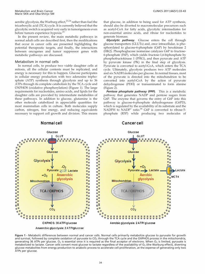
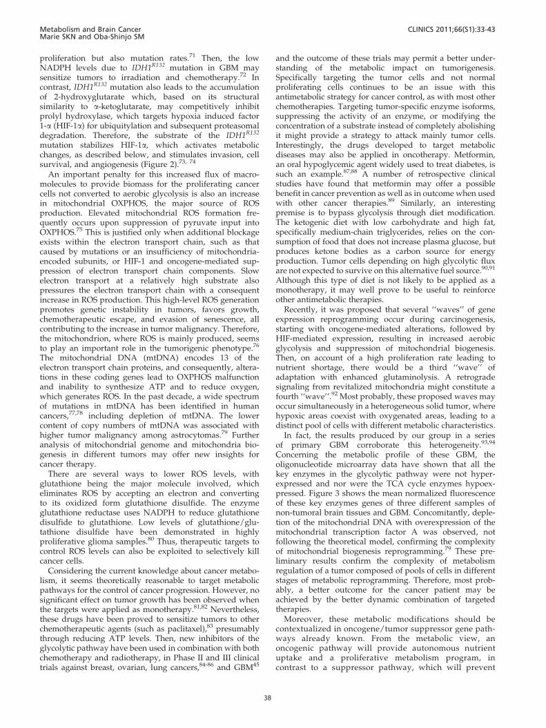
Figure 1 - Metabolic differences between normal and cancer cells. Normal cells primarily metabolize glucose to pyruvate for growthand survival, followed by complete oxidation of pyruvate to CO2 through the TCA cycle and the OXPHOS process in the mitochondria,generating 36 ATPs per glucose. O2 is essential once it is required as the final acceptor of electrons. When O2 is limited, pyruvate ismetabolized to lactate. Cancer cells convert most glucose to lactate regardless of the availability of O2 (the Warburg effect), divertingglucose metabolites from energy production to anabolic process to accelerate cell proliferation, at the expense of generating only twoATPs per glucose.
Metabolism and Brain CancerMarie SKN and Oba-Shinjo SM
CLINICS 2011;66(S1):33-43
34
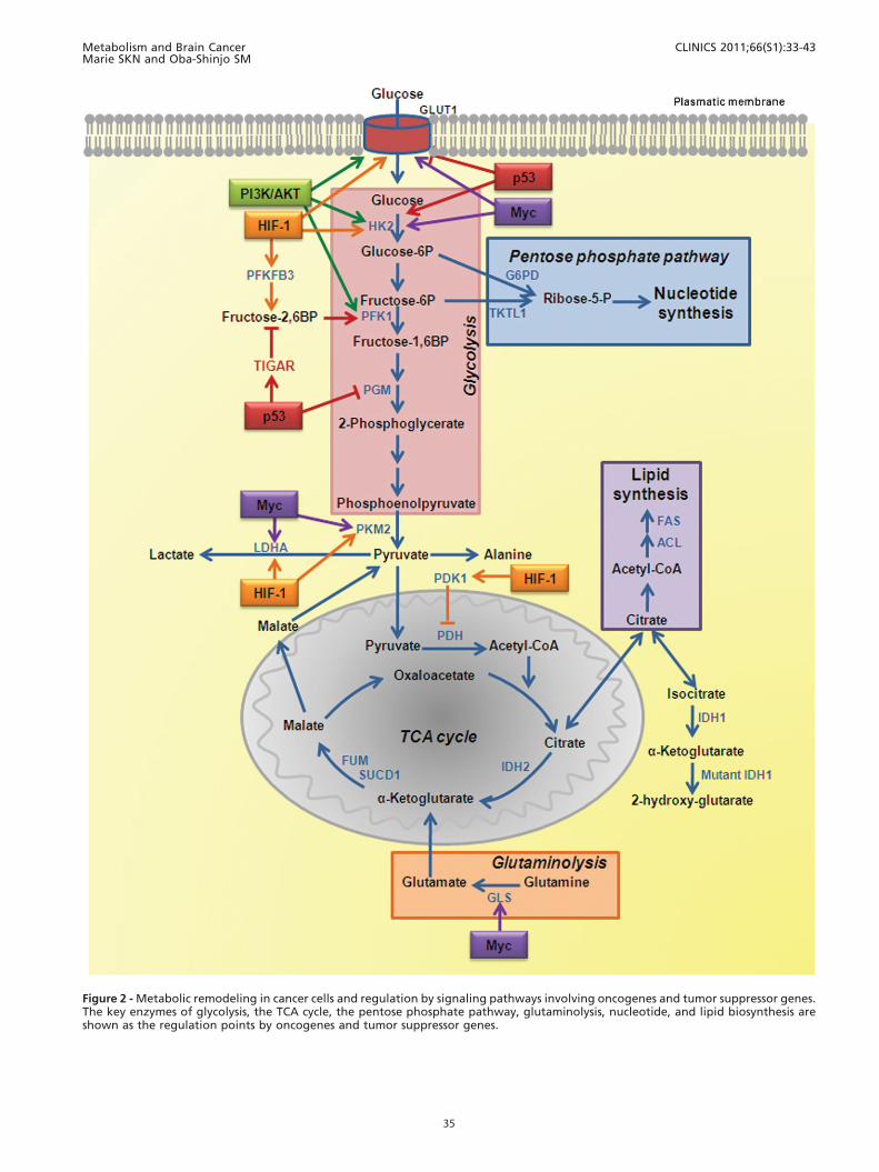
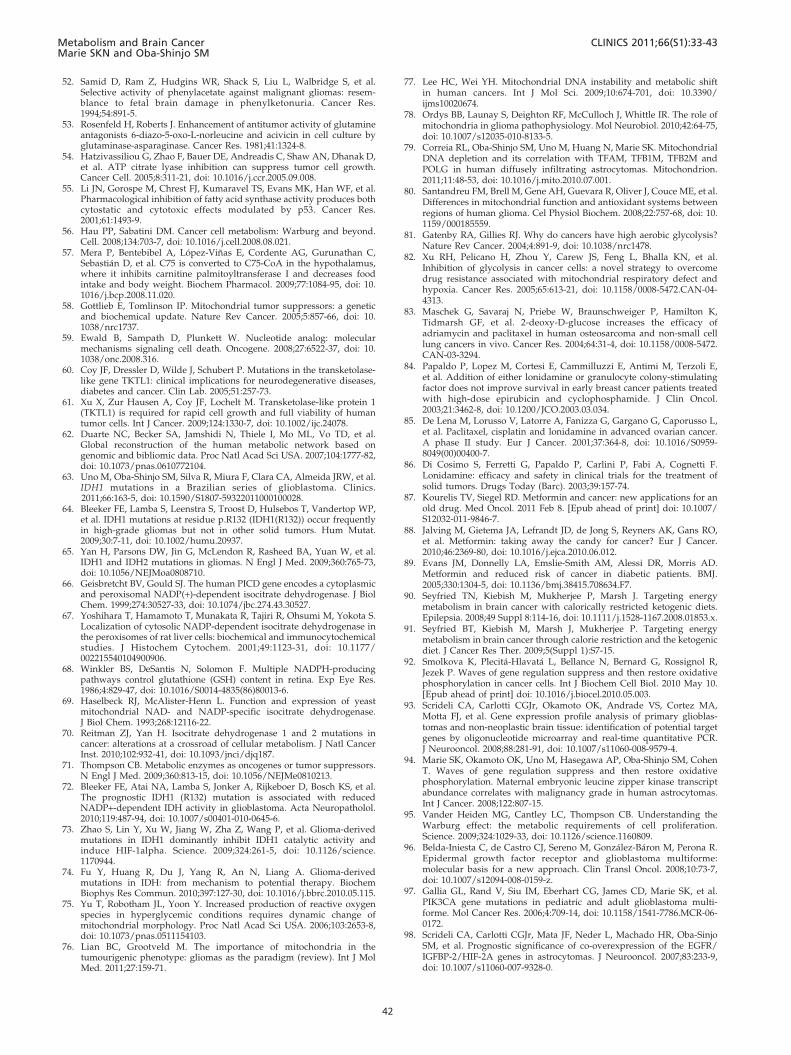
Figure 2 - Metabolic remodeling in cancer cells and regulation by signaling pathways involving oncogenes and tumor suppressor genes.The key enzymes of glycolysis, the TCA cycle, the pentose phosphate pathway, glutaminolysis, nucleotide, and lipid biosynthesis areshown as the regulation points by oncogenes and tumor suppressor genes.
Metabolism and Brain CancerMarie SKN and Oba-Shinjo SM
CLINICS 2011;66(S1):33-43
35
NADPH. NADPH is both a major cellular antioxidant,maintaining glutathione in a reduced state to preventoxidative damage, and a required cofactor in the reductivebiosynthesis of fatty acids, nucleotides, and amino acids.NADH is also used in mitochondrial OXPHOS (Figure 2).
Tricarboxylic acid (TCA) cycle. Pyruvate produced byglycolysis is converted to acetyl-CoA, which enters theTCA cycle, and citrate, a-ketoglutarate, succinyl-CoA,fumarate, malate, and oxaloacetate are produced asintermediate products. Most of the carbon for fatty acidsderives from acetyl-CoA synthesized in the mitochondrialmatrix. However, acetyl-CoA cannot cross the innermitochondrial membrane, but intramitochondrial acetyl-CoA and oxaloacetate combine to form citrate, which istransported out of the mitochondria and broken back downinto its constituents by ATP citrate lyase (ACL). Acetyl-CoAis converted to malonyl-CoA by acetyl-CoA carboxylase,and acetyl-CoA and malonyl-CoA are then both used by themulti-subunit enzyme fatty acid synthase (FAS) for thesynthesis and elongation of fatty acid chains. Oxaloacetate isused for the synthesis of non-essential amino acids.Cytosolic and nuclear acetyl-CoA is also a precursor forthe post-translational modification of proteins (for example,histones) by acetylation. Similarly to citrate, malateproduced in the TCA cycle leaves the mitochondria, and itis converted to pyruvate plus NADPH. Citrate might also beconverted to isocitrate and then to a-ketoglutarate,generating another molecule of NADPH by the action ofisocitrate dehydrogenase 1 (IDH1). In addition to glucose,amino acids can also fuel the TCA cycle. Glutamine suppliescarbon in the form of mitochondrial oxaloacetate tomaintain citrate production in the first step of the TCAcycle (Figure 2).
Glutaminolysis. Glutamine contributes both to thesubstrate needs of a dividing cell and to the control ofredox potentials through the synthesis of NADH. Afterglutamine is taken into the cell, a mitochondrial-associatedenzyme, glutaminase-1 (GLS), converts it to glutamate.Glutamate is converted to a-ketoglutarate and enters theTCA cycle in the mitochondria (Figure 2). Glutamate canalso be converted to aspartate, which contributes tonucleotide synthesis. The excessive quantity of glutamineused by the cells results in alanine and ammoniumsecretions.
Metabolic modifications in cancer cells andpotential targets for therapy
Cancer cells adapt themselves to maximize their ability tosynthesize substrates for membranes, nucleic acids, andproteins for the increased proliferative rate, a majorcharacteristic of these cells. This cannot be accomplishedwithout large amounts of energy (ATP), which are obtainedby increasing the use of glucose and glutamine many times.The cancer cells rely on aerobic glycolysis, the Warburgeffect,23,24 with a reduced use of the TCA cycle, so that thepyruvate made in glycolysis is converted to lactate27
(Figure 1). In fact, the fate of pyruvate depends on manyfactors, of which oxygen availability is one of the mostimportant. In the presence of oxygen, the pyruvate is directedinto the mitochondrion to be converted into acetyl-CoA bythe action of PDH or transaminated to form alanine. Andthen, once inside the mitochondrion, pyruvate is completelyoxidized through the TCA cycle and OXPHOS. However, incancer cells, PDH activity is blocked by the hypoxia-driven
enzyme pyruvate dehydrogenase kinase 1 (PDK1).28,29 Inaddition, an increase in lactate dehydrogenase A (LDHA)enzymatic activity is observed in cancer cells (Figure 2).30,31
These two facts determine the fate of pyruvate, which isconverted into lactate, contributing to the Warburg effectand to the enhancement of the malignant phenotype.32,33
Animal experiments knocking down LHDA or inhibitingPDK1, using interference RNA or dichloroacetate, havedemonstrated a reduction in tumor growth in xenograftmodels.30,31,34 Such results corroborate the importance ofthe decrease in the rate of pyruvate entering the TCA cycleand the concurrent increase in lactate production for thegrowth and survival of tumor cells. The lactate produced isexported from the cells by monocarboxylate transporter 4(MCT4), which allows the cell to preserve normal cellular pH(Figure 1). Oxygenated normal cells can remove lactatefrom the extracellular fluid using monocarboxylate transpor-ter 1, and convert it back to pyruvate for further oxidation,using the lactate dehydrogenase B (LDHB) isoform.However, the same is not observed in hypoxic cancer cells,and the large amounts of exported lactate create an acidictumor environment, which encourages cancer cell invasion.Thus, therapies targeting tumor acidification would inhibitglycolytic energy production and may also inhibit tumor cellinvasion.35 Nevertheless, treatment regimens directedtoward such ubiquitous transporters, such as monocarbox-ylate transporters, are also likely to affect normal tissues.Therefore, either their action must be extremely rapid or thedose very low to avoid side-effects in other tissues.
The pyruvate synthesis itself by PK is also an importantenergy-producing step in glycolysis. This step is highlyregulated by isoform selection and by allosteric regulation.Four PK isoforms have been described in humans: PKM1,PKM2, PKL, and PKR. The PKM1 isoform is expressed onlyin normal tissues and is incompatible with tumor growth.This isoform is replaced by the alternative spliced formPKM2 in highly proliferative tumor cells.36,37 PKL is foundin the liver and kidney, and PKR in erythrocytes. PKM2oscillates between inactive dimeric and active tetramericforms. The formation of PKM2 tetramers is stimulated bythe glycolytic intermediate fructose-1,6-biphosphate.38 Thedimeric form of PKM2 retards pyruvate formation andallows the accumulation of upstream glycolytic intermedi-ates, promoting their distribution into the biosyntheticpathways. The tetrameric form of PKM2 channels pyruvateto lactate. Phosphotyrosine residues bind to the sameallosteric regulatory site of fructose-1,6-biphosphate, releas-ing it.39,40 This regulation of enzyme activity may constitutea molecular switch that diverts glucose metabolites fromenergy production to anabolic processes when cells arestimulated by certain tyrosine phosphorylated growthfactors. If an inhibitor could be designed for the tumor-expressed PKM2 isoform, it might specifically inhibitglycolysis in tumor cells, killing them by energy deficit. Atherapeutic strategy inhibiting the interaction betweenphosphotyrosine residues and PKM2 to block the anabolicprocess of tumor cells would also be an interesting avenueto explore.
PFK1 is also a rate-limiting enzyme in glycolysis41 andhighly activated in tumor cells. PFK1 adds a secondphosphate group to F6P. One potent allosteric activator ofPFK1 is fructose-2,6-biphosphate (F2,6BP), the product ofthe 6-phosphofructose-2-kinase/fructose2,6-biphosphatase(PFKFB). PFKFBs are bifunctional enzymes that interconvert
Metabolism and Brain CancerMarie SKN and Oba-Shinjo SM
CLINICS 2011;66(S1):33-43
36
F6P and F2,6BP. PFK1 is normally inhibited by ATP, butF2,6BP overrides this inhibition and enhances the glycolyticflux, allowing tumor cells to maintain high glycolytic fluxdespite the presence of physiological levels of ATP. Anisoform of PFKFB, isoenzyme 3 (PFKFB3), was recentlyshown to promote proliferation through its effects on cellcycle regulators. PFKFB3 is upregulated in tumors, and itsinhibition has been proved to decrease F2,6BP levels, whichin turn decreases PFK1 activity and glycolytic flux withcytostatic effect (Figure 2).42
HK2 participates in the glycolytic pathway and also inmetabolite transport into and out of the mitochondrialintermembrane space through its association with a voltage-dependent anion channel,43 a 30 kDa pore protein insertedin the outer mitochondrial membrane.44 Therefore, HK2 isanother potential therapeutic target. In fact, two hexokinaseinhibitors, lonidamine and glucose mimetic, 2-deoxyglu-cose, are currently in clinical trials in combination withother agents.45 3-Bromopyruvate is a further inhibitor ofHK2 with promising in vivo studies, but there is no currentclinical trial.46,47
The modifications in cancer cells enumerated abovepermit an understanding of how these cells are urged to anon-profitable choice of two ATP generation by aerobicglycolysis instead of 36 ATP generation upon completeglucose oxidation by the TCA cycle and OXPHOS. In spiteof this apparently ‘‘bad’’ choice, cancer cells continue toexhibit high ratios of ATP/adenosine diphosphate (ADP)and NADH/NAD+48 due to an alternative ATP productionby converting two ADPs to one ATP and one adenosinemonophosphate (AMP) catalyzed by adenylate kinases. Thisnot only helps to maintain a viable ATP/ADP ratio as ATPproduction declines, but also to accumulate AMP, whichactivates AMP-kinase and leads to the phosphorylation ofseveral targets to improve energy charge in cells.49
Another important reason for the cancer cells to switch toaerobic glycolysis is to provide metabolic macromoleculesfor the daughter cells. 13C-nuclear magnetic resonancespectroscopy measurements show that 90% of glucose and60% of glutamine are converted into lactate or alanineby GBM cell cultures.50 Although each lactate excreted fromthe cell wastes three carbons that might otherwise beutilized for either ATP production or macromolecularprecursor biosynthesis, the tumor cells choose this methodto fasten carbon incorporation into biomass to incrementcell division velocity. Glutaminolysis also generatesreductive power required for fatty acid biosynthesis byNADPH production via the activity of NADP+-specificmalate dehydrogenase (malic enzyme), in addition tothe fundamental role in replenishing the TCA cycle.51
Blocking the fuel through this pathway for the biomass totumor proliferation seems a good therapeutic strategy.Phenylacetate is a drug that reduces the biological avail-ability of glutamine in the blood. This reagent condenseswith the c-amino group of glutamine and is excreted intourine. A previous report has demonstrated that phenylace-tate inhibits the proliferation of glioma cells and promotestheir differentiation.52 However, the removal of glutaminedirectly from the plasma may also increase the rate atwhich the body cannibalizes its own muscles (cachexia).Additionally, various other anti-glutaminolysis compoundshave been developed, but they were found to be toxic orraised immune reactions.53
Cancer biomass reduction may also be achieved byblocking fatty acid synthesis through the inhibition ofACL, which converts acetyl-CoA to malonyl-CoA,54 andFAS, a multifunctional protein that converts malonyl-CoAto palmitate over multiple steps.55 Inhibition of bothenzymes, ACL and FAS, has been shown to limit tumorcell proliferation and survival in vitro and in vivo.55,56 Onepossible negative aspect of anti-FAS therapy is its effect onfood intake and body weight, as observed in treated rodentsthat presented hypophagia and consequent weight loss.57
Deficiencies in two other enzymes participating in the TCAcycle, fumarate hydratase (FUM) and succinate dehydro-genase 1 (SUCD1), may also have a tumor suppressive effect(Figure 2).58
Nucleotide biosynthesis has also been targeted to blockbiomass production in cancer cells for several years. 5-fluorouracil, cytarabine, and methotrexate are examples ofchemotherapeutic agents known as antimetabolites. Most ofthese drugs target the final stages in the nucleotide syntheticpathway, and therefore lack specificity, leading to nucleo-tide shortage, incomplete DNA synthesis, and cell deathindistinctly of tumor and normal proliferating cells.59
Therefore, blocking the early stages of nucleotide biosynth-esis such as R5P production could provide a bettertherapeutic window than that shown by previous antimeta-bolic therapies. Transketolase-like protein 1 (TKTL1), anenzyme in the non-oxidative arm of the PPP that producesR5P, has been found to be upregulated in several tumortypes,60 and knocking it down reduced the proliferation oftumor cells, as well as decreasing lactate production andresensitizing cells to reactive oxygen species (ROS)-generat-ing compounds.61 Isolated mutations in either TKTL1 orG6PD of PPP have no impact on cancer cell growth, as bothenzymes contribute to R5P production. However, simulta-neous mutations in these two genes, blocking R5P synthesis,are lethal for cancer cells in animals (Figure 2).62
Besides the alterations described above in the mainmetabolic routes, side pathways of the TCA cycle are alsoimplicated in cancer progression. A highly prevalentmutation in an enzyme related to metabolism was uncov-ered in the recent high throughput mutation screening inGBM, which highlights the importance of energetic meta-bolism in tumor progression. This study revealed that up to12% of the GBMs harbor the same mutation in the geneencoding cytosolic IDH1.12,63 Monoallelic mutation in thesame residue 132 in IDH1 (IDH1R132) or the analogousresidue in the related enzyme IDH2 is a common feature ofgliomas, as more than 80% of indolent gliomas harbor sucha mutation.64,65 IDH1 and IDH2 couple the reversibleconversion of isocitrate to a-ketoglutarate and NADP+ toNADPH. IDH1 is located in the cytosol and the peroxi-some66,67 and produces NADPH.68 In the peroxisome,NADPH contributes to cholesterol synthesis.69 IDH2 islocated in the mitochondria and catalyzes the isocitrate to a-ketoglutarate reaction in the TCA cycle.70 The occurrence ofIDH1 mutations correlated with approximately twofolddiminished NADP+-dependent IDH activity, and totalNADPH production is hampered by 38% in GBM harboringthe IDH1R132 mutation. Therefore, mutated IDH1 consumesrather than produces NADPH. NADPH/NADH is both amajor antioxidant, maintaining glutathione in a reducedstate, protecting the cell from ROS, and a required cofactorin the biosynthesis of fatty acids, nucleotides, and aminoacids. Thus, the NADPH level may affect not only cellular
Metabolism and Brain CancerMarie SKN and Oba-Shinjo SM
CLINICS 2011;66(S1):33-43
37
proliferation but also mutation rates.71 Then, the lowNADPH levels due to IDH1R132 mutation in GBM maysensitize tumors to irradiation and chemotherapy.72 Incontrast, IDH1R132 mutation also leads to the accumulationof 2-hydroxyglutarate which, based on its structuralsimilarity to a-ketoglutarate, may competitively inhibitprolyl hydroxylase, which targets hypoxia induced factor1-a (HIF-1a) for ubiquitylation and subsequent proteasomaldegradation. Therefore, the substrate of the IDH1R132
mutation stabilizes HIF-1a, which activates metabolicchanges, as described below, and stimulates invasion, cellsurvival, and angiogenesis (Figure 2).73, 74
An important penalty for this increased flux of macro-molecules to provide biomass for the proliferating cancercells not converted to aerobic glycolysis is also an increasein mitochondrial OXPHOS, the major source of ROSproduction. Elevated mitochondrial ROS formation fre-quently occurs upon suppression of pyruvate input intoOXPHOS.75 This is justified only when additional blockageexists within the electron transport chain, such as thatcaused by mutations or an insufficiency of mitochondria-encoded subunits, or HIF-1 and oncogene-mediated sup-pression of electron transport chain components. Slowelectron transport at a relatively high substrate alsopressures the electron transport chain with a consequentincrease in ROS production. This high-level ROS generationpromotes genetic instability in tumors, favors growth,chemotherapeutic escape, and evasion of senescence, allcontributing to the increase in tumor malignancy. Therefore,the mitochondrion, where ROS is mainly produced, seemsto play an important role in the tumorigenic phenotype.76
The mitochondrial DNA (mtDNA) encodes 13 of theelectron transport chain proteins, and consequently, altera-tions in these coding genes lead to OXPHOS malfunctionand inability to synthesize ATP and to reduce oxygen,which generates ROS. In the past decade, a wide spectrumof mutations in mtDNA has been identified in humancancers,77,78 including depletion of mtDNA. The lowercontent of copy numbers of mtDNA was associated withhigher tumor malignancy among astrocytomas.79 Furtheranalysis of mitochondrial genome and mitochondria bio-genesis in different tumors may offer new insights forcancer therapy.
There are several ways to lower ROS levels, withglutathione being the major molecule involved, whicheliminates ROS by accepting an electron and convertingto its oxidized form glutathione disulfide. The enzymeglutathione reductase uses NADPH to reduce glutathionedisulfide to glutathione. Low levels of glutathione/glu-tathione disulfide have been demonstrated in highlyproliferative glioma samples.80 Thus, therapeutic targets tocontrol ROS levels can also be exploited to selectively killcancer cells.
Considering the current knowledge about cancer metabo-lism, it seems theoretically reasonable to target metabolicpathways for the control of cancer progression. However, nosignificant effect on tumor growth has been observed whenthe targets were applied as monotherapy.81,82 Nevertheless,these drugs have been proved to sensitize tumors to otherchemotherapeutic agents (such as paclitaxel),83 presumablythrough reducing ATP levels. Then, new inhibitors of theglycolytic pathway have been used in combination with bothchemotherapy and radiotherapy, in Phase II and III clinicaltrials against breast, ovarian, lung cancers,84-86 and GBM45
and the outcome of these trials may permit a better under-standing of the metabolic impact on tumorigenesis.Specifically targeting the tumor cells and not normalproliferating cells continues to be an issue with thisantimetabolic strategy for cancer control, as with most otherchemotherapies. Targeting tumor-specific enzyme isoforms,suppressing the activity of an enzyme, or modifying theconcentration of a substrate instead of completely abolishingit might provide a strategy to attack mainly tumor cells.Interestingly, the drugs developed to target metabolicdiseases may also be applied in oncotherapy. Metformin,an oral hypoglycemic agent widely used to treat diabetes, issuch an example.87,88 A number of retrospective clinicalstudies have found that metformin may offer a possiblebenefit in cancer prevention as well as in outcome when usedwith other cancer therapies.89 Similarly, an interestingpremise is to bypass glycolysis through diet modification.The ketogenic diet with low carbohydrate and high fat,specifically medium-chain triglycerides, relies on the con-sumption of food that does not increase plasma glucose, butproduces ketone bodies as a carbon source for energyproduction. Tumor cells depending on high glycolytic fluxare not expected to survive on this alternative fuel source.90,91
Although this type of diet is not likely to be applied as amonotherapy, it may well prove to be useful to reinforceother antimetabolic therapies.
Recently, it was proposed that several ‘‘waves’’ of geneexpression reprogramming occur during carcinogenesis,starting with oncogene-mediated alterations, followed byHIF-mediated expression, resulting in increased aerobicglycolysis and suppression of mitochondrial biogenesis.Then, on account of a high proliferation rate leading tonutrient shortage, there would be a third ‘‘wave’’ ofadaptation with enhanced glutaminolysis. A retrogradesignaling from revitalized mitochondria might constitute afourth ‘‘wave’’.92 Most probably, these proposed waves mayoccur simultaneously in a heterogeneous solid tumor, wherehypoxic areas coexist with oxygenated areas, leading to adistinct pool of cells with different metabolic characteristics.
In fact, the results produced by our group in a seriesof primary GBM corroborate this heterogeneity.93,94
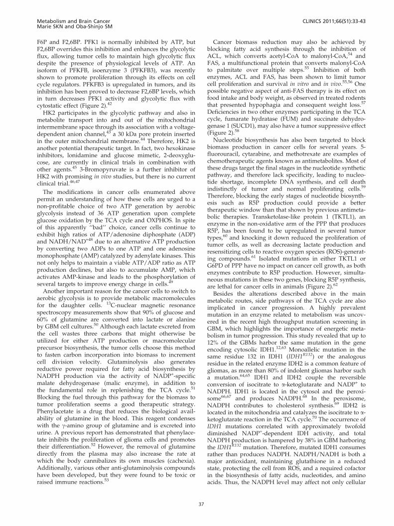
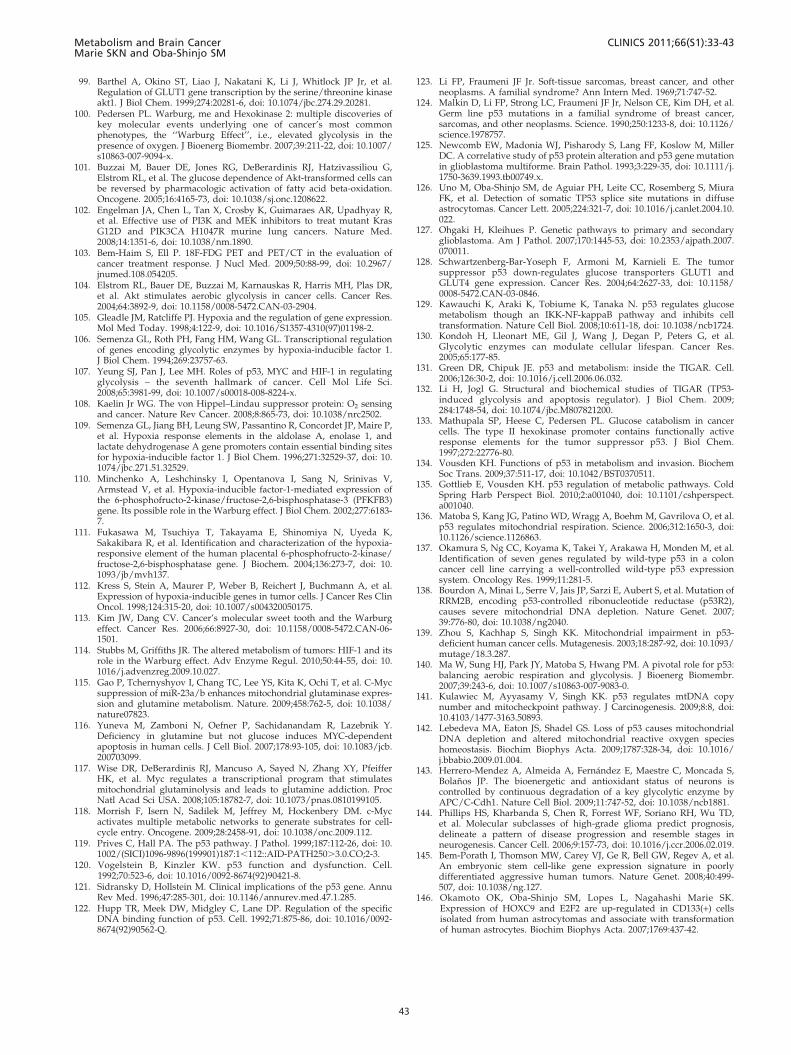
Concerning the metabolic profile of these GBM, theoligonucleotide microarray data have shown that all thekey enzymes in the glycolytic pathway were not hyper-expressed and nor were the TCA cycle enzymes hypoex-pressed. Figure 3 shows the mean normalized fluorescenceof these key enzymes genes of three different samples ofnon-tumoral brain tissues and GBM. Concomitantly, deple-tion of the mitochondrial DNA with overexpression of themitochondrial transcription factor A was observed, notfollowing the theoretical model, confirming the complexityof mitochondrial biogenesis reprogramming.79 These pre-liminary results confirm the complexity of metabolismregulation of a tumor composed of pools of cells in differentstages of metabolic reprogramming. Therefore, most prob-ably, a better outcome for the cancer patient may beachieved by the better dynamic combination of targetedtherapies.
Moreover, these metabolic modifications should becontextualized in oncogene/tumor suppressor gene path-ways already known. From the metabolic view, anoncogenic pathway will provide autonomous nutrientuptake and a proliferative metabolism program, incontrast to a suppressor pathway, which will prevent
Metabolism and Brain CancerMarie SKN and Oba-Shinjo SM
CLINICS 2011;66(S1):33-43
38

nutrient utilization for anabolic processes. Developmentof future therapies might also fit in this model, targe-ting self-limiting steps of nutrient production andmetabolism.25,95
Oncogenes and tumor suppressor genesparticipating in the metabolic switch
Oncogenes. The phosphoinositide 3-kinase (PI3K)signaling pathway is one of the major pathways linked to thedevelopment of several tumors, including primary GBM.96,97
PI3K is recruited to the cellular membrane through theactivation of epidermal growth factor receptor (EGFR), and itis phosphorylated from phosphatidylinositol-4,5-biphosphateto 3-phosphate, which activates target molecules such asprotein kinase b (AKT) and mammalian target of rapamycin(mTOR), which induces cell proliferation and cell survival by
blocking apoptosis. Several molecular participants in thispathway, such as EGFR, PTEN, and PIK3CA, presentalterations in GBM.98
The PI3K/AKT pathway may regulate glucose metabo-lism by: (1) regulating glucose transporter expressionthrough AKT, which directly stimulates transcription ofthe GLUT1;99 (2) enhancing glucose capture by HK2 andinducing aerobic glycolysis by promoting HK2 binding tovoltage-dependent anion channels;100 and (3) stimulatingPFK1 activity50 (Figure 2). In addition, activation of the PI3Kpathway renders cells dependent on high levels of glucoseflux.101 Small molecules inhibiting the PI3K pathway lead todecreased glucose uptake and correlate with tumor regres-sion;102 response to therapy has been predicted by theability to disrupt glucose metabolism measured by 18F-deoxyglucose positron emission tomography.103
Figure 3 - Oligonucleotide microarray data of genes coding for key enzymes or subunits of enzymatic complexes of glycolysis and theTCA cycle. Three samples of non-tumoral brain tissues (blue bars) and three samples of GBM (red bars) were submitted to extraction oftotal RNA and microarray analysis, as described previously.94,146 LDHA and PKM2, both enzymes of glycolysis, are upregulated in GBM,whereas the variability in the expression profile of TCA cycle genes suggests that this cycle is uncoupled. The bars represent the medianvalues of the three samples. Numbers represent the normalized fluorescence. Genes coding for glycolysis enzymes: HK2, hexokinase 2;PFK1, phosphofructokinase; PGAM1 and PGAM2, phosphoglycerate mutase 1 and 2, subunits of PGM dimer; PKM2, pyruvate kinaseM2; LDHA, lactate dehydrogenase A. Genes coding for the TCA cycle enzymes: PDHA1 and PDHA2, pyruvate dehydrogenase alpha 1and 2; PDHB, pyruvate dehydrogenase beta; DLAT, dihydrolipoamide-acetyltransferase; DLT, dihydrolipoamide dehydrogenase; PDHX,pyruvate dehydrogenase complex, component X, all six subunits of the enzymatic complex PDH; IDH2, isocitrate dehydrogenase 2;SDHA, SDHB, SDHC, and SDHD, SDH complex, subunits A, B, C, and D; and FUM, fumarate hydratase.
Metabolism and Brain CancerMarie SKN and Oba-Shinjo SM
CLINICS 2011;66(S1):33-43
39

AKT is activated when tumor cells need metabolicintermediates for rapid proliferation, and it promotes aglycolytic switch under normoxic conditions withoutaffecting the rate of OXPHOS. Thus, cancer cells turn outto be dependent on aerobic glycolysis for their growth andsurvival under AKT-mediated metabolic influence; conse-quently, activated AKT tumor cells undergo rapid cell deathwhen shifted to low-dose glucose conditions.104
HIF-1 is a pleiotropic hypoxia induced transcriptionfactor with a heterodimeric structure composed of HIF-1aas the oxygen responsive subunit and HIF-1b as theconstitutively expressed subunit.105 Hypoxia is a commonmicroenvironment feature of solid tumors, leading to HIF-1upregulation and consequently inducing a metabolicswitch. HIF-1 target genes are involved in glucose andenergy metabolism,106 including all glycolytic enzyme genesexcept phosphoglycerate mutase (PGM).107,108 Additionally,HIF-1 upregulates LDHA;109 PFK1, through activation ofPFKFB3,110,111 switches the isoform PKM1 to PKM2112 andactivates PDK1, which in turn inhibits PDH, reducing theTCA cycle.113 HIF also regulates the uptake of glucosethrough GLUT1 (Figure 2).114
MYC regulates a broad spectrum of genes involved in cellcycle control, metabolism, mitochondrial function, andregulation of apoptosis. However, the role of MYC iscomplex because its ectopic expression in cancer canconcurrently drive aerobic glycolysis and/or OXPHOSaccording to the tumor cell microenvironment. MYC maytrigger the expression of glycolytic genes or repressmicroRNAs miR-23a/b to increase GLS and stimulateOXPHOS through the activation of O2-dependent glutami-nolysis (Figure 2).115 Then, MYC driven tumor cells areparticularly sensitive to glutamine withdrawal,116 and genesinvolved in glutamine metabolism appear to be under bothdirect and indirect transcriptional control of the MYCprotein.115,117 Glutamine depletion from MYC-transformedcells results in the rapid loss of TCA cycle intermediates andcell death.115 MYC also influences glycolysis by upregulat-ing the majority of glycolytic genes, including HK2, PFK1,LDHA, PKM2, as well as GLUT1 (Figure 2).113 Deregulatedexpression of MYC collaborates with HIF-1 to confer theglycolytic (Warburg) phenotype of tumors. Additionally,MYC stimulates mitochondrial biogenesis in both normaland immortalized cells.118
Suppressor genes. Tumor suppressor pathways can alsoregulate cellular metabolism and may act to coordinatenutrient utilization with cell physiology.
TP53 is by far the most well known tumor suppressorgene. Inactivation of TP53 is observed in more than 50different types of human cancer,119 and more than 27,000mutations were enumerated in the International Agency forResearch on Cancer – TP53 Mutation Database (http://www-p53.iarc.fr/). TP53 encodes a nuclear phosphoproteinof 53 kDa, responsible for genomic integrity and cellproliferation control. p53 protein facilitates DNA repair byblocking the cell cycle and induces apoptosis when theDNA damage is unrepairable.120–122 TP53 mutation ingermline cells determines Li–Fraumeni syndrome charac-terized by predisposition to multiple types of cancer,including breast tumor, sarcoma, and central nervoussystem tumors.123,124 TP53 mutations are also frequent insecondary GBM.125–127 There are cumulative studiesdemonstrating a role for p53 in controlling metabolic
genes, such as altering glucose utilization and promotingOXPHOS, but also contributing to the induction ofapoptosis and maintaining mitochondrial health andactivity by its localization inside the organelle. The moststraightforward role of p53 in metabolism is the dampen-ing of glycolysis, which is completely consistent with itsfunction as a tumor suppressor. This limitation of glyco-lysis may occur in different ways: (1) downregulation ofexpression of several glucose transporters such as GLUT1and GLUT4 by direct transcriptional repression128 andGLUT3 by the indirect reduction of expression through theinhibition of nuclear factor kappa-b kinase;129 (2) ubiqui-tination and inactivation of PGM;130 (3) lowering F2,6BP byp53-dependent expression of the TP53-induced glycolysisand apoptosis regulator (TIGAR).131,132 However, p53 alsopromotes glycolysis by increasing HK2 and PGM expres-sion through p53-inducible promoters.133 Additionally, theincrease in HK2 activity in concert with a decrease in PFK1activity (resulting from TIGAR expression) would promotethe use of the PPP.134,135 p53 has also been shown to play arole in promoting OXPHOS through the transcriptionalactivation of cytochrome c oxidase 2,136 subunit I ofcytochrome c oxidase,137 and p52R2, a subunit of ribonu-cleotide reductase138 as well as by the post-transcriptionalregulation of cytochrome oxidase 2.139 ATP is derived inmuch higher proportion through OXPHOS in cells expres-sing p53 than in cells lacking it.140 p53 also contributes tothe maintenance of mitochondrial mass and mtDNA copynumber.141,142
In addition to the complexity of tumor cell metabolicreprogramming in a context of impulses from oncogenesand inactivation of tumor suppressor genes, the cancer stemcell hypothesis should also be considered to gain a morecomplete understanding of tumorigenesis and for theefficacy of cancer therapy.
Targeting cancer stem cell metabolism as a futureperspective
The cancer stem cell hypothesis proposes that cancerderives from a small fraction of cancer cells that constitute aself-sustaining cell reservoir, which are responsible forreseeding tumor cells after the first therapeutic approach.There are some preliminary data concerning the metabolismof the stem cells, such as increased expression of PFKFB3 incycling cells and decreased expression in terminallydifferentiated neurons.143 It has been reported that GBMcancer stem cells share gene expression signatures withprogenitor cells in the developing forebrain,144 and prefer-ential overexpression of genes normally enriched inembryonic stem cells, NANOG, OCT4, SOX2, and MYC,were activated in poorly differentiated GBM.145 Moreover, asubpopulation of cells that carry the known progenitor cellmarker CD133 has been identified in GBM, and when theexpression profiles of CD133+ GBM cells were comparedwith CD1332 cells, 16 genes, many of which had notpreviously been associated with astrocytomas, were foundto be aberrantly expressed relative to corresponding non-neoplastic brain tissue controls.146 However, the expressionprofile of genes related to energy metabolism in cancer stemcells remains to be explored.
The development of novel metabolic based drugs holdsmuch promise for the improvement of cancer therapy.
Metabolism and Brain CancerMarie SKN and Oba-Shinjo SM
CLINICS 2011;66(S1):33-43
40

REFERENCES
1. Legler JM, Ries LA, Smith MA, Warren JL, Heineman EF, Kaplan RS,et al. Cancer surveillance series: brain and other central nervous systemcancers: recent trends in incidence and mortality. J Natl Cancer Inst.1999;91:1382-90, doi: 10.1093/jnci/91.16.1382.
2. DeAngelis LM. Brain tumors. N Engl J Med. 2001;344:114-23, doi: 10.1056/NEJM200101113440207
3. Wrensch M, Minn Y, Chew T, Bondy M, Berger MS. Epidemiology ofprimary brain tumors: current concepts and review of the literature.Neuro Oncol. 2002;4:278-9.
4. Furnari FB, Fenton T, Bachoo RM, Mukasa A, Stommel JM, Stegh A,et al. Malignant astrocytic glioma: genetics, biology, and paths totreatment. Genes Dev. 2007;21:2683–710, doi: 10.1101/gad.1596707.
5. Loius DN, Ohgaki H, Wiestler OD, Cavenee WK, Burger PC, Jouvet A,et al. The 2007 WHO classification of tumours of the central nervoussystem. Acta Neuropathol. 2007;114:97-109, doi: 10.1007/s00401-007-0243-4.
6. Jemal A, Siegel R, Ward E, Hao Y, Xu J, Thun MJ. Cancer statistics, 2009.CA Cancer J Clin. 2009;59:225-49, doi: 10.3322/caac.20006.
7. Tran B, Rosenthal MA. Survival comparison between glioblastomamultiforme and other incurable cancers. J Clin Neurosci. 2010;17:417-21,doi: 10.1016/j.jocn.2009.09.004.
8. Bauchet L, Mathieu-Daude H, Fabbro-Peray P, Rigau V, Fabbro M,Chinot O, et al. Oncological patterns of care and outcome for 952patients with newly diagnosed glioblastoma in 2004. Neuro-oncol.2010;12:725-35.
9. Schneider T, Mawrin C, Scherlach C, Skalej M, Firsching R. Gliomas inadults. Dtsch Arztebl Int. 2010;107:799-807.
10. Walker MD, Alexander Jr E, Hunt WE, MacCarty CS, Mahaley MS Jr,Mealey J Jr, et al. Evaluation of BCNU and/or radiotherapy in the treatmentof anaplastic gliomas. A cooperative trial. J Neurosurg.1978;49:333-43.
11. Stupp R, Mason W, Van den Bert MJ, Weller M, Fisher B, Taphoorn MJ,et al. Radiotherapy plus concomitant and adjuvant temozolomide forglioblastoma. N Engl J Med. 2005;352:987-96, doi: 10.1056/NEJMoa043330.
12. Parsons DW, Jones S, Zhang X, Lin JC, Leary RJ, Angenendt P, et al. Anintegrated genomic analysis of human glioblastoma multiforme.Science. 2008;321:1807-12, doi: 10.1126/science.1164382.
13. Parsons DW, Li M, Zhang X, Jones S, Leary RJ, Lin JC, et al. The geneticlandscape of the childhood cancer medulloblastoma. Science.2011;331:435-9, doi: 10.1126/science.1198056.
14. Fearon ER, Vogelstein B. A genetic model for colorectal tumorigenesis.Cell. 1990;61:759-67, doi: 10.1016/0092-8674(90)90186-I.
15. The Cancer Genome Atlas Research Network. Comprehensive genomiccharacterization defines human glioblastoma genes and core pathways.Nature. 2008;455:1061-8, doi: 10.1038/nature07385.
16. Barbosa KC, Oba-Shinjo SM, Uno M, Carvalho PO, Rosemberg S,Aguiar PH, et al. Association of EGFR c.2073A.T polymorphism withdecreased risk of diffusely infiltrating astrocytoma in a Brazilian case–control study. Int J Biol Markers. 2008;23:140-6.
17. Burim RV, Teixeira SA, Colli BO, Peria FM, Tirapelli LF, Marie SK, et al.ICAM-1 (Lys469Glu) and PECAM-1 (Leu125Val) polymorphisms indiffuse astrocytomas. Clin Exp Med. 2009;9:157-63, doi: 10.1007/s10238-009-0040-6.
18. Lino M, Merlo A. Translating biology into clinics: the case ofglioblastoma. Curr Opin Cell Biol. 2009;21:311-16, doi: 10.1016/j.ceb.2008.12.009.
19. Larsen C. Genetic and molecular abnormalities of glioblastomas (GBM).Bull Cancer. 2010;97:1389-407.
20. Gerson SL. MGMT: its role in cancer aetiology and cancer therapeutics.Nature Rev Cancer. 2004;4:296-307, doi: 10.1038/nrc1319.
21. Hegi ME, Diserens AC, Gorlia T, Hamou MF, de Tribolet N, Weller M,et al. MGMT gene silencing and benefit from temozolomide inglioblastoma. N Engl J Med. 2005;352:997-1003, doi: 10.1056/NEJMoa043331.
22. Weller M, Stupp R, Reifenberger G, Brandes AA, van den Bent MJ,Wick W, Hegi ME. MGMT promoter methylation in malignant gliomas:ready for personalized medicine? Nat Rev Neurol. 2010;6:39-51.
23. Warburg O, Posener K, Negelein E. Der Tumoren de Stoffwechsel doden de Ueber. Biochem Z. 1924;152:319-44.
24. Warburg O. On the origin of cancer cells. Science. 1956;123:309-14, doi:10.1126/science.123.3191.309.
25. Tennant DA, Duran RV, Gottlieb E. Targeting metabolic transformationfor cancer therapy. Nature Rev Cancer. 2010;10:267-77, doi: 10.1038/nrc2817.
26. Saiati LM, Amir-Ahmady B. Dietary regulation of expression ofglucose-6-phosphate dehydrogenase. Annu Rev Nutr. 2001;21:121-40,doi: 10.1146/annurev.nutr.21.1.121.
27. Tennant DA, Duran RV, Boulahbel H, Gottieb E. Metabolic transforma-tion in cancer. Carcinogenesis. 2009;30:1269-80, doi: 10.1093/carcin/bgp070.
28. Kim JW, Tchernyshyov I, Semenza GL, Dang CV. HIF-1-mediatedexpression of pyruvate dehydrogenase kinase: a metabolic switch
required for cellular adaptation to hypoxia. Cell Metab. 2006;3:177-85,doi: 10.1016/j.cmet.2006.02.002.
29. Dang CV, Semenza GI. Oncogenic alteration of metabolism. TrendsBiochem Sci. 1999;24:68-72, doi: 10.1016/S0968-0004(98)01344-9.
30. Fantin VR, St-Pierre J, Leder P. Attenuation of LDH-A expressionuncovers a link between glycolysis, mitochondrial physiology, andtumor maintenance. Cancer Cell. 2006;9:425-34, doi: 10.1016/j.ccr.2006.04.023.
31. Le A, Cooper CR, Gouw AM, Dinavahi R, Maitra A, Deck LM, et al.Inhibition of lactate dehydrogenase A induces oxidative stress andinhibits tumor progression. Proc Natl Acad Sci USA. 2010;107:2037-42,doi: 10.1073/pnas.0914433107.
32. Jones RG, Thompson CB. Tumor suppressors and cell metabolism: arecipe for cancer growth. Genes Dev. 2009;23:537-48, doi: 10.1101/gad.1756509.
33. McFate T, Mohyeldin A, Lu H, Thakar J, Henriques J, Halim ND, et al.Pyruvate dehydrogenase complex activity controls metabolic andmalignant phenotype in cancer cells. J Biol Chem. 2008;283:22700-8,doi: 10.1074/jbc.M801765200.
34. Sun RC, Fadia M, Dahlstrom JE, Parish CR, Board PG, Blackburn AC.Reversal of the glycolytic phenotype by dichloroacetate inhibitsmetastatic breast cancer cell growth in vitro and in vivo. BreastCancer Res Treat. 2009;120:253-60, doi: 10.1007/s10549-009-0435-9.
35. Gatenby RA, Gawlinski ET, Gmitro AF, Kaylor B, Gillies RJ. Acid-mediated tumor invasion: a multidisciplinary study. Cancer Res.2006;66:5216-23, doi: 10.1158/0008-5472.CAN-05-4193.
36. Mazurek S, Boschel CB, Hugo F, Eigenbrodt E. Pyruvate kinase type M2and its role in tumor growth and spreading. Semin Cancer Biol.2005;15:300-8, doi: 10.1016/j.semcancer.2005.04.009.
37. Christofk HR, Vander Heiden MG, Harris MH, Ramanathan A,Gerszten RE, Wei R, et al. The M2 splice isoform of pyruvate kinaseis important for cancer metabolism and tumour growth. Nature.2008;452:230-3, doi: 10.1038/nature06734.
38. Mazurek S, Grimm H, Boschek CB, Vaupel P, Eigenbrodt E. Pyruvatekinase type M2: a crossroad in the tumor metabolome. Br J Nutr.2002;87:S23-9, doi: 10.1079/BJN2001454.
39. Christofk HR, Vander Heiden MG, Wu N, Asara JM, Cantley LC.Pyruvate kinase M2 is a phosphotyrosine-binding protein. Nature.2008;452:181-6, doi: 10.1038/nature06667.
40. Eigenbrodt E, Reinacher M, Scheefers-Borchel U, Scheefers H, Friis R.Double role for pyruvate kinase type M2 in the expansion ofphosphometabolite pools found in tumor cells. Crit Rev Oncog.1992,3:91-115.
41. Deprez J, Vertommen D, Alessi DR, Hue L, Rider MH. Phosphorylationand activation of heart 6-phosphofructo-2-kinase bt protein kinase Band other protein kinases of the insulin signaling cascades. J Biol Chem.1997;272:17269-75, doi: 10.1074/jbc.272.28.17269.
42. Clem B, Telang S, Clem A, Yalcin A, Meier J, Simmons A, et al. Small-molecule inhibition of 6-phosphofructo2-kinase activity suppressesglycolytic flux and tumor growth. Mol Cancer Ther. 2008;7:110-20,doi: 10.1158/1535-7163.MCT-07-0482.
43. Mathupala SP, Ko YH, Pedersen PL. Hexokinase II: cancer’s double-edged sword acting as both facilitator and gatekeeper of malignancywhen bound to mitochondria. Oncogene. 2006;25:4777-86, doi: 10.1038/sj.onc.1209603.
44. Colombini M. VDAC: the channel at the interface between mitochon-dria and the cytosol. Mol Cell Biochem. 2004;256-7:107-15, doi: 10.1023/B:MCBI.0000009862.17396.8d.
45. Oudard S, Carpentier A, Banu E, Fauchon F, Celerier D, Poupon MF,et al. Phase II study of lonidamine and diazepam in the treatment ofrecurrent glioblastoma multiforme. J Neurooncol. 2003;63:81-6, doi: 10.1023/A:1023756707900.
46. Cao X, Bloomston M, Zhang T, Frankel WL, Jia G, Wang B, et al.Synergistic antipancreatic tumor effect by simultaneously targetinghypoxic cancer cells with HSP90 inhibitor and glycolysis inhibitor. ClinCancer Res. 2008;14:1831-9, doi: 10.1158/1078-0432.CCR-07-1607.
47. Ko YH, Smith BL, Wang Y, Pomper MG, Rini DA, Torbenson MS, et al.Advanced cancers: eradication in all cases using 3-bromopyruvatetherapy to deplete ATP. Biochem Biophys Res Commun. 2004;324:269-75, doi: 10.1016/j.bbrc.2004.09.047.
48. DeBerardinis RJ, Lum JJ, Hatzivassiliou G, Thompson CB. The biologyof cancer: metabolic reprogramming fuels cell growth and proliferation.Cell Metab. 2008;7:11-20, doi: 10.1016/j.cmet.2007.10.002.
49. Hardie DG. AMP-activated/SNF1 protein kinases: conserved guardiansof cellular energy. Nature Rev Mol Cell Biol. 2007;8:774-85, doi: 10.1038/nrm2249.
50. DeBerardinis RJ, Mancuso A, Daikhin E, Nissim I, Yudkoff M, Wehrli S,et al. Beyond aerobic glycolysis: transformed cells can engage inglutamine metabolism that exceeds the requirement for protein andnucleotide synthesis. Proc Natl Acad Sci USA. 2007;104:19345-50, doi:10.1073/pnas.0709747104.
51. DeBerardinis RJ, Cheng T. Q’s next: the diverse functions of glutaminein metabolism, cell biology and cancer. Oncogene. 2009;29:313-24, doi:10.1038/onc.2009.358.
Metabolism and Brain CancerMarie SKN and Oba-Shinjo SM
CLINICS 2011;66(S1):33-43
41
52. Samid D, Ram Z, Hudgins WR, Shack S, Liu L, Walbridge S, et al.Selective activity of phenylacetate against malignant gliomas: resem-blance to fetal brain damage in phenylketonuria. Cancer Res.1994;54:891-5.
53. Rosenfeld H, Roberts J. Enhancement of antitumor activity of glutamineantagonists 6-diazo-5-oxo-L-norleucine and acivicin in cell culture byglutaminase-asparaginase. Cancer Res. 1981;41:1324-8.
54. Hatzivassiliou G, Zhao F, Bauer DE, Andreadis C, Shaw AN, Dhanak D,et al. ATP citrate lyase inhibition can suppress tumor cell growth.Cancer Cell. 2005;8:311-21, doi: 10.1016/j.ccr.2005.09.008.
55. Li JN, Gorospe M, Chrest FJ, Kumaravel TS, Evans MK, Han WF, et al.Pharmacological inhibition of fatty acid synthase activity produces bothcytostatic and cytotoxic effects modulated by p53. Cancer Res.2001;61:1493-9.
56. Hau PP, Sabatini DM. Cancer cell metabolism: Warburg and beyond.Cell. 2008;134:703-7, doi: 10.1016/j.cell.2008.08.021.
57. Mera P, Bentebibel A, Lopez-Vinas E, Cordente AG, Gurunathan C,Sebastian D, et al. C75 is converted to C75-CoA in the hypothalamus,where it inhibits carnitine palmitoyltransferase I and decreases foodintake and body weight. Biochem Pharmacol. 2009;77:1084-95, doi: 10.1016/j.bcp.2008.11.020.
58. Gottlieb E, Tomlinson IP. Mitochondrial tumor suppressors: a geneticand biochemical update. Nature Rev Cancer. 2005;5:857-66, doi: 10.1038/nrc1737.
59. Ewald B, Sampath D, Plunkett W. Nucleotide analog: molecularmechanisms signaling cell death. Oncogene. 2008;27:6522-37, doi: 10.1038/onc.2008.316.
60. Coy JF, Dressler D, Wilde J, Schubert P. Mutations in the transketolase-like gene TKTL1: clinical implications for neurodegenerative diseases,diabetes and cancer. Clin Lab. 2005;51:257-73.
61. Xu X, Zur Hausen A, Coy JF, Lochelt M. Transketolase-like protein 1(TKTL1) is required for rapid cell growth and full viability of humantumor cells. Int J Cancer. 2009;124:1330-7, doi: 10.1002/ijc.24078.
62. Duarte NC, Becker SA, Jamshidi N, Thiele I, Mo ML, Vo TD, et al.Global reconstruction of the human metabolic network based ongenomic and bibliomic data. Proc Natl Acad Sci USA. 2007;104:1777-82,doi: 10.1073/pnas.0610772104.
63. Uno M, Oba-Shinjo SM, Silva R, Miura F, Clara CA, Almeida JRW, et al.IDH1 mutations in a Brazilian series of glioblastoma. Clinics.2011;66:163-5, doi: 10.1590/S1807-59322011000100028.
64. Bleeker FE, Lamba S, Leenstra S, Troost D, Hulsebos T, Vandertop WP,et al. IDH1 mutations at residue p.R132 (IDH1(R132)) occur frequentlyin high-grade gliomas but not in other solid tumors. Hum Mutat.2009;30:7-11, doi: 10.1002/humu.20937.
65. Yan H, Parsons DW, Jin G, McLendon R, Rasheed BA, Yuan W, et al.IDH1 and IDH2 mutations in gliomas. N Engl J Med. 2009;360:765-73,doi: 10.1056/NEJMoa0808710.
66. Geisbretcht BV, Gould SJ. The human PICD gene encodes a cytoplasmicand peroxisomal NADP(+)-dependent isocitrate dehydrogenase. J BiolChem. 1999;274:30527-33, doi: 10.1074/jbc.274.43.30527.
67. Yoshihara T, Hamamoto T, Munakata R, Tajiri R, Ohsumi M, Yokota S.Localization of cytosolic NADP-dependent isocitrate dehydrogenase inthe peroxisomes of rat liver cells: biochemical and immunocytochemicalstudies. J Histochem Cytochem. 2001;49:1123-31, doi: 10.1177/002215540104900906.
68. Winkler BS, DeSantis N, Solomon F. Multiple NADPH-producingpathways control glutathione (GSH) content in retina. Exp Eye Res.1986;4:829-47, doi: 10.1016/S0014-4835(86)80013-6.
69. Haselbeck RJ, McAlister-Henn L. Function and expression of yeastmitochondrial NAD- and NADP-specific isocitrate dehydrogenase.J Biol Chem. 1993;268:12116-22.
70. Reitman ZJ, Yan H. Isocitrate dehydrogenase 1 and 2 mutations incancer: alterations at a crossroad of cellular metabolism. J Natl CancerInst. 2010;102:932-41, doi: 10.1093/jnci/djq187.
71. Thompson CB. Metabolic enzymes as oncogenes or tumor suppressors.N Engl J Med. 2009;360:813-15, doi: 10.1056/NEJMe0810213.
72. Bleeker FE, Atai NA, Lamba S, Jonker A, Rijkeboer D, Bosch KS, et al.The prognostic IDH1 (R132) mutation is associated with reducedNADP+-dependent IDH activity in glioblastoma. Acta Neuropatholol.2010;119:487-94, doi: 10.1007/s00401-010-0645-6.
73. Zhao S, Lin Y, Xu W, Jiang W, Zha Z, Wang P, et al. Glioma-derivedmutations in IDH1 dominantly inhibit IDH1 catalytic activity andinduce HIF-1alpha. Science. 2009;324:261-5, doi: 10.1126/science.1170944.
74. Fu Y, Huang R, Du J, Yang R, An N, Liang A. Glioma-derivedmutations in IDH: from mechanism to potential therapy. BiochemBiophys Res Commun. 2010;397:127-30, doi: 10.1016/j.bbrc.2010.05.115.
75. Yu T, Robotham JL, Yoon Y. Increased production of reactive oxygenspecies in hyperglycemic conditions requires dynamic change ofmitochondrial morphology. Proc Natl Acad Sci USA. 2006;103:2653-8,doi: 10.1073/pnas.0511154103.
76. Lian BC, Grootveld M. The importance of mitochondria in thetumourigenic phenotype: gliomas as the paradigm (review). Int J MolMed. 2011;27:159-71.
77. Lee HC, Wei YH. Mitochondrial DNA instability and metabolic shiftin human cancers. Int J Mol Sci. 2009;10:674-701, doi: 10.3390/ijms10020674.
78. Ordys BB, Launay S, Deighton RF, McCulloch J, Whittle IR. The role ofmitochondria in glioma pathophysiology. Mol Neurobiol. 2010;42:64-75,doi: 10.1007/s12035-010-8133-5.
79. Correia RL, Oba-Shinjo SM, Uno M, Huang N, Marie SK. MitochondrialDNA depletion and its correlation with TFAM, TFB1M, TFB2M andPOLG in human diffusely infiltrating astrocytomas. Mitochondrion.2011;11:48-53, doi: 10.1016/j.mito.2010.07.001.
80. Santandreu FM, Brell M, Gene AH, Guevara R, Oliver J, Couce ME, et al.Differences in mitochondrial function and antioxidant systems betweenregions of human glioma. Cel Physiol Biochem. 2008;22:757-68, doi: 10.1159/000185559.
81. Gatenby RA, Gillies RJ. Why do cancers have high aerobic glycolysis?Nature Rev Cancer. 2004;4:891-9, doi: 10.1038/nrc1478.
82. Xu RH, Pelicano H, Zhou Y, Carew JS, Feng L, Bhalla KN, et al.Inhibition of glycolysis in cancer cells: a novel strategy to overcomedrug resistance associated with mitochondrial respiratory defect andhypoxia. Cancer Res. 2005;65:613-21, doi: 10.1158/0008-5472.CAN-04-4313.
83. Maschek G, Savaraj N, Priebe W, Braunschweiger P, Hamilton K,Tidmarsh GF, et al. 2-deoxy-D-glucose increases the efficacy ofadriamycin and paclitaxel in human osteosarcoma and non-small celllung cancers in vivo. Cancer Res. 2004;64:31-4, doi: 10.1158/0008-5472.CAN-03-3294.
84. Papaldo P, Lopez M, Cortesi E, Cammilluzzi E, Antimi M, Terzoli E,et al. Addition of either lonidamine or granulocyte colony-stimulatingfactor does not improve survival in early breast cancer patients treatedwith high-dose epirubicin and cyclophosphamide. J Clin Oncol.2003;21:3462-8, doi: 10.1200/JCO.2003.03.034.
85. De Lena M, Lorusso V, Latorre A, Fanizza G, Gargano G, Caporusso L,et al. Paclitaxel, cisplatin and lonidamine in advanced ovarian cancer.A phase II study. Eur J Cancer. 2001;37:364-8, doi: 10.1016/S0959-8049(00)00400-7.
86. Di Cosimo S, Ferretti G, Papaldo P, Carlini P, Fabi A, Cognetti F.Lonidamine: efficacy and safety in clinical trials for the treatment ofsolid tumors. Drugs Today (Barc). 2003;39:157-74.
87. Kourelis TV, Siegel RD. Metformin and cancer: new applications for anold drug. Med Oncol. 2011 Feb 8. [Epub ahead of print] doi: 10.1007/S12032-011-9846-7.
88. Jalving M, Gietema JA, Lefrandt JD, de Jong S, Reyners AK, Gans RO,et al. Metformin: taking away the candy for cancer? Eur J Cancer.2010;46:2369-80, doi: 10.1016/j.ejca.2010.06.012.
89. Evans JM, Donnelly LA, Emslie-Smith AM, Alessi DR, Morris AD.Metformin and reduced risk of cancer in diabetic patients. BMJ.2005;330:1304-5, doi: 10.1136/bmj.38415.708634.F7.
90. Seyfried TN, Kiebish M, Mukherjee P, Marsh J. Targeting energymetabolism in brain cancer with calorically restricted ketogenic diets.Epilepsia. 2008;49 Suppl 8:114-16, doi: 10.1111/j.1528-1167.2008.01853.x.
91. Seyfried BT, Kiebish M, Marsh J, Mukherjee P. Targeting energymetabolism in brain cancer through calorie restriction and the ketogenicdiet. J Cancer Res Ther. 2009;5(Suppl 1):S7-15.
92. Smolkova K, Plecita-Hlavata L, Bellance N, Bernard G, Rossignol R,Jezek P. Waves of gene regulation suppress and then restore oxidativephosphorylation in cancer cells. Int J Biochem Cell Biol. 2010 May 10.[Epub ahead of print] doi: 10.1016/j.biocel.2010.05.003.
93. Scrideli CA, Carlotti CGJr, Okamoto OK, Andrade VS, Cortez MA,Motta FJ, et al. Gene expression profile analysis of primary glioblas-tomas and non-neoplastic brain tissue: identification of potential targetgenes by oligonucleotide microarray and real-time quantitative PCR.J Neurooncol. 2008;88:281-91, doi: 10.1007/s11060-008-9579-4.
94. Marie SK, Okamoto OK, Uno M, Hasegawa AP, Oba-Shinjo SM, CohenT. Waves of gene regulation suppress and then restore oxidativephosphorylation. Maternal embryonic leucine zipper kinase transcriptabundance correlates with malignancy grade in human astrocytomas.Int J Cancer. 2008;122:807-15.
95. Vander Heiden MG, Cantley LC, Thompson CB. Understanding theWarburg effect: the metabolic requirements of cell proliferation.Science. 2009;324:1029-33, doi: 10.1126/science.1160809.
96. Belda-Iniesta C, de Castro CJ, Sereno M, Gonzalez-Baron M, Perona R.Epidermal growth factor receptor and glioblastoma multiforme:molecular basis for a new approach. Clin Transl Oncol. 2008;10:73-7,doi: 10.1007/s12094-008-0159-z.
97. Gallia GL, Rand V, Siu IM, Eberhart CG, James CD, Marie SK, et al.PIK3CA gene mutations in pediatric and adult glioblastoma multi-forme. Mol Cancer Res. 2006;4:709-14, doi: 10.1158/1541-7786.MCR-06-0172.
98. Scrideli CA, Carlotti CGJr, Mata JF, Neder L, Machado HR, Oba-SinjoSM, et al. Prognostic significance of co-overexpression of the EGFR/IGFBP-2/HIF-2A genes in astrocytomas. J Neurooncol. 2007;83:233-9,doi: 10.1007/s11060-007-9328-0.
Metabolism and Brain CancerMarie SKN and Oba-Shinjo SM
CLINICS 2011;66(S1):33-43
42
99. Barthel A, Okino ST, Liao J, Nakatani K, Li J, Whitlock JP Jr, et al.Regulation of GLUT1 gene transcription by the serine/threonine kinaseakt1. J Biol Chem. 1999;274:20281-6, doi: 10.1074/jbc.274.29.20281.
100. Pedersen PL. Warburg, me and Hexokinase 2: multiple discoveries ofkey molecular events underlying one of cancer’s most commonphenotypes, the ‘‘Warburg Effect’’, i.e., elevated glycolysis in thepresence of oxygen. J Bioenerg Biomembr. 2007;39:211-22, doi: 10.1007/s10863-007-9094-x.
101. Buzzai M, Bauer DE, Jones RG, DeBerardinis RJ, Hatzivassiliou G,Elstrom RL, et al. The glucose dependence of Akt-transformed cells canbe reversed by pharmacologic activation of fatty acid beta-oxidation.Oncogene. 2005;16:4165-73, doi: 10.1038/sj.onc.1208622.
102. Engelman JA, Chen L, Tan X, Crosby K, Guimaraes AR, Upadhyay R,et al. Effective use of PI3K and MEK inhibitors to treat mutant KrasG12D and PIK3CA H1047R murine lung cancers. Nature Med.2008;14:1351-6, doi: 10.1038/nm.1890.
103. Bem-Haim S, Ell P. 18F-FDG PET and PET/CT in the evaluation ofcancer treatment response. J Nucl Med. 2009;50:88-99, doi: 10.2967/jnumed.108.054205.
104. Elstrom RL, Bauer DE, Buzzai M, Karnauskas R, Harris MH, Plas DR,et al. Akt stimulates aerobic glycolysis in cancer cells. Cancer Res.2004;64:3892-9, doi: 10.1158/0008-5472.CAN-03-2904.
105. Gleadle JM, Ratcliffe PJ. Hypoxia and the regulation of gene expression.Mol Med Today. 1998;4:122-9, doi: 10.1016/S1357-4310(97)01198-2.
106. Semenza GL, Roth PH, Fang HM, Wang GL. Transcriptional regulationof genes encoding glycolytic enzymes by hypoxia-inducible factor 1.J Biol Chem. 1994;269:23757-63.
107. Yeung SJ, Pan J, Lee MH. Roles of p53, MYC and HIF-1 in regulatingglycolysis – the seventh hallmark of cancer. Cell Mol Life Sci.2008;65:3981-99, doi: 10.1007/s00018-008-8224-x.
108. Kaelin Jr WG. The von Hippel–Lindau suppressor protein: O2 sensingand cancer. Nature Rev Cancer. 2008;8:865-73, doi: 10.1038/nrc2502.
109. Semenza GL, Jiang BH, Leung SW, Passantino R, Concordet JP, Maire P,et al. Hypoxia response elements in the aldolase A, enolase 1, andlactate dehydrogenase A gene promoters contain essential binding sitesfor hypoxia-inducible factor 1. J Biol Chem. 1996;271:32529-37, doi: 10.1074/jbc.271.51.32529.
110. Minchenko A, Leshchinsky I, Opentanova I, Sang N, Srinivas V,Armstead V, et al. Hypoxia-inducible factor-1-mediated expression ofthe 6-phosphofructo-2-kinase/fructose-2,6-bisphosphatase-3 (PFKFB3)gene. Its possible role in the Warburg effect. J Biol Chem. 2002;277:6183-7.
111. Fukasawa M, Tsuchiya T, Takayama E, Shinomiya N, Uyeda K,Sakakibara R, et al. Identification and characterization of the hypoxia-responsive element of the human placental 6-phosphofructo-2-kinase/fructose-2,6-bisphosphatase gene. J Biochem. 2004;136:273-7, doi: 10.1093/jb/mvh137.
112. Kress S, Stein A, Maurer P, Weber B, Reichert J, Buchmann A, et al.Expression of hypoxia-inducible genes in tumor cells. J Cancer Res ClinOncol. 1998;124:315-20, doi: 10.1007/s004320050175.
113. Kim JW, Dang CV. Cancer’s molecular sweet tooth and the Warburgeffect. Cancer Res. 2006;66:8927-30, doi: 10.1158/0008-5472.CAN-06-1501.
114. Stubbs M, Griffiths JR. The altered metabolism of tumors: HIF-1 and itsrole in the Warburg effect. Adv Enzyme Regul. 2010;50:44-55, doi: 10.1016/j.advenzreg.2009.10.027.
115. Gao P, Tchernyshyov I, Chang TC, Lee YS, Kita K, Ochi T, et al. C-Mycsuppression of miR-23a/b enhances mitochondrial glutaminase expres-sion and glutamine metabolism. Nature. 2009;458:762-5, doi: 10.1038/nature07823.
116. Yuneva M, Zamboni N, Oefner P, Sachidanandam R, Lazebnik Y.Deficiency in glutamine but not glucose induces MYC-dependentapoptosis in human cells. J Cell Biol. 2007;178:93-105, doi: 10.1083/jcb.200703099.
117. Wise DR, DeBerardinis RJ, Mancuso A, Sayed N, Zhang XY, PfeifferHK, et al. Myc regulates a transcriptional program that stimulatesmitochondrial glutaminolysis and leads to glutamine addiction. ProcNatl Acad Sci USA. 2008;105:18782-7, doi: 10.1073/pnas.0810199105.
118. Morrish F, Isern N, Sadilek M, Jeffrey M, Hockenbery DM. c-Mycactivates multiple metabolic networks to generate substrates for cell-cycle entry. Oncogene. 2009;28:2458-91, doi: 10.1038/onc.2009.112.
119. Prives C, Hall PA. The p53 pathway. J Pathol. 1999;187:112-26, doi: 10.1002/(SICI)1096-9896(199901)187:1,112::AID-PATH250.3.0.CO;2-3.
120. Vogelstein B, Kinzler KW. p53 function and dysfunction. Cell.1992;70:523-6, doi: 10.1016/0092-8674(92)90421-8.
121. Sidransky D, Hollstein M. Clinical implications of the p53 gene. AnnuRev Med. 1996;47:285-301, doi: 10.1146/annurev.med.47.1.285.
122. Hupp TR, Meek DW, Midgley C, Lane DP. Regulation of the specificDNA binding function of p53. Cell. 1992;71:875-86, doi: 10.1016/0092-8674(92)90562-Q.
123. Li FP, Fraumeni JF Jr. Soft-tissue sarcomas, breast cancer, and otherneoplasms. A familial syndrome? Ann Intern Med. 1969;71:747-52.
124. Malkin D, Li FP, Strong LC, Fraumeni JF Jr, Nelson CE, Kim DH, et al.Germ line p53 mutations in a familial syndrome of breast cancer,sarcomas, and other neoplasms. Science. 1990;250:1233-8, doi: 10.1126/science.1978757.
125. Newcomb EW, Madonia WJ, Pisharody S, Lang FF, Koslow M, MillerDC. A correlative study of p53 protein alteration and p53 gene mutationin glioblastoma multiforme. Brain Pathol. 1993;3:229-35, doi: 10.1111/j.1750-3639.1993.tb00749.x.
126. Uno M, Oba-Shinjo SM, de Aguiar PH, Leite CC, Rosemberg S, MiuraFK, et al. Detection of somatic TP53 splice site mutations in diffuseastrocytomas. Cancer Lett. 2005;224:321-7, doi: 10.1016/j.canlet.2004.10.022.
127. Ohgaki H, Kleihues P. Genetic pathways to primary and secondaryglioblastoma. Am J Pathol. 2007;170:1445-53, doi: 10.2353/ajpath.2007.070011.
128. Schwartzenberg-Bar-Yoseph F, Armoni M, Karnieli E. The tumorsuppressor p53 down-regulates glucose transporters GLUT1 andGLUT4 gene expression. Cancer Res. 2004;64:2627-33, doi: 10.1158/0008-5472.CAN-03-0846.
129. Kawauchi K, Araki K, Tobiume K, Tanaka N. p53 regulates glucosemetabolism though an IKK-NF-kappaB pathway and inhibits celltransformation. Nature Cell Biol. 2008;10:611-18, doi: 10.1038/ncb1724.
130. Kondoh H, Lleonart ME, Gil J, Wang J, Degan P, Peters G, et al.Glycolytic enzymes can modulate cellular lifespan. Cancer Res.2005;65:177-85.
131. Green DR, Chipuk JE. p53 and metabolism: inside the TIGAR. Cell.2006;126:30-2, doi: 10.1016/j.cell.2006.06.032.
132. Li H, Jogl G. Structural and biochemical studies of TIGAR (TP53-induced glycolysis and apoptosis regulator). J Biol Chem. 2009;284:1748-54, doi: 10.1074/jbc.M807821200.
133. Mathupala SP, Heese C, Pedersen PL. Glucose catabolism in cancercells. The type II hexokinase promoter contains functionally activeresponse elements for the tumor suppressor p53. J Biol Chem.1997;272:22776-80.
134. Vousden KH. Functions of p53 in metabolism and invasion. BiochemSoc Trans. 2009;37:511-17, doi: 10.1042/BST0370511.
135. Gottlieb E, Vousden KH. p53 regulation of metabolic pathways. ColdSpring Harb Perspect Biol. 2010;2:a001040, doi: 10.1101/cshperspect.a001040.
136. Matoba S, Kang JG, Patino WD, Wragg A, Boehm M, Gavrilova O, et al.p53 regulates mitochondrial respiration. Science. 2006;312:1650-3, doi:10.1126/science.1126863.
137. Okamura S, Ng CC, Koyama K, Takei Y, Arakawa H, Monden M, et al.Identification of seven genes regulated by wild-type p53 in a coloncancer cell line carrying a well-controlled wild-type p53 expressionsystem. Oncology Res. 1999;11:281-5.
138. Bourdon A, Minai L, Serre V, Jais JP, Sarzi E, Aubert S, et al. Mutation ofRRM2B, encoding p53-controlled ribonucleotide reductase (p53R2),causes severe mitochondrial DNA depletion. Nature Genet. 2007;39:776-80, doi: 10.1038/ng2040.
139. Zhou S, Kachhap S, Singh KK. Mitochondrial impairment in p53-deficient human cancer cells. Mutagenesis. 2003;18:287-92, doi: 10.1093/mutage/18.3.287.
140. Ma W, Sung HJ, Park JY, Matoba S, Hwang PM. A pivotal role for p53:balancing aerobic respiration and glycolysis. J Bioenerg Biomembr.2007;39:243-6, doi: 10.1007/s10863-007-9083-0.
141. Kulawiec M, Ayyasamy V, Singh KK. p53 regulates mtDNA copynumber and mitocheckpoint pathway. J Carcinogenesis. 2009;8:8, doi:10.4103/1477-3163.50893.
142. Lebedeva MA, Eaton JS, Shadel GS. Loss of p53 causes mitochondrialDNA depletion and altered mitochondrial reactive oxygen specieshomeostasis. Biochim Biophys Acta. 2009;1787:328-34, doi: 10.1016/j.bbabio.2009.01.004.
143. Herrero-Mendez A, Almeida A, Fernandez E, Maestre C, Moncada S,Bolanos JP. The bioenergetic and antioxidant status of neurons iscontrolled by continuous degradation of a key glycolytic enzyme byAPC/C-Cdh1. Nature Cell Biol. 2009;11:747-52, doi: 10.1038/ncb1881.
144. Phillips HS, Kharbanda S, Chen R, Forrest WF, Soriano RH, Wu TD,et al. Molecular subclasses of high-grade glioma predict prognosis,delineate a pattern of disease progression and resemble stages inneurogenesis. Cancer Cell. 2006;9:157-73, doi: 10.1016/j.ccr.2006.02.019.
145. Bem-Porath I, Thomson MW, Carey VJ, Ge R, Bell GW, Regev A, et al.An embryonic stem cell-like gene expression signature in poorlydifferentiated aggressive human tumors. Nature Genet. 2008;40:499-507, doi: 10.1038/ng.127.
146. Okamoto OK, Oba-Shinjo SM, Lopes L, Nagahashi Marie SK.Expression of HOXC9 and E2F2 are up-regulated in CD133(+) cellsisolated from human astrocytomas and associate with transformationof human astrocytes. Biochim Biophys Acta. 2007;1769:437-42.
Metabolism and Brain CancerMarie SKN and Oba-Shinjo SM
CLINICS 2011;66(S1):33-43
43