Embed Size (px)
Citation preview

Metabolic Studies in the African Pygmy
T. J. MEmMEE,D. L. RimoiN, and L. L. CAVALLI-SFORZA
From the Department of Medicine, Boston University School of Medicine,Boston, Massachusetts 02118; the Department of Medical Genetics, HarborGeneral Hospital, Torrance, California 90509; and the Institute of Genetics,University of Pavia, Milan, Italy
A B S T R A C T Major metabolic effects of human growthhormone (HGH) were assessed in the African Babingapygmy. Plasma free fatty acid (FFA) and glucose con-centrations were measured in pygmies, HGH-deficientdwarfs, Bantu tribesmen, and Caucasian controls aftereach received 4 mg of HGHintravenously over a 20min period. Pygmies had an early decrease of plasmaFFA and glucose concentration, but did not exhibit alater lipolytic response.
In neighboring Bantu tribesmen, American controls,and HGH-deficient dwarfs, both the early and late re-sponses to intravenous HGHwere present. The failureof plasma FFA concentration to increase in the pygmyafter intravenous HGHwas not due to a generalizeddefect in lipolysis since a normal lipolytic response wasobtained with epinephrine (2 Ag/min for 20 min).
Pygmies, like HGH-deficient dwarfs, had significantlyreduced insulin responses to both oral glucose and argi-nine. Insulin secretion was significantly reduced whencompared with either Bantu tribesmen or American con-trols and was not altered by 2 wk of a high carbohy-drate/high protein diet. HGHtreatment in pygmies (5mg b.i.d. for 5 days) failed to augment either glucoseor arginine-induced insulin secretion. Glucagon con-sistently caused normal insulin secretion in HGH-deficient dwarfs and was, likewise, effective in eachpygmy studied. In two offspring from different pygmymothers and Bantu fathers, insulin responses to glucosewere initially normal and increased in a normal mannerafter HGHtreatment.
In previous studies, HGHfailed to reduce serum ureanitrogen concentration in pygmies. Sulfation factor wasfound to be normal. A consideration of the data in totois consistent with a hypothesis that the metabolic find-ings in the pygmy may result from partial nonrespon-siveness to either HGHor to a factor generated by
Received for publication 10 May 1971 anid inl revised form26 August 1971.
HGH. This defect is not transmitted as either- anautosomal or sex-linked dominant trait.
INTRODUCTION
Pygmy subjects have normal increases of plasma im-munoassayable HGHafter appropriate stimuli and nor-mal concentration of sulfation factor. Thus, a deficiencyof human growth hormone (HGH)' can be excluded asa cause of their short stature (1). There were in ourinitial studies, however, clinical and biochemical findingssuggesting a similarity of pygmies and dwarfs with amonotropic deficiency of HGH(sexual ateliotic dwarfs)(1, 2). Both groups displayed (a) abnormally prolongedhypoglycemia after exogenous insulin and (b) insulino-penia after intravenous arginine and glucose.
Initial studies revealed, likewise, that plasma FFAconcentrations did not increase in 19 pygmy subjectsgiven HGHintravenously and that poor responsivenesswas noted after 5 days of receiving HGHtreatment.Effectiveness of HGHtreatment was judged by com-paring changes of insulin secretion and changes in serumurea nitrogen with those obtained in American controlsand HGH-deficient dwarfs.
Although we interpreted our initial data to indicateend-organ unresponsiveness to HGHin the pygmy (2,3), it was recognized that this hypothesis depended uponassumptions regarding the prior diet of these subjectsand the suitability of the control population.
The present paper will focus upon four topics: (a)studies in pygmies both prepared and unprepared byspecial diets for 2 wk; (b) comparative responses ofBantu, American controls, HGH-deficient dwarfs, andpygmy subjects with special reference to lipolytic hor-mones other than HGH; (c) the possible site of the de-
'Abbreviation used in this paper: HGH, human growthhormone.
The Journal of Clinical Investigation Volume 51 1972 395
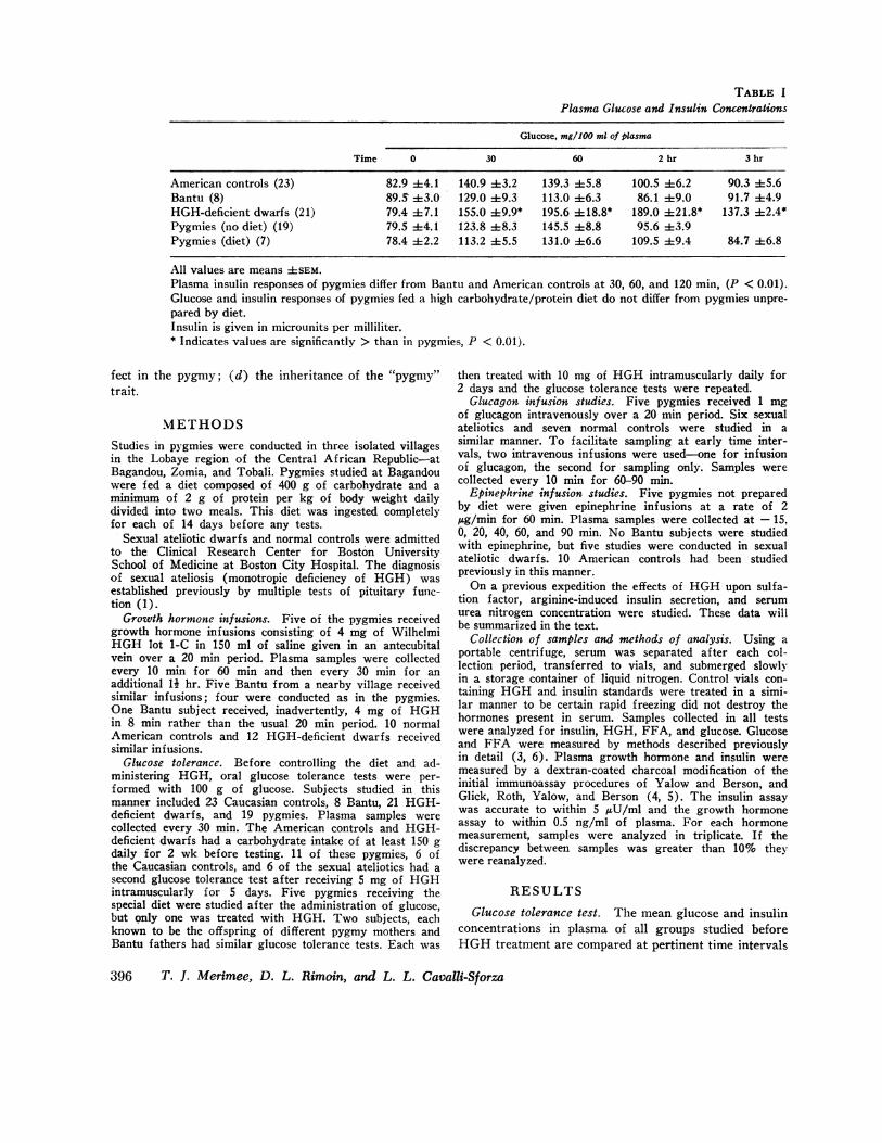
Plasma Glucose and InsulinTABLE I
Concentrations
Glucose, mg/1OO ml of plasma
Time 0 30 60 2 hr 3 hr
American controls (23) 82.9 414.1 140.9 1:-3.2 139.3 ±5.8 100.5 46.2 90.3 4:15.6Bantu (8) 89.S +3.0 129.0 ±9.3 113.0 ±6.3 86.1 ±9.0 91.7 ±4.9HGH-deficient dwarfs (21) 79.4 ±7.1 155.0 49.9* 195.6 418.8* 189.0 ±21.8* 137.3 ±2.4*Pygmies (no diet) (19) 79.5 ±4.1 123.8 ±8.3 145.5 ±8.8 95.6 43.9Pygmies (diet) (7) 78.4 42.2 113.2 ±5.5 131.0 ±6.6 109.5 ±9.4 84.7 ±6.8
All values are means ±SEM.Plasma insulin responses of pygmies differ from Bantu and American controls at 30, 60, and 120 min, (P < 0.01).Glucose and insulin responses of pygmies fed a high carbohydrate/protein diet do not differ from pygmies unpre-pared by diet.Insulin is given in microunits per milliliter.* Indicates values are significantly > than in pygmies, P < 0.01).
fect in the pygmiy; (d) the inheritance of the "pygmy"trait.
METHODSStudies in pygmies were conducted in three isolated villagesin the Lobaye region of the Central African Republic-atBagandou, Zomia, and Tobali. Pygmies studied at Bagandouwere fed a diet composed of 400 g of carbohydrate and aminimum of 2 g of protein per kg of body weight dailydivided into two meals. This diet was ingested completelyfor each of 14 days before any tests.
Sexual ateliotic dwarfs and normal controls were admittedto the Clinical Research Center for Boston UniversitySchool of Medicine at Boston City Hospital. The diagnosisof sexual ateliosis (monotropic deficiency of HGH) wasestablished previously by multiple tests of pituitary func-tion (1).
Growth hormone infusions. Five of the pygmies receivedgrowth hormone infusions consisting of 4 mg of WilhelmiHGH lot 1-C in 150 ml of saline given in an antecubitalvein over a 20 min period. Plasma samples were collectedevery 10 min for 60 min and then every 30 min for anadditional 1 hr. Five Bantu from a nearby village receivedsimilar infusions; four were conducted as in the pygmies.One Bantu subject received, inadvertently, 4 mg of HGHin 8 min rather than the usual 20 min period. 10 normalAmerican controls and 12 HGH-deficient dwarfs receivedsimilar infusions.
Glucose tolerance. Before controlling the diet and ad-ministering HGH, oral glucose tolerance tests were per-formed with 100 g of glucose. Subjects studied in thismanner included 23 Caucasian controls, 8 Bantu, 21 HGH-deficient dwarfs, and 19 pygmies. Plasma samples werecollected every 30 min. The American controls and HGH-deficient dwarfs had a carbohydrate intake of at least 150 gdaily for 2 wk before testing. 11 of these pygmies, 6 ofthe Caucasian controls, and 6 of the sexual ateliotics had asecond glucose tolerance test after receiving 5 mg of HGHintramuscularly for 5 days. Five pygmies receiving thespecial diet were studied after the administration of glucose,but only one was treated with HGH. Two subjects, eachknown to be the offspring of different pygmy mothers andBantu fathers had similar glucose tolerance tests. Each was
then treated with 10 mg of HGHintramuscularly daily for2 days and the glucose tolerance tests were repeated.
Glucagon infusion studies. Five pygmies received 1 mgof glucagon intravenously over a 20 min period. Six sexualateliotics and seven normal controls were studied in asimilar manner. To facilitate sampling at early time inter-vals, two intravenous infusions were used-one for infusionof glucagon, the second for sampling only. Samples werecollected every 10 min for 60-90 min.
Epinephrine infusion studies. Five pygmies not preparedby diet were given epinephrine infusions at a rate of 2,ug/min for 60 min. Plasma samples were collected at - 15,0, 20, 40, 60, and 90 min. No Bantu subjects were studiedwith epinephrine, but five studies were conducted in sexualateliotic dwarfs. 10 American controls had been studiedpreviously in this manner.
On a previous expedition the effects of HGHupon sulfa-tion factor, arginine-induced insulin secretion, and serumurea nitrogen concentration were studied. These data willbe summarized in the text.
Collection of samples and methods of analysis. Using aportable centrifuge, serum was separated after each col-lection period, transferred to vials, and submerged slowlyin a storage container of liquid nitrogen. Control vials con-taining HGHand insulin standards were treated in a simi-lar manner to be certain rapid freezing did not destroy thehormones present in serum. Samples collected in all testswere analyzed for insulin, HGH, FFA, and glucose. Glucoseand FFA were measured by methods described previouslyin detail (3, 6). Plasma growth hormone and insulin weremeasured by a dextran-coated charcoal modification of theinitial immunoassay procedures of Yalow and Berson, andGlick, Roth, Yalow, and Berson (4, 5). The insulin assaywas accurate to within 5 ,uU/ml and the growth hormoneassay to within 0.5 ng/ml of plasma. For each hormonemeasurement, samples were analyzed in triplicate. If thediscrepancy between samples was greater than 10% theywere reanalyzed.
RESULTS
Glucose tolerance test. The mean glucose and insulinconcentrations in plasma of all groups studied beforeHGHtreatment are compared at pertinent time intervals
396 T. J. Merimee, D. L. Rimoin, and L. L. Cavalli-Sforza

after 100 g of Glucose Orally
Insulin, pU/ml of plasma
Time 0 30 60 2 hr 3 hr
14.2 ± 1.4 76.9 ± 1 1.3* 87.1 + 12.4* 64.6 15.5* 46.3 ±6.1*27.8 ±5.4* 57.8 ±10.0* 70.0 ±4.3* 33.6 ±-3.9 28.0 ±4.6*16.1 41.5 27.4 ±4.0 37.4 44.2 43.3 ±5.8 30.8 ±7.1*15.4 ±2.1 21.0 42.7 25.3 ±3.9 22.2 ±5.013.8 ±4.5 17.2 A3.5 28.8 45.6 23.0 ±6.3 12.8 ±2.2
after administration of glucose in Table I. The maximalglucose concentrations in plasma achieved after the in-gestion of glucose were 140.9 ±3.2 mg/100 ml' inAmerican controls, 129.0 ±9.3 mg/100 ml in Bantu sub-jects, 195.6 ±18.8 mg/100 ml in sexual ateliotics, and131.0 ±6.6 mg/100 ml in pygmies prepared with thehigh carbohydrate/high protein diet. This latter figurewas similar to that noted in pygmies not prepared withthe special diet, (145.5 ±8.8 mg/100 ml of plasma).Unlike the sexual ateliotic, the pygmy did not exhibitglucose intolerance.
The insulin responses of Bantu subjects and Amer-ican controls were significantly greater than in pygmies.The mean peak insulin response to glucose in Bantuwas 70.0 ±4.2 .U/ml, in American controls, 87.1 +-12.4,oU/ml, and in 11 pygmies not prepared by diet, 25.3±3.9 /AU/ml. That these responses of the pygmy werenot related to the prior diet or to the control groupchosen is emphasized in Fig. 1. The mean maximal in-sulin response obtained in pygmies prepared by diet was28.8 ±5.6 sAU/ml.
Fig. 2 compares the mean insulin responses withglucose before and after HGH treatment. Subjectsstudied are American controls, pygmies not prepared bydiet, and HGH-deficient dwarfs. Similar responses ofa single pygmy prepared by diet and two offspring ofpygmy mothers and Bantu fathers are shown after ashorter course of HGHtreatment in Fig. 3. The indi-vidual responses of 11 pygmies are also shown in TableII which indicates not only the initial level of response,but emphasizes the complete, uniform failure of thepygmy to respond to HGH.
Both American controls and HGH-deficient dwarfshad a striking augmentation of their insulin responses
'All mean and mean maximal values are given withstandard errors of the mean.
GLUCOSE
_A,
160
120
t 80
40-INSULIN
HOURS
FIGURE 1 Plasma glucose and insulin concentrations areshown for Bantu, pygmies unprepared by diet, and pygmiesprepared by diet (D). 100 g of glucose was ingested attime 0. All points are means +SEM. Peak insulin responsesof both pygmy groups at 30 and 60 min are significantlydifferent fronm Bantu.
BBEFOREHGH
240-
200 -
% 160,S
o-:
801 T
4 0 ;/
O- -
/
0
0 2
IAFTER HGHLtSmg/dSd)
k Qj
,, / Ateiwotics
!,' '
I
Pyqmles
0 § 2
HOURSAFTERGLUCOSEINOESr/O
FIGURE 2 Plasma insulin concentrations after oral glucose(100 g) are shown before and after HGHtreatment. Allpoints are means +SEM.
Metabolic Studies in the African Pygmy 397
NP*1,1
HGH AFTER HGH (5mg/d, 2d)
Hi~~~~~~~~~~~H
2 0 2
HOURSAFTER GLUCOSEINGEST/ON
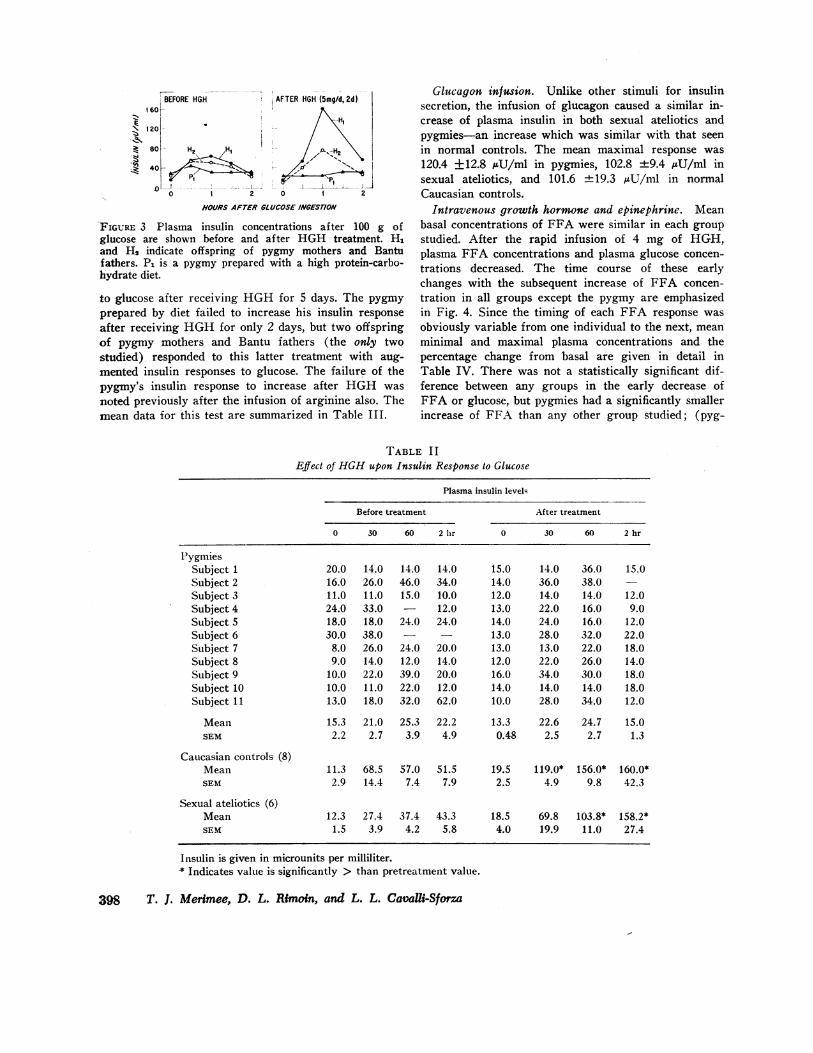
FIGURE 3 Plasma insulin concentrations after 100 g ofglucose are shown before and after HGH treatment. H.and H2 indicate offspring of pygmy mothers and Bantufathers. P1 is a pygmy prepared with a high protein-carbo-hydrate diet.
to glucose after receiving HGHfor 5 days. The pygmyprepared by diet failed to increase his insulin responseafter receiving HGHfor only 2 days, but two offspringof pygmy mothers and Bantu fathers (the only twostudied) responded to this latter treatment with aug-mented insulin responses to glucose. The failure of thepygmy's insulin response to increase after HGHwasnoted previously after the infusion of arginine also. Themean data for this test are summarized in Table III.
Glucagon infusion. Unlike other stimuli for insulinsecretion, the infusion of glucagon caused a similar in-crease of plasma insulin in both sexual ateliotics andpygmies-an increase which was similar with that seenin normal controls. The mean maximal response was120.4 ±12.8 AU/ml in pygmies, 102.8 -+-9.4 AU/ml insexual ateliotics, and 101.6 + 19.3 AU/ml in normalCaucasian controls.
Intravenous growth hormone and epinephrine. Meanbasal concentrations of FFA were similar in each groupstudied. After the rapid infusion of 4 mg of HGH,plasma FFA concentrations and plasma glucose concen-trations decreased. The time course of these earlychanges with the subsequent increase of FFA concen-tration in all groups except the pygmy are emphasizedin Fig. 4. Since the timing of each FFA response wasobviously variable from one individual to the next, meanminimal and maximal plasma concentrations and thepercentage change from basal are given in detail inTable IV. There was not a statistically significant dif-ference between any groups in the early decrease ofFFA or glucose, but pygmies had a significantly smallerincrease of FFA than any other group studied; (pyg-
TABLE I IEffect of HGHupon Insulin Response to Glucose
Plasma insulin level-
Before treatment After treatment
0 30 60 2 hlr 0 30 60 2 hr
PygmiesSubject 1 20.0 14.0 14.0 14.0 15.0 14.0 36.0 15.0Subject 2 16.0 26.0 46.0 34.0 14.0 36.0 38.0Subject 3 11.0 11.0 15.0 10.0 12.0 14.0 14.0 12.0Subject 4 24.0 33.0 - 12.0 13.0 22.0 16.0 9.0Subject 5 18.0 18.0 24.0 24.0 14.0 24.0 16.0 12.0Subject 6 30.0 38.0 - 13.0 28.0 32.0 22.0Subject 7 8.0 26.0 24.0 20.0 13.0 13.0 22.0 18.0Subject 8 9.0 14.0 12.0 14.0 12.0 22.0 26.0 14.0Subject 9 10.0 22.0 39.0 20.0 16.0 34.0 30.0 18.0Subject 10 10.0 11.0 22.0 12.0 14.0 14.0 14.0 18.0Subject 11 13.0 18.0 32.0 62.0 10.0 28.0 34.0 12.0
Mean 15.3 21.0 25.3 22.2 13.3 22.6 24.7 15.0SEM 2.2 2.7 3.9 4.9 0.48 2.5 2.7 1.3
Caucasian controls (8)Mean 11.3 68.5 57.0 51.5 19.5 119.0* 156.0* 160.0*SEM 2.9 14.4 7.4 7.9 2.5 4.9 9.8 42.3
Sexual ateliotics (6)Mean 12.3 27.4 37.4 43.3 18.5 69.8 103.8* 158.2*SEM 1.5 3.9 4.2 5.8 4.0 19.9 11.0 27.4
Insulin is given in microunits per milliliter.* Indicates value is significantly > than pretreatment value.
398 T. J. Merimee, D. L. Rimoin, and L. L. Cavalli-Sforza

TABLE IIIEffect of HGHupon Insulin Responses to Arginine*
Plasma insulin levels
Before treatment After treatment
0 30 60 90 120 0 30 60 90 120
Pygmies 12.2 26.8 22.0 13.6 15.2 12.8 18.3 15.3 13.0(19) ±1.9 ±3.6 ±3.9 ±1.7 ±2.2 ±1.6 ±1.2 ±3.5 ±1.7
Caucasian 21.5 81.5 49.2 28.0 27.7 23.0 290.01 101.01 33.0Controls (8) ±4.5 ±19.0 ±0.5 ±4.9 ±7.6 ±1.3 ±65.0 ±10.5 ±1.7
* Insulin is given in microunits per milliliter. All values are mean ±SEM.0.25 g of arginine per pound of body weight was given from time 0 to 30.1 Indicates value is significantly > than pretreatment value.
mies vs. Bantu P < 0.01, vs. normal P < 0.05, vs. HGH-deficient dwarfs P < 0.01).
The mean maximal FFA response to epinephrine wassimilar in growth hormone-deficient dwarfs and Babingapygmies. Epinephrine caused an increase in plasma FFAconcentration in each of these groups. The mean max-imal increase of FFA over basal concentration was 150±20.5% in pygmies and 158 ±16.0% in HGH-deficientdwarfs. In a previous study, similar responses werenoted in normal controls.
DISCUSSION
Wehave described previously two groups of dwarfs whohave a monotropic deficiency of HGH, and Laron hasdescribed a type of dwarfism in which high basal valuesof immunoreactive HGHare found (7-9). The presentstudy was undertaken to clarify the problems whicharose in our earlier attempts to delineate the endocrinestatus of the African pygmy.
On previous expeditions to the Central African Re-public, we established that Babinga pygmies do not havea deficiency of HGH, that their basal plasma HGHvalues are not elevated, and that several responses ex-pected after HGHtreatment were absent (1-3).
There were several problems which made it difficultto interpret the initial data. Although we had empha-sized the poor insulin response of the pygmy to glucoseafter treatment with HGH, it seemed possible that thiscould be secondary to dietary peculiarities and notsimply the result of resistance to HGH. Furthermore,we had compared the pygmy in all studies with Amer-ican controls, but if neighboring Bantu resembled thepygmy, the comparisons would be invalid. We, likewise,emphasized the lack of a lipolytic response (i.e. a laterise in FFA level above basal) after intravenous HGH.This, however, could be nonspecific or, alternatively,might reflect a relatively greater physiologic effect ofinsulin in the pygmy. In addition to these problems, we
had no information which aided in determining the siteof the defect and its manner of inheritance.
That the impoverished insulin response of the pygmynoted initially was not related to diet is evident from thedata given in Table I and Fig. 1. Despite the fact thatpygmies were prepared with a special diet for 2 wk,their plasma insulin responses to a glucose load re-mained attenuated and were almost identical with re-sponses obtained in pygmies not treated with a highcarbohydrate/protein diet. Insulin responses of Bantu toglucose were very similar to those of American controls.Glucagon, the only simulus giving consistently a normalinsulin response in HGH-deficient dwarfs, also gave asimilar response in pygmies. This would seem to furthersupport the fact that decreased insulin responses of the
2 O0C
CONTROL(Bantu
0
HGH- DEfIcENTrDWARFS
150 i/
O C _
!bt-
PrGMIES
5 0
1 00 - -
50 _-30 60 90 '20
MINUTrES
FIGURE 4 Free fatty acid concentration in plasma ex-pressed as per cent of the basal FFA concentration isshown after 4 mg of intravenous HGHgiven over 20 min.
Metabolic Studies in the African Pygmy 399

TABLE IVEffect of HGHInfusion on Plasma Glucose and FFA Concentrations*
Mean basal FFA Mean minimal FFA Mean maximal FFA Maximal decrease ofconcentration concentration concentration plasma
AM/mi JIEqlml %of pEq/ml %of mg/ %de-basal basal 100 ml creaselevel level from
basallevel
Pygmies (prepared by diet) 0.653 ±0.092 0.384 40.099* 708.0 40.074 15.4 ±2.1*(59.0 ±9.4%) (107.0 48.8%) (17.5 ±1.9%)
Bantu 0.652 ±0.084 0.394 ±0.090* 1.078 41.00* 20.5 ±6.6*(60.2 ±10.3%) (165.0 ±21.0%) (18.8 ±4.6%)
HGH-deficient dwarfs 0.800 ±0.102 0.442 ±0.083* 1.402 ±0.126* 16.9 44.3*(55.0 ±12.0%) (175.0 +16.3%) (17.0 ±2.3%)
American controls 0.608 ±0.062 0.394 ±0.070* 0.923 ±0.064*(65.0 ±9.3%) (151.0 ±7.8%)
The initial decreases of FFA and glucose concentrations occurred within 40 min. All increases of plasma FFA concentrationsoccurred between 60 and 150 min.* Indicates value differs significantly both from basal state and from maximum variation obtained in 12 control studies with theinfusion of saline.
pygmy to glucose and arginine' were somehow relatedto HGH. This finding, however, need not be overempha-sized. Indeed, the insulin output of the pygmy to glucosemay well be functionally appropriate. The point of majorimportance is not the appropriateness or inappropriate-ness of the pygmies initial insulin response to glucose,but the consistent failure of insulin secretion to increaseafter exogenous HGHwas administered. In our experi-ence, it remains striking and unique to find any groupin which every individual studied failed to augment in-sulin secretion after HGHtherapy.
The FFA responses after acute administration ofHGHalso support the view that HGHis at least par-tially ineffective in these individuals. As indicated inFig. 4, the infusion of HGHfailed to increase FFAlevels in pygmies. Increases were readily detectable incontrol subjects, (including the Bantu) and in sexualateliotics. As with insulin secretion, diet does not ex-plain these results and our "control" studies appearsuitable since Bantu from the same area do not showpatterns similar to the pygmy. As with insulin outputafter HGH therapy, it remains possible that the im-paired rise in FFA levels is related somehow to agreater physiologic activity of insulin in the pygmy. Ifso, it is an uniquely specific effect, since insulin did notalter the increase of FFA levels obtained with epi-nephrine.
The exact nature of the metabolic defect in the pygmycannot be described with complete assurance, but thedata seem to us most consistent with two possibilities:(a) An intracellular defect in HGH-mediated proteinsynthesis or (b) a similar, partial insensitivity to a
HGH-dependent substance such as sulfation factor. Al-though early responses to HGH (as we tested them)might well be pharmacologic effects, they are known tobe independent of DNA-medicated protein synthesis(10, 11). The crucial metabolic reactions responsiblefor a normal growth rate, such as the over-all proteinsynthetic rate, are probably abnormal in the pygmy asindicated by the failure of HGHto increase lipolysis,augment insulin secretion, or reduce serum urea nitro-gen concentration. These reactions are blocked readilyby agents interfering with DNA-mediated protein syn-thesis (10-12). For example, the need for HGHtoinduce intracellular protein synthesis for its lipolyticaction is fairly well documented (13, 14). Similarly, theability of HGHto alter insulin secretion is clearly aneffect requiring a minimum of hours and usually severaldays (15). There are normal basal levels of plasmaHGHin the pygmy. If the above speculation is correct,this might indicate that neural centers controlling HGHsecretion are not dependent upon a HGH-mediated pro-tein synthetic process.
It cannot be determined at present whether the in-sensitivity noted is to HGH. per se, or to a substancegenerated by HGH. It is known that sulfation factor isnormal in the pygmy, so one must consider the possibil-ity that anabolic effects attributed to HGHare inducedby an intermediary. It is not possible to speculatewhether there is an unknown antagonist to the action ofHGHor whether the HGHof the pygmy is anomalous,retaining immunoreactivity but lacking biological activ-ity. With the present data, the latter possibility is highlyunlikely. One would expect decreased responsiveness to
400 T. J. Merimee, D. L. Rimoin, and L. L. Cavalli-Sforza

exogenous HGHonly if the concentration of the anom-alous hormone were increased.
Too few subjects were studied to state conclusively-the manner in which the "pygmy" trait was inherited.Despite a considerable effort to locate such subjects,only two could be identified positively as the offspringof pygmy/Bantu parents. That the defect is not inheritedas a dominant trait is apparent from studies conductedin these two hybrids. Fig. 2 shows the plasma insulinresponse to oral glucose before and after HGHtreat-ment in controls, sexual ateliotics, and pygmies. Each ofthese subjects had received 5 mg of HGHdaily for 5days. Although the two hybrids were treated with HGHfor 2 days only, each showed an increase of their plasmainsulin response to glucose.
In summary, the pygmy has several defective re-sponses to HGH. These probably involve actions ofHGHdependent upon late protein synthetic processes.Offspring of pygmy mothers and Bantu fathers do notshow abnormal insulin responses to glucose and readilyaugment their insulin responses to HGHtreatment, indi-cating that the defect is not inherited as a dominant trait.Although isolated cases of insensitivity to a peptidehormone have been described, the defect in the pygmyappears unique in that it effects an entire race.
ACKNOWLEDGMENTSWewish to acknowledge the critical analysis of these databy Dr. John Eager Howard and Dr. Victor McKusick.
This work was supported in part by U. S. Public HealthService Grants, 5-ROI-GM10189, HD-02422, AM-13565, andRR-533 (General Clinical Research Program of the Divi-sion of Research Resources). The expedition was organizedby L. L. Cavalli-Sforza and supported by grants from theItalian I.B.P. Committee of the Consiglio Nazionale delleRicherche and by the World Health Organization.
REFERENCES1. Rimoin, D. L., T. J. Merimee, D. Rabinowitz, and V. A.
McKusick. 1968. Genetic aspects of clinical endocrin-ology. Recent Progr. Hormone Res. 24: 365.
2. Merimee, T. J., D. L. Rimoin, D. Rabinowitz, L. L.Cavalli-Sforza, and V. A. McKusick. 1968. Metaboliceffects of HGHin the African Pygmy. Lancet. 1: 194.
3. Merimee, T. J., D. L. Rimoin, D. Rabinowitz, L. L.Cavalli-Sforza, and V. A. McKusick. 1968. Metabolicstudies in the African Pygmy. Trans. Ass. Amer.Physicians Philadelphia. 81: 221.
4. Yalow, R. S., and S. A. Berson. 1960. Immunoassay ofendogenous plasma insulin in man. J. Clin. Invest. 39:1159.
5. Glick, S. M., J. Roth, R. S. Yalow, and S. A. Berson.1963. Immunoassay of growth hormone in plasma.Nature (London). 119: 784.
6. Dole, V. P. 1956. A relation between non-esterifiedfatty acids in plasma and the metabolism of glucose.J. Clin. Invest. 35: 150.
7. Merimee, T. J., J. A. Burgess, and D. Rabinowitz. 1967.Influence of growth hormone on insulin secretion. Dia-betes. 16: 478.
8. Merimee, T. J., D. L. Rimoin, J. G. Hall, and V. A.McKusick. 1969. A metabolic and hormonal basis forclassifying sexual ateliotic dwarfs. Lancet. 1: 963.
9. Laron, A., A. Pentzelon, and S. Mannheimer. 1966.Genetic pituitary dwarfism with high serum concentra-tion of growth hormone. Israel J. Med. Sci. 2: 152.
10. DeBodo, R. C., and N. Altszuler. 1957. The metaboliceffects of growth hormone and their physiologic signifi-cance. Vitamins Hormones. 15: 206.
11. Swislucki, N. I., and C. M. Szego. 1965. Acute reduc-tion of plasma nonesterified fatty acid by growth hor-mone in hypophysectomized and Houssay rats. Endo-crinology. 76: 665.
12. Goodman, H. M. 1968. Growth hormone and the metab-olism of carbohydrate and lipid in adipose tissue. Ann.N. Y. Acad. Sci. 148: 419.
13. Fain, J. N., and R. Saperstein. 1970. The involvementof RNA synthesis and cyclic AMP in the activation offat cell lipolysis by growth hormone and glucocorticoids.In Adipose Tissue Regulation and Metabolic Findings.B. Heanrenaud and D. Hepp, editors. Academic PressInc., NewYork. 20.
14. Fain, J. N., A. Dodd, and L. Novak. 1971. Relation-ship of protein synthesis and cyclic AMP to lipolyticaction of growth hormone and glucocorticoids. Metab.(Clin. Exp.). 20: 109.
15. Merimee, T. J., D. Rabinowitz, D. L. Rimoin, and V. A.McKusick. 1968. Sexual ateliotic dwarfism IV: theresponse of sexual ateliotic dwarfs to exogenous growthhormone. Metab. (Clin. Exp.). 16: 1012.
Metabolic Studies in the African Pygmy 401