Embed Size (px)
Citation preview
METABOLIC SIGNALS IN SLEEP REGULATION: THE ROLE OF
CHOLECYSTOKININ
By
LEVENTE KAPÁS, M.D.
A thesis for the degree of
DOCTOR OF PHYLOSOPHY
(Ph. D.)
Department of Physiology, Faculty of Medicine, University of Szeged
2010 Szeged

P a g e | i
To the memory of Ferenc Obál Jr.
P a g e | ii
Peer-reviewed papers directly related to the thesis
1. Kapás, L., F. Obál, Jr., P. Alföldi, G. Rubicsek, B. Penke, and F. Obál. Effects of
nocturnal intraperitoneal administration of cholecystokinin in rats: simultaneous increase
in sleep, increase in EEG slow-wave activity, reduction of motor activity, suppression of
eating, and decrease in brain temperature. Brain Res. 438: 155-164, 1988.
2. Kapás, L., F. Obál, Jr., I. Farkas, L. C. Payne, G. Sáry, G. Rubicsek, and J. M. Krueger.
Cholecystokinin promotes sleep and reduces food intake in diabetic rats. Physiol.
Behav. 50: 417-420, 1991.
3. Kapás, L., F. Obál, Jr., M. R. Opp, L. Johannsen, and J. M. Krueger. Intraperitoneal
injection of cholecystokinin elicits sleep in rabbits. Physiol. Behav. 50: 1241-1244,
1991.
4. Chang, H.-Y. and L. Kapás. The effects of CCK-4 and non-sulfated CCK-8 on sleep,
EEG slow-wave activity and brain temperature in rats, Physiol. Behav., 62: 175-179,
1997.
5. Shemyakin, A. and L. Kapás. L-364,718, a cholecystokinin-A receptor antagonist,
suppresses feeding-induced sleep in rats. Am. J. Physiol., 280: R1420-R1426, 2001.
Reviews and book chapters directly related to the thesis
1. Kapás, L., F. Obál, Jr., and J. M. Krueger. Humoral regulation of sleep. Int. Rev.
Neurobiol. 35: 131-160, 1993.
2. Kapás, L. and É. Szentirmai. Sleep regulatory factors. In: Monti, J., Sinton, C. and
Pandi-Perumal, S. R. (Eds.), The Neurochemistry of Sleep and Wakefulness. Cambridge
University Press, UK, 2008, pp. 315-336.
3. Szentirmai, É., L. Kapás and J. M. Krueger. Interactive regulation of sleep and feeding.
In: Kryger, M. H. (Ed.), Atlas of Clinical Sleep Medicine, Philadelphia: Saunders
Elsevier, 2010, pp. 53-56.
P a g e | iii
4. Szentirmai, É., L. Kapás and J. M. Krueger. Hormones and sleep. In: Breed, M. D. and
Moore, J. (Eds.), Encyclopedia of Animal Behavior, Oxford: Academic Press, 2010, in
press.
Abstracts directly related to the thesis
1. Kapás, L., F. Obál, Jr., P. Alföldi, G. Rubicsek, B. Penke, and F. Obál. Sleep elicited by
peripheral injection of cholecystokinin in rats. Neuroscience 22: p. S841, 1987.
2. Kapás, L., M. R. Opp, L. Johannsen, and J. M. Krueger. Divergent effects of central
and peripheral injections of cholecystokinin (CCK) on sleep-wake activity in rabbits.
Sleep Res. 19: p. 18, 1990.
3. Kapás, L., F. Obál, Jr., I. Farkas, L. C. Payne, G. Sáry, G. Rubicsek, and J. M. Krueger.
Hypnogenic and anorectic effects of CCK persist in vagotomized and diabetic rats. Eur.
Sleep Res. Soc. Abstracts. p. 93, 1990.
4. Kapás, L. LY-288,513, a cholecystokinin (CCK)-B receptor antagonist, inhibits CCK-
induced sleep. Sleep Res. 25: p. 12, 1996.
5. Kapás, L. Cholecystokinin (CCK)-induced sleep is suppressed by LY-288,513, a CCK-
B receptor antagonist. J. Sleep Res. 5 (suppl. 1): p. 103, 1996.
6. Chang, H.-Y. and L. Kapás. The effects of CCK-8NS and CCK-4 on sleep, slow wave
activity of the EEG and brain temperature in rats. Soc. Neurosci. Abstr., Vol. 22., Part
1., p. 147, 1996.
7. Chang, H.-Y. and L. Kapás. L-364,718, a cholecystokinin (CCK)-A receptor
antagonist, inhibits the sleep-inducing effects of CCK. Sleep Res. 26:138, 1997.
8. Kapás, L. Subdiaphragmatic vagotomy does not prevent the somnogenic and
hypothermic effects of cholecystokinin (CCK). Sleep Res. 26:76, 1997
9. Shemyakin, A. and L. Kapás. Starvation suppresses NREMS and EEG slow-wave
activity in rats. Sleep 22, Suppl. 1, S247, 1999.
P a g e | iv
10. Shemyakin, A. and L. Kapás. A CCK-A receptor antagonist inhibits sleep responses to
feeding in rats. Sleep 23, Suppl. 2, A121, 2000.
11. Shemyakin, A. and L. Kapás. L-364,718, a CCK-A receptor antagonist, suppresses
sleep responses to feeding in rats. J. Sleep Res. 9, Suppl. 1, P175, 2000.
12. Shemyakin, A. and L. Kapás. L-364,718, a CCK-A receptor antagonist does not
prevent the somnogenic effects of interleukin in rats. Sleep 24, A145, 2001.
P a g e | v
Table of Content
Summary …………………………………………………………………..…………… VI
Abbreviations …………………………………………………………...……………… VII
Introduction …………………………………………………...……………………….. 1
1. Sleep regulation ……………………………………………….……………….. 1
2. Sleep, feeding and metabolism …………………………………….………….. 4
3. Cholecystokinin ………………………………………………………………. 6
4. Aims of the present studies ……………………………………………………. 8
Materials and Methods …………………………………………………………………. 10
1. General Methods ……………………………………………………………....... 10
2. Experimental Design …………………………………………………………… 11
Results ………………………………………………………………………………….. 14
1. The effects of systemic injection of CCK in rats ……………………………….. 14
2. The effects of intraperitoneal and intracerebroventricular
injection of CCK in rabbits …………………………………………………….. 15
3. The effects of CCK2 receptor agonists in rats ………………………………….. 16
4. The effects of CCK1 receptor antagonist on CCK-induced
sleep in rats ……………………………………………………………………… 18
5. The effects of CCK in diabetic rats …………………………………………….. 21
6. The effects of CCK1 antagonist on feeding-induced sleep ……………………. 23
Discussion ………………………………………………………………………………. 27
Acknowledgements ……………………………………………………………………. 37
References ………………………………………………………………………………. 38
Attachments …………………………………………………………………………….. 48
List of all peer-reviewed publications ………………………………………………….. 83

P a g e | vi
Summary
Acute metabolic changes in response to feeding or starvation as well as long-term metabolic
shifts due to increased or decreased adiposity are influences that greatly affect the amount
and the quality of sleep. We posit that hormones of the gastrointestinal (GI) system and
adipose tissue play a key role in signaling for these adaptive sleep responses. Eating is
followed by a characteristic postprandial behavioral sequence which ends with sleep.
Several GI hormones which are released after eating, e.g., cholecystokinin and gastric leptin,
are known to suppress food intake and bring about satiety. The specific hypothesis tested in
the present work is that cholecystokinin is a sleep-inducing hormone which contributes to
signaling for postprandial sleep.
We tested this hypothesis in six sets of experiments. We determined that systemic
administration of cholecystokinin octapeptide sulfate ester (CCK) elicits dose-dependent and
selective increases in non-rapid-eye-movement sleep (NREMS) in rats. The lowest effective
dose is 10 µg/kg when administered intraperitoneally. The sleep responses are accompanied
by a decrease in brain temperature and suppressed feeding. Similar sleep and
thermoregulatory effects are observed in rabbits after systemic injection of 10 and 50 µg/kg
CCK. Central administration of CCK does not affect sleep in rabbits. CCK2 receptor
specific analogues, CCK tetrapeptide and nonsulfated CCK octapeptide, lack somnogenic and
hypothermic activities when given systemically. Both the somnogenic and thermoregulatory
effects of exogenously administered CCK are blocked by L-364,718, a selective CCK1
receptor antagonist. Lesion of pancreatic beta cells by streptozotocin does not prevent the
somnogenic effects of CCK. Enhanced feeding results in increases in NREMS in control
rats. The postprandial sleep responses are prevented by CCK1 receptor antagonist treatment.
We conclude that CCK has somnogenic activities in rats and rabbits. Selective activation of
the CCK2 receptors is not sufficient for the effects whereas the activation of CCK1 receptors
is required; these strongly suggest the involvement of CCK1 receptors both in the
somnogenic and hypothermic actions of CCK. CCK strongly stimulates insulin secretion by
the pancreas but pancreatic insulin is not a mediator of CCK-induced sleep. Endogenously
released CCK after feeding is likely a key factor for signaling postprandial sleep responses.
Present results are consistent with the hypothesis that CCK is a component of a complex
signaling mechanism which modulates sleep-wake activity according to the metabolic status
of the body.
P a g e | vii
Abbreviations
BBB Blood-brain barrier
CCK Cholecystokinin; cholecystokinin octapeptide sulfate ester
CCK-4 CCK tetrapeptide
CCK-8 -NS Nonsulfated CCK octapeptide
CCK-8-SE CCK octapeptide sulfate ester
CNS Central nervous system
EEG Electroencephalogram
EMG Electromyography
GI Gastrointestinal
icv Intracerebroventricular
ip Intraperitoneal
iv Intravenous
LH Lateral hypothalamus
NREMS Non-rapid-eye-movement sleep
NTS Nucleus tractus solitarius
PBN Parabrachial nucleus
REMS Rapid-eye-movement sleep
sc Subcutaneous
SCN Suprachiasmatic nucleus
SE Sulfate ester; standard error
Tbr Brain temperature
TNF Tumor necrosis factor
VMH Ventromedial hypothalamus
P a g e | 1
Introduction
1. Sleep Regulation.
Questions about the nature and function of sleep have interested a great number of scientists,
philosophers and common people across cultures and millennia. Aristotle wrote the following
about sleep 2,400 years ago:
“WITH regard to sleep and waking, we must consider what they are: whether they are
peculiar to soul or to body, or common to both; and if common, to what part of soul or
body they appertain: further, from what cause it arises that they are attributes of
animals, and whether all animals share in them both, or some partake of the one only,
others of the other only, or some partake of neither and some of both.” (Aristotle: “On
Sleep and Sleeplessness”, translated by J.I. Beare, Electronically Enhanced Text
Copyright 1991, World Library, Inc.)
Replacing the word ‘soul’ with ‘brain’ leads to some of the most fundamental questions in
sleep research that are still being debated in the 21st century. Is sleep for the brain or for the
body? Which part(s) of the brain is sleep related to? What is that sleeps in the brain and
what is that regulates sleep? Do all animals sleep?
All animals exhibit some form of rest-activity cycle. All mammals that have been studied so
far exhibit sleep similar to humans’ in that two basic forms of vigilance states alternate
during the sleep period: non-rapid-eye-movement sleep (NREMS) and rapid-eye-movement
sleep (REMS). The proportion of NREMS and REMS in total sleep time shows species
differences; both in humans and rats about 80% of sleep is NREMS and the rest is REMS.
There are no known differences in the fundamental nature of sleep among mammals,
including humans. Sleep of humans and rats, the most widely studied species in sleep
research, shows some difference only in its timing. Healthy young adults typically have a
single, ~8 h sleep episode at night. In the laboratory, rats sleep both during the day and night
but more sleep occurs during the light period, characteristic of nocturnal species. Rat sleep is
polyphasic, i.e., multiple short sleep and wake episodes alternate during the nychthemeron,
the length of single wake periods rarely exceeds one hour.
Most in the field of sleep research maintain that sleep is by the brain and for the brain. It is
also evident that specific changes occur in the activities of most organs during sleep. Some
P a g e | 2
of the sleep-related physiological events are slight adjustments, such as the 5-10% decline in
energy expenditure during NREMS, others are fundamental changes, such as the near
complete loss of homeothermic thermoregulation during REMS. There are three aspects of
vigilance-related alterations in physiological functions. One, most physiological adjustments
are thought to be caused by sleep or the lack thereof. For example, the decline in energy
expenditure and increases in growth hormone secretion are due to sleep itself. Two, some
changes are not caused by sleep per se but are independent manifestations of the action of a
common regulatory mechanism which they share with sleep/wakefulness. Increased feeding
and wakefulness at the beginning of the behaviorally active phase are thought to be parallel
outputs of such a shared hypothalamic circuit of sleep and feeding regulation. We posit that
there is a third aspect of body-sleep interaction: physiological changes outside of the brain
affect complex brain functions, including sleep. Part of these somatic changes is related to
eating, adiposity or changes in metabolism. Our long-term goal is to understand how the
metabolic status of the body affects brain in general and vigilance in particular. We aim to
decipher the mechanisms involved in signaling to integrative sleep centers under various
metabolic conditions. The present work focuses on one of the putative peripheral messengers
involved in signaling between the body and sleep centers, the hormone cholecystokinin
(CCK).
Sleep appears to be a robust and distributed function of the brain. Multiple brain structures
are proposed to be involved in triggering and maintaining sleep and wakefulness. While
lesions or stimulations of various structures often lead to transient changes in sleep, there is
not a single structure the lesion of which would permanently eliminate sleep if the animal
survives. Ascending arousal systems arising from the brain stem and basal forebrain as well
as arousal mechanisms originating in the lateral hypothalamus and thalamus are implicated in
the maintenance of wakefulness (Jones, 2003). Sleep-promoting regions reside within the
hypothalamus, mainly the anterior and the ventro- and dorsomedial regions (McGinty and
Szymusiak, 2003). Though much has been learned about these structures in regard to sleep
regulation, the exact function of these regions and the interaction among them are still poorly
understood and widely debated.
Our understanding of sleep regulation is more complete when we view it from a more
theoretical perspective. The most widely accepted model of sleep regulation is the “two-
process model” (Borbély, 1982). It describes the onset of sleep and waking and the intensity
P a g e | 3
of sleep as the function of two independent processes, the sleep pressure and sleep threshold.
In short, sleep occurs when sleep pressure exceeds threshold.
Sleep threshold (Process C), is the circadian component of sleep regulation driven by the
main biological clock, the suprachiasmatic nucleus (SCN). Sleep pressure (Process S) is
independent of circadian influences, its intensity solely determined by and proportional to
prior wakefulness. Changes in sleep threshold show a 24-h cycle. In essence, these changes
increase the probability of wakefulness during those hours of the day when active
engagement with the environment is likely to be the most advantageous for a given species.
Sleep pressure is the homeostatic component of sleep regulation, a mechanism that aims to
keep the amount of sleep optimal. After extended periods of wakefulness, increases in sleep
pressure lead to more prolonged and deeper sleep. In humans, high sleep pressure and low
sleep threshold normally coincide around the usual bedtime leading to sleepiness and sleep.
During sleep, sleep pressure dissipates and sleep threshold increases. Low sleep pressure
coinciding with increased sleep threshold at the habitual waking time leads to arousal.
The two-process model gives a reliable statistical approximation of sleep timing in a large
population of subjects under controlled, identical conditions but does not account for changes
in vigilance driven by acute changes in the external of internal environment of the individual.
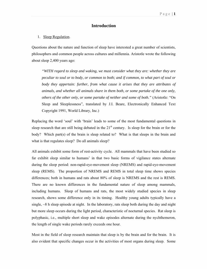
Fig. 1. Intrinsic and extrinsic factors in signaling sleep.

P a g e | 4
Both Process S and Process C are functions of the brain itself. Reflecting the fundamental
nature of these processes that they arise from within the brain, we propose to consider them
intrinsic factors in sleep regulation. The actual vigilance state of an individual subject,
however, is also a function of influences that are not inherent to the brain but arise from
outside of the central nervous system (CNS). These extrinsic factors include inputs from
sensory organs, infections, stress and others (Fig. 1). Neural and hormonal signals in
response to the external influences convey information to integrative sleep centers, which, in
turn, bring about adaptive changes in sleep-wake activity. Acute metabolic changes in
response to feeding or starvation as well as long-term metabolic shifts due to increased or
decreased adiposity are extrinsic influences that greatly affect the amount and the quality of
sleep. We posit that hormones of the gastrointestinal (GI) system and adipose tissue play a
key role in signaling for these sleep changes.
2. Sleep, feeding and metabolism.
There is a strong bidirectional interaction between sleep/vigilance and metabolism/feeding. It
has long been recognized that sleep is associated with characteristic changes in energy
expenditure and metabolism (Garby et al., 1987). Cross-species correlational studies in
mammals revealed a robust relationship between daily sleep amounts and resting metabolic
rate (Zepelin and Rechtschaffen, 1974; Allison and Cicchetti, 1976). A growing body of
evidence indicates that changes in metabolism and feeding lead to adaptive responses in
sleep. Rats are nocturnal, about 80-90% of their daily feeding takes place at night when they
are mostly awake and lipogenesis dominates their metabolic profile. In the light period, they
sleep more, feeding is minimal and energy is mainly supplied by increased lipolysis.
Reversing the lipolytic and lipogenic phases by sequential administration of lipolytic and
lipogenic hormones (Danguir and Nicolaidis, 1980a) or by restricting feeding to the light
period (Roky et al., 1999) leads to an almost complete reversal of the sleep-wake pattern of
rats. The naturally nocturnal animals become diurnal, mostly awake during the day and sleep
at night.
Acute, transient changes in the amount and/or content of food profoundly affect sleep-wake
activity in several species, including humans. In general, starvation induces marked sleep
loss (Borbély, 1977; Danguir and Nicolaidis, 1979; Szentirmai et al., 2010) while
spontaneously or experimentally increased caloric intake leads to increased sleep. In 1964,
Hockman reported that the electroencephalogram (EEG) of food-satiated animals shows a

P a g e | 5
marked increase in amount of high-voltage low-frequency activity, changes characteristic of
sleep (Hockman, 1964). Introduction of milk into the duodenum leads to sedation in cats
(Fara et al., 1969) and intragastric injection of eggnog results in postprandial EEG
synchronization in rats (Bernstein, 1974). There is a positive correlation between meal size
and the subsequent duration of sleep in normally feeding rats during the dark period (Danguir
et al., 1979). Refeeding after food deprivation in adult (Jacobs and McGinthy, 1971;
Borbely, 1977) or suckling rats (Lorenz, 1986), enhances sleep. The calorie-rich “cafeteria
diet” induces hyperphagia and increases the amount of sleep in rats (Danguir, 1987; Hansen
et al., 1998). Intravenous (iv) administration of highly nutritive composite solution greatly
enhances both NREMS and REMS in rats (Danguir and Nicolaidis, 1980b).
In humans, enhanced postprandial sleepiness is not only our every day experience but it is
also well-documented experimentally (Stahl et al., 1983; Smith et al., 1991; Zammit et al.,
1992). Fat-rich meals have a more potent effect on subjective feelings of sleepiness than
isocaloric meals in which fat is replaced by carbohydrate (Lloyd et al., 1994; Wells et al.,
1995). In humans, both sleep and plasma CCK levels are enhanced after high-fat/low-
carbohydrate diet as compared to low-fat/high-carbohydrate food (Wells et al., 1997).
Nighttime protein- and fat-rich drinks prolong sleep (Southwell et al., 1972; Brezinova and
Oswald, 1972). Intravenous infusion of amino acid mixture solutions promotes stage 3 and 4
NREMS (Lacey et al., 1978). Deep sleep profoundly increases during refeeding periods in
anorexia nervosa patients when they are gaining weight but rapidly falls back to previous
levels when normal weight is reached and stabilized (Lacey et al., 1975).
The regulation of sleep, feeding and metabolism overlaps on a structural level. Several
hypothalamic areas, such as the SCN, lateral hypothalamus (LH) and ventromedial
hypothalamic nucleus (VMH) are implicated in the regulation of both sleep and
metabolism/food intake (Grill, 2006). We propose that there is also overlap on a second,
signaling level as well; as certain signaling mechanisms, particularly GI hormones, may be
involved both in sleep and feeding/metabolism regulation. Our broad hypothesis is that
feeding-related GI hormones play a key role as metabolic signals in aligning vigilance
with the current metabolic state of the body. Fasting is accompanied by marked increases
in wakefulness and overall behavioral activity. There is strong evidence that ghrelin, a
gastrointestinal peptide produced by the stomach during fasting, plays a role in fasting-
induced arousal responses (Szentirmai et al., 2010). Eating is followed by a characteristic

P a g e | 6
postprandial behavioral sequence, called the satiety syndrome (Antin et al., 1975). Satiety
syndrome entails the cessation of eating, transiently increased non-feeding activities such as
grooming and exploration followed by reduced behavioral activity and social withdrawal
ending with complete behavioral rest (Antin et al., 1975). Several GI hormones which are
released after eating, e.g., CCK and gastric leptin, are known to suppress food intake and are
thought to bring about satiety (Wren and Bloom, 2007). Blood transfusion experiments
suggest that the increased postprandial EEG slow-wave activity (SWA), a characteristic sign
of sleep, is due to the presence of a humoral factor in the plasma (Rosen et al., 1971). This
supports the notion that increased sleep after eating is also signaled by humoral/hormonal
factors. The specific hypothesis tested in the present work is that CCK is a sleep-
inducing hormone which contributes to signaling for postprandial sleep.
3. Cholecystokinin.
The first, classic gastrointestinal effects of CCK were identified as the stimulatory effects of
small intestinal extracts on gall bladder contraction (Ivy and Oldberg, 1928) and pancreatic
exocrine secretion (Harper and Raper, 1943). Initially, the presence of two separate
hormones was assumed, one named cholecystokinin and the other pancreozymin. The
isolation and characterization of the active component of the intestinal extract led to the
recognition that a single peptide is responsible for both effects (Jorpes et al., 1964). The
name CCK prevailed and remained in general use.
There are two major, independent pools of CCK-producing cells, one in the gastrointestinal
tract and the other in the nervous system (Crawley, 1985). Intestinal CCK serves as a GI
hormone and paracrine agent while neuronal CCK is a neurotransmitter/neuromodulator
(Crawley and Corwin, 1994). CCK is synthesized first as a 115-amino acid pre-prohormon
which, in turn, is cleaved to various CCK forms of different sizes. Three of the four tyrosine
residues of proCCK are sulfated in the trans-golgi network; sulfation of the CCK octapeptide
is essential for its ability to bind to CCK1 receptors (Beinfeld, 2003). Posttranslational
processing of pre-proCCK shows significant tissue- and species-specificity. In the brain of
rats and mice, the predominant form is CCK octapeptide (Larsson and Rehfeld, 1979) while
in the circulation longer forms, such as CCK-22 and CCK-33 also exist (Beinfeld, 2003).
CCK octapeptide is the shortest form with full biological activity. Two G protein-coupled
CCK receptor subtypes, CCK1 and CCK2 receptors (formerly known as CCK-A and CCK-B
receptors, respectively), have been identified (Innis and Snyder, 1980; Jensen et al., 1980;

P a g e | 7
Saito et al., 1980). CCK1 receptors are mainly found in the GI tract but also present in select
brain regions such as the nucleus tractus solitarius (NTS), area postrema, interpeduncular
nucleus, posterior hypothalamic nuclei and posterior accumbens (Moran et al., 1986; Hill et
al., 1987). CCK1 receptors are also expressed by peripheral and central axon terminals of
vagal neurons (Lin and Miller, 1992; Corp et al., 1993) as well as by perikarya of nodose
cells (Broberger et al., 2001). CCK2 receptors, which are identical to the gastrin receptor
(Pisegna et al., 1992), are present in both the central (Innis and Snyder, 1980; Miceli and
Steiner, 1989) and peripheral nervous system, e.g., the vagus nerve (Lin and Miller, 1992;
Corp et al., 1993), as well as in various organs of the GI system.
The presence of a gastrin-like peptide in the brain was first reported in 1975 (Vanderhaeghen
et al., 1975); subsequently it was determined that mainly sulfated CCK octapeptide accounts
for the gastric-like activity (Dockray, 1976; Rehfeld, 1978). In the brain, especially high
CCK peptide and mRNA (Cain et al., 2003) concentrations occur in the cortex, hippocampus,
hypothalamic (Vanderhaeghen et al., 1980; Beinfeld and Palkovits, 1981) and thalamic
(Beinfeld and Palkovits, 1981; Hunt et al., 1987; Bhatnagar et al., 2000) nuclei, striatum
(Larsson and Rehfeld, 1979) and brain stem (Mantyh and Hunt, 1984); some of these areas
are involved in sleep regulation. Well-defined ascending, descending and intranuclear CCK-
ergic pathways have been described. Intrinsic CCK-ergic neurons are found in the
hippocampus and the cortex (Handelmann et al., 1981). Ascending CCK-ergic projections
originate from brain stem nuclei such as the parabrachial nucleus (PBN), dorsal raphe and
periaqueductal gray matter and innervate various thalamic and hypothalamic nuclei
(Bhatnagar et al., 2000). There is an extensive descending corticostriatal CCK-ergic pathway
which is thought to interact with striatal dopaminergic terminals (Morino et al., 1992). CCK
also co-localizes with classic neurotransmitters in various parts of the brain. Most notably,
mesolimbic and mesostriatal dopaminergic neurons synthesize CCK (Hokfelt et al., 1980).
CCK is also present in peripheral nerves, e.g., vagus afferents and primary spinal afferents
(Dockray et al., 1981; Dalsgaard et al., 1982).
Intestinal CCK is secreted postprandially in response to dietary fat and protein by the “I”
enteroendocrine cells of the small intestines (Liddle et al., 1985). CCK elicits a set of
coordinated GI and behavioral responses characteristic of postprandial phase. CCK creates
an alimentary environment favorable for fat and protein digestion by stimulating bile ejection
and pancreatic enzyme secretion into the duodenum. CCK inhibits gastric emptying and

P a g e | 8
secretion, thereby delaying the delivery of undigested chyme into the small intestines. These
autonomic actions in the GI system during the post-meal period are complemented by
postprandial behavioral responses, also triggered by CCK.
The best characterized behavioral effect of CCK is its suppressive action on feeding.
Administration of CCK decreases food intake in various species including rat, rabbit, mouse,
sheep, and human (Crawley and Corwin, 1994). Administration of CCK antagonists
stimulates eating (Lotti et al., 1987; Dourish et al., 1989; Weller et al., 1990). These basic
observations led to postulate a role for CCK in the short-term regulation of feeding as a
satiety hormone (Crawley and Corwin, 1994). Vagotomy prevents the food intake-
suppressing effects of systemically administered CCK (Smith et al., 1981). According to the
generally accepted view, CCK is released from the enteroendocrine cells after a meal and, by
acting in a paracrine fashion, it binds to vagal CCK 1 receptors to stimulate vagus afferents.
This leads to the activation of NTS – PBN – ventromedial hypothalamus (VMH) circuit
resulting in the inhibition of feeding. CCK is present in NTS – PBN projection neurons as
well as in the neurons from the PBN to the VMH suggesting that both peripheral, intestinal
and central, neuronal CCK may contribute to signaling satiety. CCK is released in the
hypothalamus after eating (McLaughlin et al., 1985; Schick et al., 1986). The role of central
CCK in satiety is further supported by the notions that microinjections of CCK into the NTS
and PBN and VMH suppress (Blevins et al., 2000) and centrally acting CCK receptor
antagonists facilitate eating.
In addition to its effects on feeding, CCK has a wide variety of behavioral and autonomic
actions. CCK suppresses exploratory behavior (Crawley et al., 1981b), modulates learning
and memory (Flood et al., 1987; Gulpinar and Yegen, 2004), and elicits both hypothermia
(Kapás et al., 1987; Kapás et al., 1989; Szelényi et al., 1994) and fever (Szelényi et al., 1994;
Székely et al., 1994; Szelényi et al., 2004), has antiopioid activity (Faris et al., 1983; Kapás et
al., 1989; Mollereau et al., 2005) and plays a role in opioid tolerance (Xie et al., 2005). CCK
plays a key role in anxiety (Wang et al., 2005), dopamine-mediated reward (Rotzinger and
Vaccarino, 2003) and psychostimulant sensitization (Rotzinger and Vaccarino, 2003).
4. Aims of the present studies.
At the outset of our studies, several lines of evidence suggested that CCK might signal for
postprandial sleep increases. It was known that CCK administration to fasted rats not only
P a g e | 9
suppresses eating, but it leads to the complete sequence of behavioral events characteristic of
rats after eating. This "satiety syndrome" terminates with resting. Since reduction of motor
activity does not necessarily represent sleep, resting elicited by CCK might be a
manifestation of behavioral sedation without sleep. Short episodes of sleep can often be
observed after eating periods in rats. Supposing that postprandial sleep is a component of the
behavioral manifestation of satiety, we postulated that resting observed after the injection of
CCK may correspond to sleep. The few attempts to clarify the effects of CCK on sleep in rats
produced controversial findings. Based on these observations, we set out to perform a series
of experiments to determine the effects of CCK on sleep-wake activity and its role in
postprandial sleep responses.
The following specific hypotheses were tested:
1. Systemic administration of CCK elicits sleep responses in rats.
2. Systemic but not central administration of CCK elicits sleep responses in rabbits.
3. The selective activation of CCK2 receptors by CCK tetrapeptide (CCK-4) or non-
sulfated CCK octapeptide (CCK-8-NS) is not sufficient to induce sleep in rats.
4. The activation of CCK1 receptors is required for sleep responses in rats.
5. Sleep responses to systemically administered CCK are mediated by pancreatic insulin.
6. Intact CCK signaling on the CCK1 receptors is required for feeding-induced sleep
responses.
P a g e | 10
Materials and Methods
1. General Methods
Animals. Forty-three Pasteurella-free New Zealand White rabbits, 60 CFY (Experiment 1),
54 Wistar (Experiment 5) and 99 Sprague-Dawley rats (Experiments 3,4 and 6) were used.
All animals were male. Rabbits weighed 3-5 kg and the rats 260-420 g at the time of the
experiments. Institutional guidelines for the care and use of research animals were followed
and protocols were approved by the respective institutional committees when applicable.
Surgeries. The surgeries were performed using pentobarbital [50 mg/kg intraperitoneally
(ip), Experiment 1] or ketamine-xylazine (rats: 87 and 13 mg/kg ip, respectively; rabbits: 35
and 5 mg/kg) anesthesia. For sleep recordings, animals were implanted with stainless steel
screw EEG electrodes over the parietal and frontal cortices and above the cerebellum and
electromyographic (EMG) electrodes in the nuchal muscle. With the exception of
Experiment 5, a thermistor was also implanted over the dura above the parietal cortex to
record brain temperature. A guide cannula for icv injections was also implanted into the left
lateral ventricle for rabbits. Insulated leads from the EEG and EMG electrodes and the
thermistor were routed to a plastic pedestal and cemented to the skull with dental adhesive.
Experimental conditions. After surgeries, the animals were placed into individual sleep-
recording cages inside sound-attenuated and temperature-controlled environmental chambers
for a minimum of a 1-week recovery followed by a 5-7-day habituation period. During the
habituation period and the sleep recordings, the pedestal mounted on the animal’s head was
connected to a commutator through a flexible tether. The tether allowed the animals to move
freely in their home cages. Cables from the commutator were connected to amplifiers (Grass
7D polygraphs or Coulbourn Instruments), the EEG signal was filtered below 0.5 and above
30 Hz. In Experiment 2, EEG and EMG signals were recorded on a polygraph, for the other
experiments signals were digitized (100 or 128 Hz) and collected by a computer. In all
experiments, a dark-light cycle of 12:12 h was maintained. Ambient temperature was set
between 21 and 24°C and maintained within 1°C range for the entire duration of an
experiment. Food and water were available ad libitum, unless noted otherwise.
Data analysis. Vigilance states were determined off-line by visually scoring the records in
10-30-s epochs or by an automatic analyzer (Experiment 1). Wakefulness, NREMS and
REMS were distinguished. Wakefulness was defined as low-amplitude, high-frequency

P a g e | 11
irregular EEG and high EMG activity, NREMS as high-amplitude, low-frequency EEG
waves with minimal EMG activity, and REMS as low-amplitude, relatively regular EEG
waves with pronounced theta-wave activity and the complete lack of muscle tone. The
amounts of NREMS, REMS and wakefulness were expressed as percent time spent in the
given vigilance state over a 1-, 2-, 4- or 12-h period. In four experiments, spectral analysis of
the EEG by fast-Fourier transformation (FFT) was also performed in 10-s intervals on 2-s
segments of the EEG in the 0.5- to 4-Hz (delta) frequency range. For Experiments 3, 4 and 6,
EEG power density values in the delta range were calculated separately for the three
vigilance states. Delta-wave activity of the EEG during NREMS (also called slow-wave
activity, SWA) is a measure of sleep intensity. For Experiment 1, all vigilance states were
pooled for FFT analysis; separate analyses for NREMS, REMS and wakefulness were not
performed.
Materials. Cholecystokinin octapeptide sulfate ester (synthesized by Botond Penke,
University of Szeged for Experiment 1; purchased from Bachem Inc., Torrance, CA for
Experiment 2 and Peninsula, Belmont, CA for Experiments 4 and 5), cholecystokinin
tetrapeptide (Peninsula), nonsulfated cholecystokinin octapeptide (Peninsula), L-364,718
(Merck Research Laboratories, Rahway, NJ), streptozotocin (Sigma, St. Louis, MO), insulin
radioimmunoassay kit (Incstar Corp., Stillwater, MN). L-364,718 was suspended in 4%
methylcellulose and streptozotocin was dissolved in a mixture of citric acid and Na2HPO4 at
pH 4; all other chemicals were dissolved in isotonic NaCl. Injection volumes were 2 ml/kg
for systemic treatments, and 25µl for intracerebroventricular (icv) injections in rabbits.
2. Experimental Design
When feasible, repeated measures experimental design was used. On the baseline day(s)
sleep-wake activity, temperature and motor activity were recorded; the same animals were
subjected to the experimental challenge on the test day. For statistical analysis, ANOVA for
repeated measures was applied; in most cases two factors were used (time effect and
treatment effect, both repeated measures). Paired t-test was used post hoc when appropriate.
In Experiment 5, a mixed repeated and independent measures design was used. Group effect
(control vs. diabetic) was treated as independent factor, treatment effect (baseline vs. CCK)
and time effect as repeated measures. Student’s t-test was used post hoc for independent
samples.
P a g e | 12
Experiment 1. Effects of systemic injection of CCK in rats.
a. Food intake measurements. Three groups of rats (n = 8, each) were injected with
saline ip, 10 min before dark onset on the baseline day and 4, 10 or 50 µg/kg CCK on
the test day. Pre-weighed food was placed in the cages at dark onset; after 1 h,
spillage was recovered and reweighed.
b. Sleep, temperature and motor activity measurements. Three groups of rats (n = 12,
each) were injected ip with saline on the baseline day and 4, 10 or 50 µg/kg CCK on
the test day. The order of the saline and CCK treatments was balanced. The animals
were fasted for 12 h before injections; treatments were done 5-10 min before dark
onset. Recordings were obtained for 24 h after each injection.
Experiment 2. Effects of ip and icv injection of CCK in rabbits.
Six groups of rabbits were used. On the baseline day, isotonic NaCl was injected icv or ip.
On the test day, 0.05, 0.5 or 2 µg CCK was injected icv (n = 7, 9 and 5, respectively) or 2.5,
10 or 40 µg/kg CCK was given ip (n = 11, 4 and 7, respectively). The order of saline and
CCK treatments was balanced. Injections were performed 3 h after light onset. Sleep and
brain temperature were recorded for 6 h.
Experiment 3. Effects of CCK2 receptor agonists in rats.
Six groups of rats were used. On the baseline day, the animals were injected with isotonic
NaCl ip. On the test day, 3 groups of rats received 3 different doses of CCK-8-NS ip (10, 50
and 250 µg/kg, n = 7, 11 and 6, respectively) and the other 3 groups were injected with CCK-
4 (10, 50 and 250 µg/kg, n = 7 for each). The order of the baseline and test days was
balanced. Injections were done 5-10 min before dark onset, recordings continued for 12 h
after lights-off.
Experiment 4. Effects of CCK1 receptor antagonist on CCK-induced sleep in rats.
Five groups of rats were used. On the baseline day, rats were injected with vehicle for L-
364,718 and with isotonic NaCl 20 min later. Animals were also injected twice on the test
day as follows. Group 1 received vehicle for L-364,718 followed by 10 µg/kg CCK (n = 10).
Groups 2 and 3 received 100 µg/kg (n = 9) and 500 µg/kg (n = 6) L-364,718, respectively,
followed by saline. Groups 4 and 5 received 100 µg/kg (n = 6) and 500 µg/kg (n = 7) L-
364,718, respectively, followed by 10 µg/kg CCK. All injections were given ip during the

P a g e | 13
last 30 min of the light period. The order of the baseline and test days was balanced.
Recordings started at dark onset and continued for 12 h.
Experiment 5. Effects of CCK in diabetic rats.
Diabetes was induced by iv injection of 65 mg/kg streptozotocin; control rats received
vehicle. Sleep, food intake and serum insulin measurements were done on the second and
third days after the streptozotocin treatment in three separate experiments in separate groups
of animals.
a. Food intake measurements. Groups of diabetic (n = 4) and control (n = 6) rats were
injected with saline on the baseline day and with 10 µg/kg CCK on the test day 5-10
min before dark onset. Pre-weighed food was placed in the cages at dark onset; after
1 h, spillage was recovered and reweighed.
b. Serum insulin measurements. Rats were implanted with chronic intra-atrial cannula
through the jugular vein. Five days after the surgery, 5 rats were injected with
streptozotocin and 7 with vehicle through the cannula. On days 2 and 3 after
streptozotocin injections, baseline and test sampling was performed in a balanced
order. Rats were injected with saline on the baseline and 50 µg/kg CCK on the test
day 5 min before dark onset. Immediately before treatments (time 0), and 5, 15, 30
and 60 min after the injections blood samples (0.5 ml) were taken from the freely
moving animals through the intraatrial cannula.
c. Sleep and temperature measurements. Two diabetic and two control groups of rats
were used (n = 8 for each group). On the baseline day, all animals were injected with
isotonic NaCl. On the test day, one control and one diabetic group received 10 µg/kg
CCK, the other control and diabetic group was injected with 50 µg/kg CCK. All
injections were given ip 5-10 min before dark onset. Recordings started at dark onset
and continued for 12 h. At the end of the experiment, fasting plasma levels of glucose
were determined.
Experiment 6. Effects of a CCK1 receptor antagonist on feeding-induced sleep.
The experiment consisted of 2 baseline days followed by 4 days of starvation and 2 days of
refeeding. To induce starvation, food was removed at the end of the second baseline day
(i.e., at dark onset of day 3); rat chow was returned to the animals 96 h later. The average
weight loss during starvation was 13.2 ± 1.0% of the initial body weight. Two groups of rats
were used (n = 8 for both). The control group received vehicle for L-364,718 on all 8 days.

P a g e | 14
Fig. 2. The effects of the intraperitoneal (ip) injection of cholecystokinin octapeptide
sulfate ester (CCK) on brain temperature (Tbr), motor activity, non-rapid-eye-movement
sleep (NREMS) and rapid-eye-movement sleep (REMS) in rats. Motor activity is
shown in 1-h data blocks; Tbr is shown in 30-min blocks for the first 2 h and in 1-h
blocks for the rest of the recording. Sleep is expressed as percent of recording time in
1-h data blocks for the first three hours and in 3-h time blocks for the rest of the
recording period. Horizontal dark bars: dark phase. Time 0: time of injections.
Asterisks: significant difference between baseline and CCK treatment, p < 0.05, paired
t-test. Error bars: SE. Modified from Kapás et al., 1988.
The experimental group was injected with vehicle on the baseline and starvation days and
with 500 µg/kg L-364,718 on both refeeding days. The injections were given ip 10-20 min
before light onset. Sleep was recorded on the baseline and refeeding days; on these days, 12-
h food intake was also measured separately for the dark and the light periods.
Results
Experiment 1. Effects of systemic injection of CCK in rats.

P a g e | 15
Fig. 3. The effects of ip injection of CCK on sleep and Tbr in rabbits.
Sleep is expressed as percent of recording time in 1-h data blocks,
temperature is shown in 10-min intervals. Time 0: injection time. Asterisks: significant difference between baseline and CCK treatment,
p < 0.05, paired t-test. Error bars: SE. Modified from Kapás et al., 1991.
a. Food intake. Intraperitoneal injection of CCK suppressed eating dose-dependently
[ANOVA treatment effect: F(2,21) = 4.0, p < 0.05; Attachment 1, Fig.1]. Ten and 50 µg/kg
CCK reduced food intake by 45% and 63%, respectively (p < 0.05 for both, paired t-test); the
lowest dose, 4 µg/kg, did not have significant effects.
b. Sleep, brain temperature and motor activity. Systemic injection of CCK elicited dose-
dependent increases in NREMS, decreases in brain temperature (Tbr) and suppressions in
motor activity (Fig. 2). Four µg/kg CCK was a subthreshold dose for all measured
parameters. After the middle dose, 10 µg/kg CCK, there were significant increases in
NREMS and decreases in Tbr in the first h after the injection. NREMS increased at the
expense of wakefulness, the amount of REMS was not affected. Increased NREMS was
accompanied by suppressed motor activity. The highest dose of CCK, 50 µg/kg, caused a
more than 200% increase in NREMS in the first hour (baseline: 7.2 ± 0.2 vs. CCK: 22.1 ± 1.7
min, p < 0.05). Motor activity was suppressed by ~73% and Tbr dropped by ~0.9°C during
this period. Increases in the EEG power in the 0.4-6 Hz range accompanied the sleep
enhancement after CCK injections (Attachment 1, Fig. 3).
Experiment 2. Effects of ip and icv injection of CCK in rabbits.
The experiment was
designed to test a) if
the sleep-promoting
effects of CCK are
specific to rats or
they are present in a
second species and
b) if central
injection of CCK
has also effects on
sleep-wake activity.
a. Intraperitoneal
injection of CCK.
Similar to the
effects seen in rats,

P a g e | 16
ip injection of CCK caused dose-dependent increases in NREMS and decreases in Tbr in
rabbits (Fig. 3). Ten µg/kg CCK significantly decreased wakefulness and 40 µg/kg CCK
significantly increased NREMS in the first h after injection. The lowest dose did not have
significant effects on sleep or wakefulness. Maximal EEG delta-wave amplitudes during
NREMS – a measure of NREMS intensity, analogous to SWA, see General Methods – was
not affected by CCK treatment (Attachment 2, Table 1). The somnogenic effects of CCK
were accompanied by dose-dependent decreases in Tbr. While 2.5 µg/kg CCK did not affect
Tbr, 10 µg/kg slightly decreased Tbr for about 2 h, and 40 µg/kg caused significant
hypothermia lasting for about 3 h (Fig. 3).
b. Intracerebroven-
tricular injection of
CCK. In contrast to
the effects of ip
injections, icv admi-
nistration of CCK did
not cause any signi-
ficant increase in
NREMS in rabbits.
Rather, 0.05 µg CCK
reduced REMS
across the 6-h record-
ing period [ANOVA
treatment effect:
F(1,6) = 4.2, p <
0.05] and 0.5 µg
CCK reduced
NREMS in the first h after the injection (Fig. 4). There was a slight but significant decrease
in Tbr after the central injection of 0.05 and 2 µg CCK [ANOVA treatment effect for 0.05 µg:
F(1,18) = 7.1, p < 0.05; for 2 µg: F(1,18) = 4.7, p < 0.05].
Experiment 3. Effects of CCK2 receptor agonists in rats.
The experiments aimed to determine if selective activation of CCK2 receptors is sufficient to
elicit sleep responses characteristic of CCK. CCK2 receptors are present both in the CNS
and in peripheral tissues (Hokfelt et al., 1991). There are CCK2 receptor-selective CCK
Fig. 4. The effects of intracerebroventricular injection of CCK on
sleep and Tbr in rabbits. See legend to Fig. 3 for details. Modified
from Kapás et al., 1991.
P a g e | 17
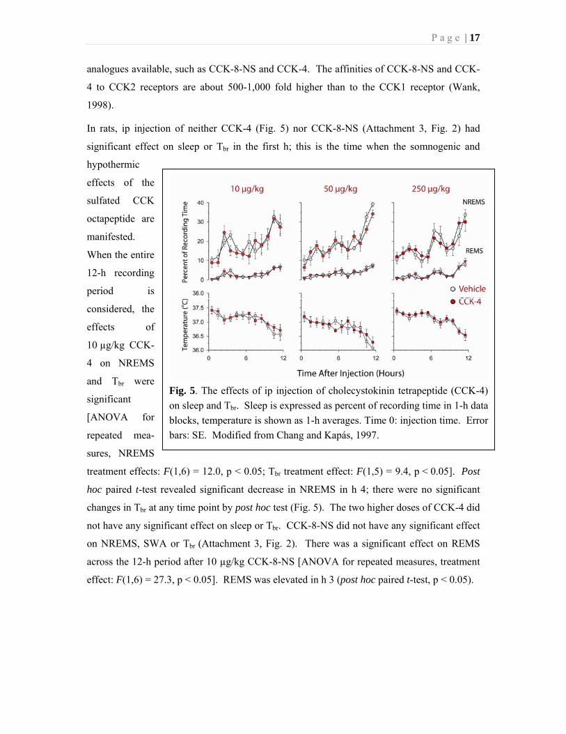
Fig. 5. The effects of ip injection of cholecystokinin tetrapeptide (CCK-4)
on sleep and Tbr. Sleep is expressed as percent of recording time in 1-h data
blocks, temperature is shown as 1-h averages. Time 0: injection time. Error
bars: SE. Modified from Chang and Kapás, 1997.
analogues available, such as CCK-8-NS and CCK-4. The affinities of CCK-8-NS and CCK-
4 to CCK2 receptors are about 500-1,000 fold higher than to the CCK1 receptor (Wank,
1998).
In rats, ip injection of neither CCK-4 (Fig. 5) nor CCK-8-NS (Attachment 3, Fig. 2) had
significant effect on sleep or Tbr in the first h; this is the time when the somnogenic and
hypothermic
effects of the
sulfated CCK
octapeptide are
manifested.
When the entire
12-h recording
period is
considered, the
effects of
10 µg/kg CCK-
4 on NREMS
and Tbr were
significant
[ANOVA for
repeated mea-
sures, NREMS
treatment effects: F(1,6) = 12.0, p < 0.05; Tbr treatment effect: F(1,5) = 9.4, p < 0.05]. Post
hoc paired t-test revealed significant decrease in NREMS in h 4; there were no significant
changes in Tbr at any time point by post hoc test (Fig. 5). The two higher doses of CCK-4 did
not have any significant effect on sleep or Tbr. CCK-8-NS did not have any significant effect
on NREMS, SWA or Tbr (Attachment 3, Fig. 2). There was a significant effect on REMS
across the 12-h period after 10 µg/kg CCK-8-NS [ANOVA for repeated measures, treatment
effect: F(1,6) = 27.3, p < 0.05]. REMS was elevated in h 3 (post hoc paired t-test, p < 0.05).

P a g e | 18
Fig. 6. The effects of ip injections of L-364,718, a CCK1
receptor antagonist on sleep and Tbr in rats. See legend to Fig.
5 for details.
Experiment 4. Effects of CCK1 receptor antagonist on CCK-induced sleep in rats.
The aim of the experiment was to determine if the activation of CCK1 receptors is necessary
for the somnogenic effects of systemically administered CCK. CCK1 receptors are expressed
in the brain, by neurons of the vagus nerve and by peripheral tissues (Hokfelt et al., 1991; Lin
and Miller, 1992; Corp et al., 1993). The food intake-suppressing effects of CCK are
mediated by the activation of CCK1 receptors on vagus nerve terminals (Dockray, 2009). L-
364,718 is a widely-used and highly selective CCK1 receptor antagonist (Chang and Lotti,
1986; Lotti et al., 1987; Hewson et al., 1988; Soar et al., 1989).
Intraperitoneal
injection of L-364,718
alone did not have
significant effects on
spontaneous sleep,
SWA and Tbr (Fig. 6,
Table 1). Ten µg/kg
CCK, ip, elicited sig-
nificant increases in
NREMS and decreases
in Tbr in the first h after
the injection (Fig. 7,
Table 1). One hundred
µg/kg L-364,718 atte-
nuated but did not
completely block
CCK-induced sleep;
NREMS was signi-
ficantly increased
across the 12-h and in
the first 2-h time block as compared to baseline (Table 1). The same dose of L-364,718
completely blocked the hypothermic effects of CCK (Fig. 7). Five hundred µg/kg of L-
364,718 completely abolished CCK-induced sleep and hypothermic responses (Fig. 7). L-
364,718 pretreatment did not affect hourly SWA values. When, however, SWA values are
averaged in 2-h time blocks, the combined treatment of CCK with either 100 or 500 g/kg L-

P a g e | 19
364,718 caused significantly increased SWA in the first 2-h time block as compared to
baseline (Fig. 7, Table 1). Neither dose of the antagonist, when given without CCK, caused
significant changes in SWA in the first 2 h (data not shown, see Table 1 for statistical
results).
Fig. 7. The effects of L-364,718 pretreatment on CCK-induced sleep and hypothermic responses.
Sleep and the slow-wave activity of the electroencephalogram during NREMS (SWA) are shown in
1-h data blocks. On the main Tbr panel, average hourly temperatures are shown for 12 h. On the
insets, Tbr is plotted for the first 2 h after the injection in 10-min intervals. Baseline: ip vehicle for
L-364,718 followed by ip saline; treatment: vehicle (left panels) or L-364,718 followed by CCK
(middle and right panels). Asterisk: significant difference between baseline and treatment (paired t-
test, p < 0.05). Error bars: SE.
P a g e | 20
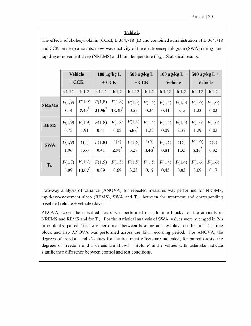
Table 1.
The effects of cholecystokinin (CCK), L-364,718 (L) and combined administration of L-364,718
and CCK on sleep amounts, slow-wave activity of the electroencephalogram (SWA) during non-
rapid-eye-movement sleep (NREMS) and brain temperature (Tbr): Statistical results.
Vehicle
+ CCK
100 g/kg L
+ CCK
500 g/kg L
+ CCK
100 g/kg L +
Vehicle
500 g/kg L +
Vehicle
h 1-12 h 1-2 h 1-12 h 1-2 h 1-12 h 1-2 h 1-12 h 1-2 h 1-12 h 1-2
NREMS F(1,9)
3.14
F(1,9)
7.49*
F(1,8)
21.96*
F(1,8)
13.49*
F(1,5)
0.57
F(1,5)
0.26
F(1,5)
0.41
F(1,5)
0.15
F(1,6)
1.23
F(1,6)
0.02
REMS F(1,9)
0.75
F(1,9)
1.91
F(1,8)
0.61
F(1,8)
0.05
F(1,5)
5.63*
F(1,5)
1.22
F(1,5)
0.09
F(1,5)
2.37
F(1,6)
1.29
F(1,6)
0.02
SWA F(1,9)
1.96
t (7)
1.66
F(1,8)
0.41
t (8)
2.78*
F(1,5)
3.29
t (5)
3.46*
F(1,5)
0.81
t (5)
1.33
F(1,6)
5.36*
t (6)
0.92
Tbr F(1,7)
6.09
F(1,7)
13.67*
F(1,5)
0.09
F(1,5)
0.69
F(1,5)
3.23
F(1,5)
0.19
F(1,4)
0.45
F(1,4)
0.03
F(1,6)
0.09
F(1,6)
0.17
Two-way analysis of variance (ANOVA) for repeated measures was performed for NREMS,
rapid-eye-movement sleep (REMS), SWA and Tbr, between the treatment and corresponding
baseline (vehicle + vehicle) days.
ANOVA across the specified hours was performed on 1-h time blocks for the amounts of
NREMS and REMS and for Tbr. For the statistical analysis of SWA, values were averaged in 2-h
time blocks; paired t-test was performed between baseline and test days on the first 2-h time
block and also ANOVA was performed across the 12-h recording period. For ANOVA, the
degrees of freedom and F-values for the treatment effects are indicated; for paired t-tests, the
degrees of freedom and t values are shown. Bold F and t values with asterisks indicate
significance difference between control and test conditions.
P a g e | 21
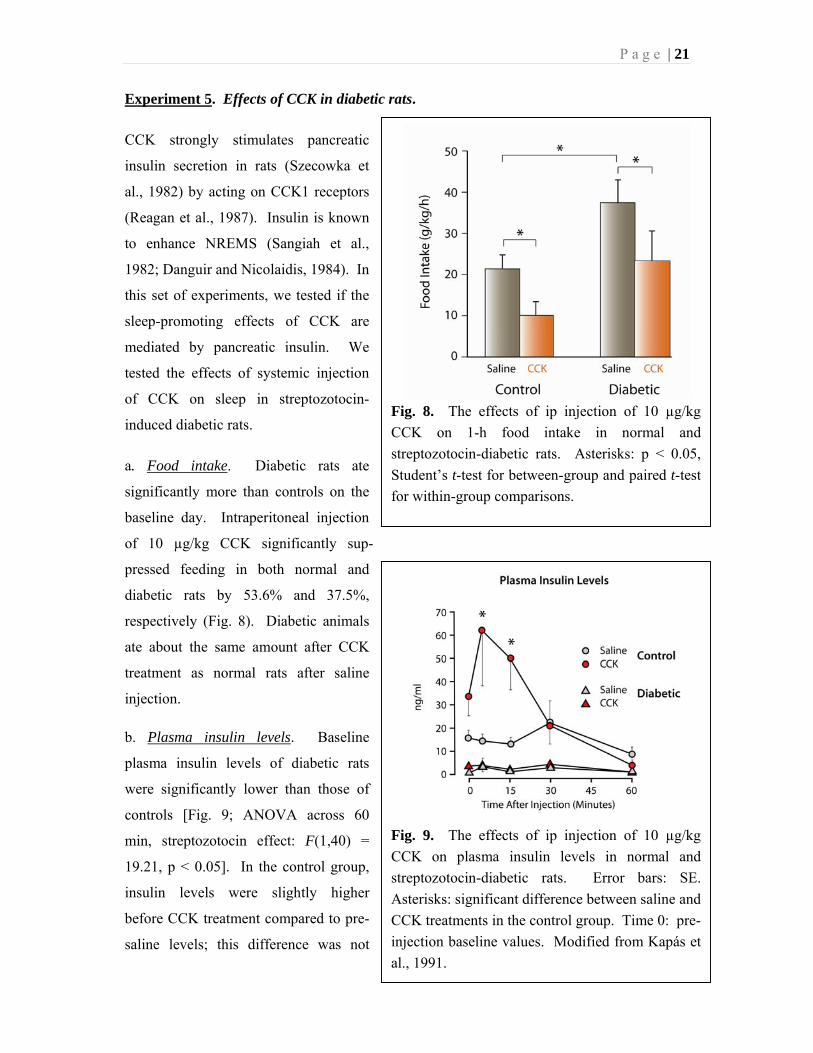
Fig. 8. The effects of ip injection of 10 µg/kg
CCK on 1-h food intake in normal and
streptozotocin-diabetic rats. Asterisks: p < 0.05,
Student’s t-test for between-group and paired t-test
for within-group comparisons.
Fig. 9. The effects of ip injection of 10 µg/kg
CCK on plasma insulin levels in normal and
streptozotocin-diabetic rats. Error bars: SE.
Asterisks: significant difference between saline and
CCK treatments in the control group. Time 0: pre-
injection baseline values. Modified from Kapás et
al., 1991.
Experiment 5. Effects of CCK in diabetic rats.
CCK strongly stimulates pancreatic
insulin secretion in rats (Szecowka et
al., 1982) by acting on CCK1 receptors
(Reagan et al., 1987). Insulin is known
to enhance NREMS (Sangiah et al.,
1982; Danguir and Nicolaidis, 1984). In
this set of experiments, we tested if the
sleep-promoting effects of CCK are
mediated by pancreatic insulin. We
tested the effects of systemic injection
of CCK on sleep in streptozotocin-
induced diabetic rats.
a. Food intake. Diabetic rats ate
significantly more than controls on the
baseline day. Intraperitoneal injection
of 10 µg/kg CCK significantly sup-
pressed feeding in both normal and
diabetic rats by 53.6% and 37.5%,
respectively (Fig. 8). Diabetic animals
ate about the same amount after CCK
treatment as normal rats after saline
injection.
b. Plasma insulin levels. Baseline
plasma insulin levels of diabetic rats
were significantly lower than those of
controls [Fig. 9; ANOVA across 60
min, streptozotocin effect: F(1,40) =
19.21, p < 0.05]. In the control group,
insulin levels were slightly higher
before CCK treatment compared to pre-
saline levels; this difference was not

P a g e | 22
Fig. 10. The effects of ip injection of 10 µg/kg CCK on sleep in normal and
streptozotocin-diabetic rats. Sleep is expressed as percent of recording time in 1-h
blocks for the first three hours and in 3-h blocks from h 4 to 12. Asterisks: significant
difference between saline and CCK treatment (paired t-test, p < 0.05). Error: SE.
Modified from Kapás et al., 1991.
statistically significant. After saline treatment, there was no significant change in plasma
insulin levels in either group of animals compared to pre-injection baseline. In control rats,
CCK significantly increased plasma insulin levels 5 and 15 min after injection (paired t-test,
p < 0.05). In diabetic rats, CCK did not have any significant effect on plasma insulin
concentrations.
c. Sleep, brain temperature and motor activity. As expected, streptozotocin-induced diabetic
rats had significantly higher plasma glucose levels compared to normal animals (17.6 ± 1.6
vs. 3.4 ± 0.2 mmol/l in diabetic and control animals, respectively). There were no significant
differences in the baseline sleep-wake activity of control and diabetic rats. Neither time spent
in NREMS during the 12-h recording period (diabetics: 25.6 ± 2.4%, controls: 25.7 ± 1.3%),
nor REMS amounts differed between the two groups (diabetics: 3.7 ± 0.5%, controls: 3.5 ±
0.5%). Intraperitoneal injections of CCK induced selective increases in NREMS in both the
control and the diabetic groups in the first h after the injection. Ten µg/kg CCK doubled the
amount of NREMS in the first h in normal rats; similar increases were observed in
P a g e | 23
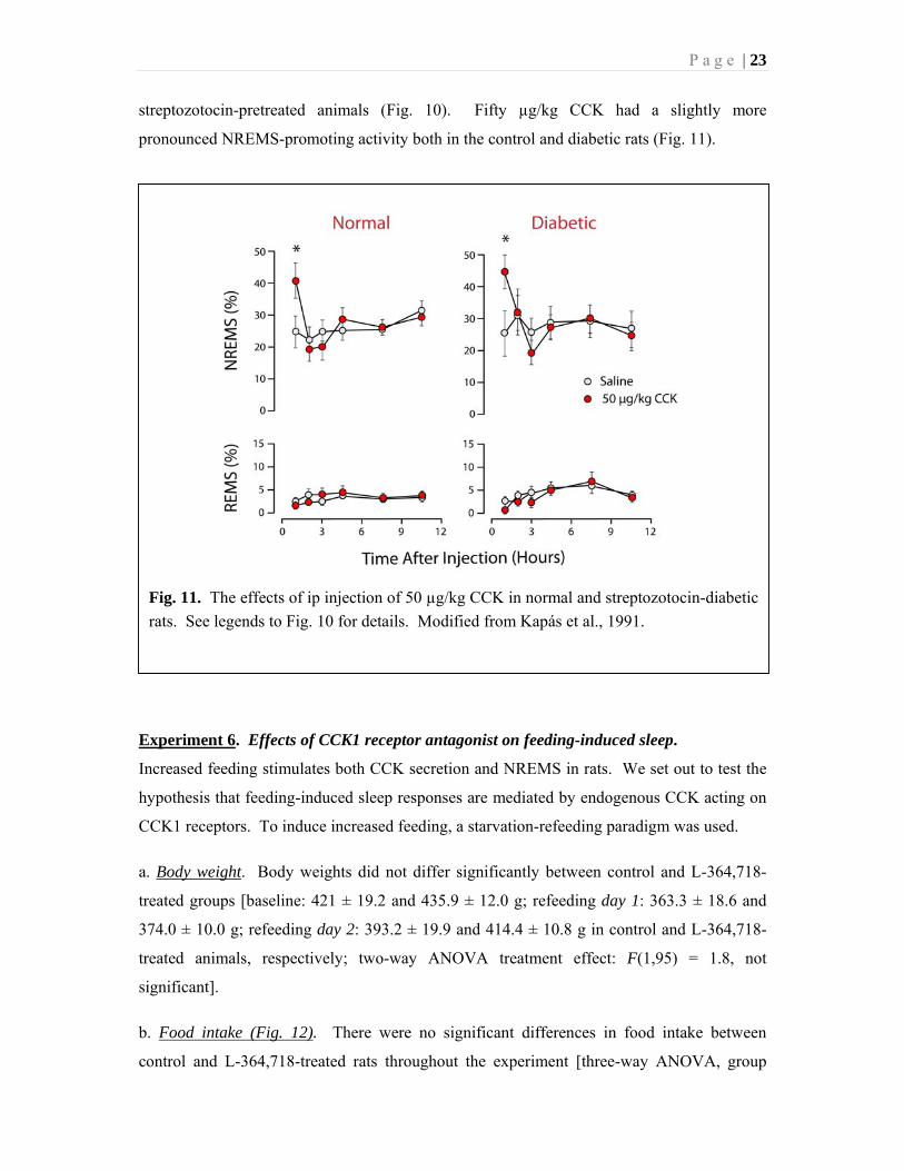
Fig. 11. The effects of ip injection of 50 µg/kg CCK in normal and streptozotocin-diabetic
rats. See legends to Fig. 10 for details. Modified from Kapás et al., 1991.
streptozotocin-pretreated animals (Fig. 10). Fifty µg/kg CCK had a slightly more
pronounced NREMS-promoting activity both in the control and diabetic rats (Fig. 11).
Experiment 6. Effects of CCK1 receptor antagonist on feeding-induced sleep.
Increased feeding stimulates both CCK secretion and NREMS in rats. We set out to test the
hypothesis that feeding-induced sleep responses are mediated by endogenous CCK acting on
CCK1 receptors. To induce increased feeding, a starvation-refeeding paradigm was used.
a. Body weight. Body weights did not differ significantly between control and L-364,718-
treated groups [baseline: 421 ± 19.2 and 435.9 ± 12.0 g; refeeding day 1: 363.3 ± 18.6 and
374.0 ± 10.0 g; refeeding day 2: 393.2 ± 19.9 and 414.4 ± 10.8 g in control and L-364,718-
treated animals, respectively; two-way ANOVA treatment effect: F(1,95) = 1.8, not
significant].
b. Food intake (Fig. 12). There were no significant differences in food intake between
control and L-364,718-treated rats throughout the experiment [three-way ANOVA, group

P a g e | 24
Fig. 12. Food intake of control and CCK1
antagonist (L-364,718)-treated rats under
baseline conditions and on refeeding days 1 and
2 after food deprivation. Asterisk: significant
difference from baseline (paired t-test,
p < 0.05). Modified from Shemyakin and
Kapás, 2001.
effect: F(1,126) = 0.30, not significant]. There was significant difference in feeding among
baseline day, refeeding day 1 and 2 [three-way ANOVA, day effect: F(2,126) = 12.2,
p < 0.05]. Food intake significantly
increased in the dark period of the first
refeeding day in both treatment groups.
In the following light phase, feeding in
the control group decreased below
baseline. Similar tendencies were
present after the CCK antagonist
treatment, but the changes were not
significant. On the second refeeding day,
both day- and night-time food intake
returned to baseline levels in both
groups.
c. The effects of refeeding on sleep and
Tbr in control, saline-treated rats.
Reintroducing food at the beginning of
the dark period after 4 days of food
deprivation led to delayed but long-
lasting increases in NREMS indicative of
postprandial sleep (Fig. 13). NREMS was significantly elevated during the second 12-h
period (light phase) of the first refeeding day (baseline: 332 ± 23 min/12 h vs. refeeding
day 1: 392 ± 7 min/12 h, p < 0.05). Strong tendencies toward increased NREMS continued
throughout the next day, but the changes did not reach the level of significance. REMS was
elevated during the light phase of the second refeeding day (baseline: 38 ± 5 min/12 h vs.
refeeding day 2: 58 ± 7 min/12 h, p < 0.05). There were significant reductions in SWA
during the second refeeding night; similar tendencies were present for the prior and
subsequent 12-h periods. Tbr was not affected by refeeding.

P a g e | 25
Fig. 13. The effects of refeeding on NREMS, REMS, SWA and Tbr in control rats.
Time spent in sleep is summed in 2-h blocks. SWA is averaged in 2-h and Tbr in 1-h
intervals. Error bar: SE. Gray shaded area: dark phase. Asterisks: significant
difference from baseline (p < 0.05, paired t-test). Modified from Shemyakin and Kapás,
2001.
d. The effects of refeeding on sleep and Tbr in L-364,718-treated rats. The NREMS-inducing
effects of refeeding on the first and the REMS-promoting effects on the second refeeding day
were completely abolished by the CCK1 receptor antagonist (Fig. 14). During the dark phase
of the second refeeding day, increases in NREMS – that were present only as a tendency after
saline injection – became significant; NREMS returned to baseline by the second part of
refeeding day 2. L-364,718 completely abolished the SWA responses to refeeding.

P a g e | 26
Fig. 14. The effects of refeeding on NREMS, REMS, SWA and Tbr in L-364,718-treated
rats. See legend to Fig. 13 for details. Modified from Shemyakin and Kapás, 2001.
P a g e | 27
Discussion
In the present experiments, we have shown that systemic injections of sulfated CCK
octapeptide selectively and dose-dependently stimulate NREMS in rats and rabbits. Central
injection of CCK in rabbits or systemic injection of CCK2 receptor agonists in rats did not
have significant effects on sleep. The somnogenic effects of exogenously administered CCK
as well as the sleep-inducing effects of refeeding after starvation were completely abolished
by a selective CCK1 receptor antagonist. Systemic, but not central, administration of CCK
elicited significant decreases in brain temperature, a response completely prevented by CCK1
receptor antagonist. The results are consistent with our hypothesis that CCK produced by the
GI system in response to eating plays a key role in eliciting postprandial sleep and thus in
aligning vigilance to the acute feeding/metabolic status of the body.
Effects of CCK on sleep
Prior to our studies, only sparse and mainly indirect data were available concerning the
effects of CCK on sleep. The first experiments suggesting a possible somnogenic effect for
CCK were performed in the late 1960s. In these studies, both intra-duodenal administration
of fat and iv injection of CCK-rich duodenum extracts caused sedation in awake in cats (Fara
et al., 1969). In 1982, Mansbach and Lorenz reported that CCK reduced sleep latency and 20
min after the ip injection of CCK, a significantly greater number of rats were asleep than after
control treatments. These effects of CCK were indistinguishable from those induced by
eating (Mansbach and Lorenz, 1983). There was one single report prior to our experiments
where the effects of systemically administered CCK were quantitatively analyzed. A reduced
latency to NREMS in rats after ip injection of 20 µg/kg CCK was reported without effects on
the duration of sleep (Rojas-Ramirez et al., 1982). CCK injections and sleep recordings were
performed during the first part of light phase in these experiments in ad libitum fed, therefore
likely satiated, animals. The light period, however, is the rest phase in rats, physiological
sleep-promoting mechanisms are fully engaged, sleep is already elevated; it is unlikely that a
physiological sleep-promoting hormone in a presumably physiological dose range could
further increase sleep amounts.
To avoid such a ceiling effect, we injected CCK immediately before dark onset in our
experiments with rats. During the first part of the dark period, the spontaneous activity of
sleep-promoting mechanisms, hence the amount of sleep, is minimal in rats. Also, in
P a g e | 28
Experiment 1, we fasted rats for 12 h before the CCK treatment to ensure that endogenous
satiety mechanisms are not already activated. In our subsequent experiments it became
apparent that such a prior fasting is not required for the manifestation of the sleep-promoting
effects of ip administered CCK. As expected, the dose-dependent NREMS-promoting effects
of CCK were mirrored by decreases in motor activity. In Experiment 1, sleep-increases were
accompanied by increased SWA (i.e., delta wave activity) of the EEG. In this experiment, all
artifact-free EEG segments were included in the SWA (FFT) analysis, including segments of
NREMS, REMS and wakefulness. During NREMS, slow (delta) waves dominate the EEG;
during REMS and wakefulness, slow waves are uncommon. Elevated SWA after CCK
injections in Experiment 1 is simply the reflection of the increased amounts of NREMS;
conclusions about the quality/intensity of NREMS cannot be drawn from the data. In our
subsequent experiments, we restricted slow-wave analysis to the NREMS segments of the
EEG. In this case, changes in SWA reflect qualitative changes in NREMS, mainly sleep
intensity. We did not find any significant effect of CCK on SWA during NREMS suggesting
that the intensity of NREMS is not affected by CCK.
The sleep-promoting effects of CCK in rats were confirmed by independent laboratories after
our initial publication (de Saint Hilaire-Kafi et al., 1989; Posadas-Andrews et al., 1989). We
also described the somnogenic actions of CCK in rabbits (Experiment 2) and mice
(Szentirmai et al., 2007b) thereby demonstrating that its sleep-promoting effects are not
species specific. In all three species, the lowest somnogenic ip dose was 10 µg/kg. Although
we did not observe any appreciable effects on duration of rapid-eye-movement sleep (REMS)
in rats, rabbits or mice (Szentirmai et al., 2007b), there are reports that in
parachlorophenylalanine-induced insomniac cats CCK restores REMS (Prospero-Garcia et
al., 1987), and in normal rats CCK increases REMS frequency (DeMesquita and Haney,
1986), and decreases REMS latency (Mansbach and Lorenz, 1983).
Effects of CCK on brain temperature
We previously found that systemic injections of CCK elicit dose-dependent hypothermia in
rats (Kapás et al., 1987; Kapás et al., 1989). These results were confirmed by the present
experiments and subsequently replicated by independent laboratories (South, 1992; Szelényi
et al., 1994; Rezayat et al., 1999). We extended these finding by showing that ip injection of
CCK also produces dose-dependent hypothermic responses in rabbits (Experiment 2). In rats,
the dose-response relationships for the somnogenic, hypothermic and food intake-suppressing

P a g e | 29
effects of CCK were similar. The effects of ip CCK on thermoregulation and sleep were also
in the same dose range in rabbits. Our data indicate that the hypothermic response to CCK is
mediated by the CCK1 receptor subtype. First, CCK2 receptor-selective CCK analogues,
CCK-4 and CCK-8-NS, did not have hypothermic activities in our present and previous
(Kapás et al., 1987) experiments in rats. Consistent with this observation, sc injection of the
same analogues did not affect body temperature in mice (Rezayat et al., 1999). Second, the
hypothermic effects of CCK were completely abolished by pretreatment with L-364,718, a
selective CCK1 receptor antagonist. Similarly, Szelényi and coworkers reported that the
hypothermic effects of sc injected CCK were attenuated by a CCK1, but not a CCK2,
receptor antagonist in rats (Szelényi et al., 1994).
While there is a broad consensus about the hypothermic effects of systemically injected CCK,
there are conflicting data about the central effects of CCK on thermoregulation. Initially, it
was found that icv (Morley et al., 1981; Katsuura et al., 1981) or intra-preoptic (Liu and Lin,
1985) injection of CCK reduces body temperature in rats. Subsequently, these effects were
not confirmed, rather, a hyperthermic response was reported in the same species (Shido et al.,
1989; Szelényi et al., 1994; Székely et al., 1994; Ghosh et al., 1997; Ghosh et al., 1998;
Sugimoto et al., 1999). In rats, hyperthermia is elicited in the 0.02-10 µg/rat dose range. In
our experiment, we did not find hyperthermic response to CCK in rabbits in the dose range of
0.05-2 µg/animal, rather, a modest, but statistically significant drop in brain temperature was
evident. This may reflect true species-specific difference in the central effects of CCK or it
may be due to differences in experimental conditions. In rabbits, we measured brain
temperature in freely moving animals, while colonic temperature was recorded in restrained
rats (Shido et al., 1989; Szelényi et al., 1994; Székely et al., 1994) or CCK was given in the
form of chronic icv infusion with telemetric recording of the abdominal temperature
(Szelényi et al., 2004). The hyperthermic response to centrally administered CCK is
attenuated by a CCK2 receptor antagonist (Szelényi et al., 1994) suggesting the involvement
of brain CCK2 receptors in CCK-induced fever. It is unlikely that the activation of peripheral
CCK2 receptors also leads to hyperthermic responses since systemic injections of CCK2
receptor-selective analogues (CCK-4 and CCK-8-NS) did not cause fever in our previous
(Kapás et al., 1987) or present (Experiment 3) studies in rats or in mice (Rezayat et al., 1999).

P a g e | 30
The mechanism of CCK-induced sleep
The two main questions regarding the mechanism of CCK-induced sleep are related to the
involvement of CCK1 vs. CCK2 receptor subtypes and the anatomical location of the target.
Our results with CCK2 agonists and CCK1 receptor antagonist indicate that CCK2 receptor
activation is not sufficient but CCK1 receptor activation is necessary for the somnogenic
effects of CCK.
CCK-8-SE binds to both CCK receptor subtypes with equal affinity. CCK2 receptors have
similar high affinity for both sulfated CCK and nonsulfated analogues such as CCK-8-NS
and CCK-4 (Wank, 1998). The affinities of CCK-8-NS and CCK-4 to CCK1 receptors are
about 500-1,000 fold less than that of sulfated CCK octapeptide (Wank, 1998). If the
somnogenic effects of CCK are due to the activation of CCK2 receptors then it is expected
that equimolar amounts of sulfated CCK octapeptide, CCK-8-NS and CCK-4 would lead to
similar sleep responses. This was not the case. The lowest effective somnogenic dose of
systemically injected CCK-8-SE is 8.7 nmol/kg (10 µg/kg) in rats. In Experiment 3, the
amount of NREMS did not increase in response to ip injection of 16.8-419.3 nmol/kg CCK-4
or 9.4-235.3 nmol/kg CCK-8-NS. These clearly show that the selective activation of CCK2
receptors is not sufficient to elicit somnogenic responses characteristic of CCK-8-SE. After
the injection of 10 µg/kg (16.8 nmol/kg) CCK-4, NREMS decreased in h 3. The biological
significance of such a delayed and slight effect is not clear, nevertheless, it is consistent with
prior findings that BC-264, another CCK2 receptor agonist, slightly enhances wakefulness
(de Saint Hilaire et al., 1991) and CCK-4 induces behavioral activation in open-field tests
(Hsiao et al., 1984).
L-364,718 is a selective antagonist of the CCK1 receptor. It is void of CCK-like agonistic
activities. CCK antagonism by L-364,718 lasts for at least 2-5 h (Lotti et al., 1987). In rats,
systemic injection of 100 µg/kg L-364,718 prevents the effects of exogenous CCK on food
intake (Hewson et al., 1988), locomotor activity (Soar et al., 1989) and gall bladder
contraction (Chang and Lotti, 1986). We found that 100 µg/kg L-364,718 nearly completely
while 500 µg/kg completely abolished the sleep-inducing effects of CCK. This indicates that
the activation of CCK1 receptors is necessary for the manifestation of sleep-inducing effects
of ip administered CCK. The CCK1 antagonist did not affect spontaneous sleep in normally
fed animals when given at dark onset suggesting that tonic activation of CCK1 receptors by
endogenous CCK plays minimal role in maintaining spontaneous sleep at the beginning of

P a g e | 31
the activity phase in rats. To make more definitive conclusions about the role of endogenous
CCK in maintaining normal amounts of sleep in the dark and light periods, additional studies
are needed by testing the effects of a wider dose range of both CCK1 and CCK2 receptor
antagonists by various routes of administration at different times of the diurnal cycle.
Regardless, we hypothesize that increased CCK secretion is likely a physiological signal for
increased sleep under certain conditions (discussed below).
Our findings that CCK2 receptor activation is not sufficient but CCK1 receptor activation is
necessary for CCK-induced sleep responses do not rule out the possibility that the activation
of CCK2 receptors also contributes to the sleep effects. The co-activation or sequential
activation of CCK1 and CCK2 receptors may be necessary for the manifestation of the
somnogenic effects of CCK. There are known effects of CCK that require the activation of
both receptor subtypes, e.g., suppression of acetylcholine release from cerebral cortex
(Kimura et al., 1995) or the potentiation of the anticonvulsive actions of morphine (Legido et
al., 1995).
The site of the somnogenic action of CCK.
The present experiments with L-364,718 do not address the question of the site of the
somnogenic effects of CCK. CCK1 receptors are present both in the brain and in the
periphery. L-364,718 crosses the BBB after systemic injection (Pullen and Hodgson, 1987)
and binds to both central as well as peripheral CCK1 receptors. Systemically injected CCK
does not cross the BBB (Passaro E Jr et al., 1982; Zhu et al., 1986) and likely acts on
peripheral targets or brain structures that lack the BBB.
Regarding peripheral targets, we considered the possibility that the sleep effects of CCK are
mediated through the release of another peripheral hormone stimulated by CCK. We
considered insulin as a potential mediator of CCK’s somnogenic action since CCK is a potent
stimulator of insulin secretion (Unger et al., 1967; Szecowka et al., 1982) and the effects of
insulin on sleep and feeding are similar to those of CCK. Exogenous administration of
insulin stimulates NREMS (Sangiah et al., 1982; Danguir and Nicolaidis, 1984), suppresses
feeding (Woods and Porte, Jr., 1983) while diabetic rats show diminished sleep (Danguir,
1984; Kapás et al., 1991) and increased feeding (Kumaresan and Turner, 1965; Booth, 1972).
To test the role of pancreatic insulin in the sleep-promoting action of CCK, we studied the
effects of CCK in streptozotocin-diabetic rats. Our findings of virtually undetectable plasma

P a g e | 32
insulin levels, elevated plasma glucose concentrations and increased feeding under baseline
conditions confirmed the lack of pancreatic insulin in streptozotocin-treated animals.
Confirming our prior results, spontaneous sleep in diabetic rats was unaltered during the dark
phase (Kapás et al., 1991). The known sleep deficiency in diabetic animals (Danguir, 1984)
is confined to the light period of the day (Kapás et al., 1991). In line with the known
stimulatory effects of CCK on insulin secretion, ip injection of CCK caused increases in
plasma insulin levels in control rats but not in diabetics. In spite of the lack of insulin
response, diabetic rats mounted similar sleep responses to CCK injection as normal animals
indicating that insulin is not involved in the sleep actions of CCK. As in normal rats, 10
µg/kg CCK suppressed 1-h food intake in diabetic animals. This confirms prior studies
suggesting that CCK-induced satiety is independent of pancreatic insulin (Vanderweele,
1982).
The role of CCK in postprandial sleep
Increased feeding or postingestive satiety elicits postprandial sleep (Mansbach and Lorenz,
1983). Delivery of nutrients into the stomach or duodenum elicits EEG synchronization in
rats (Bernstein, 1974) and cats (Fara et al., 1969). Increased eating induced by palatable,
high-energy diet (Danguir, 1987; Hansen et al., 1998) or by the lesion of the VMH (Danguir
and Nicolaidis, 1978) leads to increases of daily NREMS and REMS amounts. Genetically
hyperphagic and obese Zucker rats have large amounts of NREMS (Danguir, 1989). There is
a positive correlation between meal size and the length of the subsequent sleep period in rats
(Danguir et al., 1979). Excess eating induced by prior food deprivation for 80 or 96 h results
in sleep increases (Borbély, 1977; Danguir and Nicolaidis, 1979).
In Experiment 6, we induced excess eating by reintroducing food after 96 h of food
deprivation. In control animals, refeeding elicited significant increases in NREMS and
REMS, predominantly during the light phase of the refeeding days. Similarly, NREMS is
elevated only during the light period in cafeteria diet-fed rats (Hansen et al., 1998). Eating
and sleep cannot take place simultaneously, therefore an increase in feeding behavior may
interfere with the possible somnogenic effects of the ingested food. In the dark phase of the
first refeeding day, food intake was increased by 43% above baseline. It is likely that the
increased behavioral activity interfered with food-induced increases in sleep pressure; as a
result, sleep amounts did not change. In the following light period eating was below baseline
which allowed the somnogenic effect of previously consumed food to be manifested. In the
P a g e | 33
dark of the second refeeding day, feeding behavior was less robust than the first night. This
decline in feeding activity allowed for sleep increases in the dark, i.e., increases in REMS and
a strong tendency toward increased NREMS. Also, since less food was consumed, sleep
enhancement in the following light period were less pronounced, in fact, increases in
NREMS were not statistically significant in the light phase of refeeding day 2.
SWA was reduced during the dark period of the second refeeding day in control rats. This is
likely due to the fact that re-fed rats spent extra time in NREMS during the first light and
second dark periods. The excess sleep likely reduced the homeostatic pressure for
subsequent NREMS and the decreased SWA may be the consequence of this reduced
pressure. Previously, we described similar decreases in SWA activity that accompanied
feeding-induced sleep in the cafeteria diet model (Hansen et al., 1998).
The CCK1 receptor antagonist, L-364,718, was administered at light onset on both refeeding
days, i.e., the first injection was done 12 h after reintroducing the food. We did not inject the
antagonist immediately after the end of the starvation period for two reasons. First, sleep
responses to refeeding started only after a latency of 12 h in control rats. Second, L-364,718
itself stimulates feeding in rats (Reidelberger and O'Rourke, 1989); postponing treatments
allowed the animals to eat according to their natural needs during the first dark period after
starvation. L-364,718 completely abolished the NREMS increases during the light periods
both on refeeding day 1 and 2. This, together with the known increase of plasma CCK in
response to feeding and our observation that L-364,718 abolishes exogenous CCK-induced
sleep, strongly indicate a role of endogenously produced CCK in feeding-induced sleep
responses.
A model of feeding and metabolism and sleep - perspectives
The present work represents a segment of an ongoing broad project to test our model on the
integration of metabolism and sleep (Fig. 15). We hypothesize that in addition to the well-
established wake-dependent homeostatic and SCN-driven circadian factors, metabolic signals
also play a fundamental role in determining sleep-wake activity. We posit that CCK is such a
metabolic signal. These signals may modulate the activity of arousal mechanisms or may
modulate circadian influences by acting through the food-entrainable oscillator, an
endogenous clock independent of SCN (Antle and Silver, 2009). Some signals trigger acute
changes in sleep in response to short-term negative energy balance such as during starvation

P a g e | 34
(ghrelin) or positive energy balance such as postprandial states (CCK, gastric leptin).
Different signaling mechanisms set sleep amounts in response to long-term changes in
adiposity (adipocyte-secreted leptin and TNF) or food availability (FEO). We hypothesize
that both short- and long-term signals converge on a common integrative center in the
hypothalamus. A ghrelin-NPY-orexin circuit is thought to be a key component of this
integrative center (Szentirmai et al., 2007a; Szentirmai et al., 2009; Szentirmai et al., 2010).
Circulating CCK may modulate the activity of this hypothalamic circuit by acting through a
peripheral or central target or both. In Experiment 5, we ruled out pancreatic insulin as a
possible peripheral mediator of CCK-induced sleep. Another potential peripheral target for
CCK to induce sleep is the vagus nerve. Peripheral sensory nerve endings in the vagus
abundantly express CCK1 and CCK2 receptors (Dufresne et al., 2006). Numerous effects of
systemic CCK on brain functions are, indeed, mediated by vagal afferents. Surgical
vagotomy, or selective chemical destruction of vagal afferents by capsaicin prevents the
effects of CCK on food intake (Smith et al., 1981), memory (Flood et al., 1987), exploratory
behavior (Crawley et al., 1981a), and oxytocin secretion (Verbalis et al., 1986). Further,
sensory inputs from vagal afferents modulate sleep, the activation of vagus - NTS complex
results in generation of NREMS (Puizillout and Foutz, 1977). In our prior studies, however,
surgical vagotomy did not prevent the somnogenic effects of ip injected CCK (Kapás, 1997).
This finding makes it unlikely that peripheral vagal CCK receptors are the target for the
somnogenic action of CCK although the role of vagus in CCK-induced sleep cannot be ruled
it out completely for two main reasons. One, the fact CCK is capable of inducing sleep in the
absence of functional vagus does not necessarily mean that intact vagus does not contribute to
the effects of CCK in normal animals. Two, nodose cells express CCK1 receptors and, in
general, respond to CCK (Widdop et al., 1993). They also survive subdiaphragmal vagotomy
(Lieberman, 1971), although with a markedly reduced CCK1 receptor expression (Broberger
et al., 2001). Surviving nodose cells are not sufficient to mediate food intake-suppressing
effects of CCK as those effects are completely prevented by subdiaphragmatic vagotomy;
nevertheless, it cannot be precluded that CCK acts on surviving cell populations to elicit
sleep. While its role in signaling CCK-induced sleep is questionable, the vagus is likely to
play a role in signaling increased adiposity-induced sleep. We have shown that cafeteria-diet
induced adiposity is accompanied by increased sleep, a response that is prevented in
vagotomized animals (Hansen et al., 1998). TNF is a major adipokine, most circulating TNF
is of adipose origin in normal healthy rats (Kershaw and Flier, 2004). TNF also has potent
P a g e | 35
somnogenic actions (Kapás et al., 1992; Kapás and Krueger, 1992) that are abolished in
vagotomized rats (Kubota et al., 2001).
The findings that lateral ventricular injection of CCK does not induce sleep in rabbits
(Experiment 2) or rats (Gilligan et al., 1998) seemingly contradict a central target for CCK.
The possibility, however, remains that there is a central target site which can be reached by
circulating CCK easily but less accessible for CCK injected into the lateral ventricle. When
administered into the lateral ventricle, CCK can potentially reach all neurons in the brain
without encountering barriers such as the BBB. This, however, does not imply that CCK
indeed reaches all neurons in a physiologically meaningful concentration since, clearly, there
is a gradient for injected molecules in the cerebrospinal fluid from the site of the injection to
distant neuron populations such as in the brain stem. Those brain stem sites that are supplied
by fenestrated capillaries can be reached by circulating CCK unabatedly.
We hypothesize that circulating CCK acts on CCK receptors within NTS to elicit sleep. NTS
is the primary projection target for vagal visceral afferents arising from the GI tract thus a
major relay sites for vagus-mediated CCK effects. We posit that circulating CCK reaches the
NTS directly and may act on the NTS directly by bypassing peripheral vagal sensory nerves.
CCK1 receptors are abundantly expressed in the NTS, mainly restricted to its medial
subnucleus (Hill et al., 1987; Corp et al., 1993; Qian et al., 1997). The medial subnucleus is
also rich in highly fenestrated capillaries similar to those in the area postrema (Gross et al.,
1990). The lack of BBB makes neurons and CCK receptors in the NTS accessible to
circulating CCK and other large blood-borne molecules. CCK by acting on NTS CCK1
receptors has known physiological actions such as eliciting satiety (Blevins et al., 2000) and
modulating glutamate release from vagus afferents (Appleyard et al., 2005). The NTS may
serve as a key interface between metabolic signals, both blood-born and vagus mediated, and
sleep regulatory centers in the brain. NTS has extensive projections to the PBN (Loewy and
Burton, 1978; Saper and Loewy, 1980). PBN itself is implicated in sleep regulation, most of
its neurons show sleep-dependent activity pattern (Saito et al., 1977; Gilbert and Lydic,
1994). From the PBN, dense projections arise to the VMH, DMH, posterior LH and preoptic
hypothalamus (Bester et al., 1997), areas thought to play key role in the regulation of sleep
and wakefulness.
In summary, we have shown that systemic injection of CCK elicits dose-dependent
somnogenic and hypothermic responses in rats and rabbits. The sleep effects are

P a g e | 36
accompanied by suppressed feeding and motor activity. Selective activation of CCK2
receptors is not sufficient to elicit the responses while the activation of CCK1 receptors is
required suggesting CCK1 receptors as a primary target. Pancreatic insulin does not play a
role in CCK-induced sleep and thermoregulatory responses. Eating-induced sleep is
prevented by CCK1 receptor antagonist treatment indicating a role for CCK in the
postprandial modulation of vigilance. Present results are consistent with the hypothesis that
CCK is a component of a complex signaling mechanism which modulates sleep-wake activity
according to the metabolic status of the body.
Fig. 15. Integration of metabolic and circadian signals in the regulation of sleep, wakefulness and feeding.
P a g e | 37
Acknowledgements
I thank the Chairs of the Department of Physiology in Szeged for their unwavering support
over the years. Professor Ferenc Obál recruited me to work in the Department as a student
and offered my first post-doctoral position. Professor György Benedek gave me the
opportunity to work in his laboratory during my student years; he has been encouraging my
scientific endeavors and sharing his knowledge and wisdom generously ever since. Professor
Gábor Jancsó gave invaluable support by retaining me in the PhD program, without his help
this thesis could not have been completed.
Much of the present work was aided by my mentors, Drs. Ferenc Obál, Jr. and James M.
Krueger. Their contribution to shaping my understanding of science reaches far beyond this
thesis. Their role in my life as a mentors and friends can never be overstated.
I would like to acknowledge my coauthors’ contribution to the studies. It would not have
been possible to carry out the studies without their generous help. I thank Drs. Péter Alföldi,
Hee-Yoon Chang, Ibolya Farkas, Lars Johannsen, Mark R. Opp, Linda C. Payne, Botond
Penke, György Rubicsek, Gyula Sáry and Alexei Shemyakin.
I thank the skilled technical assistance by Klára Ormos, Ilona Ponicsán, László Kiss, Sandy
Johnson, Donna Maxwell and Gail Richmond. Special thanks to Péter Liszli for his help with
computerized data collection in Szeged and Dr. Zoltán Lelkes for his continuing help and
advice in the last thirty years.
Extended thanks to Dr. Éva Szentirmai for the inspiring discussions that helped shape the
interpretation of past and the conception of present and future studies. She gave invaluable
feedback on early versions of this thesis.
I am indebted to my parents for their selfless support, understanding and endless patience.
This work was supported by grants from the Hungarian Academy of Sciences, Hungarian
Ministry of Health, National Institutes of Health (USA), Office of Naval Research (USA), US
Army and Medical Research and Development Command and Fordham University, New
York.

P a g e | 38
References
Allison, T., Cicchetti, D.V., 1976. Sleep in mammals: ecological and constitutional correlates. Science 194, 732-734.
Antin, J., Gibbs, J., Holt, J., Young, R.C., Smith, G.P., 1975. Cholecystokinin elicits the complete behavioral sequence of satiety in rats. J Comp Physiol Psychol 89, 784-790.
Antle, M.C., Silver, R., 2009. Neural basis of timing and anticipatory behaviors. Eur J Neurosci 30, 1643-1649.
Appleyard, S.M., Bailey, T.W., Doyle, M.W., Jin, Y.H., Smart, J.L., Low, M.J., Andresen, M.C., 2005. Proopiomelanocortin neurons in nucleus tractus solitarius are activated by visceral afferents: regulation by cholecystokinin and opioids. J Neurosci 25, 3578-3585.
Beinfeld, M.C., 2003. Biosynthesis and processing of pro CCK: recent progress and future challenges. Life Sci 72, 747-757.
Beinfeld, M.C., Palkovits, M., 1981. Distribution of cholecystokinin (CCK) in the hypothalamus and limbic system of the rat. Neuropeptides 2, 123-129.
Bernstein, I.L., 1974. Post-prandial EEG synchronization in normal and hypothalamically lesioned rats. Physiol Behav 12, 535-545.
Bester, H., Besson, J.M., Bernard, J.F., 1997. Organization of efferent projections from the parabrachial area to the hypothalamus: a Phaseolus vulgaris-leucoagglutinin study in the rat. J Comp Neurol 383, 245-281.
Bhatnagar, S., Viau, V., Chu, A., Soriano, L., Meijer, O.C., Dallman, M.F., 2000. A cholecystokinin-mediated pathway to the paraventricular thalamus is recruited in chronically stressed rats and regulates hypothalamic-pituitary-adrenal function. J Neurosci 20, 5564-5573.
Blevins, J.E., Stanley, B.G., Reidelberger, R.D., 2000. Brain regions where cholecystokinin suppresses feeding in rats. Brain Res 860, 1-10.
Booth, D.A., 1972. Some characteristics of feeding during streptoxotocin-induced diabetes in the rat. J Comp Physiol Psychol 80, 238-249.
Borbély, A.A., 1977. Sleep in the rat during food deprivation and subsequent restitution of food. Brain Res124, 457-471.
Borbély, A.A., 1982. A two process model of sleep regulation. Hum Neurobiol 1, 195-204.
Brezinova, V., Oswald, I., 1972. Sleep after a bedtime beverage. Br Med J 2, 431-433.
Broberger, C., Holmberg, K., Shi, T.J., Dockray, G., Hokfelt, T., 2001. Expression and regulation of cholecystokinin and cholecystokinin receptors in rat nodose and dorsal root ganglia. Brain Res 903, 128-140.

P a g e | 39
Cain, B.M., Connolly, K., Blum, A., Vishnuvardhan, D., Marchand, J.E., Beinfeld, M.C., 2003. Distribution and colocalization of cholecystokinin with the prohormone convertase enzymes PC1, PC2, and PC5 in rat brain. J Comp Neurol 467, 307-325.
Chang, R.S., Lotti, V.J., 1986. Biochemical and pharmacological characterization of an extremely potent and selective nonpeptide cholecystokinin antagonist. Proc Natl Acad Sci USA 83, 4923-4926.
Corp, E.S., McQuade, J., Moran, T.H., Smith, G.P., 1993. Characterization of type A and type B CCK receptor binding sites in rat vagus nerve. Brain Res 623, 161-166.
Crawley, J.N., 1985. Clarification of the behavioral functions of peripheral and central cholecystokinin: two separate peptide pools. Peptides 6 Suppl 2, 129-136.
Crawley, J.N., Corwin, R.L., 1994. Biological actions of cholecystokinin. Peptides15, 731-755.
Crawley, J.N., Hays, S.E., Paul, S.M., 1981a. Vagotomy abolishes the inhibitory effects of cholecystokinin on rat exploratory behaviors. Eur J Pharmacol 73, 379-380.
Crawley, J.N., Hays, S.E., Paul, S.M., Goodwin, F.K., 1981b. Cholecystokinin reduces exploratory behavior in mice. Physiol Behav 27, 407-411.
Dalsgaard, C.J., Vincent, S.R., Hokfelt, T., Lundberg, J.M., Dahlstrom, A., Schultzberg, M., Dockray, G.J., Cuello, A.C., 1982. Coexistence of cholecystokinin- and substance P-like peptides in neurons of the dorsal root ganglia of the rat. Neurosci Lett 33, 159-163.
Danguir, J., 1984. Sleep deficits in diabetic rats: restoration following chronic intravenous or intracerebroventricular infusions of insulin. Brain Res Bull 12, 641-645.
Danguir, J., 1987. Cafeteria diet promotes sleep in rats. Appetite 8, 49-53.
Danguir, J., 1989. Sleep patterns in the genetically obese Zucker rat: effect of acarbose treatment. Am J Physiol 256, R281-R283.
Danguir, J., Nicolaidis, S., 1978. Sleep and feeding patterns in the ventromedial hypothalamic lesioned rat. Physiol Behav 21, 769-777.
Danguir, J., Nicolaidis, S., 1979. Dependence of sleep on nutrients' availability. Physiol Behav 22, 735-740.
Danguir, J., Nicolaidis, S., 1980a. Circadian sleep and feeding patterns in the rat: possible dependence on lipogenesis and lipolysis. Am J Physiol 238, E223-E230.
Danguir, J., Nicolaidis, S., 1980b. Intravenous infusions of nutrients and sleep in the rat: an ischymetric sleep regulation hypothesis. Am J Physiol 238, E307-E312.
Danguir, J., Nicolaidis, S., 1984. Chronic intracerebroventricular infusion of insulin causes selective increase of slow wave sleep in rats. Brain Res 306, 97-103.
Danguir, J., Nicolaidis, S., Gerard, H., 1979. Relations between feeding and sleep patterns in the rat. J Comp Physiol Psychol 93, 820-830.

P a g e | 40
de Saint Hilaire, Z., Roques, B.P., Nicolaidis, S., 1991. Effect of a highly selective central CCK-B receptor agonist: BC-264 on rat sleep. Pharmacol Biochem Behav 38, 545-548.
de Saint Hilaire-Kafi, Z., Depoortere, H., Nicolaidis, S., 1989. Does cholecystokinin induce physiological satiety and sleep? Brain Res 488, 304-310.
DeMesquita, S., Haney, W.H., 1986. Effect of chronic intracerebroventricular infusion of cholecystokinin on respiration and sleep. Brain Res 378, 127-132.
Dockray, G.J., 1976. Immunochemical evidence of cholecystokinin-like peptides in brain. Nature 264, 568-570.
Dockray, G.J., 2009. Cholecystokinin and gut-brain signalling. Regul Pept 155, 6-10.
Dockray, G.J., Gregory, R.A., Tracy, H.J., Zhu, W.Y., 1981. Transport of cholecystokinin-octapeptide-like immunoreactivity toward the gut in afferent vagal fibres in cat and dog. J Physiol 314, 501-511.
Dourish, C.T., Rycroft, W., Iversen, S.D., 1989. Postponement of satiety by blockade of brain cholecystokinin (CCK-B) receptors. Science 245, 1509-1511.
Dufresne, M., Seva, C., Fourmy, D., 2006. Cholecystokinin and gastrin receptors. Physiol Rev 86, 805-847.
Fara, J.W., Rubinstein, E.H., Sonnenschein, R.R., 1969. Visceral and behavioral responses to intraduodenal fat. Science 166, 110-111.
Faris, P.L., Komisaruk, B.R., Watkins, L.R., Mayer, D.J., 1983. Evidence for the neuropeptide cholecystokinin as an antagonist of opiate analgesia. Science 219, 310-312.
Flood, J.F., Smith, G.E., Morley, J.E., 1987. Modulation of memory processing by cholecystokinin: dependence on the vagus nerve. Science 236, 832-834.
Garby, L., Kurzer, M.S., Lammert, O., Nielsen, E., 1987. Energy expenditure during sleep in men and women: evaporative and sensible heat losses. Hum Nutr Clin Nutr 41, 225-233.
Ghosh, S., Geller, E.B., Adler, M.W., 1997. Interaction of cholecystokinin and somatostatin with a selective mu-opioid agonist and mu- and kappa-antagonists in thermoregulation. Brain Res 745, 152-157.
Ghosh, S., Handler, C.M., Geller, E.B., Adler, M.W., 1998. Effect of a mu-selective opioid antagonist on CCK-8-induced changes in thermoregulation in the rat. Pharmacol Biochem Behav 59, 261-264.
Gilbert, K.A., Lydic, R., 1994. Pontine cholinergic reticular mechanisms cause state-dependent changes in the discharge of parabrachial neurons. Am J Physiol 266, R136-R150.
Gilligan, J.G., Milanes, L., Chang, H.Y., Ribeiro, A.C., Kapás, L., 1998. Intracerebroventricular injections of cholecystokinin in rats does not elicit changes in sleep or brain temperature. Sleep7 (Suppl. 2), 30.

P a g e | 41
Grill, H.J., 2006. Distributed neural control of energy balance: contributions from hindbrain and hypothalamus. Obesity 14 Suppl 5, 216S-221S.
Gross, P.M., Wall, K.M., Pang, J.J., Shaver, S.W., Wainman, D.S., 1990. Microvascular specializations promoting rapid interstitial solute dispersion in nucleus tractus solitarius. Am J Physiol 259, R1131-R1138.
Gulpinar, M.A., Yegen, B.C., 2004. The physiology of learning and memory: role of peptides and stress. Curr Protein Pept Sci 5, 457-473.
Handelmann, G., Meyer, D.K., Beinfeld, M.C., Oertel, W.H., 1981. CCK-containing terminals in the hippocampus are derived from intrinsic neurons: an immunohistochemical and radioimmunological study. Brain Res 224, 180-184.
Hansen, M.K., Kapás, L., Fang, J., Krueger, J.M., 1998. Cafeteria diet-induced sleep is blocked by subdiaphragmatic vagotomy in rats. Am J Physiol 274, R168-R174.
Harper, A.A., Raper, H.S., 1943. Pancreozymin, a stimulant of the secretion of pancreatic enzymes in extracts of the small intestine. J Physiol 102, 115-125.
Hewson, G., Leighton, G.E., Hill, R.G., Hughes, J., 1988. The cholecystokinin receptor antagonist L364,718 increases food intake in the rat by attenuation of the action of endogenous cholecystokinin. Br J Pharmacol 93, 79-84.
Hill, D.R., Campbell, N.J., Shaw, T.M., Woodruff, G.N., 1987. Autoradiographic localization and biochemical characterization of peripheral type CCK receptors in rat CNS using highly selective nonpeptide CCK antagonists. J Neurosci 7, 2967-2976.
Hockman, C.H., 1964. EEG and behavioral effects of food deprivation in the albino rat. Electroencephalogr Clin Neurophysiol 17, 420-427.
Hokfelt, T., Cortes, R., Schalling, M., Ceccatelli, S., Pelto-Huikko, M., Persson, H., Villar, M.J., 1991. Distribution patterns of CCK and CCK mRNA in some neuronal and non-neuronal tissues. Neuropeptides19 Suppl, 31-43.
Hokfelt, T., Rehfeld, J.F., Skirboll, L., Ivemark, B., Goldstein, M., Markey, K., 1980. Evidence for coexistence of dopamine and CCK in meso-limbic neurones. Nature 285, 476-478.
Hsiao, S., Katsuura, G., Itoh, S., 1984. Cholecystokinin tetrapeptide, proglumide and open-field behavior in rats. Life Sci 34, 2165-2168.
Hunt, C.A., Seroogy, K.B., Gall, C.M., Jones, E.G., 1987. Cholecystokinin innervation of rat thalamus, including fibers to ventroposterolateral nucleus from dorsal column nuclei. Brain Res 426, 257-269.
Innis, R.B., Snyder, S.H., 1980. Distinct cholecystokinin receptors in brain and pancreas. Proc Natl Acad Sci U S A 77, 6917-6921.
Ivy, A.C., Oldberg, E., 1928. A hormone mechanism for gall-bladder contraction and evacuation. Am J Physiol 86, 599-613.

P a g e | 42
Jacobs, B.L., McGinthy, D.J., 1971. Effects of food deprivation on sleep and wakefulness in the rat. Exp Neurol 30, 212-222.
Jensen, R.T., Lemp, G.F., Gardner, J.D., 1980. Interaction of cholecystokinin with specific membrane receptors on pancreatic acinar cells. Proc Natl Acad Sci U S A 77, 2079-2083.
Jones, B.E., 2003. Arousal systems. Front Biosci 8, s438-s451.
Jorpes, E., Mutt, V., Toczko, K., 1964. Further purification of cholecystokinin and pancreozymin. Acta Chem Scand 18, 2408-2410.
Kapás, L., 1997. Subdiaphragmatic vagotomy does not prevent the somnogenic and hypothermic effects of cholecystokinin (CCK). Sleep Res 26, 76.
Kapás, L., Benedek, G., Penke, B., 1989. Cholecystokinin interferes with the thermoregulatory effect of exogenous and endogenous opioids. Neuropeptides14, 85-92.
Kapás, L., Hong, L., Cady, A.B., Opp, M.R., Postlethwaite, A.E., Seyer, J.M., Krueger, J.M., 1992. Somnogenic, pyrogenic, and anorectic activities of tumor necrosis factor-alpha and TNF-alpha fragments. Am J Physiol 263, R708-R715.
Kapás, L., Krueger, J.M., 1992. Tumor necrosis factor-beta induces sleep, fever, and anorexia. Am J Physiol 263, R703-R707.
Kapás, L., Obál, F., Jr., Penke, B., Obál, F., 1987. Cholecystokinin-octapeptide-induced hypothermia in rats: dose-effect and structure-effect relationships, effect of ambient temperature, pharmacological interactions and tolerance. Neuropharmacology 26, 131-137.
Kapás, L., Payne, L., Obál, F., Jr., Opp, M., Johannsen, L., Krueger, J.M., 1991. Sleep in diabetic rats: effects of interleukin 1. Am J Physiol 260, R995-R999.
Katsuura, G., Hirota, R., Itoh, S., 1981. Cholecystokinin-induced hypothermia in the rat. Experientia 37, 60.
Kershaw, E.E., Flier, J.S., 2004. Adipose tissue as an endocrine organ. J Clin Endocrinol Metab 89, 2548-2556.
Kimura, I., Wakasono, S., Kimura, M., 1995. Loxiglumide, L-364,718 and L-365,260 prevent the inhibition of spontaneous acetylcholine release from the frontal cerebral cortex of freely moving rat peripherally administered with cholecystokinin-8S. Jpn J Pharmacol 68, 129-132.
Kubota, T., Fang, J., Guan, Z., Brown, R.A., Krueger, J.M., 2001. Vagotomy attenuates tumor necrosis factor-alpha-induced sleep and EEG delta-activity in rats. Am J Physiol 280, R1213-R1220.
Kumaresan, P., Turner, C.W., 1965. Effect of alloxan on feed consumption in rats. Proc Soc Exp Biol Med 119, 400-402.
Lacey, J.H., Crisp, A.H., Kalucy, R.S., Hartmann, M.K., Chien, C.N., 1975. Weight gain and the sleeping electroencephalogram: study of 10 patients with anorexia nervosa. Br Med J 4, 556-558.
P a g e | 43
Lacey, J.H., Stanley, P., Hartmann, M., Koval, J., Crisp, A.H., 1978. The immediate effects of intravenous specific nutrients on EEG sleep. Electroencephalogr Clin Neurophysiol 44, 275-280.
Larsson, L.I., Rehfeld, J.F., 1979. Localization and molecular heterogeneity of cholecystokinin in the central and peripheral nervous system. Brain Res 165, 201-218.
Legido, A., Adler, M.W., Karkanias, C., Geller, E.B., Bradley, E., Greenstein, J.I., Grover, W.D., 1995. Cholecystokinin potentiates morphine anticonvulsant action through both CCK-A and CCK-B receptors. Neuropeptides 28, 107-113.
Liddle, R.A., Goldfine, I.D., Rosen, M.S., Taplitz, R.A., Williams, J.A., 1985. Cholecystokinin bioactivity in human plasma. Molecular forms, responses to feeding, and relationship to gallbladder contraction. J Clin Invest 75, 1144-1152.
Lieberman, A.R., 1971. The axon reaction: a review of the principal features of perikaryal responses to axon injury. Int Rev Neurobiol 14, 49-124.
Lin, C.W., Miller, T.R., 1992. Both CCK-A and CCK-B/gastrin receptors are present on rabbit vagus nerve. Am J Physiol 263, R591-R595.
Liu, H.J., Lin, M.T., 1985. Cholecystokinin-induced hypothermia: possible involvement of serotoninergic mechanisms in the rat hypothalamus. Pharmacology 31, 108-114.
Lloyd, H.M., Green, M.W., Rogers, P.J., 1994. Mood and cognitive performance effects of isocaloric lunches differing in fat and carbohydrate content. Physiol Behav 56, 51-57.
Loewy, A.D., Burton, H., 1978. Nuclei of the solitary tract: efferent projections to the lower brain stem and spinal cord of the cat. J Comp Neurol 181, 421-449.
Lorenz, D.N., 1986. Alimentary sleep satiety in suckling rats. Physiol Behav 38, 557-562.
Lotti, V.J., Pendleton, R.G., Gould, R.J., Hanson, H.M., Chang, R.S., Clineschmidt, B.V., 1987. In vivo pharmacology of L-364,718, a new potent nonpeptide peripheral cholecystokinin antagonist. J Pharmacol ExpTher 241, 103-109.
Mansbach, R.S., Lorenz, D.N., 1983. Cholecystokinin (CCK-8) elicits prandial sleep in rats. Physiol Behav 30, 179-183.
Mantyh, P.W., Hunt, S.P., 1984. Neuropeptides are present in projection neurones at all levels in visceral and taste pathways: from periphery to sensory cortex. Brain Res 299, 297-312.
McGinty, D., Szymusiak, R., 2003. Hypothalamic regulation of sleep and arousal. Front Biosci 8, s1074-s1083.
McLaughlin, C.L., Baile, C.A., Della-Fera, M.A., Kasser, T.G., 1985. Meal-stimulated increased concentrations of CCK in the hypothalamus of Zucker obese and lean rats. Physiol Behav 35, 215-220.

P a g e | 44
Miceli, M.O., Steiner, M., 1989. Novel localizations of central- and peripheral-type cholecystokinin binding sites in Syrian hamster brain as determined by autoradiography. Eur J Pharmacol 169, 215-224.
Mollereau, C., Roumy, M., Zajac, J.M., 2005. Opioid-modulating peptides: mechanisms of action. Curr Top Med Chem 5, 341-355.
Moran, T.H., Robinson, P.H., Goldrich, M.S., McHugh, P.R., 1986. Two brain cholecystokinin receptors: implications for behavioral actions. Brain Res 362, 175-179.
Morino, P., Herrera-Marschitz, M., Meana, J.J., Ungerstedt, U., Hokfelt, T., 1992. Immunohistochemical evidence for a crossed cholecystokinin corticostriatal pathway in the rat. Neurosci Lett 148, 133-136.
Morley, J.E., Levine, A.S., Lindblad, S., 1981. Intraventricular cholecystokinin-octapeptide produces hypothermia in rats. Eur J Pharmacol.74, 249-251.
Passaro E Jr, Debas, H., Oldendorf, W., Yamada, T., 1982. Rapid appearance of intraventricularly administered neuropeptides in the peripheral circulation. Brain Res 241, 335-340.
Pisegna, J.R., de, W.A., Huppi, K., Wank, S.A., 1992. Molecular cloning of the human brain and gastric cholecystokinin receptor: structure, functional expression and chromosomal localization. Biochem Biophys Res Commun 189, 296-303.
Posadas-Andrews, A., Prospero-Garcia, O., Rojas-Ramirez, J.A., 1989. CCK-8 effects on sleep and feeding. Soc Neurosci Abst15, part 2, 1068.
Prospero-Garcia, O., Ott, T., Drucker-Colin, R., 1987. Cerebroventricular infusion of cholecystokinin (CCK-8) restores REM sleep in parachlorophenylalanine (PCPA)-pretreated cats. Neurosci Lett 78, 205-210.
Puizillout, J.J., Foutz, A.S., 1977. Characteristics of the experimental reflex sleep induced by vago-aortic nerve stimulation. Electroencephalogr Clin Neurophysiol 42, 552-563.
Pullen, R.G., Hodgson, O.J., 1987. Penetration of diazepam and the non-peptide CCK antagonist, L-364,718, into rat brain. J Pharm Pharmacol 39, 863-864.
Qian, M., Johnson, A.E., Kallstrom, L., Carrer, H., Sodersten, P., 1997. Cholecystokinin, dopamine D2 and N-methyl-D-aspartate binding sites in the nucleus of the solitary tract of the rat: possible relationship to ingestive behavior. Neuroscience 77, 1077-1089.
Reagan, J.E., Robinson, J.L., Lotti, V.J., Goldman, M.E., 1987. Fasting and L-364,718 prevent cholecystokinin-induced elevations of plasma insulin levels. Eur J Pharmacol 144, 241-243.
Rehfeld, J.F., 1978. Immunochemical studies on cholecystokinin. II. Distribution and molecular heterogeneity in the central nervous system and small intestine of man and hog. J Biol Chem 253, 4022-4030.
Reidelberger, R.D., O'Rourke, M.F., 1989. Potent cholecystokinin antagonist L 364718 stimulates food intake in rats. Am J Physiol 257, R1512-R1518.
P a g e | 45
Rezayat, M., Ravandeh, N., Zarrindast, M.R., 1999. Cholecystokinin and morphine-induced hypothermia. Eur Neuropsychopharmacol 9, 219-225.
Rojas-Ramirez, J.A., Crawley, J.N., Mendelson, W.B., 1982. Electroencephalographic analysis of the sleep-inducing actions of cholecystokinin. Neuropeptides 3, 129-138.
Roky, R., Kapás, L., Taishi, T.P., Fang, J., Krueger, J.M., 1999. Food restriction alters the diurnal distribution of sleep in rats. Physiol Behav 67, 697-703.
Rosen, A.J., Davis, J.D., ladove, R.F., 1971. Electrocortical activity: modification by food ingestion and a humoral satiety factor. Comm Behav Biol 6, 323-327.
Rotzinger, S., Vaccarino, F.J., 2003. Cholecystokinin receptor subtypes: role in the modulation of anxiety-related and reward-related behaviours in animal models. J Psychiatry Neurosci 28, 171-181.
Saito, A., Sankaran, H., Goldfine, I.D., Williams, J.A., 1980. Cholecystokinin receptors in the brain: characterization and distribution. Science 208, 1155-1156.
Saito, H., Sakai, K., Jouvet, M., 1977. Discharge patterns of the nucleus parabrachialis lateralis neurons of the cat during sleep and waking. Brain Res 134, 59-72.
Sangiah, S., Caldwell, D.F., Villeneuve, M.J., Clancy, J.J., 1982. Sleep: sequential reduction of paradoxical (REM) and elevation of slow-wave (NREM) sleep by a non-convulsive dose of insulin in rats. Life Sci 31, 763-769.
Saper, C.B., Loewy, A.D., 1980. Efferent connections of the parabrachial nucleus in the rat. Brain Res 197, 291-317.
Schick, R.R., Yaksh, T.L., Go, V.L., 1986. An intragastric meal releases the putative satiety factor cholecystokinin from hypothalamic neurons in cats. Brain Res 370, 349-353.
Shido, O., Yoneda, Y., Nagasaka, T., 1989. Changes in brown adipose tissue metabolism following intraventricular vasoactive intestinal peptide and other gastrointestinal peptides in rats. Jpn J Physiol 39, 359-369.
Smith, A., Ralph, A., McNeill, G., 1991. Influences of meal size on post-lunch changes in performance efficiency, mood, and cardiovascular function. Appetite 16, 85-91.
Smith, G.P., Jerome, C., Cushin, B.J., Eterno, R., Simansky, K.J., 1981. Abdominal vagotomy blocks the satiety effect of cholecystokinin in the rat. Science213, 1036-1037.
Soar, J., Hewson, G., Leighton, G.E., Hill, R.G., Hughes, J., 1989. L364,718 antagonizes the cholecystokinin-induced suppression of locomotor activity. Pharmacol Biochem Behav 33, 637-640.
South, E.H., 1992. Cholecystokinin reduces body temperature in vehicle- but not capsaicin-pretreated rats. Am J Physiol 263, R1215-R1221.
Southwell, P.R., Evans, C.R., Hunt, J.N., 1972. Effect of a hot milk drink on movements during sleep. Br Med J 2, 429-431.
P a g e | 46
Stahl, M.L., Orr, W.C., Bollinger, C., 1983. Postprandial sleepiness: objective documentation via polysomnography. Sleep 6, 29-35.
Sugimoto, N., Simons, C.T., Romanovsky, A.A., 1999. Vagotomy does not affect thermal responsiveness to intrabrain prostaglandin E2 and cholecystokinin octapeptide. Brain Res 844, 157-163.
Szecowka, J., Lins, P.E., Efendic, S., 1982. Effects of cholecystokinin, gastric inhibitory polypeptide, and secretin on insulin and glucagon secretion in rats. Endocrinology110, 1268-1272.
Székely, M., Szelényi, Z., Balasko, M., 1994. Cholecystokinin participates in the mediation of fever. Pflugers Arch 428, 671-673.
Szelényi, Z., Barthó, L., Székely, M., Romanovsky, A.A., 1994. Cholecystokinin octapeptide (CCK-8) injected into a cerebral ventricle induces a fever-like thermoregulatory response mediated by type B CCK-receptors in the rat. Brain Res 638, 69-77.
Szelényi, Z., Hummel, Z., Székely, M., Petervari, E., 2004. CCK-8 and PGE1: central effects on circadian body temperature and activity rhythms in rats. Physiol Behav 81, 615-621.
Szentirmai, É., Kapás, L., Krueger, J.M., 2007a. Ghrelin microinjection into forebrain sites induces wakefulness and feeding in rats. Am J Physiol 292, R575-R585.
Szentirmai, E., Kapás, L., Sun, Y., Smith, R.G., Krueger, J.M., 2009. The preproghrelin gene is required for the normal integration of thermoregulation and sleep in mice. Proc Natl Acad Sci U S A 106, 14069-14074.
Szentirmai, E., Kapás, L., Sun, Y., Smith, R.G., Krueger, J.M., 2010. Restricted feeding-induced sleep, activity, and body temperature changes in normal and preproghrelin-deficient mice. Am J Physiol 298, R467-R477.
Szentirmai, É., Kapás, L., Sun, Y., Smith, R.G., Krueger, J.M., 2007b. Sleep response to ghrelin, leptin and cholecystokinin in ghrelin knockout mice. Sleep 30, A18.
Unger, R.H., Ketterer, H., Dupre, J., Eisentraut, A.M., 1967. The effects of secretin, pancreozymin, and gastrin on insulin and glucagon secretion in anesthetized dogs. J Clin Invest 46, 630-645.
Vanderhaeghen, J.J., Lotstra, F., De, M.J., Gilles, C., 1980. Immunohistochemical localization of cholecystokinin- and gastrin-like peptides in the brain and hypophysis of the rat. Proc Natl Acad Sci U S A 77, 1190-1194.
Vanderhaeghen, J.J., Signeau, J.C., Gepts, W., 1975. New peptide in the vertebrate CNS reacting with antigastrin antibodies. Nature 257, 604-605.
Vanderweele, D.A., 1982. CCK, endogenous insulin condition and satiety in free-fed rats. Physiol Behav 29, 961-964.
Verbalis, J.G., McCann, M.J., McHale, C.M., Stricker, E.M., 1986. Oxytocin secretion in response to cholecystokinin and food: differentiation of nausea from satiety. Science 232, 1417-1419.

P a g e | 47
Wang, H., Wong, P.T., Spiess, J., Zhu, Y.Z., 2005. Cholecystokinin-2 (CCK2) receptor-mediated anxiety-like behaviors in rats. Neurosci Biobehav Rev 29, 1361-1373.
Wank, S.A., 1998. G protein-coupled receptors in gastrointestinal physiology. I. CCK receptors: an exemplary family. Am J Physiol 274, G607-G613.
Weller, A., Smith, G.P., Gibbs, J., 1990. Endogenous cholecystokinin reduces feeding in young rats. Science 247, 1589-1591.
Wells, A.S., Read, N.W., Craig, A., 1995. Influences of dietary and intraduodenal lipid on alertness, mood, and sustained concentration. Br J Nutr 74, 115-123.
Wells, A.S., Read, N.W., Uvnas-Moberg, K., Alster, P., 1997. Influences of fat and carbohydrate on postprandial sleepiness, mood, and hormones. Physiol Behav 61, 679-686.
Widdop, R.E., Krstew, E., Mercer, L.D., Carlberg, M., Beart, P.M., Jarrott, B., 1993. Electrophysiological and autoradiographical evidence for cholecystokinin A receptors on rat isolated nodose ganglia. J Auton Nerv Syst 46, 65-73.
Woods, S.C., Porte, D., Jr., 1983. The role of insulin as a satiety factor in the central nervous system. Adv Metab Disord 10, 457-468.
Wren, A.M., Bloom, S.R., 2007. Gut hormones and appetite control. Gastroenterology132, 2116-2130.
Xie, J.Y., Herman, D.S., Stiller, C.O., Gardell, L.R., Ossipov, M.H., Lai, J., Porreca, F., Vanderah, T.W., 2005. Cholecystokinin in the rostral ventromedial medulla mediates opioid-induced hyperalgesia and antinociceptive tolerance. J Neurosci 25, 409-416.
Zammit, G.K., Ackerman, S.H., Shindledecker, R., Fauci, M., Smith, G.P., 1992. Postprandial sleep and thermogenesis in normal men. Physiol Behav 52, 251-259.
Zepelin, H., Rechtschaffen, A., 1974. Mammalian sleep, longevity, and energy metabolism. Brain Behav Evol 10, 425-470.
Zhu, X.G., Greeley, G.H., Jr., Lewis, B.G., Lilja, P., Thompson, J.C., 1986. Blood-CSF barrier to CCK and effect of centrally administered bombesin on release of brain CCK. J Neurosci Res 15, 393-403.

P a g e | 48
Attachment 1.

Brain Research, 438 (1988) 155-164 Elsevier
BRE 13183
155
Effects of nocturnal intraperitoneal administration of cholecystokinin in rats: simultaneous increase in sleep, increase
in EEG slow-wave activity, reduction of motor activity, suppression of eating, and decrease in brain temperature
Levente Kapfis l, Ferenc Obfil Jr.", P6ter Alf61di 1, Gy6rgy Rubicsek I , Botond Penke 3 and Ferenc O b f i l 1
t D "partment of Physiology, University Medical School of Szeged, Szeged (Hungary), 2Department of Physiology, UniversiO' of Tennessee, Memphis, TN (U. S.A.) and SDepartment of Medical Chemistry, Universi~' Medical School of Szeged, Szeged (Hungary)
(Accepted 23 June 1987)
Key words: Cholecystokinin; Sleep; Brain temperature; Feeding
Rats received an i.p. injection of cholecystokinin-octapeptide sulfate ester (CCK; 4, 10 or 50 jug/kg) or physiological saline at dark onset, and the 24-h sleep-wake cycle ( 12-h-dark and 12-h-light phases), spontaneous motor activity and brain temperature (Thr) were recorded. EEG activity was studied through spectral analysis for 2.5 h, and food intake was measured at the end of postinjection hour 1. In response to CCK, non-REM sleep increased at the expense of wakefulness, and the sleep-promoting effect was substantiated by an increase in EEG slow-wave activity. Motor activity, Tt,, and food intake decreased. The effects vanished in postinjection hour 2; the diurnal rhythms were not modified. The changes varied as a function of the dose: the effects were significant following 10 ~,g/kg, and even higher in response to 50 !!g/kg CCK. The results indicate that i.p. CCK definitely promotes non-REM sleep. This effect may be- long to the behavioral sequence elicited by the peptide, which is often attributed to satiety. As evidenced by the reduction of Tb~, CCK also exerts strong autonomic actions, which might interfere with the behavioral responses.
INTRODUCTION
Besides the well-known effects of cholecystokinin
(cholecystokinin-octapeptide sulfate ester, CCK) as
a gut hormone, exogenously administered CCK elic-
its a great number of behavioral responses. The re-
duction of food intake 17 is t;~ssibly the best docu-
mented effect; it is generally attributed to satiety in-
duced by the peptide 3s. It is assumed that endoge-
nous CCK released after the ingestion of food pro-
motes satiety too, and thereby contributes to the ces-
sation of feeding (see refs. 3 and 24 for reviews). Sa-
tiety is therefore regarded as a physiological action of
CCK, and the peptide has been implicated in the reg-
ulation of food intake. Although this concept has
been supported by several lines of evidence, it should
be noted that the role of endogenous CCK awaits fur-
ther experimental support; and some findings have
suggested explanations different from satiety for the
mechanism of inhibition of eating after CCK (see ref.
3 for a review). CCK administration to fasted rats is followed by a
sequence of behavioral events characteristic of rats
after eating-'. This "syndrome of satiety" terminates
with resting. Since the pharmacological reduction of
motor activity does not necessarily lead to sleep, rest-
ing elicited by CCK might represent behavioral seda-
tion without sleep s . Short episodes of sleep can nor-
mally often be observed after eating periods in rats.
Supposing that postprandial sleep is a component of
the behavioral manifestation of satiety, the resting
observed after the injection of CCK may correspond
Correspondence: F. Obal Jr.. Department of Physiology. University of Tennessee, 894 Union Avenue (NA426) Memphis. TN 38163. U.S.A.
0006-8993/88/$03.50 © 1988 Elsevier Science Publishers B.V. (Biomedical Division)
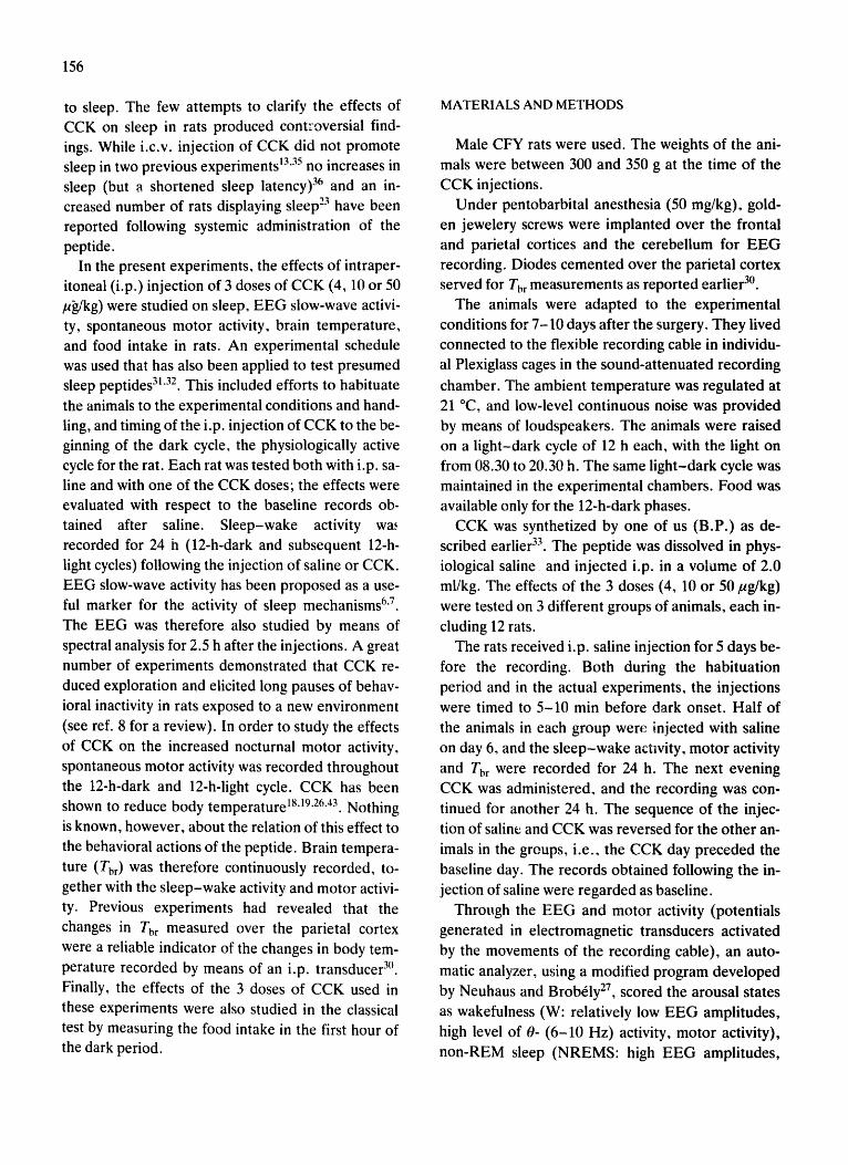
156
to sleep. The few attempts to clarify the effects of CCK on sleep in rats produced controversial find- ings. While i.c.v, injection of CCK did not promote sleep in two previous experiments t3"35 no increases in sleep (but a shortened sleep latency) 36 and an in- creased number of rats displaying sleep 23 have been reported following systemic administration of the peptide.
In the present experiments, the effects of intraper- itoneal (i.p.) injection of 3 doses of CCK (4, 10 or 50 /~/kg) were studied on sleep, EEG slow-wave activi- ty, spontaneous motor activity, brain temperature, and food intake in rats. An experimental schedule was used that has also been applied to test presumed sleep peptides 31"32. This included efforts to habituate
the animals to the experimental conditions and hand- ling, and timing of the i.p. injection of CCK to the be- ginning of the dark cycle, the physiologically active cycle for the rat. Each rat was tested both with i.p. sa- line and with one of the CCK doses; the effects were evaluated with respect to the baseline records ob- tained after saline. Sleep-wake activity wa~ recorded for 24 h (12-h-dark and subsequent 12-h- light cycles) following the injection of saline or CCK. EEG slow-wave activity has been proposed as a use- ful marker for the activity of sleep mechanisms 6"7. The EEG was therefore also studied by means of spectral analysis for 2.5 h after the injections. A great number of experiments demonstrated that CCK re- duced exploration and elicited long pauses of behav- ioral inactivity in rats exposed to a new environment (see ref. 8 for a review). In order to study the effects of CCK on the increased nocturnal motor activity, spontaneous motor activity was recorded throughout the 12-h-dark and 12-h-light cycle. CCK has been shown to reduce body temperature 18'19"26'43. Nothing
is known, however, about the relation of this effect to the behavioral actions of the peptide. Brain tempera- ture (Thr) was therefore continuously recorded, to- gether with the sleep-wake activity and motor activi- ty. Previous experiments had revealed that the changes in Tbr measured over the parietal cortex were a reliable indicator of the changes in body tem- perature recorded by means of an i.p. transducer 3°. Finally, the effects of the 3 doses of CCK used in these experiments were also studied in the classical test by measuring the food intake in the first hour of the dark period.
MATERIALS AND METHODS
Male CFY rats were used. The weights of the ani- mals were between 300 and 350 g at the time of the CCK injections.
Under pentobarbital anesthesia (50 mg/kg), gold- en jewelery screws were implanted over the frontal and parietal cortices and the cerebellum for EEG recording. Diodes cemented over the parietal cortex served for Tbr measurements as reported earlier 3°.
The animals were adapted to the experimental conditions for 7-10 days after the surgery. They lived connected to the flexible recording cable in individu- al Plexiglass cages in the sound-attenuated recording
chamber. The ambient temperature was regulated at 21 °C, and low-level continuous noise was provided by means of loudspeakers. The animals were raised on a light-dark cycle of 12 h each, with the light on from 08.30 to 20.30 h. The same light-dark cycle was maintained in the experimental chambers. Food was available only for the 12-h-dark phases.
CCK was synthetized by one of us (B.P.) as de- scribed earlier 33. The peptide was dissolved in phys- iological saline and injected i.p. in a volume of 2.0 ml/kg. The effects of the 3 doses (4, 10 or 50/zg/kg) were tested on 3 different groups of animals, each in- cluding 12 rats.
The rats received i.p. saline injection for 5 days be- fore the recording. Both during the habituation period and in the actual experiments, the injections were timed to 5-10 min before dark onset. Half of the animals in each group were injected with saline on day 6, and the sleep-wake actwity, motor activity and Tur were recorded for 24 h. The next evening CCK was administered, and the recording was con- tinued for another 24 h. The sequence of the injec- tion of saline and CCK was reversed for the other an- imals in the groups, i.e., the CCK day preceded the baseline day. The records obtained following the in- jection of saline were regarded as baseline.
Through the EEG and motor activity (potentials generated in electromagnetic transducers activated by the movements of the recording cable), an auto- matic analyzer, using a modified program developed by Neuhaus and Brob61y 27, scored the arousal states as wakefulness (W: relatively low EEG amplitudes, high level of 0- (6-10 Hz) activity, motor activity), non-REM sleep (NREMS: high EEG amplitudes,
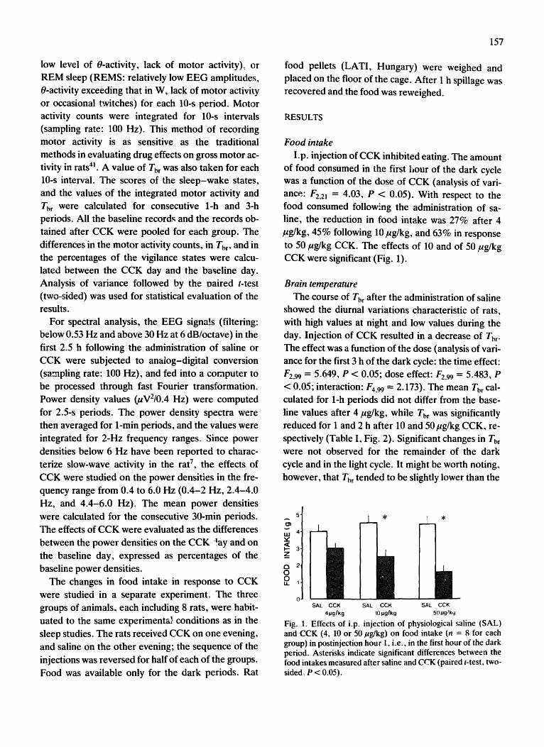
low level of 0-activity, lack of motor activity).~ or REM sleep (REMS: relatively low EEG amplitudes, 0-activity exceeding that in W, lack of motor activi W or occasional twitches) for each 10-s period. Motor activity counts were integrated for 10-s intervals (sampling rate: 100 Hz). This method of recording motor activity is as sensitive as the traditional methods in evaluating drug effects on gross motor ac- tivity in rats 41. A value of Tbr was also taken for each 10-s interval. The scores of the sleep-wake states, and the values of the integrated motor activity and Tbr were calculated for consecutive 1-h and 3-h periods. All the baseline records and the records ob- tained after CCK were pooled for each group. The differences in the motor activity counts, in Tbr, and in the percentages of tile vigilance states were calcu- lated between the CCK day and the baseline day. Analysis of variance followed by the paired t-test (two-sided) was used for statistical evaluation of the results.
For spectral analysis, the EEG signals (filtering: below 0.53 Hz and above 30 Hz at 6 dB/octave) in the first 2.5 h following the administration of saline or CCK were subjected to analog-digital conversion (sampling rate: 100 Hz), and fed into a computer to be processed through fast Fourier transformation. Power density values (gV2/0.4 Hz) were computed
for 2.5-s periods. The power density spectra were then averaged for 1-min periods, and the values were integrated for 2-Hz frequency ranges. Since power densities below 6 Hz have been reported to charac- terize slow-wave activity in the rat 7, the effects of CCK were studied on the power densities in the fre- quency range from 0.4 to 6.0 Hz (0.4-2 Hz, 2.4-4.0 Hz, and 4.4-6.0 Hz). The mean power densities were calculated for the consecutive 30-rain periods. The effects of CCK were evaluated as the differences between the power densities on the CCK lay and on the baseline day, expressed as percentages of the baseline power densities.
The changes in food intake in response to CCK were studied in a separate experiment. The three groups of animals, each including 8 rats, were habit- uated to the same experimental conditions as in the sleep studies. The rats received CCK on one evening, and saline on the other evening; the sequence of the injections was reversed for half of each of the groups. Food was available only for the dark periods. Rat
157
food pellets (LATI, Hungary) were weighed and placed on the floor of the cage. After 1 h spillage was recovered and the food was reweighed.
RESULTS
Food intake
l.p. injection of CCK inhibited eating. The amount of food consumed in the first hour of the dark cycle was a function of the dose of CCK (analysis of vari- ance: F2.21 = 4.03, P < 0.05). With respect to the food consumed following the administration of sa- line, the reduction in food intake was 27% after 4 /tg/kg, 45% following 10 pg/kg, and 63% in response to 50 #g/kg CCK. The effects of 10 and of 50 #g/kg CCK were significant (Fig. 1).
Brain temperature
The course of Tbr after the administration of saline showed the diurnal variations characteristic of rats, with high values at night and low values during the day. Injection of CCK resulted in a decrease of Tbr. The effect was a function of the dose (analysis of vari- ance for the first 3 h of the dark cycle: the time effect: F2.99 = 5.649, P < 0.05; dose effect: F2.99 = 5.483, P < 0.05; interaction: F4.99 -- 2.173). The mean Tbr cal- culated for 1-h periods did not differ from the base- line values after 4 pg/kg, while Tbr was significantly reduced for 1 and 2 h after 10 and 50 pg/kg CCK, re- spectively (Table I, Fig. 2). Significant changes in Tbr
were not observed for the remainder of the dark cycle and in the light cycle. It might be worth noting, however, that Tbr tended to be slightly lower than the
5
LLI 4'
Z
a 2. 0 0 LL 1
._J_.
/I SAL CCK
4pglkg
[ . *
/! SAL CCK
10 pglkg
1 *
I SAL CCK
50t.lglkg
Fig. 1. Effects of i.p. injection of physiological saline (SAL) and CCK (4, 10 or 50 Ftg/kg) on food intake (n = 8 for each group) in postinjection hour 1, i.e., in the first hour of the dark period. Asterisks indicate significant differences between the food intakes measured after saline and CCK (paired t-test, two- sided, P < 0.05).
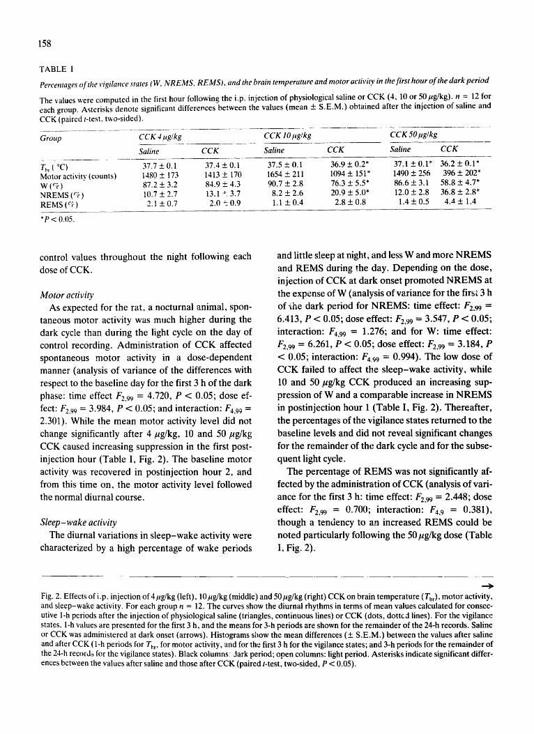
158
TABLE I
Percentages of the vigilance states (W, NREMS, REMS), and the brain temperature and motor activity in the first hour of the dark period
The values were computed in the first hour following the i.p. injection of physiological saline or CCK (4, 10 or 50/ag/kg). n = 12 for each group. Asterisks denote significant differer, ces between the values (mean +- S.E.M.) obtained after the injection of saline and CCK (paired t-test, two-sided).
Group CCK 4 ltg/kg CCK 10/tg/kg CCK 50/4g/kg
Saline C C K Saline C C K Saline C C K
Tbr(°C) 37.7+0.1 37.4+0.1 37.5+0.1 36.9+_0.2* 37.1+0.1" 36.2+0.1" Motor activity (counts) 1480 + 173 1413 + 170 1654 _ 211 1094 + 151" 1490 + 256 396 + 202* W (%) 87.2 + 3.2 84.9 + 4.3 90.7 + 2.8 76.3 +- 5.5* 86.6 + 3.1 58.8 + 4.7* NREMS (%) 10.7+2.7 13.1 ±3.7 8.2+2.6 20.9+-5.0* 12.0+2.8 36.8+-2.8* REMS(%) 2.1 +0.7 2.0 ~0.9 1.1 +-0.4 2.8+0.8 1.4+0.5 4.,1+ 1.4
*P < 0.05.
control values throughout the night following each
dose of CCK.
Motor activity
As expected for the rat, a nocturnal animal, spon-
taneous motor activity was much higher during the
dark cycle than during the light cycle on the day of
control recording. Administrat ion of CCK affected
spontaneous motor activity in a dose-dependent
manner (analysis of variance of the differences with
respect to the baseline day for the first 3 h of the dark
phase: time effect F 2 , 9 9 -'- 4.720, P < 0.05; dose ef-
fect: F2 .99 = 3.984, P < 0.05; and interaction: 1174.99 - -
2.301). While the mean motor activity level did not
change significantly after 4/~g/kg, 10 and 50/~g/kg
CCK caused increasing suppression in the first post-
injection hour (Table I, Fig. 2). The baseline motor
activity was recovered in postinjection hour 2, and
from this time on, the motor activity level followed
the normal diurnal course.
Sleep-wake activity
The diurnal variations in s leep-wake activity were
characterized by a high percentage of wake periods
and little sleep at night, and less W and more N R E M S
and REMS during the day. Depending on the dose,
injection of CCK at dark onset promoted N R E M S at
the expense of W (analysis of variance for the first 3 h
of-the dark period for NREMS: time effect: F2.99 =
6.413, P < 0.05; dose effect: F2.99 = 3.547, P < 0.05;
interaction: F4.99 : 1.276; and for W: time effect:
F2,99 = 6.261, P < 0.05; dose effect: F2,99 "- 3.184, P
< 0.05; interaction: F4.99 = 0.994). The low dose of
CCK failed to affect the s l eep-wake activity, while
l0 and 50/zg/kg CCK produced an increasing sup-
pression of W and a comparable increase in N R E M S
in postinjection hour 1 (Table I, Fig. 2). Thereaf ter ,
the percentages of the vigilance states re turned to the
baseline levels and did not reveal significant changes
for the remainder of the, dark cycle and for the subse-
quent light cycle.
The percentage of R E M S was not significantly af-
fected by the administration of CCK (analysis of vari-
ance for the first 3 h: time effect: F2,99 : 2.448; dose
effect: F2,99 "- 0 . 7 0 0 ; interaction: F4, 9 : 0.381),
though a tendency to an increased REMS could be
noted particularly following the 50 ktg/kg dose (Table
1, Fig. 2).
Fig. 2. Effects of i.p. injection of 4/~g/kg (left), 10 pg/kg (middle) and 50 pg/kg (right) CCK on brain temperature (Tbr), motor activity, and sleep-wake activity. For each group n = 12. The curves show the diurnal rhythms in terms of mean values calculated for consec- utive 1-h periods after the injection of physiological saline (triangles, continuous lines) or CCK (dots, dotted lines). For the vigilance states, 1-h values are presented for the first 3 h, and the means for 3-h periods are shown for the remainder of the 24-h records. Saline or CCK was administered at dark onset (arrows). Histograms show the mean differences (+ S.E.M.) between the values after saline and after CCK (1-h periods for Tb,, for motor activity, and for the first 3 h for the vigilance states; and 3-h periods for the remainder of the 24-h records for the vigilance states). Black columns: dark period; open columns: light period. Asterisks indicate significant differ- ences between the values after saline and those after CCK (paired t-test, two-sided, P < 0.05).

:INI'I:IS
V8
~01::I_-I
30N31:13_-I =JlO
(31~11 9N
101::!0031::1 40
39V
1N30t::13d
) .-~ :3
~o
~,
~ ,.'~ ~ o
( o0 ) (81N
nO0)
~o ~
~o ,~ ,~ ooo~
o~
o,~
""
T
~.
~
--
=1. W
- W
-~
-
2 i
-C
or)
I, Ll 7
J F.
(Oo)
(81
NR
03
) ( 3b
Nll
ON
IO~
OO
3U
~0
3
0V
IN3
OU
3d
)
3U
~lV
U~
::I~3
1
NIV
I:IB
83
1V
18
:::I),IV
M-d
33
38
A
llAIIO
V
EIO
IOIN
F t t-.
o
7_ w
w
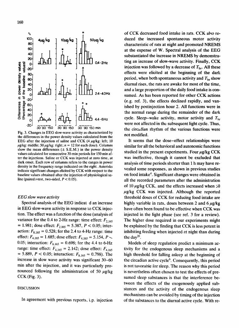
160
o ~ ._c '~
% 100 80 60. 40, 20. 0
-20
.200j
4O
20
-2
lOl, ig/kg ~ t
Y
. . . • . .
30 90 1,50 3o'9o" 150
j~i~Oi i ! i i i i~g/ko.4, g2H z
, ,y,
I ~ 2.4-4.0Hz
4.4- 6Hz 30 "90" 150 min
Fig. 3. Changes in EEG slow-wave activity as characterized by the differences in the power density values calculated from the EEG after the injection of saline and CCK (4 gg/kg: left; 10 pg/kg: middle; 50/tg/kg: right; n = 12 for each dose). Columns show the mean differences (+ S.E.M.) in the power density values calculated for consecutive 30-min periods for 150 min af- ter the injections. Saline or CCK was injected at zero time, at dark onset. Each row of columns refers to the canges in power density in the frequency range indicated on the right. Asterisks indicate significant changes elicited by CCK with respect to the baseline values obtained after the injection of physiological sa- line (paired t-test, two-sided, P < 0.05).
EEG slow wave activity Spectral analysis of the EEG indicat d an increase
in EEG slow-wave activity in response to CCK injec- tion. The effect was a function of the dose (analysis of variance for the 0.4 to 2-Hz range: time effect: F2,]65 = 1.981; dose effect: F2j65 - 5.387, P < 0.05; inter- action: Fs.165 = 0.528; for the 2.4 to 4-Hz range: time effect: F4,16 5 = 1.685; dose effect: F4.16 5 = 5.154, P .-, 0.05; interaction: Fsj65 = 0.698; for the 4.4 to 6-Hz range: time effect: F4.165 = 2.142; dose effect: F4,165 = 5.889, P < 0.05; interaction: F8.165 = 0.798). The increase in slow wave activity was significant 30-60 rain after the injection, and it was particularly pro- nounced following the administration of 50 pg/kg CCK (Fig. 3).
DISCUSSION
In agreement with previous reports, i.p. injection
of CCK decreased food intake in rats. CCK alto re- duced the increased spontaneous motor activity characteristic of rats at night and promoted NREMS at the expense of W. Spectral analysis of the EEG substantiated the increase in NREMS by demonstra- ting an increase of slow-wave activity. Finally, CCK injection was followed by a decrease of Tbr- All these effects were elicited at the beginning of the dark period, when both spontaneous activity and Tbr show diurnal rises, the rats are awake for most of the time, and a large proportion of the daily food intake is con- sumed. As has been reported for other CCK actions (e.g. ref. 3), the effects declined rapidly, and van- ished by postinjection hour 2. All functions were in the normal range during the remainder of the dark cycle. Sleep-wake activity, motor activity and Tbr were not affected in the subsequent light cycle. Thus, the circadian rhythm of the various functions were not modified.
It seems that the dose-effect relationships were similar for all the behavioral and autonomic functions studied in the present experiments. Four/~g/kg CCK was ineffective, though it cannot be excluded that analysis of time periods shorter than 1 h may have re- vealed some responses, as shown in previous studies on food intakC. Significant changes were obtained in all the recorded parameters after the administration of 10/~g/kg CCK, and the effects increased when 50 /~g/kg CCK was injected. Although the reported threshold doses of CCK for reducing food intake are highly variable in rats, doses between 2 and 6/~g/kg have often been found to be effective when CCK was injected in the light phase (see ref. 3 for a review). The higher dose required in our experiments might be explained by the finding that CCK is less potent in inhibiting feeding when injected at night than during the day :° .
Models of sleep regulation predict a minimum ac- tivity for the endogenous sleep mechanisms and a high threshold for falling asleep at the beginning of the circadian active cycle 6. Consequently, this period is not tavorabie for sleep. The reason why this period is nevertheless often chosen to test the effects of pre- sumed sleep substances is that the interference be- tween the effects of the exogenously applied sub- stances and the activity of the endogenous sleep mechanisms can be avoided by timing of the injection of the substances to the diurnal active cycle. With re-

gard to the high capacity of CCK to overcome the physiological pressure for W and thereby to increase sleep at night, it is reasonable to suppose that the peptide might also be effective when injected during the light cycle. Mansbach and Lorenz 23, in fact, found an increased number of rats displaying sleep and an increased sleep duration in response to i.p. CCK in rats during the day. Rojas-Ramirez et al. 36, however, observed a reduction in sleep latency with- out any increases in sleep following i.p. CCK during the light cycle, though the baseline percentages of the vigilance states could easily allow increases in NREMS. The results were interpreted by suggesting that CCK induced an unusual state of behavioral se- dation without concomitant EEG sleep s.
The evident increase of sleep in our experiments suggests that other explanations might also be con- sidered. Two points are worth mentioning. (1) Since the process of physiological sleep can easily be dis- turbed by any unusual stimulus, efforts were made to habituate the rats to the experimental conditioas as far as possible both in the study by Mansbach and Lo- renz 23 and in our own experiments. A habituation procedure was not mentioned in the report by Rojas- Ramirez et al. 36. In non-habituated rats, drowsiness may decrease sleep latency, but an apparent increase of sleep may be absent because of frequent arousals. This explanation implies that CCK promotes physiol- ogical NREMS. In agreement with this suggestion, human subjects injected with CCK reported drowsi- ness, but irresistible sleepiness was not expe- rienced 4°. (2) In the study by Mansbach and Lo- renz 23, the rats received food first, and were injected with CCK after a period of eating. Food was also available in our experiments, and eating was not to- tally inhibited even after the largest dose of CCK. In contrast, Rojas-Ramirez et al. 36 used fasted rats without food. It cannot be excluded therefore, that the increase of sleep results from the combined effect of CCK and some other factor(s) closely related to food ingestion. An increased availability of nutrients in the blood together with insulin release, has also been shown to promote sleep ~2, and may be an addi- tional factor in the mechanism of postprandial sleep.
Various lines of evidence indicate that the suppres- sion of eating and motor activity elicited by systemic administration of CCK are mediated through vagal afferents 9"11-22-25"39 though a direct effect of CCK on
161
the area postrema, a region with a deficient blood- brain barrier, might also be involved 42. Since i.c.v. administradgn of CCK failed to increase sleep 13.35 there is no reason to assume that the sleep effects of i.p. CCK injection were produced by a direct action of the peptide on the central nervous system, and possible peripheral sites of action should be con- sidered. Introduction of fat into the duodenum in- duced sedation in the cat, and the effect could be mimicked by i.v. administration of CCK 15. Electrical stimulation of the intestines has been shown to pro- mote sleep, and the effect seemed to be mediated, at least in part, via vagal afferents 2~. Vagal afferents are acknowledged as an important input for sleep regula- tion (see ref. 34 for a review). The increase of sleep might therefore be mediated through the vagus nerve, like the other components of the behavioral sequence elicited by CCK. It has been suggested for both the reduction of exploratory activity and the suppression of food intake that the vagal information about the peripherally administered CCK is carried to the basal forebrain through the ascending projec- tion of the solitary tract nuclei 1°. At the level of the basal forebrain, however, the mediation of the be- havioral effects of CCK seems to be separated: the paraventricular and dorsomedial nuclei of the hypo- thalamus are involved in the inhibition of eating, while a different, albeit non-specified area may be re- sponsible for the reduction of exploratory activity 8. At least in the paraventricular nucleus, CCK appears to be the neurotransmitter delivering the signal for reducing food intake 24. Another area in the basal forebrain, the olfactory tubercle, is known as one of the regions with the highest densities of CCK recep- tors 16. Unit activity in the olfactory tubercle has been reported to be responsive to hypnogenic vagal stimu- lations 14. Experiments with stimulations 5 and le- sions 29 indicated that the olfactory tubercle might be involved in sleep regulation. It is possible, therefore, that the basal forebrain provides mechanisms for the mediation of all the behavioral events of the syn- drome of satiety elicited by peripheral injection of CCK, including sleep. While feeding is inhibited in a few minutes following the administration of CCK, sleep-like resting behavior has been reported to ter- minate the behavioral sequence elicited by the pep- tide-'. The changes in EEG slow-wave activity indi- cated that NREMS increased significantly only

162
30-60 rain after CCK injection. It seems therefore that the effect of CCK on sleep has a much longer la- tency than on food intake which is in agreement with the findings reported by Rojaz-Ramirez et al. 36. The possible temporal separation of these effects may support the notion that they are elicited by different central mechanisms.
While the sleep-promoting activity of CCK can be regarded as an integral part of the satiety syndrome attributed to the peptide, the mechanism of the de- crease of Tbr and the importance of this effect in the behavioral actions are not clear. Motor inactivity and sleep have been shown to reduce Tbr; these changes, however, were much slighter than the relatively large fall found in response to C C K 3°. In fact, both intrace- rebra119'26 and systemic 18'43 administrations of CCK
have been reported to elicit hypothermia; the reduc- tion of body temperature seemed to result from a de- creased heat production 37 and an increased heat loss 18'37. It has also been speculated that the hypo- thermic effect of CCK might be closely related to the mechanism of the suppression of food intake, either as a causal factor or as a component of the satiety syndrome 26'43. These ideas, however, were not sup-
ported by the few experiments where the effects of systemic administration of CCK on body tempera- ture were studied, because the threshold dose of CCK required for the thermoregulatory effects was several times higher than the dose of CCK effective on food intake 18.43. The present results were there-
fore unexpected. The difference in the effects of CCK on Tbr and rectal temperature may be due to differences in the recording techniques; either Tbr is a more sensitive indicator of the effects of CCK than the rectal temperature, or the stress caused by the re- peated insertion of the rectal probes interfered with the hypothermic action of low doses of CCK, and this could be avoided by means of the chronic recording technique.
CCK has been shown to elicit a reduction in body temperature by acting on the thermosensitive units in the preoptic region 37. After systemic injection, how-
ever, CCK penetrates poorly through the blood- brain barrier, and a direct central action seems to be unlikely 44. In fact, some differences in the hypother- mia elicited by central and peripheral injection of CCK indicated that the structures involved were not the same TM. Although heat-sensitive afferent fibers have been identified in the vagus nerve l, there is no evidence suggesting that the thermosensitive vagal afferents are involved in the effects of CCK. Thus the mechanism of the decrease of body temperature in response to systemic administration of CCK remains to be clarified.
To summarize, in addition to reducing the high spontaneous motor activity at night and decreasing food intake, CCK promoted NREMS and increased EEG slow-wave activity. These effects may belong to the behavioral sequence of satiety, which has been proposed as the m~,jor behavioral action of the pep- tide. The effects of CCK on NREMS were similar to, or even more marked than those obtained in re- sponse to i.c.v, injection of presumed sleep peptides (6-sleep-inducing peptide (DSIP) 31, DSIP analogs 28, vasoactive intestinal polypeptide 32, growth hor- mone-releasing factor 28) in the same experimental schedule. Unfike these peptides, however, CCK also reduced Tbr. The pronounced effect on Tbr indicates that the autonomic actions of the peptide should be considered seriously in further studies on the behav- ioral responses to peripheral administration of CCK.
ACKNOWLEDGEMENTS
The authors would like to thank Dr. A.A. Borb61y for his continuous support during the experiments, including the provision of computer programs for the analysis of sleep-wake activity. The technical assist- ance by Mrs. K. Ormos, Mrs. I. Ponics~in, Mr. L. Kiss and Mr. P. Liszli is gratefully acknowledged. This work was supported by the Hungarian Academy of Sciences (OTKA 1104, OKKFT177/86) and the Ministry of Health (ETT 528).
REFERENCES
1 Adachi, A., Thermosensitive and osmoreceptive afferent fibers in the hepatic branch of the vagus nerve, J. Auton. Nerv. Syst., 10 (1984) 269-273.
2 Antin, J., Gibbs, J., Holt, J., Young, R.C. and Smith, G.P., Cholecystokinin elicits the complete behavioral se- quence of satiety in rats, J. Comp. Phsyiol. Psychol., 89 (1975) 784-790.
3 Baile, C.A., McLaughin, C.L. and Della-Fera, M.A., Role

of cholecystokinin and opioid peptides in control of food in- take, Physiol. Rev., 66 (1986) 172-234.
4 Bellinger, L.L. and Bernardis, L.L., Suppression of feed- ing by cholecystokinin but not bombesin is attenuated in dorsomedial hypothalamic nuclei lesioned rats, Peptides, 5 (1984) 547-552.
5 Benedek, G., Obfil Jr., F., Rubicsek, G. and Ob~ii, F., Sleep elicited by olfactory tubercle stimulation and the ef- fect of atropine, Behav. Brain Res., 2 (1981) 23-32.
6 Borb61y, A.A., A two process model of sleep regulation, Human NeurobioL, 1 (1982) 195-204.
7 Borb¢ly, A.A., Tobler, I. and Hanagasioglu, M., Effects of sleep deprivation on sleep and EEG power spectra in the rat, Behav. Brain Res., 14 (1984) 17!-182.
8 Crawley, J.N., Clarification of the behavioral functions of peripheral and central cholecystokinin: two separate pep- tide pools, Peptides, 6 Suppl. 2 (1985) 129-136.
9 Crawley, J.N., Hays, S.E. and Paul, S.M., Vagotomy abo- lishes the inhibitory effects of cholecystokinin on the rat ex- ploratory behaviors, Fur. J. Pharmacol., 73 (1981) 379-380.
10 Crawley, J.N., Kiss, J.Z. and Mezey, E., Bilateral mid- brain transections block the behavioral effects of cholecys- tokinin on feeding and exploration in rats, Brain Research, 322 (1984) 316-321.
11 Crawley, J.N. and Schwaber, J.S., Abolition of the behav- ioral effects of cholecystokinin following bilateral radio- frequency lesions of the parvoceilular subdivision of the nu- cleus tractus solitarius, Brain Research, 295 (1984) 289-299.
12 Danguir, J. and Nicolaidis, S., Intravenous infusions of nu- trients and sleep in the rat: an ischymetric sleep regulation hypothesis, Am. J. Physiol., 238 (1980} E301-E312.
13 DeMesquita, S. and Haney, W.H., Effect of chronic intra- cerebroventricular infusion of cholecystokinin on respira- tion and sleep, Brain Research, 378 (1986) 127-132.
14 D6t~iri, L., Juhfisz, G. and Kukorelli, T., Effect of stimula- tion of vagal and radial nerves on neuronal activity in the basal forebrain area of anaesthetized cats, Acta Physiol. Hung., 61 (1983) 147-154.
15 Fara, J.W., Rubinstein, E.H. and Sonnenscheim R.R., Visceral and behavioral responses to intraduodenal fat, Science, 166 (1969) 110-111.
16 Gaudreau, P., Quirion, R., St.-Pierre, S. and Pert, C.B., Tritium-sensitive film autoradiography of (3H)cholecysto- kinin-5/pentagastrin receptors in rat brain, Eur. J. Pharma- col., 87 (1983) 173-174.
17 Gibbs, J., Young, R.C. and Smith, G.P., Cholecystokinin decreases food intake in rats, J. Comp. Physiol. Psychol., 84 (1973) 488-495.
18 Kapfis, L., Ob~il Jr., F., Penke, B. and Obfil, F., Cholecys- tokinin-octapeptide-induced hypothermia in rats: dose-ef- fect and structure-effect relationships, effect of ambient temperature, pharmacological interactions and tolerance, Neuropharmacology, 26 (1986) 131-137.
19 Katsuura, G. and Itoh, S., Effect of cholecystokinin octa- peptide on body temperature in the rat, Jpn. J. Physiol., 3 l (1981) 849-858.
20 Kraly, F.S., A diurnal variation in the satieting potency of cholecystokinin in the rat, Appetite: J. Intake Res., 2 (1981) 177-191.
21 Kukorelli, T. and Juhfisz, G., Electroencephalographic synchronization induced by stimulation c,E small intestine and splanchnic nerve in cats, Electroenceph. Ciin. Neuro-
163
physiol., 41 (1976) 491-500. 22 Lorenz, D.N. and Goldman, S.A., Vagal mediation of the
cholecystokinin satiety effects in rats, Physiol. Behav., 29 (1982) 599-604.
23 Mansbach, R.S. and Lorenz, D.N., Cholecystokinin (CCK-8) elicits prandial sleep in rats, Physiol. Behav., 30 (1983) 179-183.
24 Morley, J.E., Levine, A.S., Gosnell, B.A., Mitchell, J.E., Krahn, D.D. and Nizielski, S.E., Peptides and feeding, Peptides, 6 Suppl. 2 (1985) 181-192.
25 Morley, .i.E., Levine, A.S., Kniep, J. and Grace, M., The effect of vagotomy on the satiety effects of neuropeptides and naloxone, Life Sci., 30 (1982) 1943-1947.
26 Morley, J.E., Levine, A.S. and Lindblad, S., Intraventricu- lar cholecystokinin-octapeptide produces hypothermia in rats, Fur. J. Pharmacol., 74 (1981) 249-251.
27 Neuhaus, H.U. and Borb61y, A.A., Sleep telemetry in the rat. II. Automatic identificatio.-, an~ recording of vigilance states, Electroenceph. Ciin. Neurophysiol., 44 (1978) 115-119.
28 Ob~il Jr., F., Effects of peptides (DSIP, DISP analogues, VIP, GRF and CCK) on sleep in the rat, Clin. Neurophar- macol., 9, Suppl. 4 (1986) 459-461.
29 Obfil Jr., F., Benedek, G., R6ti, G. and Obfil, F., Tonic hypnogenic effect of the olfactory tubercle, Exp. Neurol., 69 (1980) 202-208.
30 Ob~il Jr., F., Rubicsek, G., Alf61di, P., Silty, G. and Obfil, F., Changes in the brain and core temperaures in relation to the various arousal states in rats in the light and dark periods of the day, Pflagers Arch., 404 (1985) 73-79.
31 Obfil Jr., F., TOr6k, A., AlfOldi, P., S~iry, G., Haj6s, M. and Penke, B., Effects of intracerebroventricular injection of delta-sleep-inducing peptide (DSIP) and an analogue on sleep and brain temperature in rats at night, Pharmacol. Biochem. Behav., 23 (1985) 953-957.
32 Obfil Jr., F., Silty, G., AIf6idi, P., Rubicsek, G. and Obfil, F., Vasoactive intestinal polypeptide promotes sleep with- out effects on brain temperature in rats at night, Neurosci. Lett., 64 (1986) 236-240.
33 Penke, B., Hajnal, F., Lonovics, J., Holzinger, G., Kfidfir, T., Telegdy, G. and Rivier, J., Synthesis of potent hepta- peptide analogues of cholecystokinin, J. Med. Chem., 27 (1984) 845-849.
34 Puizillout, J.J., Gaudin-Chazal, G. and Bras, H., Vagal mechanisms in sleep regulation, Exp. Brain Res., Suppl. 8 (1984) 19-38.
35 Riou, F., Cespuglio, R. and Jouvet, M., Endogenous pep- tides and sleep in the rat. II. Peptides without significant ef- fect on the sleep-waking cycle, Neuropeptides, 2 (1982) 255-264.
36 Rojas-Ramirez, J.A., Crawley, J.N. and Mendelson, W.B., Electroencephalographic analysis of the sleep-in- ducing actions of cholecystokinin, Neuropeptides, 3 (1982) 129-138.
37 Shian, L.R. and Lin, M.T., Effects of cholecystokinin octa- peptide on thermoregulatory responses and hypothalamic neuronal activity in the rat, Naunyn-Schmiedeberg's Arch. Pharmacol., 328 (1985) 363-367.
38 Smith, G.P. and Gibbs, J.. Cholecystokinin: a putative sa- tiety signal, Pharmacol. Biochem. Behav., 3, Suppl. 1 (1975) 135-138.
39 Smith, G.P., Jerome, C. and Norgren, R., Afferent axons in abdominal vagus mediate satiety effect of cholecystoki- nin in rats, Am. J. Physiol., 249 (1985) R638-R641.
164
40 Stacher, G., Bauer, H. and Steinringer, H., Cholecystoki- nin decreases appetite and activation evoked by stimuli arising from the preparation of meal in man, Physiol. Be- hay., 23 (1979) 325-331.
41 Sv~nsson, K., Alffldi, P., Haj6s, M., Rubiscek, G., Jo- hansson, A.M., Carlsson. A. and Ob~il Jr., F., Dopamine autoreceptor antagonists: effects on sleep-wake activity in the rat, Pharmacol. Biochem. Behav., 26 (1987) 123-.129.
42 Van der Kooy. D., Area postrema: site where cholecyst,v, ki-
nin acts to decrease food intake, Brain Research, 295 (1984) 345-347.
43 Zetler, G., Cholecystokinin octapeptide, caerulein and caerulein analogizes: effects on thermoregulation in the mouse, Neuropharmacology, 21 (1982) 795-801.
44 Zhu, X.-G., Greeley Jr., G.H., Lewis, B.G., Lilja, P. and Thompson, J.C., Blood-CSF barrier to CCK and effect of centrally administered bombesin on release of brain CCK, J. Neurosci. Res., 15 (1986) 393-403.

P a g e | 59
Attachment 2.
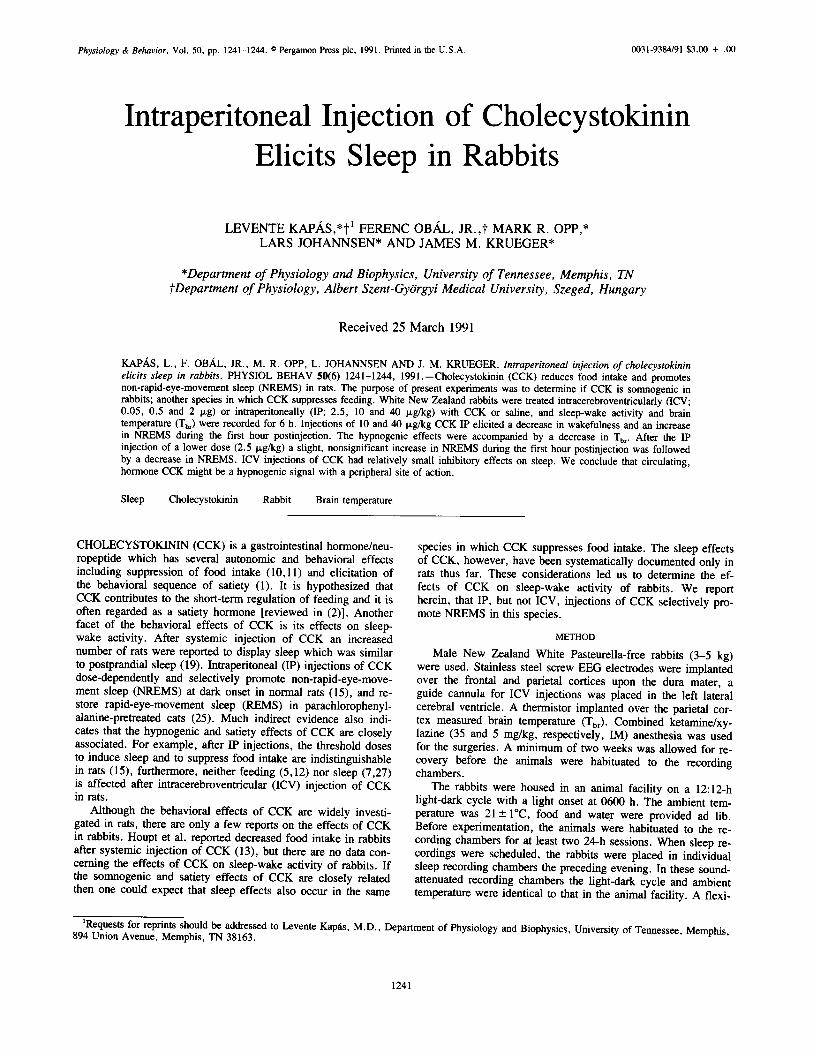
Physiology & Behavior, Vol. 50, pp. 1241-1244. © Pergamon Press plc, 1991. Printed in the U.S.A. 0031-9384/91 $3.00 + .00
Intraperitoneal Injection of Cholecystokinin Elicits Sleep in Rabbits
L E V E N T E KAP,A,S,*t t F E R E N C OB,~L, J R . , t M A R K R. OPP,* L A R S J O H A N N S E N * A N D J A M E S M. K R U E G E R *
*Department of Physiology and Biophysics, University of Tennessee, Memphis, TN ~Department of Physiology, Albert Szent-Gy6rgyi Medical University, Szeged, Hungary
Rece ived 25 March 1991
KAP/~S, L., F. OBAL, JR., M. R. OPP, L. JOHANNSEN AND J. M. KRUEGER. Intraperitoneal injection of cholecystokinin elicits sleep in rabbits. PHYSIOL BEHAV 50(6) 1241-1244, 1991.--Cholecystokinin (CCK) reduces food intake and promotes non-rapid-eye-movement sleep (NREMS) in rats. The purpose of present experiments was to determine if CCK is somnogenic in rabbits; another species in which CCK suppresses feeding. White New Zealand rabbits were treated intracerebroventricularly (ICV; 0.05, 0.5 and 2 p,g) or intraperitoneally (IP; 2.5, 10 and 40 ~g/kg) with CCK or saline, and sleep-wake activity and brain temperature (Tbr) were recorded for 6 h. Injections of 10 and 40 p,g/kg CCK IP elicited a decrease in wakefulness and an increase in NREMS during the first hour postinjection. The hypnogenic effects were accompanied by a decrease in Tbr. After the IP injection of a lower dose (2.5 p,g/kg) a slight, nonsignificant increase in NREMS during the first hour postinjection was followed by a decrease in NREMS. ICV injections of CCK had relatively small inhibitory effects on sleep. We conclude that circulating, hormone CCK might be a hypnogenic signal with a peripheral site of action.
Sleep Cholecystokinin Rabbit Brain temperature
CHOLECYSTOKININ (CCK) is a gastrointestinal hormone/neu- ropeptide which has several autonomic and behavioral effects including suppression of food intake (10,11) and elicitation of the behavioral sequence of satiety (1). It is hypothesized that CCK contributes to the short-term regulation of feeding and it is often regarded as a satiety hormone [reviewed in (2)]. Another facet of the behavioral effects of CCK is its effects on sleep- wake activity. After systemic injection of CCK an increased number of rats were reported to display sleep which was similar to postprandial sleep (19). Intraperitoneal (IP) injections of CCK dose-dependently and selectively promote non-rapid-eye-move- ment sleep (NREMS) at dark onset in normal rats (15), and re- store rapid-eye-movement sleep (REMS) in parachlorophenyl- alanine-pretreated cats (25). Much indirect evidence also indi- cates that the hypnogenic and satiety effects of CCK are closely associated. For example, after IP injections, the threshold doses to induce sleep and to suppress food intake are indistinguishable in rats (15), furthermore, neither feeding (5,12) nor sleep (7,27) is affected after intracerebroventricular (ICV) injection of CCK in rats.
Although the behavioral effects of CCK are widely investi- gated in rats, there are only a few reports on the effects of CCK in rabbits. Houpt et al. reported decreased food intake in rabbits after systemic injection of CCK (13), but there axe no data con- ceming the effects of CCK on sleep-wake activity of rabbits. If the somnogenic and satiety effects of CCK are closely related then one could expect that sleep effects also occur in the same
species in which CCK suppresses food intake. The sleep effects of CCK, however, have been systematically documented only in rats thus far. These considerations led us to determine the ef- fects of CCK on sleep-wake activity of rabbits. We report herein, that IP, but not ICV, injections of CCK selectively pro- mote NREMS in this species.
METHOD
Male New Zealand White Pasteurella-free rabbits (3-5 kg) were used. Stainless steel screw EEG electrodes were implanted over the frontal and parietal cortices upon the dura mater, a guide cannula for ICV injections was placed in the left lateral cerebral ventricle. A thermistor implanted over the parietal cor- tex measured brain temperature (Tbr). Combined ketamine/xy- lazine (35 and 5 mg/kg, respectively, IM) anesthesia was used for the surgeries. A minimum of two weeks was allowed for re- covery before the animals were habituated to the recording chambers.
The rabbits were housed in an animal facility on a 12:12-h light-dark cycle with a light onset at 0600 h. The ambient tem- perature was 21--+ I°C, food and water were provided ad lib. Before experimentation, the animals were habituated to the re- cording chambers for at least two 24-h sessions. When sleep re- cordings were scheduled, the rabbits were placed in individual sleep recording chambers the preceding evening. In these sound- attenuated recording chambers the light-dark cycle and ambient temperature were identical to that in the animal facility. A flexi-
~Requests for reprints should be addressed to Levente Kap~ls, M.D., Department of Physiology and Biophysics, University of Tennessee, Memphis, 894 Union Avenue, Memphis, TN 38163.
1241
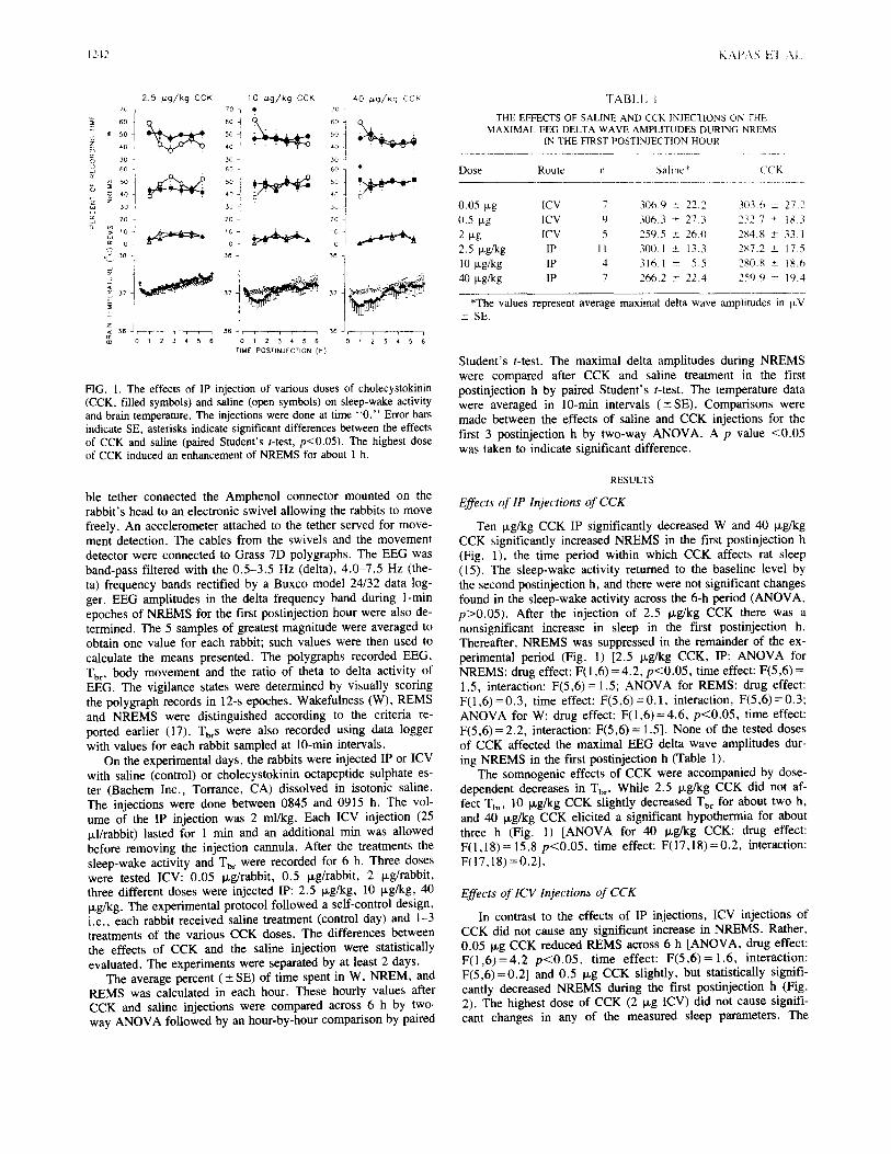
1242 KAP4S El A.i.
10 /~g/kg CCK ro q
c~
o 30 -: ' 3 6 0
; ,0 J z
~J 20~
a
0 2
3 8
2.5 //,g/kg CCK zC, 1
~: 5 0
3 0 -~
60
m z 4 0
3o ] 2o
cc 0
3 8 -,
3 7 37
e~
= j 3 6 ~ - J T , , , , 3 6
0 1 2 3 ~. 5 6
40 /.~g/Kg CCK
6 0
5o 1 '~0 J
3 o =[
6 0
4o
3o I 2o i
o I
38 ]
i i , i J , , 3 6 , , r r p I
0 1 2 3 4 5 6 0 1 2 3 4 5 6
TIME POSTINJECTION (h )
FIG. 1. The effects of IP injection of various doses of cholecystokinin (CCK, filled symbols) and saline (open symbols) on sleep-wake activity and brain temperature. The injections were done at time "0 ." Error bars indicate SE, asterisks indicate significant differences between the effects of CCK and saline (paired Student's t-test, p<0.05). The highest dose of CCK induced an enhancement of NREMS for about 1 h.
ble tether connected the Amphenol connector mounted on the rabbit's head to an electronic swivel allowing the rabbits to move freely. An accelerometer attached to the tether served for move- ment detection. The cables from the swivels and the movement detector were connected to Grass 7D polygraphs. The EEG was band-pass filtered with the 0.5-3.5 Hz (delta), 4.0-7.5 Hz (the- ta) frequency bands rectified by a Buxco model 24/32 data log- ger. EEG amplitudes in the delta frequency band during 1-min epoches of NREMS for the first postinjection hour were also de- termined. The 5 samples of greatest magnitude were averaged to obtain one value for each rabbit; such values were then used to calculate the means presented. The polygraphs recorded EEG, Tbr , body movement and the ratio of theta to delta activity of EEG. The vigilance states were determined by visually scoring the polygraph records in 12-s epoches. Wakefulness (W), REMS and NREMS were distinguished according to the criteria re- ported earlier (17). TbrS were also recorded using data logger with values for each rabbit sampled at 10-min intervals.
On the experimental days, the rabbits were injected IP or ICV with saline (control) or cholecystokinin octapeptide sulphate es- ter (Bachem Inc., Torrance, CA) dissolved in isotonic saline. The injections were done between 0845 and 0915 h. The vol- ume of the IP injection was 2 ml/kg. Each ICV injection (25 M/rabbit) lasted for 1 min and an additional min was allowed before removing the injection cannula. After the treatments the sleep-wake activity and Tb~ were recorded for 6 h. Three doses were tested ICV: 0.05 txg/rabbit, 0.5 Ixg/rabbit, 2 ~g/rabbit, three different doses were injected IP: 2.5 ~g/kg, 10 ixg/kg, 40 txg/kg. The experimental protocol followed a self-control design, i.e., each rabbit received saline treatment (control day) and 1-3 treatments of the various CCK doses. The differences between the effects of CCK and the saline injection were statistically evaluated. The experiments were separated by at least 2 days.
The average percent ( +- SE) of time spent in W, NREM, and REMS was calculated in each hour. These hourly values after CCK and saline injections were compared across 6 h by two- way ANOVA followed by an hour-by-hour comparison by paired
TABLE
THE EFFECTS OF SALINE AND CCK INJECTIONS ON THE MAXIMAL EEG DELTA WAVE AMPLITUDES DURING NREMS
IN THE FIRST POSTINJECT1ON HOUR
Dose Route n Saline* ('CK
0.05 ~xg ICV 7 306.9 -- 22.2 303.6 ± 27.2 0.5 Ixg ICV 9 306.3 m 27.3 232.7 ± t83 2 i.tg ICV 5 259.5 ± 26.0 284.8 ± 33 I 2.5 Ixg/kg IP 11 300.1 ± 13.3 287.2 _+_ 17.5 10 p.g/kg IP 4 3t6.1 ± 5.5 280.8 ± 18.6 40 txg/kg IP 7 266.2 ± 22.4 259.9 ± 19.4
*The values represent average maximal delta wave amplitudes in IxV _~ SE.
Student's t-test. The maximal delta amplitudes during NREMS were compared after CCK and saline treatment in the first postinjection h by paired Student's t-test. The temperature data were averaged in 10-min intervals (-+SE). Comparisons were made between the effects of saline and CCK injections for the first 3 postinjection h by two-way ANOVA. A p value <0.05 was taken to indicate significant difference.
RESULTS
Effects of IP Injections of CCK
Ten Fg/kg CCK IP significantly decreased W and 40 ~g/kg CCK significantly increased NREMS in the first postinjection h (Fig. 1), the time period within which CCK affects rat sleep (15). The sleep-wake activity returned to the baseline level by the second postinjection h, and there were not significant changes found in the sleep-wake activity across the 6-h period (ANOVA, p>0.05). After the injection of 2.5 p,g/kg CCK there was a nonsignificant increase in sleep in the first postinjection h. Thereafter, NREMS was suppressed in the remainder of the ex- perimental period (Fig. 1) [2.5 Ixg/kg CCK, IP: ANOVA for NREMS: drug effect: F(1,6)=4.2, p<0.05, time effect: F(5,6)= 1.5, interaction: F(5,6)= 1.5; ANOVA for REMS: drug effect: F(1,6)=0.3, time effect: F(5,6)=0.1, interaction, F(5,6)=0.3; ANOVA for W: drug effect: F(1,6)=4.6, p<0.05, time effect: F(5,6) = 2.2, interaction: F(5,6) = 1.5]. None of the tested doses of CCK affected the maximal EEG delta wave amplitudes dur- ing NREMS in the first postinjection h (Table 1).
The somnogenic effects of CCK were accompanied by dose- dependent decreases in Tbr. While 2.5 Ixg/kg CCK did not af- fect Tbr, 10 ~g/kg CCK slightly decreased Tbr for about two h, and 40 Ixg/kg CCK elicited a significant hypothermia for about three h (Fig. 1) [ANOVA for 40 ~g/kg CCK: drug effect: F( I ,18)= 15.8 p<0.05, time effect: F(17,18)=0.2, interaction: F(17,18) =0.2].
Effects of 1CV Injections of CCK
In contrast to the effects of IP injections, ICV injections of CCK did not cause any significant increase in NREMS. Rather, 0.05 i~g CCK reduced REMS across 6 h [ANOVA, drug effect: F (1 ,6 )=4 .2 p<0 .05 , time effect: F (5 ,6 )=1 .6 , interaction: F(5,6)=0.2] and 0.5 txg CCK slightly, but statistically signifi- cantly decreased NREMS during the first postinjection h (Fig. 2). The highest dose of CCK (2 Ixg ICV) did not cause signifi- cant changes in any of the measured sleep parameters. The

CCK AND SLEEP 1243
70.
"~ 6 0 r- zo ~ 5 0
4 0
0 3 0
so 1 ~5o
w 30
20
~ o 0
0 . 0 5 p,g CCK 0 . 5 p,g CCK 70
60
5o
40
311
6O •
40
30
2O
0
~ 3 6 , , ~ , , 1 1 , , , i , , ,
0 1 2 3 4 5 6 0 1 2 3 4 5 6
2 # g CCK
60
50
40
30
60
40
3O
2O
0
36 , , , j , , ,
0 I 2 3 4 5 6 TIME POSTINJECTION ( h )
FIG. 2. The effects of ICV injection of various doses of CCK and sa- line on sleep-wake activity and Tbr. See legend to Fig. 1 for details. Unlike high doses of CCK injected IP, ICV CCK failed to enhance sleep.
maximal EEG delta wave amplitudes were not affected by any doses of CCK (Table 1).
Injections of 0.05 P,g and 2 p,g, but not 0.5 p.g CCK, elic- ited statistically significant decreases in Tbr for about one h, al- though the amplitudes of these changes were minor (Fig. 2) [ANOVA for 0.05 p,g CCK: drug effect: F(1,18)=7.1 p<0.05, time effect: F(17,18)=0.2, interaction: F(17,18)=0.2; and for 2 P.g CCK: drug effect: F(1,18)=4.7 p<0.05, time effect: F(17,18) = 1.6, interaction: F(17,18) = 0.7].
DISCUSSION
The effects of CCK on sleep-wake activity of rats are well documented (6, 15, 19, 24). Although preliminary attempts to demonstrate somnogenic activity of CCK have been done in cats (8), our present work represents the first systematic description of the effects of exogenously administered CCK on normal sleep in rabbits.
We report here that 10 ~g/kg CCK decreases W, and 40 p,g/kg CCK selectively increases NREMS and elicits hypother- mia in rabbits. In contrast, ICV injections of CCK or IP injec- tion of 2.5 ~g/kg CCK did not induce NREMS in rabbits, in fact, after 0.05 ixg ICV and 2.5 Ixg/kg IP CCK NREMS was decreased and after 0.05 p,g CCK REMS was reduced, although these effects were very small. The results are similar to those obtained in rats where increases in NREMS (15) and decreases in temperature (14,15) were also found after IP administration of CCK, furthermore, ICV bolus injection (27) or chronic ICV infusion of CCK (7) failed to promote sleep. The dose-range for the hypnogenic effects of CCK is similar in both species. A threshold dose of 10 p.g/kg CCK in rats was reported (15), oth- ers also observed sleep increases after 10 Ixg/kg CCK (24), and in a third study 16, but not 8, ixg/kg CCK promoted NREMS (6). IP injection of 4 Ixg/kg (15) or 2 t~g/kg (27) CCK were ineffective on sleep of rats.
The enhanced sleep after CCK treatment was not accompa- nied with changes in EEG delta wave amplitudes. In contrast, recovery sleep following sleep deprivation (21), the sleep that accompanies acute infections (30,31) and the sleep elicited by
several sleep-promoting substances such as IL-1 (17), muramyl dipeptide (16) or growth hormone-releasing hormone (20) are characterized by increased EEG delta wave amplitudes during NREMS. This suggests that the underlying mechanisms of CCK- induced sleep and sleep induced by the above mentioned inter- ventions may be partially independent.
The dose-dependent hypothermic effects of systemically in- jected CCK are similar to those reported in rats (14,15) and mice (34). These thermoregulatory effects parallel the hypnogenic ef- fects of CCK. ICV administration of 0.05 and 2 ixg CCK also elicited a statistically significant decline in Tbr. The magnitude of these changes, however, were so minor that the physiological significance of these responses seems questionable. Similarly, Lipton and Glyn reported that ICV injection of 5 Ixg CCK does not alter the body temperature in rabbits (18).
The mechanisms mediating the effects of exogenous CCK on sleep-wake activity are not known. Since the peripheral-circulat- ing and the central-neuronal CCK pools are separated by the blood-brain barrier, which is practically impermeable to CCK (22), we assume that systemic injection of CCK mainly mimics the effects of the endogenous, peripherally circulating hormone, whereas ICV administration of CCK mimics the effects of the neurotransmitter/neuromodulator CCK. Since no somnogenic ef- fects were observed after central injection of CCK it seems that the site of hypnogenic action of CCK is peripheral. It is known that sensory inputs conveyed by the n. vagus contribute to the regulation of sleep-wake activity (26). It has also been reported that several effects of CCK are mediated by the n. vagus (3, 9, 29, 33). It is possible, therefore, that the somnogenic effects of CCK are also mediated by n. vagus. Another possibility is that CCK causes the release of other sleep-promoting substance(s) from the periphery. The role of insulin seemed likely, since in- sulin elicits sleep in rats (4,28), and CCK is a potent stimulator of insulin release [e.g., (23,32)]. Experiments in rats indicate, however, that insulin plays little if any role in the hypnogenic effects of CCK (Kap~is et al., submitted).
It is possible that the somnogenic doses of CCK are not physiological, although it is difficult to know since the concen- tration of CCK at the hypnogenic site, when its secretion is physiologically stimulated (e.g., after eating), is unknown. It is likely that when CCK is released within the small intestine it reaches high concentrations in circumscribed areas of the GI tract. To achieve such high concentrations experimentally, sys- temic injections of relatively large amounts of the peptide are needed. Regardless of the specific mechanism by which CCK affects sleep and thermoregulation, current results clearly show that the hypnogenic effects of CCK are not species specific. Furthermore, CCK enhances sleep in a species, in which it also suppresses food intake. It supports the idea that the sleep-pro- moting and food intake reducing effects are closely associated, presumably different manifestations of satiety. Finally, the expe- rience of postprandial sleepiness, when the secretion of CCK is physiologically stimulated, further suggests that one of the pri- mary actions of peripherally released CCK may be sleep promo- tion.
A C K N O W L E D G E M E N T S
The authors thank Dr. Mayumi Kimura-Takeuchi for reviewing the manuscript. The technical assistance of Sandy Johnson, Donna Maxwell and Gail Richmond and the secretarial assistance of Collean Payne is gratefully acknowledged. This work was supported by the Office of Na- val Research (N00014-90-J-1069), the U.S. Army Medical Research and Development Command (DAMD-17-86-C-6194) and the National Insti- tutes of Health (NS-25378 and NS-27250).

1244 KAPAS H AI.
REFERENCES
I. Antin, J.; Gibbs, J.; Holt, J.; Young, R. C.; Smith, G. P. Cholc- cystokinin elicits the complete behavioral sequence of satiety in rats. J. Comp. Physiol. Psychol. 89:784-790; 1975.
2. Baile, C. A.: McLaughlin, C. L.; Della-Fera, M. A. Role of chole- cystokinin and opioid peptides in control of food intake. Physiol. Rev. 66:172-234; 1986.
3. Crawley, J. N.; Hays, S. E.; Paul, S. M. Vagotomy abolishes the inhibitory effects of cholecystokinin on rat exploratory behaviors. Eur. J. Pharmacol. 73:379-380; 1981.
4. Danguir, J.; Nicolaidis, S. Chronic intracerebroventricular infusion of insulin causes selective increase of slow wave sleep in rats. Brain Res. 306:97-103: 1984.
5. Della-Fera, M. A.; Baile, C. A. Cholecystokinin octapeptide: con- tinuous injections into the cerebral ventricles of sheep suppress feeding. Science 206:471-473; 1979.
6. de Saint Hilaire-Kafi, Z.; Depoortere, H.; Nicolaidis, S. Does cholecystokinin induce physiological satiety and sleep? Brain Res. 488:304-310; 1989.
7. De Mesquita, S.; Haney, W. H. Effect of chronic intracerebroven- tricular infusion of cholecystokinin on respiration and sleep. Brain Res. 378:127-132; 1986.
8. Fara, J. W.; Rubinstein, E. H.; Sonnenschein, R. R. Visceral and behavioral responses to intraduodenal fat. Science 166:t10-111: 1969.
9. Flood, J, F.; Smith, G. E.; Morley, J. E. Modulation of memory processing by cholecystokinin: dependence on the vagus nerve. Sci- ence 236:832-834; 1987.
10. Gibbs, J.; Young, R. C.; Smith, G. P. Cholecystokinin elicits sati- ety in rats with open gastric fistulas. Nature 245:323-325; 1973.
11. Gibbs, J.; Young, R. C.; Smith, G. P. Cholecystokinin decreases food intake in rats, J. Comp. Physiol. Psychol. 84:488-495; 1973.
12. Grinker, J. A.; Schneider, B. S.; Ball, G.; Cohen, A.; Strohmayer, A.; Hirsch, J. Cholecystokinin (CCK-8) and bombesin (BBS) intra- cranial injections and satiety in rats. Fed. Proc. 39:501; 1980.
13. Houpt, T. R.; Anika, S. M.; Wolff, N. C. Satiety effects of chole- cystokinin and caerulein in rabbits. Am. J. Physiol. 235:R23-R28; 1978.
14. Kapfis, L.; Obgtl, F., Jr.; Penke, B.; Obtil, F. Cholecystokinin-oc- tapeptide-induced hypothermia in rats: dose-effect and structure-ef- fect relationships, effect of ambient temperature, pharmacological interactions and tolerance. Neuropharmacology 26:131-137; 1987.
15. Kap~is, L.; ONil, F., Jr.; Alf61di, P.; Rubicsek, G.; Penke, B.; Obgl, F. Effects of nocturnal intraperitoneal administration of cholecysto- kinin in rats: simultaneous increase in sleep, increase in EEG slow- wave activity, reduction of motor activity, suppression of eating, and decrease in brain temperature. Brain Res. 438:155-164; 1988.
16. Krueger, J. M.; Pappenheimer, J. R.; Karnovsky, M. L. Sleep-pro- moting effects of muramyl peptides. Proc. Natl. Acad. Sci. U;A 79:6102-6t06; 1982.
17. Krueger, J. M.; Walter, J.; Dinarello, C. A.; Wolff, S. M.; Che- did. L. Sleep-promoting effects of endogenous pyrogen (interleukin-
!). Am. J. Physiol. 246:R994-R999; 1984. 18. Lipton, J. M.; Glyn, J. R. Central administration ot peptidcs alters
thermoregulation in the rabbit. Peptides 1:15 18; 1980 19. Mansbach, R. S.; Lorenz, D. N. Cholecystokinin tCCK-81 elicits
prandial sleep in rats. Physiol. Behav. 30:179-183; 1983. 20. Obfil, F., Jr.; Alf61di, P.; Cady, A. B.; Johannsen, I_.: S~y, G.:
Krueger, J. M. Growth hormone-releasing factor enhances sleep in rats and rabbits. Am. J. Physiol. 255:R310-R316; 1988.
21. Pappenheimer, J. R.; Koski, G.; Fencl, V.; Karnovsky, M. L.: Krueger, J. Extraction of sleep-promoting factor S from cerebrospi- hal fluid and from brains of sleep-deprived animals. J Neurophys- iol. 38:1299-1311; 1975.
22. Passaro, E., Jr.; Debas, H.; Oldendoff, W.; Yamada, T. Rapid ap- pearance of intraventricularly administered neuropeptides in the pe- ripheral circulation. Brain Res. 241:335-340; 1982.
23. Pederson, R. A.; Brown, J. C. Effect of cholecystokinin, secretin, and gastric inhibitory polypeptide on insulin release from the iso- lated perfused rat pancreas. Can. J. Physiol. Pharmacol. 57:1233- 1237: 1979.
24. Posadas-Andrews, A.; Prosp6ro-Garcia, O.; Rojas-Ramirez, J. A. CCK-8 effects on sleep and feeding. Soc. Neurosci. Abstr. 15:1068; 1989.
25. Prosp6ro-Garcfa, O.; Ott, T.; Drucker-Colin, R. Cerebroventricutar infusion of cholecystokinin (CCK-8) restores REM sleep in parachlorophenylalanine (PCPA)-pretreated cats. Neurosci. Lett. 78: 205-210; 1987.
26. Puizillout, J. J.; Gaudin-Chazal, G.; Bras, H. Vagal mechanisms in sleep regulation. Exp. Brain Res. Suppl. 8:19-38; 1984.
27. Riou, F.; Cespuglio, R.; Jouvet, M. Endogenous peptides and sleep in the rat: II Peptides without significant effect on the sleep-waking cycle. Neuropeptides 2:255-264; 1982.
28. Sangiah, S.; Caldwell, D. F.; Villeneuve, M. J.: Clancy, J. J. Sleep: sequential reduction of paradoxical (REM) and elevation of slow-wave (NREM) sleep by a nonconvulsive dose of insulin in rats. Life Sci. 31:763-769; 1982.
29. Smith, G. P.; Jerome, C.; Cushin, B. J.; Eterno, R.; Simansky, K. J. Abdominal vagotomy blocks the satiety effect of cholecystokinin in the rat. Science 213:1036-1037; 1981.
30. Toth, L. A.; Krueger, J. M. Alteration of sleep in rabbits by Sta- phylococcus aureus infection. Infect. Immunol. 56:1785-1791; 1988.
31. Toth, L. A.; Krueger, J. M. Effects of microbial challenge on sleep in rabbits. FASEB J. 3:2062-2066; 1989.
32. Unger, R. H.; Ketterer, H.; Dupr6, J.; Eisentraut, A. M. The ef- fects of secretin, pancreozymin, and gastrin on insulin and glucagon secretion in anesthetized dogs. J. Clin. Invest. 46:630-645; 1967.
33. Verbalis, J. G.; McCann, M. J.; McHale, C. M.; Stricker, E. M. Oxytocin secretion in response to cholecystokinin and food: differ- entiation of nausea from satiety. Science 232:1417-1419; 1986.
34. Zetler, G. Cholecystokinin octapeptide, caerulein and caerulein ana- logues: effects on thermoregulation in the mouse. Neuropharmacol- ogy 21:795-801; 1982.
P a g e | 64
Attachment 3.


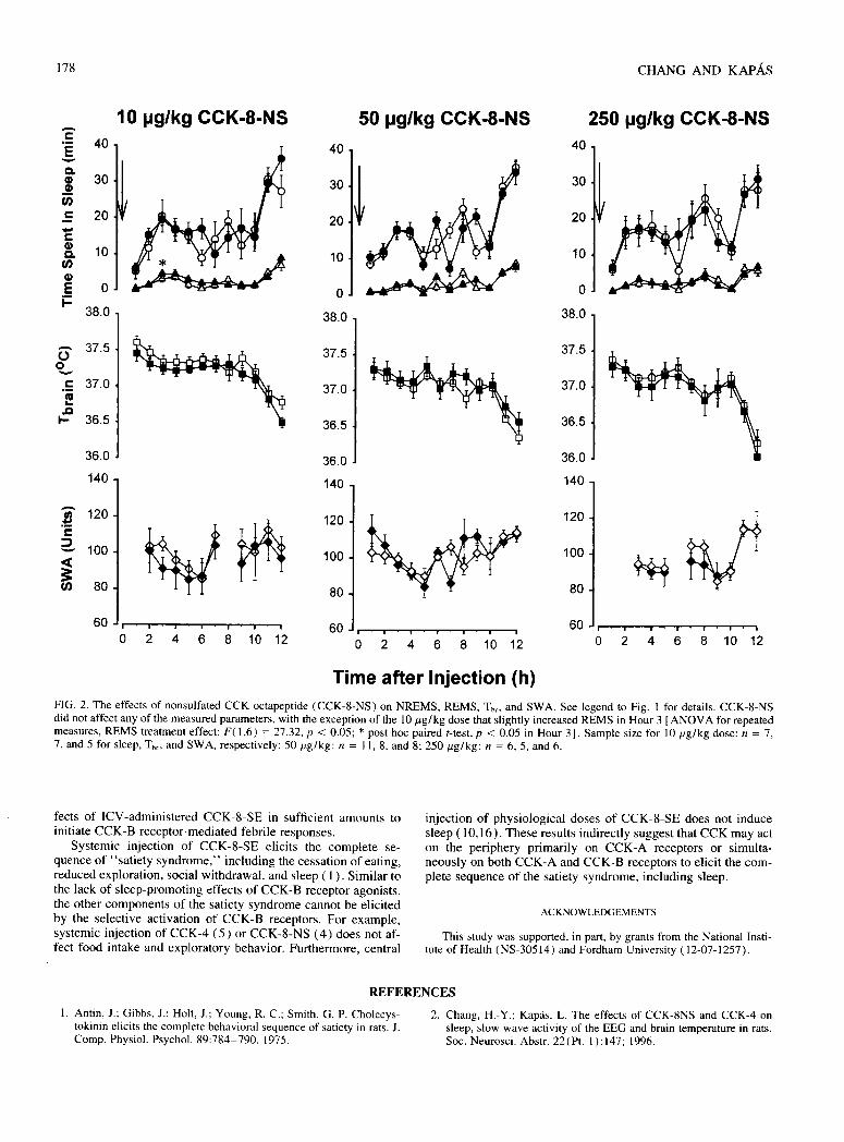

P a g e | 70
Attachment 4.

Physiology & Behavior, Vol. 50, pp. 417-420. © Pergamon Press plc, 1991. Printed in the U.S.A. 0031-9384/91 $3.00 + .00
Cholecystokinin Promotes Sleep and Reduces Food Intake in Diabetic Rats
L E V E N T E KAP/~S,*I "1 F E R E N C OBAL, JR. ,* I B O L Y A F A R K A S , * L I N D A C. P A Y N E , t G Y U L A SARY,* G Y O R G Y R U B I C S E K * A N D JAMES M. K R U E G E R t
*Department of Physiology, Albert Szent-Gy6rgyi Medical University, Szeged, Hungary ?Department of Physiology and Biophysics, University of Tennessee, Memphis, TN
Received 18 January 1991
KAPAS, L., F. OBAL, JR., I. FARKAS, L. C. PAYNE, G. SARY, G. RUBICSEK AND J. M. KRUEGER. Cholecystokinin promotes sleep and reduces food intake in diabetic rats. PHYSIOL BEHAV 50(2) 417-420, 1991.--It has been reported that systemic injections of cholecystokinin (CCK) elicit the behavioral characteristics of satiety, including sleep, in rats. CCK is a potent stimulator of insulin secretion, and insulin is hypothesized to be involved in sleep and feeding regulation. The purpose of the current experiments was to study the possible role of endogenous insulin in the food-intake-reducing and hypnogenic effects of intraperitoneally (IP) administered CCK. Normal and streptozotocin (STR)-diabetic rats were injected with isotonic saline or CCK (10 and 50 ixg,'kg) at dark onset, and sleep-wake activity was determined for the next 12 h. There were no significant differences between the baseline sleep-wake activity of normal and diabetic rats. IP injection of CCK elicited a selective increase in nonrapid- eye-movement sleep in both groups during the lust postinjection hour. In a separate experiment, the effects of CCK (10 Irg/kg) on food intake were determined in control and diabetic rats; CCK suppressed the 1-h food intake in both groups. In a third experi- ment, the effects of CCK treatment (50 p,g/kg) on plasma insulin levels were determined. In normal rats, CCK elicited a two-fold increase in plasma insulin concentration, whereas diabetic rats had a significantly lower basal insulin level which was not affected by CCK treatment. We conclude that hypnogenic and food-intake-reducing effects of exogenously administered CCK are closely associated; however, pancreatic insulin does not play a significant role in either of these effects.
Cholecystokinin Sleep Food intake Insulin Streptozotocin Rat
IT is hypothesized that humoral factors are involved in the regu- lation of sleep-wake activity and feeding behavior. A variety of endogenous substances that promote sleep [sleep factors, re- viewed in (5)] and/or reduce food intake [satiety peptides, re- viewed in (28)] have been described. Among these substances, cholecystokinin (CCK) and insulin are of special interest, since both are implicated as sleep factors and satiety hormones. A single peripheral injection of CCK suppresses food intake (10,11), elicits the whole behavioral sequence of satiety (2), and pro- motes nonrapid-eye-movement sleep (NREMS) (13,18) resem- bling that observed after eating (18). Similarly, exogenous administration of insulin selectively increases NREMS (8,23) and decreases food intake (27), whereas diabetic rats exhibit disturbed sleep (6) and increased feeding (4,16). Since CCK is a major hormonal stimulatory factor of insulin secretion (1, 22, 24, 25, 29), it seemed possible that the effects of exogenously administered CCK on sleep and feeding are, in fact, mediated by endogenously released insulin. The aim of the current experi- ments was to study the possible role of pancreatic insulin in the food-intake-suppressive and sleep-promoting effects of exoge- nously administered CCK using a streptozotocin (STR)-induced diabetes model in rats. We report here that systemic injection of CCK promotes NREMS and reduces food intake, but it is un- likely that these effects are mediated by pancreatic insulin.
METHOD
Sleep Experiments
Effects of 50 Ixg/kg CCK on sleep-wake activity. Male rats of the Wistar strain were used weighing 250-350 g at the time of the CCK injections. Gold jewelry screws were implanted over the frontal and parietal cortices, and the cerebellum for electro- encephalogram (EEG) recordings. The surgeries were performed under ketamine anesthesia (95 mg/kg). After the implantation, the animals were placed in the sleep-recording chamber and al- lowed to adapt to the experimental conditions for 7-10 days. They were connected to a flexible recording cable in individual cages in a sound-attenuated environment. A 12:12-h light:dark schedule was maintained with a dark onset at 2030 h during the adaptation and sleep-recording periods. The ambient temperaaae was 21 -0 .5°C . Water was provided ad lib, whereas food was available only during the dark period. The habituation also in- cluded the gentle handling of the animals and daily IP injections of isotonic saline 5-10 min before dark onset.
To produce diabetic animals, rats provided with EEG elec- trodes were treated with STR (65 mg/kg) through the tail vein under light ether anesthesia. Control rats using vehicle were also prepared. The sleep recordings were done on the second and
~Requests for reprints should be addressed to Levente Kap~s, M,D., Department of Physiology and Biophysics, University of Tennessee, Memphis, 894 Union Avenue, Memphis, TN 38163.
417

418 KAP/~S ET AL.
third days after the STR treatment. One day was used as a base- line day, and the animals received an IP injection of saline. On the other day, they received an IP injection of CCK. The order of the CCK and the saline treatment was such that in half of the animals of each group, the CCK day preceded the baseline day. The injections were given 5-10 min before dark onset. EEG and motor activity (potentials generated in electromagnetic transduc- ers activated by the movements of the recording cable) were re- corded on a polygraph for 12 h. The arousal states were determined by visually scoring the polygraph records in 40-s epochs accord- ing to standard criteria (13) as wakefulness (W), NREMS, or rapid-eye-movement sleep (REMS). The percent of time in the vigilance states was computed for consecutive 1-h and 3-h peri- ods, and for the entire 12-h recording period. The differences between the CCK days and baseline days within the same groups of rats, differences of the baseline sleep-wake activities, and the responsiveness to CCK between normal and diabetic rats were calculated. After the sleep recordings, fasting plasma glucose levels were measured by the o-toluidin method using 20 p,l blood obtained from the tail vein. STR-pretreated rats showed signifi- cantly higher plasma glucose concentrations than normal rats (17.6_+ 1.63 nmol/l vs. 3.4---0.20 nmol/l, p<0.001).
Effects of 10 I~g/kg CCK on sleep-wake activity. The experi- ments with the low dose of CCK were done basically under identical circumstances as listed above with the following excep- tions. The surgery was performed under ketamine-xylazine anes- thesia (87 and 13 mg/kg), and it included the implantation of a thermistor upon the dura mater above the parietal cortex for measuring brain temperature (Tbr). The dark onset during the habituation and the experiments was at 2000 h. The polygraphs (Grass 7D) recorded EEG, Tbr, body movement (detected by ul- trasonic motion detector), and the ratio of theta-to-delta activity calculated from the band-pass filtered EEG (0.5-3.5 Hz for delta, and 4.0-7.5 Hz for theta frequency rectified by a Buxco 24/32 data logger). The polygraph records were visually scored in 10-s epochs for the duration of the states of vigilance accord- ing to the criteria reported earlier (20).
Food Intake Measurement
The effects of CCK on the food intake of diabetic (n = 4) and control (n = 6) animals were studied in separate experiments on rats previously tested for the sleep effects of 50 p,g/kg CCK. After the sleep recordings, experimental conditions were main- tained for an additional week and then, on two consecutive days, food intake measurements were performed. The rats were IP in- jected with saline on one day, and with CCK (10 I~g/kg) on the other day 5-10 min before dark onset. Rat food pellets (LATI, Hungary) were weighed and placed into the cage at the start of the dark period. After 1 h, spillage was recovered and food was reweighed.
Serum Insulin Measurement
A separate set of rats was implanted with chronic intra- atrial cannula through the right jugular vein. After implantation, the animals were kept under the same conditions as in the sleep experiments. The patency of the cannula was maintained by daily flushing with heparinized saline. Five days after the sur- gery, five rats were treated with STR (65 mg/kg) and seven with vehicle through the caunula. Two days later, three of the STR- treated and three of the control rats were injected with CCK (50 p,g/kg IP) or with saline at the dark onset. The next evening, the treatment was reversed. Immediately before treatment (time 0), and 5, 15, 30 and 60 rain after the injections, blood samples (0.5 ml) were taken from the freely moving rats. The plasma
was separated and was frozen at -20°C, and the blood cells were reinjected after the next blood-taking. Plasma insulin was measured by radioimmunoassay specific to rat insulin (INCSTAR Corp., Stillwater, MN).
Statistics
The hourly percents of vigilance states throughout 12 h were compared between the diabetic and normal groups after the identical treatments using ANOVA followed by unpaired Stu- dent's t-test. The percent of vigilance states within the same ex- perimental group after saline and CCK treatment was compared by ANOVA followed by paired Student's t-test. The effects of saline and CCK on plasma insulin levels within the same groups were compared by ANOVA followed by paired Student's t-test, and the differences between the baseline insulin levels of normal and diabetic rats were analyzed by ANOVA followed by un- paired Student's t-test. The differences between the food intake of diabetic and control rats were compared by unpaired Student's t-test; the differences between the effects of saline and CCK were compared by paired Student's t-test.
Materials
Cholecystokinin octapeptide sulfate ester (Peninsula Labora- tories, Inc., Belmont, CA) was dissolved in isotonic NaC1 solu- tion. Streptozotocin (Sigma, St. Louis, MO) was dissolved in a mixture of citric acid and Na2HPO 4 at pH 4. Both of these sub- stances were dissolved immediately before the experiments and were injected in a volume of 2 ml/kg.
RESULTS
Effects on Sleep-Wake Activity
There were no significant differences between the baseline sleep-wake activity of the diabetic and the normal rats. Neither the time spent in NREMS during the total 12-h recording period (25.6---2.37% and 25.7_+ 1.32%, pooled data from the two dia- betic groups and pooled data from two control groups, respec- tively), nor the time spent in REMS (3.7 --.0.54% and 3.5 _+0.51%) differed significantly.
IP injection of CCK selectively promoted NREMS in all of the experimental groups, and this effect was conf'med to the first postinjection hour in all cases. The lower dose of CCK (I0 p,g/ kg) elicited a significant increase in NREMS of about 11% in normal rats, although the duration of REMS was not affected. This dose of CCK had the same effects in diabetic rats; the amount of NREMS was siguificanfly elevated in the first hour, whereas the time spent in REMS was not changed (Fig. 1). Ad- ministration of 50 p,g/kg CCK caused more pronounced changes in NREMS than the lower dose. In the first hour, the NREMS was elevated significantly in both normal and diabetic rats by 16 and 19%, respectively. The percent of REMS was not affected in either group (Fig. 2).
Effects on Food Intake
Diabetic rats ate significantly more in the baseline day than normal rats (37.3_+5.88 vs. 21.1_+3.57 g/kg/h, unpaired Stu- dent's t-test, p<0.05). In response to the IP injection of 10 p,g/kg CCK, the food intake of the diabetic rats decreased sig- nificantly; this value (23.3---7.32 g/kg/h, paired Student's t-test, p<0.05) approached the level observed in normal rats after sa- line treatment. CCK also suppressed the food intake in normal
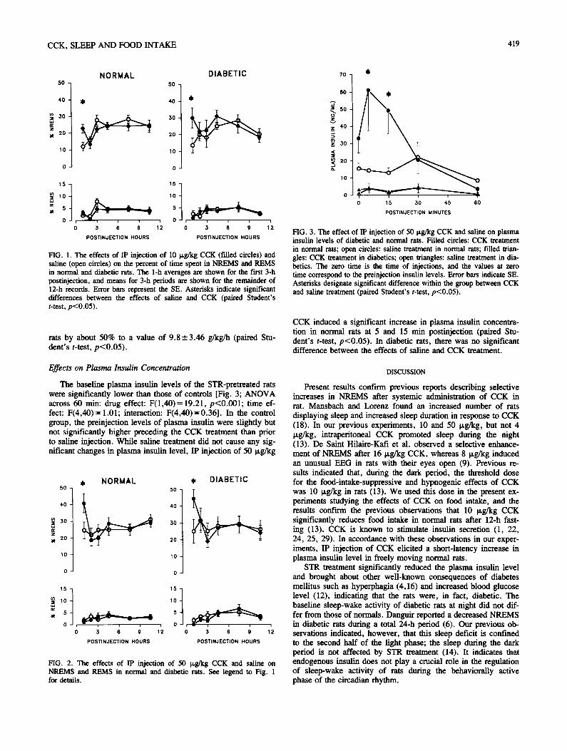
CCK, SLEEP AND FOOD INTAKE 419
50 N O R M A L
50
DIABETIC
4-0
.,~ 30
z 2O
10
4O
20
10
0
$
15 15 1
0 , , , , , 0 ~
0 3 6 9 12 0 3 6 9
POSTINJECTION HOURS POSTINJECTION HOURS
i
1 2
FIG. 1. The effects of IP injection of 10 p,g/kg CCK (t'dled circles) and saline (open circles) on the percent of time spent in NREMS and REMS in normal and diabetic rats. The 1-h averages are shown for the fh'st 3-h posfinjection, and means for 3-h periods are shown for the remainder of 12-h records. Error bars represent the SE. Asterisks indicate significant differences between the effects of saline and CCK (paired Student's t-test, p<0.05).
rats by about 50% to a value of 9.8-+ 3.46 g/kg/h (paired Stu- dent's t-test, p<O.05).
Effects on Plasma Insulin Concentration
The baseline plasma insulin levels of the STR-pretreated rats were significantly lower than those of controls [Fig. 3; ANOVA across 60 min: drug effect: F(1,40)= 19.21, p<0.001; time ef- fect: F(4,40)= 1.01; interaction: F(4,40)=0.36]. In the control group, the preinjection levels of plasma insulin were slightly but not significantly higher preceding the CCK treatment than prior to saline injection. While saline treatment did not cause any sig- nificant changes in plasma insulin level, IP injection of 50 p,g/kg
5 0
4 0
.~ 3 O
2 O
1 0
, NORMAL 50
3 O
2 0
1 0
0
$ DIABETIC
5 51 ' =. lo ] lo
5 ~ 5
0 , , , 0 , 0 3 6 9 12 0 3 6 9
POSTINJECTION HOURS POSTINJECTION HOURS
i
1 2
FIG. 2. The effects of IP injection of 50 p,g/kg CCK and saline on NREMS and REMS in normal and diabetic rats. See legend to Fig. 1 for details.
7o $
6O
.J ~. 5o
z 417 ...1
z 3 0
~ 2o o.
10
N"
i i T
0 15 30 4.5 60
POSTINJECTION MINUTES
FIG. 3. The effect of IP injection of 50 p.g/kg CCK and saline on plasma insulin levels of diabetic and normal rats. Filled circles: CCK treatment in normal rats; open circles: saline treatment in normal rats; filled trian- gles: CCK treatment in diabetics; open triangles: saline treatment in dia- betics. The zero time is the time of injections, and the values at zero time correspond to the preinjection insulin levels. Error bars indicate SE. Asterisks designate significant difference within the group between CCK and saline treatment (paired Student's t-test, p<0.05).
CCK induced a significant increase in plasma insulin concentra- tion in normal rats at 5 and 15 min postinjection (paired Stu- dent's t-test, p<0.05). In diabetic rats, there was no significant difference between the effects of saline and CCK treatment.
D I S C U S S I O N
Present results confirm previous reports describing selective increases in NREMS after systemic administration of CCK in rat. Mansbach and Lorenz found an increased number of rats displaying sleep and increased sleep duration in response to CCK (18). In our previous experiments, 10 and 50 p,g/kg, but not 4 p,g/kg, intraperitoneal CCK promoted sleep during the night (13). De Saint Hilaire-Kafi et al. observed a selective enhance- ment of NREMS after 16 p,g/kg CCK, whereas 8 p,g/kg induced an unusual EEG in rats with their eyes open (9). Previous re- suits indicated that, during the dark period, the threshold dose for the food-intake-suppressive and hypnogenic effects of CCK was 10 p,g/kg in rats (13). We used this dose in the present ex- periments studying the effects of CCK on food intake, and the results confirm the previous observations that 10 p,g/kg CCK significantly reduces food intake in normal rats after 12-h fast- ing (13). CCK is known to stimulate insulin secretion (1, 22, 24, 25, 29). In accordance with these observations in our exper- iments, IP injection of CCK elicited a short-latency increase in plasma insulin level in freely moving normal rats.
STR treatment significantly reduced the plasma insulin level and brought about other well-known consequences of diabetes mellitus such as hyperphagia (4,16) and increased blood glucose level (12), indicating that the rats were, in fact, diabetic. The baseline sleep-wake activity of diabetic rats at night did not dif- fer from those of normals. Danguir reported a decreased NREMS in diabetic rats during a total 24-h period (6). Our previous ob- servations indicated, however, that this sleep deficit is confined to the second half of the light phase; the sleep during the dark period is not affected by STR treatment (14). It indicates that endogenous insulin does not play a crucial role in the regulation of sleep-wake activity of rats during the behaviorally active phase of the circadian rhythm.

420 KAP/kS ET AL.
In previous reports in which CCK seemed to be particularly effective in promoting sleep, the rats were also provided food (13,18), and though food intake decreased after CCK treatment, it was not totally inhibited. It was speculated, therefore, that ad- ditional factors closely linked to eating might be involved in the sleep-promoting actions of CCK. Since food intake is followed by insulin release, a hormone that promotes sleep (7, 8, 23), and CCK stimulates insulin secretion, the involvement of insulin in the mediation of sleep-promoting action of CCK was postu- lated (19). According to the present results, however, this possi- bility can be ruled out, since, as in normal rats, CCK increased NREMS in diabetic rats. There was no effect on REMS. Al- though an increased [3-cell responsiveness was reported to a CCK analogue, caerulein, in perifused pancreas of diabetic rats in vitro (21), we did not find any significant insulin response in vivo in diabetic animals even after a relatively high dose (50 v,g/kg) of CCK. As in normal rats, 10 Ixg/kg CCK significantly suppressed the 1-h food intake in diabetic animals. This finding is in accordance with that of Vanderweele, who also reported food-intake-suppressive effects of CCK in STR-diabetic rats (26). The findings that CCK increases NREMS and decreases
food intake in diabetic rats without stimulating pancreatic insu- lin release indicate that plasma insulin does not mediate the hypnogenic and food-intake-suppressive effects of CCK.
A similar conclusion was drawn recently concerning the hyp- nogenic effects of another sleep factor, intedeukin 1 (IL-1). Among its biological activities, IL-1 suppresses feeding (17), enhances NREMS (15) and alters insulin release (3). Originally, we hypothesized that insulin might contribute to the hypnogenic effects of IL-1. STR pretreatment of rats, however, failed to al- ter the hypnogenic activity of IL-1, indicating that its hypno- genic effect is independent from pancreatic insulin (14).
ACKNOWLEDGEMENTS
The authors would like to thank Dr. M. Opp for reviewing the manuscript and Dr. L. Johannsen for providing his expertise in RIA. The technical assistance of Ms. Klfira Ormos, Ms. Ilona Ponics~in, Ms. Gail Richmond, Ms. Donna Maxwell and Mr. P. Liszli is gratefully ac- knowledged. This work was supported by the Hungarian Academy of Sciences (OTKA 1104, OKKFT 177/86), Hungarian Ministry of Health (ETT 528) and the National Institutes of Health (USA) NS-27250.
REFERENCES
1. Ahren, B.; Lundquist, I. Effects of two cholecystokinin variants, CCK-39 and CCK-8, on basal and stimulated insulin secretion. Acta Diabetol. Lat. 18:345-356; 1981.
2. Antin, J.; Gibbs, J.; Holt, J.; Young, R. C.; Smith, G. P. Chole- cystokinin elicits the complete behavioral sequence of satiety in rats. J. Comp. Physiol. Psychol. 89:784-790; 1975.
3. Besedovsky, H. O.; del Rey, A. Immune-neuroendocrine communi- cation: Key role of interleukin-1. In: Hadden, J. W.; Masek, K.; Nistico, G., eds. Interactions among CNS, neuroendocrine and im- mune systems. Rome: Pythagore Press; 1989:191-197.
4. Booth, D. A. Some characteristics of feeding during streptozotocin- induced diabetes in the rat. J. Comp. Psychol. Physiol. 80:238-249; 1972.
5. Borb61y, A. A.; Tobler, I. Endogenous sleep-promoting substances and sleep regulation. Physiol. Rev. 69:605-670; 1989.
6. Danguir, J. Sleep deficits in diabetic rats: Restoration following chronic intravenous or intracerebroventricular infusions of insulin. Brain Res. Bull. 12:641--645; 1984.
7. Danguir, J.; Nicolaldis, S. Circadian sleep and feeding patterns in the rat: Possible dependence on lipogenesis and lipolysis. Am. J. Physiol. 238:E223-E230; 1980.
8. Danguir, J.; Nicolaidis, S. Chronic intracerebroventricular infusion of insulin causes selective increase of slow wave sleep in rats. Brain Res. 306:97-103; 1984.
9. de Saint Hilaire-Kafi, Z.; Depoortere, H.; Nicolaidis, S. Does cholecystokinin induce physiological satiety and sleep? Brain Res. 488:304-310; 1989.
10. Gibbs, J.; Young, R. C.; Smith, G. P. Cholecystokinin elicits sati- ety in rats with open gastric fistula. Nature 245:323-325; 1973.
11. Gibbs, J.; Young, R. C.; Smith, G. P. Cholecystokinin decreases food intake in rats. J. Comp. Physiol. Psychol. 84:488-495; 1973.
12. Junod, A.; Lambert, A. E.; Stauffacher, W.; Renold, A. E. Diabe- togenic action of streptozotocin: Relationship of dose to metabolic response. J. Clin. Invest. 48:2129-2139; 1969.
13. Kapfis, L.; Obfil, F. Jr.; Alf61di, P.; Rubicsek, G.; Penke, B.; Obfil, F. Effects of nocturnal intraperitoneal administration of cholecysto- kinin in rats: Simultaneous increase in sleep, increase in EEG slow- wave activity, reduction of motor activity, suppression of eating, and decrease in brain temperature. Brain Res. 438:155-164; 1988.
14. Kap~ts, L.; Payne, L.; Ob~il, F. Jr.; Opp, M.; Johannsen, L.; Krueger, J. M. Sleep in diabetic rats: Effects of interleukin-1. Am. J. Physiol. 260:R995-R999, 1991.
15. Krueger, J. M.; Walter, J.; Dinarello, C. A.; Wolff, S. M.; Che-
did, L. Sleep-promoting effects of endogenous pyrogen (interleukin- 1). Am. J. Physiol. 246:R994-R999; 1984.
16. Kumaresan, P.; Turner, C. W. Effect of alloxan on food consump- tion in rats. Proc. Soc. Exp. Biol. Med. 119:400--402; 1965.
17. McCarthy, D. O.; Klueger, M. J.; Vander, A. J. Suppression of food intake during infection: Is interleuldn-1 involved? Am. J. Clin. Nutr. 42:1179-1182; 1985.
18. Mansbach, R. S.; Lorenz, D. N. Cholecystokinin (CCK-8) elicits prandial sleep in rats. Physiol. Behav. 30:179-183; 1983.
19. Ob~il, F. Jr.; Opp, M.; Siiry, G.; Krueger, J. M. Endocrine mecha- nisms in sleep regulation. In: Inou6, S.; Krueger, J. M., eds. En- dogenous sleep factors. The Hague: SPB Academic Publishing; 1990:109-120.
20. Obfil, F. Jr.; Alf61di, P.; Cady, A. B.; Johannsen, L.; S~iry, G.; Krueger, J. M. Growth hormone-releasing factor enhances sleep in rats and rabbits. Am. J. Physiol. 255:R310--R316; 1988.
21. Okabayashi, Y.; Otsuki, M.; Ohki, A.; Tani, S.; Baba, S. Increased 13-cell secretory responsiveness to cemletide and TPA in streptozo- cin-induced mildly diabetic rats. Diabetes 38:1042-1047; 1989.
22. Pederson, R. A.; Brown, J. C. Effect of cholecystokinin, secretin, and gastric inhibitor polypeptide on insulin release from the isolated perfnsed rat pancreas. Can. J. Physiol. Pharmacol. 57:1233-1237; 1979.
23. Sangiah, S.; Caldwell, D. F.; Villeneuve, M. J.; Clancy, J. J. Sleep: Sequential reduction of paradoxical (REM) and elevation of slow-wave (NREM) sleep by a non-convulsive dose of insulin in rats. Life Sci. 31:763-769; 1982.
24. Szec6wka, J.; Lins, P. E.; Efendic, S. Effects of cholecystokinin, gastric inhibitory polypeptide, and secretin on insulin and glucagon secretion in rats. Endocrinology 110:1268-1272; 1982.
25. Unger, R. H.; Ketterer, H.; Dupre, J.; Eisentraut, A. M. The ef- fects of secretin, pancreozymin, and gastrin on insulin and glucagon secretion in anesthetized dogs. J. Clin. Invest. 46:630-645; 1967.
26. Vanderweele, D. A. CCK, endogenous insulin condition and satiety in free-fed rats. Physiol. Behav. 29:961-964; 1982.
27. Woods, S. C.; Porte, D. Jr. The role of insulin as a satiety factor in the central nervous system. Adv. Metab. Dis. 10:457-468; 1983.
28. Woods, S. C.; Gibbs, J. The regulation of food intake by peptides. Ann. NY Acad. Sci. 575:236--243; 1989.
29. Zawalich, W. S.; Cote, S. B.; Diaz, V. A. Influence of cholecysto- kinin on insulin output from isolated perifused pancreatic islets. En- docrinology 119:616-621; 1986.
P a g e | 75
Attachment 5.
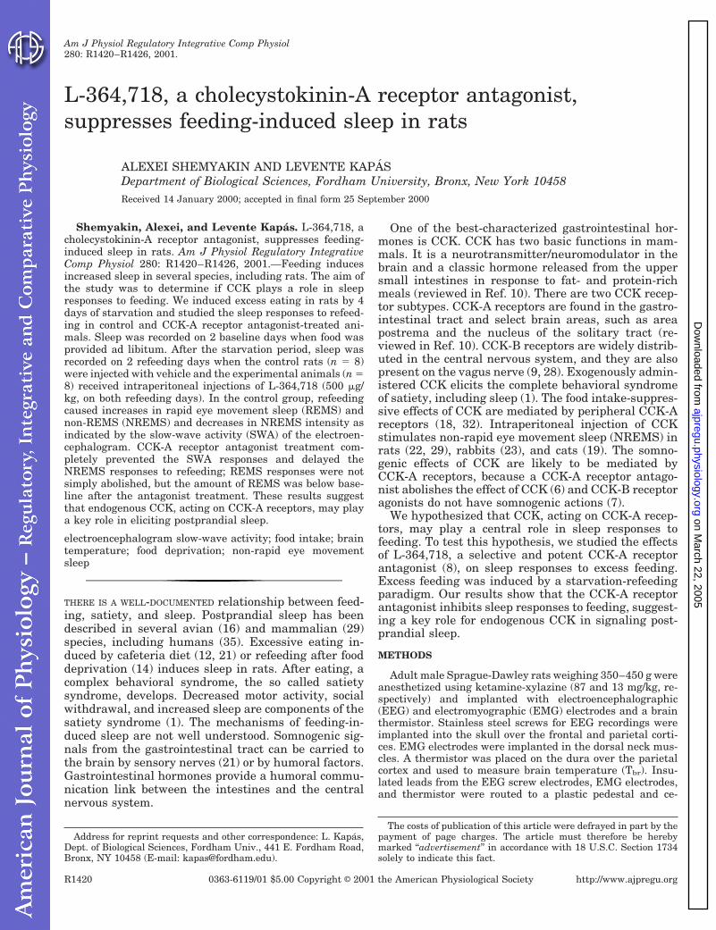
L-364,718, a cholecystokinin-A receptor antagonist,suppresses feeding-induced sleep in rats
ALEXEI SHEMYAKIN AND LEVENTE KAPASDepartment of Biological Sciences, Fordham University, Bronx, New York 10458Received 14 January 2000; accepted in final form 25 September 2000
Shemyakin, Alexei, and Levente Kapas. L-364,718, acholecystokinin-A receptor antagonist, suppresses feeding-induced sleep in rats. Am J Physiol Regulatory IntegrativeComp Physiol 280: R1420–R1426, 2001.—Feeding inducesincreased sleep in several species, including rats. The aim ofthe study was to determine if CCK plays a role in sleepresponses to feeding. We induced excess eating in rats by 4days of starvation and studied the sleep responses to refeed-ing in control and CCK-A receptor antagonist-treated ani-mals. Sleep was recorded on 2 baseline days when food wasprovided ad libitum. After the starvation period, sleep wasrecorded on 2 refeeding days when the control rats (n 5 8)were injected with vehicle and the experimental animals (n 58) received intraperitoneal injections of L-364,718 (500 mg/kg, on both refeeding days). In the control group, refeedingcaused increases in rapid eye movement sleep (REMS) andnon-REMS (NREMS) and decreases in NREMS intensity asindicated by the slow-wave activity (SWA) of the electroen-cephalogram. CCK-A receptor antagonist treatment com-pletely prevented the SWA responses and delayed theNREMS responses to refeeding; REMS responses were notsimply abolished, but the amount of REMS was below base-line after the antagonist treatment. These results suggestthat endogenous CCK, acting on CCK-A receptors, may playa key role in eliciting postprandial sleep.
electroencephalogram slow-wave activity; food intake; braintemperature; food deprivation; non-rapid eye movementsleep
THERE IS A WELL-DOCUMENTED relationship between feed-ing, satiety, and sleep. Postprandial sleep has beendescribed in several avian (16) and mammalian (29)species, including humans (35). Excessive eating in-duced by cafeteria diet (12, 21) or refeeding after fooddeprivation (14) induces sleep in rats. After eating, acomplex behavioral syndrome, the so called satietysyndrome, develops. Decreased motor activity, socialwithdrawal, and increased sleep are components of thesatiety syndrome (1). The mechanisms of feeding-in-duced sleep are not well understood. Somnogenic sig-nals from the gastrointestinal tract can be carried tothe brain by sensory nerves (21) or by humoral factors.Gastrointestinal hormones provide a humoral commu-nication link between the intestines and the centralnervous system.
One of the best-characterized gastrointestinal hor-mones is CCK. CCK has two basic functions in mam-mals. It is a neurotransmitter/neuromodulator in thebrain and a classic hormone released from the uppersmall intestines in response to fat- and protein-richmeals (reviewed in Ref. 10). There are two CCK recep-tor subtypes. CCK-A receptors are found in the gastro-intestinal tract and select brain areas, such as areapostrema and the nucleus of the solitary tract (re-viewed in Ref. 10). CCK-B receptors are widely distrib-uted in the central nervous system, and they are alsopresent on the vagus nerve (9, 28). Exogenously admin-istered CCK elicits the complete behavioral syndromeof satiety, including sleep (1). The food intake-suppres-sive effects of CCK are mediated by peripheral CCK-Areceptors (18, 32). Intraperitoneal injection of CCKstimulates non-rapid eye movement sleep (NREMS) inrats (22, 29), rabbits (23), and cats (19). The somno-genic effects of CCK are likely to be mediated byCCK-A receptors, because a CCK-A receptor antago-nist abolishes the effect of CCK (6) and CCK-B receptoragonists do not have somnogenic actions (7).
We hypothesized that CCK, acting on CCK-A recep-tors, may play a central role in sleep responses tofeeding. To test this hypothesis, we studied the effectsof L-364,718, a selective and potent CCK-A receptorantagonist (8), on sleep responses to excess feeding.Excess feeding was induced by a starvation-refeedingparadigm. Our results show that the CCK-A receptorantagonist inhibits sleep responses to feeding, suggest-ing a key role for endogenous CCK in signaling post-prandial sleep.
METHODS
Adult male Sprague-Dawley rats weighing 350–450 g wereanesthetized using ketamine-xylazine (87 and 13 mg/kg, re-spectively) and implanted with electroencephalographic(EEG) and electromyographic (EMG) electrodes and a brainthermistor. Stainless steel screws for EEG recordings wereimplanted into the skull over the frontal and parietal corti-ces. EMG electrodes were implanted in the dorsal neck mus-cles. A thermistor was placed on the dura over the parietalcortex and used to measure brain temperature (Tbr). Insu-lated leads from the EEG screw electrodes, EMG electrodes,and thermistor were routed to a plastic pedestal and ce-
Address for reprint requests and other correspondence: L. Kapas,Dept. of Biological Sciences, Fordham Univ., 441 E. Fordham Road,Bronx, NY 10458 (E-mail: [email protected]).
The costs of publication of this article were defrayed in part by thepayment of page charges. The article must therefore be herebymarked ‘‘advertisement’’ in accordance with 18 U.S.C. Section 1734solely to indicate this fact.
Am J Physiol Regulatory Integrative Comp Physiol280: R1420–R1426, 2001.
0363-6119/01 $5.00 Copyright © 2001 the American Physiological Society http://www.ajpregu.orgR1420
on March 22, 2005
ajpregu.physiology.orgD
ownloaded from
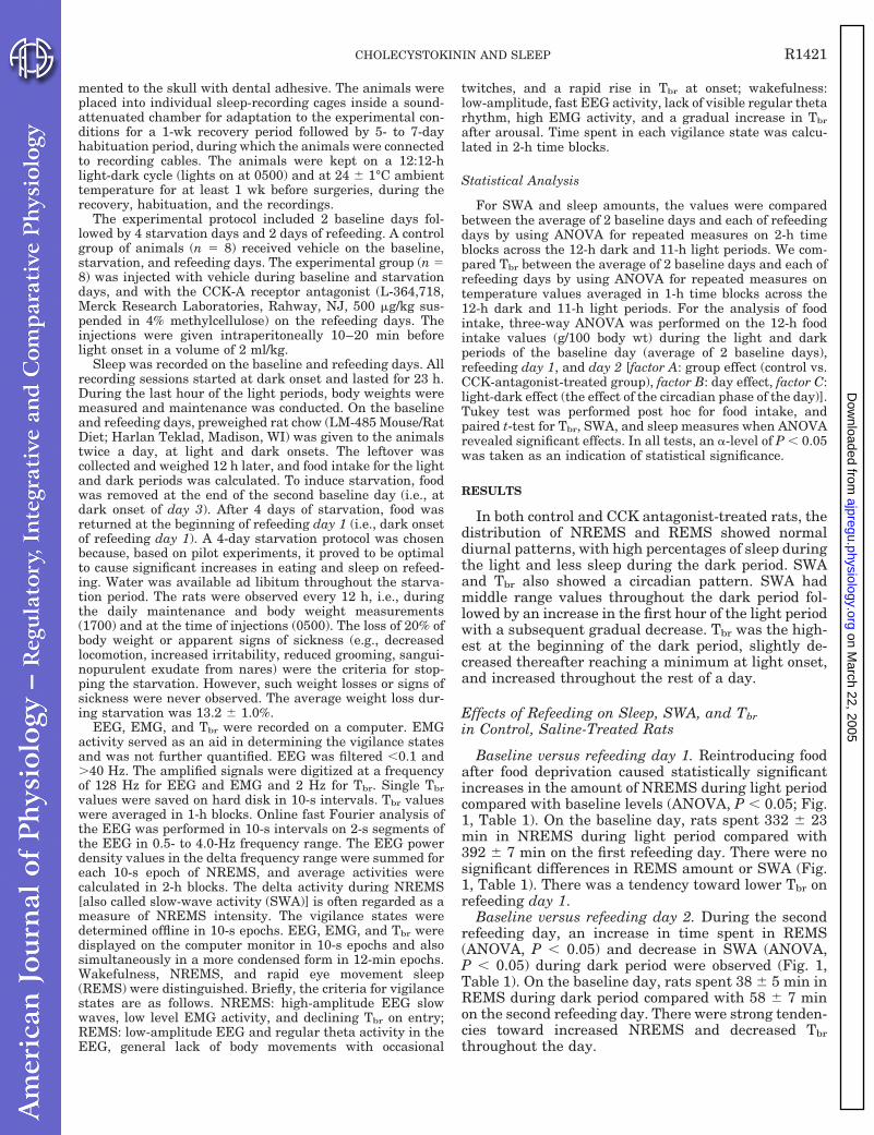
mented to the skull with dental adhesive. The animals wereplaced into individual sleep-recording cages inside a sound-attenuated chamber for adaptation to the experimental con-ditions for a 1-wk recovery period followed by 5- to 7-dayhabituation period, during which the animals were connectedto recording cables. The animals were kept on a 12:12-hlight-dark cycle (lights on at 0500) and at 24 6 1°C ambienttemperature for at least 1 wk before surgeries, during therecovery, habituation, and the recordings.
The experimental protocol included 2 baseline days fol-lowed by 4 starvation days and 2 days of refeeding. A controlgroup of animals (n 5 8) received vehicle on the baseline,starvation, and refeeding days. The experimental group (n 58) was injected with vehicle during baseline and starvationdays, and with the CCK-A receptor antagonist (L-364,718,Merck Research Laboratories, Rahway, NJ, 500 mg/kg sus-pended in 4% methylcellulose) on the refeeding days. Theinjections were given intraperitoneally 10–20 min beforelight onset in a volume of 2 ml/kg.
Sleep was recorded on the baseline and refeeding days. Allrecording sessions started at dark onset and lasted for 23 h.During the last hour of the light periods, body weights weremeasured and maintenance was conducted. On the baselineand refeeding days, preweighed rat chow (LM-485 Mouse/RatDiet; Harlan Teklad, Madison, WI) was given to the animalstwice a day, at light and dark onsets. The leftover wascollected and weighed 12 h later, and food intake for the lightand dark periods was calculated. To induce starvation, foodwas removed at the end of the second baseline day (i.e., atdark onset of day 3). After 4 days of starvation, food wasreturned at the beginning of refeeding day 1 (i.e., dark onsetof refeeding day 1). A 4-day starvation protocol was chosenbecause, based on pilot experiments, it proved to be optimalto cause significant increases in eating and sleep on refeed-ing. Water was available ad libitum throughout the starva-tion period. The rats were observed every 12 h, i.e., duringthe daily maintenance and body weight measurements(1700) and at the time of injections (0500). The loss of 20% ofbody weight or apparent signs of sickness (e.g., decreasedlocomotion, increased irritability, reduced grooming, sangui-nopurulent exudate from nares) were the criteria for stop-ping the starvation. However, such weight losses or signs ofsickness were never observed. The average weight loss dur-ing starvation was 13.2 6 1.0%.
EEG, EMG, and Tbr were recorded on a computer. EMGactivity served as an aid in determining the vigilance statesand was not further quantified. EEG was filtered ,0.1 and.40 Hz. The amplified signals were digitized at a frequencyof 128 Hz for EEG and EMG and 2 Hz for Tbr. Single Tbr
values were saved on hard disk in 10-s intervals. Tbr valueswere averaged in 1-h blocks. Online fast Fourier analysis ofthe EEG was performed in 10-s intervals on 2-s segments ofthe EEG in 0.5- to 4.0-Hz frequency range. The EEG powerdensity values in the delta frequency range were summed foreach 10-s epoch of NREMS, and average activities werecalculated in 2-h blocks. The delta activity during NREMS[also called slow-wave activity (SWA)] is often regarded as ameasure of NREMS intensity. The vigilance states weredetermined offline in 10-s epochs. EEG, EMG, and Tbr weredisplayed on the computer monitor in 10-s epochs and alsosimultaneously in a more condensed form in 12-min epochs.Wakefulness, NREMS, and rapid eye movement sleep(REMS) were distinguished. Briefly, the criteria for vigilancestates are as follows. NREMS: high-amplitude EEG slowwaves, low level EMG activity, and declining Tbr on entry;REMS: low-amplitude EEG and regular theta activity in theEEG, general lack of body movements with occasional
twitches, and a rapid rise in Tbr at onset; wakefulness:low-amplitude, fast EEG activity, lack of visible regular thetarhythm, high EMG activity, and a gradual increase in Tbr
after arousal. Time spent in each vigilance state was calcu-lated in 2-h time blocks.
Statistical Analysis
For SWA and sleep amounts, the values were comparedbetween the average of 2 baseline days and each of refeedingdays by using ANOVA for repeated measures on 2-h timeblocks across the 12-h dark and 11-h light periods. We com-pared Tbr between the average of 2 baseline days and each ofrefeeding days by using ANOVA for repeated measures ontemperature values averaged in 1-h time blocks across the12-h dark and 11-h light periods. For the analysis of foodintake, three-way ANOVA was performed on the 12-h foodintake values (g/100 body wt) during the light and darkperiods of the baseline day (average of 2 baseline days),refeeding day 1, and day 2 [factor A: group effect (control vs.CCK-antagonist-treated group), factor B: day effect, factor C:light-dark effect (the effect of the circadian phase of the day)].Tukey test was performed post hoc for food intake, andpaired t-test for Tbr, SWA, and sleep measures when ANOVArevealed significant effects. In all tests, an a-level of P , 0.05was taken as an indication of statistical significance.
RESULTS
In both control and CCK antagonist-treated rats, thedistribution of NREMS and REMS showed normaldiurnal patterns, with high percentages of sleep duringthe light and less sleep during the dark period. SWAand Tbr also showed a circadian pattern. SWA hadmiddle range values throughout the dark period fol-lowed by an increase in the first hour of the light periodwith a subsequent gradual decrease. Tbr was the high-est at the beginning of the dark period, slightly de-creased thereafter reaching a minimum at light onset,and increased throughout the rest of a day.
Effects of Refeeding on Sleep, SWA, and Tbr
in Control, Saline-Treated Rats
Baseline versus refeeding day 1. Reintroducing foodafter food deprivation caused statistically significantincreases in the amount of NREMS during light periodcompared with baseline levels (ANOVA, P , 0.05; Fig.1, Table 1). On the baseline day, rats spent 332 6 23min in NREMS during light period compared with392 6 7 min on the first refeeding day. There were nosignificant differences in REMS amount or SWA (Fig.1, Table 1). There was a tendency toward lower Tbr onrefeeding day 1.
Baseline versus refeeding day 2. During the secondrefeeding day, an increase in time spent in REMS(ANOVA, P , 0.05) and decrease in SWA (ANOVA,P , 0.05) during dark period were observed (Fig. 1,Table 1). On the baseline day, rats spent 38 6 5 min inREMS during dark period compared with 58 6 7 minon the second refeeding day. There were strong tenden-cies toward increased NREMS and decreased Tbr
throughout the day.
R1421CHOLECYSTOKININ AND SLEEP
on March 22, 2005
ajpregu.physiology.orgD
ownloaded from
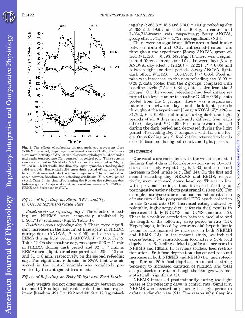
Effects of Refeeding on Sleep, SWA, and Tbrin CCK Antagonist-Treated Rats
Baseline versus refeeding day 1. The effects of refeed-ing on NREMS were completely abolished byL-364,718 treatment (Fig. 2, Table 1).
Baseline versus refeeding day 2. There were signifi-cant increases in the amount of time spent in NREMSduring dark (ANOVA, P , 0.05) and decreases inREMS during light period (ANOVA, P , 0.05; Fig. 2,Table 1). On the baseline day, rats spent 206 6 11 minin NREMS during dark period and 92 6 7 min inREMS during light period compared with 239 6 13 minand 81 6 8 min, respectively, on the second refeedingday. The significant reduction in SWA that was ob-served in the control animals was completely pre-vented by the antagonist treatment.
Effects of Refeeding on Body Weight and Food Intake
Body weights did not differ significantly between con-trol and CCK antagonist-treated rats throughout exper-iment [baseline: 421.7 6 19.2 and 435.9 6 12.0 g; refeed-
ing day 1: 363.3 6 18.6 and 374.0 6 10.0 g; refeeding day2: 393.2 6 19.9 and 414.4 6 10.8 g, in control andL-364,718-treated rats, respectively; 2-way ANOVA,group effect: F(1,95) 5 1.792, not significant (NS)].
There were no significant differences in food intakebetween control and CCK antagonist-treated ratsthroughout the experiment [3-way ANOVA, group ef-fect: F(1,126) 5 0.298, NS; Fig. 3]. There was a signif-icant difference in consumed food between days [3-wayANOVA, day effect: F(2,126) 5 12.221, P , 0.05] andbetween light and dark periods [3-way ANOVA, light-dark effect: F(1,126) 5 1064.353, P , 0.05]. Food in-take was increased on the first refeeding day (8.99 60.26 g, data pooled from the 2 groups) compared withbaseline levels (7.54 6 0.34 g, data pooled from the 2groups). On the second refeeding day, food intake re-turned to a level similar to baseline (7.29 6 0.36 g, datapooled from the 2 groups). There was a significantinteraction between days and dark-light periodsthroughout the experiment [3-way ANOVA: F(2,126) 531.792, P , 0.05]: food intake during dark and lightperiods of all 3 days significantly differed from eachother (Tukey test, P , 0.05). Food intake was increasedduring the dark period and decreased during the lightperiod of refeeding day 1 compared with baseline lev-els. On refeeding day 2, food intake returned to levelsclose to baseline during both dark and light periods.
DISCUSSION
Our results are consistent with the well-documentedfindings that 4 days of food deprivation cause 10–15%decrease in body weight (e.g., Ref. 30) and a reboundincrease in food intake (e.g., Ref. 14). On the first andsecond refeeding day, NREMS and REMS, respec-tively, were increased above baseline. This is in linewith previous findings that increased feeding orpostingestive satiety elicits postprandial sleep (29). Forexample, intragastric or intraduodenal administrationof nutrients elicits postprandial EEG synchronizationin rats (2) and cats (19). Increased eating induced bypalatable, high-energy diet (cafeteria diet) results inincreases of daily NREMS and REMS amounts (12).There is a positive correlation between meal size andthe length of the following sleep period in rats (15).Hyperphagia, induced by ventromedial hypothalamiclesion, is accompanied by increases in both NREMSand REMS (13). In the present study, we inducedexcess eating by reintroducing food after a 96-h fooddeprivation. Refeeding elicited significant increases inNREMS and REMS. In previous studies, food restitu-tion after a 96-h food deprivation also caused reboundincreases in both NREMS and REMS (14), and refeed-ing after an 80-h food deprivation caused a strongtendency to increased duration of NREMS and totalsleep episodes in rats, although the changes were notstatistically significant (3).
NREMS increased predominantly during the lightphase of the refeeding days in control rats. Similarly,NREMS was elevated only during the light period incafeteria diet-fed rats (21). The reason why sleep in-
Fig. 1. The effects of refeeding on non-rapid eye movement sleep(NREMS, circles), rapid eye movement sleep (REMS, triangles),slow-wave activity (SWA) of the electroencephalogram (diamonds),and brain temperature (Tbr, squares) in control rats. Time spent insleep is summed in 2-h blocks. SWA values are averaged in 2-h, Tbr
values in 1-h intervals. Baseline day: open symbols, refeeding day:solid symbols. Horizontal solid bars: dark period of the day. Errorbars: SE. Arrows indicate the time of injections. *Significant differ-ences between baseline and refeeding conditions (P , 0.05, pairedt-test). Time 0: the time of returning the food on the refeeding day.Refeeding after 4 days of starvation caused increases in NREMS andREMS and decreases in SWA.
R1422 CHOLECYSTOKININ AND SLEEP
on March 22, 2005
ajpregu.physiology.orgD
ownloaded from
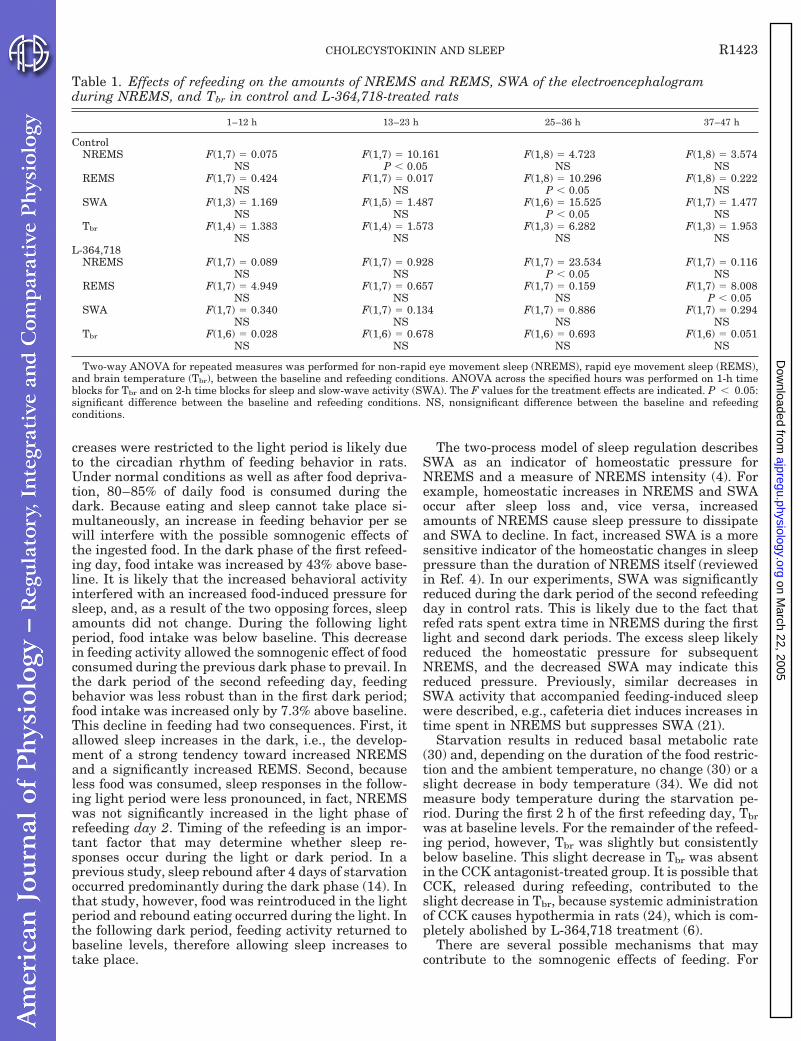
creases were restricted to the light period is likely dueto the circadian rhythm of feeding behavior in rats.Under normal conditions as well as after food depriva-tion, 80–85% of daily food is consumed during thedark. Because eating and sleep cannot take place si-multaneously, an increase in feeding behavior per sewill interfere with the possible somnogenic effects ofthe ingested food. In the dark phase of the first refeed-ing day, food intake was increased by 43% above base-line. It is likely that the increased behavioral activityinterfered with an increased food-induced pressure forsleep, and, as a result of the two opposing forces, sleepamounts did not change. During the following lightperiod, food intake was below baseline. This decreasein feeding activity allowed the somnogenic effect of foodconsumed during the previous dark phase to prevail. Inthe dark period of the second refeeding day, feedingbehavior was less robust than in the first dark period;food intake was increased only by 7.3% above baseline.This decline in feeding had two consequences. First, itallowed sleep increases in the dark, i.e., the develop-ment of a strong tendency toward increased NREMSand a significantly increased REMS. Second, becauseless food was consumed, sleep responses in the follow-ing light period were less pronounced, in fact, NREMSwas not significantly increased in the light phase ofrefeeding day 2. Timing of the refeeding is an impor-tant factor that may determine whether sleep re-sponses occur during the light or dark period. In aprevious study, sleep rebound after 4 days of starvationoccurred predominantly during the dark phase (14). Inthat study, however, food was reintroduced in the lightperiod and rebound eating occurred during the light. Inthe following dark period, feeding activity returned tobaseline levels, therefore allowing sleep increases totake place.
The two-process model of sleep regulation describesSWA as an indicator of homeostatic pressure forNREMS and a measure of NREMS intensity (4). Forexample, homeostatic increases in NREMS and SWAoccur after sleep loss and, vice versa, increasedamounts of NREMS cause sleep pressure to dissipateand SWA to decline. In fact, increased SWA is a moresensitive indicator of the homeostatic changes in sleeppressure than the duration of NREMS itself (reviewedin Ref. 4). In our experiments, SWA was significantlyreduced during the dark period of the second refeedingday in control rats. This is likely due to the fact thatrefed rats spent extra time in NREMS during the firstlight and second dark periods. The excess sleep likelyreduced the homeostatic pressure for subsequentNREMS, and the decreased SWA may indicate thisreduced pressure. Previously, similar decreases inSWA activity that accompanied feeding-induced sleepwere described, e.g., cafeteria diet induces increases intime spent in NREMS but suppresses SWA (21).
Starvation results in reduced basal metabolic rate(30) and, depending on the duration of the food restric-tion and the ambient temperature, no change (30) or aslight decrease in body temperature (34). We did notmeasure body temperature during the starvation pe-riod. During the first 2 h of the first refeeding day, Tbrwas at baseline levels. For the remainder of the refeed-ing period, however, Tbr was slightly but consistentlybelow baseline. This slight decrease in Tbr was absentin the CCK antagonist-treated group. It is possible thatCCK, released during refeeding, contributed to theslight decrease in Tbr, because systemic administrationof CCK causes hypothermia in rats (24), which is com-pletely abolished by L-364,718 treatment (6).
There are several possible mechanisms that maycontribute to the somnogenic effects of feeding. For
Table 1. Effects of refeeding on the amounts of NREMS and REMS, SWA of the electroencephalogramduring NREMS, and Tbr in control and L-364,718-treated rats
1–12 h 13–23 h 25–36 h 37–47 h
ControlNREMS F(1,7) 5 0.075 F(1,7) 5 10.161 F(1,8) 5 4.723 F(1,8) 5 3.574
NS P , 0.05 NS NSREMS F(1,7) 5 0.424 F(1,7) 5 0.017 F(1,8) 5 10.296 F(1,8) 5 0.222
NS NS P , 0.05 NSSWA F(1,3) 5 1.169 F(1,5) 5 1.487 F(1,6) 5 15.525 F(1,7) 5 1.477
NS NS P , 0.05 NSTbr F(1,4) 5 1.383 F(1,4) 5 1.573 F(1,3) 5 6.282 F(1,3) 5 1.953
NS NS NS NSL-364,718
NREMS F(1,7) 5 0.089 F(1,7) 5 0.928 F(1,7) 5 23.534 F(1,7) 5 0.116NS NS P , 0.05 NS
REMS F(1,7) 5 4.949 F(1,7) 5 0.657 F(1,7) 5 0.159 F(1,7) 5 8.008NS NS NS P , 0.05
SWA F(1,7) 5 0.340 F(1,7) 5 0.134 F(1,7) 5 0.886 F(1,7) 5 0.294NS NS NS NS
Tbr F(1,6) 5 0.028 F(1,6) 5 0.678 F(1,6) 5 0.693 F(1,6) 5 0.051NS NS NS NS
Two-way ANOVA for repeated measures was performed for non-rapid eye movement sleep (NREMS), rapid eye movement sleep (REMS),and brain temperature (Tbr), between the baseline and refeeding conditions. ANOVA across the specified hours was performed on 1-h timeblocks for Tbr and on 2-h time blocks for sleep and slow-wave activity (SWA). The F values for the treatment effects are indicated. P , 0.05:significant difference between the baseline and refeeding conditions. NS, nonsignificant difference between the baseline and refeedingconditions.
R1423CHOLECYSTOKININ AND SLEEP
on March 22, 2005
ajpregu.physiology.orgD
ownloaded from

example, the activation of gastrointestinal sensorynerves causes increases in NREMS (26). In fact, sub-diaphragmal dissection of the vagus nerve prevents thesomnogenic effects of cafeteria diet (21). Another pos-sibility is that increased sleep is a consequence ofmetabolic changes after eating (31). A third possibilityis that feeding-induced sleep is due to the release ofsomnogenic hormones from the gastrointestinal tract.One of the most studied somnogenic gastrointestinalhormones is CCK. Systemic injection of CCK causesthe complete behavioral sequence of satiety (1), includ-ing sleep (19, 22, 23, 29). We hypothesized that CCK,acting on CCK-A receptors, plays a key role in elicitingsleep responses to feeding.
L-364,718 was administered at light onset on bothrefeeding days. That is, the first injection of L-364,718was done 12 h after reintroducing the food. There aretwo reasons why we did not inject the antagonist im-mediately after the end of the starvation period. First,sleep responses to refeeding started only after a la-tency of 12 h in control rats. Second, L-364,718 itselfstimulates feeding in rats (32); delaying the injectionallowed the animals to eat according to their naturalneeds during the first dark period after starvation. Ourmajor finding is that L-364,718 completely abolishedthe NREMS increases on refeeding day 1. During thedark period of the following day, however, NREMS was
significantly increased in CCK antagonist-treated rats,a response that was present only as a tendency in thecontrol group. It is not likely that the observed effectsin the dark period are directly related to the action ofthe antagonist at that time, because the duration of theaction is shorter than 12 h. There are two explanationsfor this increase in NREMS. First, during the lightperiod of refeeding day 1, antagonist-injected animalsate ;50% more than animals in the control group.Increased feeding may have contributed to NREMSincreases in the subsequent dark period. Second, theNREMS increase seen in control rats during the lightperiod on refeeding day 1 was absent in CCK antago-nist-injected rats. It is possible that the antagonist-treated animals accumulated undischarged sleep pres-sure that was carried over to the dark period ofrefeeding day 2. Furthermore, in contrast to decreasedSWA in control rats, SWA in the antagonist-treatedanimals was not significantly suppressed in the darkperiod on refeeding day 2 likely because the antago-nist-treated rats did not accumulate excess sleep dur-ing refeeding day 1.
In summary, we found that the CCK-A receptorantagonist, L-364,718, suppresses NREMS and REMSresponses to feeding. This is consistent with the hy-pothesis that endogenous CCK plays a role in elicitingpostprandial sleep. L-364,718 acts on both central andperipheral CCK-A receptors. It is likely that the role ofperipherally located CCK-A receptors is more impor-tant for the somnogenic effects of feeding, because
Fig. 2. The effects of refeeding on NREMS, REMS, SWA, and Tbr inL-364,718-treated rats. Symbols same as in Fig. 1. L-364,718 com-pletely abolished the REMS and SWA responses to refeeding and theNREMS-promoting effects of refeeding during the light periods of therefeeding days. *Significant differences between baseline and refeed-ing conditions (P , 0.05, paired t-test).
Fig. 3. Food intake under baseline conditions and on refeeding days1 and 2 after food deprivation in control and L-364,718-treated rats.Solid bars: food intake during the dark period; open bars: food intakeduring the light period. Top: food intake in control rats. Bottom: foodintake in L-364,718-treated rats. Food intake is expressed as theamount of food consumed per 100 g body wt. Error bars: SE. Three-way ANOVA indicated that there is no significant difference betweenthe food intake of the control and L-364,718-treated rats. The circa-dian period (light-dark effect) and the refeeding (day effect) havesignificant effects on food intake. See RESULTS for details.
R1424 CHOLECYSTOKININ AND SLEEP
on March 22, 2005
ajpregu.physiology.orgD
ownloaded from

intracerebroventricular injection of CCK does not elicitchanges in sleep (20).
Perspectives
Although sleep is generated by the brain, there areseveral factors arising from outside the brain thatcontribute to the regulation of sleep. For example,somatosensory stimuli, ambient temperature, systemicinfections, and feeding all affect sleep. There are signalmechanisms from the periphery that stimulate or in-hibit somnogenic brain structures according to somaticneeds. Hormones and various neurotransmitters arepart of these signaling mechanisms. For example, cy-tokines have a key role in triggering sleep responsesduring systemic infections (reviewed in Ref. 25). Nitricoxide may have a role in signaling homeostatic needsfor sleep (33). Cytokines and CCK have overlappingbiological activities and have mutual stimulatory ef-fects on each other’s secretion or function. For example,administration of interleukin (IL)-1 increases CCKplasma levels (17, 27), IL-1 sensitizes peripheral vagusafferents to the effects of CCK (5); the effects of IL-1 onthe vagus (27) and on food intake (17) are suppressedby CCK-A receptor antagonists; and CCK stimulatesthe production of IL-1, tumor necrosis factor-a, andgranulocyte/monocyte-colony-stimulating factor frommonocytes (11). Increased feeding stimulates the secre-tion of CCK as well as the expression of IL-1b mRNA inthe liver and the brain. It is possible that CCK andcytokines form an intertwined humoral network toinduce sleep in response to eating and infections orunder other conditions.
L-364,718 was a generous gift by Dr. William Henckler (MerckSharp and Dohme).
This work was supported by National Institutes of Health GrantNS-30514.
REFERENCES
1. Antin J, Gibbs J, Holt J, Young RC, and Smith GP. Chole-cystokinin elicits the complete behavioral sequence of satiety inrats. J Comp Physiol Psychol 89: 784–790, 1975.
2. Bernstein IL. Post-prandial EEG synchronization in normaland hypothalamically lesioned rats. Physiol Behav 12: 525–545,1974.
3. Borbely AA. Sleep in the rat during food deprivation and sub-sequent restitution of food. Brain Res 124: 457–471, 1977.
4. Borbely AA. Sleep homeostasis and models of sleep regulation.In: Principles and Practice of Sleep Medicine, edited by KrygerMH, Roth T, and Dement WC. Philadelphia, PA: Saunders, 1994,p. 309–320.
5. Bucinskaite V, Kurosawa M, Miyasaka K, Funakoshi A,and Lundeberg T. Interleukin-1b sensitizes the response of thegastric vagal afferent to cholecystokinin in rat. Neurosci Lett229: 33–36, 1997.
6. Chang H-Y and Kapas L. L-364,718, a cholecystokinin (CCK)-A receptor antagonist, inhibits the sleep-inducing effects of CCK(Abstract). Sleep Res 26: 138, 1997.
7. Chang H-Y and Kapas L. Selective activation of CCK-B recep-tors does not induce sleep and does not affect EEG slow-waveactivity and brain temperature in rats. Physiol Behav 62: 175–179, 1997.
8. Chang RSL and Lotti VJ. Biochemical and pharmacologicalcharacterization of an extremely potent and selective nonpeptidecholecystokinin antagonist. Proc Natl Acad Sci USA 83: 4923–4926, 1986.
9. Corp ES, McQuade J, Moran TH, and Smith GP. Charac-terization of type A and type B CCK receptor binding sites in ratvagus nerve. Brain Res 623: 161–166, 1993.
10. Crawley JN and Corwin RL. Biological actions of cholecysto-kinin. Peptides 15: 731–755, 1994.
11. Cunningham ME, Shaw-Stiffel TA, Bernstein LH, Tin-ghitella TJ, Claus RE, Brogan DA, and McMillen MA.Cholecystokinin-stimulated monocytes produce inflammatorycytokines and eicosanoids. Am J Gastroenterol 90: 621–626,1995.
12. Danguir J. Cafeteria diet promotes sleep in rats. Appetite 8:49–53, 1987.
13. Danguir J and Nicolaidis S. Sleep and feeding patterns in theventromedial hypothalamic lesioned rat. Physiol Behav 21: 769–777, 1978.
14. Danguir J and Nicolaidis S. Dependence of sleep on nutrients’availability. Physiol Behav 22: 735–740, 1979.
15. Danguir J, Nikolaidis S, and Gerard H. Relations betweenfeeding and sleep patterns in the rat. J Comp Physiol Psychol 93:820–830, 1979.
16. Dario AJ, Lopes PR, Freitas CG, Paschoalini MA, andMarino-Neto J. Electrographic patterns of postprandial sleepafter food deprivation or intraventricular adrenaline injectionsin pigeons. Brain Res Bull 39: 249–254, 1996.
17. Daun JM and McCarthy DO. The role of cholecystokinin ininterleukin-1-induced anorexia. Physiol Behav 54: 237–241,1993.
18. Dourish CT, Ruckert AC, Tattersall FD, and Iversen SD.Evidence that decreased feeding induced by systemic injection ofcholecystokinin is mediated by CCK-A receptors. Eur J Pharma-col 173: 233–234, 1989.
19. Fara JW, Rubinstein EH, and Sonnenschein RR. Visceraland behavioral responses to intraduodenal fat. Science 166:110–111, 1969.
20. Gilligan JG, Milanes L, Chang H-Y, Ribeiro AC, and KapasL. Intracerebroventricular injection of cholecystokinin in ratsdoes not elicit changes in sleep or brain temperature (Abstract).Sleep 21, Suppl 3: 30, 1998.
21. Hansen MK, Kapas L, Fang J, and Krueger JM. Cafeteriadiet-induced sleep is blocked by subdiaphragmatic vagotomy inrats. Am J Physiol Regulatory Integrative Comp Physiol 274:R168–R174, 1998.
22. Kapas L, Obal F Jr, Alfoldi P, Rubicsek G, Penke B, andObal F. Effects of nocturnal intraperitoneal administration ofcholecystokinin in rats: simultaneous increase in sleep, increasein EEG slow-wave activity, reduction of motor activity, suppres-sion of eating, and decrease in brain temperature. Brain Res 438:155–164, 1988.
23. Kapas L, Obal F Jr, Opp MR, Johannsen L, and KruegerJM. Intraperitoneal injection of cholecystokinin elicits sleep inrabbits. Physiol Behav 50: 1241–1244, 1991.
24. Kapas L, Obal F Jr, Penke B, and Obal F. Cholecystokinin-octapeptide-induced hypothermia in rats: dose-effect and struc-ture-effect relationships, effect of ambient temperature, pharma-cological interactions and tolerance. Neuropharmacology 26:131–137, 1987.
25. Krueger JM, Obal F Jr, and Fang J. Humoral regulation ofphysiological sleep: cytokines and GHRH. J Sleep Res 8, Suppl 1:53–59, 1999.
26. Kukorelli T and Juhasz G. Sleep induced by intestinal stim-ulation in cats. Physiol Behav 19: 355–358, 1977.
27. Kurosawa M, Uvnas-Moberg K, Miyasaka K, and Lunde-berg T. Interleukin-1 increases activity of the gastric vagalafferent nerve partly via stimulation of type A CCK receptor inanesthetized rats. J Auton Nerv Syst 62: 72–78, 1997.
28. Lin CW and Miller TR. Both CCK-A and CCK-B/gastrin recep-tors are present on rabbit vagus nerve. Am J Physiol RegulatoryIntegrative Comp Physiol 263: R591–R595, 1992.
29. Mansbach RS and Lorenz DN. Cholecystokinin (CCK-8) elic-its prandial sleep in rats. Physiol Behav 30: 179–183, 1983.
30. Markussen NH and Oritsland NA. Metabolic depression andheat balance in starving Wistar rats. Comp Biochem Physiol APhysiol 84: 771–776, 1986.
R1425CHOLECYSTOKININ AND SLEEP
on March 22, 2005
ajpregu.physiology.orgD
ownloaded from

31. Nicolaidis S and Danguir J. Metabolic determinants of feed-ing and sleep. The ischymetric hypothesis. Exp Brain Res 8:171–187, 1984.
32. Reidelberger RD and O’Rourke MF. Potent cholecystokininantagonist L-364718 stimulates food intake in rats. Am J PhysiolRegulatory Integrative Comp Physiol 257: R1512–R1518, 1989.
33. Ribeiro AC, Gilligan JG, and Kapas L. Systemic injection ofa nitric oxide synthase inhibitor suppresses sleep responses to
sleep deprivation in rats. Am J Physiol Regulatory IntegrativeComp Physiol 278: R1048–R1056, 2000.
34. Severinsen T and Munch IC. Body core temperature duringfood restriction in rats. Acta Physiol Scand 165: 299–305,1999.
35. Zammit GK, Ackerman SH, Shindledecker R, Fauci M,and Smith GP. Postprandial sleep and thermogenesis in nor-mal men. Physiol Behav 52: 251–259, 1992.
R1426 CHOLECYSTOKININ AND SLEEP
on March 22, 2005
ajpregu.physiology.orgD
ownloaded from

P a g e | 83
List of Publications
Original papers
1. Kapás, L., F. Obál, Jr., B. Penke, and F. Obál. Cholecystokinin-octapeptide-induced hypothermia in rats: dose-effect and structure-effect relationships, effect of ambient temperature, pharmacological interactions and tolerance. Neuropharmacology. 26: 131-137, 1987.
2. Kapás, L., F. Obál, Jr., P. Alföldi, G. Rubicsek, B. Penke, and F. Obál. Effects of nocturnal intraperitoneal administration of cholecystokinin in rats: simultaneous increase in sleep, increase in EEG slow-wave activity, reduction of motor activity, suppression of eating, and decrease in brain temperature. Brain Res. 438: 155-164, 1988.
3. Kapás, L., G. Benedek, and B. Penke. Cholecystokinin interferes with the thermoregulatory effect of exogenous and endogenous opioids. Neuropeptides 14: 85-92, 1989.
4. Krueger, J. M., M. Opp, L. A. Toth, and L. Kapás. Immune regulation, hormones and sleep. In: Horne, J. (Ed.), Sleep '90. Bochum: Pontenagel Press, 1990, pp. 371-374.
5. Kapás, L., F. Obál, Jr., I. Farkas, L. C. Payne, G. Sáry, G. Rubicsek, and J. M. Krueger. Cholecystokinin promotes sleep and reduces food intake in diabetic rats. Physiol. Behav. 50: 417-420, 1991.
6. Kapás, L., L. Payne, F. Obál, Jr., M. Opp, L. Johannsen, and J. M. Krueger. Sleep in diabetic rats: effects of interleukin 1. Am. J. Physiol. 260: R995-R999, 1991.
7. Kapás, L., F. Obál, Jr., M. R. Opp, L. Johannsen, and J. M. Krueger. Intraperitoneal injection of cholecystokinin elicits sleep in rabbits. Physiol. Behav. 50: 1241-1244, 1991.
8. Obál, Jr., F., L. Payne, L. Kapás, M. Opp, and J. M. Krueger. Inhibition of growth hormone-releasing factor suppresses both sleep and growth hormone secretion. Brain Res. 557: 149-153, 1991.
9. Krueger, J. M., L. Kapás, M. R. Opp, and F. Obál, Jr. Prostaglandins E2 and D2 have little effect on rabbit sleep. Physiol. Behav. 51: 481-485, 1992.
10. Kapás, L., L. Hong, A. Cady, M. R. Opp, A. E. Postlethwaite, J. M. Seyer, and J. M. Krueger. Somnogenic, pyrogenic, and anorectic activities of tumor necrosis factor (TNF), and TNF fragments. Am. J. Physiol. 263: R708-R715, 1992.
11. Kapás, L., and J. M. Krueger. Tumor necrosis factor induces sleep, fever, and anorexia. Am. J. Physiol. 263: R703-R707, 1992.
12. Obál, Jr., F., L. Payne, M. Opp, P. Alföldi, L. Kapás, and J. M. Krueger. Antibodies to growth hormone-releasing hormone suppresses sleep and prevent enhancement of sleep after sleep deprivation. Am. J. Physiol. 263: R1078-R1085, 1992.
13. Kapás, L., M. Shibata, M. Kimura, and J. M. Krueger. Inhibition of nitric oxide synthesis suppresses sleep in rabbits. Am. J. Physiol. 266: R151-R157, 1994.
P a g e | 84
14. Johannsen, L., F. Obál, Jr., L. Kapás, V. Kovalzon, and J. M. Krueger. Somnogenic activity of muramyl peptide-derived immune adjuvants. Int. J. Immunopharmacol. 16:109-116, 1994.
15. Obál, F. Jr., L. Payne, B. Kacsóh, M. Opp, L. Kapás, C. E. Grosvenor, and J. M. Krueger. Involvement of prolactin in the REM sleep promoting activity of systemic vasoactive intestinal peptide (VIP). Brain Res. 645:143-149, 1994.
16. Kapás, L., J. Fang, and J. M. Krueger. Inhibition of nitric oxide synthesis suppresses sleep in rats. Brain Res. 664:189-196, 1994.
17. Knefati, M., C. Somogyi, L. Kapás, T. Bourcier, and J. M. Krueger. Acidic fibroblast growth factor (FGF) but not basic FGF induces sleep and fever in rabbits. Am. J. Physiol. 269:R87-R91, 1995.
18. Takahashi, S., L. Kapás, J. Fang, and J. M. Krueger. An anti-tumor necrosis factor antibody suppresses sleep in rats and rabbits. Brain. Res. 690:241-244, 1995.
19. Takahashi, S., D. D. Tooley, L. Kapás, J. Fang, J. M. Seyer, and J. M. Krueger. Inhibition of tumor necrosis factor in the brain suppresses rabbit sleep. Pflügers Archiv, 431:155-160, 1995.
20. Ayers, N. A., L. Kapás, and J. M. Krueger. Circadian variation in brain nitric oxide synthase activities and cytosolic protein concentrations in rats, Brain Res., 707:127-130, 1996.
21. Kapás, L., F. Obál Jr., A. A. Book, J. B. Schweitzer, R. G. Wiley, and J. M. Krueger. The effects of immunolesion of nerve growth factor-receptive neurons by 192 IgG-saporin on sleep, Brain Res., 712:53-59, 1996.
22. Fincher IV, E. F., L. Johannsen, L. Kapás, S. Takahashi, and J. M. Krueger. Microglia digest Staphylococcus aureus into low molecular weight biologically active compounds, Am. J. Physiol., 271:R149-R156, 1996.
23. Takahashi, S., L. Kapás, J. Fang, J. M. Seyer, Y. Wang, and J. M. Krueger. An interleukin-1 receptor fragment inhibits spontaneous sleep and muramyl dipeptide-induced sleep in rabbits, Am. J. Physiol., 271:R101-R108, 1996.
24. Obál, F. Jr., R. Floyd, L. Kapás, B. Bodosi, and J. M. Krueger. Effects of systemic GHRH on sleep in intact and hypophysectomized rats, Am. J. Physiol., 270:E230-E237, 1996.
25. Takahashi, S., L. Kapás, and J. M. Krueger. A tumor necrosis factor (TNF) receptor fragment attenuates TNF-- and muramyl dipeptide-induced sleep and fever in rabbits, J. Sleep Res., 5:106-114, 1996.
26. Kapás, L. and J. M. Krueger. Nitric oxide donors SIN-1 and SNAP promote non-rapid-eye-movement sleep in rats, Brain Res. Bull., 41:293-298, 1996.
27. Takahashi, S., L. Kapás, J. M. Seyer, Y. Wang, and J. M. Krueger. Inhibition of tumor necrosis factor attenuates physiological sleep in rabbits, Neuroreport, 7:642-646, 1996.
28. Ayers, N. A., L. Kapás, and J. M. Krueger. The inhibitory effects of N-nitro-L-arginine methylester on nitric oxide synthase activity vary among brain regions in vivo but not in vitro, Neurochem. Res., 22:81-86, 1997.

P a g e | 85
29. Chang, H.-Y. and L. Kapás. The effects of CCK-4 and non-sulfated CCK-8 on sleep, EEG slow-wave activity and brain temperature in rats, Physiol. Behav., 62:175-179, 1997.
30. Takahashi, S., J. Fang, L. Kapás, Y. Wang, and J. M. Krueger. Inhibition of brain interleukin-1 attenuates sleep rebound after sleep deprivation in rabbits, Am. J. Physiol., 273:R677-82, 1997.
31. Hansen, M., L. Kapás, J. Fang, and J. M. Krueger. Cafeteria diet-induced sleep is blocked by subdiaphragmatic vagotomy in rats, Am. J. Physiol., 274:R168-R174, 1998.
32. Kapás, L., M. K. Hansen, H.-Y. Chang, and J. M. Krueger. Vagotomy attenuates but does not prevent the somnogenic and febrile effects of lipopolysaccharide in rats, Am. J. Physiol., 274: R406-R411, 1998.
33. Kimura, M., L. Kapás, and J. M. Krueger. The effects of oxidized glutathione on rabbit sleep, Brain Res. Bull., 45:545-548, 1998.
34. Obál, F. Jr., L. Kapás, B. Bodosi, and J. M. Krueger. Changes in sleep response to intracerebral injection of insulin-like growth factor-1 (IGF-1) in the rat, Sleep Res. Online, 1:87-91, 1998.
35. Obál, F. Jr., L. Kapás, and J. M. Krueger. Albumin enhances sleep in the young rat, Physiol. Behav., 64:261-266, 1998.
36. Obál, F. Jr, L. Kapás, J. Gardi, P. Taishi, B. Bodosi, and J. M. Krueger. Insulin-like growth factor-1 (IGF-1)-induced inhibition of growth hormone secretion is associated with sleep suppression. Brain Res., 818:267-274, 1999.
37. Takahashi, S., L. Kapás, J. Fang, and J. M. Krueger. Somnogenic relationships between tumor necrosis factor and interleukin-1. Am. J. Physiol., 276:R1132-R1140, 1999.
38. Roky, R., L. Kapás, P. Taishi, J. Fang, and J. M. Krueger. Food restriction alters the diurnal distribution of sleep in rats. Physiol. Behav., 67:697-703, 1999.
39. Ribeiro, A. C, J. Gilligan, and L. Kapás. Systemic injection of a nitric oxide synthase inhibitor suppresses sleep responses to sleep deprivation in rats. Am. J. Physiol., 278:R1048-R1056, 2000.
40. Shemyakin, A. and L. Kapás. L-364,718, a cholecystokinin-A receptor antagonist, suppresses feeding-induced sleep in rats. Am. J. Physiol., 280:R1420-R1426, 2001.
41. Ribeiro, A.C. and L. Kapás. The effects of intracerebroventricular application of 8-Br-cGMP and LY-83,583, a guanylyl cyclase inhibitor, on sleep–wake activity in rats. Brain Res., 1049:25-33, 2005.
42. Ribeiro, A.C. and L. Kapás. Day- and night-time injection of a nitric oxide synthase inhibitor elicits opposite sleep responses. Am. J. Physiol., 289:R521-R531, 2005
43. Szentirmai, E., L. Kapás, and J.M. Krueger. Ghrelin microinjection into forebrain sites induces wakefulness and feeding in rats. Am. J. Physiol., 292: R575-R585, 2007.
44. Szentirmai, E., L. Kapás, Y. Sun, R.G. Smith, and J.M. Krueger. Spontaneous sleep and homeostatic sleep regulation in ghrelin knockout mice. Am. J. Physiol., 293(1): R510-R517, 2007.

P a g e | 86
45. Rubin, B.Y., S.L. Anderson, and L. Kapás. Can the therapeutic efficacy of tocotrienols in neurodegenerative familial dysautonomia patients be measured clinically? Antioxid. Redox Sign., 10:1-5, 2008.
46. Kapás, L., S. G. Bohnet, J. A. Majde, É. Szentirmai, P. Magrath, P. Taishi and J. M. Krueger. Spontaneous and influenza virus-induced sleep are altered in TNF-α double-receptor deficient mice. J. Appl. Physiol., 105:1187-1198, 2008.
47. Szentirmai, É., L. Kapás, Y. Sun, R.G. Smith, and J.M. Krueger. The preproghrelin gene is required for normal integration of thermoregulation and sleep in mice. Proc. Natl. Acad. Sci. USA, 106:14069-14074, 2009.
48. Majde, J. A, L. Kapás, S. G. Bohnet, A. De, and J. M. Krueger. Attenuation of the acute phase response to influenza virus in mice deficient in Toll-like receptor 3. Brain Behav. Immunol., 24:306-315, 2010.
49. Szentirmai, É., L. Kapás, Y. Sun, R.G. Smith, and J.M. Krueger. Restricted feeding-induced sleep, activity and body temperature changes in normal and preproghrelin deficient mice, Am. J. Physiol., 298:R467-R477, 2010.
Reviews – Book Chapters
5. Krueger, J. M., L. Johannsen, L. A. Toth, M. Opp, and L. Kapás. CNS Mechanisms for sleep responses to microbial challenge. In: Frederickson, R. C. A, J. L. McGaugh, and D. L. Felten (Eds.), Peripheral Signaling of the Brain: Role in Neural-Immune Interactions, Learning and Memory. Toronto: Hogrefe and Huber, 1991, pp. 179-191.
6. Krueger, J. M., M. R. Opp, L. A. Toth, L. Johannsen, and L. Kapás. Cytokines and sleep. Current Topics in Neuroendocrinology 10: 243-261, 1990.
7. Opp, M. R., L. Kapás, and L. Toth. Cytokine involvement in the regulation of sleep. Proc. Soc. Exp. Biol. Med. 201: 16-27, 1992.
8. Kapás, L., F. Obál, Jr., and J. M. Krueger. Humoral regulation of sleep. Int. Rev. Neurobiol. 35: 131-160, 1993.
9. Obál, Jr., F., L. Payne, M. Opp, L. Kapás, P. Alföldi, and J. M. Krueger. Hypothalamic releasing hormones in sleep regulation. In: Simrne, S., Franceschi, M., Ferini-Strambi, L., and Zucconi, M. (Eds.), Sleep Hormones and Immunological System. Milano: Masson, 1992, pp. 91-98.
10. Krueger, J. M., L. A. Toth, F. Obál, Jr., M. R. Opp, M. Kimura-Takeuchi, and L. Kapás. Infections, cytokines and sleep. In: Simrne, S., Franceschi, M., Ferini-Strambi, L., and Zucconi, M. (Eds.), Sleep Hormones and Immunological System. Milano: Masson, 1992, 59-70.
11. Krueger, J. M., M. R. Opp, L. Kapás, M. Kimura-Takeuchi, and L. Toth. Muramyl peptides and interleukin-1 in sleep regulation. In: Murison, R., et al. (Eds.), Endocrine and Nutritional Control of Basic Biological Functions. Hogrefe & Huber Publishers (in press).
12. Krueger, J. M., L. Kapás, M. Kimura, and M. Opp. Somnogenic cytokines: methods and overview. In: Souza, D. (Ed.), Methods in Neuroscience, Vol. 17, Neurobiology of cytokines. Orlando, FL: Academic Press, 1993, pp. 111-129.

P a g e | 87
13. Krueger, J. M., M. R. Opp, L. Kapás, and Obál F Jr. Interleukin-1 in sleep regulation. Mohan Kumar, V., H. N. Mallick, and U. Nayar (Eds.), Sleep-wakefulness. New Delhi: Wiley Eastern, 1993, pp. 11-15.
14. Krueger, J. M., L. A. Toth, R. Floyd, J. Fang, L. Kapás, S. Bredow, and F. Obál, Jr. Sleep, microbes and cytokines. Neuroimmunomodulation 1:100-109, 1994.
15. Kapás, L., M. Shibata, and J. M. Krueger. The role of cytokines in sleep, fever and anorexia. In: Aggarwal, B. B., and R. K. Puri (Eds.), Human Cytokines: Their Role in Disease and Therapy. Cambridge, MA: Blackwell, 1995, pp. 305-314.
16. Krueger, J. M., F. Obál Jr., L. Kapás, and J. Fang. Brain organization and sleep function Behav. Brain Res. 69:177-185, 1995.
17. Krueger, J. M., S. Takahashi, L. Kapás, S. Bredow, R. Roky, J. Fang, R. Floyd, K. B. Renegar, N. Guha-Thakurta, S. Novitsky, and F. Obál Jr. Cytokines in sleep regulation. Advances in Neuroimmunology. 5:171-188, 1995.
18. Kapás, L. and J. M. Krueger. Interactive effects of cytokines in the brain: sleep, fever and anorexia. In: Marsh, J. A. and Kendall, M. D. (Eds.), The Physiology of Immunity. Boca Raton, FL: CRC Press, 1996, pp. 157-167.
19. Alfoldi, P., L. Kapás, E. Szentirmai, P. Taishi, J. Gardi, Z. Peterfi, B. Kacsoh, J. M. Krueger. The somatotropic axis in sleep and thermoregulation. J. Therm. Biol. 31:30-39, 2006.
20. Kapás, L. and É. Szentirmai. Sleep regulatory factors. In: Monti, J., Sinton, C. and Pandi-Perumal, S. R. (Eds.), The Neurochemistry of Sleep and Wakefulness. Cambridge University Press, UK, 2008, pp. 315-336.
21. Krueger, J.M., É. Szentirmai and L. Kapás. The biochemistry of sleep function. In: Amlaner, C. and Fuller, P. M (Eds.), Basics of Sleep Guide, 2nd edition, Sleep Research Society, 2009, pp. 69-74.
22. Szentirmai, É., L. Kapás and J. M. Krueger. Interactive regulation of sleep and feeding. In: Kryger, M. H. (Ed.), Atlas of Clinical Sleep Medicine, Philadelphia: Saunders Elsevier, 2010, pp. 53-56.
23. Szentirmai, É., L. Kapás and J. M. Krueger. Hormones and sleep. In: Breed, M. D. and Moore, J. (Eds.), Encyclopedia of Animal Behavior, Oxford: Academic Press, 2010, in press.
![Sleep,Hormones,andCircadianRhythmsthroughout ...downloads.hindawi.com/journals/ije/2010/259345.pdfstrong circadian modulation [10]. Signals originating in the SCN generate the circadian](https://img.pdfslide.us/doc/110x75/5ebc01f64577c770205365a3/sleephormonesandcircadianrhythmsthroughout-strong-circadian-modulation-10.jpg)