Embed Size (px)
Citation preview

METABOLIC DISORDERS
Amino Acid Metabolism Disorders
Phenylketonuria (PKU) Hyperphenylalanemia is an amino acid disorder caused by decreased activity, impaired synthesis or recycling of phenylalanine hydroxylase or its cofactor, BH4. Phenylketonuria (PKU) is caused by deficiency of phenylalanine hydroxylase. Without this enzyme, the body is unable to convert phenylalanine (PHE) into tyrosine (TYR). Phenylalanine accumulates in the blood, urine, and central nervous system. If left untreated, the infant will experience profound mental retardation. She or he could also have decreased pigmentation of the skin and hair, a musty odor, unusual behavior, and/or seizures. Screening for PKU can also identify infants with benign hyperphenylalaninemia, defects of biopterin cofactor biosynthesis and defects of biopterin cofactor regeneration. Inheritance: Autosomal recessive Estimated Incidence: PKU—1:16,000 Benign hyperphenylalaninemia—unknown
Defects of biopterin cofactors biosynthesis or regeneration—unknown, thought to be very rare
Abnormal Screen Result: Elevated PHE Elevated PHE/TYR Method of Notification: All abnormal results called to provider of record Next Steps if Abnormal: Repeat amino acid profile as soon as possible on filter paper. No
formula/feeding change until results of repeat known. If PHE is still elevated in the repeat specimen, refer to pediatric metabolic specialist. Further diagnostic evaluation may be necessary to rule out BH4 defects. The metabolic specialist will initiate PHE restricted diet in coordination with metabolic dietitian.
Neonatal Presentation: None. Treatment: PKU/defects of biopterin cofactor biosynthesis or regeneration—
PHE restricted diet for life. Special metabolic formula available to all SC residents at no charge at this time. BH4 defects require additional diagnostic evaluation and treatment. Some persons with PKU are responsive to sapopterin, a pharmaceutical formulation of tetrahydrobiopterin, which can enhance residual phenylalanine hydroxylase activity.
Benign hyperphenylalaninemia—usually none
Special Considerations Maternal PKU and Hyperphenylalaninemia--Women with poorly controlled PKU have an increased risk of pregnancy loss. In studies of women with PKU, when PHE levels were not strictly controlled, the following outcomes were found in 90% of such pregnancies: intrauterine growth retardation, microcephaly, mental retardation and/or birth defects, particularly congenital heart defects. Therefore, it is vital that women with PKU maintain phenylalanine levels between 120 and 360 µM/L. Excellent control prior to conception and during pregnancy can help prevent damage to the developing fetus.
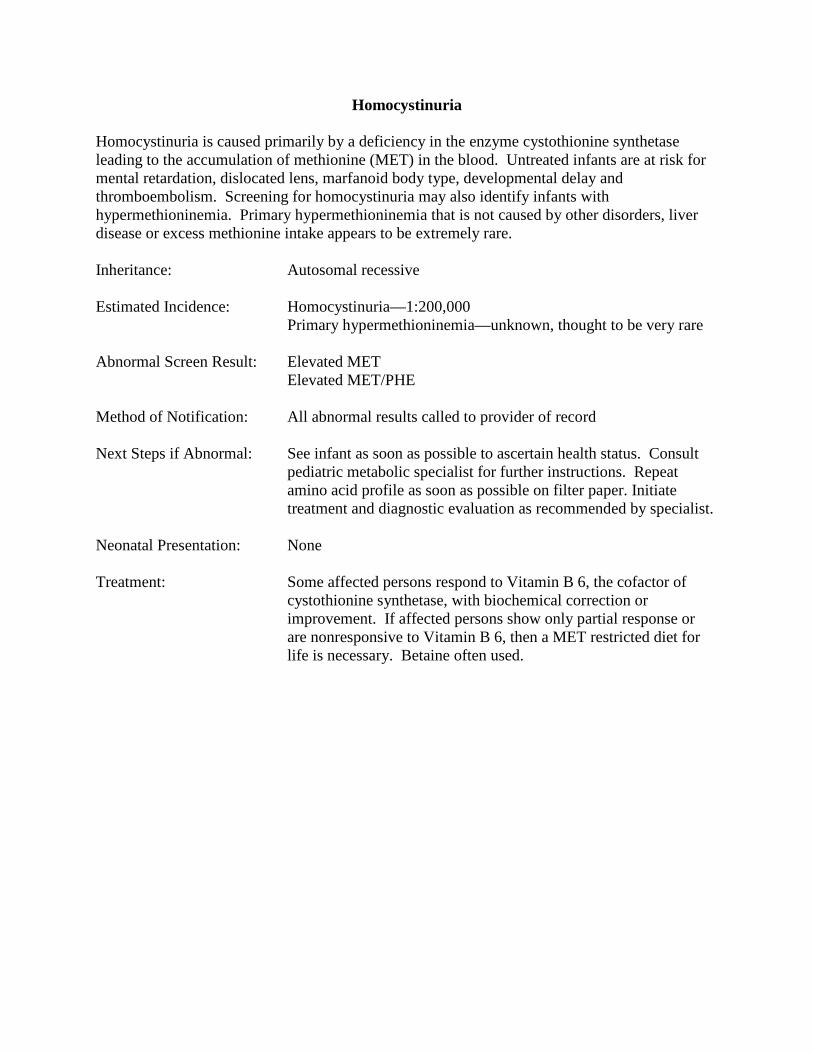
Homocystinuria
Homocystinuria is caused primarily by a deficiency in the enzyme cystothionine synthetase leading to the accumulation of methionine (MET) in the blood. Untreated infants are at risk for mental retardation, dislocated lens, marfanoid body type, developmental delay and thromboembolism. Screening for homocystinuria may also identify infants with hypermethioninemia. Primary hypermethioninemia that is not caused by other disorders, liver disease or excess methionine intake appears to be extremely rare. Inheritance: Autosomal recessive Estimated Incidence: Homocystinuria—1:200,000 Primary hypermethioninemia—unknown, thought to be very rare Abnormal Screen Result: Elevated MET Elevated MET/PHE Method of Notification: All abnormal results called to provider of record Next Steps if Abnormal: See infant as soon as possible to ascertain health status. Consult
pediatric metabolic specialist for further instructions. Repeat amino acid profile as soon as possible on filter paper. Initiate treatment and diagnostic evaluation as recommended by specialist.
Neonatal Presentation: None Treatment: Some affected persons respond to Vitamin B 6, the cofactor of
cystothionine synthetase, with biochemical correction or improvement. If affected persons show only partial response or are nonresponsive to Vitamin B 6, then a MET restricted diet for life is necessary. Betaine often used.

Maple Syrup Urine Disease (MSUD)
Maple syrup urine disease (MSUD) is caused by deficiencies in the branched chain keto-acid dehydrogenase complex leading to the accumulation of leucine (LEU), isoleucine (ILE), valine (VAL) and alloisoleucine. Cerumen, urine or sweat may smell faintly of maple syrup. Untreated infants with MSUD who survive infancy have retarded physical and mental development. Milder variants have been reported and may not be picked up by newborn screening. Inheritance: Autosomal recessive Estimated Incidence: 1:185,000 Abnormal Screen Result: Elevated LEU+ILE Elevated VAL Elevated LEU+ILE/PHE Elevated VAL/PHE Method of Notification: All abnormal results called to provider of record Next Steps if Abnormal: Potential medical emergency. See infant as soon as possible to
ascertain health status. Consult pediatric metabolic specialist for further instructions.
Repeat amino acid profile as soon as possible on filter paper.
Initiate treatment and diagnostic evaluation as recommended by specialist.
Neonatal Presentation: May show neurological impairment in first week of life. Lethargy
and poor suck are often the first signs followed by abnormal muscle tone, involuntary movements, seizures and coma.
Treatment: LEU restricted/ILE, VAL controlled diet for life. Some affected
persons with a less severe form of MSUD are thiamin responsive. Special Considerations Fasting/infection/intercurrent illness—Parents must clearly understand that minor illnesses can precipitate metabolic decompensation in an infant/child with this disorder and should seek medical attention with any concern. Urinary ketones may be monitored as a precaution during illness. Ketonuria can be an early sign of metabolic decompensation and frequently precedes clinical signs.

Citrullinemia
Citrullinemia I is a urea cycle disorder caused primarily by a deficiency of the enzyme argininosuccinic acid synthetase. Citrulline (CIT) and ammonia build up in the blood which can lead to lethargy, seizures, coma and death. Citrullinemia II is also a urea cycle disorder. It is caused by a deficiency of the protein citrin which is necessary for many metabolic processes. In the neonatal onset type of CIT II, bile flow is blocked. Inheritance: Autosomal recessive Estimated Incidence: CIT I—1:57,000
CIT II—1:100,000 primarily in persons of Japanese, East Asian or Middle Eastern ancestry
Abnormal Screen Result: Elevated CIT Method of Notification: All abnormal results called to provider of record Next Steps if Abnormal: Potential medical emergency. See infant as soon as possible to
ascertain health status. Consult pediatric metabolic specialist for further instructions. Emergency treatment may include provision of sufficient nonprotein calories to prevent catabolism; Na benzoate or Na phenylacetate; IV arginine. Dialysis may be necessary to lower ammonia level.
Repeat amino acid profile as soon as possible on filter paper.
Initiate treatment and diagnostic evaluation as recommended by specialist.
Neonatal Presentation: May show neurological deterioration in first week of life.
Lethargy, poor feeding, vomiting, grunting respirations, tachypnea, and hypothermia progress to seizures, encephalopathy and death unless quickly treated.
Treatment: High calorie, protein restricted, ARG supplemented diet. Na
benzoate, Na phenylacetate, Na phenylbutyrate may be used to help decrease accumulated toxic precursors
Special Considerations Fasting/infection/intercurrent illness—Parents must clearly understand that minor illness can precipitate metabolic decompensation in an infant/child with this disorder and should seek medical attention with any concern.

Argininosuccinic Aciduria
Argininosuccinic aciduria is a urea cycle disorder caused primarily by a deficiency of the enzyme argininosuccinic acid lyase. Argininosuccinic acid, citrulline (CIT) and ammonia build up in the blood which can lead to lethargy, seizures, coma and death. Inheritance: Autosomal recessive Estimated Incidence: 1:70,000 Abnormal Screen Result: Elevated CIT Method of Notification: All abnormal results called to provider of record Next Steps if Abnormal: Potential medical emergency. See infant as soon as possible to
ascertain health status. Consult pediatric metabolic specialist for further instructions. Emergency treatment may include provision of sufficient nonprotein calories to prevent catabolism, Na benzoate or Na phenylacetate, IV arginine. Dialysis may be necessary to lower ammonia level.
Repeat amino acid profile as soon as possible on filter paper.
Initiate treatment and diagnostic evaluation as recommended by specialist.
Neonatal Presentation: May show neurological deterioration in first week of life.
Lethargy, poor feeding, vomiting, respiratory alkalosis, and hypothermia progress to seizures, encephalopathy and death unless quickly treated.
Treatment: High calorie, protein restricted, ARG supplemented diet. Na
benzoate, Na phenylacetate, Na phenylbutyrate may be used to help decrease accumulated toxic precursors
Special Considerations Fasting/infection/intercurrent illness—Parents must clearly understand that minor illness can precipitate metabolic decompensation in an infant/child with this disorder and should seek medical attention with any concern.

Tyrosinemia Tyrosinemia I (TYR I) is caused by a deficiency in the enzyme fumarylacetoacetase. Untreated infants are at risk for liver failure, jaundice, growth retardation and eventual hepatocellular carcinoma. Tyrosinemia Type II or III (TYR II or III) can also be identified by screening. TYR II is caused by a deficiency in the enzyme tyrosine aminotransferase. TYR III is caused by a deficiency in the enzyme 4-OH phenylpyruvate dioxygenase. Untreated infants with TYR II are at risk for eye and skin lesions with neurological problems including developmental delay. The clinical features of TYR III are not well described; however, mental retardation and behavioral problems have been found in affected persons. Inheritance: Autosomal recessive Estimated Incidence: TYR I—1:100,000 TYR II—1:250,000 TYR III—unknown, thought to be very rare Abnormal Screen Result: TYR I—Elevated succinylacetone (SUAC) TYR II or III—Elevated TYR with normal SUAC Method of Notification: All abnormal SUAC results called to provider of record. TYR
results greater than 800 µM are called to the provider of record. Other abnormal TYR results are mailed to provider of record.
Next Steps if Abnormal: See infant as soon as possible to ascertain health status. Consult
pediatric metabolic specialist for further instructions. Repeat SUAC and amino acid profile as soon as possible on filter
paper. Initiate treatment and diagnostic evaluation as recommended by specialist.
Neonatal Presentation: All forms—usually none. Treatment: TYR I—TYR and PHE restricted diet for life. NTBC (Nitisinone)
also used to inhibit the degradation of tyrosine and the formation of toxic metabolites. Liver transplantation if indicated.
TYR II or III—TYR and PHE restricted diet for life. Special Considerations Premature/sick infants—Transient Tyrosinemia of the Newborn is the most common amino acid disorder found in infants, especially those who are premature and/or sick. However, prompt repeat screening is needed as a precaution.

Carbohydrate Metabolism Disorders
Galactosemia Galactosemia is a condition of abnormal galactose metabolism caused by deficient functioning of any of three separate enzymes. These include galctose-1-P-uridyl transferase (GALT) deficiency or classical galactosemia; galactokinase deficiency (GALK); and UDP galactose-4-epimerase deficiency (GALE). Individuals with galactosemia are unable to break down and use the sugar galactose (a component of lactose found primarily in dairy products and human milk). If undiagnosed, the affected infant with classical galactosemia may develop gastrointestinal disturbances, fail to gain weight and become jaundiced. Life-threatening infection can occur in the newborn period. Mental retardation and delayed physical growth occur in untreated infants who survive. Some infants with low levels of GALT are subsequently diagnosed with a form of galactosemia called Duarte variant galactosemia. Almost all cases of Duarte variant galactosemia are benign; however, a few affected infants may be treated during the first year of life as a precaution. Infants with GALK deficiency only have cataracts. Infants with GALE deficiency will have varying outcomes. If the GALE deficiency is localized in the red blood cell, the infant does not have any symptoms of disease and no treatment is necessary. If the GALE deficiency involves other tissues, the clinical course is similar to that of GALT deficiency. The diagnostic work-up may also identify infants who are genetic carriers for one of the forms of galactosemia. Inheritance: Autosomal recessive Estimated Incidence: GALT (classical galactosemia)—1:60,000 Duarte variant galactosemia—1:16,000 GALK unknown, thought to be rare GALE unknown, thought to be very rare Abnormal Screen Result: Elevated total galactose with low GALT—at risk for classical
galactosemia Normal total galactose with low GALT—at risk for Duarte galactosemia or at risk for classical galactosemia if infant on non-lactose feeding at time of screening Elevated total galactose with normal GALT—at risk for GALK or GALE deficiency
Method of Notification: All results where the GALT is low and total galactose is elevated
are called to provider of record. Other combinations of results are mailed to provider of record.

Next Steps if Abnormal: Potential medical emergency when GALT is low and total galactose is elevated. See infant as soon as possible to ascertain health status. Change to soy based formula when GALT is low and total galactose is elevated. If total galactose is not elevated, consider change to soy based formula based upon clinical observation and recommendation from pediatric metabolic specialist. In most circumstances, at least partial breastfeeding is possible as long as total galactose is not elevated.
Repeat galactosemia screening as soon as possible. Consult
pediatric metabolic specialist for further instructions and diagnostic evaluation.
If GALT is normal in the initial specimen, repeat galactosemia
screening as soon as possible. NO NEED TO STOP BREAST FEEDING OR CHANGE FORMULA TYPE at this time. If total galactose remains elevated in the repeat specimen or if the GALT result is now low, consult pediatric metabolic specialist for further diagnostic evaluation and feeding recommendations.
Neonatal Presentation: GALT—hypoglycemia, jaundice, sepsis, failure to thrive Duarte variant galactosemia—None GALK—None GALE—Usually none Treatment: Galactose restricted diet for life. Special Considerations Reporting of Feeding Type--It is crucial that staff report whether the infant is on a lactose containing feeding (breast milk or cow's milk based infant formula), a soy based infant formula or any other non-lactose containing feeding (including IV fluids or total parenteral nutrition/hyperalimentation) so that the lab test can be interpreted appropriately. Exposure of the Specimen to Heat/Humidity--Both heat and humidity can affect the test for GALT. The enzyme activity can be diminished causing a false positive result for galactosemia. NEVER put a newborn screening specimen in a plastic bag as this can increase exposure to both heat and humidity! Transfusion—Transfusion of red blood cells prior to drawing the newborn screening specimen may affect the GALT result. Repeat screening for galactosemia should be done 120 days after the last transfusion. If the date of the last transfusion is unknown, put the date of hospital discharge on the collection form.

Organic Acid Metabolism Disorders
Propionic Acidemia (PA) Propionic acidemia is a disorder of isoleucine (ILE), methionine (MET), threonine (THR), valine (VAL), and odd chain fatty acid metabolism caused by deficient activity of the enzyme propionyl coenzyme A carboxylase. This enzyme deficiency leads to the accumulation of toxic organic acid metabolites when the affected infant is ingesting a normal diet or is under catabolic stress. Inheritance: Autosomal recessive Estimated Incidence: 1:100,000 Abnormal Screen Result: Elevated C3 (propionyl carnitine) Elevated C3/C2 Elevated C3/C16 Method of Notification: All results where the C3 is greater than 10 μM and the C3/C2
and/or C3/C16 is elevated are called to provider of record. All results where the C3 is greater than 15 μM are called to the provider of record regardless of the ratio levels. Any other abnormal C3 results are mailed to the provider of record.
Next Steps if Abnormal: Potential medical emergency when the C3 is greater than 10
μM and the C3/C2 and/or C3/C16 is elevated or when the C3 is greater than 15 μM regardless of the ratio levels. See infant as soon as possible to ascertain health status. Consult pediatric metabolic specialist for further instructions.
Repeat acyl carnitine profile as soon as possible on filter paper.
Initiate treatment and diagnostic evaluation as recommended by specialist.
Neonatal Presentation: Poor feeding, vomiting, tachypnea, lethargy, abnormal muscle
tone, involuntary movements, seizures, coma Treatment: Protein restricted diet. Use of metabolic formula without ILE,
MET, THR, VAL. Carnitine supplementation. Biotin trial. Special Considerations Fasting/infection/intercurrent illness—Parents must clearly understand that minor illnesses can precipitate metabolic decompensation in an infant/child with an organic acid disorder and should seek medical attention with any concern. Urinary ketones may be monitored as a precaution

during illness. Ketonuria can be an early sign of metabolic decompensation and frequently precedes clinical signs.

Malonic Acidemia (MA)
Malonic acidemia is a disorder of ketone metabolism arising from a deficiency of the enzyme malonyl coA decarboxylase. Almost all affected infants have developmental delay. Other findings include hypotonia, seizures, hypoglycemia and cardiomyopathy. Fewer than 30 cases of malonic acidemia have been reported. Inheritance: Autosomal recessive Estimated Incidence: Unknown—thought to be very rare Abnormal Screen Result: Elevated C3DC (malonyl carnitine) + C4OH (3-OH butyryl
carnitine)/C10 (decaonyl carnitine) Method of Notification: All results where the C3DC + C4OH/C10 is greater than 5 are
called to provider of record. Next Steps if Abnormal: See infant as soon as possible to ascertain health status. Consult
pediatric metabolic specialist for further instructions. Repeat acyl carnitine profile as soon as possible on filter paper.
Initiate treatment and diagnostic evaluation as recommended by specialist.
Neonatal Presentation: May have hypotonia, hypoglycemia, hypertrophic cardiomyopathy,
diarrhea, vomiting, ketosis and/or seizures. Infants are at risk for metabolic decompensation/crisis.
Treatment: Carnitine supplementation. May be prescribed fat controlled diet
with MCT as major fat source. Avoid fasting.

Methylmalonic Acidemia (MMA) Methylmalonic academia is a disorder of isoleucine (ILE), methionine (MET), threonine (THR), valine (VAL), and odd chain fatty acid metabolism caused by deficient methylmalonyl CoA mutase, deficient Vitamin B12 (cobalamin) or defects in absorption, transport or processing of cobalamin. Toxic organic acid metabolites accumulate when the affected infant is ingesting a normal diet or is under catabolic stress. Inheritance: Autosomal recessive Estimated Incidence: Vit B 12 non-responsive 1:48,000 Other types, unknown incidence Abnormal Screen Result: Elevated C3 (propionyl carnitine) Elevated C3/C2 Elevated C3/C16 Method of Notification: All results where the C3 is greater than 10 μM and the C3/C2
and/or C3/C16 is elevated are called to provider of record. All results where the C3 is greater than 15 μM are called to the provider of record regardless of the ratio levels. Any other abnormal C3 results are mailed to the provider of record.
Next Steps if Abnormal: Potential medical emergency when the C3 is greater than 10
μM and the C3/C2 and/or C3/C16 is elevated or when the C3 is greater than 15 μM regardless of the ratio levels. See infant as soon as possible to ascertain health status. Consult pediatric metabolic specialist for further instructions.
Repeat acyl carnitine profile as soon as possible on filter paper.
Initiate treatment and diagnostic evaluation as recommended by specialist.
Neonatal Presentation: Poor feeding, vomiting, tachypnea, lethargy, abnormal muscle
tone, involuntary movements, seizures, coma Treatment: Trial of hydroxycobalamin as soon as suspected. Protein restricted
diet. Use of metabolic formula without ILE, MET, THR, VAL. Carnitine supplementation.
Special Considerations Fasting/infection/intercurrent illness—Parents must clearly understand that minor illnesses can precipitate metabolic decompensation in an infant/child with an organic acid disorder and should seek medical attention with any concern. Urinary ketones may be monitored as a precaution

during illness. Ketonuria can be an early sign of metabolic decompensation and frequently precedes clinical signs.

Isovaleric Acidemia (IVA) Isovaleric acidemia is a disorder of leucine (LEU) metabolism caused by deficiency of the enzyme isovaleryl coA dehydrogenase. This enzyme deficiency leads to the accumulation of toxic organic acid metabolites when the affected infant is ingesting a normal diet or is under catabolic stress. A chronic, intermittent form of IVA can present later in infancy of childhood with episodes of metabolic acidosis, usually associated with an intercurrent illness or increased protein intake. Inheritance: Autosomal recessive Estimated Incidence: 1:230,000 Abnormal Screen Result: Elevated C5 (isovaleryl carnitine) Method of Notification: All abnormal results called to provider of record Next Steps if Abnormal: Potential medical emergency. See infant as soon as possible to
ascertain health status. Consult pediatric metabolic specialist for further instructions.
Repeat acyl carnitine profile as soon as possible on filter paper.
Initiate treatment and diagnostic evaluation as recommended by specialist.
Neonatal Presentation: Poor feeding, vomiting, tachypnea, lethargy, abnormal muscle
tone, involuntary movements, seizures, coma Treatment: Protein restricted diet. Use of metabolic formula without LEU.
Glycine (GLY) supplementation. Carnitine supplementation. Special Considerations Fasting/infection/intercurrent illness—Parents must clearly understand that minor illnesses can precipitate metabolic decompensation in an infant/child with an organic acid disorder and should seek medical attention with any concern. Urinary ketones may be monitored as a precaution during illness. Ketonuria can be an early sign of metabolic decompensation and frequently precedes clinical signs.

2-Methylbutyryl coA Dehydrogenase Deficiency (2-MBCDD) 2-Methylbutyryl coA dehydrogenase deficiency is a disorder of isoleucine (ILE) metabolism. Infants with this disorder may be asymptomatic or may have an episode of metabolic decompensation with subsequent neurological deficits. Inheritance: Presumed autosomal recessive Estimated Incidence: Unknown, thought to be very rare outside of persons of Hmong
ancestry Abnormal Screen Result: Elevated C5 (isovaleryl carnitine) Method of Notification: All abnormal results called to provider of record Next Steps if Abnormal: See infant as soon as possible to ascertain health status. Consult
pediatric metabolic specialist for further instructions. Repeat acyl carnitine profile as soon as possible on filter paper. Initiate treatment and diagnostic evaluation as recommended by specialist.
Neonatal Presentation: Hypotonia, lethargy, apnea, hypoglycemia Treatment: Carnitine supplementation. Moderate protein restriction. Avoid
fasting.

3-Methylcrotonyl coA Carboxylase Deficiency (3-MCC) 3-Methylcrotonyl coA carboxylase deficiency (3-MCC) is a disorder of leucine (LEU) metabolism. Infants may have a Reye-like illness with hypoketotic hypoglycemia, hypotonia, hepatic encephalopathy, and metabolic acidosis. Symptomatic infants may have a “cat’s urine” odor. Inheritance: Autosomal recessive Estimated Incidence: 1:50,000 Abnormal Screen Result: Elevated C4DC (methylmalonyl carnitine) + C5OH (3-OH
isovaleryl carnitine) Method of Notification: All abnormal results called to provider of record Next Steps if Abnormal: See infant as soon as possible to ascertain health status. Consult
pediatric metabolic specialist for further instructions. Repeat acyl carnitine profile as soon as possible on filter paper. Initiate treatment and diagnostic evaluation as recommended by specialist.
Neonatal Presentation: Usually none. May present with seizures. Treatment: Carnitine supplementation. Moderate protein and LEU restriction.
Glycine supplementation. Avoid fasting. NOTE: Biotin is not effective in isolated 3-MCC.
Special Considerations: Maternal 3-MCC—In some newborns, the elevated C4DC+C5OH is reflective of maternal 3-MCC.

Beta Ketothiolase Deficiency Beta ketothiolase deficiency (SKAT) is a disorder of isoleucine (ILE) metabolism and of ketolysis. Infants with this disorder are at risk for episodes of severe ketoacidosis with subsequent neurological deficits. This disorder is sometimes called 2-methyl 3-OH butyric aciduria. Inheritance: Autosomal recessive Estimated Incidence: Unknown Abnormal Screen Result: Elevated C4DC (methylmalonyl carnitine) + C5OH (3-OH
isovaleryl carnitine) Elevated C5:1 (tiglyl carnitine) Method of Notification: All abnormal results called to provider of record Next Steps if Abnormal: See infant as soon as possible to ascertain health status. Consult
pediatric metabolic specialist for further instructions. Repeat acyl carnitine profile as soon as possible on filter paper. Initiate treatment and diagnostic evaluation as recommended by specialist.
Neonatal Presentation: Poor feeding, vomiting, tachypnea, lethargy Treatment: Carnitine supplementation. Protein restricted/fat controlled diet.
Avoid fasting. May require long term bicarbonate. Special Considerations Fasting/infection/intercurrent illness—Parents must clearly understand that minor illnesses can precipitate metabolic decompensation in an infant/child with an organic acid disorder and should seek medical attention with any concern. Urinary ketones should be monitored at home. Ketonuria can be an early sign of metabolic decompensation and frequently precedes clinical signs.
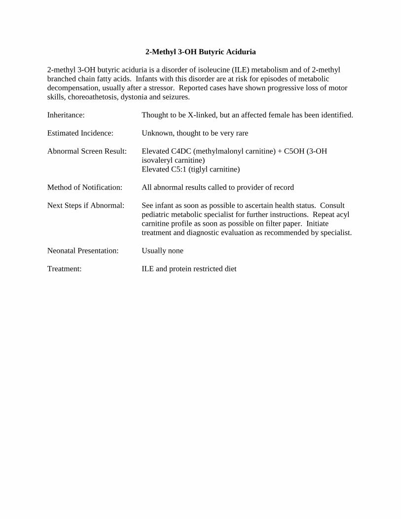
2-Methyl 3-OH Butyric Aciduria
2-methyl 3-OH butyric aciduria is a disorder of isoleucine (ILE) metabolism and of 2-methyl branched chain fatty acids. Infants with this disorder are at risk for episodes of metabolic decompensation, usually after a stressor. Reported cases have shown progressive loss of motor skills, choreoathetosis, dystonia and seizures. Inheritance: Thought to be X-linked, but an affected female has been identified. Estimated Incidence: Unknown, thought to be very rare Abnormal Screen Result: Elevated C4DC (methylmalonyl carnitine) + C5OH (3-OH
isovaleryl carnitine) Elevated C5:1 (tiglyl carnitine) Method of Notification: All abnormal results called to provider of record Next Steps if Abnormal: See infant as soon as possible to ascertain health status. Consult
pediatric metabolic specialist for further instructions. Repeat acyl carnitine profile as soon as possible on filter paper. Initiate treatment and diagnostic evaluation as recommended by specialist.
Neonatal Presentation: Usually none Treatment: ILE and protein restricted diet

3-Methyl 3-OH Glutaryl co-A Lyase Deficiency (HMGLD)
3-methyl 3-OH glutaryl co-A lyase deficiency is a disorder of leucine (LEU) metabolism and of ketogenesis. Infants with this disorder may present with hypoketotic hypoglycemia and are at risk for subsequent neurological deficits. Inheritance: Autosomal recessive Estimated Incidence: Unknown, thought to be rare Abnormal Screen Result: Elevated C4DC (methylmalonyl carnitine) + C5OH (3-OH
isovaleryl carnitine) Elevated C6DC (3-methyl glutaryl carnitine)
Method of Notification: All abnormal results called to provider of record Next Steps if Abnormal: See infant as soon as possible to ascertain health status. Consult
pediatric metabolic specialist for further instructions. Repeat acyl carnitine profile as soon as possible on filter paper. Initiate treatment and diagnostic evaluation as recommended by specialist.
Neonatal Presentation: One-third of affected newborns will have hypoketotic
hypoglycemia, severe metabolic acidosis, vomiting, lethargy, hypotonia
Treatment: Protein restricted diet. Use of metabolic formula without LEU.
Carnitine supplementation. Fat controlled diet when older. Avoid fasting.
Special Considerations Fasting/illness/protein loading—Parents must clearly understand that minor illness can
precipitate metabolic decompensation in an infant/child with this disorder and should seek medical attention with any concern. Protein loading or fasting can also lead to hypoglycemic episodes result in seizures or coma.

3-Methylglutaconyl co-A Hydratase Deficiency 3-Methylglutaconyl co-A hydratase deficiency is a disorder of leucine (LEU) metabolism. It is sometimes known as 3-methylglutaconic aciduria type I. Three other types of 3-methylglutaconic aciduria have also been described. Mildly affected persons have speech retardation and short attention span. Severely affected persons have had acidosis and more severe neurological problems, hypotonia, spastic dystonia, irritability, developmental delay and mental retardation. Inheritance: Autosomal recessive Estimated Incidence: Unknown, thought to be very rare Abnormal Screen Result: Elevated C4DC (methylmalonyl carnitine) + C5OH (3-OH
isovaleryl carnitine) Method of Notification: All abnormal results called to provider of record Next Steps if Abnormal: See infant as soon as possible to ascertain health status. Consult
pediatric metabolic specialist for further instructions. Repeat acyl carnitine profile as soon as possible on filter paper. Initiate treatment and diagnostic evaluation as recommended by specialist
Neonatal Presentation: None reported Treatment: Carnitine supplementation. Moderate protein and LEU restriction.
Avoid fasting.

Multiple Carboxylase Deficiency (MCD) or Holocarboxylase Synthetase Deficiency
Multiple carboxylase deficiency is caused by a deficiency of the enzyme holocarboxylase synthetase. This enzyme activates four carboxylases by attaching biotin. These carboxylases are involved in amino acid metabolism, gluconeogenesis, and fatty acid synthesis. Affected infants may develop severe metabolic acidosis leading to coma. Skin rash and hair loss occur at later stages. Inheritance: Autosomal recessive Estimated Incidence: 1:87,000 Abnormal Screen Result: Elevated C3 (propionyl carnitine)
Elevated C4DC (methylmalonyl carnitine) + C5OH (3-OH isovaleryl carnitine)
Method of Notification: All results where both C3 and C4DC+C5OH are elevated are
called to the provider of record. Next Steps if Abnormal: See infant as soon as possible to ascertain health status. Consult
pediatric metabolic specialist for further instructions. Repeat acyl carnitine profile as soon as possible on filter paper. Initiate treatment and diagnostic evaluation as recommended by specialist
Neonatal Presentation: May show food refusal, vomiting, lethargy, seizures, hypotonia,
tachypnea Treatment: Biotin supplementation Special Considerations Enzymes necessary for carboxylase activity—Two enzymes are necessary for normal activity of four carboxylases: holocarboxylase synthetase to attach biotin to the carboxylases and biotinidase to free the protein bound biotin.

Glutaric Aciduria Type I (GA I) Glutaric aciduria type I is caused by a deficiency in the enzyme glutaryl coA dehydrogenase. Seventy percent of infants will have macrocephaly at or shortly after birth. Infants may remain asymptomatic until an encephalopathic crisis. Others gradually develop motor delay and hypotonia without any apparent acute crisis. No loss of intellectual capacity develops unless a neurological crisis occurs. Inheritance: Autosomal recessive Estimated Incidence: 1:40,000 Abnormal Screen Result: Elevated C5DC (glutaryl carnitine) + C6OH (3-OH hexanoyl
carnitine) Method of Notification: All abnormal results as determined likely positive by the Region 4
Genetics Collaborative (R4GC) Post Analytical Tool are called to provider of record. Other abnormal results are mailed to the provider of record.
Next Steps if Abnormal: Potential medical emergency as determined by score using the
R4GC Post Analytical Tool. See infant as soon as possible to ascertain health status. Consult pediatric metabolic specialist for further instructions. A portion of the initial specimen will be sent to the Greenwood Genetic Center Laboratory for secondary testing. If the R4GC tool indicates lower risk for GA I, then secondary testing will not occur unless the C5DC+C6OH is elevated in a second specimen.
Repeat acyl carnitine profile as soon as possible on filter paper. Collection of other specimens may be indicated depending upon the results of secondary testing. Initiate treatment and diagnostic evaluation as recommended by specialist.
Neonatal Presentation: Macrocephaly, irritability, jitteriness, hypotonia Treatment: Prompt treatment of catabolic events. Aggressive fever control.
Watch fluid intake, as affected persons may have profuse sweating. Riboflavin trial. Carnitine supplementation. Protein restricted diet. Use of metabolic formula without lysine (LYS) and tryptophan (TRP).
Special Considerations

Fasting/infection/intercurrent illness—Parents must clearly understand that minor illnesses can precipitate encephalopathic or metabolic decompensation in an infant/child with this disorder. Hospital admission may be considered mandatory for IV fluids with any vomiting illness. Fever—Poorly controlled/untreated persons with GA I may have recurrent fever not related to illness. Death from hyperthermia has been reported in children with GA I. Acute subdural and/or retinal hemorrhages—Infants with GA I are prone to acute subdural and/or retinal hemorrhage after minor head trauma (ie, minor childhood falls that can occur when infant is learning to walk) that can be misdiagnosed as child abuse.

Fatty Acid Metabolism Disorders
Medium Chain Acyl co-A Dehydrogenase Deficiency (MCAD) Medium chain acyl co-A dehydrogenase deficiency (MCAD) is an inborn error of fatty acid oxidation that can cause significant morbidity and mortality in the newborn. It usually presents in infancy or early childhood with hypoketotic hypoglycemia and encephalopathy after an intercurrent illness and/or period of poor oral intake. Approximately 20% of infants with MCAD die before diagnosis, and a substantial proportion of the survivors have significant residual problems from an initial crisis. Children who survive the initial crises may have developmental delay, seizures, speech/language delays, chronic muscle weakness, failure to thrive, cerebral palsy and attention deficit disorder. Inheritance: Autosomal recessive Estimated Incidence: 1:16,000 Abnormal Screen Result: Primary Markers
Elevated C8 (octanoyl carnitine) Elevated C8/C10 Secondary Markers Elevated C6 (hexanoyl carnitine)
Elevated C10 (decanoyl carnitine) Elevated C10:1 (decenoyl carnitine) Method of Notification: All abnormal results where the C8 is elevated are called to
provider of record. Isolated elevations of secondary markers have no clinical significance and are not reported.
Next Steps if Abnormal: See infant as soon as possible to ascertain health status. Consult
pediatric metabolic specialist for further instructions. Repeat acyl carnitine profile as soon as possible on filter paper. Initiate treatment and diagnostic evaluation as recommended by specialist.
Neonatal Presentation: Usually none Treatment: Avoid fasting. Supplementation with high energy carbohydrate
drinks during periods of illness. Seek medical attention if these liquids are refused. Oral rehydration fluids are not adequate. If oral feedings are not possible, then intravenous fluids with concentrated dextrose should be started without waiting for symptoms of decompensation to develop.
Infants with MCAD must be fed at least every three to four hours,
including at night. Infants with MCAD should not be fed

formulas that have medium chain triglycerides (MCT) as the primary fat source if a safe alternative is available. Feeding intervals can be lengthened as the infant gets older.
Carnitine supplementation if helpful. Special Considerations Fasting/infection/intercurrent illness—Parents must clearly understand that prolonged fasting and infection/intercurrent illnesses can cause serious complications in an infant with MCAD. These outcomes include hypoketotic hypoglycemia, vomiting, lethargy, seizures and coma. Infants experiencing stressful events such as trauma, infections/illnesses, and even receiving immunizations should be monitored to prevent hypoglycemic episodes. Blood glucose may be monitored as a precaution. Premature/sick infants—Some special formulas and breast milk fortifiers fed to premature/sick infants contain medium chain triglycerides (MCT) as the primary fat source. These feedings may cause false elevations of some acyl carnitines analyzed in MCAD screening, particularly C8, C10:1 and C8/C10.

Medium Chain Ketoacyl co-A Thiolase Deficiency Medium chain ketoacyl co-A thiolase deficiency (MCKAT) is an inborn error of fatty acid oxidation. One infant with this disorder has been detected worldwide. This male neonate presented with vomiting, dehydration, metabolic acidosis, liver dysfunction, and terminal rhabdomyolysis with myoglobinuria. Inheritance: Unknown Estimated Incidence: Extremely rare Abnormal Screen Result: Elevated C8 (octanoyl carnitine) Elevated C6DC (adipyl carnitine) Method of Notification: All abnormal results called to provider of record Next Steps if Abnormal: See infant as soon as possible to ascertain health status. Consult
pediatric metabolic specialist for further instructions. Repeat acyl carnitine profile as soon as possible on filter paper. Initiate treatment and diagnostic evaluation as recommended by specialist.
Neonatal Presentation: Vomiting, dehydration, metabolic acidosis, liver dysfunction Treatment: Only known affected infant died at 13 days of life. Presumed
treatment is same as that for other fatty acid metabolism disorders: Avoid fasting. Supplementation with high energy carbohydrate drinks during periods of illness. Seek medical attention if these liquids are refused. Oral rehydration fluids are not adequate. If oral feedings are not possible, then intravenous fluids with concentrated dextrose should be started without waiting for symptoms of decompensation to develop.
Feed at least every three to four hours, including at night. Special Considerations Fasting/infection/intercurrent illness—Parents must clearly understand that prolonged fasting and infection/intercurrent illnesses can cause serious complications in an infant with a fatty acid oxidation disorder. Infants experiencing stressful events such as trauma, infections/illnesses, and even receiving immunizations should be monitored to prevent metabolic decompensation.

Medium/Short Chain 3-OH acyl coA Dehydrogenase Deficiency (M/SCHAD) Medium/Short chain 3-OH acyl co-A dehydrogenase deficiency (M/SCHAD) is an inborn error of fatty acid oxidation. Infants may have poor feeding, vomiting and lethargy. Individuals with M/SCHAD are at risk for seizures, life threatening heart and breathing problems, coma and sudden death. Inheritance: Autosomal recessive Estimated Incidence: Unknown, thought to be very rare Abnormal Screen Result: Elevated C3DC (malonyl carnitine) + C4OH (3-OH butyryl carnitine) Method of Notification: All abnormal results called to provider of record Next Steps if Abnormal: See infant as soon as possible to ascertain health status. Consult
pediatric metabolic specialist for further instructions. Repeat acyl carnitine profile as soon as possible on filter paper. Initiate treatment and diagnostic evaluation as recommended by specialist.
Neonatal Presentation: Poor feeding, vomiting, lethargy, seizures. Infants are at risk for
metabolic decompensation/crisis, hypoglycemia. Plasma insulin may be elevated.
Treatment: Avoid fasting. Supplementation with high energy carbohydrate
drinks during periods of illness. Seek medical attention if these liquids are refused. Oral rehydration fluids are not adequate. If oral feedings are not possible, then intravenous fluids with concentrated dextrose should be started without waiting for symptoms of decompensation to develop.
Feed at least every three to four hours, including at night. May
need cornstarch supplementation at bedtime to maintain blood glucose levels overnight. Carnitine supplementation if helpful. Consider medication for infants with documented hyperinsulinisum.
Special Considerations Fasting/infection/intercurrent illness—Parents must clearly understand that prolonged fasting and infection/intercurrent illnesses can cause serious complications in an infant with a fatty acid oxidation disorder. Infants experiencing stressful events such as trauma, infections/illnesses, and even receiving immunizations should be monitored to prevent metabolic decompensation.

Dienoyl co-A Reductase Deficiency 2, 4-Dienoyl co-A reductase deficiency is an inborn error of fatty acid oxidation. At least one infant with this disorder has been detected. This neonate presented with short trunk, arms and fingers; microcephaly; hypotonia. Inheritance: Autosomal recessive Estimated Incidence: Unknown, thought to be extremely rare Abnormal Screen Result: Elevated C10:2 (decadienoyl carnitine) Method of Notification: All abnormal results called to provider of record Next Steps if Abnormal: See infant as soon as possible to ascertain health status. Consult
pediatric metabolic specialist for further instructions. Repeat acyl carnitine profile as soon as possible on filter paper. Initiate treatment and diagnostic evaluation as recommended by specialist.
Neonatal Presentation: Hypotonia, possible ventricular septal defect, microcephaly Treatment: Presumed treatment is same as that for other fatty acid metabolism
disorders: Avoid fasting. Supplementation with high energy carbohydrate drinks during periods of illness. Seek medical attention if these liquids are refused. Oral rehydration fluids are not adequate. If oral feedings are not possible, then intravenous fluids with concentrated dextrose should be started without waiting for symptoms of decompensation to develop. Fat restricted diet with use of MCT oil as fat source. Carnitine if helpful. Feed at least every three to four hours, including at night.
Special Considerations Fasting/infection/intercurrent illness—Parents must clearly understand that prolonged fasting and infection/intercurrent illnesses can cause serious complications in an infant with a fatty acid oxidation disorder. Infants experiencing stressful events such as trauma, infections/illnesses, and even receiving immunizations should be monitored to prevent metabolic decompensation.

Long Chain 3-OH Acyl co-A Dehydrogenase Deficiency (LCHAD) Trifunctional Protein Deficiency (TFP)
Long chain 3-OH acyl co-A dehydrogenase is one of three enzymes in trifunctional protein deficiency (TFP). Deficiencies of these enzymes overlap clinically. Affected persons may present with hypoketotic hypoglycemia, hepatic encephalopathy and muscle weakness usually associated with cardiomyopathy. Other features include rhabdomyolysis, myoglobinuria and peripheral neuropathy. They may also show retinal pigmentation with vision loss in childhood. Symptoms may present as early as the first days of life. Inheritance: Autosomal recessive Estimated Incidence: Unknown Abnormal Screen Result: Primary Marker
Elevated C16-OH (3-OH palmitoyl carnitine) Secondary Markers Elevated C14:1 (tetradecenoyl carnitine)
Elevated C16 (palmitoyl carnitine) Elevated C18:1 (oleyl carnitine)
Elevated C18:1-OH (3-OH oleyl carnitine) Method of Notification: All abnormal results as determined by the Region 4 Genetics
Collaborative Post-Analytical Tool are called to provider of record Next Steps if Abnormal: See infant as soon as possible to ascertain health status. Consult
pediatric metabolic specialist for further instructions. Repeat acyl carnitine profile as soon as possible on filter paper. Initiate treatment and diagnostic evaluation as recommended by specialist.
Neonatal Presentation: Hypoketotic hypoglycemia. Some infants will have hepatic
encephalopathy and muscle weakness associated with cardiomyopathy.
Treatment: Avoid fasting. Supplementation with high energy carbohydrate
drinks during periods of illness. Seek medical attention if these liquids are refused. Oral rehydration fluids are not adequate. If oral feedings are not possible, then intravenous fluids with concentrated dextrose should be started without waiting for symptoms of decompensation to develop.
Feed at least every three to four hours, including at night. May
need cornstarch supplementation via tube feeding overnight in older infancy/childhood. Fat restricted diet with use of MCT oil as fat source. Essential fatty acid supplementation. DHA

(docosahexanoic acid) supplementation to prevent retinal degeneration may be used. Carnitine supplementation if helpful.
Special Considerations Fasting/infection/intercurrent illness—Parents must clearly understand that prolonged fasting and infection/intercurrent illnesses can cause serious complications in an infant with a fatty acid oxidation disorder. Infants experiencing stressful events such as trauma, infections/illnesses, and even receiving immunizations should be monitored to prevent metabolic decompensation. HELLP Syndrome/AFLP—HELLP syndrome (hemolysis, elevated liver enzymes, low platelets)/AFLP (acute fatty liver of pregnancy) occurs in 20% of pregnancies where the fetus is affected by LCHAD. TFP is thought to be a less common cause of HELLP syndrome.

Very Long Chain Acyl co-A Dehydrogenase Deficiency (VLCAD) Very long chain acyl co-A dehydrogenase deficiency (VLCAD) is an inborn error of fatty acid oxidation. Infants may have hypoketotic hypoglycemia, hypotonia, hepatic dysfunction and cardiomyopathy. 20% of affected persons present as adolescents or adults with muscle fatigue, rhabdomyolysis and myoglobinuria triggered by exercise or fasting. Inheritance: Autosomal recessive Estimated Incidence: Unknown Abnormal Screen Result: Primary Markers Elevated C14:1 (tetradecenoyl carnitine) Elevated C14:1/C2 Secondary Markers Elevated C14 (tetradecanoyl carnitine) Elevated C14:2 (tetradecadienoyl carnitine) Method of Notification: All abnormal results called to provider of record Next Steps if Abnormal: See infant as soon as possible to ascertain health status. Consult
pediatric metabolic specialist for further instructions. A portion of the initial specimen will be sent to the Greenwood Genetic Center Laboratory for secondary testing. Repeat acyl carnitine profile as soon as possible on filter paper. Collection of other specimens may be indicated depending upon the results of secondary testing. Initiate treatment and diagnostic evaluation as recommended by specialist.
Neonatal Presentation: Hypoketotic hypoglycemia, hepatic dysfunction, hypotonia and
cardiomyopathy Treatment: Avoid fasting. Supplementation with high energy carbohydrate
drinks during periods of illness. Seek medical attention if these liquids are refused. Oral rehydration fluids are not adequate. If oral feedings are not possible, then intravenous fluids with concentrated dextrose should be started without waiting for symptoms of decompensation to develop.
Feed at least every three to four hours, including at night. May
need cornstarch supplementation via tube feeding overnight in older infancy/childhood. Fat restricted diet with use of MCT oil as fat source. Essential fatty acid supplementation. Carnitine supplementation if helpful.

Special Considerations Fasting/infection/intercurrent illness—Parents must clearly understand that prolonged fasting and infection/intercurrent illnesses can cause serious complications in an infant with a fatty acid oxidation disorder. Infants experiencing stressful events such as trauma, infections/illnesses, and even receiving immunizations should be monitored to prevent metabolic decompensation.
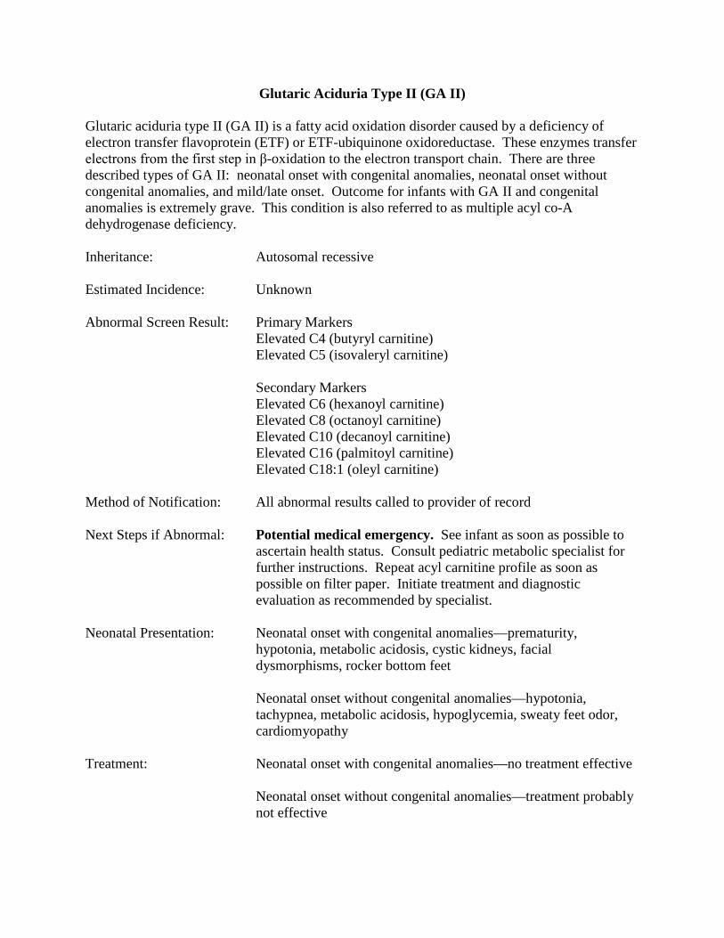
Glutaric Aciduria Type II (GA II)
Glutaric aciduria type II (GA II) is a fatty acid oxidation disorder caused by a deficiency of electron transfer flavoprotein (ETF) or ETF-ubiquinone oxidoreductase. These enzymes transfer electrons from the first step in β-oxidation to the electron transport chain. There are three described types of GA II: neonatal onset with congenital anomalies, neonatal onset without congenital anomalies, and mild/late onset. Outcome for infants with GA II and congenital anomalies is extremely grave. This condition is also referred to as multiple acyl co-A dehydrogenase deficiency. Inheritance: Autosomal recessive Estimated Incidence: Unknown Abnormal Screen Result: Primary Markers Elevated C4 (butyryl carnitine) Elevated C5 (isovaleryl carnitine) Secondary Markers Elevated C6 (hexanoyl carnitine) Elevated C8 (octanoyl carnitine) Elevated C10 (decanoyl carnitine) Elevated C16 (palmitoyl carnitine) Elevated C18:1 (oleyl carnitine) Method of Notification: All abnormal results called to provider of record Next Steps if Abnormal: Potential medical emergency. See infant as soon as possible to
ascertain health status. Consult pediatric metabolic specialist for further instructions. Repeat acyl carnitine profile as soon as possible on filter paper. Initiate treatment and diagnostic evaluation as recommended by specialist.
Neonatal Presentation: Neonatal onset with congenital anomalies—prematurity,
hypotonia, metabolic acidosis, cystic kidneys, facial dysmorphisms, rocker bottom feet
Neonatal onset without congenital anomalies—hypotonia,
tachypnea, metabolic acidosis, hypoglycemia, sweaty feet odor, cardiomyopathy
Treatment: Neonatal onset with congenital anomalies—no treatment effective Neonatal onset without congenital anomalies—treatment probably
not effective

Mild/late onset—Avoid fasting. Supplementation with high energy carbohydrate drinks during periods of illness. Seek medical attention if these liquids are refused. Oral rehydration fluids are not adequate. If oral feedings are not possible, then intravenous fluids with concentrated dextrose should be started without waiting for symptoms of decompensation to develop.
Feed at least every three to four hours, including at night. May
need cornstarch supplementation via tube feeding overnight in older infancy/childhood. May be prescribed diet restricted in fat, controlled in protein. Riboflavin supplementation if helpful.
Special Considerations Fasting/infection/intercurrent illness—Parents must clearly understand that prolonged fasting and infection/intercurrent illnesses can cause serious complications in an infant with a fatty acid oxidation disorder. Infants experiencing stressful events such as trauma, infections/illnesses, and even receiving immunizations should be monitored to prevent metabolic decompensation.

Carnitine Palmitoyl Transferase I Deficiency (CPT I)
Carnitine palmitoyl transferase I (CPT I) is necessary for the conversion of long chain fatty acyl co-A molecules to their corresponding acylcarnitine molecules. Deficiency of this enzyme reduces the availability of acylcarnitines for transport into the mitochondrial matrix for fatty acid oxidation. Inheritance: Autosomal recessive Estimated Incidence: Unknown, thought to be more common in persons of Inuit or
Hutterite origins Abnormal Screen Result: Elevated Free Carnitine/C16 (palmitoyl carnitine) + C18
(octadecanoyl carnitine) Method of Notification: All abnormal results called to provider of record Next Steps if Abnormal: Potential medical emergency. See infant as soon as possible to
ascertain health status. Consult pediatric metabolic specialist for further instructions. Repeat acyl carnitine profile as soon as possible on filter paper. Initiate treatment and diagnostic evaluation as recommended by specialist.
Neonatal Presentation: Seizures, hepatomegaly, hypoketotic hypoglycemia Treatment: Avoid fasting. Supplementation with high energy carbohydrate
drinks during periods of illness. Seek medical attention if these liquids are refused. Oral rehydration fluids are not adequate. If oral feedings are not possible, then intravenous fluids with concentrated dextrose should be started without waiting for symptoms of decompensation to develop. Feed at least every three to four hours, including at night. Feeding regimen may be adjusted when the infant is older. May be prescribed diet restricted in fat. MCT oil may be used as a fat source after diagnosis is clearly established.
Carnitine is contraindicated in treatment of CPT I. Special Considerations Fasting/infection/intercurrent illness—Parents must clearly understand that prolonged fasting and infection/intercurrent illnesses can cause serious complications in an infant with a fatty acid oxidation disorder. Infants experiencing stressful events such as trauma, infections/illnesses, and even receiving immunizations should be monitored to prevent metabolic decompensation.

HELLP Syndrome/AFLP—HELLP syndrome (hemolysis, elevated liver enzymes, low platelets)/AFLP (acute fatty liver of pregnancy) can occur in pregnancies where the fetus is affected by CPT I.

Carnitine Palmitoyl Transferase II Deficiency (CPT II)
Carnitine palmitoyl transferase II deficiency (CPT II) is a disorder of fatty acid transport. In classic CPT II, the onset is during adolescence or early adulthood. It presents with muscle weakness, pain and myoglobinuria usually prompted by exercise, but sometimes by fasting, infection or stress. Renal failure from myoglobinuria occurs in 25% of affected persons. The neonatal type, hepatocardiomuscular CPT II, is extremely rare. Symptoms include hypoketotic hypoglycemia, hepatomegaly, skeletal muscle involvement and marked lipid accumulation in muscle. These infants may also have dysmorphic features. Inheritance: Autosomal recessive Estimated Incidence: Unknown Abnormal Screen Result: Elevated C16 (palmitoyl carnitine) Elevated C18:1 (oleyl carnitine) Method of Notification: All abnormal results called to provider of record Next Steps if Abnormal: Potential medical emergency. See infant as soon as possible to
ascertain health status. Consult pediatric metabolic specialist for further instructions. Repeat acyl carnitine profile as soon as possible on filter paper. Initiate treatment and diagnostic evaluation as recommended by specialist.
Neonatal Presentation: Classic CPT II—none
Hepatocardiomuscular CPT II—hypoketotic hypoglycemia, hepatomegaly, skeletal muscle involvement, marked lipid accumulation in muscle, dysmorphic features.
Treatment: Classic CPT II—Avoid fasting. Supplementation with high energy
carbohydrate drinks during periods of illness. Seek medical attention if these liquids are refused. Oral rehydration fluids are not adequate. If oral feedings are not possible, then intravenous fluids with concentrated dextrose should be started without waiting for symptoms of decompensation to develop. Feed at least every three to four hours, including at night. Feeding regimen may be adjusted when the infant is older. May be prescribed diet restricted in fat. MCT oil may be used as a fat source after diagnosis is clearly established.
Hepatocardiomuscular CPT II—treatment probably not effective
Special Considerations

Fasting/infection/intercurrent illness—Parents must clearly understand that prolonged fasting and infection/intercurrent illnesses can cause serious complications in an infant with a fatty acid oxidation disorder. Infants experiencing stressful events such as trauma, infections/illnesses, and even receiving immunizations should be monitored to prevent metabolic decompensation.

Carnitine/Acylcarnitine Translocase Deficiency (CACT)
Carnitine/acylcarnitine translocase deficiency (CACT) is a disorder of fatty acid and carnitine transport. Two types have been described: one with neonatal onset and the other with onset later in infancy/early childhood. In neonatal onset CACT, the affected infant presents with a metabolic crisis that often results in death from cardiopulmonary complications and/or liver failure. When the onset is later in infancy/early childhood, the affected person presents with hypoglycemia, but not cardiomyopathy. Inheritance: Autosomal recessive Estimated Incidence: Unknown, thought to be very rare Abnormal Screen Result: Elevated C16 (palmitoyl carnitine) Elevated C18:1 (oleyl carnitine) Method of Notification: All abnormal results called to provider of record Next Steps if Abnormal: Potential medical emergency. See infant as soon as possible to
ascertain health status. Consult pediatric metabolic specialist for further instructions. Repeat acyl carnitine profile as soon as possible on filter paper. Initiate treatment and diagnostic evaluation as recommended by specialist.
Neonatal Presentation: Neonatal onset CACT—hypoketotic hypoglycemia,
hyperammonemia, hypotonia, liver dysfunction, cardiomyopathy Later infancy/early childhood onset CACT—none Treatment: Neonatal onset CACT—treatment probably not effective Later infancy/early childhood onset CACT—Avoid fasting.
Supplementation with high energy carbohydrate drinks during periods of illness. Seek medical attention if these liquids are refused. Oral rehydration fluids are not adequate. If oral feedings are not possible, then intravenous fluids with concentrated dextrose should be started without waiting for symptoms of decompensation to develop.
Feed at least every three to four hours, including at night. Feeding
regimen may be adjusted when the infant is older. May be prescribed diet restricted in fat. MCT oil may be used as a fat source after diagnosis is clearly established.
Special Considerations

Fasting/infection/intercurrent illness—Parents must clearly understand that prolonged fasting and infection/intercurrent illnesses can cause serious complications in an infant with a fatty acid oxidation disorder. Infants experiencing stressful events such as trauma, infections/illnesses, and even receiving immunizations should be monitored to prevent metabolic decompensation.

Carnitine Uptake/Transport Deficiency
In carnitine uptake/transport deficiency, carnitine transport across the plasma membrane is inhibited. The reduction in carnitine limits the formation of acylcarnitine and subsequently limits energy production. Skeletal and heart muscle tissues are particularly affected in this process. Inheritance: Autosomal recessive Estimated Incidence: 1:100,000 Abnormal Screen Result: Low free carnitine Method of Notification: All abnormal results where the sum of C3 (propionyl carnitine) and
C16 (palmitoyl carnitine) is less than 2 are called to provider of record. Other low free carnitine results are mailed to the provider of record.
Next Steps if Abnormal: Potential medical emergency when the free carnitine is low and
the sum of C3 and C16 is less than 2. See infant as soon as possible to ascertain health status. Consult pediatric metabolic specialist for further instructions. Repeat acyl carnitine profile as soon as possible on filter paper. Initiate treatment and diagnostic evaluation as recommended by specialist.
Neonatal Presentation: Tachycardia, hepatomegaly, reduced muscle tone, poor feeding Treatment: Carnitine supplementation. Avoid fasting. Supplementation with
high energy carbohydrate drinks during periods of illness. Seek medical attention if these liquids are refused. Oral rehydration fluids are not adequate. If oral feedings are not possible, then intravenous fluids with concentrated dextrose should be started without waiting for symptoms of decompensation to develop.
Feed at least every three to four hours, including at night. Feeding
regimen may be adjusted when the infant is older. May be prescribed diet restricted in fat.
Special Considerations Fasting/infection/intercurrent illness—Parents must clearly understand that prolonged fasting and infection/intercurrent illnesses can cause serious complications in an infant with a fatty acid oxidation disorder. Infants experiencing stressful events such as trauma, infections/illnesses, and even receiving immunizations should be monitored to prevent metabolic decompensation. Maternal CUD—In some newborns, the low free carnitine is reflective of maternal CUD.