Embed Size (px)
Citation preview
Metabolic bone diseases
By Dr. Abdelaty Shawky Dr. Gehan Abdel-Monem
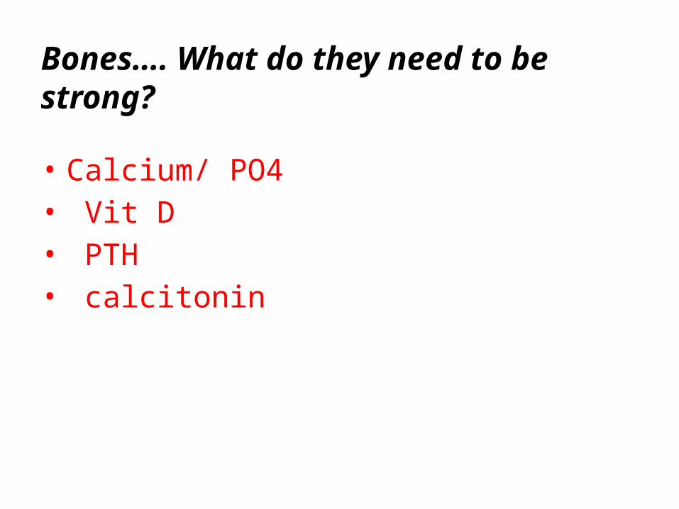
Bones…. What do they need to be strong?
• Calcium/ PO4• Vit D• PTH• calcitonin
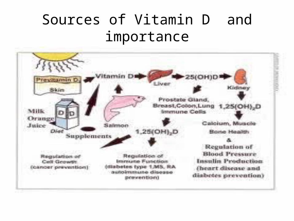
Sources of Vitamin D and importance
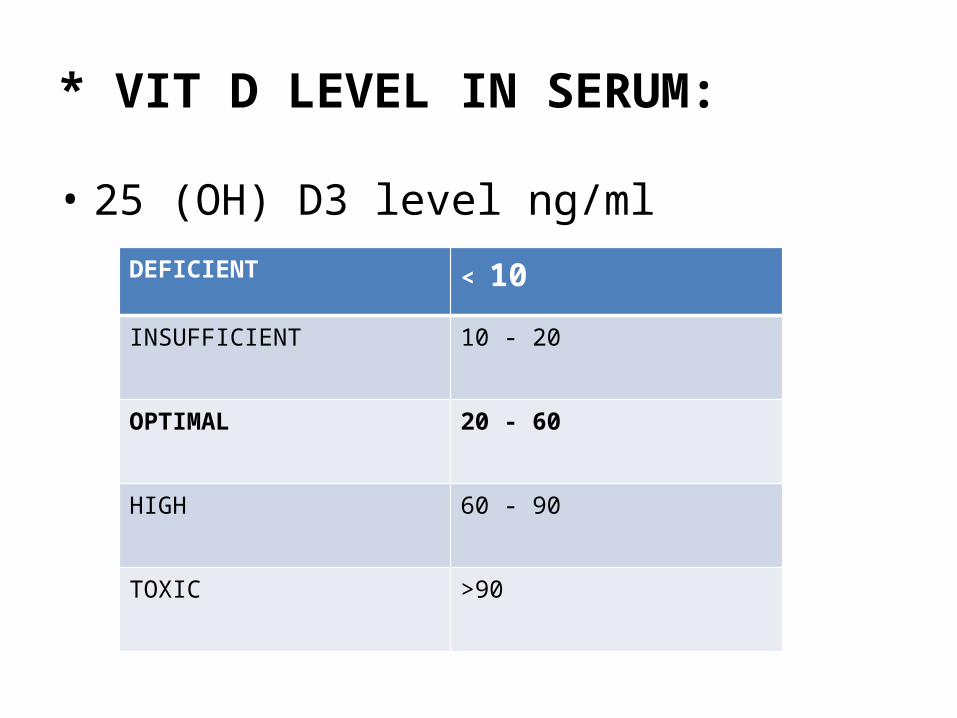
* VIT D LEVEL IN SERUM:
• 25 (OH) D3 level ng/mlDEFICIENT < 10
INSUFFICIENT 10 - 20
OPTIMAL 20 - 60
HIGH 60 - 90
TOXIC >90
Vitamin D Deficiency in Saudi Arabia
Group mostly affected are:• Breast- Fed infants because Human milk contains
little vitamin D, • Age < 2 years• Darked –skin children because darkly pigmented
skin, which blocks penetration of ultraviolet light necessary for formation of cholecalciferol (Vit. D3) from cholesterol in the skin.
• Low socio-economic Class• Urban > Rural
PARATHYROID HORMONE
• Stimulus for its secretion : fall in serum Ca.
• PTH promotes bone resorption process and is adversely affected by
calcitonin.
• PTH also stimulates the excretion of phosphates by the kidneys; this
inhibition of phosphate resorption in turn enables calcium resorption.
• In GIT - indirectly increases calcium absorption by increasing the synthesis of
active vit D 3 by stimulating alpha hydroxylase
CALCITONIN
• It is produced by para follicular c cells of thyroid.
• It is a calcium lowering hormone in serum by inhibiting
bone resorption by decreasing the no & activity of
osteoclasts .
• So calcitonin acts counter to PTH. Calcitonin inhibits
bone resorption thus causing serum calcium levels to fall.
Metabolic bone Diseases
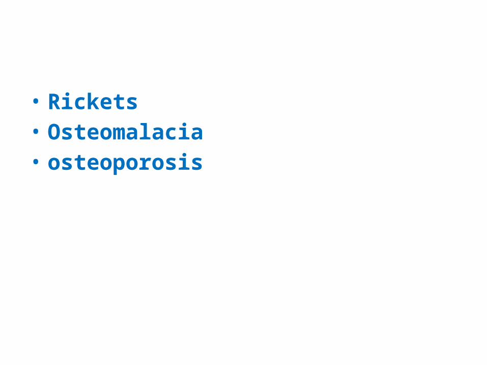
• Rickets• Osteomalacia• osteoporosis
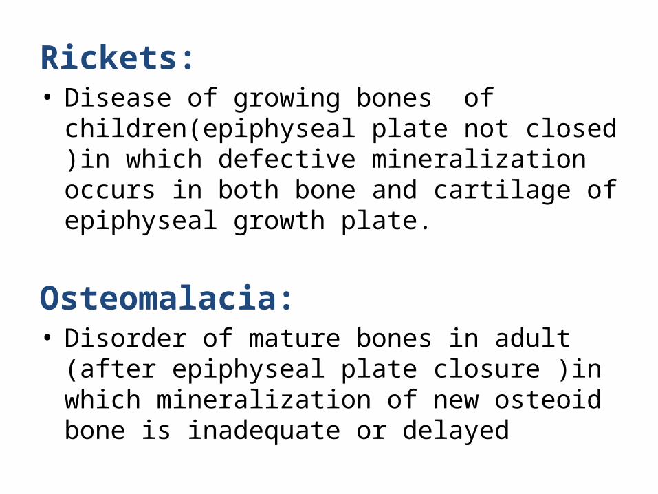
Rickets:• Disease of growing bones of children(epiphyseal
plate not closed )in which defective mineralization occurs in both bone and cartilage of epiphyseal growth plate.
Osteomalacia:• Disorder of mature bones in adult (after epiphyseal
plate closure )in which mineralization of new osteoid bone is inadequate or delayed
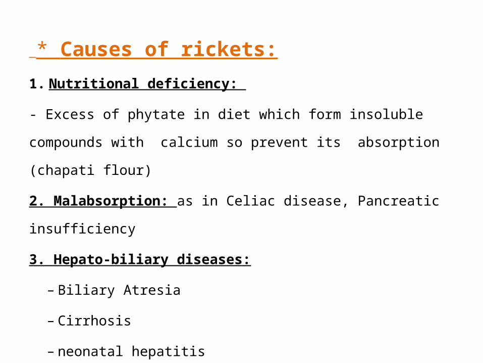
* Causes of rickets:
1. Nutritional deficiency:
- Excess of phytate in diet which form insoluble compounds
with calcium so prevent its absorption (chapati flour)
2. Malabsorption: as in Celiac disease, Pancreatic insufficiency
3. Hepato-biliary diseases:
– Biliary Atresia
– Cirrhosis
– neonatal hepatitis
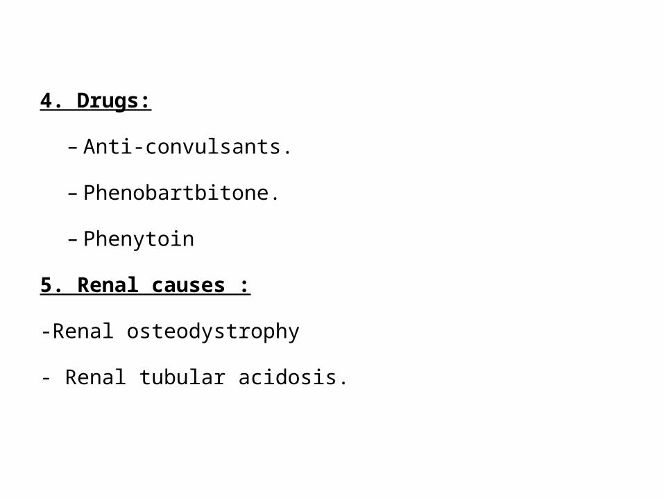
4. Drugs:
– Anti-convulsants.
– Phenobartbitone.
– Phenytoin
5. Renal causes :
-Renal osteodystrophy
- Renal tubular acidosis.
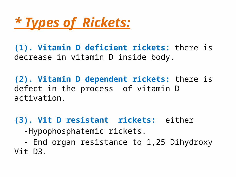
* Types of Rickets:
(1). Vitamin D deficient rickets: there is decrease in vitamin D inside body.
(2). Vitamin D dependent rickets: there is defect in the process of vitamin D activation. (3). Vit D resistant rickets: either -Hypophosphatemic rickets. - End organ resistance to 1,25 Dihydroxy Vit D3.
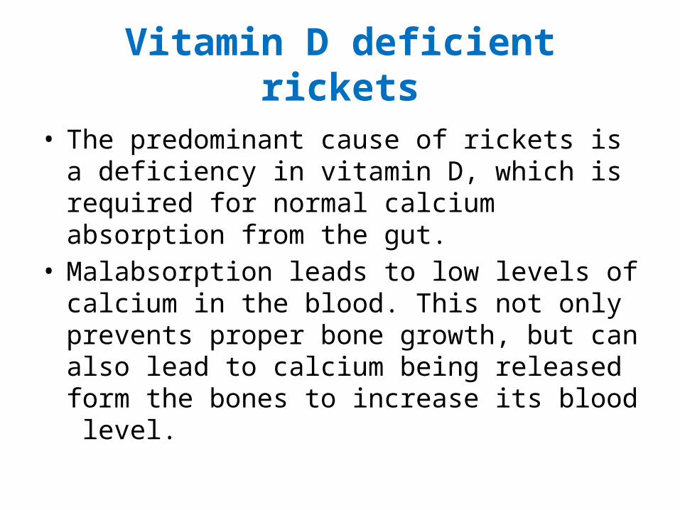
Vitamin D deficient rickets
• The predominant cause of rickets is a deficiency in vitamin D, which is required for normal calcium absorption from the gut.
• Malabsorption leads to low levels of calcium in the blood. This not only prevents proper bone growth, but can also lead to calcium being released form the bones to increase its blood level.
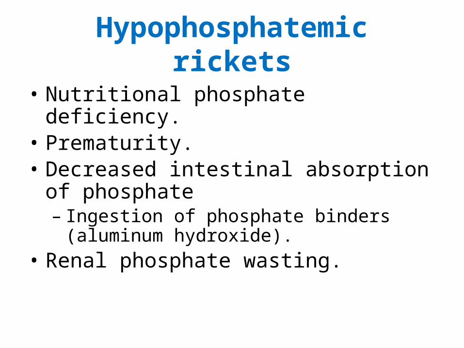
Hypophosphatemic rickets
• Nutritional phosphate deficiency.• Prematurity. • Decreased intestinal absorption of phosphate
– Ingestion of phosphate binders (aluminum hydroxide).
• Renal phosphate wasting.
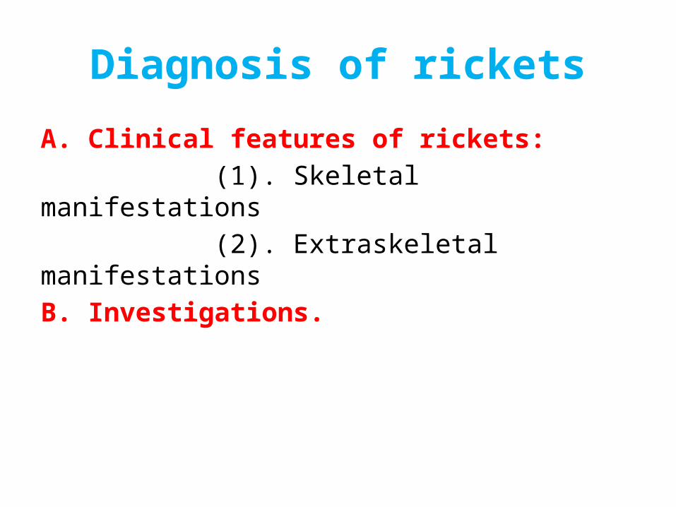
Diagnosis of rickets
A. Clinical features of rickets: (1). Skeletal manifestations (2). Extraskeletal manifestationsB. Investigations.
(1). Skeletal manifestations
• Craniotabes: The earliest sign of rickets in infant, is (abnormal softness of skull)
• Delayed closure of anterior fontanel.• Frontal and parietal bossing: Rounded prominence of the
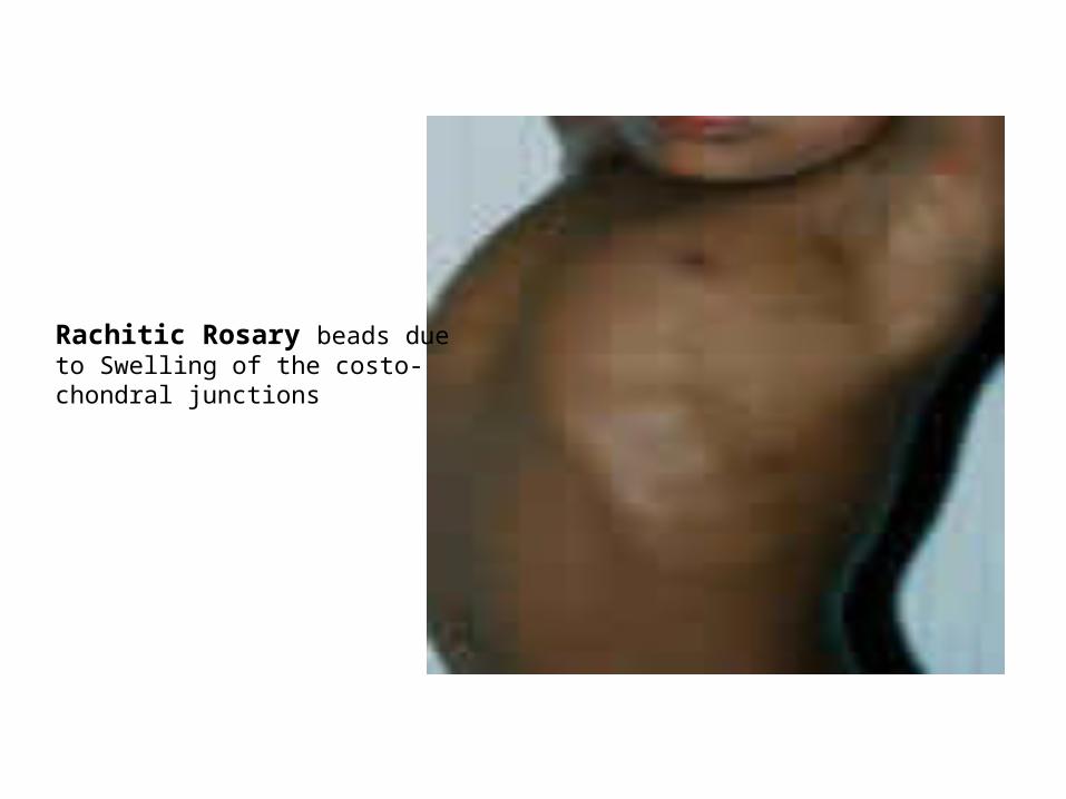
frontal and parietal bones in an infant’s cranial vault• Delayed eruption of primary teeth.• Enamel defects and caries teeth.• Rachitic rosary
– Swelling of the costo-chondral junctions.
• Harrison’s groove:– Lateral indentation of the chest wall at the site of
attachment of diaphragm because the patients lack the mineralized calcium in their bones necessary to harden them; thus the diaphragm, which is always in tension, pulls the softened bone inward.
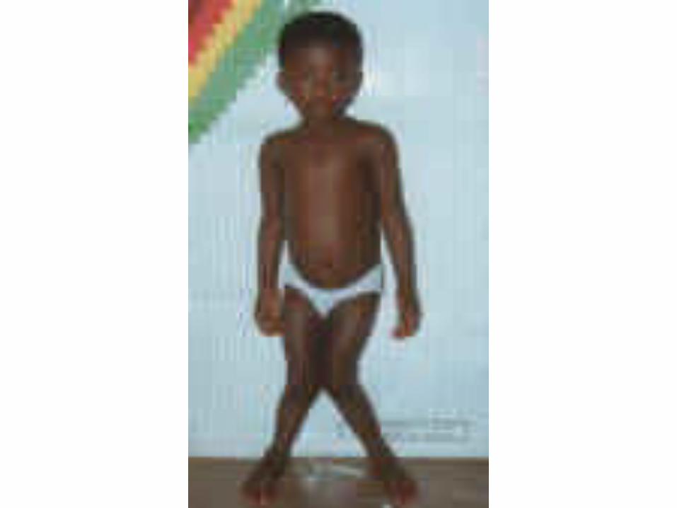
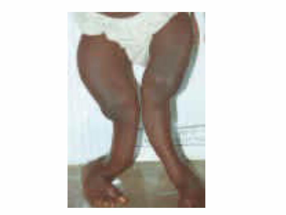
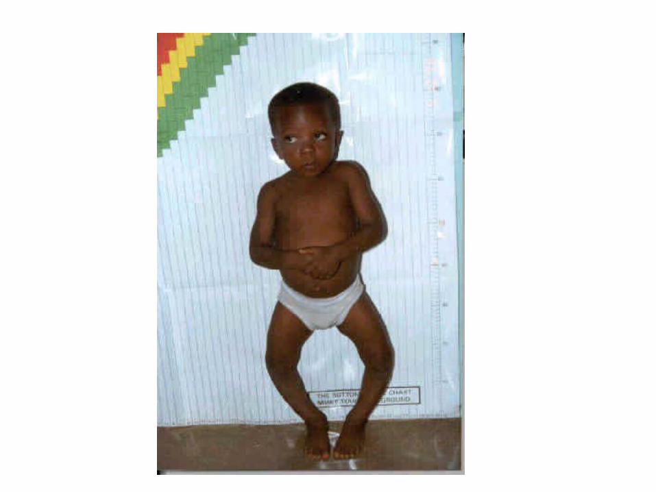
• Enlargement of long bones around wrists and ankles• Bow legs • knock knees• Green stick fractures• Deformities of spine, pelvis and leg – rachitic dwarfism• Growth retardation due to impaired calcification of bone
epiphysis.
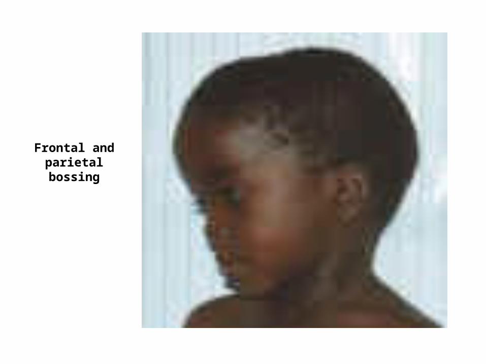
Frontal and parietal bossing
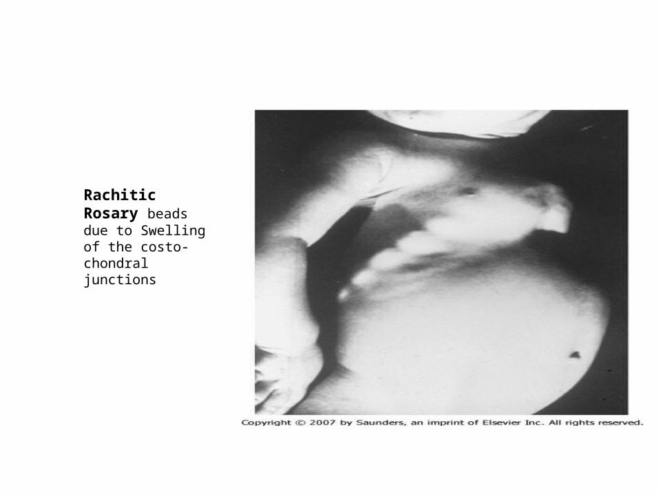
Rachitic Rosary beads due to Swelling of the costo-chondral junctions
Rachitic Rosary beads due to Swelling of the costo-chondral junctions
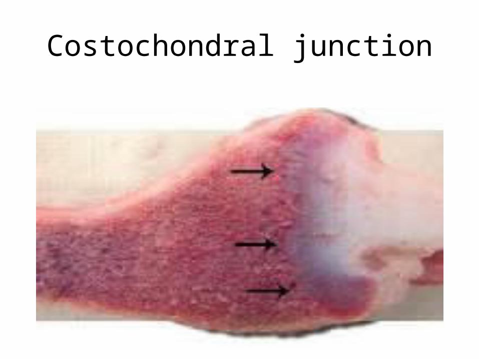
Costochondral junction
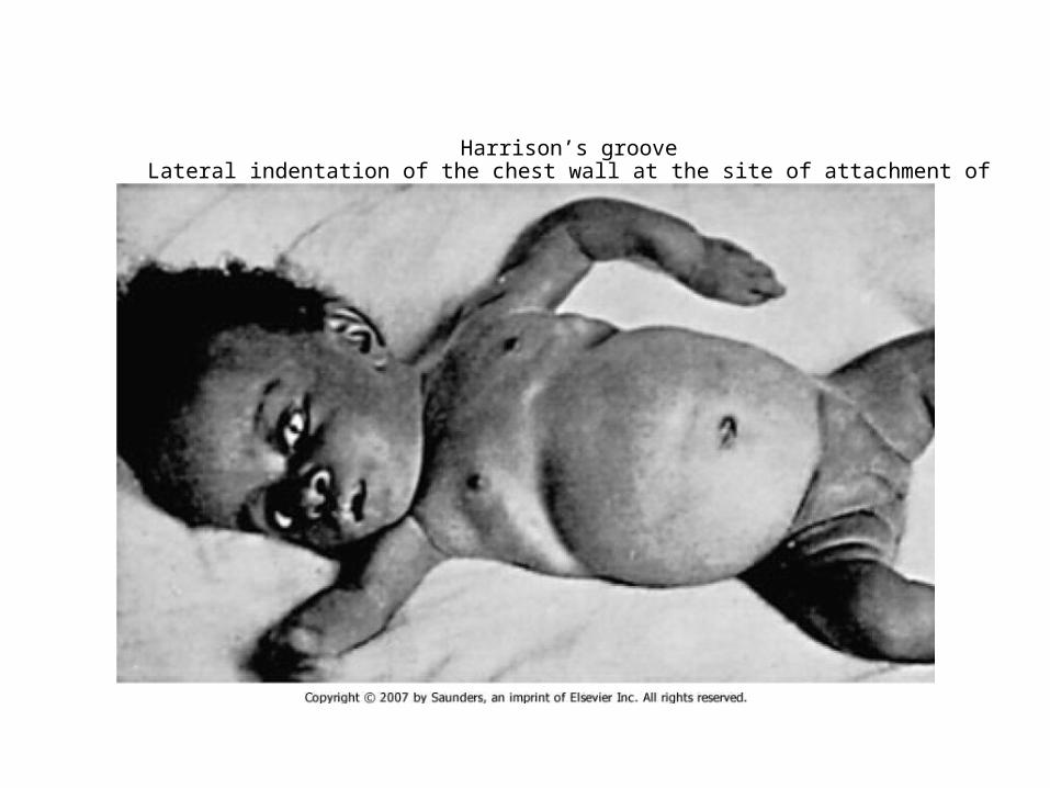
Harrison’s grooveLateral indentation of the chest wall at the site of attachment of diaphragm
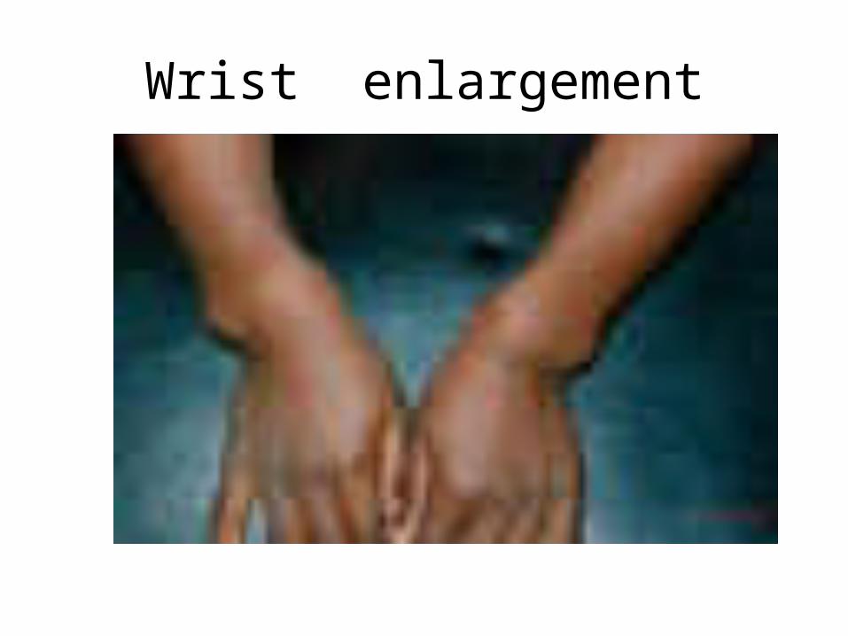
Wrist enlargement
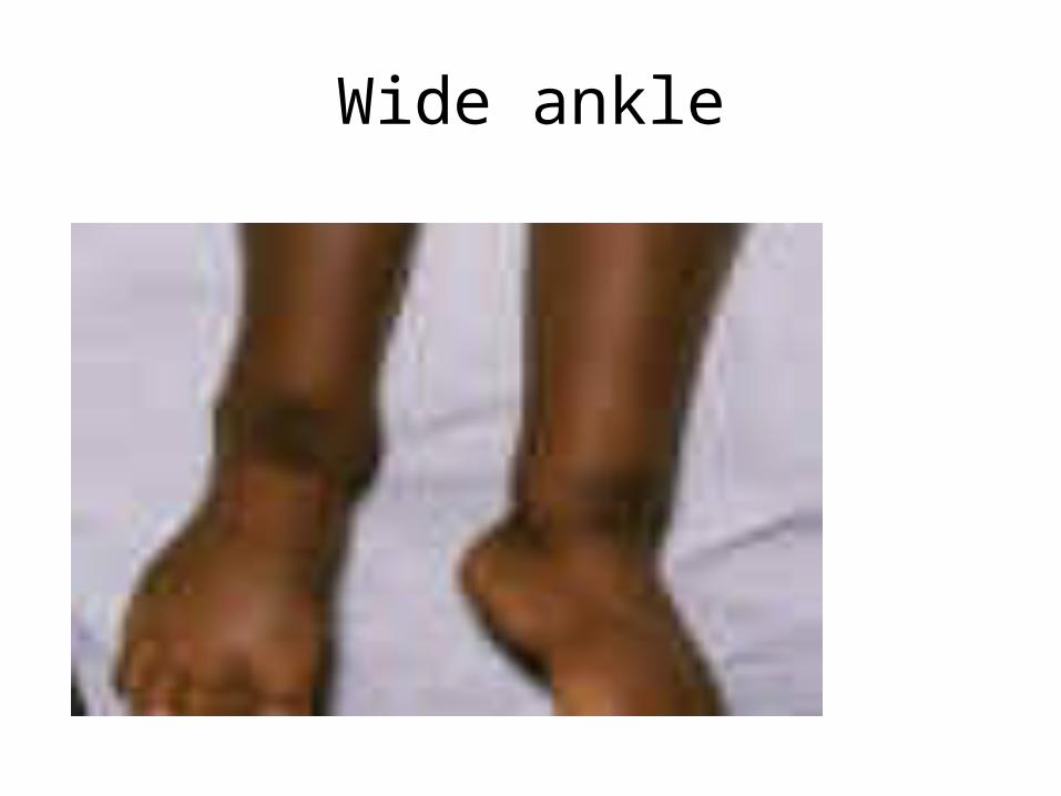
Wide ankle
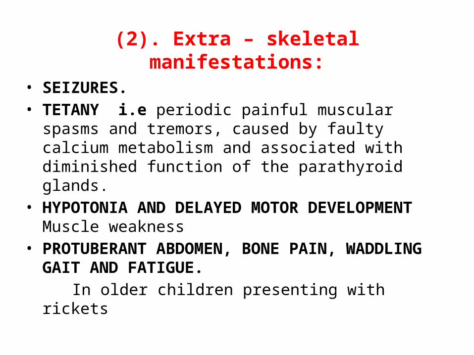
(2). Extra – skeletal manifestations:• SEIZURES. • TETANY i.e periodic painful muscular spasms and tremors,
caused by faulty calcium metabolism and associated with diminished function of the parathyroid glands.
• HYPOTONIA AND DELAYED MOTOR DEVELOPMENT Muscle weakness
• PROTUBERANT ABDOMEN, BONE PAIN, WADDLING GAIT AND FATIGUE.
In older children presenting with rickets
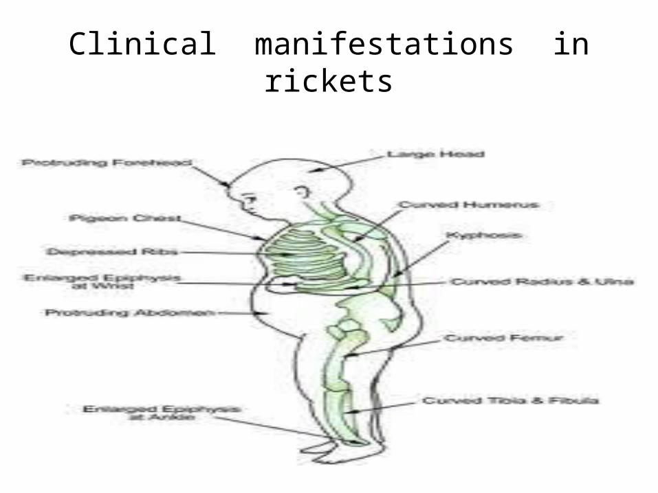
Clinical manifestations in rickets
B. Investigations
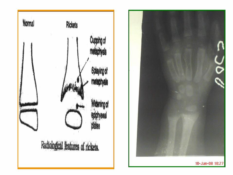
* BASIC INVESTIGATIONS TO CONFIRM RICKETS:• Serum Ca, P: Hypocalcemia if Serum Calcium
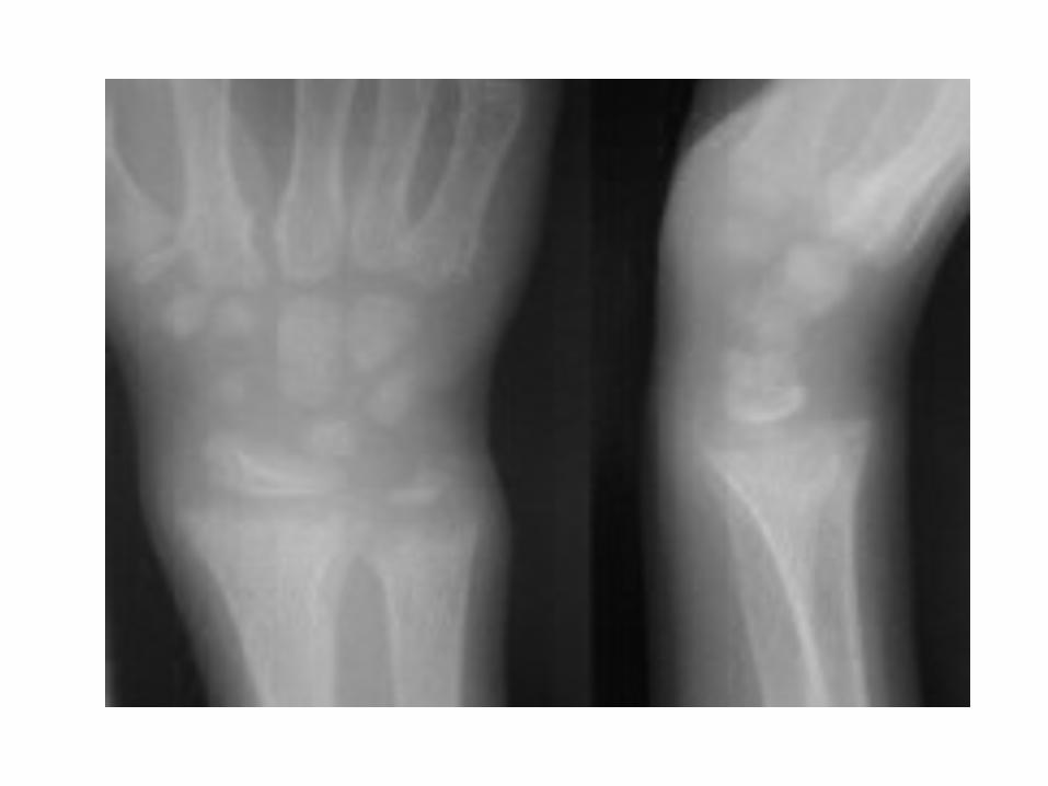
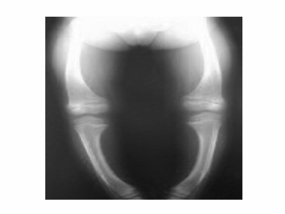
less than 8.0 mg/dl • X rays of ends of long bones at knees or wrists: Widening and cupping of the distal ends of shaft.
Osteoporosis & Osteomalacia
Difference Between Osteoporosis & Osteomalacia
• Osteoporosis refers to the degeneration of already constructed bone, making them brittle, while osteomalacia is an abnormality in the building process of bone, making them soft.
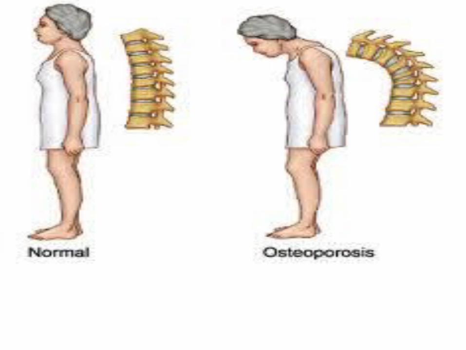
Osteoporosis The word osteoporosis literally means porous bones . It
occurs when rate of degeneration of already constructed bone is rapid and exceed rate of bone building .
This leads to loss of an excessive amount of their protein and mineral content .
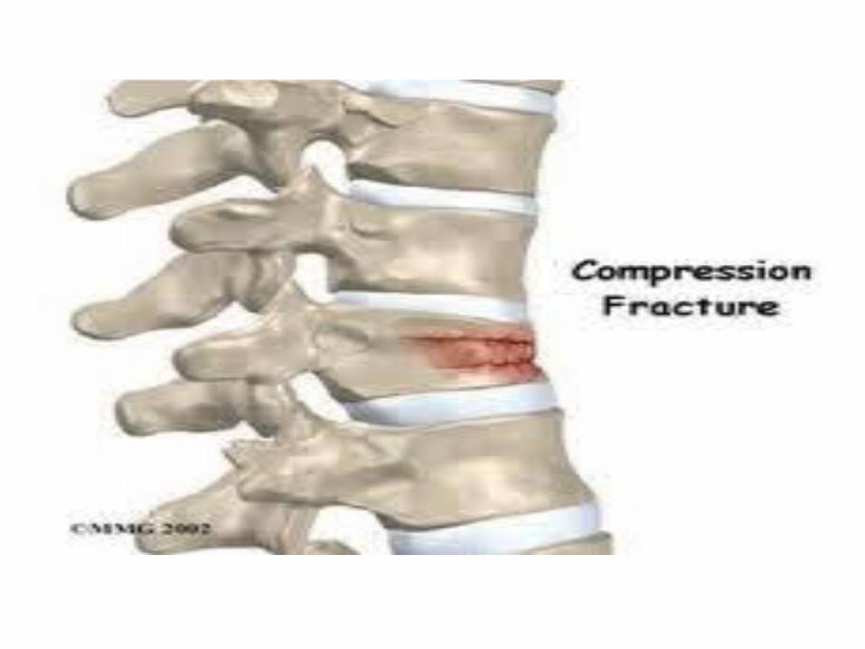
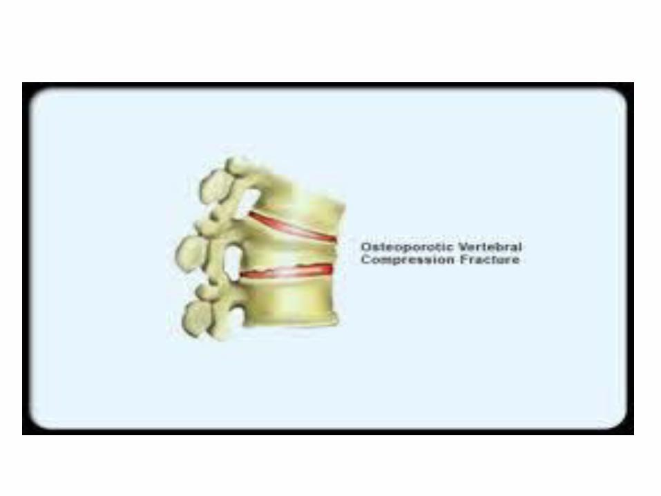
An osteoporotic bone characterized by:– Thinning of bone trabecula– low bone mass and volume.– Leading to enhanced bone fragility and increase in fracture
risk even with any trivial trauma.
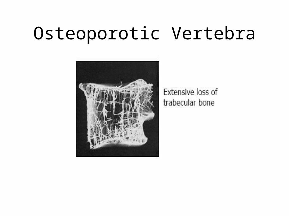
Osteoporotic Vertebra
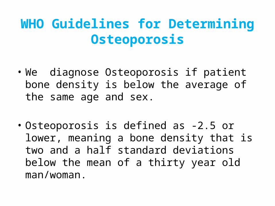
WHO Guidelines for Determining Osteoporosis
• We diagnose Osteoporosis if patient bone density is below the average of the same age and sex.
• Osteoporosis is defined as -2.5 or lower, meaning a bone density that is two and a half standard deviations below the mean of a thirty year old man/woman.
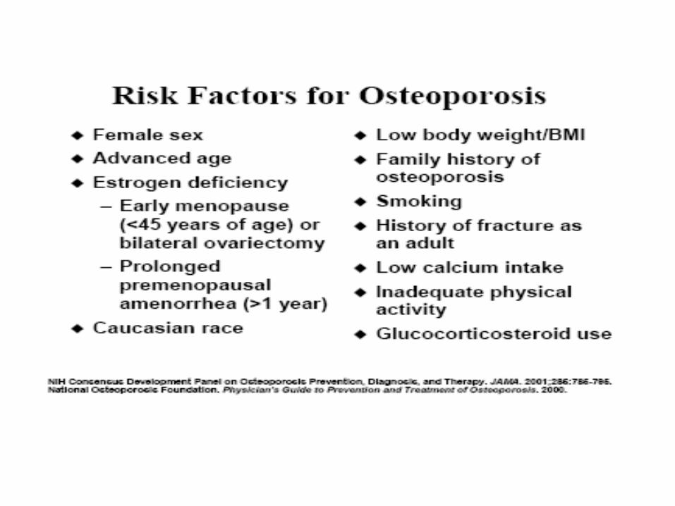
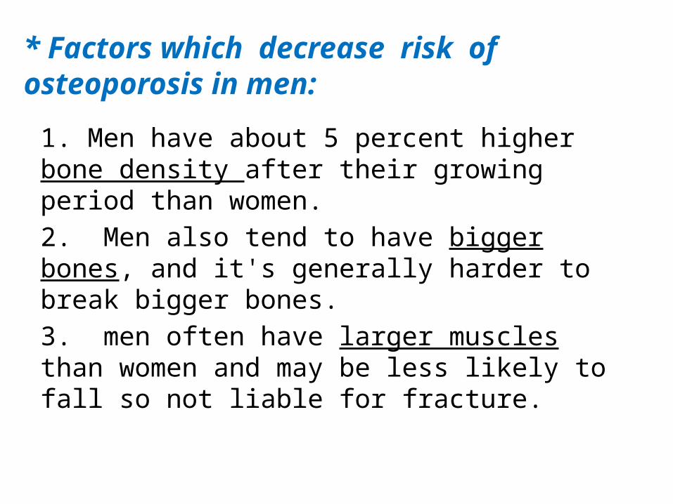
* Factors which decrease risk of osteoporosis in men:
1. Men have about 5 percent higher bone density after their growing period than women.2. Men also tend to have bigger bones, and it's generally harder to break bigger bones.3. men often have larger muscles than women and may be less likely to fall so not liable for fracture.
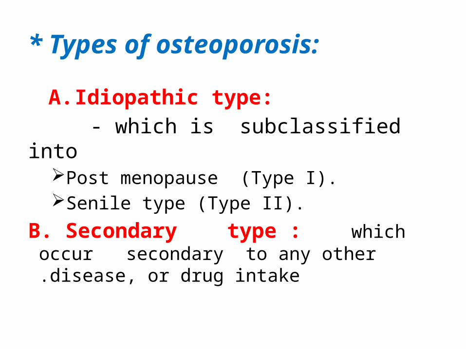
* Types of osteoporosis:
A. Idiopathic type: - which is subclassified into
Post menopause (Type I).Senile type (Type II).
B. Secondary type : which occur secondary to any other disease, or drug intake.
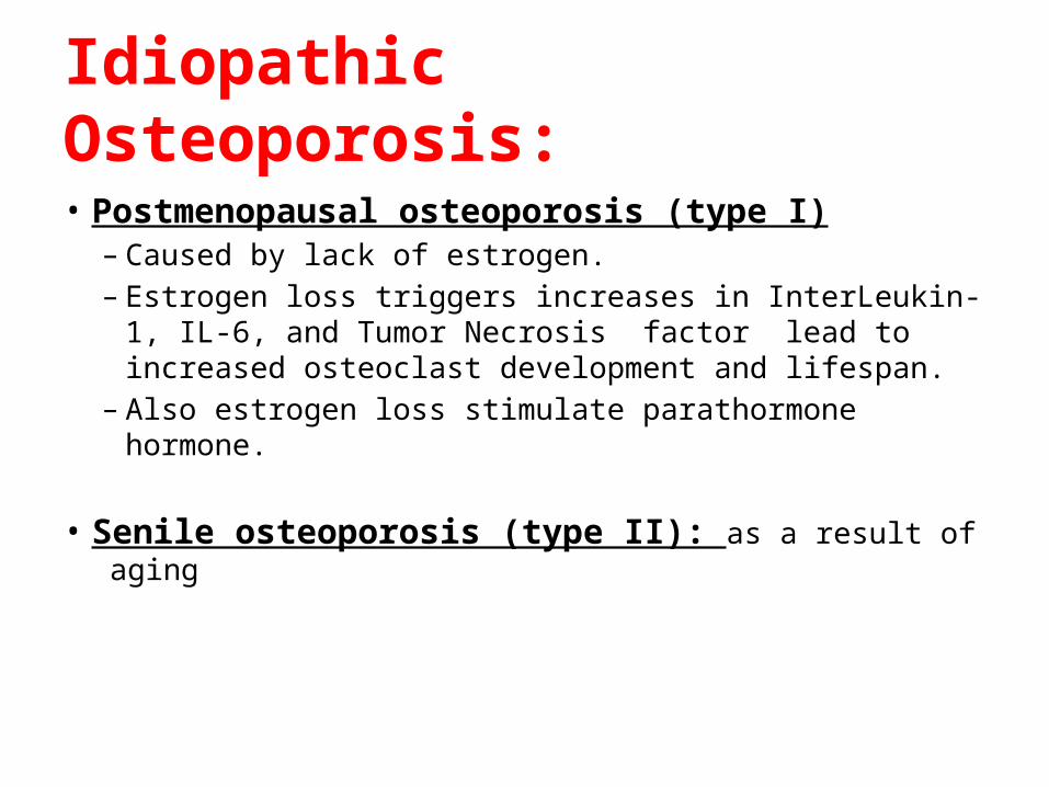
Idiopathic Osteoporosis:
• Postmenopausal osteoporosis (type I)– Caused by lack of estrogen.– Estrogen loss triggers increases in InterLeukin-1, IL-6,
and Tumor Necrosis factor lead to increased osteoclast development and lifespan.
– Also estrogen loss stimulate parathormone hormone.
• Senile osteoporosis (type II): as a result of aging
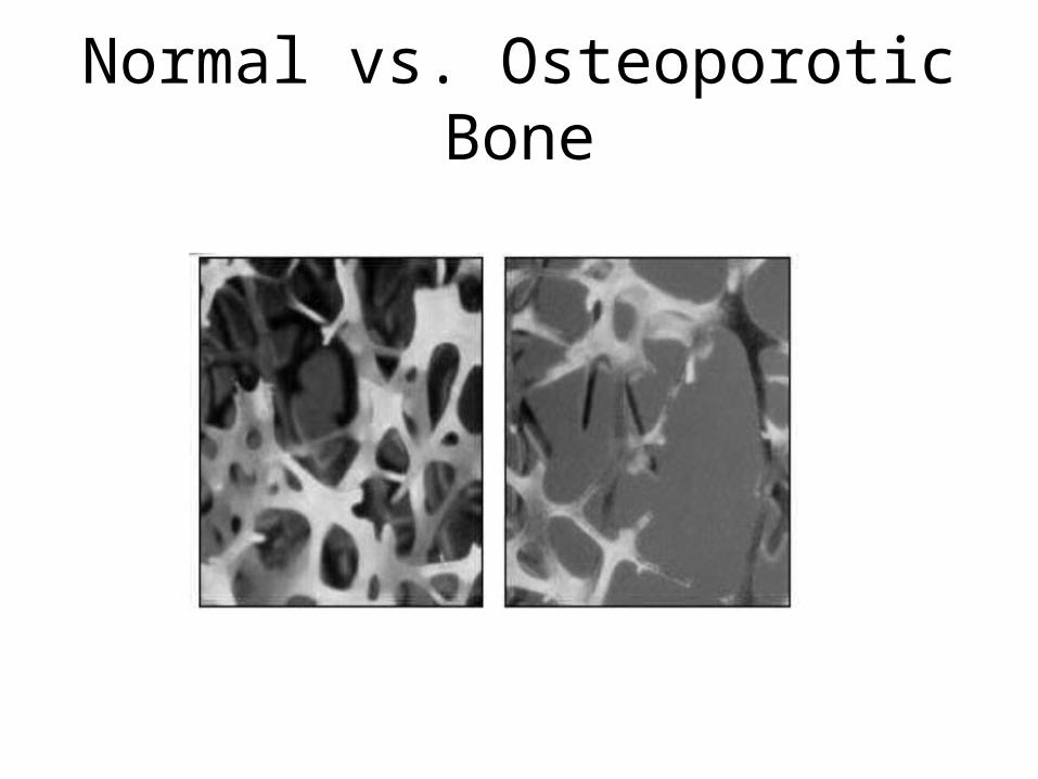
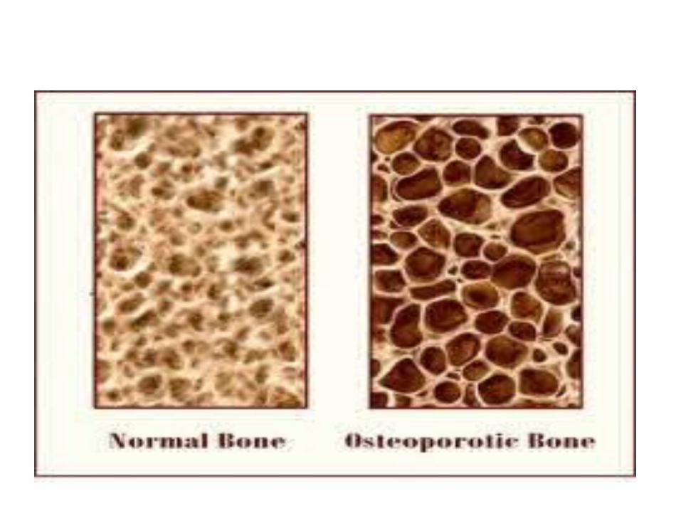
Normal vs. Osteoporotic Bone
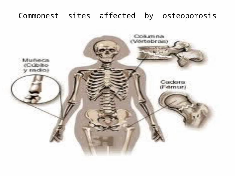
Commonest sites affected by osteoporosis
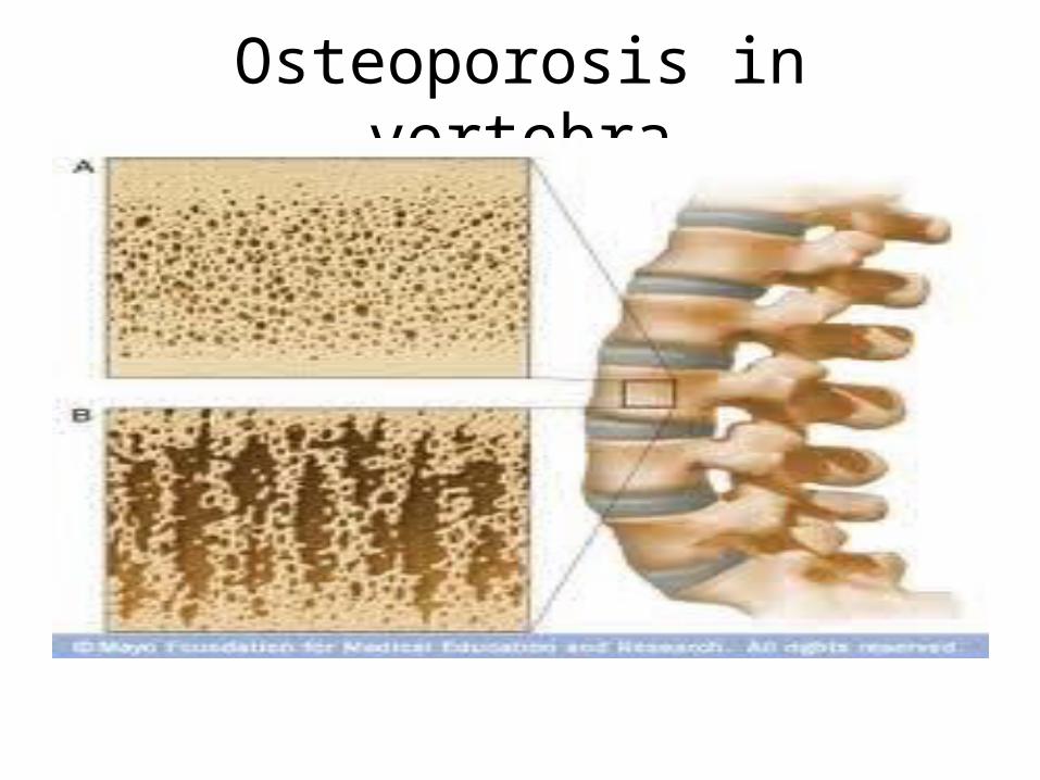
Osteoporosis in vertebra
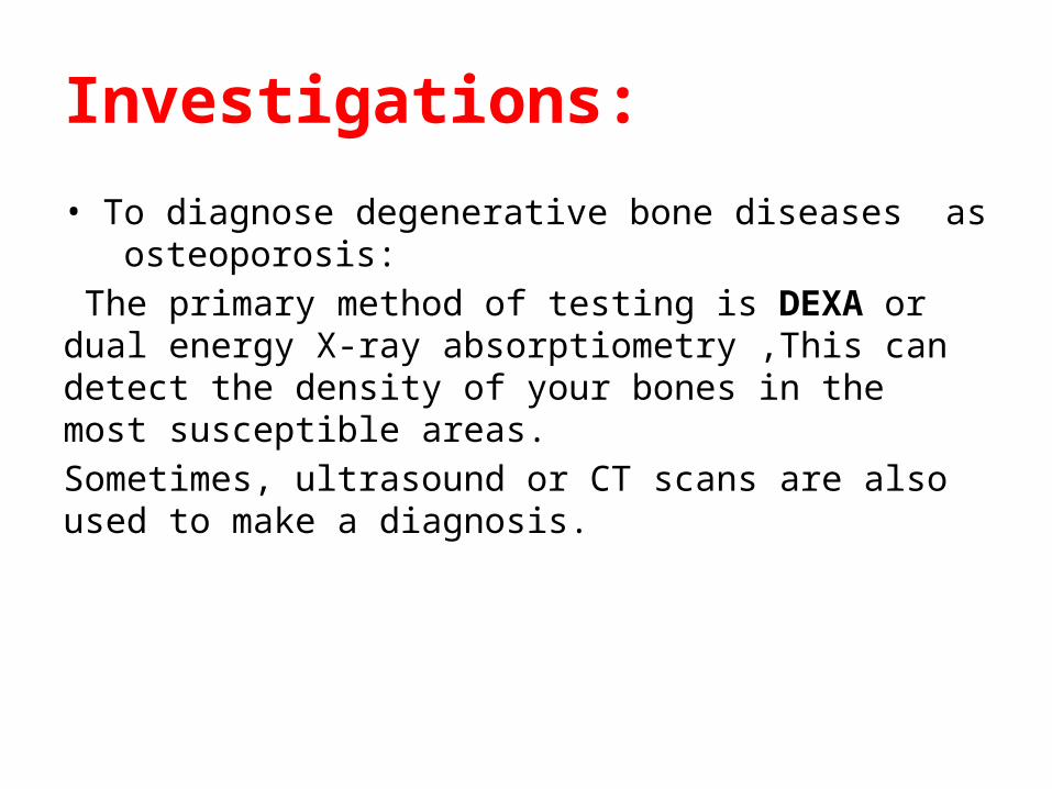
Investigations:
• To diagnose degenerative bone diseases as osteoporosis: The primary method of testing is DEXA or dual energy X-ray absorptiometry ,This can detect the density of your bones in the most susceptible areas. Sometimes, ultrasound or CT scans are also used to make a diagnosis.
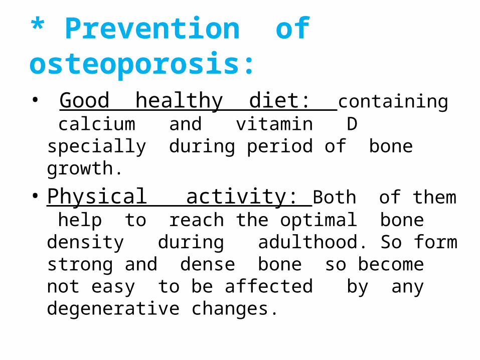
* Prevention of osteoporosis:
• Good healthy diet: containing calcium and vitamin D specially during period of bone growth.
• Physical activity: Both of them help to reach the optimal bone density during adulthood. So form strong and dense bone so become not easy to be affected by any degenerative changes.