Embed Size (px)
Citation preview
Proc. Nadl. Acad. Sci. USAVol. 83, pp. 7503-7507, October 1986Neurobiology
Messenger RNA coding for only the a subunit of the rat brain Nachannel is sufficient for expression of functional channels inXenopus oocytes
(hybrid selection/sucrose-gradient fractionation/voltage clamp)
ALAN L. GOLDIN*, TERRY SNUTCH*, HERMANN LUBBERT*, ANDREW DOWSETT*, JOHN MARSHALLt,VANESSA AULDt, WILLIAM DOWNEYf, LARRY C. FRITZ§¶, HENRY A. LESTER§, ROBERT DUNNt,WILLIAM A. CATTERALLO, AND NORMAN DAVIDSON**Church Chemical Laboratories, §Division of Biology, California Institute of Technology, Pasadena, CA 91125; tDepartment of Medical Genetics, Universityof Toronto, Toronto, Canada M5S 1A8; and *Department of Pharmacology, University of Washington, Seattle, WA 98195
Contributed by Norman Davidson, October 28, 1985
ABSTRACT Several cDNA clones coding for the highmolecular weight (a) subunit of the voltage-sensitive Na chan-nel have been selected by immunoscreening a rat brain cDNAlibrary constructed in the expression vector Xgtll. As will bereported elsewhere, the amino acid sequence translated fromthe DNA sequence shows considerable homology to that re-ported for the Electrophorus eketricus electroplax Na channel.Several of the cDNA inserts hybridized with a low-abundance9-kilobase RNA species from rat brain, muscle, and heart.Sucrose-gradient fractionation of rat brain poly(A) RNA yield-ed a high molecular weight fraction containing this mRNA,which resulted in functional Na channels when injected intooocytes. This fraction contained undetectable amounts of lowmolecular weight RNA. The high molecular weight Na channelRNA was selected from rat brain poly(A)RNA by hybridizationto a single-strand antisense cDNA clone. Translation of thisRNA in Xenopus oocytes resulted in the appearance oftetrodotoxin-sensitive voltage-sensitive Na channels in theoocyte membrane. These results demonstrate that mRNAencoding the a subunit of the rat brain Na channel, in theabsence of any ,B-subunit mRNA, is sufficient for translation togive functional channels in oocytes.
The initial depolarization event in the propagation of anaction potential in nerve and muscle is due to the opening ofvoltage-sensitive Na channels. This channel-protein com-plex has been purified from Electrophorus electricus elec-troplax membranes (1), rat muscle (2), chicken cardiacmuscle (3), and rat brain (4). As isolated, the electroplaxprotein consists of a single large subunit of Mr 260,000 (5-7)encompassing 1820 amino acids (8). Similarly, the chickenheart protein comprises one subunit of Mr 230,000-270,000(3). However, as isolated, the rat brain Na-channel complexcontains one large subunit (a) with a Mr of =260,000 asdetermined by NaDodSO4/PAGE, and two associated smallsubunits, (B1 and ,82, ofMr 36,000 and 33,000, respectively (9,10). The rat skeletal muscle channel also contains both a andat least one /3 subunit (11, 12). In adult rat brain, the a and ,82subunits are covalently attached by disulfide bonds (9). Boththe rat and eel protein complexes have been reconstitutedinto phospholipid vesicles, with restoration of toxin-inducedactivation of Na permeability (13-16).The cDNA coding for the electroplax protein has been
molecularly cloned and its sequence has been determined (8).Furthermore, while the present manuscript was in the proofstage, the cloning and sequencing of cDNAs coding for ratbrain Na channels was reported (17). This latter study shows
that there are at least two related a-subunit Na-channelmRNAs in rat brain, denoted I and II, and there is possiblya third.The functional significance of the small subunits in rat
brain is not known. Schmidt et al. (18) present data suggest-ing that disulfide linkage of the a and ,2 subunits, insertioninto the cell-surface membrane, and attainment of a func-tional conformation are closely related late events in thebiogenesis of functional Na channels in rat brain. However,Sumikawa et al. (19) have shown that when rat brain poly(A)RNA is fractionated by velocity centrifugation on a nonde-naturing sucrose gradient, a high molecular weight fractionby itself was sufficient to induce Na-channel function wheninjected into oocytes. A similar experiment, albeit at a lowerresolution, was carried out by Hirono et al. (35) with the sameresult. These results suggest that the small subunits are notnecessary for channel function in oocytes.
In this report, we have isolated cDNA clones encodingportions of the coding region of the large subunit of the Nachannel. We have used these clones to analyze a sedimen-tation fractionation experiment similar to that ofSumikawa etal. (19). We found that only those fractions that by hybrid-ization contained the high molecular weight a-specificmRNA [9 kilobases (kb)] were translated in the oocyte to givefunctional channels. Furthermore, the fractions containingthe a-subunit mRNA had undetectable quantities of lowmolecular weight RNA, as determined by hybridization withan unrelated probe that detects 2.2- and 2.4-kb brain mRNAs.We have also used the cDNA clones to hybrid-select a-subunit-specific mRNA. This mRNA induced synthesis ofvoltage-sensitive tetrodotoxin-inhibited Na channels wheninjected into Xenopus oocytes. These results indicate thata-subunit mRNA, in the absence of any P-subunit mRNA, istranslated in Xenopus oocytes to give functional Na chan-nels.
EXPERIMENTAL PROCEDURES
Isolation of Rat Brain RNA and Na Channel Clones. RNAwas isolated from the brains of 4- to 14-day-old rats by thelithium chloride/urea procedure (20) or by a modification ofthe procedure of Chirgwin et al. (21) using guanidine hydro-chloride. The procedures for construction ofcDNA librariesin the vector Xgt11 (22, 23) for immunological screening withan antibody to the rat brain channel (24) and for character-ization of the clones at the sequence level will be describedelsewhere.
Abbreviation: kb, kilobase(s).VPresent address: California Biotechnology, Inc., Palo Alto, CA94043.
7503
The publication costs of this article were defrayed in part by page chargepayment. This article must therefore be hereby marked "advertisement"in accordance with 18 U.S.C. §1734 solely to indicate this fact.
Proc. Natl. Acad. Sci. USA 83 (1986)
RNA Transfer. RNA was electrophoresed through a 1.1%agarose gel containing 2.2 M formaldehyde at 3.3 V/cm for8 hr as described (25). After electrophoresis, the gel wassoaked in water for 20 min, then transferred to nitrocelluloseor Hybond N (Amersham) (26). Hybridization was carriedout at 630C-680C as described (27).
Preparation of SP6 RNA Transcripts. Labeled hybridiza-tion probes were synthesized from 1 ,ug of linearized templateDNA in 20 ul with 500 ;kM each ATP, GTP, and UTP, and100 ,uCi of [32P]CTP (400 Ci/mmol; 1 Ci = 37 GBq;Amersham) (27, 28).Hybrid Selection. Nitrocellulose filters (Schleicher &
Schuell; 9-mm diameter) were loaded with 40 ,tg of DNA asdescribed (29). Filters were air-dried, washed three timeswith 6x SSC (lx SSC = 0.15 M NaCl/0.015 M Na citrate)and baked for 2 hr at 80'C under vacuum. To release anyDNA that was not properly bound, filters were immersed in0.3 ml of H20 and placed in a boiling water bath for 1 min.Prehybridizations were performed overnight at 650C in 1 MNaCl/0.1 M Pipes, pH 6.4/2 mM EDTA/yeast RNA (300,ug/ml; type III; Sigma). For hybridizations, 100 ,ul of thesame buffer was used, but the yeast RNA was replaced with30 ,ug ofpoly(A) RNA from the brains of 4- or 14-day-old rats.Hybridization time was 1 or 3 hr. Afterwards, RNA remain-ing in the hybridization solution was precipitated twice withethanol. The filters were washed three times with 0.2xSSC/0.2% NaDodSO4 at 65°C and three times with 10 mMTris HCl, pH 7.5/2 mM EDTA at room temperature. RNAbound on the filter was eluted by shaking the filters in 0.3 mlofH20 for 1 min in a boiling water bath. Subsequently, filterswere quick-frozen in an ethanol/dry ice bath, thawed on ice,and RNA in the water was precipitated twice with ethanoltogether with S ,ug of yeast RNA as carrier. The final pelletsof the bound RNA and of the RNA precipitated from thehybridization solutions were each dissolved in 3 ul of H20,and 0.06 ,lI of each was injected into Xenopus oocytes.RNA Fractionation by Sucrose Gradient Sedimentation.
Poly(A) rat brain RNA (150 ,ug in 100 ,ul of H20) was heatedto 65°C for 3 min and then layered on a 6-20% linear sucrosegradient containing 15 mM Pipes, pH 6.4/5 mm EDTA/0.25% sarcosyl. The gradient was centrifuged in an SW 27.1rotor at 24,000 rpm at 4°C for 18 hr. Thirty-four 0.5-mlfractions were collected and the RNA was precipitated withsodium acetate and ethanol. The RNA from every threefractions was then pooled for all further experiments. RNAgel blots utilized 1/20th of each pool per lane (equivalent to7.5 ,tg of unfractionated RNA) and oocyte microinjectionsutilized 1/1200th of each pool per oocyte (equivalent to 125ng of unfractionated RNA).RNA Injections into Oocytes and Electrophysiological Pro-
cedures. These procedures were carried out as described (28).After removing follicle cells by incubating the oocytes for 3hr in Ca2l-free OR-2 solution (30) containing collagenase (2mg/ml) (Sigma; type IA), 60 nl ofRNA solution was injectedinto the cytoplasm by using a device similar to that describedby Contreras et al. (31). The oocytes were kept in ND 96 (96mM NaCl/2 mM KC1/1.8 mM CaCl2/1 mM MgCl2/5 mMHepes NaOH, pH 7.5) supplemented with penicillin at 100units/ml, streptomycin at 100 ,ug/ml, and 0.5 mM theophyl-line at room temperature until testing (usually 40 hr).A standard two-microelectrode voltage clamp (model 8500,
Dagan Instruments, Minneapolis, MN), was used to test theoocytes in a recording chamber continuously perfused withND 96. The electrodes were filled with 3 M potassium
RESULTSIsolation and Characterization ofNa Channel cDNA Clones.
A rat brain cDNA library in the expression vector Xgtll wasscreened with antibody against the purified rat brain Nachannel by standard methods (23, 24). Additional clones wereisolated by hybridization screening of cDNA libraries withthe initial clones. The isolation and characterization at thesequence level of the cDNA clones will be described else-where.Sequence analysis revealed the expected moderate degree
ofhomology with the eel electroplax sequence (8), ranging upto 80% at the nucleotide level and 100% at the amino acidlevel. Comparison with the subsequently published se-quences of the rat brain Na channels (17) revealed that all ofthe clones isolated by us correspond to the rat channel II ofNoda et al. (17). However, the clones used in this paperwould all hybridize strongly to both rat channels I and II atthe stringencies used.RNA Gel Blots. The mRNA coding for the rat brain Na
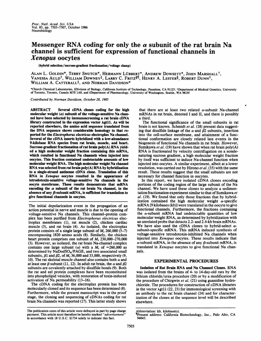
channel was identified by RNA gel blot hybridization. Highlylabeled single-strandRNA probes, =400 nucleotides long andcorresponding to the antisense strand, were synthesized inthe SP6 system. As shown in Fig. 1, a combination of twoantisense probes complementary to coding sequences in theamino-terminal and carboxyl-terminal regions of the rat II asubunit hybridized to anRNA species of9.0 kb. The intensityof hybridization indicates that this is an RNA of low abun-dance. There is a second fainter component of 8.0 kb. Stillfainter bands of 10.5 and 11.5 kb can be seen with appropriateexposures. In support of the interpretation that these bandscode for a voltage-sensitive Na channel, we observed that thesame probes hybridized to bands of comparable length but oflower intensity for RNA from rat heart and of still lowerintensity for skeletal muscle (Fig. 1), but that no signal wasseen with RNA from rat liver, kidney, or spleen (data notshown).
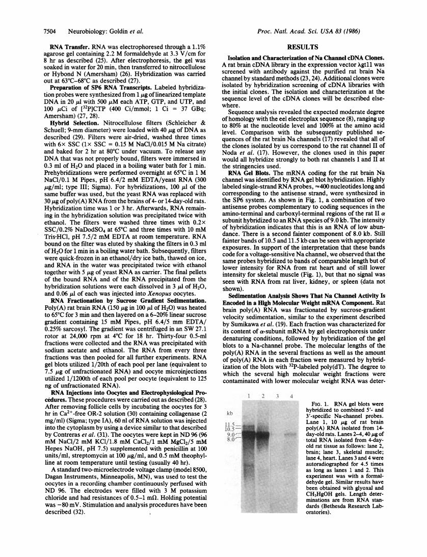
Sedimentation Analysis Shows That Na Channel Activity IsEncoded in a High Molecular Weight mRNA Component. Ratbrain poly(A) RNA was fractionated by sucrose-gradientvelocity sedimentation, similar to the experiment describedby Sumikawa et al. (19). Each fraction was characterized forits content of a-subunit mRNA by gel electrophoresis underdenaturing conditions, followed by hybridization of the gelblots to a Na-channel probe. The molecular lengths of thepoly(A) RNA in the several fractions as well as the amountof poly(A) RNA in each fraction were measured by hybrid-ization of the blots with 32P-labeled poly(dT). The degree towhich the several high molecular weight fractions werecontaminated with lower molecular weight RNA was deter-
1 2 3 4
kb
chloride and had resistances of 0.5-1 mfl. Holding potentialwas -80 mV. Stimulation and analysis procedures have beendescribed (32). i
FIG. 1. RNA gel blots werehybridized to combined 5'- and3'-specific Na-channel probes.Lane 1, 10 itg of rat brainpoly(A) RNA isolated from 14-day-old rats. Lanes 2-4, 40 ,ug oftotal RNA isolated from 4-day-old rat tissue as follows: lane 2,brain; lane 3, skeletal muscle;lane 4, heart. Lanes 3 and 4 wereautoradiographed for 4.5 timesas long as lanes 1 and 2. Thisexperiment was with a formal-dehyde gel. Similar results havebeen obtained with glyoxal andCH3HgOH gels. Length deter-minations are from RNA stan-dards (Bethesda Research Lab-oratories).
7504 Neurobiology: Goldin et al.
Proc. Natl. Acad. Sci. USA 83 (1986) 7505
mined by hybridization of the same blots with a cloned probethat was known to hybridize to two specific brain poly(A)RNA molecules of 2.2 and 2.4 kb.The results are presented in Figs. 2 and 3. Fig. 2 Left shows
that most of the discrete 9.0-kb a-subunit mRNA was infraction D, which contained poly(A) RNA in the range of7-12kb (Fig. 2 Right) and 1.5% of the poly(A) RNA recoveredfrom the gradient. This and other high molecular weightfractions contained no detectable amount of 2.2- to 2.4-kbpoly(A) RNA (Fig. 2 Center). Many of the fractions showedhybridization to degraded Na-channel RNA of lower molec-ular weight. Comparison with the unfractionated RNA laneshows that some random degradation had occurred duringsedimentation. We estimate by densitometric scans that50%o of the Na-channel hybridization is retained in the
discrete 9.0- and 8.0-kb bands.A quantity of 1/1200th of each fraction (see Experimental
Procedures) was injected into each of several oocytes and thepeak Na-channel current was measured. The data in Fig. 3show that all of the Na-channel activity as assayed by oocyteinjection resided in fractions C and D. As can be seen (Figs.2 and 3), these two fractions contained full-length Na-channelmRNA. The specific activity in fraction D had been enrichedat least 6-fold compared to unfractionated RNA, based on thesize of the peak current and the amount of RNA in thatfraction. No Na-channel activity was detectable in any of thelower molecular weight fractions E-K. No lower molecularweight RNA was detectable in fractions C and D (Figs. 2 and3), indicating that the Na channels synthesized from mRNAin these fractions did not contain any subunits coded for bylower molecular weight RNAs.
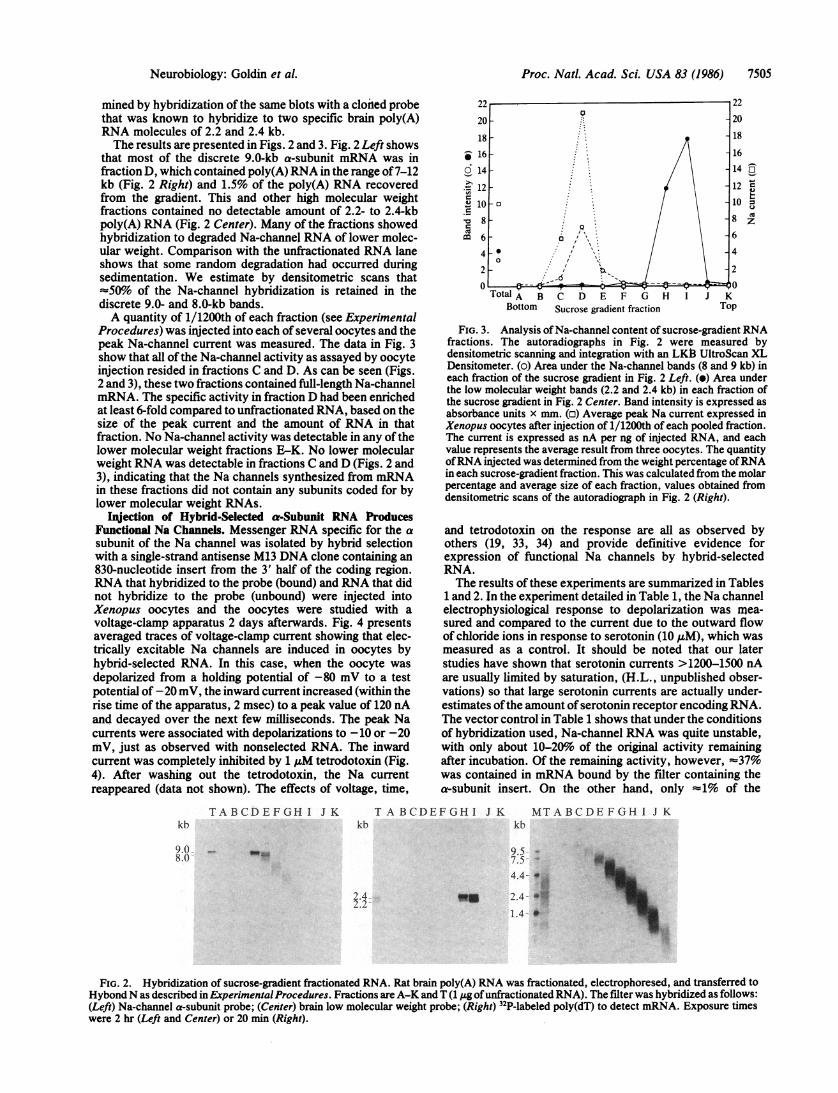
14jection of Hybrid-Selected a-Subunit RNA ProducesFunctional Na Channels. Messenger RNA specific for the asubunit of the Na channel was isolated by hybrid selectionwith a single-strand antisense M13 DNA clone containing an830-nucleotide insert from the 3' half of the coding region.RNA that hybridized to the probe (bound) and RNA that didnot hybridize to the probe (unbound) were injected intoXenopus oocytes and the oocytes were studied with avoltage-clamp apparatus 2 days afterwards. Fig. 4 presentsaveraged traces of voltage-clamp current showing that elec-trically excitable Na channels are induced in oocytes byhybrid-selected RNA. In this case, when the oocyte wasdepolarized from a holding potential of -80 mV to a testpotential of -20 mV, the inward current increased (within therise time of the apparatus, 2 msec) to a peak value of 120 nAand decayed over the next few iilliseconds. The peak Nacurrents were associated with depolarizations to -10 or -20mV, just as observed with nonselected RNA. The inwardcurrent was completely inhibited by 1 ,uM tetrodotoxin (Fig.4). After washing out the tetrodotoxin, the Na currentreappeared (data not shown). The effects of voltage, time,
I1-
0
60.1=1.5cu
mi
Ta T "I leT aTotalA B C D E F G H IBottom Sucrose gradient fraction
20
18
16
14 z_12 'r-U10 =e10
6
4
2
J KTop
FIG. 3. Analysis of Na-channel content of sucrose-gradient RNAfractions. The autoradiographs in Fig. 2 were measured bydensitometric scanning and integration with an LKB3 UltroScan XLDensitometer. (o) Area under the Na-channel bands (8 and 9 kb) ineach fraction of the sucrose gradient in Fig. 2 Left. (e) Area underthe low molecular weight bands (2.2 and 2.4 kb) in each fraction ofthe sucrose gradient in Fig. 2 Center. Band intensity is expressed asabsorbance units x mm. (o) Average peak Na current expressed inXenopus oocytes after injection of 1/1200th of each pooled fraction.The current is expressed as nA per ng of injected RNA, and eachvalue represents the average result from three oocytes. The quantityofRNA injected was determined from the weight percentage ofRNAin each sucrose-gradient fraction. This was calculated from the molarpercentage and average size of each fraction, values obtained fromdensitometric scans of the autoradiograph in Fig. 2 (Right).
and tetrodotoxin on the response are all as observed byothers (19, 33, 34) and provide definitive evidence forexpression of functional Na channels by hybrid-selectedRNA.The results of these experiments are summarized in Tables
1 and 2. In the experiment detailed in Table 1, the Na channelelectrophysiological response to depolarization was mea-sured and compared to the current due to the outward flowof chloride ions in response to serotonin (10 ,uM), which wasmeasured as a control. It should be noted that our laterstudies have shown that serotonin currents >1200-1500 nAare usually limited by saturation, (H.L., unpublished obser-vations) so that large serotonin currents are actually under-estimates ofthe amount ofserotonin receptor encoding RNA.The vector control in Table 1 shows that under the conditionsof hybridization used, Na-channel RNA was quite unstable,with only about 10-20%o of the original activity remainingafter incubation. Of the remaining activity, however, =37%was contained in mRNA bound by the filter containing thea-subunit insert. On the other hand, only -1% of the
TABCDEFGHI J K T A BCDEFGHI J Kkb
9.08.0
2.4.,;-4
kbMTABCDEFGH1 JK
7.5-4.4- 0
us 24-- 0
1.4- 0
FIG. 2. Hybridization of sucrose-gradient fractionated RNA. Rat brain poly(A) RNA was fractionated, electrophoresed, and transferred toHybondN as described in ExperimentalProcedures. Fractions are A-K and T (1 j.g ofunfractionated RNA). The filter was hybridized as follows:(Left) Na-channel a-subunit probe; (Center) brain low molecular weight probe; (Right) 32P-labeled poly(dT) to detect mRNA. Exposure timeswere 2 hr (Left and Center) or 20 mm (Right).
20 o
1816
14
12
LO -oc
8
6
4.0
2 .
kb
Neurobiology: Goldin et al.

Proc. Natl. Acad. Sci. USA 83 (1986)
125
nA 4
-125 010
-+TTX
mS
10
FIG. 4. Na-channel current induced by hybrid-selected RNA.Poly(A) RNA from the brains of 4-day-old rats was hybrid-selectedwith an antisense cDNA clone for the Na channel and injected intoXenopus oocytes. The oocytes were tested 40 hr later for voltage-sensitive Na currents. These tracings represent voltage-clamp cur-rents associated with a jump from -80 to -20 mV. The transientupward deflection is the current required to charge the membranecapacitance upon depolarization. This is followed by the downwarddeflection, which is due to the inward flow ofNa ions. The Na currentwas completely abolished in the presence of 1 ,uM tetrodotoxin(TTX).
serotonin activity was contained in the mRNA bound to theNa-channel probe.
In the experiments summarized in Table 2, the averageNa-channel signal from mRNA bound to the hybridizationfilter was divided by the average signal from mRNA that didnot bind to the filter to determine a bound-to-unbound ratio.As can be seen, this ratio of bound to unbound Na-channelmRNA activity was 3.1 for hybrid selection with a Nachannel a probe, whereas the ratio was only 0.16 for hybridselection with a vector control. These experiments furtherdemonstrate that RNA selected by hybridization to a Nachannel a-subunit insert is sufficient for expression of func-tional Na channels when injected into Xenopus oocytes.
DISCUSSION
We have isolated clones encoding the high molecular weight(a) subunit of the rat brain voltage-sensitive Na channel byimmunological screening of cDNA libraries. The structuralcharacterization of these clones will be reported indepen-dently.
Table 1. Na-channel and serotonin receptor expressionby hybrid-selected RNA
Na-channel Serotonin Na/serotonin,tpeak current,* peak normalized
RNA nA current,* nA ratio
Brain A' 664 155(7) 1125 377(4) 1.00(±0.40)Bound
(a insert) 37 5(11) 12 5(6) 5.20(±2.00)Unbound
(a insert) 64 9(9) 1000 184(8) 0.11(±0.03)Unbound
(vector) 107 23(3) 1920 ± 650(3) 0.09(±0.04)
*Average peak values ± SEM for the number of oocytes surviving(in parentheses). Holding potential for the serotonin-induced cur-rent was -60 mV.tRatios were obtained by dividing Na peak current by serotonin peakcurrent, and then normalizing so that the value for rat brain poly(A)(Brain Al) was equal to 1.0.
Table 2. Na-channel expression by hybrid-selected RNA
Na-channel peakcurrent, nA*
RNA Exp. 1 Exp. 2 Bound/unboundtBrain A+t 313 ± 82(7) 288 ± 74(6)Bound
(a insert) 17 ± 10(24) 63 ± 29(3) 1Unbound 3.10
(a insert) 0 (10) 38 ± 8(3) JBound
(vector) 0 (10) 22 ± 8(6) 1Unbound 0.16
(vector) 31 ± 14(9) 113 ± 20(3) J
*Average peak values ± SEM for the number of oocytes surviving(in parentheses).
tRatios were obtained by averaging all values for experiments 1 and2 and then dividing the peak Na current for bound RNA by that forunbound RNA.tRat brain poly(A) RNA.
Using the Na-channel clones, we have analyzed the frac-tionation ofNa-channelmRNA by sucrose-gradient sedimen-tation. Sumikawa et al. (19) and Hirono et al. (35) hadpreviously shown that after nondenaturing sucrose-gradientcentrifugation of rat brain poly(A) RNA, Na-channel activity(as assayed by the oocyte injection assay) resided in a highmolecular weight fraction. In a similar experiment in whichwe also tested the effectiveness of the size fractionation byhybridization with Na channel and other probes, we alsofound that injection of a high molecular weight fraction gavefunctional channels. We further demonstrated by RNA gelblots that this fraction was enriched in undegraded 9-kbNa-channel mRNA, that it contained only RNA in the rangeof 7-12 kb in detectable amounts, and, specifically, thatRNAof the length that might be expected for a subunit of Mr33,000-36,000 was not present in the fraction (see below).To confirm that a-subunit RNA alone is sufficient to
encode functional Na channels, we performed positive hy-brid-selection experiments with an a-subunit antisensecDNA clone affixed to a membrane filter. These experimentsdemonstrated that a-subunit RNA selected by hybridizationand injected into Xenopus oocytes did result in Na channelsthat showed the expected time response and voltage-sensi-tive characteristics and were inhibited by tetrodotoxin.However, rather small Na-channel activities were recoveredby hybrid selection because of the instability of Na-channelRNA. Messenger RNA encoding the serotonin receptor wasquite stable under the same hybridization conditions, and<1% of serotonin mRNA activity was recovered in thehybrid-selected fraction (Table 1). In addition, the ratio ofhybridized to unhybridized mRNA encoding Na-channelactivity was =z20-fold greater when an a-subunit DNA probewas used compared to the vector DNA alone (Table 2).Therefore, it is quite unlikely that any other mRNA, forexample Na-channel (3-subunit mRNA, was present in thehybrid-selected material. Thus, a-subunit mRNA by itselfwas sufficient for the synthesis of functional Na channels inthe oocyte.The A3 subunits of the rat brain Na channel have M, values
of 36,000 and 33,000 when glycosylated and 23,000 and 21,000when deglycosylated (10). Thus, the required coding length is<0.7 kb ofRNA. Based on other precedents, it is improbable(but not impossible) that ,3-subunit mRNA could lie in the sizerange of 7-12 kb included in the active fraction. It is morelikely that RNA encoding a protein of this size will be in the1- to 3-kb size range. No RNA of this size could be detectedin the sucrose-gradient fractions, which contained the RNAactive for expression of functional channels in the oocyte
7506 Neurobiology: Goldin et al.

Proc. Natl. Acad. Sci. USA 83 (1986) 7507
(Fig. 3). Therefore, the sedimentation experiment supportsthe hybridization-selection experiment in showing that a-subunit RNA by itself is sufficient for functional expressionin oocytes.What then is the role of the (1 and 832 subunits in the rat
brain channel? The (31 subunit is covalently labeled byphotoreactive derivatives of a- and (subunit scorpion tox-ins, suggesting that it is located at or near the receptor sitesat which these toxins modify Na-channel gating (36, 37).Selective removal of the 31 subunit from purified brain Nachannels in detergent solution is accompanied by loss ofsaxitoxin binding activity (38) and of the ability to reconsti-tute neurotoxin-activated 22Na' influx on incorporation intolipid vesicles (D.J. Messner and W.A.C., unpublished data),suggesting that a complex of a and (31 subunit is required tomaintain a functional state of the detergent-solubilized andpurified Na channel. In contrast, selective removal of the (32subunit has no apparent effect on the functional properties ofpurified Na channels. Schmidt et al. (18) offer evidence thatdisulfide-bond linkage of the (82 subunit with the a subunitoccurs before the incorporation of intracellular a chains intothe cell-surface membrane. This linkage is evidently unnec-essary for insertion into the oocyte membrane. It is conceiv-able that the oocyte has endogenous polypeptides that playthe role of the 83 subunits, but there is no evidence for thishypothesis. Further analysis of the physiological and phar-macological properties ofNa channels produced by a-subunitmRNA in oocytes may reveal differences that result from theabsence of (1 and /32 subunits.
We thank Nathan Dascal for his expert assistance and advice inperforming the electrophysiological procedures. This research hasbeen supported by grants from the National Institutes of Health toN.D., W.A.C., and H.A.L.; by a grant from the Medical ResearchCouncil of Canada to R.D.; and by fellowship support from theAmerican Heart Association to A.D. and T.S.; from the NaturalSciences and Engineering Research Council of Canada to T.S.; fromthe Deutsche Forschungsgemeinschaft to H.L.; and from the Na-tional Multiple Sclerosis Society to A.L.G.
1. Agnew, W. S., Levinson, S. R., Brabson, J. S. & Raftery,M. A. (1978) Proc. Nat!. Acad. Sci. USA 75, 2606-2610.
2. Barchi, R. L., Cohen, S. A. & Murphy, L. E. (1980) Proc.Nat!. Acad. Sci. USA 77, 1306-1310.
3. Lombet, A. & Lazdunski, M. (1984) Eur. J. Biochem. 141,651-660.
4. Hartshorne, R. P. & Catterall, W. A. (1981) Proc. Nat!. Acad.Sci. USA 78, 4620-4624.
5. Agnew, W. A., Moore, A. C., Levinson, S. R. & Raftery,M. A. (1980) Biochem. Biophys. Res. Commun. 92, 860-866.
6. Nakayama, H., Withy, R. M. & Raftery, M. A. (1982) Proc.Nat!. Acad. Sci. USA 79, 7575-7579.
7. Miller, J. A., Agnew, W. A. & Levinson, S. R. (1983) Bio-chemistry 22, 462-470.
8. Noda, M., Shimizu, S., Tanabe, T., Takai, T., Kayano, T.,Ikeda, T., Takahashi, H., Nakayama, H., Kanaoka, Y.,Minamino, N., Kangawa, K., Matsuo, H., Raftery, M. A.,Hirose, T., Inayama, S., Hayashida, H., Miyata, T. & Numa,S. (1984) Nature (London) 312, 121-127.
9. Hartshorne, R. P., Messner, D. J., Coppersmith, J. C. &Catterall, W. A. (1982) J. Biol. Chem. 257, 13888-13891.
10. Messner, D. J. & Catterall, W. A. (1986) J. Biol. Chem. 260,10597-10604.
11, Barchi, R. L. & Murphy, L. E. (1981) J. Neurochem. 36,207-210.
12. Casadei, J., Gordon, R., Lampson, L., Schotland, D. &Barchi, R. (1984) Proc. Nat!. Acad. Sci. USA 81, 6227-6231.
13. Talvenheimo, J. A., Tamkun, M. M. & Catterall, W. A. (1982)J. Biol. Chem. 257, 11868-11871.
14. Weigele, J. B. & Barchi, R. L. (1982) Proc. Natl. Acad. Sci.USA 79, 3651-3655.
15. Rosenberg, R. L., Tomiko, S. A. & Agnew, W. S. (1984)Proc. Natl. Acad. Sci. USA 81, 1239-1243.
16. Rosenberg, R. L., Tomiko, S. A. & Agnew, W. S. (1984)Proc. Nat!. Acad. Sci. USA 81, 5594-5598.
17. Noda, M., Ikeda, T., Kayano, T., Suzuki, H., Takeshima, H.,Kurasaki, M., Takahashi, H. & Numa, S. (1986) Nature(London) 320, 188-192.
18. Schmidt, J., Rossie, S. & Catterall, W. A. (1985) Proc. Nat!.Acad. Sci. USA 83, 4847-4851.
19. Sumikawa, K., Parker, I. & Miledi, R. (1984) Proc. Nat!.Acad. Sci. USA 81, 7994-7998.
20. Dierks, P., van Ooyen, A.-, Manatei, N. & Weissman, C. (1981)Proc. Nat!. Acad. Sci. USA 78, 1411-1415.
21. Chirgwin, J. M., Przybyla, A. E., MacDonald, R. J. & Rutter,W. J. (1979) Biochemistry 18, 5294-5299.
22. Young, R. A. & Davis, R. W. (1983) Proc. Natl. Acad. Sci.USA 80, 1194-1198.
23. Young, R. A. & Davis, R. W. (1983) Science 222, 778-782.24. Costa, M. R. C. & Catterall, W. A. (1984) J. Biol. Chem. 159,
8210-8218.25. Maniatis, T., Fritsch, E. F. & Sambrook, J. (1982) Molecular
Cloning: A Laboratory Manual (Cold Spring Harbor Labora-tory, Cold Spring Harbor, NY).
26. Thomas, P. S. (1980) Proc. Nat!. Acad. Sci. USA 77,5201-5205.
27. Melton, D. A., Krieg, P. A., Rebagliati, M. R., Maniatis, T.,Zinn, K. & Green, M. R. (1984) Nucleic Acids Res. 12,7035-7056.
28. White, M. M., Mixter-Mayne, K., Lester, H. A. & Davidson,N. (1985) Proc. Nat!. Acad. Sci. USA 82, 4852-4856.
29. Kafatos, F. C., Jones, C. W. & Efstradiatis, A. (1979) NucleicAcids Res. 7, 1541-1552.
30. Wallace, R. A., Jared, D. W., Dumont, J. N. & Sega, M. W.(1973) J. Exp. Zool. 184, 321-334.
31. Contreras, R., Cheroutre, H. & Fiers, W. (1981) Anal.Biochem. 113, 185-187.
32. Kegel, D. R., Wolf, B. D., Sheridan, R. E. & Lester, H. A.(1985) J. Neurosci. Methods 12, 317-330.
33. Gunderson, C. B., Miledi, R. & Parker, I. (1984) Nature(London) 308, 421-424.
34. Leonard, J., Snutch, T., Lubbert, H., Davidson, N. & Lester,H. A. (1986) Biophys. J. 49, 386a (abstr.).
35. Hirono, C., Yamagishi, S., Ohara, R., Hisanaga, Y.,Nakayama, T. & Sugiyama, H. (1985) Brain Res. 359, 57-64.
36. Beneski, D. A. & Catterall, W. A. (1980) Proc. Nat!. Acad.Sci. USA 77, 639-643.
37. Darbon, H., Jover, E., Couraud, F. & Riochat, H. (1983)Biochem. Biophys. Res. Commun. 115, 415-422.
38. Messner, D. J. & Catterall, W. A. (1986) J. Biol. Chem. 161,211-215.
39. Schmidt, J. & Catterall, W. A. (1986) Cell 46, 437-445.
Neurobiology: Goldin et aL