Embed Size (px)
Citation preview
Plant Physiol. (1977) 60, 313-316
Mesophyll Cell Protoplasts of PotatoISOLATION, PROLIFERATION, AND PLANT REGENERATION1
Received for publication January 17, 1977 and in revised form April 11, 1977
JAMES F. SHEPARD AND ROGER E. TOTrENDepartment of Plant Pathology, Kansas State University, Manhattan, Kansas 66506
ABSTRACT
Mesophyll cell protoplasts were isolated from potato (Solanum tuber-osum L. cv. Russet Burbank) leaves and induced to proliferate inculture. Protoplast division was observed only among preparations iso-lated from plants previously conditioned under short periods of lowintensity illumination. Sustained growth and development of protoplast-derived calli (p-calli) occurred when they were maintained on definedmedia at 24 C under 500 lux lighting. Shoot bud development within p-calli was controiled by a number of factors including light, temperature,basic medium composition, nature and source of phytohormones, thecontinued presence of an osmoticum, low concentrations of a utiLizablecarbohydrate, and the developmental stage of the p-callus.
Plant cell protoplasts have advanced considerably as modelsystems for the study of numerous biochemical and geneticphenomena (4). Especially leaves, but also other plant tissues,offer a ready source of genetically stable cells which may expedi-tiously be converted into mass populations of protoplasts. Whencultured under appropriate conditions, protoplasts isolated di-rectly from tobacco (12) and a limited number of other species(e.g. 2, 6, 8, 9) have been induced to reform their cell walls,undergo sustained proliferation, and ultimately redifferentiatewhole plants. This feature of protoplast totipotency among theexperimental plant species studied thus far suggests a viable newapproach toward crop and varietal improvement (3) once similarsuccess is achieved for species of major economic consequence.The potato, which ranks fourth among world food crops (19),
has been recalcitrant in tissue culture. In only a few cases (5, 10,14, 20) has plant regeneration been achieved from excisedtissues other than shoot tips. Recently, however, there has beendefinite progress toward the development of an in vitro regener-ation system from single cells. Upadhya (18) cultured proto-plasts from potato leaves and obtained calli which differentiatedroots but not shoots. Behnke (1) plated cells from dihaploidsuspension cultures and reported that "small shoots" emerged ina high percentage of individual calli, although mention was notmade of whether whole plants were obtained. In the presentstudy, calli raised from single mesophyll protoplasts of potatowere induced to undergo shoot formation and eventually regen-erate whole plants. The developmental sequence, while depend-ent upon the proper balance of phytohormones, was also sensi-tive to numerous other constituents of the culture medium.
1 This work was supported by National Science Foundation GrantsBMS74-14563 and PCM76-11007 and was largely completed at theDepartment of Plant Pathology, Montana State University, Bozeman,Montana 59715. Montana Agricultural Experiment Station Journal Se-ries Paper No. 608.
MATERIALS AND METHODS
Source Plants. The potato (Solanum tuberosum L. cv. RussetBurbank) was used throughout this investigation. Propagativesource material had originally been freed from potato viruses X(PVX) and S (PVS) by shoot tip culture (17), and was increasedthereafter without demonstrable reinfection.
Potato tubers were cut into small (10-15 g) pieces possessing asingle "eye" or small sprout. Seed pieces were planted in ver-miculite in large (25 cm diameter) plastic pots. Upon emer-gence, plants were grown to a height of about 30 cm under15,000 lux of mercury vapor lamp illumination (Sylvania Color-Improved and Brite-White Delux bulbs) with a 12-hr dark pe-riod. The environmentally controlled growth room was main-tained at a constant temperature of 24 C and a relative humidityof 70 to 75%. Pots were watered daily with approximately 100ml of a soluble 20-20-20 fertilizer solution (Peters, Inc., Allen-town, Pa.) at 1 g/l. Plants were then transferred to a secondgrowth room and maintained from 4 to 10 days at the sametemperature and humidity as before but under 6-hr photoperiodsof 7,000 lux white fluorescent light.
Isolation of Protoplasts. Well expanded leaflets (9-13 cm inlength) were collected from plants conditioned under short pho-toperiods and surface-sterilized with 0.53% sodium hypochloriteand 70% ethanol (16). Lower leaflet surfaces were gentlystroked with a soft nylon brush until they appeared light green(15) and were then cut into squares approximately 2 cm indiameter. Four g tissue were placed in a 500-ml evacuation flaskcontaining 200 ml of medium A (see Table I) without sucrose oragar and then incubated in the dark at 4 C. After 16 to 24 hr, theconditioning medium was replaced with 100 ml of a mixedenzyme solution consisting of 0.3 M sucrose, 0.1 g MacerozymeR-10 (Kinki Yakult Co., Nishinomiya, Japan), 0.5 g CellulaseR-10 (Kinki Yakult Co.), 2 g PVP (mol wt 10,000, SigmaChemical Co., St. Louis, Mo.), mineral salts of medium A, 0.01M MES (Sigma Chemical Co.) buffer, pH was adjusted to 5.6with KOH. Enzyme solutions were then vacuum-infused into theleaf tissue. After 4 hr of incubation at 28 C on a gyratory shaker,40 rpm, complete digestion of the leaf tissue had occurred andthe suspension was poured into a funnel containing four layers ofcheesecloth. Protoplasts were collected in Babcock bottles andcentrifuged (International model HN-S) at 350g for 8 min.During centrifugation, viable protoplasts floated to the meniscuswhile debris settled to the bottom or remained suspended. Pro-toplasts were collected, placed in a liquid medium A rinse, andcentrifuged as before, but for 5 min. Protoplasts were collectedand held in liquid medium A at a density of 6 x 10⁵ cells/ml.
Plating Protoplasts. Medium B (Table I) was poured (20 ml)into 100-mm plastic Petri dishes and allowed to solidify, then 1.5ml of liquid medium A was pipetted on each plate in order to wetthe entire agar surface. Subsequently, all remaining free liquidwas removed. Protoplasts were diluted with medium A resultingin a final concentration of 3 x 10⁴ cells/ml. Suspended cells (2
313
SHEPARD AND TOTTEN Plant Physiol. Vol. 60, 1977TABLE I. COMPBOSITION OF CULTURE ME¶DIA Ao ei, o hc oiiain h
These media were from Lam (10). The pH after autoclaving was5.8.A varietyofmda most o hc weremoicaon ofthConstituent Medium A Medium 0 Medium C Medium. D Medium E Murashige and Skoog (11) formulation, were tested for proto-mg/S plast culture. Of these, the basal composition described by LamNH4No 0 0 0 1650 1650 (10) which includes the inorganic salts of Murashige and SkoogKNO3 ~~190 950 1900 1900 1900 and the organic addenda of Nitsch and Nitsch (13) was the mostCaClO2 2020 44 220 4110 4oC 110 effective when sutbymodified. Tedeletion ofNHLNOC3 andMgSO 4 7020 37 185 370 370 370 sial h f554~./
KH2 PO4 17 85 170 10 170 reduction in strength to that shown in medium A (Table I) wasNa2 'ECTA 3.7 10.5 37.3 37.3 37.3 most favorable to protoplast viability. When protoplasts in me-FeSO 4 7H20 2.8 13.9 27.8 27.8 27.8 dium A were dispersed over a reservoir of medium B, a platingH3BO3 0.6 3.1 6.2 6.2 6.2 efficiency of from 20 to 30% was regularly achieved. The bilayerMnClO H2 20 9.91.6 9.2 19.2 19.2 system produced higher plating efficiencies than either medium'K1 ~~~0.00 0.1)2 0.83 0.83 0.83 A or medium B with equivalent final concentrations of sucrose.NMo 200 0.03 0.13 0.25 0.25 0.25 aho h organic an hthroecomponents soninCu2C14 5020 0.003 0.013 3.025 0.025 0.025 Table I for media A and B were essential for optimum protoplast
foso 4 70221 0.003 0.315 0.030 0.030 0.030 proliferation.
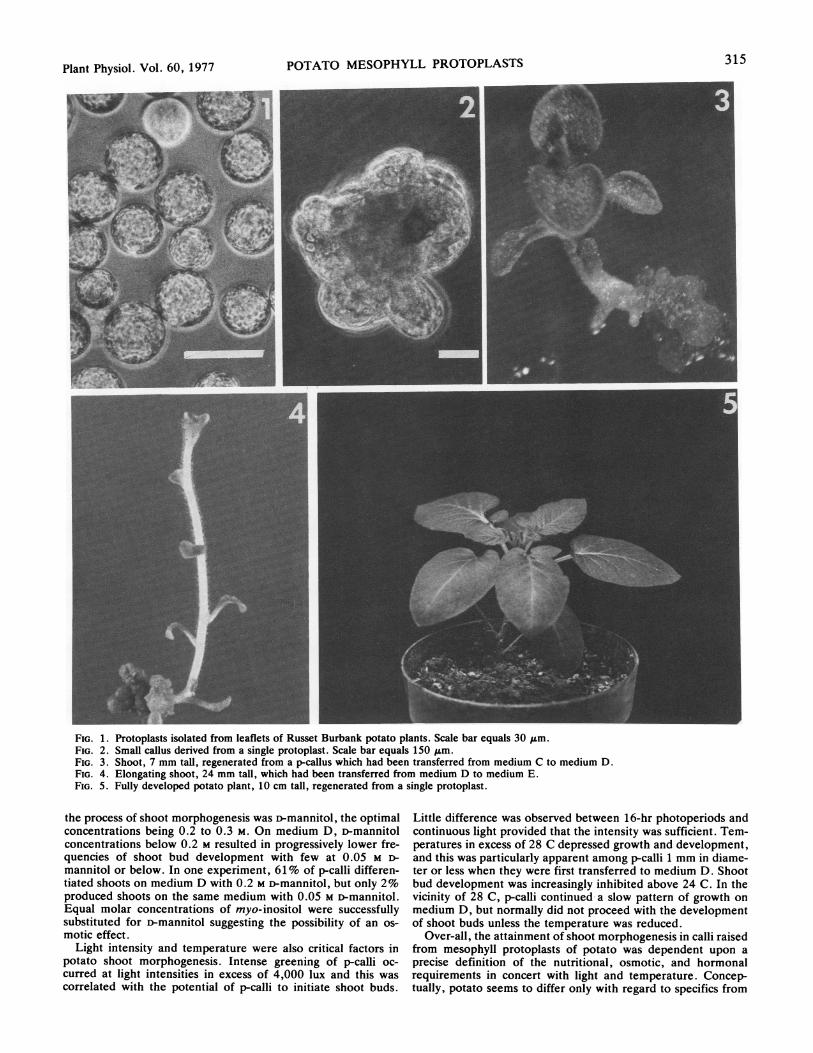
Myo-inositol 10 50 100 100 100 Shoot Morphogenesis. The general sequence of plant develop-Thiamine 801C 0.05 0.25 0.5 0.5 0.5 ment from mesophyll protoplasts of potato is illustrated in Fig-Glycine 0.2 1 2 22Nicotimic acid 0.5 2.5 5 5 5 ures 1 to 5. Shoot bud primordia first became identifiable asPyridoxine -.0..: 0.05 0.25 0.5 0.5 0.5Folio acid 0.05 0.25 0.5 0.5 0.5 dark green nodules located at the base of either small (1-2 mm)Biotin ~~~0.005 0.025 0.05 0.05 0.05 orlre(uto1mmdaee)clindrctotctwhteCaenhydrolyzaute 100 500 1000 1000 1000 orlre(uto1mmdaee)clindrctotctwhteAdenine sulfate 0 0 40 10 10 agar surface. This finding was in contrast to root initials which1-Naphthaleneacetic2 1 0.05 0 0
developed away from the points of contact. Once organized, theacid 2 10O shoot bud (when all other conditions were optimal) continued
Indole-3-acetic acid 0 0 2 0.1 0 dvlpetit ml re ho ihpioda evs6-Benzylamimopurine 0.5 0.5 0.5 0) 0.5 dvlpetit ml re ho ihpioda evsZeatin
I 0 0 0 0.1 0 Mutpesotemrefrmsmpcal,btoefeqnlyGihherellic acid 0 0 0 0 0.1
either one or two shoots became fully differentiated. As shootsD-Manni-tol0 0.35M 0.3M 0.2M 0 elongated, they became pubescent and differentiated fullySuz5ose 0.35M 0 15mM 3-15mM 15mM
MES3 0 0 5mM.: Sn!. 5M formed leaves.Agar 0.5% 1.5% 2.0% 2.0% .% Of the basic media tested which were supportive of both
1Gihherellicacid added after autoclavnig. proliferative growth and of root morphogenesis, only the basal2kNMorhtolino-ethn sulfonic acid. composition of the Lam medium with specific additions andtight:volume,DifoPurifiedAgar.deletions (see Table I, medium D) would also promote shootmorphogenesis. Two features of this medium were apparently
nl) were pipetted over the surface of plates containing medium responsible for its superiority: the presence of casein hydroly-B. Plates containing protoplasts were sealed with Parafilm and sate, and the organic addenda of Nitsch and Nitsch (13). Theincubated at 24 C under continuous illumination of 500 lux. omission of the casamino acids from the medium and/or substi-Plant Regeneration. Initial cell division occurred within 4 to 6 tution of numerous other organic compounds for those formu-
lays, followed by continued proliferation into p-calli (proto- lated by Nitsch and Nitsch depressed the ability of p-calli toplast-derived calli). Before p-calli were more than 2 mm in develop shoot buds. When other factors were held constant,liameter they were transferred to Petri plates containing 20 ml these additives permitted a slow pattern of growth among p-calli)f medium C (Table I), and incubated at 24 C under continuous in the presence of 0.1 mg/l IAA as the sole auxin source. In their),000 lux illumination for approximately 14 days. Shoot mor- absence, IAA was generally insufficient below 1 mg/l with NAAphogenesis was induced by transferring p-calli from medium C to at 0.1 mg/l being far superior for growth. However, IAA atrnedium D (Table I), and returning them to the same environ- concentrations in excess of 0.1 mg/l or NAA at all concentra-nental conditions. P-calli with developed shoots, approximately tions tested was inhibitory to shoot bud development. The modi-Icm in length, were transferred to medium E (Table I) for final fied L medium (medium D) promoted the growth and develop-;tages of shoot development and root initiation. After the shoots ment of p-calli at exogenous cytokinin (zeatin) and auxin (IAA)touched the lid, the Petri plates (25 x 100 mm) were incubated concentrations permissive of shoot morphogenesis whereas theit 24 C under dim light (about 200 lux) to promote root initia- other combinations tested did not.ion. Shoots that were inconveniently slow in rooting were ex- Inadtotohergicded,csmnocd,adm--ised from p-calli, and the cut surface dipped in a commercial eral salts, other medium components were also found to influ-)reparation of Rootone (Amchem Products, Fremont, Calif.). ence the development of shoot buds critically. Paramount of;5hoots were then placed in moist sand under high humidity with these was the concentration of a utilizable carbohydrate source.-onstant 5,000 lux of illumination. All shoots which had rooted At phytohormone concentrations permissive of shoot bud devel-vertrnsfrret pet cps onainng ertlied ermculte opment (i.e. those of medium D), sucrose levels profoundlyLndeplacsedrinea enionmentalup chnambigerati22zC, 70%riclatie influenced the growth pattern of p-calli. At sucrose concentra-iumdiy under
i13hnvrofn13,000chmerux. 70 eltv
tions of 2% (58 mm) or greater, small (1-2 mm diameter) p-callilumidity,uner13 hr of13,000lux.grew for only a brief period after which they assumed a brownRESULTS AND DISCUSSION color and became inhibited. The same effect was recorded be-
tween 1 and 2% sucrose (29-58 mm), but the time required forProtoplast Isolation and Culture. The environmental and nu- its manifestation varied with the size of the callus. Larger (5-10
:ritive conditions under which source plants were grown were a mm diameter) p-calli grew for a longer time before tissue regions-ritical aspect of protoplast isolation. The exposure of plants to contacting the medium turned brown.- Upper callus regions often;hort photoperiods of dim light was especially beneficial for remained green and initiated roots but no shoots. However,)btaining consistently high yields (i.e. 2-3 x 10⁶/g tissue) of when the sucrose concentration in medium D was reduced touiable protoplasts. The conditioning of leaf tissue in cold me- between 0.2 and 0.5% (6-15 mM), p-calli became deep greenhium A (without sucrose) prior to protoplast isolation generally and shoot buds emerged in either small (1-2 mm) or larger (5-mnhanced the stability of protoplast populations before and after 10 mm) calli.)lating. Another medium ingredient which played a significant role in
t
I
t
C
SC
314
Plant Physiol. Vol. 60, 1977315POTATO MESOPHYLL PROTOPLASTS
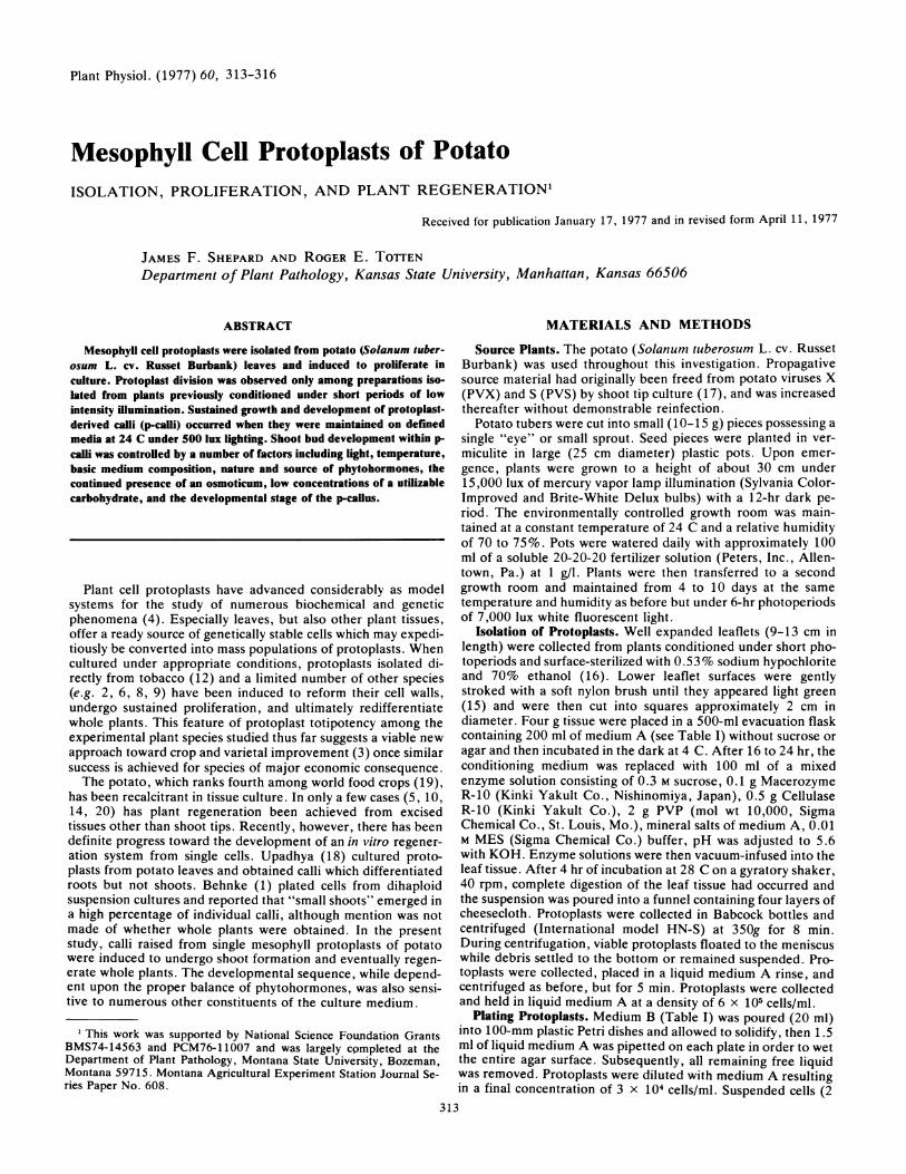
FIG. 1. Protoplasts isolated from leaflets of Russet Burbank potato plants. Scale bar equals 30 ,um.FIG. 2. Small callus derived from a single protoplast. Scale bar equals 150 ,um.FIG. 3. Shoot, 7 mm tall, regenerated from a p-callus which had been transferred from medium C to medium D.FIG. 4. Elongating shoot, 24 mm tall, which had been transferred from medium D to medium E.FIG. 5. Fully developed potato plant, 10 cm tall, regenerated from a single protoplast.
the process of shoot morphogenesis was D-mannitol, the optimalconcentrations being 0.2 to 0.3 M. On medium D, i-mannitolconcentrations below 0.2 M resulted in progressively lower fre-quencies of shoot bud development with few at 0.05 M D-mannitol or below. In one experiment, 61% of p-calli differen-tiated shoots on medium D with 0.2 M D-mannitol, but only 2%produced shoots on the same medium with 0.05 M D-mannitol.Equal molar concentrations of myo-inositol were successfullysubstituted for D-mannitol suggesting the possibility of an os-motic effect.
Light intensity and temperature were also critical factors inpotato shoot morphogenesis. Intense greening of p-calli oc-curred at light intensities in excess of 4,000 lux and this wascorrelated with the potential of p-calli to initiate shoot buds.
Little difference was observed between 16-hr photoperiods andcontinuous light provided that the intensity was sufficient. Tem-peratures in excess of 28 C depressed growth and development,and this was particularly apparent among p-calli 1 mm in diame-ter or less when they were first transferred to medium D. Shootbud development was increasingly inhibited above 24 C. In thevicinity of 28 C, p-calli continued-a slow pattern of growth onmedium D, but normally did not proceed with the developmentof shoot buds unless the temperature was reduced.
Over-all, the attainment of shoot morphogenesis in calli raisedfrom mesophyll protoplasts of potato was dependent upon aprecise definition of the nutritional, osmotic, and hormonalrequirements in concert with light and temperature. Concep-tually, potato seems to differ only with regard to specifics from
316 SHEPARD A
the other species studied thus far. But arriving at the optimumfor each of the possible categories can be a tedious process andnutritional medium components although sometimes describedas unnecessary (7) played a key role in determining whethershoot bud morphogenesis would occur.
Shoot Initiation from Tuber Tissue. To ascertain whether ornot the cultural conditions found to be determinative for shootmorphogenesis in p-calli were also appropriate for other tissuetypes, preliminary attempts were made to stimulate shoot devel-opment in excised tuber tissue. Potato (cv. Russet Burbank)tubers were surface-sterilized for 60 min in 1.5% sodium hypo-chlorite, rinsed in sterile H20, and cut in half. Tuber plugs, 4mm in diameter and 10 mm long, were taken with a cork borerand placed on medium C to stimulate a controlled callus devel-opment. In 3 to 4 weeks at 24 C under continuous 5,000 luxillumination, compact and light green callus developed amongportions of tuber pieces in direct contact with the agar. The tuberplugs were then transferred to medium D and positioned as theywere on medium C. Shoots began to emerge within 4 weeks anddeveloped in manner akin to those from p-calli.
Acknowledgment -The authors gratefully acknowledge the excellent technical assistance ofR. Guthrie.
LITERATURE CITED
1. BEHNKE M 1976 Kulturen isolierter Zellen von einigen dihaploiden Solanum tuberosum -
Klonen und ihre Regeneration. Z Pflanzenphysiol 78: 177-1812. But-DANc-HA D, IA MACKENZIE 1973 The division of protoplasts from Asparagus officin-
alis L. and their growth and differentiation. Protoplasma 78: 215-221
!qD TOTTEN Plant Physiol. Vol. 60, 1977
3. CARLSON PS, JC POLACCO 1975 Plant cell cultures: genetic aspects of crop improvement.Sciencel88: 622-625
4. CHALEFF RS, PS CARLSON 1974 Somatic cell genetics of higher plants. Annu Rev Genet 8:267-278
5. DUNWELL JM, N SUNDERLAND 1973 Anther culture of Solanum tuberosum L. Euphytica22: 317-323
6. DURAND J, I POTRYKUS, G DONN 1973 Plantes issues de protoplastes de Petunia. ZPflanzenphysiol 69: 26-34
7. GAMBORG OL, T MURASHIGE, TA THORPE, IK VASIL 1976 Plant tissue culture media. InVitro 12: 473-478
8. KAMEYA T 1975 Culture of protoplasts from chimeral plant tissue of nature. Jap J Genet 50:417-420
9. KARTHA KK, MR MICHAYLUK, KN KAO, OL GAMBORG, F CONSTABLE 1974 Callusformation and plant regeneration from mesophyll protoplasts of rape plants (Brassicanapus L. W. Zephyr). Plant Sci Lett 3: 265-271
10. LAM S 1975 Shoot formation in potato tuber discs in tissue culture. Am Potato J 52: 103-106
11. MURASHIGE T, F SKOOG 1962 A revised medium for rapid growth and bioassays withtobacco tissue cultures. Physiol Plant 15: 473-497
12. NAGATA T, I TAKEBE 1971 Plating of isolated tobacco mesophyll protoplasts on agarmedium. Planta 99: 12-20
13. NrrsCH JP, C NrrscH 1969 Haploid plants from pollen grains. Science 163: 85-8714. ROEST S, GS BOKELMANN 1976 Vegetative propagation of Solanum tuberosum in vitro.
Potato Res 19: 173-17815. SHEPARD JF 1975 Regeneration of plants from protoplasts of potato virus X-infected
tobacco leaves. Virology 66: 492-50116. SHEPARD JF, RE TOTTEN 1975 Isolation and regeneration of tobacco mesophyll cell
protoplasts under low osmotic conditions. Plant Physiol 55: 689-69417. STACE-SMrrH R, FC MELLOR 1968 Eradication of potato viruses X and S by thermotherapy
and axillary bud culture. Phytopathology 58: 199-20318. UPADHYA MD 1975 Isolation and culture of mesophyll protoplasts of potato (Solanum
tuberosum L.). Potato Res 18: 438-44519. WADE N 1975 International agricultural research. Science 188: 585-58920. WANG P-J, L-C HUANG 1975 Callus cultures from potato tissues and the exclusion of potato
virus X from plants regenerated from stem tips. Can J Bot 53: 2565-2567
k 1.


![Estimating Mesophyll Conductance from Measurements of ... · Estimating Mesophyll Conductance from Measurements of C18OO Photosynthetic Discrimination and Carbonic Anhydrase Activity1[OPEN]](https://img.pdfslide.us/doc/110x75/5e218e60b49cd34ffe11f49e/estimating-mesophyll-conductance-from-measurements-of-estimating-mesophyll-conductance.jpg)