Embed Size (px)
Citation preview
ARTICLES
Mesoangioblast stem cells amelioratemuscle function in dystrophic dogsMaurilio Sampaolesi1,2*, Stephane Blot3*, Giuseppe D’Antona2, Nicolas Granger3, Rossana Tonlorenzi1,Anna Innocenzi1, Paolo Mognol4, Jean-Laurent Thibaud3, Beatriz G. Galvez1, Ines Barthelemy3, Laura Perani1,Sara Mantero4, Maria Guttinger5, Orietta Pansarasa2, Chiara Rinaldi2, M. Gabriella Cusella De Angelis2,Yvan Torrente6, Claudio Bordignon1, Roberto Bottinelli2 & Giulio Cossu1,5,7
Duchenne muscular dystrophy remains an untreatable genetic disease that severely limits motility and life expectancy inaffected children. The only animal model specifically reproducing the alterations in the dystrophin gene and the full spectrumof human pathology is the golden retriever dog model. Affected animals present a single mutation in intron 6, resulting incomplete absence of the dystrophin protein, and early and severe muscle degeneration with nearly complete loss of motilityand walking ability. Death usually occurs at about 1 year of age as a result of failure of respiratory muscles. Here we reportthat intra-arterial delivery of wild-type canine mesoangioblasts (vessel-associated stem cells) results in an extensiverecovery of dystrophin expression, normal muscle morphology and function (confirmed by measurement of contractionforce on single fibres). The outcome is a remarkable clinical amelioration and preservation of active motility. These dataqualify mesoangioblasts as candidates for future stem cell therapy for Duchenne patients.
Duchenne muscular dystrophy primarily affects skeletal muscle,causing fibre degeneration, progressive paralysis and death1. Noeffective treatment exists although novel therapeutic strategies, rang-ing from new drugs to gene and cell therapy, hold promise for sig-nificant advance in the future2. In particular, different types of stemcell have been shown to induce dystrophin synthesis and partialrescue of the pathology in dystrophic mice3–8. However, dystrophicmice do not display clinical signs of the disease, and to proceed to aclinical trial it is imperative to show efficacy in a large, non-syngeneicanimal model of muscular dystrophy. Golden retriever musculardystrophy (GRMD)9,10 is a very severe form of dystrophy, which
affects not only limb, respiratory and heart muscles but also pharyn-geal muscles, resulting in a severe involvement of the digestive tract;although variability exists between individuals, by 8 months of agemost dogs walk with great difficulty (Supplementary Movie 1). Totest the efficacy of cell or gene therapy, we transplanted GRMD dogswith either autologous genetically corrected or donor wild-typemesoangioblasts, under different regimes of immune suppression.
Ten dystrophic dogs were treated in three experiments and a gen-eral scheme of treatments and outcome is reported in Table 1. Fourdogs received autologous mesoangioblasts, transduced in vitro with alentiviral vector expressing human microdystrophin (Supplementary
*These authors contributed equally to this work.
1San Raffaele Scientific Institute, Universita Vita e Salute, Stem Cell Research Institute, Via Olgettina 58, 20132 Milan, Italy. 2Department of Experimental Medicine and InteruniversityInstitute of Myology, University of Pavia, Via Forlanini 6-8, 27100 Pavia, Italy. 3Neurobiology Laboratory, Ecole Veterinaire d’Alfort, 7 Avenue General de Gaulle, 94704 Maisons-Alfortcedex, France. 4Department of Bioengineering, Politecnico di Milano, Piazza Leonardo Da Vinci, 20130 Milan, Italy. 5Institute of Cell Biology and Tissue Engineering, San RaffaeleBiomedical Science Park of Rome, Via Castel Romano 100, 00128 Rome, Italy. 6IRCCS Fondazione Policlinico di Milano, Department of Neurological Sciences, University of Milan, ViaSforza 35, 20122 Milan, Italy. 7Department of Biology and Centre for Stem Cell Research, University of Milan, Via Celoria 28, 20130 Milan, Italy.
Table 1 | Summary of treatment
Dog Dog Cell treatment Lentiviral vector Onset oftreatment
Immune suppression(time)
Dystrophinexpression
Motility Outcome of experiment(at time P400)no. name
01A Ucal Autologous, gene therapy CK-mdys-ires–GFP P118 – 1/2 Loss Euthanasia (P272)02H Vrillie Heterologous, WT donor – P80 CYC A (P78) 1 Loss Euthanasia (P235)03H Valgus Heterologous, WT donor – P75 CYC A (P73) 111 No decline Alive and well04H Varus Heterologous, WT donor – P75 RAP (P73) 111 Modest decline Alive and well05H Viko Heterologous, WT donor – P77 RAP 1 IL-10 (P74) ND ND (sudden death) Myocarditis (P186)06A Vaccin Autologous, gene therapy MLC1F-mdys P113 – 11 Major decline Euthanasia (P326)07A Valium Autologous, gene therapy MLC1F-mdys P113 – ND Loss Pneumonia (P245)08A Vampire Autologous, gene therapy MLC1F-mdys P113 – 11 Major decline Pneumonia (P154)09H Azur Heterologous, WT donor – P159 CYC A (P157) 11 Restored Alive and well10H Azor Heterologous, WT donor – P159 CYC A (P157) 111 Restored Alive and well11U Akan None – – – 2 Loss Euthanasia (P380)12U Vulcano None – – – 2 Loss Euthanasia (P376)13U Viking None – – – 2 Loss Euthanasia (P340)
Each dog was given a specific name and a sequential number, followed by A (transplanted with autologous cells), H (transplanted with heterologous cells) or U (untreated). The nature of the lentiviralvector is indicated (CK-mdys-ires–GFP, creatine kinase promoter driving microdystrophin-ires–GFP; MLCF1-mdys, myosin light chain 1 fast promoter driving microdystrophin). Dystrophin expressionwas quantified as follows: 2, average dystrophin (or micro-dystrophin) expression in less than 1% of positive fibres; 1/2, less than 10% of positive fibres; 1, less than 20% of positive fibres; 11,less than 50% of positive fibres; 111, more than 50% of positive fibres. CYC A, cyclosporine; ND, not determined; RAP, rapamycin; WT, wild-type. Euthanasia was administered when clinicalconditions worsened.
Vol 444 | 30 November 2006 | doi:10.1038/nature05282
574Nature Publishing Group ©2006
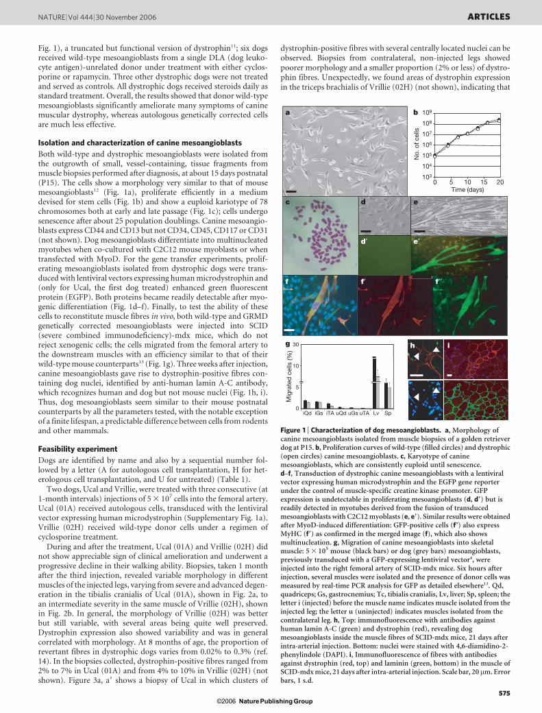
Fig. 1), a truncated but functional version of dystrophin11; six dogsreceived wild-type mesoangioblasts from a single DLA (dog leuko-cyte antigen)-unrelated donor under treatment with either cyclos-porine or rapamycin. Three other dystrophic dogs were not treatedand served as controls. All dystrophic dogs received steroids daily asstandard treatment. Overall, the results showed that donor wild-typemesoangioblasts significantly ameliorate many symptoms of caninemuscular dystrophy, whereas autologous genetically corrected cellsare much less effective.
Isolation and characterization of canine mesoangioblasts
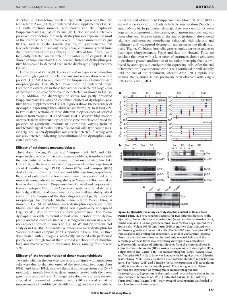
Both wild-type and dystrophic mesoangioblasts were isolated fromthe outgrowth of small, vessel-containing, tissue fragments frommuscle biopsies performed after diagnosis, at about 15 days postnatal(P15). The cells show a morphology very similar to that of mousemesoangioblasts12 (Fig. 1a), proliferate efficiently in a mediumdevised for stem cells (Fig. 1b) and show a euploid kariotype of 78chromosomes both at early and late passage (Fig. 1c); cells undergosenescence after about 25 population doublings. Canine mesoangio-blasts express CD44 and CD13 but not CD34, CD45, CD117 or CD31(not shown). Dog mesoangioblasts differentiate into multinucleatedmyotubes when co-cultured with C2C12 mouse myoblasts or whentransfected with MyoD. For the gene transfer experiments, prolif-erating mesoangioblasts isolated from dystrophic dogs were trans-duced with lentiviral vectors expressing human microdystrophin and(only for Ucal, the first dog treated) enhanced green fluorescentprotein (EGFP). Both proteins became readily detectable after myo-genic differentiation (Fig. 1d–f). Finally, to test the ability of thesecells to reconstitute muscle fibres in vivo, both wild-type and GRMDgenetically corrected mesoangioblasts were injected into SCID(severe combined immunodeficiency)-mdx mice, which do notreject xenogenic cells; the cells migrated from the femoral artery tothe downstream muscles with an efficiency similar to that of theirwild-type mouse counterparts13 (Fig. 1g). Three weeks after injection,canine mesoangioblasts gave rise to dystrophin-positive fibres con-taining dog nuclei, identified by anti-human lamin A-C antibody,which recognizes human and dog but not mouse nuclei (Fig. 1h, i).Thus, dog mesoangioblasts seem similar to their mouse postnatalcounterparts by all the parameters tested, with the notable exceptionof a finite lifespan, a predictable difference between cells from rodentsand other mammals.
Feasibility experiment
Dogs are identified by name and also by a sequential number fol-lowed by a letter (A for autologous cell transplantation, H for het-erologous cell transplantation, and U for untreated) (Table 1).
Two dogs, Ucal and Vrillie, were treated with three consecutive (at1-month intervals) injections of 5 3 107 cells into the femoral artery.Ucal (01A) received autologous cells, transduced with the lentiviralvector expressing human microdystrophin (Supplementary Fig. 1a).Vrillie (02H) received wild-type donor cells under a regimen ofcyclosporine treatment.
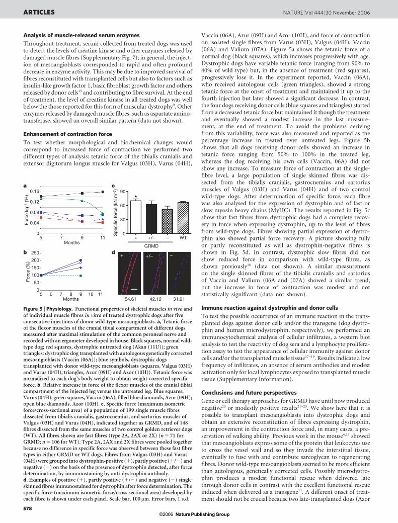
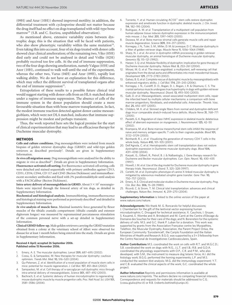
During and after the treatment, Ucal (01A) and Vrillie (02H) didnot show appreciable sign of clinical amelioration and underwent aprogressive decline in their walking ability. Biopsies, taken 1 monthafter the third injection, revealed variable morphology in differentmuscles of the injected legs, varying from severe and advanced degen-eration in the tibialis cranialis of Ucal (01A), shown in Fig. 2a, toan intermediate severity in the same muscle of Vrillie (02H), shownin Fig. 2b. In general, the morphology of Vrillie (02H) was betterbut still variable, with several areas being quite well preserved.Dystrophin expression also showed variability and was in generalcorrelated with morphology. At 8 months of age, the proportion ofrevertant fibres in dystrophic dogs varies from 0.02% to 0.3% (ref.14). In the biopsies collected, dystrophin-positive fibres ranged from2% to 7% in Ucal (01A) and from 4% to 10% in Vrillie (02H) (notshown). Figure 3a, a9 shows a biopsy of Ucal in which clusters of
dystrophin-positive fibres with several centrally located nuclei can beobserved. Biopsies from contralateral, non-injected legs showedpoorer morphology and a smaller proportion (2% or less) of dystro-phin fibres. Unexpectedly, we found areas of dystrophin expressionin the triceps brachialis of Vrillie (02H) (not shown), indicating that
c d e
fM
igra
ted
cel
ls (%
)
iQd0
5
10
30
iGs iTA uQd uGs uTA Lv Sp
g i
Time (days)10 15 2050
No.
of c
ells
h
d′ e′
f′ f′′
109
108
107
106
105
104
103
a b
Figure 1 | Characterization of dog mesoangioblasts. a, Morphology ofcanine mesoangioblasts isolated from muscle biopsies of a golden retrieverdog at P15. b, Proliferation curves of wild-type (filled circles) and dystrophic(open circles) canine mesoangioblasts. c, Karyotype of caninemesoangioblasts, which are consistently euploid until senescence.d–f, Transduction of dystrophic canine mesoangioblasts with a lentiviralvector expressing human microdystrophin and the EGFP gene reporterunder the control of muscle-specific creatine kinase promoter. GFPexpression is undetectable in proliferating mesoangioblasts (d, d9) but isreadily detected in myotubes derived from the fusion of transducedmesoangioblasts with C2C12 myoblasts (e, e9). Similar results were obtainedafter MyoD-induced differentiation: GFP-positive cells (f0) also expressMyHC (f9) as confirmed in the merged image (f), which also showsmultinucleation. g, Migration of canine mesoangioblasts into skeletalmuscle: 5 3 105 mouse (black bars) or dog (grey bars) mesoangioblasts,previously transduced with a GFP-expressing lentiviral vector4, wereinjected into the right femoral artery of SCID-mdx mice. Six hours afterinjection, several muscles were isolated and the presence of donor cells wasmeasured by real-time PCR analysis for GFP as detailed elsewhere13. Qd,quadriceps; Gs, gastrocnemius; Tc, tibialis cranialis, Lv, liver; Sp, spleen; theletter i (injected) before the muscle name indicates muscle isolated from theinjected leg; the letter u (uninjected) indicates muscles isolated from thecontralateral leg. h, Top: immunofluorescence with antibodies againsthuman lamin A-C (green) and dystrophin (red), revealing dogmesoangioblasts inside the muscle fibres of SCID-mdx mice, 21 days afterintra-arterial injection. Bottom: nuclei were stained with 4,6-diamidino-2-phenylindole (DAPI). i, Immunofluorescence of fibres with antibodiesagainst dystrophin (red, top) and laminin (green, bottom) in the muscle ofSCID-mdx mice, 21 days after intra-arterial injection. Scale bar, 20 mm. Errorbars, 1 s.d.
NATURE | Vol 444 | 30 November 2006 ARTICLES
575Nature Publishing Group ©2006
injected cells must to a certain extent transit through the capillaries ofthe injected leg and then through the filter organs, finally entering thearterial circulation to reach the forelimb muscles. Overall, the intens-ity of staining was weak; indeed, western blot analysis confirmed verylow, barely detectable, levels of microdystrophin and dystrophin,respectively. Immunohistochemistry detected several inflammatoryinfiltrates containing mainly macrophages and lymphocytes (Supple-mentary Fig. 2a, b). Although these data demonstrate donor-cell-dependent dystrophin expression in dystrophic dogs and thus thefeasibility of this therapeutic approach, the results were modestoverall. To improve the efficacy of the treatment, the number ofinjections was increased to five (always at 1-month intervals), anew lentivector was produced in which muscle-specific creatinekinase was replaced by the stronger myosin light chain 1F pro-moter and GFP was deleted because we could not detect GFP inunfixed cryostat sections (Supplementary Fig. 1b).
Efficacy of heterologous wild-type mesoangioblasts
Three dogs, namely Valgus, Varus and Viko (03H, 04H and 05H,respectively), were treated with donor cells. One of these dogs(Valgus, 03H) received five arterial systemic injections (5 3 107 cellseach) through a catheter that was introduced in the left femoralisartery and reached the aortic arch at the level of the left subclavia: cellswere released mainly in the two large arteries. From a clinical point ofview, Valgus (03H) had optimal performance and was still walkingwell 5 months after the last injection and the termination of immunesuppression, at the age of 13 months (Supplementary Movie 2).Valgus (03H) was treated with cyclosporine, whereas the other twodogs, Varus (04H) and Viko (05H), were treated with rapamycin andwith rapamycin and interleukin (IL)-10, respectively. Different pro-tocols of immune suppression were tested to evaluate efficacy versustoxicity in this model, but the results did not show significant differ-ences between cyclosporine and rapamycin. In fact, Varus (04H;Supplementary Movie 3) and Viko (05H) also had good clinicalperformance but after 2 months of treatment Viko (05H) died sud-denly of a fulminans myocarditis whose cause remained unexplained.Varus (04H), in contrast, progressively lost walking ability after the
end of immune suppression. Unexpectedly, however, this animal hadno detectable anti-dystrophin antibodies and his circulating lympho-cytes did not react to donor mesoangioblasts or to protein extractsfrom the transplanted muscle (Supplementary Fig. 1c, d).
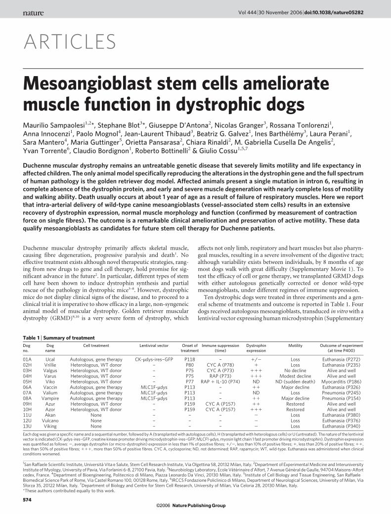
At the end of the treatment, biopsies were taken from severalmuscles of the injected and contralateral leg of these dogs, treatedwith heterologous wild-type cells. Histological analysis of biopsiesfrom Valgus (03H) revealed generally well-preserved morphology(Fig. 2c), although areas of degeneration and regeneration weredetected infrequently. Supplementary Fig. 3 shows a large area ofthe tibialis cranialis of Valgus (03H) (Supplementary Fig. 3a, a9),better preserved than a corresponding area from Vampire (08A), adog transplanted with autologous cells (Supplementary Fig. 3b, b9),
c c′
b b′
a′a
d′d
e′e
Figure 3 | Immunofluorescence analysis of tissue from treated dogs.Double immunofluorescence analysis of muscle biopsies from the tibialiscranialis of Ucal (01A; a, a9), Valgus (03H; b, b9), Varus (04H; c, c9), Vampire(08A; d, d9) and Azor (10H; e, e9), stained with anti-laminin antibody (greenin a9–e9 and inset in d9) and anti-dystrophin (red in a–d) or anti-b-sarcoglycan (red in inset in d). Nuclear staining with DAPI appears in blue.Note clusters of dystrophin-expressing fibres in Ucal, 01A (a) and extensivereconstitution of dystrophin-expressing fibres in Valgus (03H; b), Varus(04H; c) and Azor (10H; e). Several fibres expressing microdystrophin inVampire, 08H (d) also express b-sarcoglycan in the same fibres stained onserial, non-adjacent sections (inset in d). Scale bar, 100mm.
d
b
c
a
Figure 2 | Morphology of muscle in treated dogs. Azan Mallory staining ofseveral muscle biopsies from the tibialis cranialis of dogs transplanted withheterologous mesoangioblasts or autologous, genetically corrected,mesoangioblasts. a–c, Examples show variability from severely affectedtissue of Ucal (01A; a) and Vrillie (02H; b), with many infiltrates, collagenand fat deposition, to the almost normal appearance of tissue from Valgus(03H; c), showing only thickened interstitial tissue. d, A biopsy from Vaccin(06A) shows an intermediate situation with well-preserved morphology.Bar 5 100 mm.
ARTICLES NATURE | Vol 444 | 30 November 2006
576Nature Publishing Group ©2006
described in detail below, which is itself better preserved than thebiopsy from Akan (11U), an untreated dog (Supplementary Fig. 3c,c9). Both forelimb muscles (not shown) and the diaphragm(Supplementary Fig. 5a) of Valgus (03H) also showed a relativelypreserved morphology. Similarly, dystrophin was expressed in mostof the examined biopsies from several different muscles of Valgus(03H9), such as tibialis cranialis (Fig. 3b, b9), gastrocnemius andbiceps femoralis (not shown). Large areas, containing several hun-dred dystrophin-expressing fibres (up to 70% of total fibres), werefrequently detected: an example of the sartorius of Valgus (03H) isshown in Supplementary Fig. 5. Several clusters of dystrophin pos-itive fibres could be detected even in the diaphragm (SupplementaryFig. 4e).
The biopsies of Varus (04H) also showed well-preserved morpho-logy although signs of muscle necrosis and regeneration were stillpresent (Fig. 2d). Overall, most of the biopsies in all muscles weremorphologically less affected than those of untreated dogs.Dystrophin expression in these biopsies was variable but large areasof dystrophin-positive fibres could be detected, as shown in Fig. 3c,c9. In addition, the diaphragm of Varus was partly preserved(Supplementary Fig. 4b) and contained clusters of dystrophin-pos-itive fibres (Supplementary Fig. 4f). Figure 4 shows the percentage ofdystrophin-expressing fibres, which ranged from 10% to at least 70%in two distant sections of three different biopsies each of selectedmuscles from Valgus (03H) and Varus (04H). Western blot analysesof extracts from different biopsies of the same muscles confirmed thepresence of significant amounts of dystrophin, varying from anundetectable signal to about 60% of a control wild-type canine mus-cle (Fig. 4c). When dystrophin was clearly detected, b-sarcoglycanwas also detected, indicating reconstitution of the dystrophin-asso-ciated complex.
Efficacy of autologous mesoangioblasts
Three dogs, Vaccin, Valium and Vampire (06A, 07A and 08A,respectively), received their own mesoangioblasts, transduced withthe new lentiviral vector expressing human microdystrophin. LikeUcal (01A) in the first experiment, they received the first injection atabout 4 months of age (P113). Valium (07A) and Vampire (08A)died of pneumonia after the third and fifth injection, respectively.Because of early death, no force measurement was performed but amovie showing reduced walking ability in Vampire (08A) was takenfew days before his death (Supplementary Movie 4) and biopsies weretaken at autopsy. Valium (07A) received systemic arterial delivery,like Valgus (03H), and maintained a certain walking ability, albeitvery stiff. The biopsies of the three dogs revealed partly preservedmorphology; for example, tibialis cranialis from Vaccin (06A) isshown in Fig. 2d. In addition, microdystrophin expression in thetibialis cranialis of Vampire (08A) was significantly widespread(Fig. 3d, d9), despite the poor clinical performance. The micro-dystrophin was able to recruit at least some member of the dystro-phin-associated complex such as b-sarcoglycan (shown in a serial,non-adjacent section in insets to Fig. 3d, d9 and by western blotanalysis in Fig. 4b). A quantitative analysis of microdystrophin forVaccin (06A) and Vampire (08A) is reported in Fig. 4. Thus, all threedogs treated with autologous, genetically corrected cells performedpoorly, even though two of them showed amelioration of morpho-logy and microdystrophin-expressing fibres, ranging from 5% to50%.
Efficacy of late transplantation of donor mesoangioblasts
To verify whether the less effective results obtained with autologouscells were due to the later onset of the treatment, two dogs, Azur(09H) and Azor (10H), received the first of five injections at P159 (5months), 1 month later than those animals treated with their owngenetically modified cells. Although both dogs were already severelyaffected at the onset of treatment, Azor (10H) showed a strikingimprovement of motility (while still limping) and was even able to
run at the end of treatment (Supplementary Movie 5). Azur (09H)showed a less evident but clearly detectable amelioration (Supplem-entary Movie 6). In particular, although there was variation betweendogs in the progression of the disease, spontaneous improvement wasnever observed. Biopsies taken at the end of treatment also showedrelatively well-preserved morphology (although with sclerosis andinfiltrates) and widespread dystrophin expression in the tibialis cra-nialis (Fig. 4e, e9), biceps femoralis, gastrocnemius, sartorius and evendiaphragm (Supplementary Fig. 6 and data not shown). Thus, weconclude that even with a later onset of treatment, donor cells seemto produce a greater amelioration of muscular dystrophy than is pro-duced by autologous microdystrophin-expressing cells. After the endof treatment with cyclosporine, Azor (10H) continued to walk activelyuntil the end of the experiment, whereas Azur (09H) rapidly lostwalking ability, much as had previously been observed with Valgus(03H) and Varus (04H).
Vampire
Vaccin
Valgus
u iSar
u iTC
u iGas
u iSar
u iTC
u iGas
u iSar
u iTC
u iGas
u iBF
u iSar
u iTC
u iGas
u iBF
100
80
60
40
20
0100
80
60
40
20
0
Dys
MyHC
Dys
Dys
β-SG
MyHC
MyHC
µDys
µDys
β-SG
MyHC
MyHC
Vacc
inVa
mp
ire
Varu
sVa
lgus
Valgu
s
Varu
sW
TAka
n
100
a
b
c
80
60
40
20
0100
80
60
40
20
0
VarusD
ystr
ophi
n-p
ositi
ve fi
bre
s (%
) Vampire
Vaccin
Valgus
Varus
Figure 4 | Quantitative analysis of dystrophin content in tissue fromtreated dogs. a, Three separate sections for two different biopsies of theinjected (i, blue symbols) and non-injected (u, red symbols) sartorius (Sar),tibialis cranialis (TC) and gastrocnemius (Gas) for two dogs injected withdonor cells (Valgus (03H) and Varus (04H)) and two dogs injected withautologous, genetically corrected, cells (Vaccin (06A) and Vampire (08A))were analysed for dystrophin expression. A total of 200 laminin-positivefibres (of any size) were counted in randomly selected fields, and thepercentage of these fibres also expressing dystrophin was calculated.b, Western blot analysis of different biopsies from the muscles shown ina plus the biceps femoralis (BF) showing the expression of dystrophin (Dys,Valgus (03H) and Varus (04H)) or microdystrophin (mDys, Vaccin (06A)and Vampire (08A)). Each lane was loaded with 80mg of proteins. Myosinheavy chains (MyHC) are also shown as an internal standard in the bottompanel. For Varus (03H) and Vampire (08A) the expression of b-sarcoglycan(b-SG) is also shown in the middle panel. There is a good correlationbetween the expression of dystrophin or microdystrophin andb-sarcoglycan. c, Expression of dystrophin and myosin heavy chains in theinjected biceps femoralis of GMRD untreated (Akan (U11)), wild type,Varus (04H) and Valgus (03H); only 30 mg of total proteins was loaded ineach lane for direct comparison.
NATURE | Vol 444 | 30 November 2006 ARTICLES
577Nature Publishing Group ©2006
Analysis of muscle-released serum enzymes
Throughout treatment, serum collected from treated dogs was usedto detect the levels of creatine kinase and other enzymes released bydamaged muscle fibres (Supplementary Fig. 7); in general, the inject-ion of mesoangioblasts corresponded to rapid and often profounddecrease in enzyme activity. This may be due to improved survival offibres reconstituted with transplanted cells but also to factors such asinsulin-like growth factor 1, basic fibroblast growth factor and othersreleased by donor cells15 and contributing to fibre survival. At the endof treatment, the level of creatine kinase in all treated dogs was wellbelow the those reported for this form of muscular dystrophy9. Otherenzymes released by damaged muscle fibres, such as aspartate amino-transferase, showed an overall similar pattern (data not shown).
Enhancement of contraction force
To test whether morphological and biochemical changes wouldcorrespond to increased force of contraction we performed twodifferent types of analysis: tetanic force of the tibialis cranialis andextensor digitorum longus muscle for Valgus (03H), Varus (04H),
Vaccin (06A), Azur (09H) and Azor (10H), and force of contractionon isolated single fibres from Varus (03H), Valgus (04H), Vaccin(06A) and Valium (07A). Figure 5a shows the tetanic force of anormal dog (black squares), which increases progressively with age.Dystrophic dogs have variable tetanic force (ranging from 90% to40% of wild type) but, in the absence of treatment (red squares),progressively lose it. In the experiment reported, Vaccin (06A),who received autologous cells (green triangles), showed a strongtetanic force at the onset of treatment and maintained it up to thefourth injection but later showed a significant decrease. In contrast,the four dogs receiving donor cells (blue squares and triangles) startedfrom a decreased tetanic force but maintained it though the treatmentand eventually showed a modest increase in the last measure-ment, at the end of treatment. To avoid the problems derivingfrom this variability, force was also measured and reported as thepercentage increase in treated over untreated legs. Figure 5bshows that all dogs receiving donor cells showed an increase intetanic force ranging from 50% to 100% in the treated leg,whereas the dog receiving his own cells (Vaccin, 06A) did notshow any increase. To measure force of contraction at the single-fibre level, a large population of single skinned fibres was dis-sected from the tibialis cranialis, gastrocnemius and sartoriusmuscles of Valgus (03H) and Varus (04H) and of two controlwild-type dogs. After determination of specific force, each fibrewas also analysed for the expression of dystrophin and of fast orslow myosin heavy chains (MyHC). The results reported in Fig. 5cshow that fast fibres from dystrophic dogs had a complete recov-ery in force when expressing dystrophin, up to the level of fibresfrom wild-type dogs. Fibres showing partial expression of dystro-phin also showed partial force recovery. A picture showing fullyor partly reconstituted as well as dystrophin-negative fibres isshown in Fig. 5d. In contrast, dystrophic slow fibres did notshow reduced force in comparison with wild-type fibres, asshown previously16 (data not shown). A similar measurementon the single skinned fibres of the tibialis cranialis and sartoriusof Vaccin and Valium (06A and (07A) showed a similar trend,but the increase in force of contraction was modest and notstatistically significant (data not shown).
Immune reaction against dystrophin and donor cells
To test the possible occurrence of an immune reaction in the trans-planted dogs against donor cells and/or the transgene (dog dystro-phin and human microdystrophin, respectively), we performed animmunocytochemical analysis of cellular infiltrates, a western blotanalysis to test the reactivity of dog sera and a lymphocyte prolifera-tion assay to test the appearance of cellular immunity against donorcells and/or the transplanted muscle tissue17–19. Results indicate a lowfrequency of infiltrates, an absence of serum antibodies and modestactivation only for local lymphocytes exposed to transplanted muscletissue (Supplementary Information).
Conclusions and future perspectives
Gene or cell therapy approaches for GRMD have until now producednegative20 or modestly positive results21–23. We show here that it ispossible to transplant mesoangioblasts into dystrophic dogs andobtain an extensive reconstitution of fibres expressing dystrophin,an improvement in the contraction force and, in many cases, a pre-servation of walking ability. Previous work in the mouse4,13 showedthat mesoangioblasts express some of the protein that leukocytes useto cross the vessel wall and so they invade the interstitial tissue,eventually to fuse with and contribute sarcoglycan to regeneratingfibres. Donor wild-type mesoangioblasts seemed to be more efficientthan autologous, genetically corrected cells. Possibly microdystro-phin produces a modest functional rescue when delivered latethrough donor cells in contrast with the excellent functional rescueinduced when delivered as a transgene11. A different onset of treat-ment should not be crucial because two late-transplanted dogs (Azor
0.16a c
b d
0.12
0.08
0.04
0
250
200
150
100
50
0
5 11
5 76 8 109 11
+ +/–
GRMD
– WT
Forc
e kg
–1 (%
)
0
30
60
90
Sp
ecifi
c fo
rce
(kN
m–2
)
Forc
e (%
)
+ +/– –
7 9Months
Months 42.12 31.9154.61
–+ +/–
Figure 5 | Physiology. Functional properties of skeletal muscles in vivo andof individual muscle fibres in vitro of treated dystrophic dogs after fiveconsecutive injections of donor wild-type mesoangioblasts. a, Tetanic forceof the flexor muscles of the cranial tibial compartment of different dogsmeasured after maximal stimulation of the common peroneal nerve andrecorded with an ergometer developed in house. Black squares, normal wild-type dog; red squares, dystrophic untreated dog (Akan (11U)); greentriangles: dystrophic dog transplanted with autologous genetically correctedmesoangioblasts (Vaccin (06A)); blue symbols, dystrophic dogstransplanted with donor wild-type mesoangioblasts (squares, Valgus (03H)and Varus (04H); triangles, Azur (09H) and Azor (10H)). Tetanic force wasnormalized to each dog’s body weight to obtain weight-corrected specificforce. b, Relative increase in force of the flexor muscles of the cranial tibialcompartment of the injected leg versus the untreated leg. Blue squares,Varus (04H); green squares, Vaccin (06A); filled blue diamonds, Azur (09H);open blue diamonds, Azor (10H). c, Specific force (maximum isometricforce/cross-sectional area) of a population of 199 single muscle fibresdissected from tibialis cranialis, gastrocnemius, and sartorius muscles ofValgus (03H) and Varus (04H), indicated together as GRMD, and of 148fibres dissected from the same muscles of two control golden retriever dogs(WT). All fibres shown are fast fibres (type 2A, 2AX or 2X) (n 5 71 forGRMD; n 5 106 for WT). Type 2A, 2AX and 2X fibres were pooled togetherbecause no difference in specific force was observed between these fast fibretypes in either GRMD or WT dogs. Fibres from Valgus (03H) and Varus(04H) were grouped into dystrophin-positive (1), partly positive (1/2) andnegative (2) on the basis of the presence of dystrophin detected, after forcedetermination, by immunostaining by anti-dystrophin antibody.d, Examples of positive (1), partly positive (1/2) and negative (2) singleskinned fibres immunostained for dystrophin after force determination. Thespecific force (maximum isometric force/cross sectional area) developed byeach fibre is shown under each panel. Scale bar, 100mm. Error bars, 1 s.d.
ARTICLES NATURE | Vol 444 | 30 November 2006
578Nature Publishing Group ©2006
(09H) and Azur (10H)) showed improved motility; in addition, thedifferential treatment with cyclosporine should not matter becausethe drug itself had no effect on dogs transplanted with wild-type bonemarrow19 (S.B. and C. Escriou, unpublished observation).
As mentioned above, extensive variability exists between dys-trophic dogs; this is the situation that will be faced with patients,who also show phenotypic variability within the same mutation24.Even taking this into account, four of six dogs treated with donor cellsshowed clear clinical amelioration; of the remaining two, Viko (05H)died early and could not be analysed in detail and Vrillie (02H)probably received too few cells. At the end of immune suppression,two of the four dogs showing amelioration, namely Valgus (03H) andAzor (10H), continued to walk well until the end of the experiment,whereas the other two, Varus (04H) and Azur (09H), rapidly lostwalking ability. We do not have an explanation for this difference,which may reflect the different survival of transplanted organs afterthe end of immune suppression25.
Extrapolation of these results to a possible future clinical trialwould suggest starting with donor cells from an HLA-matched donorunder a regime of immune suppression. The absence of cells of theimmune system in the donor population should create a morefavourable situation than with bone marrow transplantation. In fact,the modest immune reaction detected against donor canine mesoan-gioblasts, which were not DLA matched, indicates that immune sup-pression might be modest and perhaps transient.
Thus, the work reported here sets the logical premise for the startof clinical experimentation that may lead to an efficacious therapy forDuchenne muscular dystrophy.
METHODSCells and culture conditions. Dog mesoangioblasts were isolated from muscle
biopsies of golden retriever dystrophic dogs (GRMD) and wild-type golden
retrievers as described previously4. Details are given in Supplementary
Information.
In vivo cell migration assay. Dog mesoangioblasts were analysed for the ability to
migrate in vivo as described13. Details are given in Supplementary Information.
Fluorescence-activated cell sorting. For fluorescence-activated cell sorting, cells
were detached and stained sequentially with primary antibodies against CD13,
CD31, CD34, CD44, CD 117 and CD45 (Becton Dickinson) and immunofluor-
escent secondary antibodies and fixed with 2% paraformaldehyde until analysis
with a FACSCalibur (Becton Dickinson).
Intra-artery delivery of mesoangioblasts in GRMD. About 5 3 107 mesoangio-
blasts were injected through the femoral artery of ten dogs, as detailed in
Supplementary Information.
Biochemical and histological analysis. Immunoblotting, immunofluorescence
and histological staining were performed as previously described4 and detailed in
Supplementary Information.
In vivo analysis of muscle force. Maximal isometric force generated by flexor
muscles of the tibialis cranialis compartment (tibialis cranialis and extensor
digitorum longus) was measured by supramaximal percutaneous stimulation
of the common peroneal nerve with a set-up detailed in Supplementary
Information.
Clinical GRMD follow-up. Male golden retrievers carrying the GRMD mutation
obtained from a colony at the veterinary school of Alfort were observed for
disease for at least 1 month before being entered into the study. Details are given
in Supplementary Information.
Received 4 April; accepted 26 September 2006.Published online 15 November 2006.
1. Emery, A. E. The muscular dystrophies. Lancet 359, 687–695 (2002).
2. Cossu, G. & Sampaolesi, M. New therapies for muscular dystrophy: cautiousoptimism. Trends Mol. Med. 10, 516–520 (2004).
3. Qu-Petersen, Z. et al. Identification of a novel population of muscle stem cells inmice: potential for muscle regeneration. J. Cell Biol. 157, 851–864 (2002).
4. Sampaolesi, M. et al. Cell therapy of a-sarcoglycan null dystrophic mice throughintra-arterial delivery of mesoangioblasts. Science 301, 487–492 (2003).
5. Bachrach, E. et al. Systemic delivery of human microdystrophin to regeneratingmouse dystrophic muscle by muscle progenitor cells. Proc. Natl Acad. Sci. USA 101,3581–3586 (2004).
6. Torrente, Y. et al. Human circulating AC1331 stem cells restore dystrophinexpression and ameliorate function in dystrophic skeletal muscle. J. Clin. Invest.114, 182–195 (2004).
7. Rodriguez, A. M. et al. Transplantation of a multipotent cell population fromhuman adipose tissue induces dystrophin expression in the immunocompetentmdx mouse. J. Exp. Med. 201, 1397–1405 (2005).
8. Dezawa, M. et al. Bone marrow stromal cells generate muscle cells and repairmuscle degeneration. Science 309, 314–317 (2005).
9. Kornegay, J. N., Tuler, S. M., Miller, D. M. & Levesque, D. C. Muscular dystrophy ina litter of golden retriever dogs. Muscle Nerve 11, 1056–1064 (1988).
10. Sharp, N. J. et al. An error in dystrophin mRNA processing in golden retrievermuscular dystrophy, an animal homologue of Duchenne muscular dystrophy.Genomics 13, 115–121 (1992).
11. Harper, S. Q. et al. Modular flexibility of dystrophin: implication for gene therapy ofDuchenne muscular dystrophy. Nature Med. 8, 253–261 (2004).
12. Minasi, M. G. et al. The meso-angioblast: a multipotent, self-renewing cell thatoriginates from the dorsal aorta and differentiates into most mesodermal tissues.Development 129, 2773–2783 (2002).
13. Galvez, B. G. et al. Complete rescue of dystrophic muscle by mesoangioblasts withenhanced migratory ability. J. Cell Biol. 174, 231–243 (2006).
14. Kornegay, J. N., Cundiff, D. D., Bogan, D. J., Bogan, J. R. & Okamura, C. S. Thecranial sartorius muscle undergoes true hypertrophy in dogs with golden retrievermuscular dystrophy. Neuromuscul. Disord. 13, 493–500 (2003).
15. Galli, D. et al. Mesoangioblasts, vessel-associated multipotent stem cells, repairthe infarcted heart by multiple cellular mechanisms: a comparison with bonemarrow progenitors, fibroblasts, and endothelial cells. Arterioscler. Thromb. Vasc.Biol. 25, 692–697 (2005).
16. Childers, M. K. et al. Skinned single fibers from normal and dystrophin-deficientdogs incur comparable stretch-induced force deficits. Muscle Nerve 31, 768–771(2005).
17. Pavlath, G. K. Regulation of class I MHC expression in skeletal muscle: deleteriouseffect of aberrant expression on myogenesis. J. Neuroimmunol. 125, 42–50(2002).
18. Krampera, M. et al. Bone marrow mesenchymal stem cells inhibit the response ofnaive and memory antigen-specific T cells to their cognate peptide. Blood 101,3722–3729 (2003).
19. Reinhardt, R. L. et al. Visualizing the generation of memory CD4 T cells in thewhole body. Nature 410, 101–105 (2001).
20. Dell’Agnola, C. et al. Hematopoietic stem cell transplantation does not restoredystrophin expression in Duchenne muscular dystrophy dogs. Blood 104,4311–4318 (2004).
21. Karpati, G., Gilbert, R., Petrof, B. J. & Nalbantoglu, J. Gene therapy research forDuchenne and Becker muscular dystrophies. Curr. Opin. Neurol. 10, 430–435(1997).
22. Howell, J. M. et al. Use of the dog model for Duchenne muscular dystrophy in genetherapy trials. Neuromuscul. Disord. 7, 325–328 (1997).
23. Cerletti, M. et al. Dystrophic phenotype of canine X-linked muscular dystrophy ismitigated by adenovirus-mediated utrophin gene transfer. Gene Ther. 10,750–757 (2003).
24. Emery, A. E. Clinical and molecular studies in Duchenne muscular dystrophy. Prog.Clin. Biol. Res. 306, 15–28 (1989).
25. Ricordi, C. & Strom, T. M. Clinical islet transplantation: advances and clinicalchallenges. Nature Rev. Immunol. 4, 259–270 (2004).
Supplementary Information is linked to the online version of the paper atwww.nature.com/nature.
Acknowledgements We thank M. G. Roncarolo for helpful discussions;J. Chamberlain for the gift of the lentiviral vector expressing humanmicrodystrophin; C. Drougard for technical assistance; X. Cauchois, I. Gruyer,S. Kouame, E. Wembe and A. Brindejont and M. Carre at the Centre d’Elevage duDomaine des Souches for their care of the dogs; and N. Borenstein for the systemicdelivery of cells. M.S. and G.C. thank P. Luban for a donation. This work wassupported by grants from the Association Francaise contre les Myopathies,Telethon, the Muscular Dystrophy Association, the Parent Project Onlus, theEuropean Community ‘Eurostemcell’, the Cariplo Foundation and the ItalianMinistries of Health and Research. B.G.G. was supported by a 313 fellowship fromthe Centro Nacional de Investigationes Cardiovasculares, Spain.
Author Contributions M.S. coordinated the work on cells with R.T. and M.G.C.D.;S.B. coordinated the work on dogs with N.G., J.L.T. and I.B.; R.B. and G.D.A.coordinated the physiology experiments with O.P., C.R. and P.M., who alsodeveloped, with S.M., the instrument to measure dog tetanic force; A.I. did thehistology work; B.G.G. performed the homing experiments; L.P. and M.S.conducted the western blot analysis; M.G. did the immunology experiment; Y.T.and C.B. evaluated the clinical aspects of the work; G.C. coordinated the wholeproject.
Author Information Reprints and permissions information is available atwww.nature.com/reprints. The authors declare no competing financial interests.Correspondence and requests for materials should be addressed to C.G.([email protected]) or R.B. ([email protected]).
NATURE | Vol 444 | 30 November 2006 ARTICLES
579Nature Publishing Group ©2006


![Stem Cell Therapy in India - Clinical Study A Clinical …...Stem Cells International cells ameliorate the functional de cits in animal models of cerebral palsy [ , ]. Amongst these,](https://img.pdfslide.us/doc/110x75/5f3ff32f366e8b666156ad45/stem-cell-therapy-in-india-clinical-study-a-clinical-stem-cells-international.jpg)