Embed Size (px)
Citation preview
184Inflammation and Regeneration Vol.34 No.4 September 2014
Special Issue: Mesenchymal Stem Cells and Immunomodulation
Mini Review
Mesenchymal stem cells for the treatment of inflammatory bowel disease: from experimental models to clinical application
Rhian Stavely, Samy Sakkal, Vanesa Stojanovskaand Kulmira Nurgali*College of Health and Biomedicine, Victoria University, Western Centre for Health Research & Education, VIC, Australia
Inflammatory Bowel Disease (IBD) is a highly debilitating and potentially fatal idiopathic disorder of the intestinal tract which is exceedingly prevalent in westernized society; however there is concern of an IBD epidemic in Asia due to increasing incidence rates. There is no cure for IBD with current treatments limited by their inefficacy, toxicity and adverse side-effects; thus necessitating the search for novel therapies. In the past decade mesenchymal stem cells (MSCs) have become attractive candidates for the cellular based therapy of IBD. MSCs are easily isolated and expanded from adult bone-marrow and adipose tissue; they possess unique therapeutic characteristics including the ability to home to sites of tissue damage and inflammation, facilitate tissue repair and modulate the immune system. The administration of MSCs in animal models of experimental colitis and clinical trials of fistulising and luminal Crohn's disease have yielded promising results, however an unequivocal therapeutic mechanism remains elusive. This review will explore the clinical application of MSCs in IBD and current evidence from experimental models of colitis elucidating their potential to ameliorate intestinal inflammation.
Rec.5/30/2014, Acc.8/21/2014, pp184-197
*Correspondence should be addressed to:Kulmira Nurgali, College of Health and Biomedicine, Victoria University, Western Centre for Health Research & Education, 176 Furlong Road, St Albans, 3021, VIC, Australia. Phone: +61-3-8395-8223, E-mail: [email protected]
Key words clinical trials, experimental colitis, inflammatory bowel disease, mesenchymal stem cells
Special Issue (Mini Review) Mesenchymal stem cells and IBD
Introduction Inflammatory Bowel Disease (IBD) is comprised of two main pathologies, Crohn’s disease (CD) and ulcerative colitis (UC), which are characterised by the presentation
of recurrent idiopathic intestinal inflammation. In UC inflammation is localised in the mucosa ascending con-tinuously from the rectum to the colon. Conversely, inflam-mation in CD is transmural and manifests discontinuously
Inflammation and Regeneration Vol.34 No.4 September 2014 185
Special Issue (Mini Review) Mesenchymal stem cells and IBD
in skip lesions throughout the gastrointestinal tract with formation of granulomas1). All phenotypes of IBD greatly affect quality of life with symptoms including ulcerations, fistulae, strictures, perianal fissures, bloody stool, persistent diarrhea or constipation, abdominal pain and cramps2). Potential complications in IBD such as perforation, excessive bleeding from ul-cerations, obstruction of the bowel and intestinal scarring resulting in malnutrition can lead to fatality. Furthermore, the risk of developing cancers including colorectal cancer3) and lymphoma4) are increased in IBD resulting in an indirect escalation in the mortality rate. Although IBD is predominantly a disease of westernized society, dramatic increases in the incidence of IBD have been observed throughout Asia5) which may reach epidemic proportions6). The cause of IBD is unknown but concordant twin studies have revealed that the development of IBD is likely to require a multi-genetic predisposition and an environmental perturbation7). Numerous predisposing genes for CD and UC have been uncovered with around 30% of loci overlapping for both diseases suggesting similarities in pathological mechanisms8). Although the pathogenesis of IBD is unknown, it is predicted that antigens of commensal bacteria in the gut instigate an exaggerated immune response9). The role of epithelial permeability and leuko- cyte dysregulation in the exuberant antigen response is currently under investigation10, 11). Current treatment strategies do not provide a cure and are limited by their inefficacy, toxicity and adverse side-effects12-14); thus necessitating the search for novel thera-pies. One of the most promising treatments currently being investigated is mesenchymal stem cell (MSC) therapy. MSCs are easily isolated and expansively cultured from adult adipose tissue and bone marrow15). Furthermore, MSCs possess many unique properties making them an ideal candidate therapy for IBD. MSCs are immune evasive and can be transplanted between individuals and across species16, 17). Once administered, MSCs migrate through chemotaxis towards sites of inflammation; thus specifically targeting pathological manifestations18). After homing to the site of inflammation, MSCs facilitate tissue regeneration through secretion of pro-angiogenic and trophic factors which have been shown to promote endogenous repair mechanisms19-21). Moreover, MSCs are immunomodulatory and secrete anti-inflammatory factors suppressing the im-mune response and inflammation22). The clinical application of MSCs in CD and evidence for the possible mechanisms
elucidating their potential to regenerate intestinal epithelium and reduce inflammation in experimental models of colitis will be reviewed.
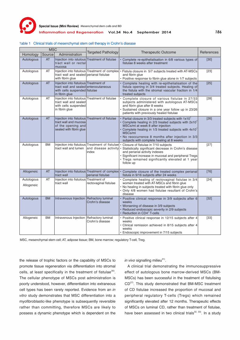
Efficacy of MSCs in Clinical Trials Clinical trials using MSCs for the treatment of CD fistulae and luminal inflammation have demonstrated that MSC therapy in IBD is both efficacious and feasible (summarised in Table 1). Predominantly, clinical trials of MSC therapy have focused on the treatment of fistulae caused by CD rather than CD manifestations as a whole. Primarily MSCs derived from adipose tissue (AT-MSCs) have been used for the treatment of fistulising CD and have resulted in the complete re-epithelialisation of rectovaginal, enterocutaneous and complex perianal fistulae in the majority of subjects. Fibrin glue was regularly used in conjunction with local MSC administration in fistulising CD, however evidence of a therapeutic benefit is not likely to be a result of fibrin glue alone23, 24). One clinical trial demonstrated that in vitro expansion is likely to be essential in harnessing the therapeutic potential of AT-MSCs rather than treating patients with the primary stromal vascular fraction of lipoaspirate25) . The therapeutic outcome of MSC therapy in fistulising CD may be dose-dependent with greater efficacy achieved by doses of 2x10⁷ or 4x10⁷ MSCs/ml compared to 1x10⁷
MSCs/ml26). Long-term effects have been reported with CD and perianal activity index scores declining 12 months post treatment27). Furthermore, sustained closure of fistulae has been achieved in 88-100% of subjects 8-12 months after a course of MSC therapy26, 28); however these effects are relatively transient given that only 58% of subjects maintain closure after 3 years29). This suggests that repeated treatment may be required to maintain the therapeutic benefits of MSC therapy. While autologous and allogeneic AT-MSCs have both demonstrated efficacy in the healing of fistulae, further evidence is required to determine long-term immune toler-ance in patients with repeated allogeneic MSC exposure. Bacterial contamination has posed a problem in the ex-pansion of autologous MSCs in the past causing delay in treatment24, 30). If allogeneic MSCs are determined to be equally efficacious, pre-prepared sources for treatment could prevent such setbacks. One clinical trial has determined that MSCs were as effective in treating fistulae of cryptoglandular origin as they were for fistulae resultant of CD23). This result supports the view that the therapeutic value of MSCs can be attributed to
186Inflammation and Regeneration Vol.34 No.4 September 2014
MSC Targeted Pathology Therapeutic Outcome ReferencesHomology Source AdministrationAutologous AT Injection into istulous
tract wall or rectal mucosa
Treatment of fistulae ・Complete re-epithelialisation in 6/8 various types of fistulae 8 weeks after treatment
[30]
Autologous AT Injection into fistulous tract wall and sealed with fibrin glue
Treatment of complex perianal fistulae
・Fistula closure in 5/7 subjects treated with AT-MSCs and fibrin glue
・Positive response to fibrin glue alone in 1/7 subjects
[23]
Autologous AT Injection into fistulous tract wall and sealed with cells suspended in fibrin glue
Treatment of enterocutaneousfistulae
・Complete healing with re-epithelialisation of the fistula opening in 3/4 treated subjects. Healing of the fistula with the stromal vascular fraction in 1/4 treated subjects
[25]
Autologous AT Injection into fistulous tract wall and sealed with cells suspended in fibrin glue
Treatment of fistulae ・Complete closure of various fistulae in 27/33 subjects administered with autologous AT-MSCs and fibrin glue after 8 weeks
・Sustained closure in a one year follow up in 23/26 patients with previously healed fistulae
[28]
Autologous AT Injection into fistulous tract wall and mucosa of the opening and sealed with fibrin glue
Treatment of fistulae ・Partial closure in 3/3 treated subjects with 1x10⁷
・Complete healing in 2/3 treated subjects with 2x10⁷ MSCs/ml at week 8 after injection
・Complete healing in 1/3 treated subjects with 4x10⁷ MSCs/ml
・No reoccurrence 8 months after injection in 3/3 subjects with complete healing at 8 weeks
[26]
Autologous BM Injection into fistulous tract wall and lumen
Treatment of fistulae and disease activity index
・Closure of fistulae in 7/10 subjects・Statistically significant decrease in Crohn’s disease
and perianal activity indexes・Significant increase in mucosal and peripheral Tregs ・Tregs remained significantly elevated at 1 year
follow up
[27]
Allogeneic AT Injection into fistulous tract wall
Treatment of complex perianal fistulae
・Complete closure of the treated complex perianal fistula in 9/16 subjects after 24 weeks
[76]
AutologousAllogeneic
AT Injection into fistulous tract wall
Treatment ofrectovaginal fistulae
・Complete healing of rectovaginal fistulae in 3/4 women treated with AT-MSCs and fibrin glue
・No healing in subjects treated with fibrin glue only ・Only 4/8 women had fistulae resultant of Crohn’s
disease
[24]
Autologous BM Intravenous Injection Refractory luminalCrohn’s disease
・Positive clinical response in 3/9 subjects after 6 weeks
・Worsening of disease in 3/9 subjects・Reduced endoscopic severity in 2/9 subjects・Reduction in CD4+ T-cells
[32]
Allogeneic BM Intravenous Injection Refractory luminalCrohn’s disease
・Positive clinical response in 12/15 subjects after 4 weeks
・Clinical remission achieved in 8/15 subjects after 4 weeks
・Endoscopic improvement in 7/15 subjects
[33]
Special Issue (Mini Review) Mesenchymal stem cells and IBD
the release of trophic factors or the capability of MSCs to promote tissue regeneration via differentiation into stromal cells, at least specifically in the treatment of fistulae30). The cellular phenotype of MSCs post administration is poorly understood, however, differentiation into extraneous cell types has been rarely reported. Evidence from an in vitro study demonstrates that MSC differentiation into a myofibroblastic-like phenotype is subsequently reversible rather than committing, therefore MSCs are likely to possess a dynamic phenotype which is dependent on the
in vivo signalling milieu31). A clinical trial demonstrating the immunosuppressive effect of autologous bone marrow-derived MSCs (BM-MSCs) has been successful in the treatment of fistulising CD27). This study demonstrated that BM-MSC treatment of CD fistulae increased the proportion of mucosal and peripheral regulatory T-cells (Tregs) which remained significantly elevated after 12 months. Therapeutic effects of MSCs on luminal CD, rather than treatment of fistulae, have been assessed in two clinical trials32, 33). In a study
Table 1 Clinical trials of mesenchymal stem cell therapy in Crohn’s disease
MSC, mesenchymal stem cell; AT, adipose tissue; BM, bone marrow; regulatory T-cell, Treg.
Inflammation and Regeneration Vol.34 No.4 September 2014 187
Special Issue (Mini Review) Mesenchymal stem cells and IBD
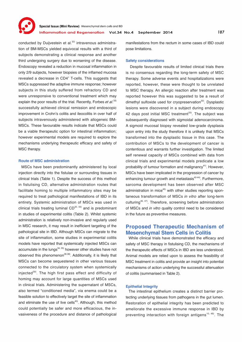
conducted by Duijvestein et al.32) intravenous administra-tion of BM-MSCs yielded equivocal results with a third of subjects demonstrating a clinical response and another third undergoing surgery due to worsening of the disease. Endoscopy revealed a reduction in mucosal inflammation in only 2/9 subjects, however biopsies of the inflamed mucosa revealed a decrease in CD4+ T-cells. This suggests that MSCs suppressed the adaptive immune response; however subjects in this study suffered from refractory CD and were unresponsive to conventional treatment which may explain the poor results of the trial. Recently, Forbes et al.33)
successfully achieved clinical remission and endoscopic improvement in Crohn’s colitis and ileocolitis in over half of subjects intravenously administered with allogeneic BM-MSCs. These favourable results indicate that MSCs could be a viable therapeutic option for intestinal inflammation; however experimental models are required to explore the mechanisms underlying therapeutic efficacy and safety of MSC therapy.
Route of MSC administration MSCs have been predominantly administered by local injection directly into the fistulae or surrounding tissues in clinical trials (Table 1). Despite the success of this method in fistulising CD, alternative administration routes that facilitate homing to multiple inflammatory sites may be required to treat pathological manifestations of IBD in its entirety. Systemic administration of MSCs was used in clinical trials treating luminal CD32, 33) and is predominant in studies of experimental colitis (Table 2). Whilst systemic administration is relatively non-invasive and regularly used in MSC research, it may result in inefficient targeting of the pathological site in IBD. Although MSCs can migrate to the site of inflammation, some studies in experimental colitis models have reported that systemically injected MSCs can accumulate in the lungs34, 35) however other studies have not observed this phenomenon36-38). Additionally, it is likely that MSCs can become sequestered in other various tissues connected to the circulatory system when systemically injected39). The high first pass effect and difficulty of homing may account for large quantities of MSCs used in clinical trials. Administering the supernatant of MSCs, also termed “conditioned media”, via enema could be a feasible solution to effectively target the site of inflammation and eliminate the use of live cells40). Although, this method could potentially be safer and more efficacious, the in-vasiveness of the procedure and distance of pathological
manifestations from the rectum in some cases of IBD could pose limitations.
Safety considerations Despite favourable results of limited clinical trials there is no consensus regarding the long-term safety of MSC therapy. Some adverse events and hospitalizations were reported, however, these were thought to be unrelated to MSC therapy. An allergic reaction after treatment was reported however this was suggested to be a result of dimethyl sulfoxide used for cryopreservation32). Dysplastic lesions were discovered in a subject during endoscopy 42 days post initial MSC treatment33). The subject was subsequently diagnosed with sigmoidal adenocarcinoma. A sigmoid mucosal biopsy revealed low-grade dysplasia upon entry into the study therefore it is unlikely that MSCs transformed into the dysplastic tissue in this case. The contribution of MSCs to the development of cancer is contentious and warrants further investigation. The limited self renewal capacity of MSCs combined with data from clinical trials and experimental models predicate a low probability of tumour formation and malignancy41). However, MSCs have been implicated in the progression of cancer by enhancing tumour growth and metastasis42-44). Furthermore, sarcoma development has been observed after MSC administration in mice45) with other studies reporting spon-taneous transformation of MSCs in vitro after long-term culturing46, 47). Therefore, screening before administration of MSCs and in vitro quality control need to be considered in the future as preventive measures.
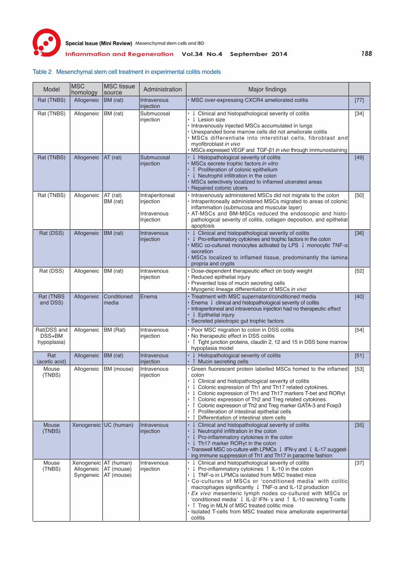
Proposed Therapeutic Mechanism of Mesenchymal Stem Cells in Colitis While clinical trials have demonstrated the efficacy and safety of MSC therapy in fistulising CD, the mechanisms of the therapeutic effects of MSCs in IBD are less understood. Animal models are relied upon to assess the feasibility of MSC treatment in colitis and provide an insight into potential mechanisms of action underlying the successful attenuation of colitis (summarised in Table 2).
Epithelial Integrity The intestinal epithelium creates a distinct barrier pro-tecting underlying tissues from pathogens in the gut lumen. Restoration of epithelial integrity has been predicted to ameliorate the excessive immune response in IBD by preventing interaction with foreign antigens10, 48). The
188Inflammation and Regeneration Vol.34 No.4 September 2014
Special Issue (Mini Review) Mesenchymal stem cells and IBD
Model MSChomology
MSC tissuesource Administration Major findings
Rat (TNBS) Allogeneic BM (rat) Intravenousinjection
・MSC over-expressing CXCR4 ameliorated colitis [77]
Rat (TNBS) Allogeneic BM (rat) Submucosal injection
・↓ Clinical and histopathological severity of colitis・↓ Lesion size ・Intravenously injected MSCs accumulated in lungs・Unexpanded bone marrow cells did not ameliorate colitis・MSCs differentiate into interstit ial cells, f ibroblast and
myofibroblast in vivo・MSCs expressed VEGF and TGF-β1 in vivo through immunostaining
[34]
Rat (TNBS) Allogeneic AT (rat) Submucosalinjection
・↓ Histopathological severity of colitis・MSCs secrete trophic factors in vitro ・↑ Proliferation of colonic epithelium・↓ Neutrophil infiltration in the colon・MSCs selectively localized to inflamed ulcerated areas・Repaired colonic ulcers
[49]
Rat (TNBS) Allogeneic AT (rat)BM (rat)
Intraperitonealinjection
IntravenousInjection
・Intravenously administered MSCs did not migrate to the colon・Intraperitoneally administered MSCs migrated to areas of colonic
inflammation (submucosa and muscular layer) ・AT-MSCs and BM-MSCs reduced the endoscopic and histo-
pathological severity of colitis, collagen deposition, and epithelial apoptosis
[50]
Rat (DSS) Allogeneic BM (rat) Intravenousinjection
・↓ Clinical and histopathological severity of colitis・↓ Pro-inflammatory cytokines and trophic factors in the colon ・MSC co-cultured monocytes activated by LPS ↓ monocytic TNF-α
secretion ・MSCs localized to inflamed tissue, predominantly the lamina
propria and crypts
[36]
Rat (DSS) Allogeneic BM (rat) Intravenousinjection
・Dose-dependent therapeutic effect on body weight・Reduced epithelial injury・Prevented loss of mucin secreting cells・Myogenic lineage differentiation of MSCs in vivo
[52]
Rat (TNBSand DSS)
Allogeneic Conditionedmedia
Enema ・Treatment with MSC supernatant/conditioned media・Enema ↓ clinical and histopathological severity of colitis・Intraperitoneal and intravenous injection had no therapeutic effect・↓ Epithelial injury・Secreted pleiotropic gut trophic factors
[40]
Rat(DSS and DSS+BM
hypoplasia)
Allogeneic BM (Rat) Intravenous injection
・Poor MSC migration to colon in DSS colitis・No therapeutic effect in DSS colitis・↑ Tight junction proteins, claudin 2, 12 and 15 in DSS bone marrow
hypoplasia model
[54]
Rat(acetic acid)
Allogeneic BM (rat) Intravenous injection
・↓ Histopathological severity of colitis・↑ Mucin secreting cells
[51]
Mouse(TNBS)
Allogeneic BM (mouse) Intravenous injection
・Green fluorescent protein labelled MSCs homed to the inflamed colon
・↓ Clinical and histopathological severity of colitis・↓ Colonic expression of Th1 and Th17 related cytokines.・↓ Colonic expression of Th1 and Th17 markers T-bet and RORγt・↑ Colonic expression of Th2 and Treg related cytokines.・↑ Colonic expression of Th2 and Treg marker GATA-3 and Foxp3・↑ Proliferation of intestinal epithelial cells・↑ Differentiation of intestinal stem cells
[53]
Mouse(TNBS)
Xenogeneic UC (human) Intravenousinjection
・↓ Clinical and histopathological severity of colitis・↓ Neutrophil infiltration in the colon・↓ Pro-inflammatory cytokines in the colon ・↓ Th17 marker RORγt in the colon ・Transwell MSC co-culture with LPMCs ↓ IFN-γ and ↓ IL-17 suggest-
ing immune suppression of Th1 and Th17 in paracrine fashion
[35]
Mouse(TNBS)
XenogeneicAllogeneicSyngeneic
AT (human)AT (mouse)AT (mouse)
Intravenousinjection
・↓ Clinical and histopathological severity of colitis・↓ Pro-inflammatory cytokines ↑ IL-10 in the colon ・↓ TNF-α in LPMCs isolated from MSC treated mice・Co-cultures of MSCs or ‘conditioned media’ with colitic
macrophages significantly ↓ TNF-α and IL-12 production ・Ex vivo mesenteric lymph nodes co-cultured with MSCs or
‘conditioned media’ ↓ IL-2/ IFN- γ and ↑ IL-10 secreting T-cells ・↑ Treg in MLN of MSC treated colitic mice・Isolated T-cells from MSC treated mice ameliorate experimental
colitis
[37]
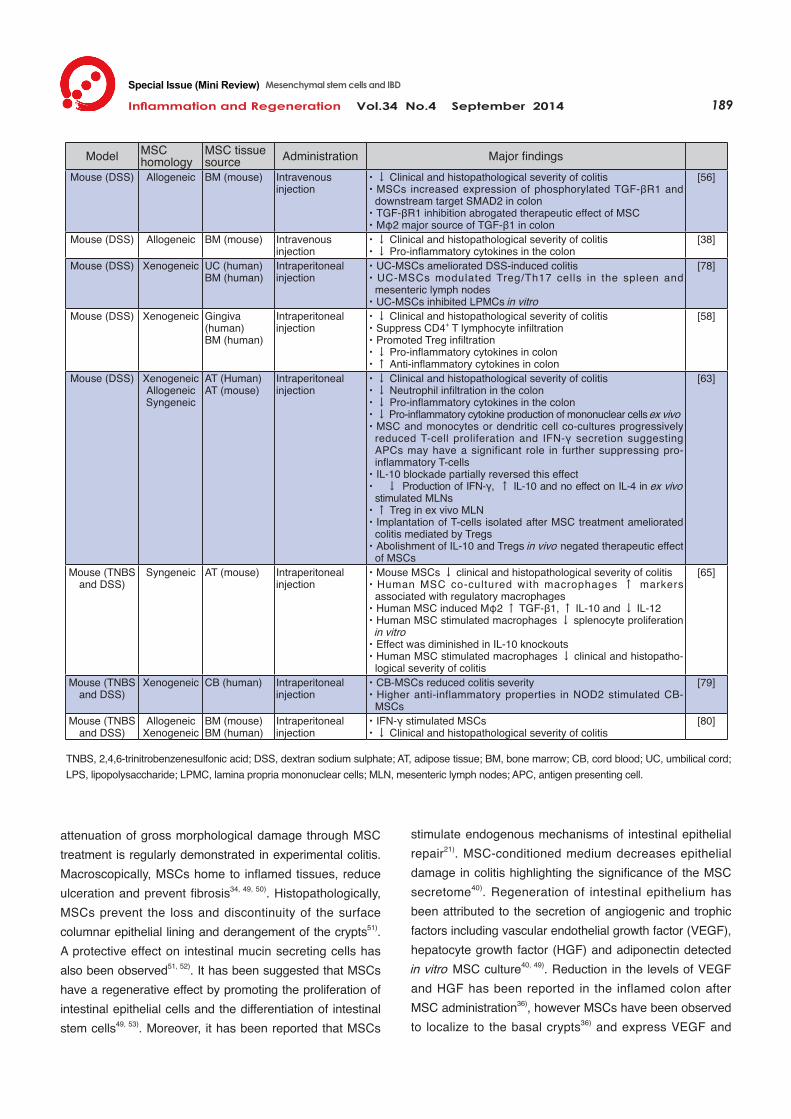
Table 2 Mesenchymal stem cell treatment in experimental colitis models
Inflammation and Regeneration Vol.34 No.4 September 2014 189
Special Issue (Mini Review) Mesenchymal stem cells and IBD
attenuation of gross morphological damage through MSC treatment is regularly demonstrated in experimental colitis. Macroscopically, MSCs home to inflamed tissues, reduce ulceration and prevent fibrosis34, 49, 50). Histopathologically, MSCs prevent the loss and discontinuity of the surface columnar epithelial lining and derangement of the crypts51). A protective effect on intestinal mucin secreting cells has also been observed51, 52). It has been suggested that MSCs have a regenerative effect by promoting the proliferation of intestinal epithelial cells and the differentiation of intestinal stem cells49, 53). Moreover, it has been reported that MSCs
stimulate endogenous mechanisms of intestinal epithelial repair21). MSC-conditioned medium decreases epithelial damage in colitis highlighting the significance of the MSC secretome40). Regeneration of intestinal epithelium has been attributed to the secretion of angiogenic and trophic factors including vascular endothelial growth factor (VEGF), hepatocyte growth factor (HGF) and adiponectin detected in vitro MSC culture40, 49). Reduction in the levels of VEGF and HGF has been reported in the inflamed colon after MSC administration36), however MSCs have been observed to localize to the basal crypts36) and express VEGF and
Model MSChomology
MSC tissuesource Administration Major findings
Mouse (DSS) Allogeneic BM (mouse) Intravenousinjection
・↓ Clinical and histopathological severity of colitis ・MSCs increased expression of phosphorylated TGF-βR1 and
downstream target SMAD2 in colon・TGF-βR1 inhibition abrogated therapeutic effect of MSC・Mφ2 major source of TGF-β1 in colon
[56]
Mouse (DSS) Allogeneic BM (mouse) Intravenousinjection
・↓ Clinical and histopathological severity of colitis・↓ Pro-inflammatory cytokines in the colon
[38]
Mouse (DSS) Xenogeneic UC (human)BM (human)
Intraperitonealinjection
・UC-MSCs ameliorated DSS-induced colitis・UC-MSCs modulated Treg/Th17 cells in the spleen and
mesenteric lymph nodes ・UC-MSCs inhibited LPMCs in vitro
[78]
Mouse (DSS) Xenogeneic Gingiva(human)BM (human)
Intraperitonealinjection
・↓ Clinical and histopathological severity of colitis ・Suppress CD4+ T lymphocyte infiltration ・Promoted Treg infiltration・↓ Pro-inflammatory cytokines in colon・↑ Anti-inflammatory cytokines in colon
[58]
Mouse (DSS) XenogeneicAllogeneicSyngeneic
AT (Human) AT (mouse)
Intraperitonealinjection
・↓ Clinical and histopathological severity of colitis・↓ Neutrophil infiltration in the colon・↓ Pro-inflammatory cytokines in the colon ・↓ Pro-inflammatory cytokine production of mononuclear cells ex vivo・MSC and monocytes or dendritic cell co-cultures progressively
reduced T-cell proliferation and IFN-γ secretion suggesting APCs may have a significant role in further suppressing pro-inflammatory T-cells
・IL-10 blockade partially reversed this effect・ ↓ Production of IFN-γ, ↑ IL-10 and no effect on IL-4 in ex vivo
stimulated MLNs・↑ Treg in ex vivo MLN・Implantation of T-cells isolated after MSC treatment ameliorated
colitis mediated by Tregs・Abolishment of IL-10 and Tregs in vivo negated therapeutic effect
of MSCs
[63]
Mouse (TNBS and DSS)
Syngeneic AT (mouse) Intraperitonealinjection
・Mouse MSCs ↓ clinical and histopathological severity of colitis ・Human MSC co-cultured with macrophages ↑ markers
associated with regulatory macrophages ・Human MSC induced Mφ2 ↑ TGF-β1, ↑ IL-10 and ↓ IL-12 ・Human MSC stimulated macrophages ↓ splenocyte proliferation
in vitro ・Effect was diminished in IL-10 knockouts ・Human MSC stimulated macrophages ↓ clinical and histopatho-
logical severity of colitis
[65]
Mouse (TNBS and DSS)
Xenogeneic CB (human) Intraperitonealinjection
・CB-MSCs reduced colitis severity・Higher anti-inflammatory properties in NOD2 stimulated CB-
MSCs
[79]
Mouse (TNBS and DSS)
AllogeneicXenogeneic
BM (mouse)BM (human)
Intraperitonealinjection
・IFN-γ stimulated MSCs ・↓ Clinical and histopathological severity of colitis
[80]
TNBS, 2,4,6-trinitrobenzenesulfonic acid; DSS, dextran sodium sulphate; AT, adipose tissue; BM, bone marrow; CB, cord blood; UC, umbilical cord; LPS, lipopolysaccharide; LPMC, lamina propria mononuclear cells; MLN, mesenteric lymph nodes; APC, antigen presenting cell.

190Inflammation and Regeneration Vol.34 No.4 September 2014
Special Issue (Mini Review) Mesenchymal stem cells and IBD
transforming growth factor-β1 (TGF-β1) in vivo34), therefore local paracrine signalling may still play a role. In addition to facilitating epithelial regeneration, MSCs promote the expression of tight junction proteins, claudin 2, 12 and 1554), which may prevent inflammation-induced increase in epithelial permeability; and thus avert antigenic insult (Fig. 1).
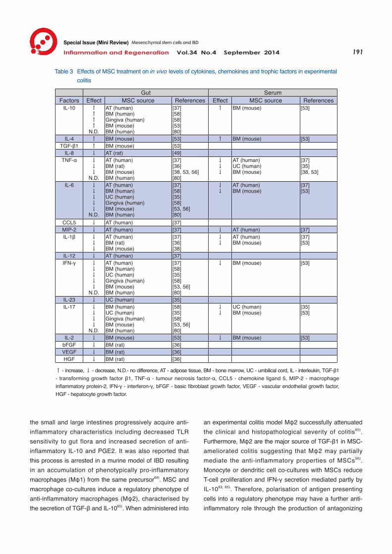
Immunomodulation The immunomodulatory properties of MSCs in models of experimental colitis have been well documented. MSCs reduce in vivo levels of pro-inflammatory cytokines and increase the production of anti-inflammatory cytokines in the gut and serum (Table 3). Conditioned medium has been shown to ameliorate the effects of colitis suggesting that the therapeutic value of MSCs is harnessed from secreted factors present in the secretome. TGF-β1 secreted by MSCs in vitro55) is elevated in the intestinal homogenate after in vivo MSC administration53); inhibition of TGF-β1 signalling abrogates the therapeutic effect of MSC in ex-perimental colitis56). Therefore TGF-β1 may be a common link between the therapeutic effect of conditioned medium and administered MSCs in vivo. Additionally, interleukin (IL)-10, which is theorized to be a target in the amelioration of enterocolitis57), was elevated in the intestine and serum
after MSC treatment of experimental colitis37, 53, 58). However, there is no evidence of IL-10 secretion by unstimulated MSCs in culture59, 60). It has been established that MSCs can be potentiated by tumour necrosis factor (TNF)-α, interferon (IFN)-γ and toll-like receptor (TLR) activation to induce an anti-inflammatory phenotype61, 62); therefore the possibility that the inflammatory microenvironment or the gut flora could upregulate anti-inflammatory factors including IL-10, indoleamine 2,3-dioxygenase (IDO) and prostaglandin E2 (PGE2) cannot be disregarded. MSCs instigate leukocytes to mediate their immuno-modulatory effects in gastrointestinal inflammation (Fig. 2). The innate immune system is critical in IBD pathology and mass neutrophil infiltration is utilized in CD diagnosis. In experimental colitis, MSCs prevent neutrophil invasion and thus damage from cytotoxic granules35, 49, 63) therefore ameliorating a key pathological marker of CD. Other cellular components of the innate immune system such as monocytes and macrophages have been postulated to be responsive to MSC secreted factors; this is highly plausible given that macrophages are highly receptive to both pro and anti-inflammatory signals. MSCs decrease the secretion of pro-inflammatory cytokines TNF-α and IL-12 from monocytes and macrophages in vitro 36, 37). Bain et al .64) suggested that resident macrophages in
Fig.1 Effects of mesenchymal stem cells on epithelial integrity in intestinal inflammation
Mesenchymal stem cells (MSCs) facilitate the repair of the intestinal epithelial barrier via the (A) secretion of trophic factors in-cluding vascular endothelial growth factor (VEGF), hepatocyte growth factor (HGF) and transforming growth factor (TGF)-β which promote re-epithelialisation and angiogenesis. (B) MSCs home to the basal crypts where they induce endogenous repair mechanisms by promoting intestinal stem cell migration and differentiation and aid in the regeneration of mucin secreting goblet cells. (C) Additionally MSCs promote the expression of tight junction proteins between epithelial cells which may attenuate inflammation-induced permeability. The regeneration of intestinal epithelium and promotion of epithelial barrier integrity prevents luminal antigens/pathogens from invading into the mucosa and submucosa; thus averting
pathogen-associated molecular pattern activation of innate leukocytes and antigen presentation to T-cells which elicit the inflammatory response.
Inflammation and Regeneration Vol.34 No.4 September 2014 191
Special Issue (Mini Review) Mesenchymal stem cells and IBD
Gut SerumFactors Effect MSC source References Effect MSC source References
IL-10 ↑↑↑↑
N.D.
AT (human)BM (human)Gingiva (human)BM (mouse)BM (human)
[37][58][58][53][80]
↑ BM (mouse) [53]
IL-4 ↑ BM (mouse) [53] ↑ BM (mouse) [53]TGF-β1 ↑ BM (mouse) [53]
IL-8 ↓ AT (rat) [49]TNF-α ↓
↓↓
N.D.
AT (human)BM (rat)BM (mouse)BM (human)
[37][36][38, 53, 56][80]
↓↓↓
AT (human)UC (human)BM (mouse)
[37] [35][38, 53]
IL-6 ↓↓↓↓↓
N.D.
AT (human)BM (human)UC (human)Gingiva (human)BM (mouse)BM (human)
[37][58][35][58][53, 56][80]
↓↓
AT (human)BM (mouse)
[37] [53]
CCL5 ↓ AT (human) [37]MIP-2 ↓ AT (human) [37] ↓ AT (human) [37]IL-1β ↓
↓↓
AT (human)BM (rat)BM (mouse)
[37][36][38]
↓↓
AT (human)BM (mouse)
[37] [53]
IL-12 ↓ AT (human) [37]IFN-γ ↓
↓↓↓↓
N.D.
AT (human)BM (human)UC (human)Gingiva (human)BM (mouse)BM (human)
[37][58][35][58][53, 56][80]
↓ BM (mouse) [53]
IL-23 ↓ UC (human) [35]IL-17 ↓
↓↓↓
N.D.
BM (human)UC (human)Gingiva (human)BM (mouse)BM (human)
[58][35][58][53, 56][80]
↓↓
UC (human)BM (mouse)
[35][53]
IL-2 ↓ BM (mouse) [53] ↓ BM (mouse) [53]bFGF ↓ BM (rat) [36]VEGF ↓ BM (rat) [36]HGF ↓ BM (rat) [36]
↑ - increase, ↓ - decrease, N.D.- no difference, AT - adipose tissue, BM - bone marrow, UC - umbilical cord, IL - interleukin, TGF-β1 - transforming growth factor β1, TNF-α - tumour necrosis factor-α, CCL5 - chemokine ligand 5, MIP-2 - macrophage inflammatory protein-2, IFN-γ - interferon-γ, bFGF - basic fibroblast growth factor, VEGF - vascular endothelial growth factor, HGF - hepatocyte growth factor.
Table 3 Effects of MSC treatment on in vivo levels of cytokines, chemokines and trophic factors in experimental colitis
the small and large intestines progressively acquire anti-inflammatory characteristics including decreased TLR sensitivity to gut flora and increased secretion of anti-inflammatory IL-10 and PGE2. It was also reported that this process is arrested in a murine model of IBD resulting in an accumulation of phenotypically pro-inflammatory macrophages (Mφ1) from the same precursor64). MSC and macrophage co-cultures induce a regulatory phenotype of anti-inflammatory macrophages (Mφ2), characterised by the secretion of TGF-β and IL-1065). When administered into
an experimental colitis model Mφ2 successfully attenuated the clinical and histopathological severity of colitis65). Furthermore, Mφ2 are the major source of TGF-β1 in MSC-ameliorated colitis suggesting that Mφ2 may partially mediate the anti-inflammatory properties of MSCs56). Monocyte or dendritic cell co-cultures with MSCs reduce T-cell proliferation and IFN-γ secretion mediated partly by IL-1063, 65). Therefore, polarisation of antigen presenting cells into a regulatory phenotype may have a further anti-inflammatory role through the production of antagonizing
192Inflammation and Regeneration Vol.34 No.4 September 2014
Special Issue (Mini Review) Mesenchymal stem cells and IBD
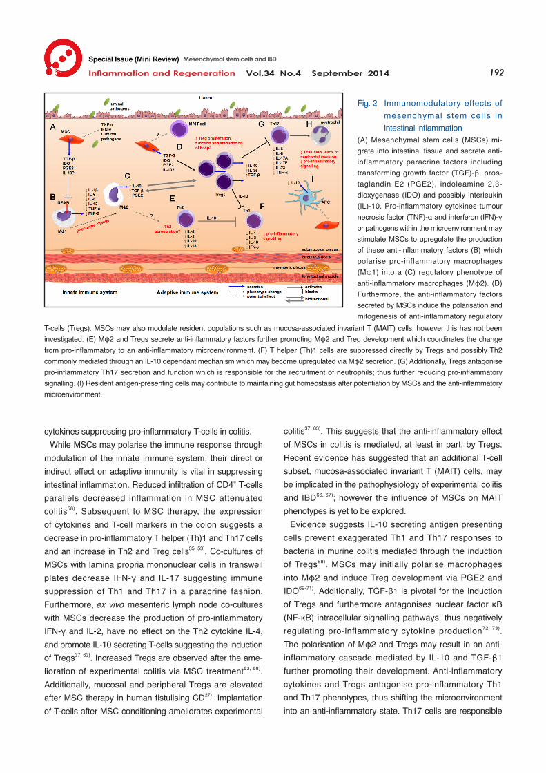
Fig. 2 Immunomodulatory effects of mesenchymal stem cells in intestinal inflammation
(A) Mesenchymal stem cells (MSCs) mi-grate into intestinal tissue and secrete anti-inflammatory paracrine factors including transforming growth factor (TGF)-β, pros-taglandin E2 (PGE2), indoleamine 2,3- dioxygenase (IDO) and possibly interleukin (IL)-10. Pro-inflammatory cytokines tumour necrosis factor (TNF)-α and interferon (IFN)-γ or pathogens within the microenvironment may stimulate MSCs to upregulate the production of these anti-inflammatory factors (B) which polarise pro-inflammatory macrophages (Mφ1) into a (C) regulatory phenotype of anti-inflammatory macrophages (Mφ2). (D) Furthermore, the anti-inflammatory factors secreted by MSCs induce the polarisation and mitogenesis of anti-inflammatory regulatory
T-cells (Tregs). MSCs may also modulate resident populations such as mucosa-associated invariant T (MAIT) cells, however this has not been investigated. (E) Mφ2 and Tregs secrete anti-inflammatory factors further promoting Mφ2 and Treg development which coordinates the change from pro-inflammatory to an anti-inflammatory microenvironment. (F) T helper (Th)1 cells are suppressed directly by Tregs and possibly Th2 commonly mediated through an IL-10 dependant mechanism which may become upregulated via Mφ2 secretion. (G) Additionally, Tregs antagonise pro-inflammatory Th17 secretion and function which is responsible for the recruitment of neutrophils; thus further reducing pro-inflammatory signalling. (I) Resident antigen-presenting cells may contribute to maintaining gut homeostasis after potentiation by MSCs and the anti-inflammatory microenvironment.
cytokines suppressing pro-inflammatory T-cells in colitis. While MSCs may polarise the immune response through modulation of the innate immune system; their direct or indirect effect on adaptive immunity is vital in suppressing intestinal inflammation. Reduced infiltration of CD4+ T-cells parallels decreased inflammation in MSC attenuated colitis58). Subsequent to MSC therapy, the expression of cytokines and T-cell markers in the colon suggests a decrease in pro-inflammatory T helper (Th)1 and Th17 cells and an increase in Th2 and Treg cells35, 53). Co-cultures of MSCs with lamina propria mononuclear cells in transwell plates decrease IFN-γ and IL-17 suggesting immune suppression of Th1 and Th17 in a paracrine fashion. Furthermore, ex vivo mesenteric lymph node co-cultures with MSCs decrease the production of pro-inflammatory IFN-γ and IL-2, have no effect on the Th2 cytokine IL-4, and promote IL-10 secreting T-cells suggesting the induction of Tregs37, 63). Increased Tregs are observed after the ame-lioration of experimental colitis via MSC treatment53, 58). Additionally, mucosal and peripheral Tregs are elevated after MSC therapy in human fistulising CD27). Implantation of T-cells after MSC conditioning ameliorates experimental
colitis37, 63). This suggests that the anti-inflammatory effect of MSCs in colitis is mediated, at least in part, by Tregs. Recent evidence has suggested that an additional T-cell subset, mucosa-associated invariant T (MAIT) cells, may be implicated in the pathophysiology of experimental colitis and IBD66, 67); however the influence of MSCs on MAIT phenotypes is yet to be explored. Evidence suggests IL-10 secreting antigen presenting cells prevent exaggerated Th1 and Th17 responses to bacteria in murine colitis mediated through the induction of Tregs68). MSCs may initially polarise macrophages into Mφ2 and induce Treg development via PGE2 and IDO69-71). Additionally, TGF-β1 is pivotal for the induction of Tregs and furthermore antagonises nuclear factor κB (NF-κB) intracellular signalling pathways, thus negatively regulating pro-inflammatory cytokine production72, 73). The polarisation of Mφ2 and Tregs may result in an anti-inflammatory cascade mediated by IL-10 and TGF-β1 further promoting their development. Anti-inflammatory cytokines and Tregs antagonise pro-inflammatory Th1 and Th17 phenotypes, thus shifting the microenvironment into an anti-inflammatory state. Th17 cells are responsible
Inflammation and Regeneration Vol.34 No.4 September 2014 193
Special Issue (Mini Review) Mesenchymal stem cells and IBD
for neutrophil recruitment74), therefore Treg suppression of Th17 may coincide with decreased neutrophil invasion observed after MSC treatment of experimental colitis35). Additionally, Tregs have been demonstrated to promote anti-inflammatory properties in neutrophils75). Thus, Tregs may further suppress the inflammatory response by directly acting on the innate immune system.
Future Outlook Currently there are more than 200 ongoing clinical trials testing MSCs as a viable treatment option, however, only a few clinical trials tested MSCs in IBD patients. These trials demonstrated that MSC treatment is effective for refractory fistulising and luminal CD after local and systemic administration. Clinical data combined with studies in experimental models are indicative of the potential of MSC therapy to ameliorate IBD. These findings have been attributed to immunomodulation and restoration of epithelial barrier integrity through paracrine trophic factors and cytokines secreted by MSCs. Outstanding questions include assessing the role of MSCs in tumour formation and progression; this remains contentious and further studies are warranted. Evidence of MSC entrapment in filtering organs after systemic injection necessitates the need for further research into more effi-cient methods of administration. Future studies should com-paratively assess administration techniques to effectively target gastrointestinal inflammation. Additionally, long-term immune tolerance to MSCs received from donors needs to be discerned. The effects of MSC treatment on inflammation-induced damage to the enteric nervous system embedded in the gastrointestinal wall has not been studied, but is important for understanding pathophysiology of disease reoccurrence and severity. Thus, therapeutic mechanisms of MSCs need to be elucidated taking into account the complexity of the intestinal microenvironment.
Acknowledgement and Source of funding This study was supported by a Victoria University Research and Development grant.
Conflict of interests None
References 1) Baumgart DC, Sandborn WJ: Crohn’s disease. Lancet.
2012; 380: 1590-1605.
2) Strober W, Fuss I, Mannon P: The fundamental basis of inflammatory bowel disease. J Clin Invest. 2007; 117: 514-521.
3) Landgren AM, Landgren O, Gridley G, Dores GM, Linet MS, Morton LM: Autoimmune disease and subsequent risk of developing alimentary tract cancers among 4.5 million US male veterans. Cancer. 2011; 117: 1163-1171.
4) de Ridder L, Turner D, Wilson DC, Koletzko S, Martin-de-Carpi J, Fagerberg UL, Spray C, Sladek M, Shaoul R, Roma-Giannikou E: Malignancy and mortality in pediatric patients with inflammatory bowel disease: A multinational study from the Porto Pediatric IBD group. Inflammatory Bowel Diseases. 2014; 20: 291-300.
5) Ng SC: Epidemiology of inflammatory Bowel disease: focus on Asia. Best Pract Res Clin Gastroenterol. 2014; 28: 363-372.
6) Gearry RB, Leong RW: Inflammatory bowel disease in Asia: The start of the epidemic? J Gastroenterol Hepatol. 2013; 28: 899-900.
7) Mayer L: Evolving paradigms in the pathogenesis of IBD. J Gastroenterol. 2010; 45: 9-16.
8) Khor B, Gardet A, Xavier RJ: Genetics and pathogenesis of inflammatory bowel disease. Nature. 2011; 474: 307-317.
9) Manichanh C, Borruel N, Casellas F, Guarner F: The gut microbiota in IBD. Nat Rev Gastroenterol Hepatol. 2012; 9: 599-608.
10) Fries W, Belvedere A, Vetrano S: Sealing the broken barrier in IBD: intestinal permeability, epithelial cells and junctions. Current Drug Targets. 2013; 14: 1460-1470.
11) Abraham C, Medzhitov R: Interactions between the host innate immune system and microbes in in-flammatory bowel disease. Gastroenterology. 2011; 140: 1729-1737.
12) Curkovic I, Egbring M, Kullak-Ublick GA: Risks of inflammatory bowel disease treatment with glucocortico-steroids and aminosalicylates. Digestive Diseases. 2013; 31: 368-373.
13) Colombel JF, Sandborn WJ, Reinisch W, Mantzaris GJ, Kornbluth A, Rachmilewitz D, Lichtiger S, D’Haens G, Diamond RH, Broussard DL, Tang KL, van der Woude CJ, Rutgeerts P: Infliximab, azathioprine, or combination therapy for Crohn’s disease. New Engl J Med. 2010; 362: 1383-1395.
14) Zenlea T, Peppercorn MA: Immunosuppressive therapies for inflammatory bowel disease. World J Gastroenterol.
194Inflammation and Regeneration Vol.34 No.4 September 2014
2014; 20: 3146-3152. 15) Mosna F, Sensebe L, Krampera M: Human bone
marrow and adipose tissue mesenchymal stem cells: a user’s guide. Stem Cells and Development. 2010; 19: 1449-1470.
16) Ankrum JA, Ong JF, Karp JM: Mesenchymal stem cells: immune evasive, not immune privileged. Nat Biotechnol. 2014; 32: 252-260.
17) Li J, Ezzelarab MB, Cooper DK: Do mesenchymal stem cells function across species barriers? Relevance for xenotransplantation. Xenotransplantation. 2012; 19: 273-285.
18) Karp JM, Leng Teo GS: Mesenchymal stem cell homing: the devil is in the details. Cell Stem Cell. 2009; 4: 206-216.
19) Wu Y, Chen L, Scott PG, Tredget EE: Mesenchymal stem cells enhance wound healing through differentia-tion and angiogenesis. Stem Cells. 2007; 25: 2648- 2659.
20) Chen L, Tredget EE, Wu PY, Wu Y: Paracrine factors of mesenchymal stem cells recruit macrophages and endothelial lineage cells and enhance wound healing. PloS One. 2008; 3: e1886.
21) Sémont A, Demarquay C, Bessout R, Durand C, Benderitter M, Mathieu N: Mesenchymal stem cell therapy stimulates endogenous host progenitor cells to improve colonic epithelial regeneration. PloS One. 2013; 8: e70170.
22) Ma S, Xie N, Li W, Yuan B, Shi Y, Wang Y: Immuno-biology of mesenchymal stem cells. Cell Death Differ. 2013; 21: 216-225.
23) Garcia-Olmo D, Herreros D, Pascual I, Pascual JA, Del-Valle E, Zorrilla J, De-La-Quintana P, Garcia-Arranz M, Pascual M: Expanded adipose-derived stem cells for the treatment of complex perianal fistula: a phase II clinical trial. Dis Colon Rectum. 2009; 52: 79-86.
24) García-Olmo D, Herreros D, De-La-Quintana P, Guadalajara H, Trébol J, Georgiev-Hristov T, García-Arranz M: Adipose-derived stem cells in Crohn's rectovaginal fistula. Case Rep Med. 2010; doi: 10.1155/ 2010/961758.
25) Garcia-Olmo D, Herreros D, Pascual M, Pascual I, De-La-Quintana P, Trébol J, García-Arranz M: Treatment of enterocutaneous fistula in Crohn’s disease with adipose-derived stem cells: a comparison of protocols with and without cell expansion. Int J Colorectal Dis. 2009; 24: 27-30.
26) Cho YB, Lee WY, Park KJ, Kim M, Yoo H-W, Yu CS: Autologous Adipose Tissue-Derived Stem Cells for the Treatment of Crohns Fistula: A Phase I Clinical Study. Cell Transplantation. 2013; 22: 279-285.
27) Ciccocioppo R, Bernardo ME, Sgarella A, Maccario R, Avanzini MA, Ubezio C, Minelli A, Alvisi C, Vanoli A, Calliada F: Autologous bone marrow-derived mes-enchymal stromal cells in the treatment of fistulising Crohn’s disease. Gut. 2011; 60: 788-798.
28) Lee WY, Park KJ, Cho YB, Yoon SN, Song KH, Kim DS, Jung SH, Kim M, Yoo HW, Kim I: Autologous adipose tissue‐derived stem Cells treatment demonstrated fa-vorable and sustainable therapeutic effect for Crohn’s fistula. Stem Cells. 2013; 31: 2575-2581.
29) Guadalajara H, Herreros D, De-La-Quintana P, Trebol J, Garcia-Arranz M, Garcia-Olmo D: Long-term follow-up of patients undergoing adipose-derived adult stem cell administration to treat complex perianal fistulas. Int J Colorectal Dis. 2012; 27: 595-600.
30) García-Olmo D, García-Arranz M, Herreros D, Pascual I, Peiro C, Rodríguez-Montes JA: A phase I clinical trial of the treatment of Crohn’s fistula by adipose mes-enchymal stem cell transplantation. Dis Colon Rectum. 2005; 48: 1416-1423.
31) Desai VD, Hsia HC, Schwarzbauer JE: Reversible modulation of myofibroblast differentiation in adipose-derived mesenchymal stem cells. PloS One. 2014; 9: e86865.
32) Duijvestein M, Vos ACW, Roelofs H, Wildenberg ME, Wendrich BB, Verspaget HW, Kooy-Winkelaar EM, Koning F, Zwaginga JJ, Fidder HH: Autologous bone marrow-derived mesenchymal stromal cell treatment for refractory luminal Crohn’s disease: results of a phase I study. Gut. 2010; 59: 1662-1669.
33) Forbes GM, Sturm MJ, Leong RW, Sparrow MP, Segarajasingam D, Cummins AG, Phillips M, Herrmann RP: A phase 2 study of allogeneic mesenchymal stromal cells for luminal Crohn’s disease refractory to biologic therapy. Clin Gastroenterol Hepatol. 2014; 12: 64-71.
34) Hayashi Y, Tsuji S, Tsujii M, Nishida T, Ishii S, Iijima H, Nakamura T, Eguchi H, Miyoshi E, Hayashi N: Topical implantation of mesenchymal stem cells has beneficial effects on healing of experimental colitis in rats. J Pharmacol Exp Ther. 2008; 326: 523-531.
35) Liang L, Dong C, Chen X, Fang Z, Xu J, Liu M, Zhang X, Gu DS, Wang D, Du W: Human umbilical cord mes-
Special Issue (Mini Review) Mesenchymal stem cells and IBD
Inflammation and Regeneration Vol.34 No.4 September 2014 195
enchymal stem cells ameliorate mice trinitrobenzene sulfonic acid (TNBS)-induced colitis. Cell Transplant. 2011; 20: 1395-1408.
36) Tanaka F, Tominaga K, Ochi M, Tanigawa T, Watanabe T, Fujiwara Y, Ohta K, Oshitani N, Higuchi K, Arakawa T: Exogenous administration of mesenchymal stem cells ameliorates dextran sulfate sodium-induced colitis via anti-inflammatory action in damaged tissue in rats. Life Sci. 2008; 83: 771-779.
37) González MA, Gonzalez-Rey E, Rico L, Büscher D, Delgado M: Adipose-derived mesenchymal stem cells alleviate experimental colitis by inhibiting inflammatory and autoimmune responses. Gastroenterology. 2009; 136: 978-989.
38) He X-W, He X-S, Lian L, Wu X-J, Lan P: Systemic infusion of bone marrow-derived mesenchymal stem cells for treatment of experimental colitis in mice. Dig Dis Sci. 2012; 57: 3136-3144.
39) Ankrum J, Karp JM: Mesenchymal stem cell therapy: Two steps forward, one step back. Trends Mol Med. 2010; 16: 203-209.
40) Watanabe S, Arimura Y, Nagaishi K, Isshiki H, Onodera K, Nasuno M, Yamashita K, Idogawa M, Naishiro Y, Murata M: Conditioned mesenchymal stem cells produce pleiotropic gut trophic factors. J Gastroenterol. 2013; 49: 270-282.
41) Prockop DJ, Brenner M, Fibbe WE, Horwitz E, Le Blanc K, Phinney DG, Simmons PJ, Sensebe L, Keating A: Defining the risks of mesenchymal stromal cell therapy. Cytotherapy. 2010; 12: 576-578.
42) Karnoub AE, Dash AB, Vo AP, Sullivan A, Brooks MW, Bell GW, Richardson AL, Polyak K, Tubo R, Weinberg RA: Mesenchymal stem cells within tumour stroma promote breast cancer metastasis. Nature. 2007; 449: 557-563.
43) Touboul C, Lis R, Al Farsi H, Raynaud CM, Warfa M, Althawadi H, Mery E, Mirshahi M, Rafii A: Mesenchy-mal stem cells enhance ovarian cancer cell infiltration through IL6 secretion in an amniochorionic membrane based 3D model. J Transl Med. 2013;11: 28.
44) Bergfeld SA, Blavier L, DeClerck YA: Bone marrow-derived mesenchymal stromal cells promote survival and drug resistance in tumor cells. Mol Cancer Ther. 2014; 13: 962-975.
45) Tolar J, Nauta AJ, Osborn MJ, Panoskaltsis Mortari A, McElmurry RT, Bell S, Xia L, Zhou N, Riddle M, Schroeder TM: Sarcoma derived from cultured
mesenchymal stem cells. Stem Cells. 2007; 25: 371-379. 46) Rubio D, Garcia-Castro J, Martín MC, de la Fuente R,
Cigudosa JC, Lloyd AC, Bernad A: Spontaneous human adult stem cell transformation. Cancer Res. 2005; 65: 3035-3039.
47) Donnelly JM, Engevik A, Feng R, Xiao C, Boivin GP, Li J, Houghton J, Zavros Y, Zavros Y: Mesenchymal stem cells induce epithelial proliferation within the inflamed stomach. Am J Physiol Gastrointest Liver Physiol.2014; 306: G1075-G1088.
48) Okamoto R: Epithelial regeneration in inflammatory bowel diseases. Inflamm Regen. 2011; 31: 275-281.
49) Ando Y, Inaba M, Sakaguchi Y, Tsuda M, Quan GK, Omae M, Okazaki K, Ikehara S: Subcutaneous adipose tissue-derived stem cells facilitate colonic mucosal recovery from 2, 4, 6‐trinitrobenzene sulfonic acid (TNBS)-induced colitis in rats. Inflammatory Bowel Diseases. 2008; 14: 826-838.
50) Castelo-Branco MT, Soares ID, Lopes DV, Buongusto F, Martinusso CA, do Rosario Jr A, Souza SA, Gutfilen B, Fonseca LMB, Elia C: Intraperitoneal but not intravenous cryopreserved mesenchymal stromal cells home to the inflamed colon and ameliorate experimental colitis. PloS One. 2012; 7: e33360.
51) Fawzy SA, El-Din Abo-Elnou R, Abd-El-Maksoud El-Deeb D, Yousry Abd-Elkader M: The possible role of mesenchymal stem cells therapy in the repair of experimentally induced colitis in male albino rats. Int J Stem Cells. 2013; 6: 92-103.
52) Tanaka H, Arimura Y, Yabana T, Goto A, Hosokawa M, Nagaishi K, Yamashita K, Yamamoto H, Sasaki Y, Fujimiya M: Myogenic lineage differentiated mesen-chymal stem cells enhance recovery from dextran sulfate sodium-induced colitis in the rat. J Gastroenterol. 2011; 46: 143-152.
53) Chen Q-Q, Yan L, Wang C-Z, Wang W-H, Shi H, Su B-B, Zeng Q-H, Du H-T, Wan J: Mesenchymal stem cells alleviate TNBS-induced colitis by modulating inflammatory and autoimmune responses. World J Gastroenterol. 2013; 19: 4702-4717.
54) Yabana T, Arimura Y, Tanaka H, Goto A, Hosokawa M, Nagaishi K, Yamashita K, Yamamoto H, Adachi Y, Sasaki Y: Enhancing epithelial engraftment of rat mesenchymal stem cells restores epithelial barrier integrity. J Pathol. 2009; 218: 350-359.
55) Amable PR, Teixeira MV, Carias RB, Granjeiro JM, Borojevic R: Protein synthesis and secretion in human
Special Issue (Mini Review) Mesenchymal stem cells and IBD
196Inflammation and Regeneration Vol.34 No.4 September 2014
mesenchymal cells derived from bone marrow, adipose tissue and Wharton’s jelly. Stem Stem Cell Res Ther. 2014; 5: 53.
56) Wang C, Chen J, Sun L, Liu Y: TGF-beta signaling-dependent alleviation of dextran sulfate sodium-induced colitis by mesenchymal stem cell transplantation. Mol Biol Rep. 2014; 41: 4977-4983.
57) Shah N, Kammermeier J, Elawad M, Glocker EO: Interleukin-10 and interleukin-10-receptor defects in inflammatory bowel disease. Curr Allergy Asthma Rep. 2012; 12: 373-379.
58) Zhang Q, Shi S, Liu Y, Uyanne J, Shi Y, Shi S, Le AD: Mesenchymal stem cells derived from human gingiva are capable of immunomodulatory functions and ameliorate inflammation-related tissue destruction in experimental colitis. J Immunol. 2009; 183: 7787-7798.
59) Melief SM, Geutskens SB, Fibbe WE, Roelofs H: Multipotent stromal cells skew monocytes towards an anti-inflammatory interleukin-10-producing phenotype by production of interleukin-6. Haematologica. 2013; 98: 888-895.
60) Hwang JH, Shim SS, Seok OS, Lee HY, Woo SK, Kim BH, Song HR, Lee JK, Park YK: Comparison of cytokine expression in mesenchymal stem cells from human placenta, cord blood, and bone marrow. J Korean Med Sci. 2009; 24: 547-554.
61) Waterman RS, Tomchuck SL, Henkle SL, Betancourt AM: A new mesenchymal stem cell (MSC) paradigm: polarization into a pro-inflammatory MSC1 or an Immunosuppressive MSC2 phenotype. PloS One. 2010; 5: e10088.
62) English K, Barry FP, Field-Corbett CP, Mahon BP: IFN-γ and TNF-α differentially regulate immunomodulation by murine mesenchymal stem cells. Immuno Lett. 2007; 110: 91-100.
63) Gonzalez-Rey E, Anderson P, González MA, Rico L, Büscher D, Delgado M: Human adult stem cells derived from adipose tissue protect against experimental colitis and sepsis. Gut. 2009; 58: 929-939.
64) Bain C, Scott C, Uronen-Hansson H, Gudjonsson S, Jansson O, Grip O, Guilliams M, Malissen B, Agace W, Mowat AM: Resident and pro-inflammatory macro-phages in the colon represent alternative context-dependent fates of the same Ly6Chi monocyte pre-cursors. Mucosal Immunol. 2013; 6: 498-510.
65) Anderson P, Souza-Moreira L, Morell M, Caro M, O’Valle F, Gonzalez-Rey E, Delgado M: Adipose-derived
mesenchymal stromal cells induce immunomodulatory macrophages which protect from experimental colitis and sepsis. Gut. 2013; 62: 1131-1141.
66) Ruijing X, Mengjun W, Xiaoling Z, Shu P, Mei W, Yingcheng Z, Yuling H, Jinquan T: Jα33+ MAIT cells play a protective role in TNBS induced intestinal inflammation. Hepatogastroenterology. 2012; 59: 762- 767.
67) Serriari NE, Eoche M, Lamotte L, Fumery M, Marcelo P, Chatelain D, Barre A, Nguyen‐Khac E, Lantz O, Dupas JL: Innate mucosal‐associated invariant T (MAIT) cells are activated in inflammatory bowel diseases. Clin Exp Immunol. 2014; 176: 266-274.
68) Liu B, Tonkonogy SL, Sartor RB: Antigen-presenting cell production of IL-10 inhibits T-helper 1 and 17 cell responses and suppresses colitis in mice. Gastro-enterology. 2011; 141: 653-662.
69) Nemeth K, Leelahavanichkul A, Yuen PS, Mayer B, Parmelee A, Doi K, Robey PG, Leelahavanichkul K, Koller BH, Brown JM, Hu X, Jelinek I, Star RA, Mezey E: Bone marrow stromal cells attenuate sepsis via prostaglandin E(2)-dependent reprogramming of host macrophages to increase their interleukin-10 production. Nat Med. 2009; 15: 42-49.
70) Francois M, Romieu-Mourez R, Li M, Galipeau J: Human MSC suppression correlates with cytokine induction of indoleamine 2,3-dioxygenase and by-stander M2 macrophage differentiation. Mol Ther. 2012; 20: 187-195.
71) Baratelli F, Lin Y, Zhu L, Yang SC, Heuze-Vourc’h N, Zeng G, Reckamp K, Dohadwala M, Sharma S, Dubinett SM: Prostaglandin E2 induces FOXP3 gene expression and T regulatory cell function in human CD4+ T cells. J Immunol. 2005; 175: 1483-1490.
72) Biancheri P, Giuffrida P, Docena GH, MacDonald TT, Corazza GR, Di Sabatino A: The role of transforming growth factor (TGF)-β in modulating the immune re-sponse and fibrogenesis in the gut. Cytokine Growth Factor Rev. 2013; 25: 45-55.
73) Nguyen T-LM, Sullivan NL, Ebel M, Teague RM, DiPaolo RJ: Antigen-Specific TGF-β−Induced Regu-latory T Cells Secrete Chemokines, Regulate T Cell Trafficking, and Suppress Ongoing Autoimmunity. J Immunol. 2011; 187: 1745-1753.
74) Pelletier M, Maggi L, Micheletti A, Lazzeri E, Tamassia N, Costantini C, Cosmi L, Lunardi C, Annunziato F, Romagnani S, Cassatella MA: Evidence for a cross-
Special Issue (Mini Review) Mesenchymal stem cells and IBD
Inflammation and Regeneration Vol.34 No.4 September 2014 197
talk between human neutrophils and Th17 cells. Blood. 2010; 115: 335-343.
75) Lewkowicz N, Klink M, Mycko MP, Lewkowicz P: Neutrophil--CD4+CD25+ T regulatory cell interactions: a possible new mechanism of infectious tolerance. Immunobiology. 2013; 218: 455-464.
76) de la Portilla F, Alba F, García-Olmo D, Herrerías J, González F, Galindo A: Expanded allogeneic adipose-derived stem cells (eASCs) for the treatment of complex perianal fistula in Crohn’s disease: results from a multi-center phase I/IIa clinical trial. Int J Colorectal Dis. 2013; 28: 313-323.
77) Liu X, Zuo D, Fan H, Tang Q, Shou Z, Cao D, Zou Z: Over-expression of CXCR4 on mesenchymal stem cells protect against experimental colitis via immuno-modulatory functions in impaired tissue. J Mol Histol. 2013; 45: 181-193.
Special Issue (Mini Review) Mesenchymal stem cells and IBD
78) Li L, Liu S, Xu Y, Zhang A, Jiang J, Tan W, Xing J, Feng G, Liu H, Huo F: Human umbilical cord-derived mesenchymal stem cells downregulate inflammatory responses by shifting the Treg/Th17 profile in experi-mental colitis. Pharmacology. 2013; 92: 257-264.
79) Kim HS, Shin TH, Lee BC, Yu KR, Seo Y, Lee S, Seo MS, Hong IS, Choi SW, Seo KW: Human umbilical cord blood mesenchymal stem cells reduce colitis in mice by activating NOD2 signaling to COX2. Gastroenterology. 2013; 145: 1392-1403. e1-e8.
80) Duijvestein M, Wildenberg ME, Welling MM, Hennink S, Molendijk I, van Zuylen VL, Bosse T, Vos ACW, de Jonge‐Muller ES, Roelofs H: Pretreatment with interferon‐γ enhances the therapeutic activity of mes-enchymal stromal cells in animal models of colitis. Stem Cells. 2011; 29: 1549-1558.