Embed Size (px)
Citation preview
IMPORTANT
EVENTS:
UKRCO is being held in
Manchester on 12-14 June.
Register at http://
ukrco.org.uk/registration/
MSR Annual Research
Meeting & Dinner will be
held on Wed 28 June. Sub-
mission date for abstracts
is Fri 9 June.
RCR17 comes to Liverpool
on 11-13 Sept. Register at
https://www.rcr.ac.uk/
college/rcr17
FRCR Aintree Bones and
Chest Course on 18 & 19
Sept—email joanne.
Closing dates for FRCR
exam applications:
Part 2A—(old format)
Fri 21 July @ 4pm
Part 2A—(new format)
Fri 20 October
Part 2B—Fri 4 August @
4pm
In this edition…. Dear all
This month the newsletter is co-authored by Dr Roz Joseph and Dr Jim Hare, ST3, with contributions by Dr Flavius Parvulescu. We have reported on the first regional registrar learning and discrepancy meeting which was held in April.
This will be the last of the newsletters overseen by your current Ed. this year—I hope you’ve en-joyed this new venture and I wish the new editor well in taking over the reins of this publication.
As usual send your sub-missions to:
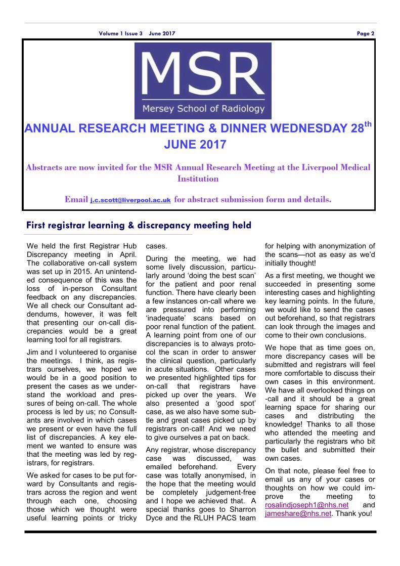
RCR Ceremony
Congratulations to our registrars who passed their Part 2B exam at the recent sitting, and those who completed Part 1 and 2A modules
Their graduation ceremony was held on 19 May at the RCR in London.
Also present at the ceremony was Dr Curtis, who was presented with the RCR’s Gold Medal for his contribu-tion to teaching.
Volume 1 Issue 3 June 2017
Mersey School of Radiology (HEE North West) Summer Newsletter
Radiology news and
updates:
General Election 2017:
RCR calls for ‘an NHS
equipped for 21st cen-
tury'
iRefer8 is released, the
newest version of the
diagnostic imaging
guidelines for referring
clinicians
A federal report from
the USA warns of a
shortage of Molyb-
denum-99 (Mo-99) and
Technetium-99m (Tc-
99m) isotopes
RCR thanks hardwork-
ing staff who mini-
mized disruption to
patients during the
recent ransomware
attack.
Inside this issue:
RCR ceremony 1
Discrepancy meeting 2
2B Course review 3
Global health study day 3
Visiting lecturers 4-5
Research 6-7
New appointments 8
Minerva 9
Dr Roz Joseph Co-author, ST3
Dr James Hare Co-author, ST3
Dr Flavius Parvulescu, Contributor,ST4
Dr Jen Jou Wong, Co-author and editor, Lead SpR
First registrar learning & discrepancy meeting held
Page 2 Volume 1 Issue 3 June 2017
We held the first Registrar Hub Discrepancy meeting in April. The collaborative on-call system was set up in 2015. An unintend-ed consequence of this was the loss of in-person Consultant feedback on any discrepancies. We all check our Consultant ad-dendums, however, it was felt that presenting our on-call dis-crepancies would be a great learning tool for all registrars.
Jim and I volunteered to organise the meetings. I think, as regis-trars ourselves, we hoped we would be in a good position to present the cases as we under-stand the workload and pres-sures of being on-call. The whole process is led by us; no Consult-ants are involved in which cases we present or even have the full list of discrepancies. A key ele-ment we wanted to ensure was that the meeting was led by reg-istrars, for registrars.
We asked for cases to be put for-ward by Consultants and regis-trars across the region and went through each one, choosing those which we thought were useful learning points or tricky
cases.
During the meeting, we had some lively discussion, particu-larly around ‘doing the best scan’ for the patient and poor renal function. There have clearly been a few instances on-call where we are pressured into performing ‘inadequate’ scans based on poor renal function of the patient. A learning point from one of our discrepancies is to always proto-col the scan in order to answer the clinical question, particularly in acute situations. Other cases we presented highlighted tips for on-call that registrars have picked up over the years. We also presented a ‘good spot’ case, as we also have some sub-tle and great cases picked up by registrars on-call! And we need to give ourselves a pat on back.
Any registrar, whose discrepancy case was discussed, was emailed beforehand. Every case was totally anonymised, in the hope that the meeting would be completely judgement-free and I hope we achieved that. A special thanks goes to Sharron Dyce and the RLUH PACS team
for helping with anonymization of the scans—not as easy as we’d initially thought!
As a first meeting, we thought we succeeded in presenting some interesting cases and highlighting key learning points. In the future, we would like to send the cases out beforehand, so that registrars can look through the images and come to their own conclusions.
We hope that as time goes on, more discrepancy cases will be submitted and registrars will feel more comfortable to discuss their own cases in this environment. We have all overlooked things on-call and it should be a great learning space for sharing our cases and distributing the knowledge! Thanks to all those who attended the meeting and particularly the registrars who bit the bullet and submitted their own cases.
On that note, please feel free to email us any of your cases or thoughts on how we could im-prove the meeting to [email protected] and [email protected]. Thank you!
ANNUAL RESEARCH MEETING & DINNER WEDNESDAY 28th
JUNE 2017
Abstracts are now invited for the MSR Annual Research Meeting at the Liverpool Medical
Institution
Email [email protected] for abstract submission form and details.
Few 2B courses are more fa-mous than the Edinburgh course, and even fewer have a longer waiting list. Having at-tended recently, I understand
why.
Taking place over two days in the Edinburgh Business School, the course inspires professionalism from the start. The venue is mod-ern, clean and looks slick with nice furniture and comfy chairs. The clear and tight schedule is adhered to and there is an over-all feeling of organisational com-petency. There are the usual breaks with coffee, tea, etc. and the unremarkable lunch is served on site. A free dinner with wine and good food is included in a restaurant that is also on site – a nice touch and good opportunity to meet some of the faculty and
candidates (the course is small with only 24 registrants). For £450 you get a very good deal.
Over two days you will get two written mock exams, i.e. long cases followed by a rapids pack. The content felt appropriate com-pared to the exam, although the rapids were slightly harder (like in most courses, except Bristol). What is (probably) unique among 2B courses is that you actually have your papers marked by the faculty, with individualised feed-back for the long cases. You also get six specialty-specific hour-long small group tutorials on MSK, neuro, chest, paeds, breast and nuclear medicine – extreme-ly useful!
And that’s not all. The viva is where they really earn their mon-ey. You get one hour in a small,
dark, quiet room, just you, all alone, with two examiners. And you get it three times! Nothing can replicate the stress you will be under in the actual exam, but listening to the deafening silence of your own inability to articulate a logical sentence and feeling the examiners’ burning gaze of disappointment in that small, dark, quiet room will certainly be as close as it gets to the real thing. It can be a tough and pain-ful experience to go through (have tissues handy), which is precisely why it is so good!
So if you are planning to spend money on a single 2B course, you are unlikely to find a better deal than Edinburgh. Just keep in mind that waiting lists begin 12 months in advance!
Careers Fair
Page 3 Volume 1 Issue 3 June 2017
The Edinburgh 2B Course—Review by Dr Flavius Parvulescu
Page 3 Volume 1 Issue 3 June 2017
Global Health and Infectious Diseases Radiology Study Day
The first global health and in-fectious diseases study day will be held at the Institute in the Park at Alder Hey Chil-
dren’s Hospital on 19 July.
The event has been organised by two ST3s, Dr Roz Joseph and Dr Radhika Prasad along with Dr Karen Chetcuti, Consult-ant Paediatric Radiologist.
This unique one-day course will comprise an educational morn-
ing session on imaging of infec-tious diseases in a systems-based approach, with both pae-diatric and adult topics being covered. The afternoon will be a series of lectures from expert speakers, high lighting the prac-tical aspects of creating and de-livering an outreach programme abroad, with an emphasis on radiology.
Places are limited. To book your place for £50, please email: [email protected] (deadline 9 June).
Page 4 Volume 1 Issue 3 June 2017
As part of our Mersey school of radiology evening lecture series, on 15th March we were delighted to welcome Prof Margaret Hall-Craggs, RCR roentgen professor and clinical radiologist from UCLH. She is active in research with over 170 publications and taught widely in a number of are-as including breast MRI, imaging of the female re-productive tract and imaging inflammatory disease of the bone and joints.
Dr Hall-Craggs gave us a very interesting talk on the imaging of disorders of sex development. The talk raised interesting discussion points regarding the medical, psychological and social impact and also management of these disorders, and how ra-
diology plays a role in this.
Margaret is an active member of the International Society of Magnetic Resonance in Medicine. She is currently Secretary of the Society, has served 3 terms on the Board of Trustees. She is Chair of several committees within the ISMRM and has been active in promoting opportunities and mentor-ship of women radiologists and scientists within the Society. She is a full member of the Centre for Medical Imaging at UCL (Academic Radiology), and is currently supervising 3 PhD and 1MD stu-dents.
Prof Margaret Hall-Craggs
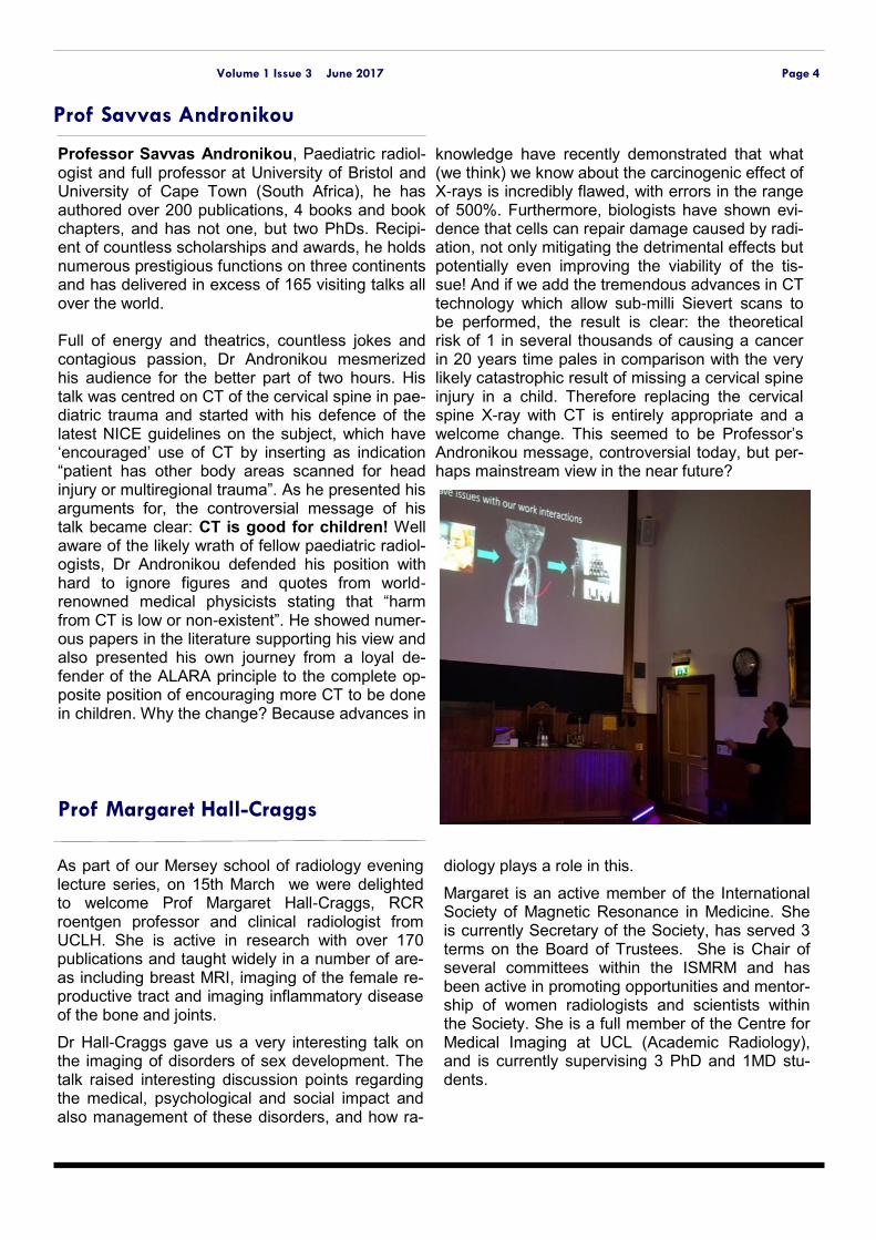
Professor Savvas Andronikou, Paediatric radiol-ogist and full professor at University of Bristol and University of Cape Town (South Africa), he has authored over 200 publications, 4 books and book chapters, and has not one, but two PhDs. Recipi-ent of countless scholarships and awards, he holds numerous prestigious functions on three continents and has delivered in excess of 165 visiting talks all over the world. Full of energy and theatrics, countless jokes and contagious passion, Dr Andronikou mesmerized his audience for the better part of two hours. His talk was centred on CT of the cervical spine in pae-diatric trauma and started with his defence of the latest NICE guidelines on the subject, which have ‘encouraged’ use of CT by inserting as indication “patient has other body areas scanned for head injury or multiregional trauma”. As he presented his arguments for, the controversial message of his talk became clear: CT is good for children! Well aware of the likely wrath of fellow paediatric radiol-ogists, Dr Andronikou defended his position with hard to ignore figures and quotes from world-renowned medical physicists stating that “harm from CT is low or non-existent”. He showed numer-ous papers in the literature supporting his view and also presented his own journey from a loyal de-fender of the ALARA principle to the complete op-posite position of encouraging more CT to be done in children. Why the change? Because advances in
knowledge have recently demonstrated that what (we think) we know about the carcinogenic effect of X-rays is incredibly flawed, with errors in the range of 500%. Furthermore, biologists have shown evi-dence that cells can repair damage caused by radi-ation, not only mitigating the detrimental effects but potentially even improving the viability of the tis-sue! And if we add the tremendous advances in CT technology which allow sub-milli Sievert scans to be performed, the result is clear: the theoretical risk of 1 in several thousands of causing a cancer in 20 years time pales in comparison with the very likely catastrophic result of missing a cervical spine injury in a child. Therefore replacing the cervical spine X-ray with CT is entirely appropriate and a welcome change. This seemed to be Professor’s Andronikou message, controversial today, but per-haps mainstream view in the near future?
Prof Savvas Andronikou
Dr Miranda Harvie
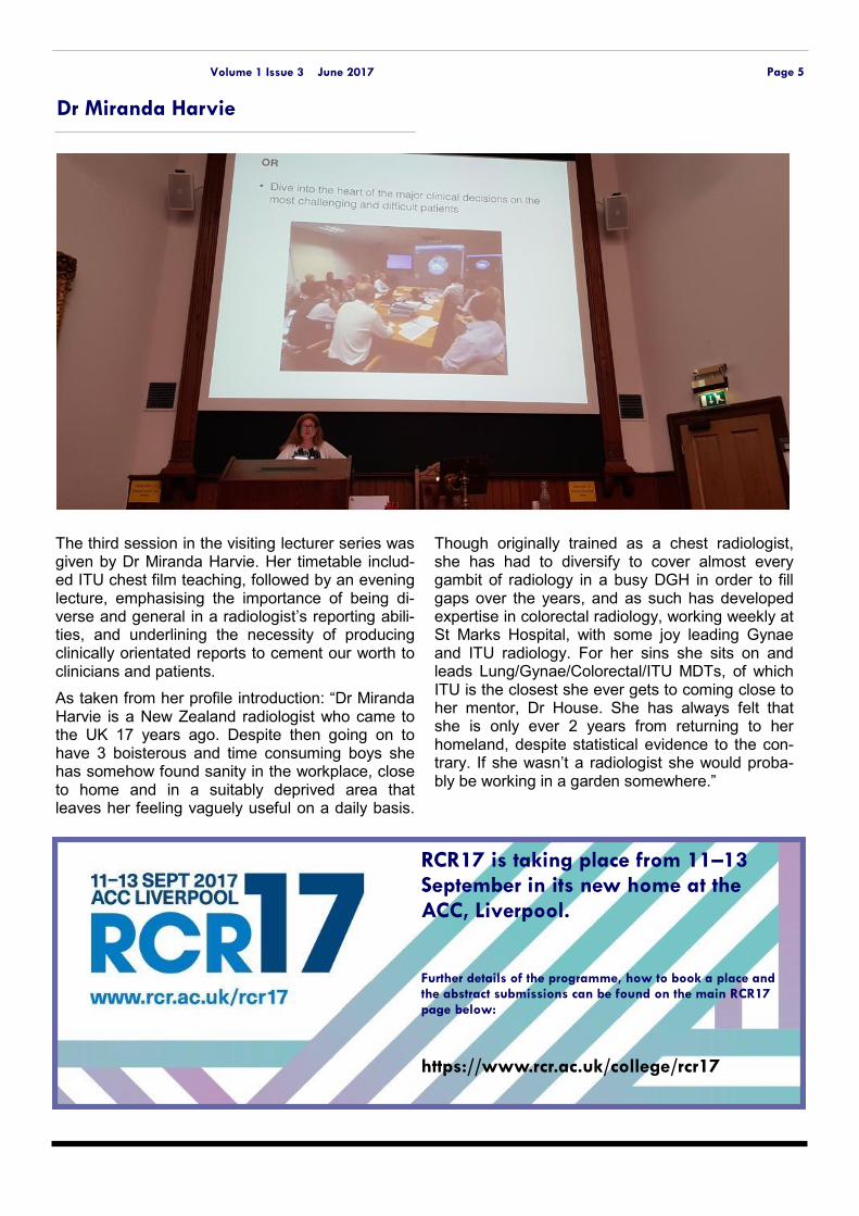
The third session in the visiting lecturer series was given by Dr Miranda Harvie. Her timetable includ-ed ITU chest film teaching, followed by an evening lecture, emphasising the importance of being di-verse and general in a radiologist’s reporting abili-ties, and underlining the necessity of producing clinically orientated reports to cement our worth to clinicians and patients.
As taken from her profile introduction: “Dr Miranda Harvie is a New Zealand radiologist who came to the UK 17 years ago. Despite then going on to have 3 boisterous and time consuming boys she has somehow found sanity in the workplace, close to home and in a suitably deprived area that leaves her feeling vaguely useful on a daily basis.
Though originally trained as a chest radiologist, she has had to diversify to cover almost every gambit of radiology in a busy DGH in order to fill gaps over the years, and as such has developed expertise in colorectal radiology, working weekly at St Marks Hospital, with some joy leading Gynae and ITU radiology. For her sins she sits on and leads Lung/Gynae/Colorectal/ITU MDTs, of which ITU is the closest she ever gets to coming close to her mentor, Dr House. She has always felt that she is only ever 2 years from returning to her homeland, despite statistical evidence to the con-trary. If she wasn’t a radiologist she would proba-bly be working in a garden somewhere.”
RCR17 is taking place from 11–13 September in its new home at the
ACC, Liverpool. Further details of the programme, how to book a place and the abstract submissions can be found on the main RCR17 page below:
https://www.rcr.ac.uk/college/rcr17
Page 5 Volume 1 Issue 3 June 2017

Research Spotlight
Page 6 Volume 1 Issue 3 June 2017
Background We will audit this against the cur-rent NICE guidelines and com-pare the accuracy of the Aintree scanograms to detect peripheral fractures against our local set target of 75%
Initial CRIS Search performed detecting all patients with whole body CT Trauma scans
100 patients using a 4 month time frame were selected
Radiologist viewed all scout images to detect peripheral frac-tures
Any central/axial fractures were excluded
Comparison made against plain radiograph report 36% of scouts were performed as full acquisition (continuous from vertex to pelvis). The re-mainder (64%) were partial acqui-sitions.
45 patients had subsequent
plain films within 20 minutes to 2 days after CT
In this column we aim to present, publicize and highlight the academic achievements of our registrars and consult-ants. This month we feature two poster presentations that will be displayed at UKRCO 2017, and an article recently published in Clinical Radiology
The Experience of Radiology at a Major Trauma Centre:
An Audit to evaluate Limb Fracture Detection on Scano-grams of Whole Body Trau-ma CTs
Authors
Dina Hikmat—ST4 Laura Smith—ST1 William Boswell & Ganesh Retnasingam —Consultant Radiologists, Ain-tree Hospital Learning objectives
To assess the sensitivity of scout images from whole body trauma scans in identifying pe-ripheral fractures, and deter-mine if this is a reasonable rec-ommendation in NICE guide-lines to guide further diagnos-tic tests.
Only 36% of scouts includ-ed the full continuous body limited to just below the pelvis in most cases. This is due to radiographer preference and also the higher radiation dose associated with a full scano-gram acquisition.
2/20 patients had peripheral fractures identified on scano-gram, which were extended at time of scan to include region of interest. Both of these were femoral displaced fractures. One of those patients had fur-ther imaging (plain film).
Only 5.5% of scouts match the plain film (1/18)
9/18 scanograms showed no fracture, which was then seen on plain film. These were mainly upper limb frac-tures.
8 out of 18 – scout did not extend low enough to include tibia/fibula, ankles and feet. Therefore we cannot assess whether those fractures would have been identified.
Scanogram in trauma CT cannot be used to assess for upper limb, small bones of the hand and wrist or undisplaced fractures.
Most scanograms resolution are too grainy for diagnostic use.
Imaging to exclude fracture should be guided by initial his-tory and examination of pa-tient.
Page 7 Volume 1 Issue 3 June 2017
more TCD exams. The sole reason for incomplete examinations was restless patients under the age of 5 years old. Most common vessels un-able to measure the peak velocities leading to incomplete examinations were identified to be at the site of bifurcation of ICAs and ACAs.
Conclusion:
This was an annual audit conducted at our institution using local PACS and following STOP criteria for analy-sis of intracranial vessels using TCD. By measuring peak systolic velocities in a co-operative child, TCD screen-ing is the best screening tool to de-tect high risk of stroke in children with underlying diagnosis of sickle cell disease. Performing TCD in younger, restless children can be difficult. Pro-vision of this service for a small but rapidly growing population presents many challenges in maintaining an accessible and responsive clinical service while ensuring sufficient ex-perience and training for practition-ers.”
Audit of Transcranial Dop-pler screening in children with Sickle cell disease
Authors: Shruthi Patel—ST3 Lawrence Abernethy—Consultant radiologist, Alder Hey Hospital Aims and objectives:
To audit the performance of a Transcranial Doppler (TCD) service for children with sickle cell disease
Background and method:
Stroke in young children is one of the complications of sickle cell dis-ease. Transcranial Doppler helps in monitoring these children by identi-fying those at high risk and guiding subsequent pharmacological thera-py. Our study describes the practi-cal procedure of patient evaluation and illustrates through STOP guide-lines, the importance of uniform methodology and operator experi-ence in a Centre with a small but rapidly growing population of affect-ed children.
The annual study cohort at our insti-tution was 56 patients with a total of 58 TCD examinations. Two exami-nations were excluded from data analysis as they were done for known intracranial pathology (non-sickle cell disease). These patients had a mean age of 7.6 years with a range from 2-18 years of age. 20 of the 56 patients (35%) had two or
An insightful article published recently in Clinical Radi-ology, by Dr Sherry Zaman ST4, Consultants Dr Landes and Dr Harave at Alder Hey hospital and Great Ormond St Hospital fellow and former Mersey trainee Dr Pete Logan, suggested the low index of suspicion that should be employed when vetting CT brains in paediatric trauma.
Children <3 years of age presenting with soft-tissue evidence of head injury between May 2011 and Oct 2014 and who subsequently underwent head CT were retrospectively identified from radiology requests. The CT images and clinical notes were used to identify those with skull fracture or intracranial haemorrhage and to determine whether the child was subsequently admitted or discharged from A&E. eighty-five CT head examinations met the criteria for inclusion. Of these, 45 examinations demonstrated skull fractures and four
examinations identified intracranial haemorrhage. Thir-ty-eight requests included soft-tissue evidence of head injury as the sole reason indicated for CT head exami-nation. Of these, 22 examinations demonstrated skull fractures and one examination identified intracranial haemorrhage.
Soft-tissue evidence of head injury as the sole reason for CT head examination appears to be justified in the present patient population. Furthermore, this study sug-gests that CT head examination should also be consid-ered for children with soft-tissue injuries of <5 cm and for children aged between 1 and 3 years if identification of a skull fracture would alter the child's management.
Soft-tissue evidence of head injury in infants and young children: is CT head examination justified?
Page 8 Volume 1 Issue 3 June 2017
New appointments and CCT’ers
We welcome back some old faces, say goodbye to others and say hello to new consultants who have
risen through the ranks in the region after many long and arduous years of training—well done to them!
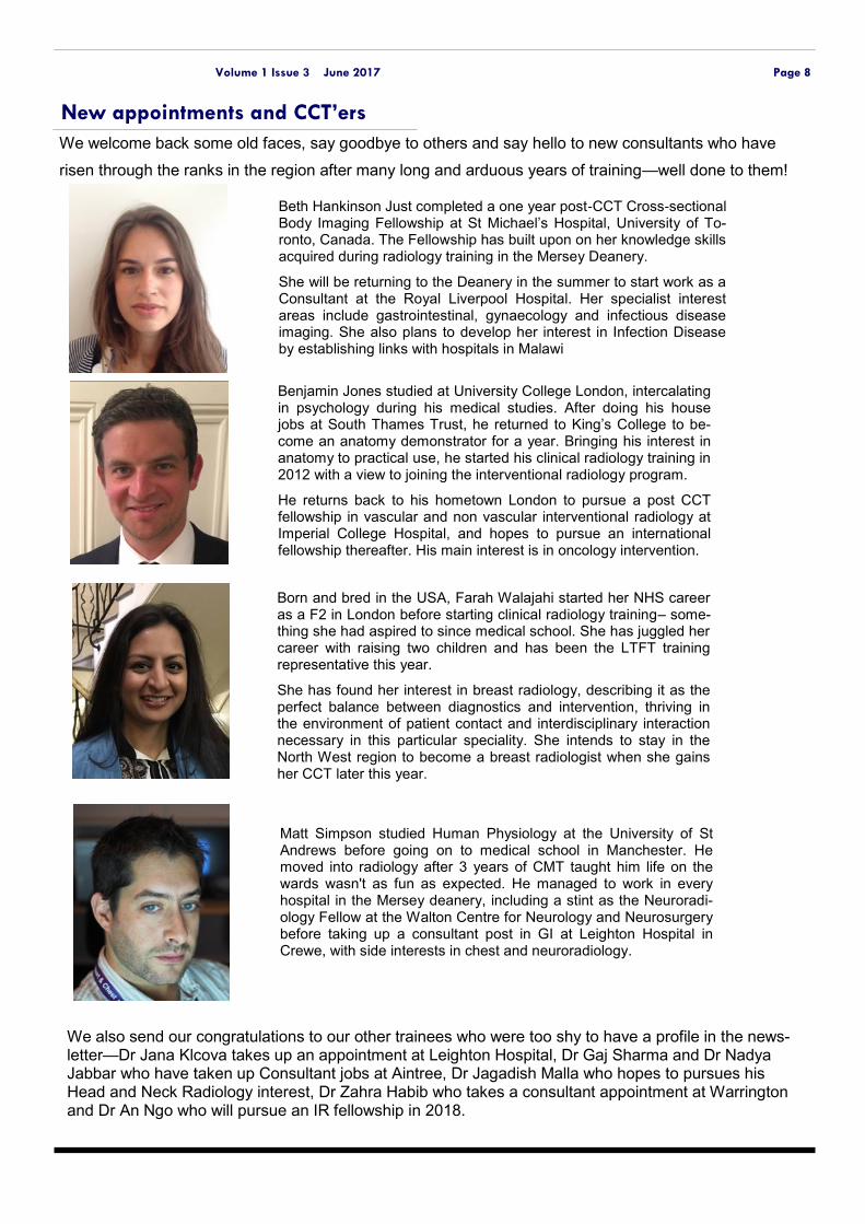
Benjamin Jones studied at University College London, intercalating in psychology during his medical studies. After doing his house jobs at South Thames Trust, he returned to King’s College to be-come an anatomy demonstrator for a year. Bringing his interest in anatomy to practical use, he started his clinical radiology training in 2012 with a view to joining the interventional radiology program.
He returns back to his hometown London to pursue a post CCT fellowship in vascular and non vascular interventional radiology at Imperial College Hospital, and hopes to pursue an international fellowship thereafter. His main interest is in oncology intervention.
Beth Hankinson Just completed a one year post-CCT Cross-sectional Body Imaging Fellowship at St Michael’s Hospital, University of To-ronto, Canada. The Fellowship has built upon on her knowledge skills acquired during radiology training in the Mersey Deanery.
She will be returning to the Deanery in the summer to start work as a Consultant at the Royal Liverpool Hospital. Her specialist interest areas include gastrointestinal, gynaecology and infectious disease imaging. She also plans to develop her interest in Infection Disease by establishing links with hospitals in Malawi
Born and bred in the USA, Farah Walajahi started her NHS career as a F2 in London before starting clinical radiology training– some-thing she had aspired to since medical school. She has juggled her career with raising two children and has been the LTFT training representative this year.
She has found her interest in breast radiology, describing it as the perfect balance between diagnostics and intervention, thriving in the environment of patient contact and interdisciplinary interaction necessary in this particular speciality. She intends to stay in the North West region to become a breast radiologist when she gains her CCT later this year.
Matt Simpson studied Human Physiology at the University of St Andrews before going on to medical school in Manchester. He moved into radiology after 3 years of CMT taught him life on the wards wasn't as fun as expected. He managed to work in every hospital in the Mersey deanery, including a stint as the Neuroradi-ology Fellow at the Walton Centre for Neurology and Neurosurgery before taking up a consultant post in GI at Leighton Hospital in Crewe, with side interests in chest and neuroradiology.
We also send our congratulations to our other trainees who were too shy to have a profile in the news-letter—Dr Jana Klcova takes up an appointment at Leighton Hospital, Dr Gaj Sharma and Dr Nadya Jabbar who have taken up Consultant jobs at Aintree, Dr Jagadish Malla who hopes to pursues his Head and Neck Radiology interest, Dr Zahra Habib who takes a consultant appointment at Warrington and Dr An Ngo who will pursue an IR fellowship in 2018.
Page 9 Volume 1 Issue 3 June 2017
Minerva
Dr Flavius Parvulescu and his wife are proud to announce the birth of their second child, Darius, in January 2017. Flavius tells us that mother and baby are doing well. Congratulations to An Ngo and his wife who also had a baby boy last month.
Dr Chris Keegan will be attempting the famous three peak challenge in June all in one day! Do-nate and check his progress: http:www.twodoctorsthreepeaks.co.uk/
A big well done goes out to Dhivya Murthy, con-sultant radiologist at Warrington hospital who did her training in the region, for completing the Chester half marathon while raising funds to pro-vide educational materials for children in Karna-taka.
We are also pleased to announce your new rep-resentatives commencing next year—Drs James Chambers and Flavius Parvulescu will be the sorely missed clinical lecturers after the post was unfilled this year. Dr Emma Hall who has helped extensively with the rota, and Jehan Ghany will take on the shared lead SpR role, Dr Azadeh Taheri will be happy to give advice on less than full time training as the new LTFT representative, and finally your outgoing editor becomes the IR training representative. We look forward to hear-ing more from them in the future. STC Leads for 2017/18 will be announced in the next edition. A big thank you to all trainees who worked very hard in helping out during the recent NHS
cyber attack.
Radiology training 101
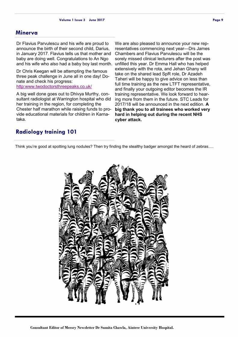
Think you’re good at spotting lung nodules? Then try finding the stealthy badger amongst the heard of zebras….
Consultant Editor of Mersey Newsletter Dr Sumita Chawla, Aintree University Hospital.


![Aintree twitter ppt [autosaved] [autosaved]](https://img.pdfslide.us/doc/110x75/55d7693dbb61ebc6238b466d/aintree-twitter-ppt-autosaved-autosaved.jpg)