Embed Size (px)
Citation preview
MENINGOENCEPHALOMYELITIS OF UNKNOWN
AETIOLOGY IN DOGS
A diagnostic, therapeutic and prognostic challenge
Ine Cornelis
Dissertation submitted in fulfilment of the requirements for the degree of Doctor of
Philosophy (PhD) in Veterinary Sciences, Small Animal Department, Faculty of
Veterinary Medicine
Ghent University
2017
Promotoren: Dr. Sofie Bhatti
Prof. Dr. Luc Van Ham
Dr. Ingrid Gielen
Dr. Steven De Decker
This thesis was performed in collaboration with:
The Royal Veterinary College, University of London.
Ine Cornelis
Meningoencephalomyelitis of unknown aetiology in dogs – a diagnostic,
therapeutic and prognostic challenge.
Universiteit Gent, Faculteit Diergeneeskunde
Vakgroep Kleine Huisdieren
Coverfoto’s: Sali en Wiebe door Fotostudio Wo

Printing of this thesis was enabled by the generous support of:

Table of Contents
List of abbreviations 7
General Introduction 13
Introduction 15
Aetiology 16
Clinical presentation 17
Diagnostic findings 18
Treatment 32
Prognostic factors 39
Outcome 41
Conclusions 42
Scientific Aims 43
Research Studies 47
Part I: Clinical Presentation And Diagnostic Findings 49
Chapter 1 51
Clinical presentation, diagnostic findings and long-term survival in
large dogs with meningoencephalomyelitis of unknown aetiology 51
Abstract 52
Introduction 53
Materials and methods 53
Results 56
Discussion 64
Conclusions 68
Chapter 2 69
Clinical presentation, diagnostic findings and outcome in dogs with
spinal-only meningoencephalomyelitis of unknown aetiology 69
Abstract 70
Introduction 71
Materials and methods 72
Results 75
Discussion 91
Conclusions 94
Part II: Treatment Options 95
Chapter 3 97
Sole prednisolone therapy in canine meningoencephalomyelitis of
unknown aetiology 97
Abstract 97
Introduction 99
Materials and methods 100
Results 102
Discussion 111
Chapter 4 113
Sole prednisolone therapy versus combination therapy with
ciclosporine in dogs with meningoencephalomyelitis of unknown
aetiology 113
Abstract 114
Introduction 115
Materials and methods 116
Results 119
Discussion 130
Conclusions 133
Part III: Prognostic Factors and Short-term Outcome 135
Chapter 5 137
Prognostic factors for one-week survival in dogs diagnosed with
meningoencephalomyelitis of unknown aetiology 137
Abstract 138
Introduction 139
Materials and methods 140
Results 143
Discussion 152
Conclusions 154
General Discussion and Conclusions 155
Clinical presentation and diagnostic findings 158
Treatment 162
Prognostic factors and outcome 165
Limitations 168
Future perspectives 170
Conclusions 171
Reference list 173
Summary 183
Samenvatting 187
Bibliography 191
Scientific publications 195
Participation to national and international conferences 198
Curriculum Vitae 201
Dankwoord 204

List of abbreviations

9
1H MRS Single Voxel Proton Magnetic Resonance Spectroscopy
ANNPE Acute Noncompressive Nucleus Pulposus Extrusion
AUC Area Under the Curve
BAR Bright Alert Responsive
CBC Complete Blood Count
CDV Canine Distemper Virus
CI Confidence Interval
CNS Central Nervous System
CRI Constant Rate Infusion
CSF Cerebrospinal Fluid
EME Eosinophilic Meningoencephalitis
FCEM Fibrocartilagenous Embolic Myelopathy
FDG-PET Fluorodeoxyglucose - Positron Emission Tomography
FDR False Discovery Rate
FLAIR Fluid Attenuation Inversion Recovery
GME Granulomatous Meningoencephalomyelitis
ICP Intracranial Pressure
IM Ischaemic Myelopathy
IQR Interquartile Ranges
IV Intravenous
MRI Magnetic Resonance Imaging
MST Median Survival Time
MUA Meningoencephalomyelitis of Unknown Aetiology
MUO Meningoencephalomyelitis of Unknown Origin
NE Necrotizing Encephalitis
10
NIME Non-Infectious Meningoencephalitis
NLE Necrotizing Leucoencephalitis
NME Necrotizing Meningoencephalomyelitis
NR Not Related
OR Odds Ratio
PCR Polymerase Chain Reaction
PET Positron Emission Tomography
PM Post Mortem
PSV Peak Systolic Velocity
QAR Quiet Alert Responsive
RI Restrictive Index
ROC Receiver Operating Characteristics
RTA Road Traffic Accident
SC Subcutaneous
SE Standard Error
SO-MUA Spinal-Only Meningoencephalomyelitis of Unknown Aetiology
SRMA Steroid Responsive Meningitis Arteritis
ST Survival Time
STIR Short Tau Inversion Recovery
T1W T1-weighted
T1WI T1-weighted Images
T2W T2-weighted
T2WI T2-weigthed Images
TNCC Total Nucleated Cell Count
TE Echo Time

11
TP Total Protein
TR Repetition Time
WBC White Blood Cell


General Introduction

14
Adapted from: Cornelis I, Van Ham L, Gielen I, De Decker S, Bhatti S. Clinical
presentation, diagnostic findings, prognostic factors, treatment and outcome in
dogs diagnosed with meningoencephalomyelitis of unknown aetiology.
Submitted to The Veterinary Journal.
General Introduction
15
Introduction
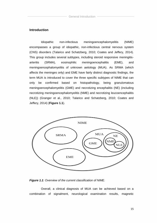
Idiopathic non-infectious meningoencephalomyelitis (NIME)
encompasses a group of idiopathic, non-infectious central nervous system
(CNS) disorders (Talarico and Schatzberg, 2010; Coates and Jeffery, 2014).
This group includes several subtypes, including steroid responsive meningitis-
arteritis (SRMA), eosinophilic meningoencephalitis (EME), and
meningoencephalomyelitis of unknown aetiology (MUA). As SRMA (which
affects the meninges only) and EME have fairly distinct diagnostic findings, the
term MUA is introduced to cover the three specific subtypes of NIME that can
only be confirmed based on histopathology, being granulomatous
meningoencephalomyelitis (GME) and necrotizing encephalitis (NE) (including
necrotizing meningoencephalomyelitis (NME) and necrotizing leucoencephalitis
(NLE)) (Granger et al., 2010; Talarico and Schatzberg, 2010; Coates and
Jeffery, 2014) (Figure 1.1).
Figure 1.1: Overview of the current classification of NIME.
Overall, a clinical diagnosis of MUA can be achieved based on a
combination of signalment, neurological examination results, magnetic
General Introduction
16
resonance imaging (MRI) findings and cerebrospinal fluid (CSF) analysis
(Munana and Luttgen, 1998; Adamo et al., 2007; Granger et al., 2010; Talarico
and Schatzberg, 2010; Coates and Jeffery, 2014), although these findings might
vary substantially between patients (Wong et al., 2010).
This group of diseases offers both a diagnostic and therapeutic
challenge to owners and veterinarians. As the condition is considered fatal
without initiation of appropriate treatment (Munana and Luttgen, 1998; Granger
et al., 2010), recent studies have evaluated different treatment modalities and
potential prognostic factors.
Aetiology
The exact aetiology and pathophysiology of MUA are currently
unknown and the most current theories were covered and discussed in a recent
literature review (Coates and Jeffery, 2014). Although MUA has most likely a
multifactorial pathogenesis, the combination of a genetic predisposition and
factors triggering an excessive immunologic response are considered the two
most important factors in the development of this disorder (Kipar et al., 1998;
Talarico and Schatzberg, 2010; Flegel et al., 2011; Coates and Jeffery, 2014).
Suspected triggering factors include environmental factors or infectious
antigenic triggers that might activate autoreactive cells in the CNS, although no
such agent has yet been identified (Schatzberg et al., 2005; Barbet et al., 2010;
Greer et al., 2010; Barber et al., 2012). Combination of this information with the
generally positive response to immunosuppressive treatment suggests that
conditions comprising MUA are immune-mediated diseases (Wong et al., 2010),
and the cornerstone of medical treatment is therefore considered
immunosuppressive therapy (Kipar et al., 1998; Talarico and Schatzberg, 2010;
Coates and Jeffery, 2014).
General Introduction
17
Clinical presentation
Middle-aged toy and terrier breeds are considered predisposed for
GME (Munana and Luttgen, 1998; Adamo et al., 2007; Talarico and
Schatzberg, 2010) whilst NE predominantly affects younger toy and small breed
dogs including Pug, Yorkshire Terrier, Maltese, Chihuahua, Pekingese,
Papillon, Shih Tzu, Coton de Tulear and Brussels Griffon (Talarico and
Schatzberg, 2010; Cooper and others, 2014). However, it is stated that dogs of
any breed and age can be affected (Granger et al., 2010; Coates and Jeffery,
2014).
Statistical analysis on 173 GME cases, 53 MUA cases and 69 NE
cases revealed a significant difference in age distribution between dogs affected
with GME and NE; dogs affected with NE were predominantly under 4 years old
whereas the peak age for GME was 4-8 years (Granger et al., 2010).
Historically, NME was described in Pug dogs with ages ranging from 6 months
to 7 years (Cordy and Holliday, 1989), whilst dogs with a histopathological
diagnosis of GME had ages ranging from 6 months to 12 years (Munana and
Luttgen, 1998). In a series of 60 Pugs with NE (Levine et al., 2008), the median
age was 18 months. In Pugs, fawn females were significantly more often
diagnosed with NME compared to black males (Greer et al., 2009). Although
female predominance is a widely held belief in GME (Cordy, 1979; Russo,
1979; Braund, 1985; Bailey, 1986; Sorjonen, 1990; Munana and Luttgen, 1998),
no statistical difference in female:male ratio could be found in more recent
studies (Talarico and Schatzberg, 2010; Granger et al., 2010).
Extraneural signs are rare, but pyrexia can occasionally accompany
CNS inflammation (Talarico and Schatzberg, 2010). Common laboratory tests
(complete blood count, biochemistry profile, urinalysis) are often within normal
limits, however, results consistent with both inflammation and stress have been
reported in dogs with GME (Thomas and Eger, 1989; Sorjonen, 1990; Tipold,
1995). Concurrent myocardial necrosis has been reported in two Pug dogs with
NME, which was thought to be due to catecholamine release by the
sympathetic nervous system (Bradley, 1991; Kobayashi et al., 1994).
General Introduction
18
On neurological examination, disease localisation was categorized as
a) mainly forebrain, brainstem or multifocal for GME, b) focal (forebrain,
brainstem) or multifocal in MUA, and c) mainly forebrain in NE (Granger et al.,
2010; Talarico and Schatzberg, 2010; Coates and Jeffery, 2014). Eight percent
of dogs diagnosed with GME were presented with neurological deficits
suggestive of a myelopathy (Granger et al., 2010), that could be located
anywhere along the spinal cord with clinical signs ranging from general
proprioceptive ataxia to paresis or plegia, with spinal hyperesthesia as a
common finding (Griffin et al., 2008; Wong et al., 2010).
Diagnostic findings
As previously stated, MUA is a clinical diagnosis that can be achieved
based on a combination of signalment, neurological examination results, cross-
sectional intracranial imaging findings and CSF analysis (Munana and Luttgen,
1998; Adamo et al., 2007; Talarico and Schatzberg, 2010; Coates and Jeffery,
2014). The study of Granger et al. (2010) systematically reviewed 457
published cases with NIME (including MUA, GME and NE) and formulated
guidelines to recruit cases diagnosed with MUA in the absence of a
histopathological diagnosis. The 4 following inclusion criteria have been
formulated: 1) dogs older than 6 months of age, 2) multiple, single or diffuse
intra-axial hyperintense lesions on T2-weighted (T2W) magnetic resonance
images (MRI), 3) pleocytosis on CSF analysis with >50% of
monocytes/lymphocytes, 4) ruling out infectious diseases commonly occurring
in the specific geographic area (Granger et al., 2010). As stated previously, a
definitive diagnosis can only be obtained by histopathological examination. The
authors refer to a recently published review article on pathological and
immunological features of GME and NME in dogs for further details (Uchida et
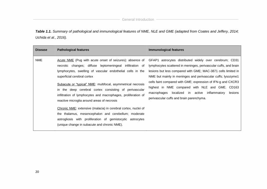
al., 2016). The most important findings are summarized in table 1.1.
Both stereotactic computed tomography (CT) - guided brain biopsy
procedures (Koblik et al., 1999) and free-hand biopsies through a mini-burr hole
(Flegel et al., 2012) have been described in dogs with inflammatory CNS
disease. Diagnostic accuracy ranged from 82% (n=17) (Flegel et al., 2012) to
General Introduction
19
100% (n=3) (Koblik et al., 1999) though results should be interpreted with
caution due to the relative small sample sizes. Complications occurred in 12-
29% of dogs, with associated signs being: transient epistaxis, transient
exacerbation of neurological signs, obtundation progressing to coma, medically
uncontrollable seizures, tetraparesis, hemiparesis, ataxia and loss of conscious
proprioception (Koblik et al., 1999; Flegel et al., 2012). Although most of these
signs resolved within 3-14 days, an indirect fatality rate of 6% was noted (Flegel
et al., 2012).
General Introduction
20
Table 1.1. Summary of pathological and immunological features of NME, NLE and GME (adapted from Coates and Jeffery, 2014;
Uchida et al., 2016).
Disease Pathological features Immunological features
NME Acute NME (Pug with acute onset of seizures): absence of
necrotic changes; diffuse leptomeningeal infiltration of
lymphocytes, swelling of vascular endothelial cells in the
superficial cerebral cortex
Subacute or “typical” NME: multifocal, asymmetrical necrosis
in the deep cerebral cortex consisting of perivascular
infiltration of lymphocytes and macrophages, proliferation of
reactive microglia around areas of necrosis
Chronic NME: extensive (malacia) in cerebral cortex, nuclei of
the thalamus, mesencephalon and cerebellum; moderate
astrogliosis with proliferation of gemistocytic astrocytes
(unique change in subacute and chronic NME).
GFAP1 astrocytes distributed widely over cerebrum; CD31
lymphocytes scattered in meninges, perivascular cuffs, and brain
lesions but less compared with GME; MAC-3871 cells limited in
NME but mainly in meninges and perivascular cuffs; lysozyme1
cells faint compared with GME; expression of IFN-g and CXCR3
highest in NME compared with NLE and GME. CD163
macrophages localized in active inflammatory lesions
perivascular cuffs and brain parenchyma.
General Introduction
21
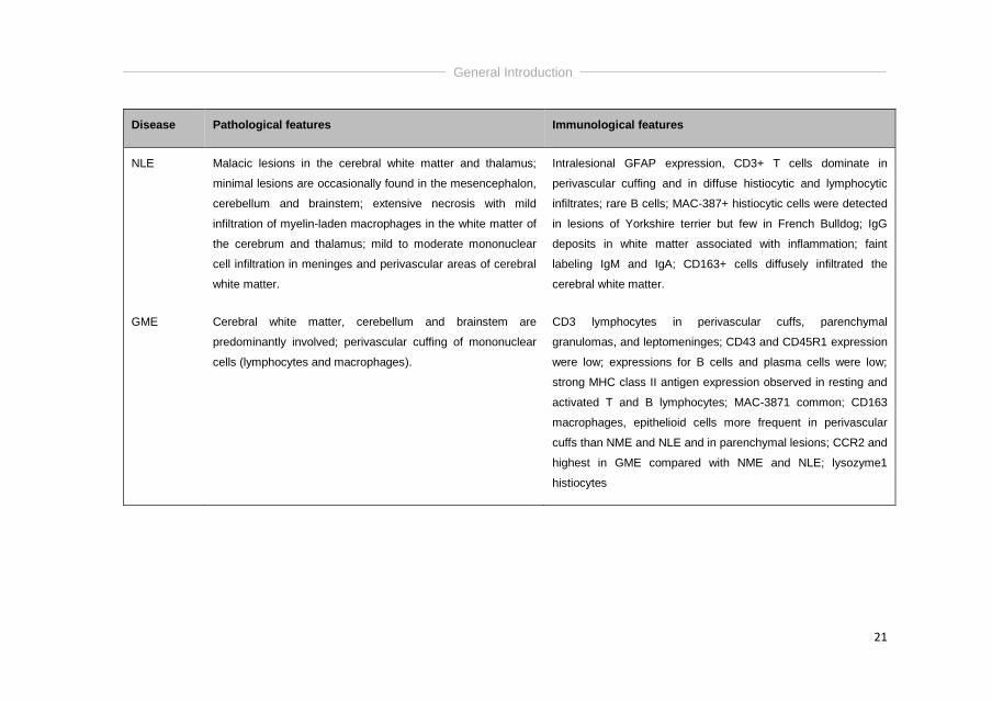
Disease Pathological features Immunological features
NLE Malacic lesions in the cerebral white matter and thalamus;
minimal lesions are occasionally found in the mesencephalon,
cerebellum and brainstem; extensive necrosis with mild
infiltration of myelin-laden macrophages in the white matter of
the cerebrum and thalamus; mild to moderate mononuclear
cell infiltration in meninges and perivascular areas of cerebral
white matter.
Intralesional GFAP expression, CD3+ T cells dominate in
perivascular cuffing and in diffuse histiocytic and lymphocytic
infiltrates; rare B cells; MAC-387+ histiocytic cells were detected
in lesions of Yorkshire terrier but few in French Bulldog; IgG
deposits in white matter associated with inflammation; faint
labeling IgM and IgA; CD163+ cells diffusely infiltrated the
cerebral white matter.
GME Cerebral white matter, cerebellum and brainstem are
predominantly involved; perivascular cuffing of mononuclear
cells (lymphocytes and macrophages).
CD3 lymphocytes in perivascular cuffs, parenchymal
granulomas, and leptomeninges; CD43 and CD45R1 expression
were low; expressions for B cells and plasma cells were low;
strong MHC class II antigen expression observed in resting and
activated T and B lymphocytes; MAC-3871 common; CD163
macrophages, epithelioid cells more frequent in perivascular
cuffs than NME and NLE and in parenchymal lesions; CCR2 and
highest in GME compared with NME and NLE; lysozyme1
histiocytes
General Introduction
22
Cross-sectional imaging
MRI has been reported to be 94.4% sensitive and 95.5% specific for
detecting a brain lesion with similarly high performance for classifying neoplastic
and inflammatory disease. On the contrary, MRI has been only 38.9% sensitive
for classifying cerebrovascular disease. In general, high specificity but no
sensitivity was retained for MR diagnosis of specific brain diseases (Wolff et al.,
2012).
MR imaging is considered the most sensitive imaging modality for
detecting intracranial lesions, but up to 7% (2/25 dogs, one diagnosed with
GME and one with MUA) of scans showed no lesion on T2W images (T2WI)
(Talarico and Schatzberg, 2010; Granger et al., 2010). Up to 14% (5/36 dogs,
specific diagnosis not specified) of CT scans revealed no lesion (Granger et al.,
2010). Overall, the sensitivity of imaging in identifying all inflammatory lesions
suspected from the neurological examination remains quite low (<60%)
(Granger et al, 2010). Additionally, MRI abnormalities were only seen in 76% of
cases with inflammatory CSF findings in 1 study (Lamb et al, 2005). Although
the use of cross-sectional imaging might aid in differentiating between the
different types of idiopathic meningoencephalitides (Talarico and Schatzberg,
2010), no study is currently available looking into the use of MRI to differentiate
between histopathologically confirmed cases of GME, NME and NLE.
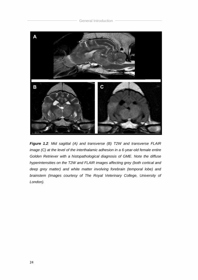
In the literature, one study specifically focuses on the MRI findings in
11 dogs with histopathologically confirmed GME (Cherubini et al., 2006). The
focal, multifocal or diffuse lesions were located in the forebrain, brainstem or
cerebellum, and were hyperintense on T2W and fluid attenuating inversion
recovery (FLAIR) images (Figure 1.2). Lesions were scattered throughout grey
and white matter, showed variable intensities on T1-weighted images (T1WI)
and variable degrees of contrast enhancement. Vasogenic oedema in the white
matter was commonly present on T2-weighted images (T2WI), where
meningeal enhancement was not commonly apparent and minimal if present
(Cherubini et al., 2006; Talarico and Schatzberg, 2010; Coates and Jeffery,
General Introduction
23
2014). The lesion distribution (grey/white matter) was consistent with the
histopathological findings (Cherubini et al., 2006).
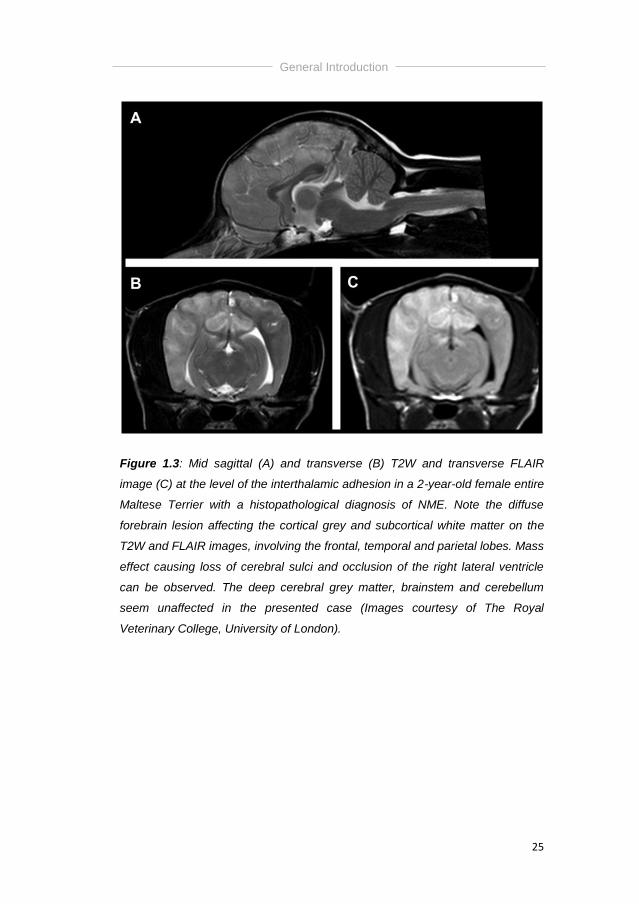
The most common MRI abnormalities in dogs with NME include
bilateral but asymmetrical, multifocal forebrain lesions (more severe lesions in
parietal and occipital lobes have been described), hyperintense on T2W and
FLAIR images, typically affecting the cortical grey and subcortical white matter
with loss of grey/white matter demarcation and variable degrees of contrast
enhancement of the parenchymal lesions on T1-weighted (T1W) post-contrast
images (Flegel et al., 2008; Young et al., 2009; Talarico and Schatzberg, 2010)
(Figure 1.3). However, cerebellar and brainstem lesions were additionally
detected in 4/18 and 3/18 cases in one study, respectively (Young et al., 2009).
Meningeal enhancement could be present, accompanied by mass effect and
varying degrees of ventriculomegaly (Coates and Jeffery, 2014).
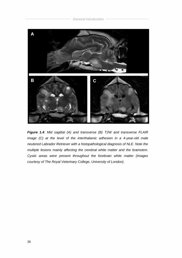
In NLE, multiple, bilateral but asymmetrical cerebral white matter and
brainstem lesions have been detected (von Praun et al., 2006). These lesions
were typically hyperintense on T2W and FLAIR images and often included
multiple cystic areas of necrosis. Contrast enhancement of parenchymal lesions
was minimal (Talarico and Schatzberg, 2010; Coates and Jeffery, 2014). There
was lack of meningeal enhancement and mass effect, with varying degrees of
ventriculomegaly (Coates and Jeffery, 2014) (Figure 1.4).
General Introduction
24
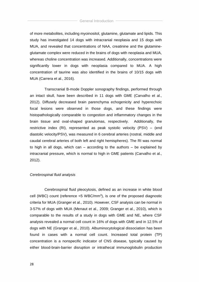
Figure 1.2: Mid sagittal (A) and transverse (B) T2W and transverse FLAIR
image (C) at the level of the interthalamic adhesion in a 6-year-old female entire
Golden Retriever with a histopathological diagnosis of GME. Note the diffuse
hyperintensities on the T2W and FLAIR images affecting grey (both cortical and
deep grey matter) and white matter involving forebrain (temporal lobe) and
brainstem (Images courtesy of The Royal Veterinary College, University of
London).
General Introduction
25
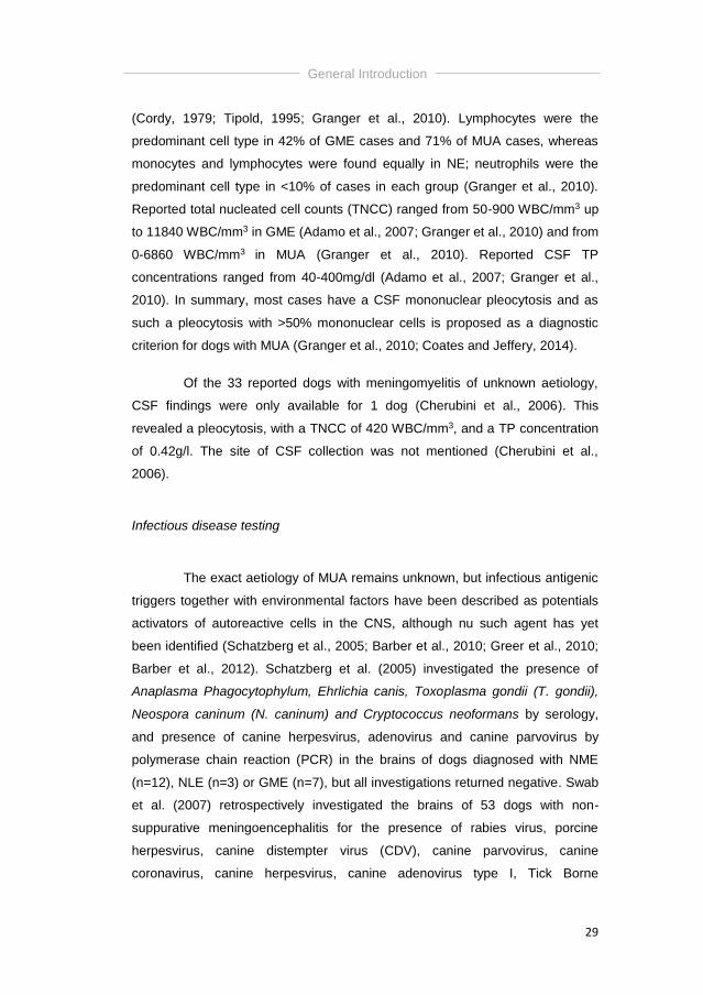
Figure 1.3: Mid sagittal (A) and transverse (B) T2W and transverse FLAIR
image (C) at the level of the interthalamic adhesion in a 2-year-old female entire
Maltese Terrier with a histopathological diagnosis of NME. Note the diffuse
forebrain lesion affecting the cortical grey and subcortical white matter on the
T2W and FLAIR images, involving the frontal, temporal and parietal lobes. Mass
effect causing loss of cerebral sulci and occlusion of the right lateral ventricle
can be observed. The deep cerebral grey matter, brainstem and cerebellum
seem unaffected in the presented case (Images courtesy of The Royal
Veterinary College, University of London).
General Introduction
26
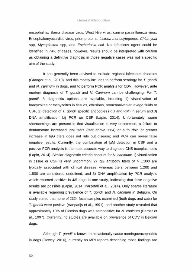
Figure 1.4: Mid sagittal (A) and transverse (B) T2W and transverse FLAIR
image (C) at the level of the interthalamic adhesion in a 4-year-old male
neutered Labrador Retriever with a histopathological diagnosis of NLE. Note the
multiple lesions mainly affecting the cerebral white matter and the brainstem.
Cystic areas were present throughout the forebrain white matter (Images
courtesy of The Royal Veterinary College, University of London).
General Introduction
27
Thirty-three dogs with MUA only involving the spinal cord have been
reported, including 3 dogs with GME (Cherubini et al., 2006; Griffin et al., 2008;
Wong et al., 2010). Imaging findings were available for 15 of these 33 cases,
using different types of imaging modalities. Twelve dogs underwent
myelography alone or CT-myelography, revealing no abnormalities in 11 dogs
and a ventral extradural spinal cord compression in 1 dog (Wong et al., 2010).
MRI was performed in 3 dogs, revealing no abnormalities in 1 dog, and
multifocal poorly demarcated intramedullary T2W hyperintensities with variable
contrast enhancement in 2 dogs (Cherubini et al., 2006; Wong et al., 2010).
Other imaging modalities, including positron emission tomography
(PET) in NME, fluorodeoxyglucose PET (FDG-PET) and single voxel proton
magnetic resonance spectroscopy (1H MRS) in MUA, and transcranial
sonographic findings in GME were investigated as diagnostic modalities (Eom
et al., 2008; Kang et al., 2009; Carvalho et al., 2012; Carrera et al., 2016).
FDG-PET is a new imaging technique evaluating in-vivo tissue
metabolism with the use of a metabolic tracer that acts as a glucose molecule.
By this means, the tracer is transported into the tissue and trapped, which can
afterwards be visualized. In both studies, a total of 5 dogs with NME, 1 dog with
GME and 1 dog with MUA have been studied (Eom et al., 2008; Kang et al.,
2009). Interestingly, all dogs with NME showed glucose hypometabolism (most
likely attributed to the presence of malacia and necrosis), whereas glucose
hypermetabolism was seen in GME (most likely due to strong granulomatous
inflammatory reaction). In conclusion, the authors stated that further studies
with larger sample sizes are necessary to confirm the associations (Eom et al.,
2008; Kang et al., 2009).
1H MRS is a non-invasive imaging diagnostic technique that provides
specific biochemical information on numerous intracellular metabolites by
measuring the signal that is emitted by proton nuclei because of their high
magnetic sensitivity and presence in all tissues of the body. Long echo time
sequences (typically >144 milliseconds) allow the determination of
concentrations of N-acetyl aspartate (NAA), choline, creatinine and lactate,
where short echo time sequences (typically <35 milliseconds) permit evaluation
General Introduction
28
of more metabolites, including myoinositol, glutamine, glutamate and lipids. This
study has investigated 14 dogs with intracranial neoplasia and 15 dogs with
MUA, and revealed that concentrations of NAA, creatinine and the glutamine-
glutamate complex were reduced in the brains of dogs with neoplasia and MUA,
whereas choline concentration was increased. Additionally, concentrations were
significantly lower in dogs with neoplasia compared to MUA. A high
concentration of taurine was also identified in the brains of 10/15 dogs with
MUA (Carrera et al., 2016).
Transcranial B-mode Doppler sonography findings, performed through
an intact skull, have been described in 11 dogs with GME (Carvalho et al.,
2012). Diffusely decreased brain parenchyma echogenicity and hyperechoic
focal lesions were observed in those dogs, and these findings were
histopathologically comparable to congestion and inflammatory changes in the
brain tissue and oval-shaped granulomas, respectively. Additionally, the
restrictive index (RI), represented as peak systolic velocity (PSV) – (end
diastolic velocity/PSV), was measured in 6 cerebral arteries (rostral, middle and
caudal cerebral arteries of both left and right hemispheres). The RI was normal
to high in all dogs, which can – according to the authors – be explained by
intracranial pressure, which is normal to high in GME patients (Carvalho et al.,
2012).
Cerebrospinal fluid analysis
Cerebrospinal fluid pleocytosis, defined as an increase in white blood
cell (WBC) count (reference <5 WBC/mm3), is one of the proposed diagnostic
criteria for MUA (Granger et al., 2010). However, CSF analysis can be normal in
3-57% of dogs with MUA (Menaut et al., 2009; Granger et al., 2010), which is
comparable to the results of a study in dogs with GME and NE, where CSF
analysis revealed a normal cell count in 16% of dogs with GME and in 12.5% of
dogs with NE (Granger et al., 2010). Albuminocytological dissociation has been
found in cases with a normal cell count. Increased total protein (TP)
concentration is a nonspecific indicator of CNS disease, typically caused by
either blood-brain-barrier disruption or intrathecal immunoglobulin production
General Introduction
29
(Cordy, 1979; Tipold, 1995; Granger et al., 2010). Lymphocytes were the
predominant cell type in 42% of GME cases and 71% of MUA cases, whereas
monocytes and lymphocytes were found equally in NE; neutrophils were the
predominant cell type in <10% of cases in each group (Granger et al., 2010).
Reported total nucleated cell counts (TNCC) ranged from 50-900 WBC/mm3 up
to 11840 WBC/mm3 in GME (Adamo et al., 2007; Granger et al., 2010) and from
0-6860 WBC/mm3 in MUA (Granger et al., 2010). Reported CSF TP
concentrations ranged from 40-400mg/dl (Adamo et al., 2007; Granger et al.,
2010). In summary, most cases have a CSF mononuclear pleocytosis and as
such a pleocytosis with >50% mononuclear cells is proposed as a diagnostic
criterion for dogs with MUA (Granger et al., 2010; Coates and Jeffery, 2014).
Of the 33 reported dogs with meningomyelitis of unknown aetiology,
CSF findings were only available for 1 dog (Cherubini et al., 2006). This
revealed a pleocytosis, with a TNCC of 420 WBC/mm3, and a TP concentration
of 0.42g/l. The site of CSF collection was not mentioned (Cherubini et al.,
2006).
Infectious disease testing
The exact aetiology of MUA remains unknown, but infectious antigenic
triggers together with environmental factors have been described as potentials
activators of autoreactive cells in the CNS, although nu such agent has yet
been identified (Schatzberg et al., 2005; Barber et al., 2010; Greer et al., 2010;
Barber et al., 2012). Schatzberg et al. (2005) investigated the presence of
Anaplasma Phagocytophylum, Ehrlichia canis, Toxoplasma gondii (T. gondii),
Neospora caninum (N. caninum) and Cryptococcus neoformans by serology,
and presence of canine herpesvirus, adenovirus and canine parvovirus by
polymerase chain reaction (PCR) in the brains of dogs diagnosed with NME
(n=12), NLE (n=3) or GME (n=7), but all investigations returned negative. Swab
et al. (2007) retrospectively investigated the brains of 53 dogs with non-
suppurative meningoencephalitis for the presence of rabies virus, porcine
herpesvirus, canine distempter virus (CDV), canine parvovirus, canine
coronavirus, canine herpesvirus, canine adenovirus type I, Tick Borne
General Introduction
30
encephalitis, Borna disease virus, West Nile virus, canine parainfluenza virus,
Encephalomyocarditis virus, prion proteins, Listeria monocytogenes, Chlamydia
spp, Mycoplasma spp, and Escherichia coli. No infectious agent could be
identified in 74% of cases, however, results should be interpreted with caution
as obtaining a definitive diagnosis in those negative cases was not a specific
aim of the study.
It has generally been advised to exclude regional infectious diseases
(Granger et al., 2010), and this mostly includes to perform serology for T. gondii
and N. caninum in dogs, and to perform PCR analysis for CDV. However, ante
mortem diagnosis of T. gondii and N. Caninum can be challenging. For T.
gondii, 3 diagnostic options are available, including 1) visualization of
bradyzoites or tachyzoites in tissues, effusions, bronchoalveolar lavage fluids or
CSF, 2) detection of T. gondii specific antibodies (IgG and IgM) in serum and 3)
DNA amplification bij PCR on CSF (Lapin, 2014). Unfortunately, some
shortcomings are present in that visualization is very uncommon, a failure to
demonstrate increased IgM titers (titer above 1:64) or a fourfold or greater
increase in IgG titers does not rule out disease; and PCR can reveal false
negative results. Currently, the combination of IgM detection in CSF and a
positive PCR analysis is the most accurate way to diagnose CNS toxoplasmosis
(Lapin, 2014). Similar diagnostic criteria account for N. caninum: 1) visualization
in tissue or CSF is very uncommon, 2) IgG antibody titers of > 1:800 are
typically associated with clinical disease, whereas titers between 1:200 and
1:800 are considered undefined, and 3) DNA amplification by PCR analysis
which returned positive in 4/5 dogs in one study, indicating that false negative
results are possible (Lapin, 2014; Parzefall et al., 2014). Only sparse literature
is available regarding prevalence of T. gondii and N. caninum in Belgium. On
study stated that none of 2324 fecal samples examined (both dogs and cats) for
T. gondii were positive (Vanparijs et al., 1991), and another study revealed that
approximately 10% of Flemish dogs was seropositive for N. caninum (Barber et
al., 1997). Currenlty, no studies are available on prevalence of CDV in Belgian
dogs.
Although T. gondii is known to occasionally cause meningoencephalitis
in dogs (Dewey, 2016), currently no MRI reports describing those findings are
General Introduction
31
available. On the contrary, some literature is available for cats, describing the
presence of an intracranial granuloma that might resolve after appropriate
treatment, consisting of surgical removal and/or antibiotic therapy (Pfohl and
Dewey, 2005; Falzoni et al., 2008). N. caninum has previously been associated
with necrotizing cerebellitis and cerebellar atrophy (Garosi et al., 2010) as well
as with spinal cord changes, mesencephalic and metencephalic lesions, and
with multifocal brain lesions on MR imaging in dogs (Parzefall et al., 2014).
Three forms of CDV encephalitis are currently described, including acute CDV
infection, CDV infection in mature dogs and “old dog encephalitis” (Dewey,
2016). Dogs with acute CDV infection are mostly very young (<1 year) and
mainly present with forebrain signs, where histopathology reveals a
polioencephalopathy. Mature dogs (>1 year) with CDV infection tend to develop
inflammatory demyelinating white matter disease primarily affecting the
brainstem, cerebellum and spinal cord (leucoencephalomyelopathy), whereas
“old dog encephalitis” mainly affects older dogs (>5 years) and presents with
signs of forebrain dysfunction (visual deficits, behavioral changes) (Dewey,
2016). Currently, MRI findings are only described in a cohort of 5 puppies,
revealing hyperintense lesions and loss of contrast between grey and white
matter on T2-weighted images in the cerebellum and/or brainstem. The majority
of lesions were located in the temporal lobe of the cerebrum, so results should
be interpreted with caution as all 5 dogs were presented with seizures (Bathen-
Noethen et al., 2008).
General Introduction
32
Treatment
Although the criterion-referenced standard for a clinical trial is a
randomized, placebo-controlled, double-blinded, prospective study, it is
generally accepted that use of a placebo control treatment group is unethical
because dogs with MUA have a poor outcome without treatment (Coates et al.,
2007; Smith et al., 2009; Coates and Jeffery, 2014). Historically, different
inclusion criteria have been used, and because in some studies
immunomodulary medication was only initiated later, e.g. after results for
infectious disease testing returned negative, treatment results and outcomes
are difficult to compare (Adamo et al., 2007; Coates et al., 2007; Wong et al.,
2010).
As previously stated, the exact aetiology and pathophysiology of MUA
remains unknown, but the cornerstone of medical treatment is
immunosuppressive therapy. Several treatment protocols using different
immunomodulating drugs, resulting in different long-term survival times have
been reported (Sisson et al. 1989; Gregory et al., 1998; Munana and Luttgen,
1998; Adamo and O’Brien, 2004; Gnirs, 2006; Zarfoss et al., 2006; Adamo et
al., 2007; Coates et al., 2007; de Stefani et al., 2007; Feliu-Pascual et al., 2007;
Uriarte et al., 2007; Jung et al., 2007; Menaut et al., 2008; Pakozdy et al., 2009;
Smith et al., 2009; Granger et al., 2010; Kang et al., 2009; Wong et al., 2010;
Flegel et al., 2011; Jung et al., 2012; Jung et al., 2013; Lowrie et al., 2013;
Beckmann et al., 2015; Mercier and Barnes Heller, 2015; Barnoon et al., 2015;
Lowrie et al., 2016). A comprehensive overview of all immunomodulatory
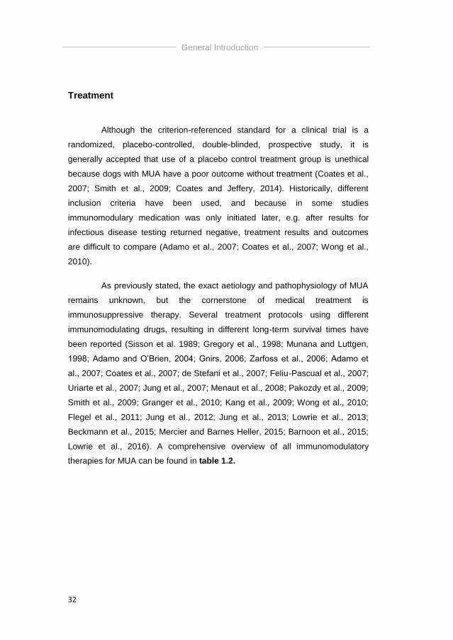
therapies for MUA can be found in table 1.2.
General Introduction
33
Table 1.2. Summary of immunomodulatory drug therapies for MUA, including
the reported number of dogs and the initial drug dosages. Abbreviations: PO =
per os; IV = intravenous. (Adapted from: Coates and Jeffery, 2014).
Drug Number
of dogs
Initial dosages References
Azathioprine +
prednisolone
40 2mg/kg PO q24h for 2 weeks,
then decrease to 2mg/kg q48h
for azathioprine
Wong et al., 2010
Ciclosporine 5 6-30mg/kg PO q24h Adamo and O’Brien, 2004;
Adamo et al., 2007
Ciclosporine +
ketoconazole
3 5-12mg/kg PO q24h
ciclosporine + 8mg/kg PO
ketoconazole
Adamo et al., 2007
Ciclosporine +
prednisolone
23 6-30mg/kg PO q24h
ciclosporine
Adamo and O’Brien, 2004;
Gnirs, 2006; Adamo et al.,
2007; Jung et al., 2007;
Pakozdy et al., 2009; Kang
et al., 2009; Jung et al.,
2012; Jung et al., 2013
Cyclophosphamide
and vincristine +
prednisolone
10 Cyclophosphamide: 50mg/m2
PO q48h for 8 weeks, then
every other week
Vincristine: 0.5mg/m2 IV, every
7 days for 8 weeks, then every
14 days
Smith et al., 2009
Cytosine
arabinoside +
prednisolone
158 50mg/m2 SC, q12h for 2
consecutive days, then repeat
every 3 weeks for 4 cycles
IV infusion: 200 mg/m2 over 8
hours
Zarfoss et al., 2006; de
Stefani et al., 2007;
Menaut et al., 2008; Smith
et al., 2009; Lowrie et al.,
2013; Lowrie et al., 2016
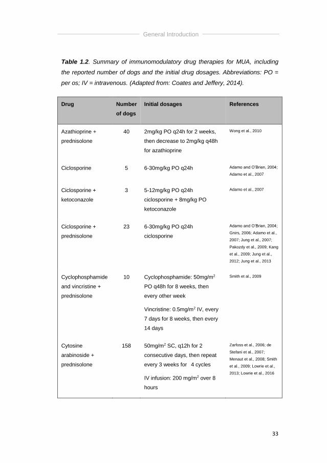
General Introduction
34
Drug Number
of dogs
Initial dosages References
Leflunomide +
prednisolone
5 1.5-4mg/kg PO q24h Gregory et al., 1998
Lomustine +
prednisolone
32 60mg/m2 PO every 6 weeks Uriarte et al., 2007; Flegel
et al., 2011
Mycophenolate
mofetil +
prednisolone
30 10-20mg/kg PO q12h, reduce
after 1 month to 5-10mg/kg
q12h
Feliu-Pascual et al., 2007;
Barnoon et al., 2015
Procarbazine +
prednisolone
31 25-50mg/m2 PO q24h Coates et al., 2007
Prednisolone 78 1 to 2mg/kg PO q12h for 3–4
weeks; 0.5–1mg/kg q12h for 6
weeks; then 0.25–0.5mg/kg
q12h for 3 weeks; then 0.25–
0.5mg/kg q24h for 3 weeks;
then 0.25–0.5mg/kg q48h
indefinitely
Coates et al., 2007;
Pakozdy et al., 2009;
Granger et al., 2010;
Flegel et al., 2011; Mercier
and Barnes Heller, 2015
Overall, treatment effect is monitored by clinical response and
resolution of neurologic deficits, and occasionally by repeated CSF analysis and
MR imaging (Coates and Jeffrey 2014). In a small cohort of dogs, Lowrie et al.
(2013) suggested that a combination of MR imaging and CSF analysis provided
greater sensitivity for prediction of relapse than one modality alone. The authors
therefore suggested that treatment should only be tapered once MR imaging
and CSF analysis (including TP concentration) returned to normal (Lowrie et al.,
2013).
General Introduction
35
Glucocorticosteroids
Treatment with glucocorticosteroids (mostly prednisolone) only is
generally associated with shorter survival times (ST) compared to combination
therapy with other immunosuppressive agents (Munana and Luttgen, 1998;
Jung et al., 2007; Pakozdy et al., 2009; Granger et al., 2010; Flegel et al., 2011;
Mercier and Barnes Heller, 2015). However, in a clinical setting, adding more
expensive immunosuppressive therapies to the glucocorticoid protocol might be
financially prohibitive.
Glucocorticosteroids bind to a cytosolic glucocorticosteroid receptor,
which then moves to the nucleus, binds to DNA, and influences gene
transcription. Cellular effects include stabilization of cell membranes, inhibition
of phospholipase A2 with resultant inhibition of the cyclooxygenase and
lipoxygenase pathways, decreased release of cytokines interleukin (IL) -1 and
IL-6, and downregulation of Fc receptor expression on macrophages. The early
effects of corticosteroids are believed to predominantly result from a rapid
decrease in phagocytic activity of splenic and hepatic macrophages, whereas
the long-term effects result primarily from suppression of cell-mediated
immunity (suppression of T-helper cells) (Nelson and Couto, 2014). Most
common adverse effects include polyuria, polydipsia, panting, muscle
weakness, dermatological changes, predisposition to infections, and muscle
atrophy. Glucocorticosteroids might cause insulin resistance, hyperglycaemia,
vacuolar hepatopathy, and hypercoagulability (Nelson and Couto, 2014).
Glucocorticosteroids are typically initiated at immunosuppressive doses,
followed by tapering to a minimal effective dose while maintaining fair (or better)
quality of life (Zarfoss et al., 2006).
In the literature, 78 dogs diagnosed with MUA and receiving sole
prednisolone therapy have been reported, and survival data were available for
all dogs (Coates et al., 2007; Pakozdy et al., 2009; Granger et al., 2010; Flegel
et al., 2011; Mercier and Barnes Heller, 2015). Median survival times (MSTs)
ranged from 28 – 357 days (43/78) (Granger et al., 2010), 91 - 329 days (19/78)
(Flegel et al., 2011) and 602 days (16/78) (Mercier and Barnes Heller, 2015) in
General Introduction
36
dogs receiving dosages ranging from 0.5 – 30 mg/kg/day (Coates et al., 2007;
Pakozdy et al., 2009; Granger et al., 2010; Flegel et al., 2011; Mercier and
Barnes Heller, 2015).
Cytosine arabinoside
Six studies evaluated treatment with cytosine arabinoside as an
adjunctive treatment option to prednisolone in dogs diagnosed with MUA,
covering a total of 158 cases. Cytosine arabinoside can be administered either
as a continuous rate infusion (CRI) (doses ranging from 100-300 mg/m2 over 8-
24 hours) or as 4 subcutaneous (SC) injections of 50 mg/m2 in 48 hours (200
mg/m2 in 48h) (Zarfoss et al., 2006; de Stefani et al., 2007; Menaut et al., 2008;
Smith et al., 2009; Lowrie et al., 2013; Lowrie et al., 2016). Crook et al. (2013)
showed that CRI administration of cytosine arabinoside provided a steady state
concentration over the time it was administered compared to a rapid absorption
and elimination when administered subcutaneously. A recent clinical study
revealed a significantly better 3-month-survival in dogs initially receiving a CRI
of cytosine arabinoside compared to the SC route (Lowrie et al., 2016).
Cytosine arabinoside is a synthetic nucleoside analogue, which
crosses the blood-brain-barrier, undergoes enzymatic activation, competes for
incorporation into nucleic acids and then competitively inhibits DNA polymerase
in mitotically active cells. Additionally, it causes topoisomerase dysfunction and
prevents DNA repair; inhibits ribonucleotide reductase; inhibits membrane
glycoprotein synthesis; and promotes leukemic cell differentiation in culture. All
the effects are dependent on both cell cycle (S-phase) and rate of DNA
synthesis (Zarfoss et al., 2006; Coates and Jeffery, 2014).
Side effects in dogs were dose dependent and mainly included
myelosuppression and gastro-intestinal upset (Zarfoss et al., 2006). However,
no dose-limiting toxicities were mentioned in most cases (Lowrie et al., Menaut
et al., 2009; Smith et al., 2009). Transient post-treatment lethargy, dysphagia or
limb tremors (3/10); mild coat and skin changes (increased shedding or
alopecia, mild localised dermatitis) (4/10); and transient to intermittent pelvic
General Introduction
37
limb weakness (3/10) was noticed in one study (Zarfoss et al., 2006). Three
case reports described three additional side effects of cytosine arabinoside
therapy. One dog developed infiltrative lung disease 24h after the 4th cytosine
arabinoside infusion (300mg/m2), where after the dog was euthanized (Hart and
Waddell, 2016). The second dog developed anterior uveitis three weeks after
the 5th cytosine arabinoside treatment (50mg/m2 q12h for 48h), which resolved
after treatment with topical antibiotics, non-steroidal anti-inflammatory drugs,
artificial tears and local atropine (Bianchi and Dodi, 2007). At last, development
of severe calcinosis cutis and deep pyoderma at the cytosine arabinoside
injection site was described in three dogs (Volk et al., 2012).
Reported MSTs with additional cytosine arabinoside ranged from 26 to
1063 days (n=69) (Zarfoss et al., 2006; de Stefani et al., 2007; Menaut et al.,
2008; Smith et al., 2009; Lowrie et al., 2013; Lowrie et al., 2016).
Ciclosporine
Ciclosporine therapy has been described as sole treatment for MUA
(n=5) (Adamo and O’Brien, 2004; Adamo et al., 2007), or as combination
therapy with prednisolone (n=23) (Adamo and O’Brien, 2004; Gnirs, 2006;
Adamo et al., 2007; Jung et al., 2007; Pakozdy et al., 2009; Kang et al., 2009;
Jung et al., 2012; Jung et al., 2013), ketoconazole (n=3) (Adamo et al, 2007), or
cytosine arabinoside and prednisolone (n=1) (Behr et al., 2009). Overall, 32
dogs have been reported receiving initial doses ranging from 3 – 15 mg/kg PO
every 12 hours, resulting in MSTs ranging from 236 to 930 days (Adamo and
O’Brien, 2004; Gnirs, 2006; Adamo et al., 2007; Jung et al., 2007; Pakozdy et
al., 2009; Kang et al., 2009; Jung et al., 2013). One dog receiving combination
therapy survived for 1096 days (Jung et al., 2012).
Ciclosporine is a fungal polypeptide that interferes with macrophage
and monocyte activation by inhibiting the transcription of alfa-interferon. It
suppresses T-cell mediated immune responses through inhibition of synthesis
of IL-2 and other cytokines. Although ciclosporine is lipophilic, it has poor blood-
brain-barrier permeability (Adamo and O’Brien, 2004; Gnirs, 2006; Adamo et al.,
General Introduction
38
2007). Because the blood-brain-barrier might be disrupted during inflammation
in MUA, therapeutic ciclosporine concentrations may be present in affected
areas of the CNS (Adamo et al., 2007). Commercial ciclosporine is available in
2 very different types of oral formulations. A vegetable-oil based preparation
that caused marked intraindividual and interindividual variations in blood drug
concentrations, and a microemulsified form that results in a more consistent and
predictable absorption. Oral bioavailability of the microemulsion has improved
by up to 50% compared with the oil-based formulation (Archer et al., 2014).
Reported side effects include mild hypertrichosis and transient
lymphopenia (Adamo and O’Brien, 2004), vomiting during first 2 weeks of
treatment (5/10) (Gnirs et al., 2006; Pakozdy et al., 2009) and severe gastro-
intestinal adverse effects with life-threatening anaemia (1/7) (Pakozdy et al.,
2009). A range of side effects has also been described in dogs receiving
5mg/kg q24h (the approved atopy dosage), additionally including anorexia,
urinary tract infections, persistent otitis externa, gingival hyperplasia and
lymphadenopathy (Archer et al., 2014).
Other immunosuppressive agents
Other immunosuppressive agents have been described in combination
with prednisolone for treatment of MUA, including azathioprine (n=40) (Wong et
al., 2010), procarbazine (n=31) (Coates et al., 2007), lomustine (n=32) (Uriarte
et al., 2007; Flegel et al., 2011), vincristine and cyclophosphamide (n=10)
(Smith et al., 2009), leflunomide (n=5) (Gregory et al., 1998), and
mycophenolate mofetil (n=30) (Feliu-Pascual et al., 2007; Barnoon et al., 2015).
Following side effects were described in those studies:
myelosuppression (19%) and haemorrhagic enteritis (15%) with procarbazine
(Coates et al., 2007); leucopenia, severe thrombocytopenia and haemorrhagic
gastro-enteritis with lomustine (Flegel et al., 2011); myelosuppression,
haemorrhagic cystitis and pyometra with vincristine and cyclophosphamide
(Smith et al., 2009); and haemorrhagic diarrhoea within the first 2 weeks of
treatment with mycophenolate mofetil (Feliu-Pascual et al., 2007; Barnoon et
General Introduction
39
al., 2015). The side effects encountered with the combination of vincristine and
cyclophosphamide were unacceptable to the author, excluding this protocol for
further investigation (Smith et al., 2009). On treatment with azathioprine (n=40),
major adverse events were infrequent and included poor coat or thin skin
(13/40), urinary tract infection (3/40), vomiting (3/40), corneal ulcers (2/40),
diabetes mellitus (2/40), renal failure, keratoconjunctivitis sicca, cruciate
ligament rupture, hepatic mass, mammary gland adenoma, lymphoma,
demodectic mange and septic arthritis of a single joint. However, many of the
adverse effects, such as weight gain, poor coat, hypertriglyceridemia,
thrombocytosis, and elevated liver enzyme activities, could have been
associated with concurrent administration of corticosteroids (Wong et al., 2010).
MSTs were available for some studies, being 425 days for
procarbazine (Coates et al., 2007), 150-740 days for lomustine (Uriarte et al.,
2007; Flegel et al., 2011), 198 days for vincristine and cyclophosphamide
(Smith et al., 2009), 250 days for mycophenolate mofetil (Barnoon et al., 2015),
and 1834 days for azathioprine (Wong et al., 2010).
Radiation therapy
Three studies comprising 17 dogs examined the additional effect of
radiation therapy (Sisson et al., 1989; Munana and Luttgen, 1998; Beckmann et
al., 2015). This resulted in MSTs of 404-476 days, without occurrence of early
or late radiotherapy reactions (Munana and Luttgen, 1998; Beckmann et al.,
2015).
Prognostic factors
As MUA is generally considered a fatal disease (Munana and Luttgen,
1998), multiple studies attempted to identify prognostic factors for dogs
diagnosed with MUA. Unfortunately, different studies revealed conflicting
results, making the majority of findings inapplicable in a clinical setting.
General Introduction
40
Younger age at time of diagnosis was significantly associated with
improved survival in dogs with MUA (Oliphant et al., 2016). Munana and
Luttgen (1998) found significant longer survival times with focal versus
multifocal neurological signs in dogs with GME. Additionally, dogs with focal
forebrain signs had a significantly longer survival time compared to dogs with
focal signs related to other areas of the CNS. Dogs with focal forebrain signs
that underwent radiation therapy had a significantly longer survival time
compared to dogs with focal forebrain signs that did not undergo radiation
therapy (Munana and Luttgen, 1998). The finding of increased survival for dogs
with focal neurological signs was, however, not repeated in more recent studies
in dogs with MUA (Coates et al., 2007; Lowrie et al., 2013). Dogs presenting
specifically with seizures or altered mentation had significantly shorter survival
times (Bateman and Parent, 1999; Coates et al., 2007; Granger et al., 2010).
Also, a significantly longer MST was recorded in dogs that were presented
within 7 days of onset of clinical signs, compared to those presented after more
than 7 days, suggesting that early diagnosis and treatment might influence
survival time (Barnoon et al., 2015).
One study identified a lower CSF TNCC to be significantly associated
with improved survival in dogs with MUA (Oliphant et al., 2016), whilst others
found that neither CSF TNCC nor protein concentration had an effect on
survival time in dogs with MUA (Coates et al., 2007). One study suggested that
serial monitoring of CSF TNCC and protein concentrations is a sensitive
indicator of successful treatment of inflammatory disease; however, clinical
relapse was not evaluated statistically (Cizinauskas et al., 2000). The study of
Lowrie et al. (2013) failed to demonstrate an association between normal CSF
analysis and improved outcome, but did find an association between abnormal
CSF analysis at three months and relapse or poor outcome in dogs with MUA
(Lowrie et al., 2013). In the study of Mercier and Barnes Heller (2015) CSF
analysis was repeated 1 month after diagnosis, and their results suggested that
serial CSF analysis might be a valid tool for monitoring success or failure of
treatment in dogs diagnosed with MUA and treated with glucocorticoid
monotherapy.
General Introduction
41
Different findings on MR imaging were evaluated for their possible
prognostic value, but so far midline brain shift (Oliphant et al., 2016) and
contrast enhancement on T1WI and lesion burden (lesion volume compared to
parenchymal volume) (Young et al., 2009) could not be associated with survival
in dogs with MUA and Pug dogs with NME, respectively. On the contrary, mass
effect, loss of cerebral sulci and foramen magnum herniation were all
significantly associated with death in dogs with MUA, however the clinical
prognostic power was low for those findings and none of them was predictive of
long-term outcome (Lowrie et al., 2013; Lowrie et al., 2016). Resolution of MRI
lesions three months after diagnosis was indicative of a good outcome (Lowrie
et al., 2013).
Outcome
Approximately 15% of dogs with GME will die before being treated
(Munana and Luttgen, 1998; Granger et al., 2010). Despite initiation of
appropriate and aggressive immunosuppressive treatment, 56% of dogs in one
study died or was euthanized because of MUA, and 33% of deceased dogs did
so within 3 days after diagnosis (Lowrie et al., 2013). Levine et al. (2008)
showed that dogs with NME that had received any form of treatment had a
significantly longer mean ST than those that received no treatment.
Nevertheless, most dogs with MUA or GME that die, do so within the first 3
months after diagnosis (Thomas and Eger, 1989; Smith et al., 2009; Lowrie et
al., 2013). Eighteen/nineteen dogs (95%) survived for one month in one study
(Smith et al., 2009), and only 1 of those dogs failed to survive for 1 year.
Additionally, dogs that survived for 1 year often lived for a relatively long period
beyond this, suggesting that animals alive after 1 month might have a relatively
good chance of living several more years (Smith et al., 2009).
In one study, relapse was recorded in 65% of dogs with MUA within a
median of 210 days following diagnosis (Lowrie et al., 2013). This study
revealed that abnormal CSF analysis at three months was associated with
higher risk of relapse, but the combination of MRI and CSF analysis provided a
greater sensitivity for predicting relapse than one modality alone. Discontinuing
General Introduction
42
treatment before resolution of MRI lesions always resulted in relapse (Lowrie et
al., 2013).
Follow-up information was available for 29 out of 33 dogs described
with spinal MUA (Griffin et al., 2008; Wong et al., 2010). Overall, 17/33 dogs
died or were euthanized because of MUA, and 9 dogs were alive at time of data
capture (Griffin et al., 2008; Wong et al., 2010). No significant difference in ST
could be identified between dogs with neurological signs of a myelopathy or an
encephalopathy (Wong et al., 2010).
Conclusions
In the absence of an immediate histopathological diagnosis, the
clinician should rely on previously established clinical diagnostic criteria used for
the diagnosis of MUA. MRI is considered the gold standard for diagnosing
intracranial inflammatory lesions, but studies looking into differentiation between
GME, NME and NLE are currently unavailable. Immunosuppressive drugs are
considered the cornerstone of treatment. Several studies have been performed
regarding long-term outcome in MUA, but no information is available about
short-term survival in those dogs. Further studies are therefore necessary to
explore the underlying aetiology and pathophysiology of MUA, with optimisation
of clinical diagnosis and treatment protocols, and to identify clinically reliable
prognostic indicators.

Scientific Aims

Scientific Aims
45
MUA is a complex neurological disorder that causes diagnostic,
therapeutic and prognostic challenges to both clinical neurologists and
investigators. Currently, there is controversy and discussion about possible
aetiologies, diagnostic criteria, different therapeutic options, prognostic factors
and outcome in dogs diagnosed with MUA.
Therefore, the general aim of this thesis was to gain more insight in the
diagnosis (including clinical presentation and cross-sectional imaging findings),
treatment, possible prognostic factors and outcome of dogs with MUA.
The specific aims of this study were:
To evaluate the clinical presentation, diagnostic findings and long-term
outcome in large dogs diagnosed with MUA.
To evaluate the clinical presentation, diagnostic findings and long-term
outcome in dogs diagnosed with MUA only affecting the spinal cord.
To compare the efficacy of three sole prednisolone treatment schedules
in dogs with MUA, and to describe their associated long-term outcome.
To compare the efficacy of sole prednisolone therapy and combination
therapy with ciclosporine in dogs with MUA.
To evaluate prognostic factors for short-term outcome in dogs diagnosed
with MUA.


Research Studies

Part I
Clinical Presentation And Diagnostic
Findings

Chapter 1
CLINICAL PRESENTATION, DIAGNOSTIC FINDINGS
AND LONG-TERM SURVIVAL IN LARGE DOGS WITH
MENINGOENCEPHALOMYELITIS OF UNKNOWN
AETIOLOGY
Ine Cornelisa, Holger A. Volkb, Steven De Deckerb
aSmall Animal Department, Faculty of Veterinary Medicine, Ghent
University, Merelbeke, Belgium.
bClinical Science and Services, The Royal Veterinary College,
University of London, Hatfield, United Kingdom.
Adapted from: Cornelis I, Volk HA, De Decker S. (2016). Clinical
presentation, diagnostic findings and long-term survival in large breed
dogs with meningoencephalomyelitis of unknown aetiology. Veterinary
Record 179, 147.
Chapter 1: Large dogs
52
Abstract
Although several studies indicate that MUA might affect every dog breed at
every age, little is known about clinical presentation, diagnostic findings and long-
term survival in large dogs. The aim of this study was therefore to compare the
clinical presentation, diagnostic findings and long-term survival between large and
small dogs diagnosed with MUA. One hundred and eleven dogs met the inclusion
criteria. Twenty-eight (25%) dogs were considered large dogs, compared to 83
(75%) small dogs. Large dogs presented significantly more often with a decreased
mentation. Age, gender, duration of clinical signs prior to diagnosis, presence of
seizures or cluster seizures, variables on complete blood count and cerebrospinal
fluid analysis, and all variables on MRI were not significantly different between small
and large dogs. Median survival time was 281 and 106 days for the large and small
dogs respectively, with no significant difference in survival curves for both groups.
Although considered not typically affected by MUA, 25% of dogs included in this
study were considered large dogs. Therefore, MUA should be included in the
differential diagnosis for large dogs presenting with intracranial neurological signs. If
diagnosed with MUA, large dogs also carried a guarded prognosis.
Chapter 1: Large dogs
53
Introduction
As stated in the introduction of this thesis, middle-aged female toy and
terrier breeds are considered predisposed to develop GME (Munana and Luttgen,
1998; Adamo et al., 2007; Talarico and Schatzberg, 2010). Necrotising encephalitis
(including NME and NLE) predominantly affects toy and small dogs including
Yorkshire Terrier, Maltese Terrier, French Bulldog, Shih Tzu, Lhasa Apso,
Chihuahua, Pug, Pekingese, Papillon, Coton de Tulear and Brussels Griffon
(Talarico and Schatzberg, 2010; Cooper et al., 2014). Although it is stated that dogs
of any breed and age can be affected by MUA (Coates and Jeffery, 2014), literature
regarding differences in clinical presentation, diagnostic findings and long-term
survival between small and large dogs is currently unavailable. It is unknown
whether the non-infectious inflammatory encephalopathies diagnosed in large dogs
are just a variation on a common etiologic theme or represent a truly different
aetiology compared to the various almost breed-specific encephalitides regularly
diagnosed in small dogs. The aims of this study where therefore to describe the
clinical presentation, diagnostic findings and long-term survival in large dogs
diagnosed with MUA compared to small dogs. We hypothesized that no differences
would be detected in clinical presentation, diagnostic findings and long-term survival
between small and large diagnosed with MUA.
Materials and methods
Case selection
The electronic medical database of the Small Animal Referral Hospital,
Royal Veterinary College, University of London, was searched between January
2006 and April 2015 for dogs diagnosed with “ meningoencephalitis of unknown
origin (MUO)”, “MUA”, “GME”, “NME”, “NLE”, “inflammatory CNS disease”, “non-
Chapter 1: Large dogs
54
infectious meningoencephalitis” and the fully written versions of the above-
mentioned abbreviations. Dogs were included based on the criteria used by Granger
et al. (2010), if they had (1) complete medical records available, (2) a complete
neurological examination performed leading to a focal or multifocal intracranial
neuroanatomical localisation, (3) inflammatory CSF analysis, (4) MR imaging of the
brain demonstrating single, multiple or diffuse intra-axial hyperintense lesions on
T2WI, and (5) outcome data available through revision of medical records or
contacting the referring veterinarian by email or telephone. Dogs were excluded if
(1) the clinical records or imaging studies were incomplete or not available for
review, (2) dogs were diagnosed with meningomyelitis without clinical signs of
intracranial involvement, (3) no pleocytosis was found on CSF analysis with the
exception of dogs with signs of raised intracranial pressure (ICP) on imaging
studies, in which case CSF collection was not performed, and if (4) outcome data
were unavailable. Dogs with histopathological confirmation of the disease only
needed to fulfil inclusion criteria (1) and (5).
Dogs were divided in two groups based on their body weight: dogs <15kg,
in this paper referred to as small dogs; and dogs >15kg, in this paper referred to as
large dogs. For dogs with a body weight around 15kg, mean body weight for male
and female dogs as reported on the Kennel Club website
(http://www.thekennelclub.org.uk/services/public/breed/standard-find.aspx) were
used to consider them small or large dogs. Information retrieved from the medical
records included breed, age at diagnosis, gender, body weight, results of general
physical and neurological examination and neuroanatomical localisation, duration of
clinical signs prior to diagnosis, results of CBC and biochemistry profile, results of
CSF analysis, and lactate concentration on venous blood gas analysis. Duration of
clinical signs prior to diagnosis was classified as peracute (<2 days), acute (2–7
days) or chronic (>7 days). For dogs that had CSF analysis performed, site of
collection (cisternal or lumbar), TNCC, TP concentration and nucleated cell
differential count were recorded. Total nucleated cell count was considered normal if
<5 WBC/mm3. Total protein concentration was considered normal for a cisternal
collection if <0.25g/l and for a lumbar collection if <0.4g/l. Possible neuroanatomical
localisations included forebrain, brainstem or cerebellum. Dogs with vestibular signs
Chapter 1: Large dogs
55
attributable to an intracranial lesion were diagnosed with central vestibular signs. If
more than 2 of the above mentioned regions appeared to be affected on the
neurological examination, dogs were given a multifocal neuroanatomical localisation,
where dogs with only one region affected were given a focal neuroanatomical
localisation. Magnetic resonance imaging was performed under general anaesthesia
with a permanent 1.5T magnet (Intera, Philips Medical Systems, Eindhoven, the
Netherlands) and all images were reviewed by a board certified neurologist (SDD)
using Osirix DICOM viewer (Osirix Foundation, V.5.5.2 Geneva, Switzerland). The
reviewer was blinded for signalment, results of the neurological examination and
necropsy findings if available. Sequences could vary, but studies included a
minimum of T2WI (TR (ms) / ET (ms), 3000/120), T1WI (TR/TE, 400/8) and FLAIR
images of the entire brain in a sagittal, transverse and dorsal plane. The T1WI were
acquired before and after IV administration of paramagnetic contrast medium
(0.1mg/kg, gadoterate meglumine, Dotarem, Guerbet, Milton Keynes, UK). Variables
recorded were lesion localisation and distribution, presence of parenchymal or
meningeal contrast enhancement and presence of mass effect (brain herniation,
midline shift, flattening of gyri/sulci).
Statistical analysis
Data analysis was performed with the aid of a standard statistical software
package (Prism, Graphpad Software Inc, La Jolla, California, USA). A Mann-
Whitney U test was used to compare age, duration of clinical signs prior to
diagnosis, venous blood lactate levels, white blood cell (total, neutrophil and
lymphocyte) count on complete blood count (CBC), TNCC and TP concentration in
CSF between small and large dogs. A fisher’s exact test was used to compare
differences in gender, presence of seizures and cluster seizures, neuroanatomical
localisation (mentation, forebrain, brainstem, central vestibular) and imaging findings
(lesion localisation, meningeal or parenchymal contrast enhancement, mass effect,
brain herniation, flattening gyri/sulci, rostral or caudal transtentorial herniation,
foramen magnum herniation) between small and large dogs. Numeric variables were
expressed as median and IQR. A false discovery rate (FDR) as used by Benjamini
Chapter 1: Large dogs
56
et al. (2001) was applied to control for the increased risk of falsely significant results
in the multiple comparisons. Values of P<0.05 were considered significant. Survival
analysis was performed using both a Log-rank (Mantel-Cox) and Gehan-Breslow-
Wilcoxin test, resulting in MST calculation and Kaplan-Meier survival curves
comparing survival percentage in small and large dogs. Survival was defined as time
from diagnosis to death or euthanasia, including whether this happened because of
disease progression or due to unrelated causes, or time from diagnosis to data
collection for dogs that were alive at time of data capture. Dogs that died because of
unrelated causes and dogs that were still alive at time of data capture were
censored for calculations.
Results
Signalment
Database research revealed 549 results. Dogs were excluded if they did
not match the inclusion criteria, or if the combination of signalment, clinical
presentation and imaging findings was more suggestive for another intracranial
disorder (neoplasia, vascular lesion). Finally, 111 dogs were included in this study.
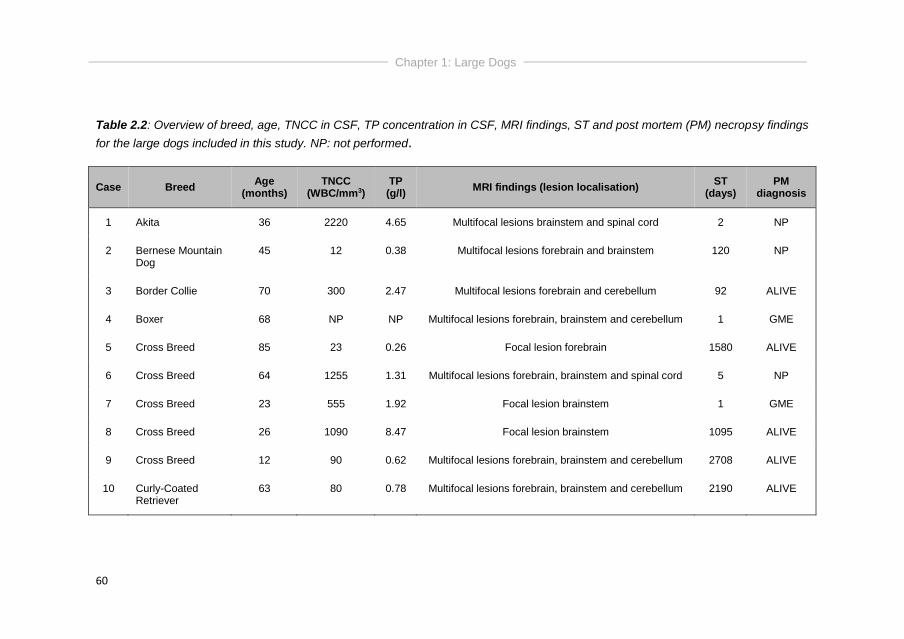
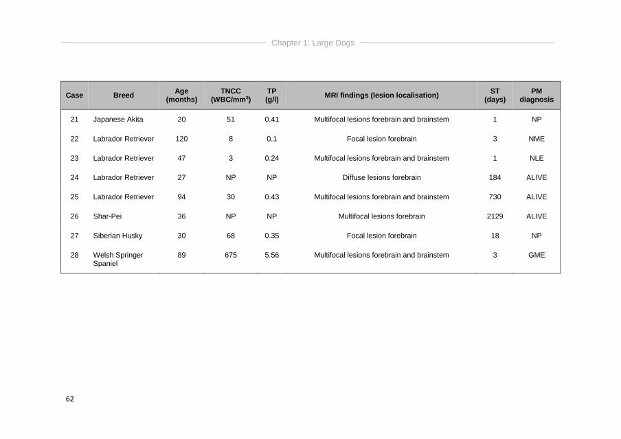
These included 28 (25%) large and 83 (75%) small dogs. Large dogs represented
were English Springer Spaniel (n=6), cross breed (n=5), Labrador Retriever (n=4),
Golden Retriever (n=2), Akita (n=2), and one each of the following breeds: Border
Collie, Boxer, Bernese Mountain dog, Curly-Coated Retriever, German Wirehaired
pointer, Great Dane, Shar-Pei, Siberian Husky, Welsh Springer Spaniel. Compared
to the general hospital population admitted between January 2006 and April 2015,
English Springer Spaniels were not significantly overrepresented (P=0.196). Small
dogs included West Highland White terrier (n=22), Chihuahua (n=8), Maltese terrier
(n=8), Pug (n=8), French Bulldog (n=7), Cavalier King Charles Spaniel (n=6),
crossbreed (n=6), Yorkshire terrier (n=5), Border terrier (n=2), Boston terrier (n=2),
Pomeranian (n=2), Poodle (n=2) and one each of the following breeds: Bichon Frise,
Welsh Corgi Cardigan, Lhasa Apso, Papillon and Sheltie.
Chapter 1: Large dogs
57
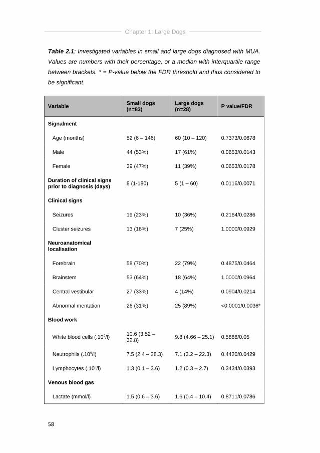
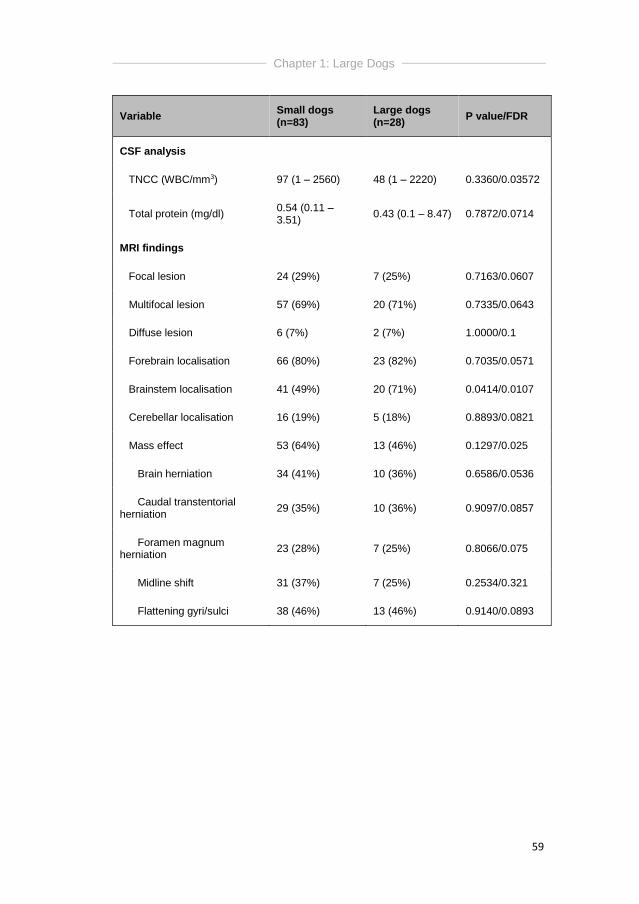
Clinical presentation and diagnostic findings
Large dogs had a significant shorter duration of clinical signs prior to
diagnosis (P=0.012), more often presented with decreased mentation (<0.0001) and
less often with cranial nerve deficits (P=0.027), and were more often diagnosed with
a brainstem lesion on MRI (P=0.039) compared to small dogs. However, when a
FDR of 10% was applied, only decreased mentation was found to be significantly
different between both groups. Gender, age at presentation, neuroanatomical
localisation, presence of seizures and cluster seizures, lactate concentration on
venous blood gas analysis, TNCC and TP concentration on CSF analysis and the
remaining MRI findings (meningeal or parenchymal contrast enhancement, mass
effect, brain herniation, flattening gyri/sulci, rostral or caudal transtentorial herniation,
foramen magnum herniation) were not different between small and large dogs. All
statistical results can be consulted in table 2.1, and a clinical summery regarding the
large dogs can be consulted in table 2.2.
Chapter 1: Large Dogs
58
Table 2.1: Investigated variables in small and large dogs diagnosed with MUA.
Values are numbers with their percentage, or a median with interquartile range
between brackets. * = P-value below the FDR threshold and thus considered to
be significant.
Variable Small dogs (n=83)
Large dogs (n=28)
P value/FDR
Signalment
Age (months) 52 (6 – 146) 60 (10 – 120) 0.7373/0.0678
Male 44 (53%) 17 (61%) 0.0653/0.0143
Female 39 (47%) 11 (39%) 0.0653/0.0178
Duration of clinical signs prior to diagnosis (days)
8 (1-180) 5 (1 – 60) 0.0116/0.0071
Clinical signs
Seizures 19 (23%) 10 (36%) 0.2164/0.0286
Cluster seizures 13 (16%) 7 (25%) 1.0000/0.0929
Neuroanatomical localisation
Forebrain 58 (70%) 22 (79%) 0.4875/0.0464
Brainstem 53 (64%) 18 (64%) 1.0000/0.0964
Central vestibular 27 (33%) 4 (14%) 0.0904/0.0214
Abnormal mentation 26 (31%) 25 (89%) <0.0001/0.0036*
Blood work
White blood cells (.109/l) 10.6 (3.52 – 32.8)
9.8 (4.66 – 25.1) 0.5888/0.05
Neutrophils (.109/l) 7.5 (2.4 – 28.3) 7.1 (3.2 – 22.3) 0.4420/0.0429
Lymphocytes (.109/l) 1.3 (0.1 – 3.6) 1.2 (0.3 – 2.7) 0.3434/0.0393
Venous blood gas
Lactate (mmol/l) 1.5 (0.6 – 3.6) 1.6 (0.4 – 10.4) 0.8711/0.0786
Chapter 1: Large Dogs
59
Variable Small dogs (n=83)
Large dogs (n=28)
P value/FDR
CSF analysis
TNCC (WBC/mm3) 97 (1 – 2560) 48 (1 – 2220) 0.3360/0.03572
Total protein (mg/dl) 0.54 (0.11 – 3.51)
0.43 (0.1 – 8.47) 0.7872/0.0714
MRI findings
Focal lesion 24 (29%) 7 (25%) 0.7163/0.0607
Multifocal lesion 57 (69%) 20 (71%) 0.7335/0.0643
Diffuse lesion 6 (7%) 2 (7%) 1.0000/0.1
Forebrain localisation 66 (80%) 23 (82%) 0.7035/0.0571
Brainstem localisation 41 (49%) 20 (71%) 0.0414/0.0107
Cerebellar localisation 16 (19%) 5 (18%) 0.8893/0.0821
Mass effect 53 (64%) 13 (46%) 0.1297/0.025
Brain herniation 34 (41%) 10 (36%) 0.6586/0.0536
Caudal transtentorial herniation
29 (35%) 10 (36%) 0.9097/0.0857
Foramen magnum herniation
23 (28%) 7 (25%) 0.8066/0.075
Midline shift 31 (37%) 7 (25%) 0.2534/0.321
Flattening gyri/sulci 38 (46%) 13 (46%) 0.9140/0.0893
Chapter 1: Large Dogs
60
Table 2.2: Overview of breed, age, TNCC in CSF, TP concentration in CSF, MRI findings, ST and post mortem (PM) necropsy findings
for the large dogs included in this study. NP: not performed.
Case Breed Age
(months) TNCC
(WBC/mm3) TP (g/l)
MRI findings (lesion localisation) ST
(days) PM
diagnosis
1 Akita 36 2220 4.65 Multifocal lesions brainstem and spinal cord 2 NP
2 Bernese Mountain Dog
45 12 0.38 Multifocal lesions forebrain and brainstem 120 NP
3 Border Collie 70 300 2.47 Multifocal lesions forebrain and cerebellum 92 ALIVE
4 Boxer 68 NP NP Multifocal lesions forebrain, brainstem and cerebellum 1 GME
5 Cross Breed 85 23 0.26 Focal lesion forebrain 1580 ALIVE
6 Cross Breed 64 1255 1.31 Multifocal lesions forebrain, brainstem and spinal cord 5 NP
7 Cross Breed 23 555 1.92 Focal lesion brainstem 1 GME
8 Cross Breed 26 1090 8.47 Focal lesion brainstem 1095 ALIVE
9 Cross Breed 12 90 0.62 Multifocal lesions forebrain, brainstem and cerebellum 2708 ALIVE
10 Curly-Coated Retriever
63 80 0.78 Multifocal lesions forebrain, brainstem and cerebellum 2190 ALIVE

Chapter 1: Large Dogs
61
Case Breed Age
(months) TNCC
(WBC/mm3) TP (g/l)
MRI findings (lesion localisation) ST
(days) PM
diagnosis
11 English Springer Spaniel
61 62 0.6 Multifocal lesions forebrain and brainstem 2950 ALIVE
12 English Springer Spaniel
60 17 0.75 Diffuse lesions forebrain 1 NP
13 English Springer Spaniel
47 12 0.16 Multifocal lesions brainstem 72 ALIVE
14 English Springer Spaniel
61 29 0.23 Multifocal lesions forebrain and brainstem 2039 ALIVE
15 English Springer Spaniel
39 173 0.68 Focal lesion brainstem 1187 ALIVE
16 English Springer Spaniel
78 NP NP Multifocal lesions forebrain, brainstem and cerebellum 700 ALIVE
17 German Wirehaired Pointer
10 12 0.23 Multifocal lesions forebrain and brainstem 975 ALIVE
18 Golden Retriever 75 1 0.21 Multifocal lesions forebrain and brainstem 1 GME
19 Golden Retriever 60 22 0.46 Multifocal lesions forebrain and brainstem 1856 ALIVE
20 Great Dane 60 40 0.1 Multifocal lesions forebrain 1 NP
Chapter 1: Large Dogs
62
Case Breed Age
(months) TNCC
(WBC/mm3) TP (g/l)
MRI findings (lesion localisation) ST
(days) PM
diagnosis
21 Japanese Akita 20 51 0.41 Multifocal lesions forebrain and brainstem 1 NP
22 Labrador Retriever 120 8 0.1 Focal lesion forebrain 3 NME
23 Labrador Retriever 47 3 0.24 Multifocal lesions forebrain and brainstem 1 NLE
24 Labrador Retriever 27 NP NP Diffuse lesions forebrain 184 ALIVE
25 Labrador Retriever 94 30 0.43 Multifocal lesions forebrain and brainstem 730 ALIVE
26 Shar-Pei 36 NP NP Multifocal lesions forebrain 2129 ALIVE
27 Siberian Husky 30 68 0.35 Focal lesion forebrain 18 NP
28 Welsh Springer Spaniel
89 675 5.56 Multifocal lesions forebrain and brainstem 3 GME
Chapter 1: Large Dogs
63
Infectious disease testing was performed in 78 dogs, including
serology for Toxoplasma gondii and Neospora caninum in 58 dogs (16 large
dogs and 42 small dogs), and/or and PCR analysis for Toxoplasma gondii,
Neospora caninum and Canine Distemper Virus in 73 dogs (13 large dogs and
41 small dogs). Overall, infectious disease testing was lacking in 9 large dogs
from which 1 dog had complete necropsy performed and 4 dogs were still alive
at time of data capture with survival times ranging from 184 – 2039 days. These
4 dogs were all treated with an immunosuppressive treatment protocol. The 4
remaining large dogs died within 20 days after diagnosis and breeds included
English Springer Spaniel, crossbreed, Siberian Husky and Akita.
Post-mortem examination was performed in 14 dogs, 8 small and 6
large dogs. Results included GME (5 small dogs, 4 large dogs), NME (3 small
dogs, 1 large dog) and NLE (1 large dog).
Outcome
All 111 dogs were initiated on immunosuppressive doses of
glucocorticosteroids at time of diagnosis, combined with cytosine arabinoside in
66 (80%) small dogs and 18 (64%) large dogs. Most dogs on cytosine
arabinoside therapy had regular (3-4 weekly) re-examinations at a dedicated
cytosine arabinoside clinic. Overall, dogs that were alive at time of data capture,
were dogs that received sole prednisolone therapy or combined prednisolone
and cytosine arabinoside treatment.
At time of data capture, 11 (39%) large dogs and 27 (33%) small dogs
were alive. Conversely, 17 (61%) large and 56 (67%) small dogs had died. Of
the deceased dogs, 15 (88%) large dogs and 47 (84%) small dogs died or were
euthanized because of disease progression, compared to 2 (12%) large dogs
and 9 (16%) small dogs that died or were euthanized for unrelated causes.
Overall, 54% of the large dogs and 57% of the small dogs died or were
euthanized because of disease progression. The MST was 281 days and 106
days for the small and large dogs respectively. There was no significant
Chapter 1: Large Dogs
64
difference in survival curves between small and large dogs (P=0.664) (Figure
2.1).
Figure 2.1: Kaplan-Meier survival curve comparing the percentage of survival in
small (full black line) and large (dotted line) dogs. Results were censored for
dogs that were still alive at time of data capture (single little blocks).
Discussion
This study evaluated the differences in clinical presentation, diagnostic
findings and long-term survival between small and large dogs diagnosed with
MUA. Large dogs were found to present significantly more often with a
decreased mentation. No significant difference was seen in survival curves
between small and large dogs. To the best of our knowledge, this is the first
study describing clinical, diagnostic and outcome data in large dogs with MUA.
Meningoencephalitis of unknown aetiology is generally considered a syndrome
affecting small, toy and terrier breed dogs aged between approximately 3 and 7
years (Munana and Luttgen, 1998; Adamo et al., 2007; Talarico and
Schatzberg, 2010; Coates and Jeffery, 2014). However, a total of 45 large dogs
are present in the literature from 1998 to 2015 (Gregory et al., 1998; Munana
and Luttgen, 1998; Cherubini et al., 2006; Fliegner et al., 2006; Zarfoss et al.,

Chapter 1: Large Dogs
65
2006; Coates et al., 2007; Pakozdy et al., 2009; Smith et al., 2009; Wong et al.,
2010; Flegel et al., 2011; Lowrie et al., 2013; Estey et al., 2014; Barnoon et al.,
2015; Beckmann et al., 2015; Mercier and Barnes Heller 2015). The distribution
of the breeds in our study appears to be similar. Although the English Springer
Spaniel was the most common large dog breed in our study, it was not
significantly overrepresented compared to the general hospital population. The
higher number of this breed in our study therefore likely reflects its popularity in
the United Kingdom. MUA occurred, as expected, more often in small dogs, but
still a quarter of dogs in the presented study were large dogs. Ignoring this
population of dogs would underestimate the prevalence of MUA in the overall
canine population. Therefore, MUA should be considered as a differential
diagnosis in dogs other than small or toy breeds that have signs suggestive of
inflammatory brain disease.
Large dogs were more likely to present with a decreased mentation
compared to their small counterparts. Abnormal mentation in dogs can be
caused by lesions in the forebrain (telencephalon and diencephalon) and/or
brainstem (mesencephalon, metencephalon, myelencephalon) (Garosi, 2013).
A lesion in the brainstem is considered a typical MRI finding in cases of GME
and NLE, but is considered an uncommon finding in dogs with NME (Coates
and Jeffery, 2014) although histopathological lesions have been identified in the
brainstem of Chihuahua and Pug dogs with NME (Higgins et al., 2008; Park et
al., 2012). In the presented study, both small and large dogs were
histopathologically diagnosed with GME and NME, and 1 large dog was
diagnosed with NLE, which has not yet been previously described. In literature,
NLE is mainly affecting Yorkshire terriers and a French Bulldog (Schatzberg,
2005; Higginbotham et al., 2007; Timmann et al., 2007; Spitzbarth et al., 2010),
which are all considered small dogs in this study. Necrotising
meningoencephalitis has previously only been once described in a large dog; a
26kg Staffordshire Bull Terrier mix (Estey et al., 2014).
A limitation of the current study is the lack of histopathological
confirmation in the majority of cases, which makes inclusion of other diseases
than MUA possible (mainly cerebrovascular or neoplastic disease). Based on
intracranial MRI, seven imaging abnormalities have been associated with
Chapter 1: Large Dogs
66
neoplastic brain disease in one study (Cherubini et al., 2005). These included
the presence of a single lesion, regular lesion shape, presence of mass effect,
dural contact, dural tail sign, lesions affecting adjacent bone and contrast
enhancement (Cherubini et al., 2005). Another study demonstrated MRI to be
94.4% sensitive and 95.5% specific for detection of a brain lesion and for
classifying neoplastic and inflammatory disease correctly, but was only 38.9%
sensitive for classifying cerebrovascular disease (Wolff et al., 2012). For the
presented study, 6 large dogs had a focal lesion on MRI of the brain, where
inflammatory lesions are more typically associated with multifocal or diffuse
lesions on intracranial imaging (Cherubini et al., 2006). Two of those lesions
were histopathologically confirmed as GME or NME. Of the 4 remaining dogs,
one dog (Siberian husky) had a focal lesion in the frontal lobe that showed
presence of mass effect, parenchymal contrast enhancement and dural contact.
Although abnormalities on CSF analysis do not exclude neoplastic or
vascular disease (Bohn et al., 2006), this dog had an increased TNCC (35
WBC/mm3) and TP concentration (0.35g/l) on cisternal CSF analysis. The dog
was only 30 months old, and died acutely at home 18 days after diagnosis after
initial improvement on immunosuppressive therapy. The second, third and
fourth dog (2 cross breeds and an English Springer Spaniel) were 26 – 85
months old at time of diagnosis and had a focal lesion in the piriform lobe
(crossbreed) or brainstem (cross breed and English Springer Spaniel). All three
dogs had increased TNCC and TP concentrations on cisternal CSF analysis,
and all were alive 1095 – 1580 days after diagnosis. All dogs had an acute
onset of neurological signs, which might still be compatible with cerebrovascular
disease, which might be supported by the long survival without treatment of
those dogs. However, in cerebrovascular disease, contrast enhancement
should only be visible after 7-10 days in dogs with ischaemic infarcts, and after
6 days to 6 weeks in dogs with haemorrhagic infarcts (Garosi, 2012). All dogs
with a focal lesion had intracranial imaging within 5 days after onset of
neurological signs, and all lesions showed parenchymal contrast enhancement,
which was considered less typical for cerebrovascular disease. Neoplastic
disease can however not be excluded in those cases, although all 4 dogs were
still young at time of diagnosis (2-5y of age) where brain tumours are typically
Chapter 1: Large Dogs
67
affecting middle-aged to older (over 5 years) dogs and cats, with the majority
being older than 9 years of age (Dickinson, 2014; Snyder et al., 2006).
Infectious disease testing was lacking in approximately 30% of both
small and large dogs. The 9 large dogs that were not tested had focal (n=2),
multifocal (n=5) or diffuse forebrain (n=2) lesions on MR imaging. Necrotising
cerebellitis and cerebellar atrophy (Garosi et al., 2010) as well as multifocal
brain involvement (Parzefall et al., 2014) have been described in association
with Neospora caninum infection in dogs. In the study of Parzefall et al., (2014),
mild cerebellar atrophy was additionally seen in 3 of 4 dogs. In the presented
study, 11 large dogs died within 20 days after diagnosis and after
immunosuppressive treatment. Necropsy confirming GME, NLE or NME was
performed in 6/11 dogs. Of the 5 remaining dogs, all but 1 (Siberian Husky) had
multifocal or diffuse forebrain lesions without cerebellar involvement, and
showed no signs of perilesional edema on FLAIR images as described by
Parzefall et al., (2014). Magnetic resonance imaging features of Toxoplasma
gondii encephalitis have not yet been described for dogs, but it is known to
cause focal granuloma formation both in the forebrain (Pfohl et al., 2005;
Falzone et al., 2008) and the spinal cord (Alves et al., 2011) of cats. The 4 dogs
with multifocal or diffuse brain lesions all had an increased TNCC with a
mononuclear pleocytosis but without signs of presence of lymphoblastic cells.
No further diagnostic investigations for lymphoma were performed. Gliomatosis
cerebri is a central nervous system neoplasia that is mainly affecting
brachycephalic breeds (Boxer, Boston Terrier, English Bulldog, Bull Mastiff) and
MRI mainly reveals single lesions in thalamus and/or brainstem, although the
cerebrum can be involved and sometimes no lesions are visible (Bentley et al.,
2014). This differential diagnosis seems less likely in these 4 dogs because of
their breeds and the presence of multifocal or diffuse lesions, but necropsy
confirmation is lacking in those cases. Overall, pitfalls of this study are its
retrospective character, the lack of histopathological confirmation and infectious
disease testing in a number of cases. In contrary, MUA is considered a clinical
and imaging diagnosis in cases were definitive brain histology is lacking.
Chapter 1: Large Dogs
68
Conclusions
Twenty-five per cent of dogs diagnosed with MUA in this study were
considered large dogs. Ignoring this population of dogs would underestimate
the prevalence of MUA in the overall canine population. Large dogs presented
significantly more often with a decreased mentation compared to small dogs.
The MST was not significantly different between small and large dogs, 281 and
106 days respectively, leading to a guarded prognosis for all dogs diagnosed
with MUA and receiving immunosuppressive treatment. In conclusion, MUA
should be considered as a differential diagnosis in dogs other than small or toy
breeds that have signs suggestive of inflammatory brain disease.
Chapter 2
CLINICAL PRESENTATION, DIAGNOSTIC FINDINGS
AND OUTCOME IN DOGS WITH SPINAL-ONLY
MENINGOENCEPHALOMYELITIS OF UNKNOWN
AETIOLOGY
Ine Cornelisa, Holger A. Volkb, Luc Van Hama, Steven De Deckerb
aSmall Animal Department, Faculty of Veterinary Medicine, Ghent
University, Merelbeke, Belgium.
bClinical Science and Services, The Royal Veterinary College,
University of London, Hatfield, United Kingdom.
Adapted from: Cornelis I, Volk HA, Van Ham L, De Decker S. (2017).
Clinical presentation, diagnostic findings and outcome in dogs
diagnosed with spinal-only meningoencephalomyelitis of unknown
aetiology. Journal of Small Animal Practice 58, 174-182.
Chapter 2: SO-MUA
70
Abstract
Although difficult to clinically diagnose, spinal-only
meningoencephalomyelitis of unknown aetiology (SO-MUA) is an important
differential diagnosis for dogs presenting with signs of spinal cord dysfunction.
The aim of this study was to evaluate clinical presentation, diagnostic findings
and long-term outcome for dogs clinically diagnosed with SO-MUA.
Twenty-one dogs were included in the study. The majority of dogs
presented with an acute (43%) or chronic (52%) onset of neurological signs.
Ambulatory paresis was the most common neurological presentation (67%).
Neurological examination most commonly revealed a T3-L3 myelopathy, and
spinal hyperaesthesia was a common finding (71%). A spinal cord lesion was
visible in 90% of cases on MR imaging. Lesions were typically extensive, ill-
defined, hyperintense on T2WI and isointense on T1W images (T1WI).
Eighteen lesions (86%) showed parenchymal contrast enhancement and 17
lesions (81%) showed contrast enhancement of overlying meninges. All dogs
were treated with immunosuppressive doses of glucocorticosteroids, sometimes
combined with cytosine arabinoside. At time of data capture, 10/21 dogs (48%)
had died or been euthanized because of SO-MUA. Overall median survival time
was 669 days.
SO-MUA should be considered in the differential diagnosis of dogs
presenting with an acute or chronic, progressive, and potentially painful
myelopathy. MRI features can possible help to distinguish presumptive SO-
MUA from other more common spinal diseases. Overall, long-term survival is
guarded as approximately 50% of dogs will die or be euthanized because of
SO-MUA regardless of immunosuppressive treatment.
Chapter 2: SO-MUA
71
Introduction
Pure myelitis (inflammation of spinal cord parenchyma) or
meningomyelitis (inflammation of spinal cord parenchyma and surrounding
meninges) are rare diseases in small animals and occur commonly in
combination with inflammatory brain disease (Tipold and Stein, 2010). Viruses
(canine distemper virus, feline coronavirus), bacteria (Staphylococcus spp.,
Streptococcus spp., Pasteurella, coliforms, Actinomyces, Nocardia spp.), fungi
(Cryptococcus, Coccidioides spp., Blastomyces, Histoplasma), rickettsiae
(Ehrlichia, Rickettsia, Rocky Mountain spotted fever), protozoa (Toxoplasma
gondii, Neospora caninum), parasites (Dirofilaria immitis, Cuterebra,
Angiostrongylus vasorum) and algae (Prototheca wickerhamii, Prototheca
zopfii) are known causes of meningomyelitis in dogs and cats, with or without
concurrent intracranial signs (Dewey and Da Costa, 2016; Csebi et al., 2010;
Parry et al., 2009; Griffin et al., 2008). Apart from infectious causes, non-
infectious meningomyelitis including GME, pyogranulomatous
meningoencephalomyelitis and SRMA are described (Dewey 2016; Parry et al.
2009; Griffin et al. 2008; Meric 1988). In agreement with the terminology for
MUA, dogs clinically diagnosed with non-infectious inflammatory myelitis that
did not have positive infectious disease testing, that were not classified as
SRMA or eosinophilic meningomyelitis, and that were not histopathologically
confirmed, were named SO-MUA. A clinical diagnosis of SO-MUA is typically
made by a combination of clinical presentation, imaging of the vertebral column,
and results of CSF analysis (Griffin et al., 2008).
Currently, only one previous study has focused specifically on the
clinical presentation, diagnostic findings, and outcome in dogs with
meningomyelitis caused by a variety of underlying aetiologies (Griffin et al.,
2008). Twenty-eight cases were included, of which 15 dogs were diagnosed
with SO-MUA. Clinical signs reflected the affected spinal cord segments, and
younger dogs, toy breeds, and hound breeds were suggested to be
predisposed for meningomyelitis. Although results of myelography, CT, and CT-
myelography have been reported, little is known about MRI findings in dogs with
SO-MUA. The aims of this study were therefore to describe the signalment,
Chapter 2: SO-MUA
72
clinical presentation, diagnostic findings, including results of MRI, and long-term
survival in dogs diagnosed with presumptive SO-MUA without concurrent
clinical signs of intracranial involvement.
Materials and methods
Case selection
The electronic medical database of the Small Animal Referral Hospital,
Royal Veterinary College, University of London, was searched between March
2006 and February 2015 for dogs diagnosed with “MUA”, “MUO”, “GME”,
“myelitis”, and “inflammatory spinal cord disease”. Dogs were included based
on the criteria used by Granger et al. (2010), if they had (1) complete medical
records available, (2) a complete neurological examination performed leading to
a spinal cord localisation, (3) inflammatory CSF analysis, (4) MRI of the spinal
cord, and if (5) long-term follow-up information was available through revision of
medical records or through contacting the referring veterinarian by telephone.
Dogs were excluded if (1) the clinical records or imaging studies were
incomplete or not available for review, (2) dogs showed clinical or neurological
signs of intracranial involvement at time of presentation, (3) they had a peracute
onset of clinical signs that were not progressive after 12-24 hours, (4) they had
signs of extradural or intradural/extramedullary spinal cord compression on MRI
and if (5) they had positive infectious disease titres or if clinical presentation,
CSF analysis or necropsy findings were suggestive of SRMA or eosinophilic
meningoencephalomyelitis (>10% eosinophils in CSF) (Dewey and Da Costa,
2016). Typical clinical presentation for SRMA was considered to be a dog less
than 2 years of age of a typical dog breed (Boxer, Beagle, Bernese Mountain
dog, Nova Scotia Duck Tolling Retriever, Golden Retriever, German
Shorthaired Pointed) presenting with pyrexia and cervical hyperesthesia. CSF
analysis in SRMA is typically revealing a predominantly neutrophilic pleocytosis
(Dewey and Da Costa, 2016). Dogs with histopathological confirmation of the
disease (GME or NME) only needed to fulfil inclusion criteria (1) and (5).
Information retrieved from the medical records included breed, gender, age at
Chapter 2: SO-MUA
73
diagnosis, body weight, results of neurological examination including
neuroanatomical localisation, duration of clinical signs prior to diagnosis, results
of CBC and biochemistry profile, results of CSF analysis, treatment received,
and outcome. For dogs that had CSF analysis performed, site of collection
(cisternal or lumbar), TNCC, TP and cytological differentiation were recorded.
Total nucleated cell count was considered normal if <5 cells/mm3. Total protein
concentration was considered normal for a cisternal collection if < 0.25g/l and
for a lumbar collection if <0.4g/l. Duration of clinical signs prior to diagnosis
(days) was classified as peracute (<2 days), acute (2–7 days) or chronic (>7
days).
Neurological assessment
The neurological status was classified from 0 to 5 according to the
clinical examination (adapted from Scott et al., 1997): grade 0 = neurologically
normal; grade 1 = spinal hyperesthesia without neurological deficits; grade 2 =
ataxia, ambulatory para- or tetraparesis; grade 3 = non- ambulatory para- or
tetraparesis; grade 4 = para- or tetraplegia with or with- out bladder control, and
intact deep pain sensation; grade 5 = para- or tetraplegia, urine retention or
overflow, and deep pain sensation loss.
Possible neuroanatomical localisations included C1-C5, C6-T2, T3-L3
or L4-S3 spinal cord segments. Dogs were diagnosed with a focal lesion if only
one spinal cord segment was affected, and with a multifocal lesion if more than
one spinal cord segment appeared to be affected on the neurological
examination.
Magnetic resonance imaging
Magnetic resonance imaging was performed under general
anaesthesia with a permanent 1.5T magnet (Intera, Philips Medical Systems,
Eindhoven, the Netherlands) and all images were reviewed by the same author
(IC) using Osirix Dicom viewer (Osirix Foundation, V.5.5.2 Geneva,
Switzerland). Sequences could vary, but studies included a minimum of T2W
Chapter 2: SO-MUA
74
(repetition time (ms) (TR)/echo time (ms) (TE), 3000/120) and T1W (TR/TE,
400/8) images of the affected spinal cord region in a sagittal and transverse
plane. The T1W images were acquired before and after IV administration of
paramagnetic contrast medium (0.1mg/kg, gadoterate meglumine, Dotarem,
Guerbet). If MR images of the brain were present, they were reviewed
concurrently. Variables recorded were lesion intensity on T2WI and T1WI,
lesion localisation and distribution, lesion length and presence of parenchymal
and/or meningeal contrast enhancement. Lesion length was measured using
Osirix Dicom viewer, and performed on sagittal T2WI for dogs that had focal
lesions. Lesion length was measured twice, and the mean value between both
was used. To compensate for differences in body size, values were corrected
towards length of vertebral body of C6 (for cervical lesions) or L2 (for
thoracolumbar lesions). Vertebral body length was measured on T1W sagittal
images.
Treatment and follow-up
For all dogs, the specific treatment protocol was recorded. During
hospitalisation, all dogs underwent daily at least one general physical and
complete neurological examination by a board-certified neurologist or neurology
resident. The results of the neurological examination as well as response to
treatment (improvement, deterioration or static status) were systematically
recorded on the kennel sheets. Follow-up information during hospitalisation was
collected from the medical records, and afterwards through medical records of
re-examination visits or telephone contact with the referring veterinarian. A
successful outcome was defined as the dog being ambulatory, faecal and
urinary continent and, according to the owners, without signs of overt spinal
hyperaesthesia. An unsuccessful outcome was defined as (1) deterioration in
neurological status by one or more grades after diagnosis and treatment, or (2)
if the dog was not independently ambulatory, possibly with previously non-
existing or worsening faecal and/or urinary incontinence, or was experiencing
spinal hyperaesthesia as defined by the owner.
Chapter 2: SO-MUA
75
Statistical analysis
Data analysis was performed with the aid of a standard statistical
software package (Prism, Graphpad Software Inc). Regarding outcome, a
Mann-Whitney U test was used to evaluate the effect of relative lesion length on
long-term outcome. A fisher’s exact test was used to evaluate the effect of pain,
presence of lymphopenia and additional administration of cytosine arabinoside
on outcome. Numeric variables were expressed as median and interquartile
ranges (IQR). Values of P<0.05 were considered significant. Survival analysis
was performed using both a Log-rank (Mantel-Cox) and Gehan-Breslow-
Wilcoxin test, resulting in MST calculation and a Kaplan-Meier survival curve.
Survival was defined as time from diagnosis to death or euthanasia, including
whether this happened because of disease progression or due to unrelated
causes, or time from diagnosis to data collection for dogs that were alive at time
of data capture. Dogs that died because of unrelated causes and dogs that
were still alive at time of data capture were censored for survival analysis.
Results
Signalment
Twenty-one dogs were included in the study. Represented breeds
included French Bulldog (n=2), Jack Russell Terrier (n=2), Lhasa Apso (n=2)
and one each of Akita, Bearded Collie, Boxer, Bull Mastiff, Chihuahua, cross
breed, English Springer Spaniel, Giant Schnauzer, Labrador Retriever, Maltese
Terrier, Rhodesian Ridgeback, Rottweiler, Shih Tzu, West Highland White
Terrier and Yorkshire Terrier. Overall, median age at presentation was 56
months (10 – 128 months). Thirteen dogs (62%) were male and 8 dogs (38%)
were female. Compared to the general hospital population between March 2006
and February 2015, there was no significant difference in sex distribution in the
group of dogs with SO-MUA (P=0.075). Median duration of clinical signs prior to
diagnosis was 8 days (ranging from 1-90 days). One dog (5%) presented with
Chapter 2: SO-MUA
76
peracute, 9 dogs (43%) with acute and 11 dogs (52%) with a chronic onset of
neurological signs.
Neurological examination
Thirteen (62%) and 8 (38%) dogs were diagnosed with a focal and
multifocal spinal lesion on neurological examination, respectively. Regarding
dogs with focal spinal lesions (n=13), 3 dogs were diagnosed with a lesion
affecting the C1-C5 spinal cord segments, 2 dogs with a lesion affecting the C6-
T2 spinal cord segments, 6 dogs with a lesion affecting the T3-L3 spinal cord
segments and 2 dogs with a lesion affecting the L4-S3 spinal cord segments. At
time of diagnosis, no dogs presented as grade 0; 2 dogs (10%) were grade 1;
14 dogs (67%) grade 2; and 5 dogs (24%) grade 3. No dogs were found to have
paraplegia or tetraplegia at time of presentation. Pain on direct spinal palpation
was present in 15 (71%) dogs. Urinary retention was seen in 2 dogs (10%), and
a combination of urinary and faecal incontinence was noticed in 2 dogs (10%).
One dog (5%) developed seizures 669 days after diagnosis of SO-MUA. An
overview of the clinical and diagnostic findings of the 21 included dogs can be
consulted in table 3.1.
Chapter 2: SO-MUA
77
Table 3.1: Clinical details of the 21 dogs diagnosed with SO-MUA. Dogs marked with * had additional intracranial abnormalities
detected on MR imaging. FE = female entire, FN = female neutered, ME = male entire, MN = male neutered.
Case Breed Gender Age
(months) Clinical presentation
Neuroanatomical localisation
Spinal hyperaesthesia
CSF TNCC
(WBC/mm3) MRI lesion
1 Akita FE 36 Non ambulatory paraparesis
Multifocal Yes 1740 Focal
2 Rottweiler ME 123 Ataxia T3-L3 No 209 Focal
3 Bull Mastiff ME 42 Ambulatory paraparesis
T3-L3 Yes 6 No lesion
visible
4 Labrador Retriever
MN 105 Ambulatory paraparesis
L4-S3 Yes 123 Focal
5 JRT MN 89 Ambulatory paraparesis
T3-L3 No 200 Focal
6 Lhasa Apso FE 48 Ambulatory tetraparesis
C1-C5 Yes 900 Focal
7 Shih Tzu MN 50 Ambulatory tetraparesis
C6-T2 Yes 5 Focal
8 Giant Schnauzer ME 32 Non ambulatory paraparesis
Multifocal No 1345 Focal
Chapter 2: SO-MUA
78
Case Breed Gender Age
(months) Clinical presentation
Neuroanatomical localisation
Spinal hyperaesthesia
CSF TNCC
(WBC/mm3) MRI lesion
9 Yorkshire Terrier FN 36 Ambulatory tetraparesis
C1-C5 Yes 7 Focal
10 English Springer Spaniel
ME 85 Ataxia Multifocal No 455 Focal
11 Rhodesian Ridgeback
FE 123 Normal gait C1-C5 Yes 89 Focal*
12 Bearded Collie MN 136 Ambulatory paraparesis
Multifocal Yes 162 No lesion
visible
13 Boxer ME 26 Normal gait Multifocal Yes 6000 Focal
14 Lhasa Apso MN 128 Ambulatory paraparesis
L4-S3 Yes 1540 Multifocal
15 Chihuahua ME 19 Ataxia T3-L3 Yes 9 Multifocal
16 Cross Breed FN 83 Ambulatory paraparesis
Multifocal No 1230 Multifocal
17 French Bulldog ME 13 Ambulatory paraparesis
T3-L3 No 250 Multifocal
18 Maltese Terrier FN 104 Ataxia Multifocal Yes 95 Focal
Chapter 2: SO-MUA
79
Case Breed Gender Age
(months) Clinical presentation
Neuroanatomical localisation
Spinal hyperaesthesia
CSF TNCC
(WBC/mm3) MRI lesion
19 Jack Russell Terrier
FN 56 Non ambulatory tetraparesis
C6-T2 Yes 2690 Focal*
20 French Bulldog ME 10 Non ambulatory paraparesis
T3-L3 Yes 43 Focal
21 West Highland White Terrier
FE 103 Non ambulatory tetraparesis
Multifocal Yes 1980 Multifocal
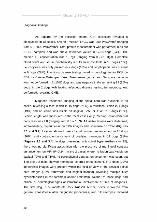
Chapter 2: SO-MUA
80
Diagnostic findings
As required by the inclusion criteria, CSF collection revealed a
pleocytosis in all cases. Overall, median TNCC was 209 WBC/mm3 (ranging
from 6 – 6000 WBC/mm3). Total protein measurement was performed in all but
3 CSF samples, and was above reference values in 17/18 dogs (94%). The
median TP concentration was 1.67g/l (ranging from 0.21-16.3g/l). Complete
blood count and serum biochemistry results were available in 16 dogs (76%).
Leucocytosis was only present in 2 dogs (10%) and lymphopenia was present
in 6 dogs (29%). Infectious disease testing based on serology and/or PCR on
CSF for Canine Distemper Virus, Toxoplasma gondii, and Neospora caninum
was not performed in 2 (10%) dogs and was negative in the remaining 19 (90%)
dogs. In the 2 dogs with lacking infectious disease testing, full necropsy was
performed, revealing GME.
Magnetic resonance imaging of the spinal cord was available in all
cases, revealing a focal lesion in 15 dogs (71%), a multifocal lesion in 4 dogs
(19%) and no lesion was visible on sagittal T2WI or T1WI in 2 dogs (10%).
Lesion length was measured in the focal cases only. Median lesion/vertebral
body ratio was 4.8 (ranging from 0.6 – 10.9). All visible lesions were ill-defined,
intramedullary, hyperintense on T2W images and isointense on T1WI (Figures
3.1 and 3.2). Lesions showed parenchymal contrast enhancement in 18 dogs
(86%), and contrast enhancement of overlying meninges in 17 dogs (81%)
(Figures 3.3 and 3.4). In dogs presenting with spinal hyperaesthesia (n=15),
there was no significant association with the presence of meningeal contrast
enhancement on MRI (P=0.24). In the 2 cases where no lesion was visible on
sagittal T2WI and T1WI, no parenchymal contrast enhancement was seen, but
1 of those 2 dogs showed meningeal contrast enhancement. In 2 dogs (10%)
intracranial images were present within the field of view of the cervical spinal
cord images (T2W transverse and sagittal images), revealing multiple T2W
hyperintensities in the forebrain and/or brainstem. Neither of those dogs had
clinical or neurological signs of intracranial involvement at time of diagnosis.
The first dog, a 56-month-old Jack Russell Terrier, never recovered from
general anaesthesia after diagnostic procedures, and full necropsy revealed

Chapter 2: SO-MUA
81
GME. The second dog, a 123-month-old Rhodesian Ridgeback, developed
seizures 669 days after diagnosis and was euthanized without further
investigations.
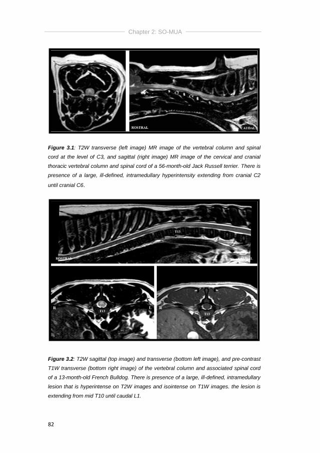
Chapter 2: SO-MUA
82
Figure 3.1: T2W transverse (left image) MR image of the vertebral column and spinal
cord at the level of C3, and sagittal (right image) MR image of the cervical and cranial
thoracic vertebral column and spinal cord of a 56-month-old Jack Russell terrier. There is
presence of a large, ill-defined, intramedullary hyperintensity extending from cranial C2
until cranial C6.
Figure 3.2: T2W sagittal (top image) and transverse (bottom left image), and pre-contrast
T1W transverse (bottom right image) of the vertebral column and associated spinal cord
of a 13-month-old French Bulldog. There is presence of a large, ill-defined, intramedullary
lesion that is hyperintense on T2W images and isointense on T1W images. the lesion is
extending from mid T10 until caudal L1.
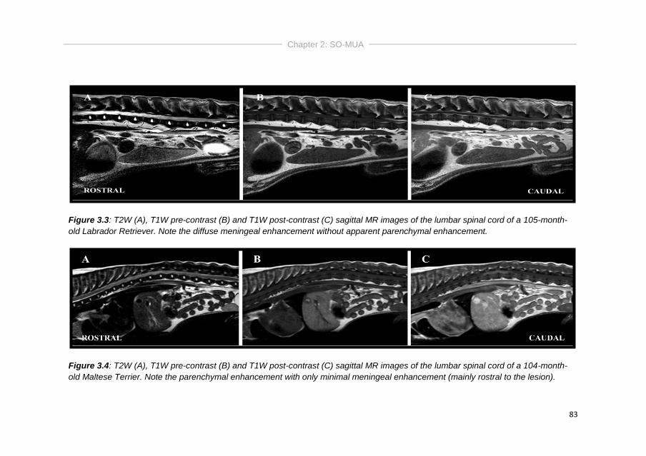
Chapter 2: SO-MUA
83
Figure 3.3: T2W (A), T1W pre-contrast (B) and T1W post-contrast (C) sagittal MR images of the lumbar spinal cord of a 105-month-
old Labrador Retriever. Note the diffuse meningeal enhancement without apparent parenchymal enhancement.
Figure 3.4: T2W (A), T1W pre-contrast (B) and T1W post-contrast (C) sagittal MR images of the lumbar spinal cord of a 104-month-
old Maltese Terrier. Note the parenchymal enhancement with only minimal meningeal enhancement (mainly rostral to the lesion).
Chapter 2: SO-MUA
84
Treatment and outcome
As required by the inclusion criteria, outcome was available in all dogs.
As described above, one dog never recovered from general anaesthesia for
MRI of the spinal cord, and this dog was censored for survival analysis. Mean
duration of hospitalisation was 5 days (ranging from 1 – 19 days), with 17 dogs
(81%) showing improvement in neurological status within those days. One dog
(5%) remained neurologically stable (no improvement nor deterioration), and 3
dogs (14%) showed deterioration of their neurological status. All dogs, including
the dog that never recovered from anaesthesia, were treated with
immunosuppressive doses of glucocorticosteroids immediately after diagnosis.
This consisted of IV dexamethasone (dose ranging from 0.3 – 0.5mg/kg/day) in
9 dogs (43%), and oral prednisolone (dose ranging from 2 – 4mg/kg/day) in 12
dogs (57%). Fourteen dogs (67%) received additional treatment with cytosine
arabinoside as a CRI of 200mg/m2 over 8 hours in 1 dog (7%) and as 4 SC
injections of 50mg/m2 every 12 hours for 2 consecutive days in 13 dogs (93%).
Twenty dogs (95%) survived to discharge. Of these dogs, 9 dogs
(45%) were still alive at time of data capture. Of these 9 dogs, 8 dogs were
neurologically normal according to follow-up information, and 1 dog was still
showing ataxia and ambulatory paraparesis. Of the 8 normal dogs, 2 dogs were
still receiving ciclosporine 5mg/kg every 24 hours, 1 dog was receiving cytosine
arabinoside 50mg/ m2 every 12 hours for 2 consecutive days every 9 weeks, 1
dog was receiving prednisolone 0.2mg/kg every 24 hours, 1 dog was receiving
prednisolone 1mg/kg every 24 hours and cytosine arabinoside 50mg/ m2 every
12 hours for 2 consecutive days every 4 weeks, and 3 dogs were not receiving
any treatment at time of data capture. The dog that was still showing
neurological abnormalities was receiving 0.5mg/kg prednisolone every other
day and cytosine arabinoside 50mg/ m2 every 12 hours for 2 consecutive days
every 5 weeks. Regarding the 11/20 dogs (55%) that had deceased at time of
data capture, 3 dogs died or were euthanized because of disease progression,
6 dogs were euthanized because of acute neurological deterioration after initial
neurological improvement, and 2 dogs were euthanized because of unrelated
causes (complications after stifle surgery and development of aggression).
Chapter 2: SO-MUA
85
Dogs that showed acute neurological deterioration after initial improvement did
so within a median of 171 days after diagnosis (ranging from 30-669 days). Of
those 6 dogs, 1 dog showed acute deterioration after discontinuation of
prednisolone treatment, and 5 dogs were still receiving treatment consisting of
prednisolone 1mg/kg every 24 hours, prednisolone 0.5mg/kg every 24 hours,
prednisolone 2mg/kg every 24 hours and azathioprine 2mg/kg every 24 hours,
or cytosine arabinoside 50mg/m2 every 12 hours for 2 consecutive days every 7
weeks. Overall, we can conclude that 10/21 dogs (48%) died or were
euthanized because of SO-MUA.
No difference was seen in long-term survival between dogs receiving
sole prednisolone therapy or combination therapy with cytosine arabinoside
(P=0.31). Overall, the MST was 669 days (ranging from 1-2250 days) (Figure
3). No significant difference was seen in relative lesion length on MR imaging
between dogs that are alive and dogs that died or were euthanized because of
SO-MUA (P=0.91). Post mortem confirmation was available in 3 dogs, revealing
GME in 2 dogs and necrotising meningomyelitis in 1 dog. An overview of the
treatment schedules, follow-up, ST and necropsy information can be consulted
in table 3.2.
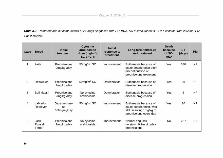
Chapter 2: SO-MUA
86
Table 3.2: Treatment and outcome details of 21 dogs diagnosed with SO-MUA. SC = subcutaneous, CRI = constant rate infusion, PM
= post mortem.
Case Breed Initial
treatment
Cytosine arabinoside
dose (mg/m2), SC or CRI
Initial response to
treatment
Long-term follow-up and treatment
Death because of SO-MUA
ST (days)
PM
1 Akita Prednisolone 2mg/kg /day
50mg/m2 SC Improvement Euthanasia because of acute deterioration after discontinuation of prednisolone treatment
Yes 380 NP
2 Rottweiler Prednisolone 2mg/kg /day
50mg/m2 SC Deterioration Euthanasia because of disease progression
Yes 20 NP
3 Bull Mastiff Prednisolone 2mg/kg /day
No cytosine arabinoside
Deterioration Euthanasia because of disease progression
Yes 6 NP
4 Labrador Retriever
Dexamethasone
0.3mg/kg/day
50mg/m2 SC Improvement Euthanasia because of acute deterioration, was still receiving 1mg/kg of prednisolone every day
Yes 30 NP
5 Jack Russell Terrier
Prednisolone 2mg/kg /day
No cytosine arabinoside
Improvement Normal dog, still receiving 0.2mg/kg/day prednisolone
No 237 NA
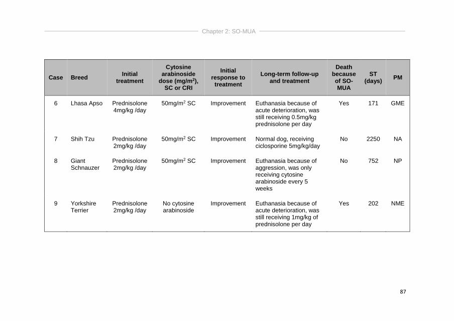
Chapter 2: SO-MUA
87
Case Breed Initial
treatment
Cytosine arabinoside
dose (mg/m2), SC or CRI
Initial response to
treatment
Long-term follow-up and treatment
Death because of SO-MUA
ST (days)
PM
6 Lhasa Apso Prednisolone 4mg/kg /day
50mg/m2 SC Improvement Euthanasia because of acute deterioration, was still receiving 0.5mg/kg prednisolone per day
Yes 171 GME
7 Shih Tzu Prednisolone 2mg/kg /day
50mg/m2 SC Improvement Normal dog, receiving ciclosporine 5mg/kg/day
No 2250 NA
8 Giant Schnauzer
Prednisolone 2mg/kg /day
50mg/m2 SC Improvement Euthanasia because of aggression, was only receiving cytosine arabinoside every 5 weeks
No 752 NP
9 Yorkshire Terrier
Prednisolone 2mg/kg /day
No cytosine arabinoside
Improvement Euthanasia because of acute deterioration, was still receiving 1mg/kg of prednisolone per day
Yes 202 NME
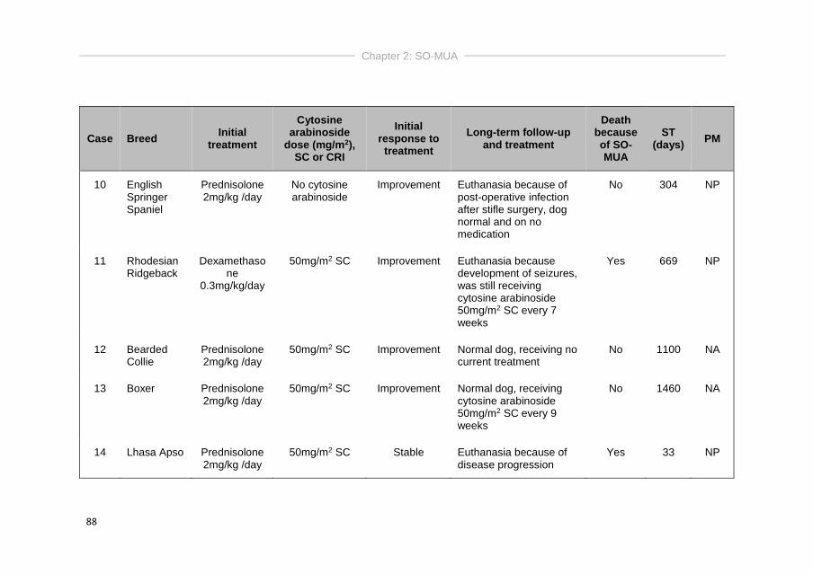
Chapter 2: SO-MUA
88
Case Breed Initial
treatment
Cytosine arabinoside
dose (mg/m2), SC or CRI
Initial response to
treatment
Long-term follow-up and treatment
Death because of SO-MUA
ST (days)
PM
10 English Springer Spaniel
Prednisolone 2mg/kg /day
No cytosine arabinoside
Improvement Euthanasia because of post-operative infection after stifle surgery, dog normal and on no medication
No 304 NP
11 Rhodesian Ridgeback
Dexamethasone
0.3mg/kg/day
50mg/m2 SC Improvement Euthanasia because development of seizures, was still receiving cytosine arabinoside 50mg/m2 SC every 7 weeks
Yes 669 NP
12 Bearded Collie
Prednisolone 2mg/kg /day
50mg/m2 SC Improvement Normal dog, receiving no current treatment
No 1100 NA
13 Boxer Prednisolone 2mg/kg /day
50mg/m2 SC Improvement Normal dog, receiving cytosine arabinoside 50mg/m2 SC every 9 weeks
No 1460 NA
14 Lhasa Apso Prednisolone 2mg/kg /day
50mg/m2 SC Stable Euthanasia because of disease progression
Yes 33 NP
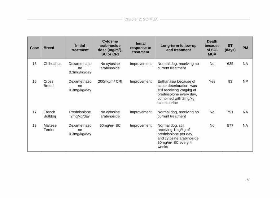
Chapter 2: SO-MUA
89
Case Breed Initial
treatment
Cytosine arabinoside
dose (mg/m2), SC or CRI
Initial response to
treatment
Long-term follow-up and treatment
Death because of SO-MUA
ST (days)
PM
15 Chihuahua Dexamethasone
0.3mg/kg/day
No cytosine arabinoside
Improvement Normal dog, receiving no current treatment
No 635 NA
16 Cross Breed
Dexamethasone
0.3mg/kg/day
200mg/m2 CRI Improvement Euthanasia because of acute deterioration, was still receiving 2mg/kg of prednisolone every day, combined with 2mg/kg azathioprine
Yes 93 NP
17 French Bulldog
Prednisolone 2mg/kg/day
No cytosine arabinoside
Improvement Normal dog, receiving no current treatment
No 791 NA
18 Maltese Terrier
Dexamethasone
0.3mg/kg/day
50mg/m2 SC Improvement Normal dog, still receiving 1mg/kg of prednisolone per day, and cytosine arabinoside 50mg/m2 SC every 4 weeks
No 577 NA
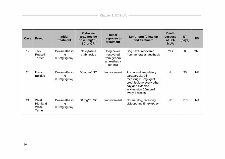
Chapter 2: SO-MUA
90
Case Breed Initial
treatment
Cytosine arabinoside
dose (mg/m2), SC or CRI
Initial response to
treatment
Long-term follow-up and treatment
Death because of SO-MUA
ST (days)
PM
19 Jack Russell Terrier
Dexamethasone
0.5mg/kg/day
No cytosine arabinoside
Dog never recovered
from general anaesthesia
for MRI
Dog never recovered from general anaesthesia
Yes 0 GME
20 French Bulldog
Dexamethasone
0.3mg/kg/day
50mg/m2 SC Improvement Ataxia and ambulatory paraparesis, still receiving 0.5mg/kg of prednisolone every other day and cytosine arabinoside 50mg/m2 every 5 weeks
No 90 NP
21 West Highland White Terrier
Dexamethasone
0.3mg/kg/day
50 mg/m2 SC Improvement Normal dog, receiving ciclosporine 5mg/kg/day
No 210 NA
Chapter 2: SO-MUA
91
Discussion
This study evaluated the clinical presentation, diagnostic findings and
long-term survival in 21 dogs diagnosed with presumptive SO-MUA. Dogs had a
median age of 5 years at time of diagnosis. A lesion affecting the T3-L3 spinal
cord segments resulting in ambulatory paraparesis was considered the most
common clinical presentation. Although the overall MST was 669 days, 48% of
dogs diagnosed with SO-MUA died or were euthanized because of this disease,
indicating a guarded long-term prognosis.
To be included in the study, dogs were not allowed to have clinical
signs or neurological examination abnormalities suggestive of intracranial
involvement. Interestingly, additional MR images of the brain were included in
the field of view of the cervical MRI in 2 dogs, showing additional lesions in both
cases. One of those dogs, a 123-month-old Rhodesian Ridgeback, developed
seizures 669 days after diagnosis despite on-going cytosine arabinoside
treatment, and was therefore euthanized. No necropsy was performed, but
because intracranial lesions were already present at time of diagnosis,
development of MUA was assumed. The other dog, a 56-month-old Jack
Russell Terrier, never recovered from general anaesthesia for MR imaging.
Necropsy was performed, revealing the presence of GME. Because intracranial
MR images were only available in 2 dogs, it is currently unclear (1) if these brain
abnormalities represent a multifocal nature of the disease or cranial extension
of the cervical inflammatory lesions, and (2) if inflammatory brain lesions are
currently under diagnosed in dogs with SO-MUA and if SO-MUA could therefore
be considered a more generalised inflammatory disease process, a
meningoencephalomyelitis.
Pain on direct spinal palpation was present in 71% of dogs. Spinal pain
reflects the involvement of the meninges, and/or vertebrae (vertebral
periosteum), and/or nerve roots or spinal nerves (Da Costa, 2012). In the
present study, the lesions showed meningeal contrast enhancement in 18/21
dogs, but there was no significant association between spinal hyperaesthesia
and the presence of meningeal enhancement on MRI.
Chapter 2: SO-MUA
92
MRI of the spinal cord revealed no lesion on sagittal T2WI and T1WI in
10% of dogs (n=2), which appears similar to the 7% described for the brain in
dogs with MUA (Granger et al., 2010). In the retrospective study of Griffin et al.
(2008), only 1 dog with meningomyelitis had MRI performed, revealing no
abnormalities. Based on these findings, presence of SO-MUA cannot be ruled
out based on unremarkable MRI findings. The first dog was a 42-month-old Bull
Mastiff with a one-month history of slowly progressive T3-L3 spinal cord lesion.
After diagnostic procedures, the dog was treated with oral prednisolone but
continued to deteriorate and was euthanized after 6 days. No necropsy was
performed. The second dog was a 136-month-old Bearded Collie with a one-
week history of a progressive multifocal spinal cord neuroanatomical
localisation (T3-S3 spinal cord lesion). The dog showed improvement on
treatment with prednisolone and cytosine arabinoside (see table 3.2) after
diagnostic investigations, and was still alive without current treatment 1100 days
after diagnosis. Both dogs had inflammatory CSF analysis (increased TNCC
and TP concentration). For both dogs, the presence of vascular, degenerative
and neoplastic spinal cord lesions cannot be excluded. As both dogs had a
progressive disease course, a vascular (ischaemic) lesion seemed less likely. A
neoplastic lesion cannot be excluded, although this seems rather unlikely in the
Bull Mastiff considering his young age. The second dog had a lymphocytic
pleocytosis on CFS analysis, but no signs of lymphoma were seen on
microscopical examination, however no specific test to look for clonality was
performed.
If a lesion was visible on MRI, all lesions were extensive, ill-defined,
intramedullary, hyperintense on T2WI and isointense on T1WI. Other spinal
conditions, including acute non-compressive nucleus pulposus extrusion
(ANNPE) and ischaemic myelopathy (IM), are also associated with
intraparenchymal hyperintensities on MRI. These conditions are however
associated with other clinical and MRI characteristics, which could potentially
aid in differentiating between these conditions (Cardy et al., 2015; Fenn et al.,
2016). Looking into a recent study (Cardy et al., 2015), the clinical presentation
of dogs with spinal cord dysfunction, IM (most commonly fibrocartilagenous
embolic myelopathy (FCEM)) and ANNPE are typically characterised by a
peracute onset of non-progressive clinical signs and affected dogs do not
Chapter 2: SO-MUA
93
commonly demonstrate overt spinal hyperaesthesia at time of admission. This
is in contrast with the clinical presentation of dogs with SO-MUA, which was
characterised by an acute onset of progressive and mainly symmetrical
neurological deficits, with pain on spinal palpation or manipulation in 86% of
dogs (Cardy et al., 2015), which is comparable with the 71% of dogs presenting
with spinal hyperaesthesia in the presented study. Although CSF analysis in
dogs with IM is most often within normal limits, affected dogs can demonstrate
an increased TP concentration and mild neutrophilic or mixed cell pleocytosis
with a median TNCC of 12 WBC/mm3 (De Risio et al., 2007). A marked
pleocytosis with a median TNCC of 209 WBC/mm3 was seen in the presented
study, although results should be interpreted with caution as presence of a CSF
pleocytosis was considered one of the inclusion criteria. To conclude, the
presentation of a dog with an acute or chronic onset of a progressive and
painful T3-L3 myelopathy in combination with an extensive, ill defined,
intramedullary lesion with presence of parenchymal and/or meningeal contrast
enhancement on MRI, and presence of a marked pleocytosis on CSF analysis,
can be presumptively diagnosed with SO-MUA. The importance of
differentiating between these conditions is highlighted by the differences in
treatment and prognosis between dogs with presumptive SO-MUA and dogs
with ANNPE or IM.
A previous study demonstrated that short tau inversion recovery
(STIR) hyperintensities in the cervical epaxial musculature of dogs with
meningoencephalomyelitis had a sensitivity of 78% and a specificity of 92% in
predicting inflammatory CSF results (Eminaga et al., 2013). In the presented
study, STIR images were unfortunately only available in 3/21 cases. Adding this
sequence to the protocol in dogs with presence of a focal or multifocal, ill-
defined T2W intramedullary hyperintensity might be considered in the future.
Several studies have evaluated survival times of dogs diagnosed with
MUA (Granger et al., 2010, Coates and Jeffery, 2014). Overall, dogs with MUA
appear to have a guarded prognosis. A large systematic review of dogs with
MUA revealed an overall reported MST of 240-590 days in 96 dogs treated with
corticosteroids plus any other immunosuppressive protocol, compared to a MST
of 28-357 days for 43 dogs receiving corticosteroids alone (Granger et al.,
Chapter 2: SO-MUA
94
2010). In the presented study, dogs with presumptive SO-MUA had a MST of
669 days (2 years), but ultimately, 48% of dogs died or were euthanized
because of SO-MUA, indicating a more guarded long-term prognosis.
Limitations of this study are the relative small sample size and
retrospective character, which limited standardisation of patient assessment
and treatment. Although dogs were all treated with glucocorticosteroids, it
cannot be excluded that specific differences in treatment have influenced our
results. Despite including cases over a relative large period and from a busy
referral hospital, only 21 dogs could be included. This could indicate that SO-
MUA should be considered a rare disorder.
Conclusions
SO-MUA can be diagnosed in every dog breed of every age that is
presented with signs of a mainly acute or chronic, possibly painful, myelopathy.
Although clinical signs can vary, affected animals most typically present with
ambulatory paraparesis and ataxia, localizing to T3-L3 spinal cord segments.
MRI typically reveals an extensive, ill-defined and intramedullary lesion that
appears hyperintense on T2WI and isointense on T1WI. Most lesions showed
parenchymal contrast enhancement and/or enhancement of the overlying
meninges on post-contrast T1WI which can possibly differentiate dogs with SO-
MUA from other more common spinal diseases. In 10% of cases, no lesion was
visible on sagittal T2WI and T1WI. Almost 50% of dogs died or were euthanized
because of SO-MUA, with a MST of 669 days for all dogs. Future studies
should be performed looking into intracranial imaging in dogs diagnosed with
presumptive SO-MUA and its prognostic value, extensive infectious disease
testing in all cases and outcome using a standard treatment protocol to give
more information about this condition.
Part II
Treatment Options

Chapter 3
SOLE PREDNISOLONE THERAPY IN CANINE
MENINGOENCEPHALOMYELITIS OF UNKNOWN
AETIOLOGY
Ine Cornelisa, Luc Van Hama, Steven De Deckerb, Kaatje Kromhoutc, Klara
Goethalsd, Ingrid Gielenc, Sofie Bhattia
aSmall Animal Department, Faculty of Veterinary Medicine, Ghent
University, Merelbeke, Belgium.
bClinical Science and Services, The Royal Veterinary College, University of
London, Hatfield, United Kingdom.
cDepartment of Veterinary Medical Imaging and Small Animal Orthopaedics,
Ghent University, Merelbeke, Belgium
dDepartment of Comparative Physiology and Biometrics, Ghent University,
Merelbeke, Belgium
Adapted from: Cornelis I, Van Ham L, De Decker S, Kromhout K, Goethals,
K, Gielen I, Bhatti S (2017). Sole prednisolone therapy in canine
meningoencephalitis of unknown aetiology. Vlaams Diergeneeskundig
Tijdschrift 86, 24-28.
Chapter 3: Sole prednisolone therapy
98
Abstract
MUA is a frequently diagnosed and often fatal disease in veterinary
neurology. The aim of this retrospective study was to assess the efficacy of 3
different sole prednisolone treatment schedules in dogs diagnosed with MUA. Dogs
were diagnosed clinically with MUA based on previously described inclusion criteria,
and treated with a 3, 8 or 18-week tapering prednisolone schedule. Thirty-eight dogs
were included in the study, and 17, 15 and 6 dogs received the 3, 8 and 18-week
tapering schedule, respectively. Overall, 37% of dogs died or were euthanized
because of MUA, and a significant difference in survival time was seen between the
three treatment schedules. Surprisingly, the highest number of dogs that died
because of MUA was seen in the 8-week treatment schedule (56%), followed by the
3-week (26%) and 18-week (0%) treatment schedule. Based on the results of this
study, no definitive conclusions can be drawn regarding the ideal prednisolone
dosing protocol for dogs diagnosed with MUA, but a more aggressive and
immunosuppressive treatment protocol might lead to a better outcome.

Chapter 3: Sole prednisolone therapy
99
Introduction
MUA is a group of non-infectious central nervous system diseases, with a
likely multifactorial pathogenesis (Coates et al., 2014). Making a definitive diagnosis
requires histopathological examination of central nervous tissue, but a presumptive
ante-mortem clinical diagnosis can be achieved based on a combination of
neurological examination results, MRI findings and CSF abnormalities (Coates et al.,
2014). The exact aetiology and pathophysiology of MUA are currently unknown, but
the cornerstone of medical treatment is considered to be immunosuppressive drugs.
Several treatment protocols with different associated long-term survival times have
been reported, whereby treatment with glucocorticosteroids only is generally
associated with shorter survival times (Munana and Luttgen, 1998; Jung et al., 2007;
Granger et al., 2010; Flegel et al., 2011; Mercier and Barnes Heller, 2015) compared
to combination therapy with other immunosuppressive therapies, including cytosine
arabinoside, ciclosporine, leflunomide, lomustine, azathioprine, procarbazine,
mycophenolate mofetil, vincristine and cyclophosphamide or radiation therapy
(Sisson et al. 1989; Gregory et al., 1998; Munana and Luttgen, 1998; Adamo and
O’Brien, 2004; Gnirs, 2006; Zarfoss et al., 2006; Adamo et al., 2007; Coates et al.,
2007; de Stefani et al., 2007; Feliu-Pascual et al., 2007; Uriarte et al., 2007; Jung et
al., 2007; Menaut et al., 2008; Pakozdy et al., 2009; Smith et al., 2009; Granger et
al., 2010; Kang et al., 2009; Wong et al., 2010; Flegel et al., 2011; Jung et al., 2012;
Jung et al., 2013; Lowrie et al., 2013; Beckmann et al., 2015; Mercier and Barnes
Heller, 2015; Barnoon et al., 2015; Lowrie et al., 2016). However, in a clinical
setting, adding more expensive immunosuppressive therapies to the glucocorticoid
protocol might be financially impossible. A recently published prospective trial in
dogs with MUA reported an overall median survival time of 602 days for dogs
receiving immunosuppressive doses of glucocorticosteroids starting at a dose of
1mg/kg twice daily, supporting the use of monotherapy with glucocorticosteroids in
the treatment of MUA (Mercier and Barnes Heller, 2015). Therefore, the aims of this
study were to retrospectively evaluate the efficacy of 3 different prednisolone
treatment protocols that were historically used (3, 8 and 18 week tapering schedule)
in dogs diagnosed with MUA. We hypothesized that a longer survival time would be
achieved with a longer and more immunosuppressive treatment protocol.
Chapter 3: Sole prednisolone therapy
100
Materials and methods
The electronic medical database of Ghent University, Small Animal
Department, was searched between March 2006 and September 2014, and owner
contact was performed in October 2014. Adapted inclusion criteria were used
(Granger et al., 2010), considering dogs suitable for inclusion if following data were
available: (1) signalment, (2) localisation by neurological examination, (3)
inflammatory CSF analysis, (4) intracranial MR and/or CT imaging results, (5)
negative infectious disease testing, and (6) long-term follow-up through research of
medical records or through telephone contact with the owner or referring
veterinarian. Neurological status was recorded at time of admission, and further on a
daily basis. Results were recorded daily in a computer program if the dog was
hospitalized. Outcome was defined as successful if dogs were not showing the
previously reported neurological signs or if improvement was seen according to the
owner or referring veterinarian. Unsuccessful outcome was defined as death or
euthanasia because of disease progression or if no change in neurological signs
was seen. Relapse was defined as a sudden deterioration in neurological status
after an initial improvement after diagnosis and initiation of treatment. All dogs were
only receiving glucocorticosteroids as immunomodulating therapy, and a tapering
prednisolone treatment schedule consisting of 3, 8 or 18 weeks was used (table
4.1).
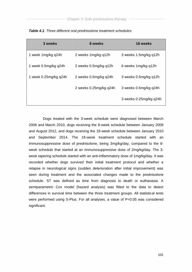
Chapter 3: Sole prednisolone therapy
101
Table 4.1: Three different oral prednisolone treatment schedules
3 weeks 8 weeks 18 weeks
1 week 1mg/kg q24h 2 weeks 1mg/kg q12h 3 weeks 1.5mg/kg q12h
1 week 0.5mg/kg q24h 2 weeks 0.5mg/kg q12h 6 weeks 1mg/kg q12h
1 week 0.25mg/kg q24h 2 weeks 0.5mg/kg q24h 3 weeks 0.5mg/kg q12h
2 weeks 0.25mg/kg q24h 3 weeks 0.5mg/kg q24h
3 weeks 0.25mg/kg q24h
Dogs treated with the 3-week schedule were diagnosed between March
2006 and March 2010, dogs receiving the 8-week schedule between January 2009
and August 2012, and dogs receiving the 18-week schedule between January 2010
and September 2014. The 18-week treatment schedule started with an
immunosuppressive dose of prednisolone, being 3mg/kg/day, compared to the 8-
week schedule that started at an immunosuppressive dose of 2mg/kg/day. The 3-
week tapering schedule started with an anti-inflammatory dose of 1mg/kg/day. It was
recorded whether dogs survived their initial treatment protocol and whether a
relapse in neurological signs (sudden deterioration after initial improvement) was
seen during treatment and the associated changes made to the prednisolone
schedule. ST was defined as time from diagnosis to death or euthanasia. A
semiparametric Cox model (hazard analysis) was fitted to the data to detect
differences in survival time between the three treatment groups. All statistical tests
were performed using S-Plus. For all analyses, a value of P<0.05 was considered
significant.
Chapter 3: Sole prednisolone therapy
102
Results
Thirty-eight dogs met the inclusion criteria. Breeds represented included
Maltese terrier (n=10), Yorkshire terrier (n=3), Golden Retriever (n=3), Chihuahua
(n=3), French bulldog (n=3), Pug (n=2), Labrador retriever (n=2), West Highland
White terrier (n=2), Shih Tzu (n=2), Boston terrier (n=2), American Staffordshire
terrier (n=2) and 4 other individual breeds. Most common presenting neurologic
signs were abnormal behaviour (n=19), altered mentation (n=18) and central
vestibular signs (n=19). Brain imaging (CT and/or MRI) was available in all cases.
Thirteen dogs (34%) only underwent CT imaging (Figure 4.1), 24 dogs (63%) only
underwent MR imaging and 1 dog (3%) underwent both CT and MR imaging. No
lesion was visible in 7 dogs (18%), based on CT (n=4) or MR (n=3) imaging. As
required by the inclusion criteria, TNCC of the CSF was above reference limits (> 5
WBC/mm3 after cisternal collection) in all cases with counts ranging from 9-1189
WBC/mm3 (median: 55 WBC/mm3). An overview of the most important diagnostic
findings can be consulted in table 4.2.
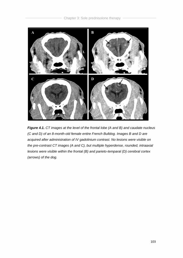
Chapter 3: Sole prednisolone therapy
103
Figure 4.1. CT images at the level of the frontal lobe (A and B) and caudate nucleus
(C and D) of an 8-month-old female entire French Bulldog. Images B and D are
acquired after administration of IV gadolinium contrast. No lesions were visible on
the pre-contrast CT images (A and C), but multiple hyperdense, rounded, intraaxial
lesions were visible within the frontal (B) and parieto-temparal (D) cerebral cortex
(arrows) of the dog.
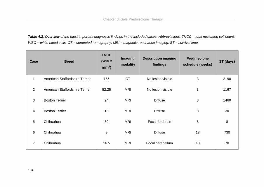
Chapter 3: Sole Prednisolone Therapy
104
Table 4.2: Overview of the most important diagnostic findings in the included cases. Abbreviations: TNCC = total nucleated cell count,
WBC = white blood cells, CT = computed tomography, MRI = magnetic resonance imaging, ST = survival time
Case Breed
TNCC
(WBC/
mm3)
Imaging
modality
Description imaging
findings
Prednisolone
schedule (weeks) ST (days)
1 American Staffordshire Terrier 165 CT No lesion visible 3 2190
2 American Staffordshire Terrier 52.25 MRI No lesion visible 3 1167
3 Boston Terrier 24 MRI Diffuse 8 1460
4 Boston Terrier 15 MRI Diffuse 8 30
5 Chihuahua 30 MRI Focal forebrain 8 8
6 Chihuahua 9 MRI Diffuse 18 730
7 Chihuahua 16.5 MRI Focal cerebellum 18 70
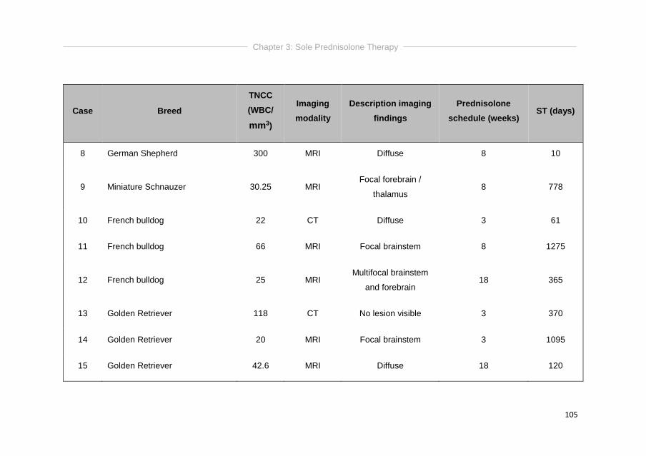
Chapter 3: Sole Prednisolone Therapy
105
Case Breed
TNCC
(WBC/
mm3)
Imaging
modality
Description imaging
findings
Prednisolone
schedule (weeks) ST (days)
8 German Shepherd 300 MRI Diffuse 8 10
9 Miniature Schnauzer 30.25 MRI Focal forebrain /
thalamus 8 778
10 French bulldog 22 CT Diffuse 3 61
11 French bulldog 66 MRI Focal brainstem 8 1275
12 French bulldog 25 MRI Multifocal brainstem
and forebrain 18 365
13 Golden Retriever 118 CT No lesion visible 3 370
14 Golden Retriever 20 MRI Focal brainstem 3 1095
15 Golden Retriever 42.6 MRI Diffuse 18 120
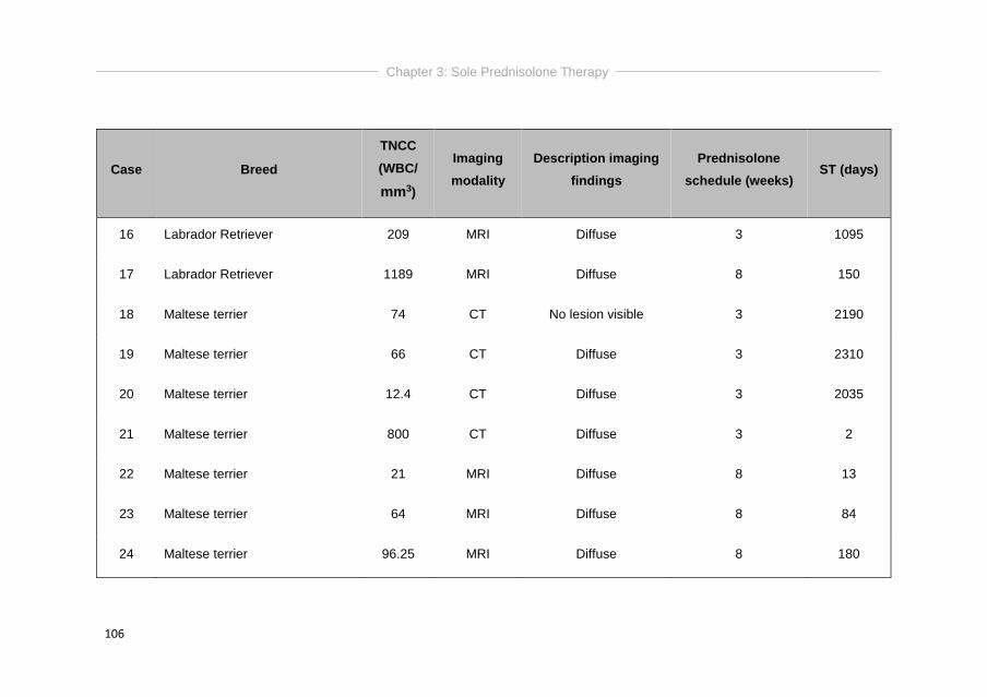
Chapter 3: Sole Prednisolone Therapy
106
Case Breed
TNCC
(WBC/
mm3)
Imaging
modality
Description imaging
findings
Prednisolone
schedule (weeks) ST (days)
16 Labrador Retriever 209 MRI Diffuse 3 1095
17 Labrador Retriever 1189 MRI Diffuse 8 150
18 Maltese terrier 74 CT No lesion visible 3 2190
19 Maltese terrier 66 CT Diffuse 3 2310
20 Maltese terrier 12.4 CT Diffuse 3 2035
21 Maltese terrier 800 CT Diffuse 3 2
22 Maltese terrier 21 MRI Diffuse 8 13
23 Maltese terrier 64 MRI Diffuse 8 84
24 Maltese terrier 96.25 MRI Diffuse 8 180
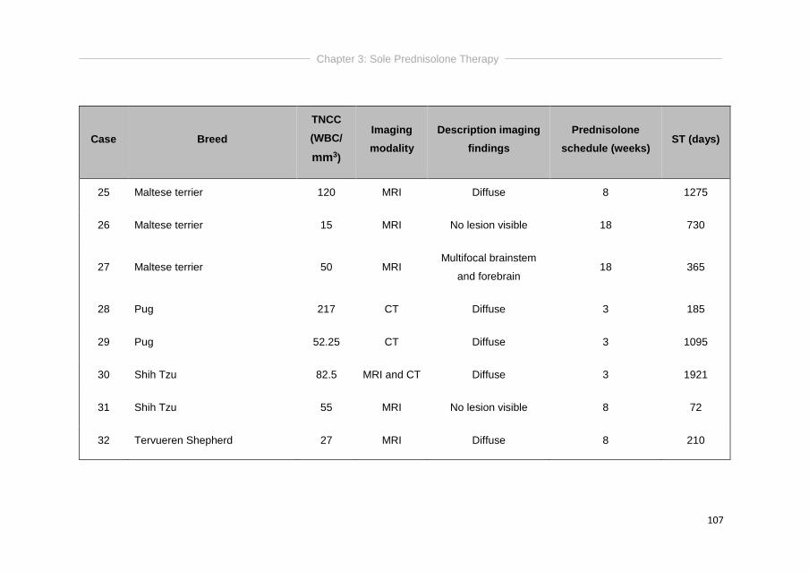
Chapter 3: Sole Prednisolone Therapy
107
Case Breed
TNCC
(WBC/
mm3)
Imaging
modality
Description imaging
findings
Prednisolone
schedule (weeks) ST (days)
25 Maltese terrier 120 MRI Diffuse 8 1275
26 Maltese terrier 15 MRI No lesion visible 18 730
27 Maltese terrier 50 MRI Multifocal brainstem
and forebrain 18 365
28 Pug 217 CT Diffuse 3 185
29 Pug 52.25 CT Diffuse 3 1095
30 Shih Tzu 82.5 MRI and CT Diffuse 3 1921
31 Shih Tzu 55 MRI No lesion visible 8 72
32 Tervueren Shepherd 27 MRI Diffuse 8 210
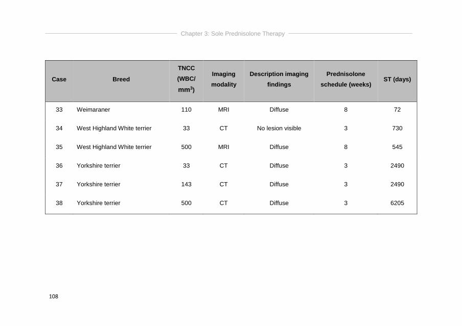
Chapter 3: Sole Prednisolone Therapy
108
Case Breed
TNCC
(WBC/
mm3)
Imaging
modality
Description imaging
findings
Prednisolone
schedule (weeks) ST (days)
33 Weimaraner 110 MRI Diffuse 8 72
34 West Highland White terrier 33 CT No lesion visible 3 730
35 West Highland White terrier 500 MRI Diffuse 8 545
36 Yorkshire terrier 33 CT Diffuse 3 2490
37 Yorkshire terrier 143 CT Diffuse 3 2490
38 Yorkshire terrier 500 CT Diffuse 3 6205
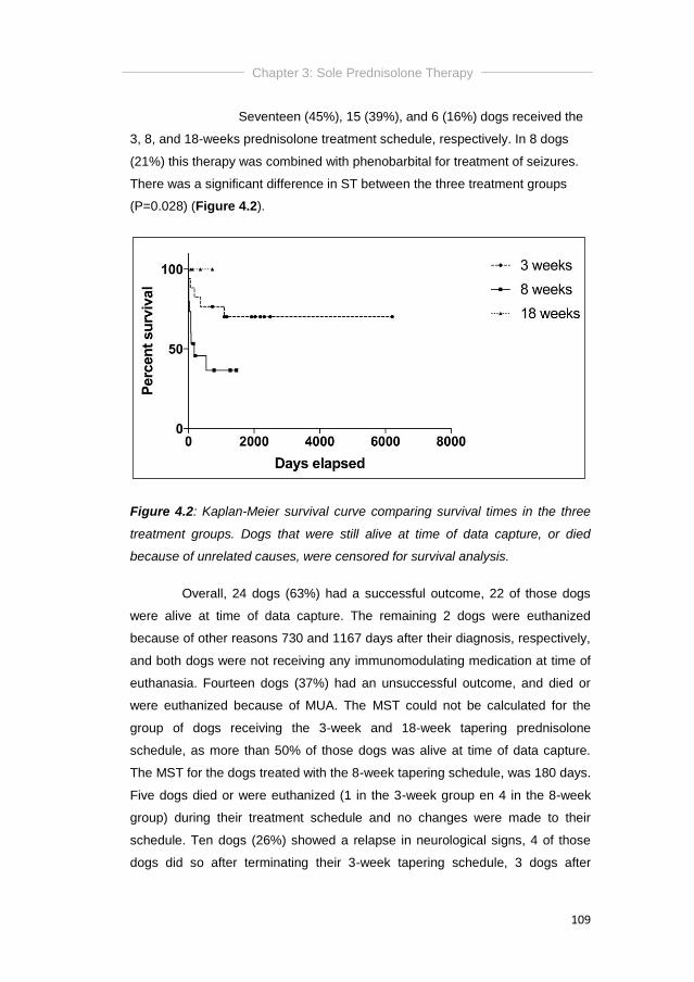
Chapter 3: Sole Prednisolone Therapy
109
Seventeen (45%), 15 (39%), and 6 (16%) dogs received the
3, 8, and 18-weeks prednisolone treatment schedule, respectively. In 8 dogs
(21%) this therapy was combined with phenobarbital for treatment of seizures.
There was a significant difference in ST between the three treatment groups
(P=0.028) (Figure 4.2).
Figure 4.2: Kaplan-Meier survival curve comparing survival times in the three
treatment groups. Dogs that were still alive at time of data capture, or died
because of unrelated causes, were censored for survival analysis.
Overall, 24 dogs (63%) had a successful outcome, 22 of those dogs
were alive at time of data capture. The remaining 2 dogs were euthanized
because of other reasons 730 and 1167 days after their diagnosis, respectively,
and both dogs were not receiving any immunomodulating medication at time of
euthanasia. Fourteen dogs (37%) had an unsuccessful outcome, and died or
were euthanized because of MUA. The MST could not be calculated for the
group of dogs receiving the 3-week and 18-week tapering prednisolone
schedule, as more than 50% of those dogs was alive at time of data capture.
The MST for the dogs treated with the 8-week tapering schedule, was 180 days.
Five dogs died or were euthanized (1 in the 3-week group en 4 in the 8-week
group) during their treatment schedule and no changes were made to their
schedule. Ten dogs (26%) showed a relapse in neurological signs, 4 of those
dogs did so after terminating their 3-week tapering schedule, 3 dogs after
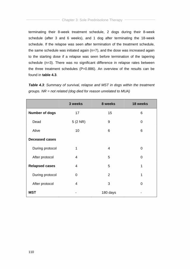
Chapter 3: Sole Prednisolone Therapy
110
terminating their 8-week treatment schedule, 2 dogs during their 8-week
schedule (after 3 and 6 weeks), and 1 dog after terminating the 18-week
schedule. If the relapse was seen after termination of the treatment schedule,
the same schedule was initiated again (n=7), and the dose was increased again
to the starting dose if a relapse was seen before termination of the tapering
schedule (n=3). There was no significant difference in relapse rates between
the three treatment schedules (P=0.886). An overview of the results can be
found in table 4.3.
Table 4.3: Summary of survival, relapse and MST in dogs within the treatment
groups. NR = not related (dog died for reason unrelated to MUA)
3 weeks 8 weeks 18 weeks
Number of dogs 17 15 6
Dead 5 (2 NR) 9 0
Alive 10 6 6
Deceased cases
During protocol 1 4 0
After protocol 4 5 0
Relapsed cases 4 5 1
During protocol 0 2 1
After protocol 4 3 0
MST - 180 days -
Chapter 3: Sole Prednisolone Therapy
111
Discussion
Thirty-eight dogs diagnosed with MUA received oral prednisolone
therapy in 3 different tapering schedules. Overall, 37% of dogs were euthanized
because of MUA, and survival curves for the three treatment schedules were
significantly different. Surprisingly, the highest mortality rate was seen in the 8-
week (immunosuppressive) treatment group (56%), followed by the 3-week
(anti-inflammatory) (28%) and the 18-week (immunosuppressive) treatment
(0%) schedule. Possible explanations might be that (1) all dogs at the institution
historically received the 3-weeks treatment protocol when a suspicion of non-
infectious encephalitis was made. However, as new literature became available
on a possibly immune-mediated origin, dogs diagnosed with MUA admitted with
severe neurological signs may have received a longer, more
immunosuppressive (8-weeks) treatment schedule and (2) the 18-week tapering
schedule was only introduced in the last 4 years of inclusion, so the recently
included cases might still decease in the following weeks to months, now falsely
(positively) influencing the results.
In literature, the MST in dogs diagnosed with MUA and receiving sole
prednisolone therapy ranges from 28-357 days (Granger et al., 2010), 91-329
days (Flegel et al., 2011) and 602 days (Mercier and Barnes Heller, 2015). As
more than 50% of the dogs was alive or censored for outcome calculations at
time of data capture, no overall MST could be calculated in the presented study.
However, the MST was 180 days in the 8-week treatment group, which appears
to be the group with the highest percentage of deceased and relapsed dogs.
Further prospective studies should be performed, including more dogs receiving
the more immunosuppressive treatment schedules, although one can have
ethical problems with comparing an immunosuppressive and anti-inflammatory
treatment protocol for a presumed immune-mediated disease.
Pitfalls of the current study are the lack of histopathological
confirmation in all cases, the low number of cases and the relatively high
percentage of dogs that was diagnosed using CT imaging (34%). However 18%
Chapter 3: Sole Prednisolone Therapy
112
of dogs had no visible lesion on CT or MR imaging which is comparable to the
7% for MRI and 14% for CT described previously (Granger et al., 2010).
Prednisolone therapy is associated with common side effects, and to overcome
these, other immunosuppressive drugs can be added to the protocol. Looking at
the side effects of the prednisolone treatment was beyond the scope of this
article, as not all side effects were systematically recorded and these are
difficult to trace back through telephone contact with the owner or referring
veterinarian.
To conclude, overall prognosis for dogs diagnosed with MUA and
treated with sole prednisolone therapy is guarded. Almost 40% of dogs will
succumb due to the disease. A long and immunosuppressive treatment
schedule is advised, but larger studies are needed to support this.
Chapter 4
SOLE PREDNISOLONE THERAPY VERSUS
COMBINATION THERAPY WITH CICLOSPORINE IN
DOGS WITH MENINGOENCEPHALOMYELITIS OF
UNKNOWN AETIOLOGY
Ine Cornelisa, Ingrid Gielenb, Luc Van Hama, Valentine Martléa, Kenny
Bossensa, Bart Broeckxc, Emilie Royauxa, Kimberley Steea, Sofie Bhattia
aSmall Animal Department, Faculty of Veterinary Medicine, Ghent
University, Merelbeke, Belgium.
bDepartment of Veterinary Medical Imaging and Small Animal
Orthopaedics, Ghent University, Merelbeke, Belgium.
cDepartment of Nutrition, Genetics and Ethology, Ghent University,
Merelbeke, Belgium.
Adapted from: Cornelis I, Gielen I, Van Ham L, Martlé V, Bossens K,
Royaux E, Stee K, Bhatti S. Sole prednisolone therapy versus
combination therapy with ciclosporine in dogs with
meningoencephalomyelitis of unknown aetiology: investigation of
prognostic factors and outcome. In preparation for submission.
Chapter 4: Prednisolone and Ciclosporine
114
Abstract
Although ciclosporine has previously been investigated as sole or add-
on treatment for dogs diagnosed with MUA, no specific treatment protocols
have been defined. The aims of this study were 1) to compare sole
prednisolone therapy with combination therapy with ciclosporine, 2) to describe
two clinically useful treatment protocols, and 3) to evaluate previously
investigated prognostic factors.
A prospective, randomized clinical study including client-owned dogs
was performed. Dogs were clinically diagnosed with MUA using previously
described inclusion criteria. Dogs were re-admitted 2, 4 and 6 months after
diagnosis for a complete neurological examination and analysis of cerebrospinal
fluid. Four months after diagnosis, MR imaging was repeated. Twelve dogs met
the inclusion criteria. Seven dogs were treated with a sole prednisolone
treatment schedule, and 5 dogs with combination therapy of prednisolone and
ciclosporine. A previously described 18-week tapering immunosuppressive
prednisolone treatment protocol was compared with combination of the same
protocol with ciclosporine. Treatment resulted in a MST of 87 and 567.5 days
for dogs treated with sole prednisolone and combination therapy, respectively,
but no difference in survival curves was found between both treatment groups.
Previously established prognostic factors including duration of clinical signs
prior to diagnosis, presentation within one week of onset of clinical signs,
presence of seizures or abnormal mentation at time of diagnosis, presence of
focal versus multifocal signs and presence of focal forebrain versus focal
brainstem signs at time of diagnosis, results of CSF analysis, and MRI findings
including mass effect, loss of cerebral sulci and foramen magnum herniation,
were not associated with outcome in the presented study.
Overall, 67% of dogs died because of MUA. Two clinically useful
treatment protocols were described, but no significant difference in MST could
be demonstrated comparing both protocols. Additionally, previously established
prognostic factors could not be confirmed.
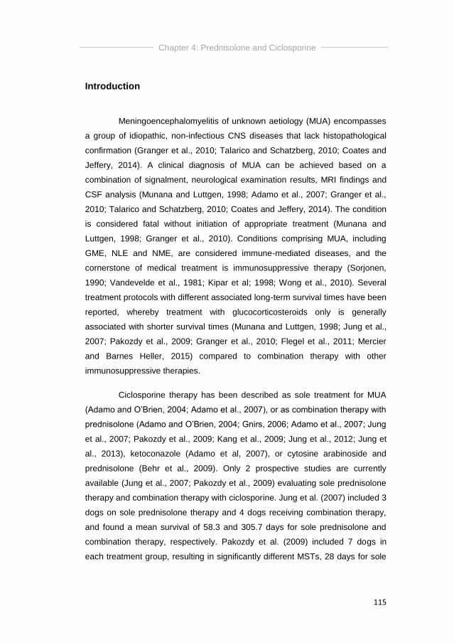
Chapter 4: Prednisolone and Ciclosporine
115
Introduction
Meningoencephalomyelitis of unknown aetiology (MUA) encompasses
a group of idiopathic, non-infectious CNS diseases that lack histopathological
confirmation (Granger et al., 2010; Talarico and Schatzberg, 2010; Coates and
Jeffery, 2014). A clinical diagnosis of MUA can be achieved based on a
combination of signalment, neurological examination results, MRI findings and
CSF analysis (Munana and Luttgen, 1998; Adamo et al., 2007; Granger et al.,
2010; Talarico and Schatzberg, 2010; Coates and Jeffery, 2014). The condition
is considered fatal without initiation of appropriate treatment (Munana and
Luttgen, 1998; Granger et al., 2010). Conditions comprising MUA, including
GME, NLE and NME, are considered immune-mediated diseases, and the
cornerstone of medical treatment is immunosuppressive therapy (Sorjonen,
1990; Vandevelde et al., 1981; Kipar et al; 1998; Wong et al., 2010). Several
treatment protocols with different associated long-term survival times have been
reported, whereby treatment with glucocorticosteroids only is generally
associated with shorter survival times (Munana and Luttgen, 1998; Jung et al.,
2007; Pakozdy et al., 2009; Granger et al., 2010; Flegel et al., 2011; Mercier
and Barnes Heller, 2015) compared to combination therapy with other
immunosuppressive therapies.
Ciclosporine therapy has been described as sole treatment for MUA
(Adamo and O’Brien, 2004; Adamo et al., 2007), or as combination therapy with
prednisolone (Adamo and O’Brien, 2004; Gnirs, 2006; Adamo et al., 2007; Jung
et al., 2007; Pakozdy et al., 2009; Kang et al., 2009; Jung et al., 2012; Jung et
al., 2013), ketoconazole (Adamo et al, 2007), or cytosine arabinoside and
prednisolone (Behr et al., 2009). Only 2 prospective studies are currently
available (Jung et al., 2007; Pakozdy et al., 2009) evaluating sole prednisolone
therapy and combination therapy with ciclosporine. Jung et al. (2007) included 3
dogs on sole prednisolone therapy and 4 dogs receiving combination therapy,
and found a mean survival of 58.3 and 305.7 days for sole prednisolone and
combination therapy, respectively. Pakozdy et al. (2009) included 7 dogs in
each treatment group, resulting in significantly different MSTs, 28 days for sole
Chapter 4: Prednisolone and Ciclosporine
116
prednisolone therapy and 620 days for combination therapy. No clinically
applicable treatment schedule has been described in both studies.
Because MUA generally has a guarded prognosis, multiple studies
attempted to identify prognostic factors, often resulting in conflicting results.
Currently, the following established prognostic factors for dogs with MUA are
available in the literature: 1) younger age at time of diagnosis was significantly
associated with improved survival (Oliphant et al., 2016), 2) presence of
seizures or altered mentation were significantly associated with shorter survival
(Bateman and Parent, 1999; Coates et al., 2007; Granger et al., 2010); 3)
presentation within 7 days of onset of clinical signs was significantly associated
with longer survival (Barnoon et al., 2015); 4) abnormal serial CSF analysis was
significantly associated with relapse and poor outcome (Lowrie et al., 2013);
and 5) mass effect, loss of cerebral sulci and foramen magnum herniation on
MRI were all significantly associated with death within 3 months (Lowrie et al.,
2013).
The aims of this study were 1) to compare sole prednisolone therapy
with combination therapy with ciclosporine, 2) to describe two clinically useful
treatment protocols, and 3) to investigate possible prognostic factors including
clinical factors and factors identified on serial MRI and CSF analysis. We
hypothesized that combination therapy with ciclosporine, and
resolution/improvement of MRI lesions and/or CSF abnormalities would lead to
an improved survival and a decreased risk for relapse.
Materials and methods
Case selection
A prospective recruitment of dogs diagnosed clinically with MUA was
performed at Ghent University between January 2014 and September 2015.
Dogs were included based on the criteria used by Granger et al. (2010), if they
had (1) a complete neurological examination performed leading to a focal or
multifocal intracranial neuroanatomical localisation, (2) MR imaging of the brain
Chapter 4: Prednisolone and Ciclosporine
117
demonstrating single, multiple or diffuse intra-axial hyperintense lesions on
FLAIR and T2WI, (3) cisternal CSF examination results, and if (4) infectious
disease testing returned negative. The study was approved by the Ethical
Committee of Ghent University (EC 2013/103) and all owners signed an
informed consent form before inclusion. Parameters recorded included
signalment, duration of clinical signs prior to diagnosis, neurological
examination findings, MRI of the brain, CSF analysis, treatment schedule used,
presence of relapses and long-term outcome. Neurological examination on time
of admission and 2, 4 and 6 months after diagnosis, was performed by a board
certified neurologist or a neurology resident. On the neurological examination,
mentation was divided into bright alert responsive (BAR), quiet alert responsive
(QAR), obtundation, stupor and coma, representing decreasing mental status in
this order. Possible neuroanatomical localisations included forebrain, brainstem
or cerebellum. Dogs with vestibular signs attributable to a brainstem-associated
lesion were diagnosed with central vestibular disease. If 2 or more of the above
mentioned neuroanatomical regions appeared to be affected on the
neurological examination, dogs were given a multifocal neuroanatomical
localisation, where dogs with only one region affected were given a focal
neuroanatomical localisation. Long-term outcome data were collected through
revision of medical records or by contacting the owner or referring veterinarian
by email or telephone in September 2016.
Diagnostic investigations
MRI was performed under general anaesthesia with a permanent 0.2T
magnet (Airis Mate, Hitachi Ltd, Tokyo, Japan) and included a minimum of T2W
and T1W images in a sagittal and transverse plane, and transverse FLAIR
images of the entire brain. T1WI were acquired before and after administration
of paramagnetic contrast (Dotarem, Guerbet, Brussels, Belgium). Variables
recorded were presence of mass effect (brain herniation, midline shift, flattening
of gyri/sulci). MR imaging was repeated 4 months after diagnosis following the
same protocol, and images were reviewed for evolution of the previously visible
lesion(s) (improved, static, or deteriorated).
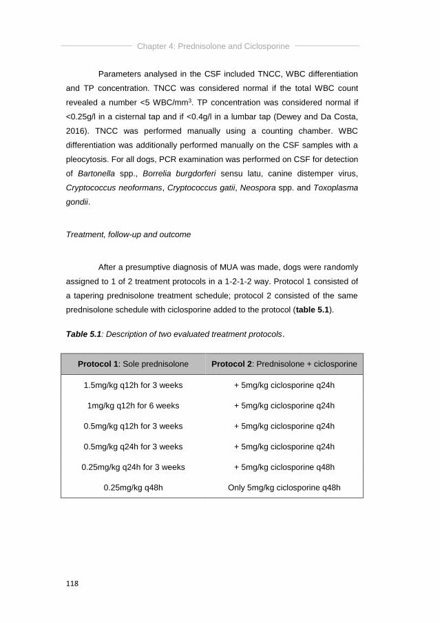
Chapter 4: Prednisolone and Ciclosporine
118
Parameters analysed in the CSF included TNCC, WBC differentiation
and TP concentration. TNCC was considered normal if the total WBC count
revealed a number <5 WBC/mm3. TP concentration was considered normal if
<0.25g/l in a cisternal tap and if <0.4g/l in a lumbar tap (Dewey and Da Costa,
2016). TNCC was performed manually using a counting chamber. WBC
differentiation was additionally performed manually on the CSF samples with a
pleocytosis. For all dogs, PCR examination was performed on CSF for detection
of Bartonella spp., Borrelia burgdorferi sensu latu, canine distemper virus,
Cryptococcus neoformans, Cryptococcus gatii, Neospora spp. and Toxoplasma
gondii.
Treatment, follow-up and outcome
After a presumptive diagnosis of MUA was made, dogs were randomly
assigned to 1 of 2 treatment protocols in a 1-2-1-2 way. Protocol 1 consisted of
a tapering prednisolone treatment schedule; protocol 2 consisted of the same
prednisolone schedule with ciclosporine added to the protocol (table 5.1).
Table 5.1: Description of two evaluated treatment protocols.
Protocol 1: Sole prednisolone Protocol 2: Prednisolone + ciclosporine
1.5mg/kg q12h for 3 weeks + 5mg/kg ciclosporine q24h
1mg/kg q12h for 6 weeks + 5mg/kg ciclosporine q24h
0.5mg/kg q12h for 3 weeks + 5mg/kg ciclosporine q24h
0.5mg/kg q24h for 3 weeks + 5mg/kg ciclosporine q24h
0.25mg/kg q24h for 3 weeks + 5mg/kg ciclosporine q48h
0.25mg/kg q48h Only 5mg/kg ciclosporine q48h
Chapter 4: Prednisolone and Ciclosporine
119
Neurological examination and CSF analysis were repeated 2, 4 and 6
months after diagnosis, MRI of the brain 4 months after diagnosis. A relapse
was defined as a sudden deterioration after initial improvement, mainly
consisting of recurrence of the initial or additional neurological signs. If a
relapse was noted clinically, the prednisolone schedule was restarted as a
rescue protocol. Follow-up information was retrieved from the clinical program
or through telephone contact with the owner or referring veterinarian in
September 2016.
Statistical Analysis
Data analysis was performed with the aid of a standard statistical
software package (Prism, Graphpad Software Inc, La Jolla, California, USA). A
Mann-Whitney U test was used to compare age, duration of clinical signs prior
to diagnosis, TNCC and TP concentration in CSF with long-term outcome. A
fisher’s exact test was used to compare differences in long-term outcome for
presence of seizures and abnormal mentation, neuroanatomical localisation
(focal versus multifocal, and focal forebrain versus focal brainstem) and imaging
findings (mass effect, foramen magnum herniation and loss of cerebral sulci).
Numeric variables were expressed as median and IQR. Values of P<0.05 were
considered significant.
Results
Signalment and clinical presentation
Fifteen dogs were included in the study. However, three dogs had to
be excluded because of a necropsy diagnosis of intracranial lymphoma (n=2) or
because of lack of owner compliance to the treatment plan (n=1). For the
remaining 12 dogs, breeds included Chihuahua (n=4), Pug dog (n=3), Yorkshire
Terrier (n=2), and 1 each of the following breeds: Lhasa Apso, Shih Tzu and
Maltese terrier. Nine dogs (75%) were female (of which 3 were neutered) and 3
dogs (25%) were entire males. Median weight at time of diagnosis was 4.45kg
Chapter 4: Prednisolone and Ciclosporine
120
(ranging from 1.75 tot 8.6kg). Median age at time of diagnosis was 28 months
(ranging from 11 to 78 months). Median duration of clinical signs prior to
diagnosis was 14 days (ranging from 1 to 30 days).
Diagnostic investigations at time of diagnosis
On neurological examination, 2 dogs (17%) were BAR, 3 dogs (25%)
were QAR, 6 dogs (50%) were obtunded and 1 dog (8%) was stuporous. Four
dogs (33%) had generalized tonic-clonic epileptic seizures of which 1 dog (25%)
presented with cluster seizures. Nine dogs (75%) were diagnosed with a focal
and 3 dogs (25%) with a multifocal neuroanatomical localisation. Of the dogs
with the focal localisation, 4 dogs (44%) presented with a central vestibular
lesion and 4 dogs (44%) with a focal forebrain lesion.
As required by the inclusion criteria, MRI of the brain was available for
all dogs, revealing multifocal or diffuse lesions affecting the forebrain, brainstem
and/or cerebellum. Mass effect was noted in 11 dogs (92%), including loss of
cerebral sulci in 7 dogs (64%) and foramen magnum herniation in 4 dogs (36%)
(Figure 5.1).
Cisternal CSF was obtained in all dogs at time of diagnosis, revealing
a normal TNCC in one dog (8%), and an increased TNCC in the remaining
eleven dogs (92%), with a median of 283 WBC/mm3 (ranging from 0-1413.5
WBC/mm3). Blood contamination was present in two dogs (16%). Manual
differentiation mainly revealed a mononuclear pleocytosis. Protein concentration
was measured in all samples, and was elevated in all but 1 sample. The median
TP concentration was 0.92g/l (ranging from 0.22-17.20g/l). As required by the
inclusion criteria, infectious disease testing was negative in all CSF samples. An
overview of the clinical and serial CSF findings can be consulted in table 5.2.
Diagnostic investigation during treatment
Seven dogs (58%) were alive at the 2-month re-examination, and 5
dogs (42%) at the 4-month and 6-month re-examination.
Chapter 4: Prednisolone and Ciclosporine
121
In the 7 dogs that were alive at the 2-month re-examination, the
neurological examination revealed no abnormalities in 4 dogs (57%), residual
central vestibular signs in 2 dogs (29%) initially diagnosed with a multifocal
lesion, and a mild residual head tilt in 1 dog (14%) initially diagnosed with a
central vestibular lesion.
In 5 dogs (42%), follow-up MRI 4 months after diagnosis was available.
This revealed an improvement of the lesions in 4 dogs (80%), and resolution of
all visible lesions in 1 dog (20%) (Figure 5.2).
Follow-up CSF analysis was performed after 2 months in 7 dogs, and
after 4 and 6 months in 5 dogs. Results of serial neurological examinations,
CSF analysis and imaging findings can be consulted in table 5.2.
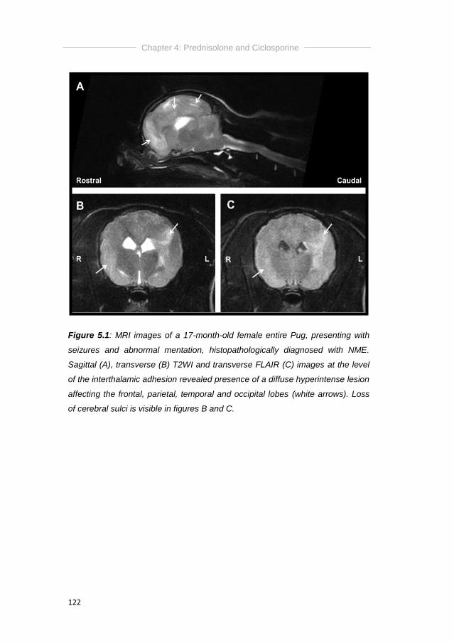
Chapter 4: Prednisolone and Ciclosporine
122
Figure 5.1: MRI images of a 17-month-old female entire Pug, presenting with
seizures and abnormal mentation, histopathologically diagnosed with NME.
Sagittal (A), transverse (B) T2WI and transverse FLAIR (C) images at the level
of the interthalamic adhesion revealed presence of a diffuse hyperintense lesion
affecting the frontal, parietal, temporal and occipital lobes (white arrows). Loss
of cerebral sulci is visible in figures B and C.
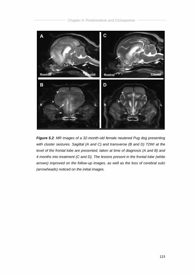
Chapter 4: Prednisolone and Ciclosporine
123
Figure 5.2: MR images of a 32-month-old female neutered Pug dog presenting
with cluster seizures. Sagittal (A and C) and transverse (B and D) T2WI at the
level of the frontal lobe are presented, taken at time of diagnosis (A and B) and
4 months into treatment (C and D). The lesions present in the frontal lobe (white
arrows) improved on the follow-up images, as well as the loss of cerebral sulci
(arrowheads) noticed on the initial images.
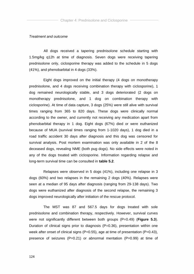
Chapter 4: Prednisolone and Ciclosporine
124
Treatment and outcome
All dogs received a tapering prednisolone schedule starting with
1.5mg/kg q12h at time of diagnosis. Seven dogs were receiving tapering
prednisolone only, ciclosporine therapy was added to the schedule in 5 dogs
(41%), and phenobarbital in 4 dogs (33%).
Eight dogs improved on the initial therapy (4 dogs on monotherapy
prednisolone, and 4 dogs receiving combination therapy with ciclosporine), 1
dog remained neurologically stable, and 3 dogs deteriorated (2 dogs on
monotherapy prednisolone, and 1 dog on combination therapy with
ciclosporine). At time of data capture, 3 dogs (25%) were still alive with survival
times ranging from 365 to 820 days. These dogs were clinically normal
according to the owner, and currently not receiving any medication apart from
phenobarbital therapy in 1 dog. Eight dogs (67%) died or were euthanized
because of MUA (survival times ranging from 1-1020 days), 1 dog died in a
road traffic accident 30 days after diagnosis and this dog was censored for
survival analysis. Post mortem examination was only available in 2 of the 8
deceased dogs, revealing NME (both pug dogs). No side effects were noted in
any of the dogs treated with ciclosporine. Information regarding relapse and
long-term survival time can be consulted in table 5.2.
Relapses were observed in 5 dogs (41%), including one relapse in 3
dogs (60%) and two relapses in the remaining 2 dogs (40%). Relapses were
seen at a median of 95 days after diagnosis (ranging from 29-138 days). Two
dogs were euthanized after diagnosis of the second relapse, the remaining 3
dogs improved neurologically after initiation of the rescue protocol.
The MST was 87 and 567.5 days for dogs treated with sole
prednisolone and combination therapy, respectively. However, survival curves
were not significantly different between both groups (P=0.49) (Figure 5.3).
Duration of clinical signs prior to diagnosis (P=0.36), presentation within one
week after onset of clinical signs (P=0.55), age at time of presentation (P=0.43),
presence of seizures (P=0.21) or abnormal mentation (P=0.99) at time of
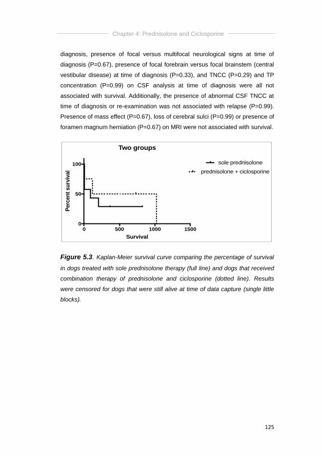
Chapter 4: Prednisolone and Ciclosporine
125
diagnosis, presence of focal versus multifocal neurological signs at time of
diagnosis (P=0.67), presence of focal forebrain versus focal brainstem (central
vestibular disease) at time of diagnosis (P=0.33), and TNCC (P=0.29) and TP
concentration (P=0.99) on CSF analysis at time of diagnosis were all not
associated with survival. Additionally, the presence of abnormal CSF TNCC at
time of diagnosis or re-examination was not associated with relapse (P=0.99).
Presence of mass effect (P=0.67), loss of cerebral sulci (P=0.99) or presence of
foramen magnum herniation (P=0.67) on MRI were not associated with survival.
Figure 5.3. Kaplan-Meier survival curve comparing the percentage of survival
in dogs treated with sole prednisolone therapy (full line) and dogs that received
combination therapy of prednisolone and ciclosporine (dotted line). Results
were censored for dogs that were still alive at time of data capture (single little
blocks).
0 500 1000 15000
50
100
Two groups
Survival
Perc
en
t su
rviv
al
sole prednisolone
prednisolone + ciclosporine
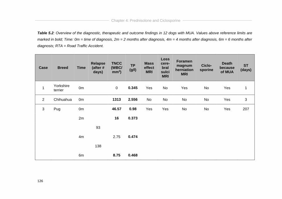
Chapter 4: Prednisolone and Ciclosporine
126
Table 5.2: Overview of the diagnostic, therapeutic and outcome findings in 12 dogs with MUA. Values above reference limits are
marked in bold; Time: 0m = time of diagnosis, 2m = 2 months after diagnosis, 4m = 4 months after diagnosis, 6m = 6 months after
diagnosis; RTA = Road Traffic Accident.
Case Breed Time Relapse (after # days)
TNCC (WBC/ mm3)
TP (g/l)
Mass effect MRI
Loss cere-bral sulci MRI
Foramen magnum
herniation MRI
Ciclo-sporine
Death because of MUA
ST (days)
1 Yorkshire terrier
0m
0 0.345 Yes No Yes No Yes 1
2 Chihuahua 0m
1313 2.556 No No No No Yes 3
3 Pug 0m
46.57 0.98 Yes Yes No No Yes 207
2m
16 0.373
93
4m
2.75 0.474
138
6m
8.75 0.468
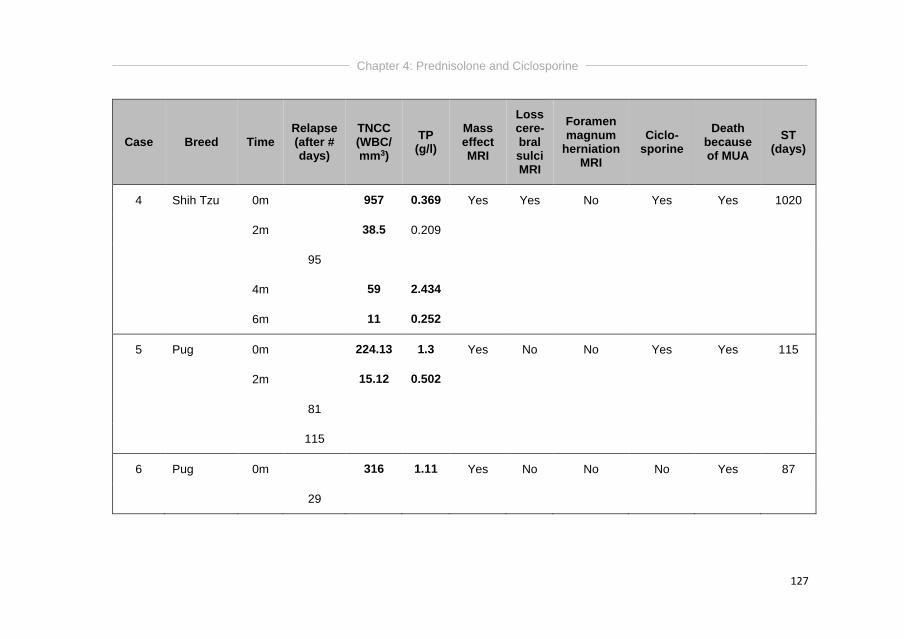
Chapter 4: Prednisolone and Ciclosporine
127
Case Breed Time Relapse (after # days)
TNCC (WBC/ mm3)
TP (g/l)
Mass effect MRI
Loss cere-bral sulci MRI
Foramen magnum
herniation MRI
Ciclo-sporine
Death because of MUA
ST (days)
4 Shih Tzu 0m
957 0.369 Yes Yes No Yes Yes 1020
2m
38.5 0.209
95
4m
59 2.434
6m
11 0.252
5 Pug 0m
224.13 1.3 Yes No No Yes Yes 115
2m
15.12 0.502
81
115
6 Pug 0m
316 1.11 Yes No No No Yes 87
29
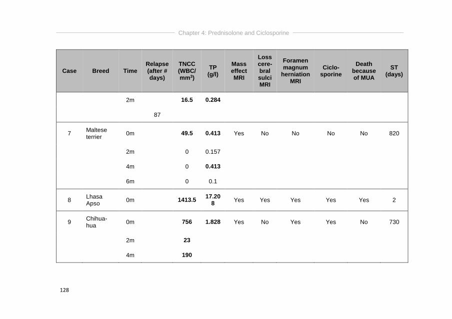
Chapter 4: Prednisolone and Ciclosporine
128
Case Breed Time Relapse (after # days)
TNCC (WBC/ mm3)
TP (g/l)
Mass effect MRI
Loss cere-bral sulci MRI
Foramen magnum
herniation MRI
Ciclo-sporine
Death because of MUA
ST (days)
2m
16.5 0.284
87
7 Maltese terrier
0m
49.5 0.413 Yes No No No No 820
2m
0 0.157
4m
0 0.413
6m
0 0.1
8 Lhasa Apso
0m
1413.5 17.20
8 Yes Yes Yes Yes Yes 2
9 Chihua-hua
0m
756 1.828 Yes No Yes Yes No 730
2m
23
4m
190
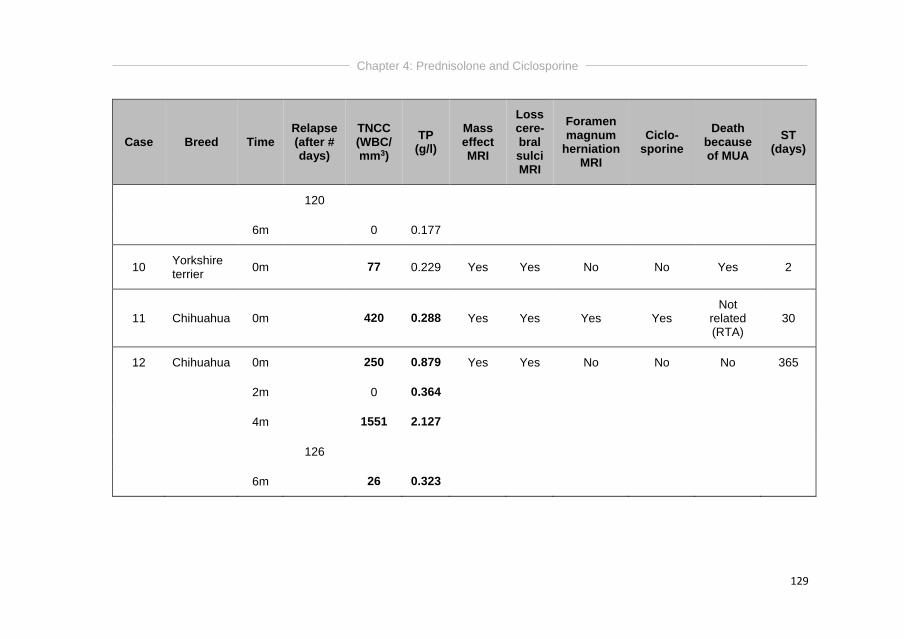
Chapter 4: Prednisolone and Ciclosporine
129
Case Breed Time Relapse (after # days)
TNCC (WBC/ mm3)
TP (g/l)
Mass effect MRI
Loss cere-bral sulci MRI
Foramen magnum
herniation MRI
Ciclo-sporine
Death because of MUA
ST (days)
120
6m
0 0.177
10 Yorkshire terrier
0m
77 0.229 Yes Yes No No Yes 2
11 Chihuahua 0m
420 0.288 Yes Yes Yes Yes Not
related (RTA)
30
12 Chihuahua 0m
250 0.879 Yes Yes No No No 365
2m
0 0.364
4m
1551 2.127
126
6m
26 0.323
Chapter 4: Prednisolone and Ciclosporine
130
Discussion
This study evaluated and provided two treatment regimens for dogs
with MUA, and evaluated possible prognostic factors for survival. Although it
was hypothesized that combination therapy would result in improved survival
compared to sole prednisolone therapy, no significant difference in MST could
be detected, most likely caused by the low case number. Additionally,
previously established prognostic factors could not be confirmed in the present
study.
Ciclosporine therapy has previously been described as sole treatment
for MUA (Adamo and O’Brien, 2004; Adamo et al., 2007), or as combination
therapy with prednisolone (Adamo and O’Brien, 2004; Gnirs, 2006; Adamo et
al., 2007; Jung et al., 2007; Pakozdy et al., 2009; Kang et al., 2009; Jung et al.,
2012; Jung et al., 2013), ketoconazole (Adamo et al, 2007), or cytosine
arabinoside and prednisolone (Behr et al., 2009). Twenty-six dogs have been
reported receiving prednisolone doses ranging from 2-30mg/kg/d combined with
ciclosporine doses ranging from 6-30mg/kg/d (Adamo and O’Brien, 2004; Gnirs,
2006; Adamo et al., 2007; Jung et al., 2007; Pakozdy et al., 2009; Kang et al.,
2009; Granger et al., 2010; Jung et al., 2012; Jung et al., 2013). In the
presented study, a more immunosuppressive prednisolone schedule as
previously described by Talarico and Schatzberg (2010) was used, combined
with 5mg/kg/d of ciclosporine. This is the approved dose for atopic dermatitis in
dogs (a common pruritic dermatologic problem affecting dogs, associated with
IgE antibodies targeting environmental antigens). The same dose was advised
in dogs with pemphigus foliaceus, an autoimmune skin disease. It was however
anecdotally reported that dogs receiving this lower dosages of ciclosporine
(≤5mg/kg q24h) can develop severe secondary infections (Archer et al., 2014).
On the contrary, higher doses of ciclosporine (5-10mg/kg q12h) have been
advised in dogs with acute and possibly life-threatening auto-immune diseases,
including immune-mediated thrombocytopenia and immune-mediated
haemolytic anaemia, to achieve clinically relevant depression of T-lymphocytes
and as such immunosuppression (Archer et al., 2014). Therefore, we wanted to
investigate whether this lower dose of ciclosporine combined with an
Chapter 4: Prednisolone and Ciclosporine
131
immunosuppressive schedule of prednisolone would be of additional value in
dogs with MUA, which we could not prove in the presented study.
The discrepancy in number of dogs included in both treatment groups
(7 dogs were treated with sole prednisolone therapy and 5 dogs with
combination therapy with ciclosporine) is mainly caused by the 3 cases that
were initially included in the study and that were mainly addressed to the
combination therapy group. After exclusion, there was no equal randomization
between groups.
None of the previously established prognostic factors for dogs with
MUA, including age and presence of seizures or altered mentation at time of
diagnosis, presentation within 7 days after onset of clinical signs, abnormal CSF
analysis at time of diagnosis, and presence of mass effect, foramen magnum
herniation or loss of cerebral sulci on MRI, could be confirmed in the presented
study. Munana and Luttgen (1998) found significant longer survival times for
dogs with focal neurological signs versus dogs with multifocal neurological
signs. Additionally, dogs with focal forebrain signs had a significantly longer
survival time compared to dogs with focal signs related to other areas of the
CNS. This finding was not repeated in more recent studies (Coates et al., 2007;
Lowrie et al., 2013) nor in the presented study.
One study identified a lower CSF TNCC to be significantly associated
with improved survival in dogs with MUA (Oliphant et al., 2016), whilst others
found that neither CSF TNCC nor TP concentration had an effect on survival
time in dogs with MUA (Coates et al., 2007). The study of Lowrie et al. (2013)
failed to demonstrate an association between normal CSF analysis and
improved outcome, but did find an association between abnormal CSF analysis
at three months and relapse or poor outcome (Lowrie et al., 2013). In the study
of Mercier and Barnes Heller (2015) CSF analysis was repeated 1 month after
diagnosis, and their results suggested that serial CSF analysis might be a valid
tool for monitoring success or failure of treatment in dogs diagnosed with MUA
and treated with glucocorticoid monotherapy. In the presented study however,
TNCC and TP of CSF were not predictive for outcome or relapse.
Chapter 4: Prednisolone and Ciclosporine
132
Overall, 26 dogs have been previously reported receiving initial doses
of ciclosporine ranging from 3-15mg/kg PO every 12 hours, resulting in MSTs
ranging from 236 to 930 days (Adamo and O’Brien, 2004; Gnirs, 2006; Adamo
et al., 2007; Jung et al., 2007; Pakozdy et al., 2009; Kang et al., 2009; Jung et
al., 2013). One dog receiving combination therapy survived for 1096 days (Jung
et al., 2012). In the presented study, the MST was 87 and 567.5 days for dogs
treated with sole prednisolone and combination therapy, respectively, however
survival curves were not significantly different between both groups. Overall,
only 2 previous studies revealed a mean ST of 305 days (Jung et al., 2007) or a
MST of 602 days (Pakozdy et al., 2009) for dogs treated with ciclosporine and
prednisolone, which is comparable to the MST of 567.5 days calculated in the
presented study. It is however important to state that both studies are
performed with a comparable small number of dogs, and a similar ciclosporine
dose (6mg/kg/day for the study of Pakozdy et al. (2009) and 5mg/kg/d for the
study of Jung et al. (2007)). On the contrary, the initial doses of steroids used in
these studies differ from the initial dose used in the presented study (3mg/kg/d),
being 5-30mg/kg/d and 2mg/kg/d, respectively. Survival times on combination
therapy in the presented study ranged from 2 – 1020 days, which is comparable
to the 2 – 1096 days reported in literature (Adamo et al., 2007; Kang et al.,
2009; Jung et al., 2012; Jung et al., 2013).
Results of the presented study should however be interpreted with
caution, mainly because of the small sample size and the limited number of
dogs that was alive at time of data capture (1 of the 4 dogs alive was censored
for survival analysis). Definitive histopathology is lacking in all but 2 cases,
revealing NME. Interestingly, all dogs included in the study can be considered
breeds that are predisposed to develop NE including Pug, Yorkshire Terrier,
Maltese, Chihuahua, Pekingese, Papillon, Shih Tzu, Coton de Tulear and
Brussels Griffon (Talarico and Schatzberg, 2010; Cooper and others, 2014). It is
however stated that dogs of any breed and age can be affected by all subtypes
of MUA (Granger et al., 2010; Coates and Jeffery, 2014). Additionally, one dog
with a normal TNCC was included in the study. Normally, a pleocytosis (>5
WBC/mm3) with monocytic predominancy is one of the inclusion criteria for
MUA in dogs. On the contrary, it is previously stated that CSF examination can
Chapter 4: Prednisolone and Ciclosporine
133
be normal in 3-57% of dogs with MUA (Menaut et al., 2009; Granger et al.,
2010), in 16% of dogs with GME and in 12.5% of dogs with NE (Granger et al.,
2010). Albuminocytological dissociation has been found in cases with a normal
cell count (Granger et al., 2010). The included dog was a 78-month-old male
neutered Yorkshire terrier with multifocal lesions mainly affecting the subcortical
white matter, and so a presumptive diagnosis of NLE was suspected and the
dog was included in the study.
A larger prospective, multi-centre study should be performed including
more dogs to establish the previously published prognostic factors and the here
described treatment protocol.
Conclusions
In the presented study, 67% of dogs died because of MUA with no
difference in outcome seen between dogs treated with sole prednisolone
therapy and combination therapy with ciclosporine. Overall, initiation of
treatment resulted in a MST of 115 days. Several described prognostic factors
to select for optimal treatment candidates and to manage owner expectance,
could not be confirmed. Further studies including more cases are warranted to
evaluate the efficacy of the two described treatment protocols.

Part III
Prognostic Factors and Short-term
Outcome

Chapter 5
PROGNOSTIC FACTORS FOR ONE-WEEK SURVIVAL
IN DOGS DIAGNOSED WITH
MENINGOENCEPHALOMYELITIS OF UNKNOWN
AETIOLOGY
Ine Cornelisa, Holger A. Volkb, Luc Van Hama, Steven De Deckerb
aSmall Animal Department, Faculty of Veterinary Medicine, Ghent
University, Merelbeke, Belgium.
bClinical Science and Services, The Royal Veterinary College,
University of London, Hatfield, United Kingdom.
Adapted from: Cornelis I, Volk HA, Van Ham L, De Decker S (2016).
Prognostic factors for one-week survival in dogs diagnosed with
meningoencephalomyelitis of unknown aetiology. The Veterinary
Journal 214, 91-95.
Chapter 5: one week survival
138
Abstract
Although several studies have evaluated the long-term outcome of
MUA in dogs, little is known about short-term survival and initial response to
therapy. The aim of this study was therefore to evaluate possible prognostic
factors for 7-day survival after diagnosis of MUA in dogs.
Medical records were reviewed for dogs diagnosed with MUA between
2006 and 2015. Previously described inclusion criteria were used, and for all
dogs 7-day survival data needed to be available. A poor outcome was defined
as death within 1 week. One hundred and sixteen dogs met the inclusion
criteria. Thirty dogs (26%) died within 7 days after making a presumptive
diagnosis of MUA. Assessed variables included age, sex, weight, duration of
clinical signs and treatment prior to diagnosis, venous blood glucose and lactate
levels, white blood cell count on complete blood count, total nucleated cell count
/ total protein concentration / white blood cell differentiation on cerebrospinal
fluid analysis, presence of seizures and cluster seizures, mentation at
presentation, neuroanatomical localisation, imaging findings and treatment after
diagnosis. Univariate statistical analysis was performed to identify variables to
be included in a multivariate model. Multivariate analysis identified three
variables significantly associated with poor outcome, including decreased
mentation at presentation, presence of seizures, and increased percentage of
neutrophils on cerebrospinal fluid analysis.
Despite initiation of appropriate treatment, more than a quarter of dogs
died within one week after making a presumptive diagnosis of MUA,
emphasizing the need for evaluation of short-term prognostic factors. The
information of this study can aid in the management of expectations of both
clinical staff and owners with dogs diagnosed with MUA.
Chapter 5: one week survival
139
Introduction
MUA describes all clinically diagnosed cases of GME, NME and NLE
that lack histopathological confirmation (Coates and Jeffery, 2014). A clinical
diagnosis can be achieved based on a combination of neurological examination
results, MRI findings and CSF abnormalities (Coates and Jeffery, 2014). The
exact aetiology and pathophysiology of MUA are currently unknown, but the
cornerstone of medical treatment is immunosuppressive therapy. Several
treatment protocols using different immunomodulating drugs, resulting in
different long-term survival times have been reported (Sisson et al. 1989;
Gregory et al., 1998; Munana and Luttgen, 1998; Adamo and O’Brien, 2004;
Gnirs, 2006; Zarfoss et al., 2006; Adamo et al., 2007; Coates et al., 2007; de
Stefani et al., 2007; Feliu-Pascual et al., 2007; Uriarte et al., 2007; Jung et al.,
2007; Menaut et al., 2008; Pakozdy et al., 2009; Smith et al., 2009; Granger et
al., 2010; Kang et al., 2009; Wong et al., 2010; Flegel et al., 2011; Jung et al.,
2012; Jung et al., 2013; Lowrie et al., 2013; Beckmann et al., 2015; Mercier and
Barnes Heller, 2015; Barnoon et al., 2015; Lowrie et al., 2016).
Although several studies have focused on long-term survival, little is
known about early survival and initial response to therapy of dogs diagnosed
with MUA. The primary aim of this study was therefore to evaluate early survival
and initial response to immunosuppressive therapy in those dogs. The
secondary aim of this study was to investigate possible prognostic factors for 7-
day survival after diagnosis of MUA. It was hypothesized that a substantial part
of dogs with MUA would succumb in the first week after diagnosis despite
appropriate treatment and monitoring. It was further hypothesized that specific
characteristics of the clinical presentation, neurological examination, clinical
pathology abnormalities, imaging findings and type of treatment would be
associated with 7-day survival in dogs with a presumptive diagnosis of MUA.
Chapter 5: one week survival
140
Materials and methods
Case selection
The electronic medical database of the Small Animal Referral Hospital,
Royal Veterinary College, University of London, was searched between January
2006 and April 2015 for dogs diagnosed with MUA. Dogs were included based
on the criteria used by Granger et al. (2010), if they had (1) complete medical
records available, (2) a complete neurological examination performed leading to
a focal or multifocal intracranial neuroanatomical localisation, (3) inflammatory
CSF analysis, (4) MR imaging of the brain demonstrating single, multiple or
diffuse intra-axial hyperintense lesions on T2W images, and if (5) 7-day follow-
up information was available. Dogs were excluded if (1) the clinical records or
imaging studies were incomplete or not available for review, (2) dogs were
diagnosed with meningomyelitis without clinical signs of intracranial
involvement, (3) no pleocytosis was found on CSF analysis with the exception
of dogs with signs of raised ICP on imaging studies, in which case CSF
collection was not performed, and if (4) positive infectious diseases titres were
found on serology or PCR examination for canine distemper virus, Toxoplasma
gondii or Neospora caninum. Dogs with histopathological confirmation of the
disease only needed to fulfil inclusion criteria (1) and (5). Information retrieved
from the medical records included breed, gender, age at diagnosis, sex, body
weight, results of neurological examination and neuroanatomical localisation,
duration of clinical signs and treatment prior to diagnosis, presence of
concurrent diseases, results of CBC and biochemistry profile, results of CSF
analysis including TNCC, WBC differentiation and TP concentration, lactate and
glucose concentration on venous blood gas analysis, treatment received and 7-
day survival time.
Dogs were divided in two groups based on their body weight: dogs
<15kg, in this paper referred to as small dogs; and dogs >15kg, in this paper
referred to as large dogs. Mentation was divided into bright alert responsive
(BAR), quiet alert responsive (QAR), obtundation, stupor and coma,
Chapter 5: one week survival
141
representing decreasing mental status in this order. Possible neuroanatomical
localisations included forebrain, brainstem or cerebellum. Dogs with vestibular
signs attributable to a brainstem-associated lesion were diagnosed with central
vestibular signs. If 2 or more of the above mentioned regions appeared to be
affected on the neurological examination, dogs were given a multifocal
neuroanatomical localisation, where dogs with only 1 region affected were given
a focal neuroanatomical localisation. MRI was performed under general
anaesthesia with a permanent 1.5T magnet (Intera, Philips Medical Systems,
Eindhoven, the Netherlands) and all images were reviewed by a board certified
neurologist (SDD) using Osirix Dicom viewer (Osirix Foundation, V.5.5.2
Geneva, Switzerland). The reviewer was blinded for the results of the
neurological examination, outcome after 7 days and histopathological findings.
Sequences could vary, but studies included a minimum of T2WI (TR/TE,
3000/120), T1WI (TR/TE, 400/8) and FLAIR images of the entire brain in a
sagittal, transverse and dorsal plane. The T1WI were acquired before and after
IV administration of paramagnetic contrast medium (0.1mg/kg, gadoterate
meglumine, Dotarem, Guerbet, Milton Keynes, UK). Variables recorded were
lesion localisation and distribution, presence of parenchymal or meningeal
contrast enhancement and presence of mass effect (brain herniation, midline
shift, flattening of gyri/sulci). For dogs that had CSF analysis performed, site of
collection (cisternal or lumbar), TNCC, TP and cytological differentiation were
recorded. Total nucleated cell count was considered normal if the TNCC <5
WBC/mm3. Total protein concentration was considered normal for a cisternal
collection if <0.25g/l and for a lumbar collection if <0.4g/l (Dewey and Da Costa,
2016).
Treatment and follow-up
For all dogs, the specific treatment protocol was recorded
(corticosteroids with or without cytosine arabinoside). During hospitalisation, all
dogs underwent daily at least one general physical and complete neurological
examination by a board-certified neurologist or neurology resident. The results
of the neurological examination as well as response to treatment (improvement,
deterioration or static) were systematically recorded on the kennel sheets.
Chapter 5: one week survival
142
Follow-up information for the first 7 days after diagnosis was collected from the
medical records. If dogs were discharged within the first seven days, medical
records were searched for the presence of a re-examination or the presence of
owner communication confirming the dog being alive. Dogs were excluded from
the study if this information was not available. A successful outcome was
defined as survival for at least 7 days after making a diagnosis of MUA, while an
unsuccessful outcome was defined as death in the first 7 days after diagnosis.
For dogs that died in the first week after diagnosis, it was recorded if dogs were
euthanized at owner request after a diagnosis was made and without treatment,
if dogs failed to recover from general anaesthesia after MRI, or if dogs died or
were euthanized due to progression of disease after successful recovery from
general anaesthesia. Dogs that did not survive general anaesthesia or were
euthanized at owner request after a diagnosis was made without treatment
were not included for further analysis.
Statistical analysis
Outcome was defined as death or alive 7 days after diagnosis. Data
analysis was performed with the aid of a standard statistical software package
(Prism, Graphpad Software Inc, La Jolla, California, USA). A Mann-Whitney U
test was used to compare age, weight, duration of clinical signs prior to
diagnosis, venous blood glucose and lactate levels, white blood cell (total,
neutrophil and lymphocyte) count on CBC, TNCC/TP/neutrophil percentage in
CSF, between dogs that were death or alive one week after diagnosis. A
fisher’s exact test was used to compare differences in sex, treatment prior to
diagnosis, presence of seizures and cluster seizures, mentation (BAR, QAR,
obtundation, stupor, coma), neuroanatomical localisation (multifocal, forebrain,
brainstem, central vestibular), treatment after diagnosis (steroids, cytosine
arabinoside, mannitol) and imaging findings (lesion localisation, meningeal or
parenchymal contrast enhancement, mass effect, brain herniation, flattening
gyri/sulci, rostral or caudal transtentorial herniation, foramen magnum
herniation) between dogs that were death and alive one week after diagnosis.
Chapter 5: one week survival
143
A binary response mixed model was carried out using SPSS
(Statistical Package for the Social Sciences v. 21.0.1; SPSS Inc.). Whether the
dog was dead or alive 7 days after diagnosis was the binary response variable.
Factors found to be significant at the univariate level were taken forward for
multivariate analysis. Body weight, duration of clinical signs, lactate
concentration on venous blood gas analysis, TNCC on CSF analysis and
percentage of neutrophils in CSF were modelled as continuous fixed effects.
Mentation was modelled as a categorical fixed effect, and the presence of
seizures, cluster seizures and cytosine arabinoside administration were
modelled as binomial fixed effects. Breed was included as a random effect, with
cross breeds coded plainly as ‘cross breed’ due to the unknown parentage of
many of these dogs. This random effect took into account the genetic non-
independence of multiple members of the same breed in the study population,
and possible demographic and environmental factors. Multicollinearity was
checked for in all models, identified from inflated standard errors in the models,
and thus avoided. Model fit was assessed using the deviance and Akaike's
information criterion. Numeric variables were expressed as median and IQR.
Values of P<0.05 were considered significant. Receiver operating characteristic
(ROC) analysis was performed to examine the performance of the significant
continuous variables on multivariate analysis as an indicator of prognosis, by
determining the power of the test by measuring the area under the curve (AUC).
A perfect test has an AUC value of 1.0, with an AUC of 0.5 means the test
performs no better than chance.
Results
Signalment
One hundred and sixteen dogs met the inclusion criteria and were
included in the study. Eighty-seven small dogs (75%) and 29 large dogs (25%)
were included in the study. Median age at time of presentation was 52.5 months
(4 – 146 months) and median body weight was 9.2kg (1.65-94 kg). Fifty dogs
(43%) were female, of which 30 were neutered, compared to 66 males (57%),
Chapter 5: one week survival
144
of which 40 were neutered. Median duration of clinical signs prior to diagnosis
was 7 days (range 1-180 days). Twenty dogs (17%) were treated with anti-
inflammatory doses of glucocorticosteroids (dose ranging from 0.5-1mg/kg
every 12-24 hours) prior to diagnosis, with a median duration of 3.5 days
(ranging from 1-90 days).
Neurological examination
Mentation was classified as BAR in 30 dogs (26%), QAR in 21 dogs
(18%), obtundation in 59 dogs (51%) and stupor in 6 dogs (5%). No dogs
presented comatose. Twenty-nine dogs (25%) presented with seizures, of
which 20 dogs (69%) presented with cluster seizures and 2 dogs (31%) with
status epilepticus. Sixty-six dogs (57%) presented with multifocal neurological
signs, 50 dogs (43%) with focal neurological signs. Of those 50 dogs, 39 dogs
(78%) presented with focal forebrain signs, 8 dogs (16%) with focal brainstem
signs, 2 dogs (4%) with focal cerebellar signs, and 1 dog (2%) with central
vestibular signs.
Diagnostic findings
Results of CBC and biochemistry profile were available in 97 dogs
(84%). Leucocytosis was present in 13 dogs (13%) and lymphopenia in 32 dogs
(33%). Serology and/or PCR analysis for Toxoplasma gondii, Neospora
caninum and canine distemper virus were available and negative in 82 dogs
(71%). Lactate and glucose concentrations on venous blood gas analysis were
available in 49 dogs (42%), revealing an increased lactate and/or glucose
concentration in 9 (18%) and 12 (24%) dogs, respectively. CSF analysis was
not performed in 20 dogs (17%), revealed no abnormalities in 3 dogs (3%) and
a pleocytosis in the remaining 93 dogs (80%). In the 3 dogs with a normal
TNCC, complete necropsy revealed GME (n=1), NME (n=1) or NLE (n=1). For
the dogs where a pleocytosis was found (n=93), median TNCC was 80
WBC/mm3 (ranging from 6-2560 WBC/mm3). For the dogs that died in the first
week after diagnosis, median percentage of lymphocytes, neutrophils and
Chapter 5: one week survival
145
monocytes/macrophages was 54%, 5% and 24%, respectively, compared to
dogs that survived the first week after diagnosis where percentages were 66%,
1% and 23%, respectively. Pre-treatment with glucocorticosteroids did not
significantly influence the TNCC on CSF analysis (P=0.9116).
Magnetic resonance imaging revealed a focal lesion in 31 dogs (27%),
a multifocal lesion in 77 dogs (66%) and a diffuse lesion in 8 dogs (7%). Mass
effect was seen in 66 dogs (57%), consisting of brain herniation (n=44), midline
shift (n=38) and/or flattening of gyri or sulci (n=51).
Treatment and outcome
All but two dogs were alive after MR imaging. Spontaneous breathing
did not return in one dog (1%) after anaesthesia, treatment was initiated with
dexamethasone but the dog was euthanized after 1 hour at owners request. As
only an attempt of 1 hour was made, this dog was excluded for further analysis.
One dog (1%) was euthanized during general anaesthesia at owners request
because of severe neurological signs, without an attempt for treatment. The
remaining 114 dogs (98%) were treated with glucocorticosteroids. Detailed
treatment data were only available in 104 cases, and treatment consisted
mainly of an intravenous (IV) dose of dexamethasone within hours after
reaching a diagnosis (dose ranging from 0.3-0.6mg/kg), followed by oral
prednisolone (dose ranging from 1-2mg/kg every 12-24 hours) therapy (n=79),
or of oral prednisolone therapy (dose ranging from 1-2mg/kg every 12-24 hours)
that was initiated within hours after diagnosis (n=25). Eighty-eight of 114 dogs
(85%) received additional treatment with cytosine arabinoside. This was given
as subcutaneous (SC) injections (50mg/m2 SC every 12 hours for 2 consecutive
days) in 69 dogs (78%) and as an IV constant rate infusion (CRI) (200mg/m2
over 8 hours) in 19 dogs (22%). Twenty-seven dogs (23%) required mannitol
(0.5-1g/kg IV over 15-20 minutes) administration during hospitalisation for
clinical signs suggestive of raised ICP. This was administered immediately after
intracranial MRI in 9 dogs (33%) and during hospitalisation in the remaining 18
dogs (67%) at a median time after diagnosis of 1 hour (ranging from 1-48
hours).
Chapter 5: one week survival
146
Of the 114 dogs in which treatment was initiated, 84 (74%) survived
and, 30 dogs (26%) died or were euthanized during the first 7 days after making
a presumptive diagnosis of MUA. These dogs died (n=10) or were euthanized
(n=20) due to deteriorating neurological signs. A median survival time (MST) of
1 day was calculated for all dogs that died within one week after diagnosis.
Overall, histopathological confirmation (necropsy) was available in 14 dogs,
revealing a diagnosis of GME (n=9), NME (n=4) or NLE (n=1). Dogs that
demonstrated neurological improvement did so within a median time of 24
hours after diagnosis (ranging from 12-72 hours) and clinical improvement
within this time period was significantly associated with 7-day survival
(P<0.0001).
Factors associated with survival
Univariate analysis revealed that higher body weight (P=0.027), shorter
duration of clinical signs prior to diagnosis (P=0.042), decreased mentation at
presentation (P=0.048), presence of seizures (P=0.0015) or cluster seizures
(P=0.0050), increased lactate concentration on venous blood gas analysis
(P=0.026), higher TNCC on CSF analysis (P=0.031), higher percentage of
neutrophils in CSF (P=0.0224), administration of IV dexamethasone
(P=0.0019), and no administration of cytosine arabinoside (P=0.012), all were
associated with a poor outcome. Administration of a cytosine arabinoside CRI
was significantly associated (P<0.0001) with a poor outcome compared to
administration of subcutaneous (SC) cytosine arabinoside. None of the other
evaluated clinical, clinical pathology, or imaging variables were significantly
associated with outcome in this model (Table 6.1).
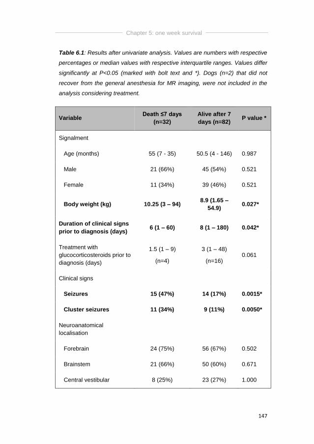
Chapter 5: one week survival
147
Table 6.1: Results after univariate analysis. Values are numbers with respective
percentages or median values with respective interquartile ranges. Values differ
significantly at P<0.05 (marked with bolt text and *). Dogs (n=2) that did not
recover from the general anesthesia for MR imaging, were not included in the
analysis considering treatment.
Variable Death ≤7 days
(n=32)
Alive after 7
days (n=82) P value *
Signalment
Age (months) 55 (7 - 35) 50.5 (4 - 146) 0.987
Male 21 (66%) 45 (54%) 0.521
Female 11 (34%) 39 (46%) 0.521
Body weight (kg) 10.25 (3 – 94) 8.9 (1.65 –
54.9) 0.027*
Duration of clinical signs
prior to diagnosis (days) 6 (1 – 60) 8 (1 – 180) 0.042*
Treatment with
glucocorticosteroids prior to
diagnosis (days)
1.5 (1 – 9)
(n=4)
3 (1 – 48)
(n=16) 0.061
Clinical signs
Seizures 15 (47%) 14 (17%) 0.0015*
Cluster seizures 11 (34%) 9 (11%) 0.0050*
Neuroanatomical
localisation
Forebrain 24 (75%) 56 (67%) 0.502
Brainstem 21 (66%) 50 (60%) 0.671
Central vestibular 8 (25%) 23 (27%) 1.000
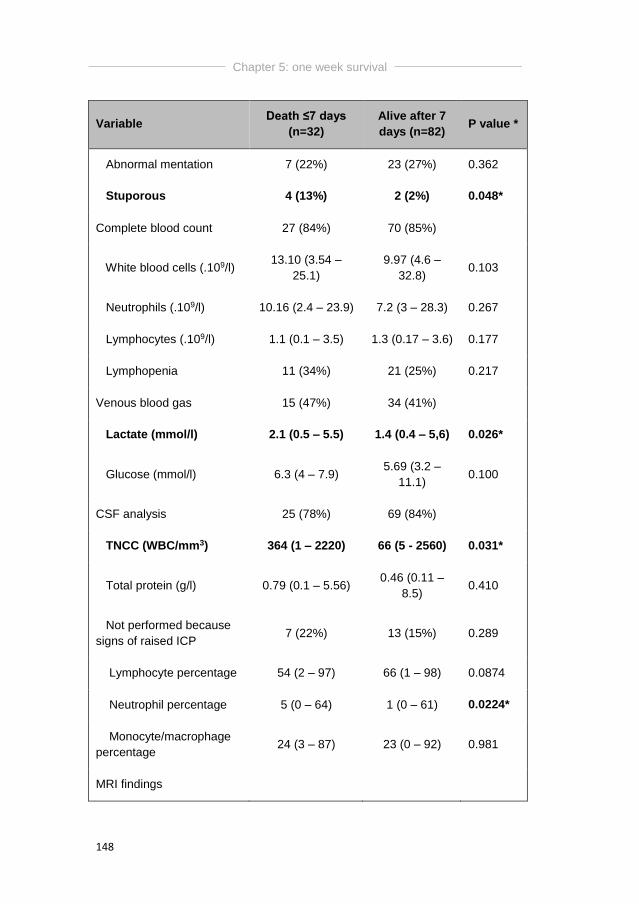
Chapter 5: one week survival
148
Variable Death ≤7 days
(n=32)
Alive after 7
days (n=82) P value *
Abnormal mentation 7 (22%) 23 (27%) 0.362
Stuporous 4 (13%) 2 (2%) 0.048*
Complete blood count 27 (84%) 70 (85%)
White blood cells (.109/l) 13.10 (3.54 –
25.1)
9.97 (4.6 –
32.8) 0.103
Neutrophils (.109/l) 10.16 (2.4 – 23.9) 7.2 (3 – 28.3) 0.267
Lymphocytes (.109/l) 1.1 (0.1 – 3.5) 1.3 (0.17 – 3.6) 0.177
Lymphopenia 11 (34%) 21 (25%) 0.217
Venous blood gas 15 (47%) 34 (41%)
Lactate (mmol/l) 2.1 (0.5 – 5.5) 1.4 (0.4 – 5,6) 0.026*
Glucose (mmol/l) 6.3 (4 – 7.9) 5.69 (3.2 –
11.1) 0.100
CSF analysis 25 (78%) 69 (84%)
TNCC (WBC/mm3) 364 (1 – 2220) 66 (5 - 2560) 0.031*
Total protein (g/l) 0.79 (0.1 – 5.56) 0.46 (0.11 –
8.5) 0.410
Not performed because
signs of raised ICP 7 (22%) 13 (15%) 0.289
Lymphocyte percentage 54 (2 – 97) 66 (1 – 98) 0.0874
Neutrophil percentage 5 (0 – 64) 1 (0 – 61) 0.0224*
Monocyte/macrophage
percentage 24 (3 – 87) 23 (0 – 92) 0.981
MRI findings
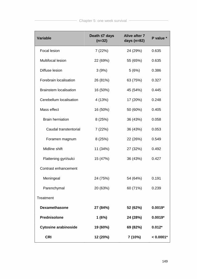
Chapter 5: one week survival
149
Variable Death ≤7 days
(n=32)
Alive after 7
days (n=82) P value *
Focal lesion 7 (22%) 24 (29%) 0.635
Multifocal lesion 22 (69%) 55 (65%) 0.635
Diffuse lesion 3 (9%) 5 (6%) 0.386
Forebrain localisation 26 (81%) 63 (75%) 0.327
Brainstem localisation 16 (50%) 45 (54%) 0.445
Cerebellum localisation 4 (13%) 17 (20%) 0.248
Mass effect 16 (50%) 50 (60%) 0.405
Brain herniation 8 (25%) 36 (43%) 0.058
Caudal transtentorial 7 (22%) 36 (43%) 0.053
Foramen magnum 8 (25%) 22 (26%) 0.549
Midline shift 11 (34%) 27 (32%) 0.492
Flattening gyri/sulci 15 (47%) 36 (43%) 0.427
Contrast enhancement
Meningeal 24 (75%) 54 (64%) 0.191
Parenchymal 20 (63%) 60 (71%) 0.239
Treatment
Dexamethasone 27 (84%) 52 (62%) 0.0019*
Prednisolone 1 (6%) 24 (28%) 0.0019*
Cytosine arabinoside 19 (60%) 69 (82%) 0.012*
CRI 12 (20%) 7 (10%) < 0.0001*
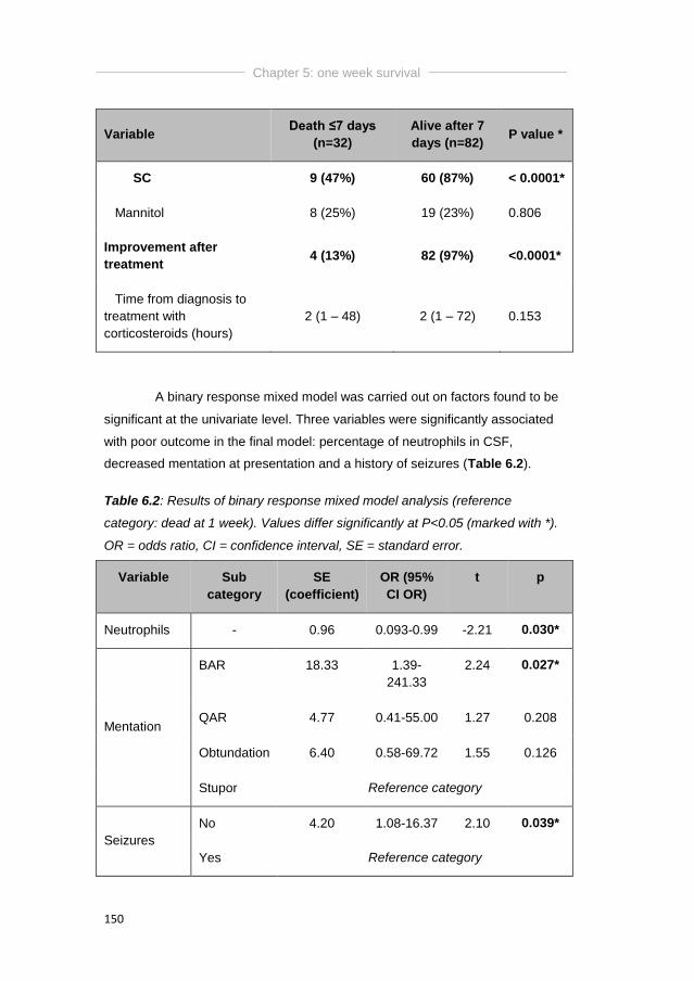
Chapter 5: one week survival
150
Variable Death ≤7 days
(n=32)
Alive after 7
days (n=82) P value *
SC 9 (47%) 60 (87%) < 0.0001*
Mannitol 8 (25%) 19 (23%) 0.806
Improvement after
treatment 4 (13%) 82 (97%) <0.0001*
Time from diagnosis to
treatment with
corticosteroids (hours)
2 (1 – 48) 2 (1 – 72) 0.153
A binary response mixed model was carried out on factors found to be
significant at the univariate level. Three variables were significantly associated
with poor outcome in the final model: percentage of neutrophils in CSF,
decreased mentation at presentation and a history of seizures (Table 6.2).
Table 6.2: Results of binary response mixed model analysis (reference
category: dead at 1 week). Values differ significantly at P<0.05 (marked with *).
OR = odds ratio, CI = confidence interval, SE = standard error.
Variable Sub
category
SE
(coefficient)
OR (95%
CI OR)
t p
Neutrophils - 0.96 0.093-0.99 -2.21 0.030*
Mentation
BAR 18.33 1.39-
241.33
2.24 0.027*
QAR 4.77 0.41-55.00 1.27 0.208
Obtundation 6.40 0.58-69.72 1.55 0.126
Stupor Reference category
Seizures
No 4.20 1.08-16.37 2.10 0.039*
Yes Reference category
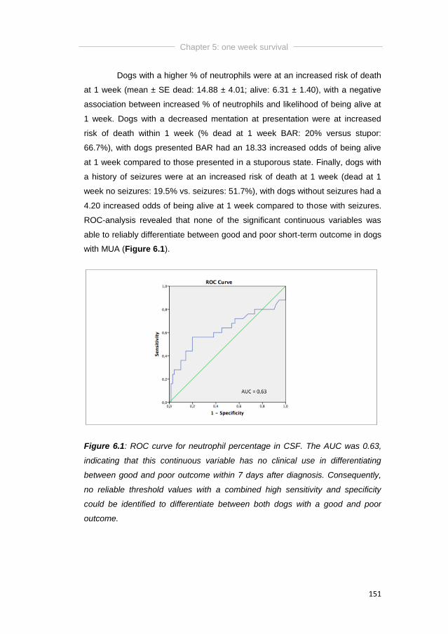
Chapter 5: one week survival
151
Dogs with a higher % of neutrophils were at an increased risk of death
at 1 week (mean ± SE dead: 14.88 ± 4.01; alive: 6.31 ± 1.40), with a negative
association between increased % of neutrophils and likelihood of being alive at
1 week. Dogs with a decreased mentation at presentation were at increased
risk of death within 1 week (% dead at 1 week BAR: 20% versus stupor:
66.7%), with dogs presented BAR had an 18.33 increased odds of being alive
at 1 week compared to those presented in a stuporous state. Finally, dogs with
a history of seizures were at an increased risk of death at 1 week (dead at 1
week no seizures: 19.5% vs. seizures: 51.7%), with dogs without seizures had a
4.20 increased odds of being alive at 1 week compared to those with seizures.
ROC-analysis revealed that none of the significant continuous variables was
able to reliably differentiate between good and poor short-term outcome in dogs
with MUA (Figure 6.1).
Figure 6.1: ROC curve for neutrophil percentage in CSF. The AUC was 0.63,
indicating that this continuous variable has no clinical use in differentiating
between good and poor outcome within 7 days after diagnosis. Consequently,
no reliable threshold values with a combined high sensitivity and specificity
could be identified to differentiate between both dogs with a good and poor
outcome.
Chapter 5: one week survival
152
Discussion
This study evaluated the prevalence and potential risk factors for 1-
week survival in dogs diagnosed with MUA. Although it was hypothesized that a
proportion of dogs would not survive the first week after obtaining a diagnosis of
MUA, a high proportion (26%) of dogs died within this specific time period
despite initiation of appropriate treatment and close monitoring. Despite the lack
of specific studies evaluating short-term survival in dogs with MUA, our findings
indicate better outcomes compared with previous findings. It has been stated
that approximately 15% of dogs diagnosed with GME died before being treated
(Granger et al., 2010), compared to only 1/116 dogs (0.9%) in the presented
study where the owner decided to euthanize the dog without attempting to treat.
A recent study reported that 56% of dogs diagnosed with and treated for MUA
died or were euthanized with a median survival time of 2 days (Lowrie et al.,
2013), which is almost double the percentage of the presented study. The
results of our study demonstrated that 25% of dogs would die in the first week
after diagnosis and initiating appropriate treatment for MUA. It seems therefore
important to include this group of dogs when considering the overall prognosis
of dogs with MUA.
In a recent study by Sharma and Holowaychuk (2015), increased
venous lactate concentrations were a risk factor for non-survival to hospital
discharge in dogs with head trauma. Additionally, hyperglycaemia has been
associated with severity of injury in cases of head trauma in dogs and cats, but
not with outcome (Syring et al., 2001). In the presented study, blood glucose
and lactate levels were measured on admission or before MR imaging on
standard venous blood gas analysis. No significant difference was found in
blood glucose levels between dogs that did or did not survive the first week after
diagnosis. In the univariate analysis, lactate concentrations were significantly
increased in dogs with a poor outcome, but this result was not confirmed in the
multivariate analysis. As both measurements were only available for review in
approximately 20% of the cases, further studies should be performed to draw
further conclusions.
Chapter 5: one week survival
153
This study identified some potential risk factors for death in the first 7
days after obtaining a presumptive diagnosis of MUA. In agreement with
previous findings, dogs presenting with seizures and/or a decreased mentation
were at increased risk of not surviving the first week after diagnosis (Bateman
and Parent, 1999; Coates et al., 2007). Although it is possible these dogs
represent a group of animals with a worse clinical phenotype, it cannot be
excluded that the necessity of administering anti-epileptic drugs in these
patients was associated with increased sedation and therefore contributed to a
further decline of their neurological function. In contrast to results of a recent
study (Lowrie et al., 2013), a higher neutrophil percentage on CSF analysis was
significantly associated with an increased risk of death in the first week after a
diagnosis of MUA. However, the constructed ROC-curve could not demonstrate
a reliable threshold value with a combined high sensitivity and specificity to
predict survival and so the exact neutrophil percentage should not be
considered a useful tool for assessing prognosis in individual animals with MUA.
In previous studies, it was stated that adding another
immunosuppressive agent or radiation therapy to the standard treatment
protocol with glucocorticosteroids improves survival of dogs with MUA (Munana
and Luttgen, 1998; Jung et al., 2007; Coates et al., 2007; Granger et al., 2010;
Flegel et al., 2011; Beckmann et al., 2015; Barnoon et al., 2015) but this could
not be confirmed in the present study. Unexpectedly, treatment with SC
injections of cytosine arabinoside and oral prednisolone therapy were both
significantly associated with a better short-term outcome. Most likely, this is
highly biased at the level of the clinician, who would possibly opt for IV
dexamethasone and an additional CRI of cytosine arabinoside in dogs with
more severe neurological signs. Additionally, a previous study (Crook et al.,
2013) indicated more favourable pharmacokinetic properties of intravenous
continuous infusion of cytosine arabinoside compared to subcutaneous
injections. Both findings could not be confirmed in the multivariate analysis
performed in our study.
This study is obviously limited by its retrospective character. Inclusion
criteria were based on previously reported studies, but they are not restrictive.
In the present study, dogs were excluded if TNCC on CSF analysis and/or
Chapter 5: one week survival
154
intracranial MRI were within normal limits, infectious disease testing was not
required for inclusion and no standardisation of the treatment protocol was
obtained. Medical management was also tailored to individual needs and
therefore some dogs might have received additional medication, such as anti-
epileptic drugs and mannitol. Additionally, dogs did receive different treatment
protocols prior to admission and diagnosis. Results might have been biased
towards more severely affected dogs by only including dogs with a CSF
pleocytosis, dogs with abnormal intracranial imaging and dogs with clinical
signs of raised ICP when CSF analysis was not performed. Definitive post
mortem diagnosis was available in almost half of the dogs (14/30) that died
within 1 week after diagnosis in the hospital. This might also implicate a bias
towards the more severely affected cases, as dogs that die or are euthanized
within a hospital environment are more likely to have post-mortem examination
performed.
Conclusions
Twenty-six percent of dogs diagnosed with MUA in this study died
within one week after diagnosis, emphasizing the need for evaluation of short-
term prognostic factors. Presence of a decreased mentation at time of
presentation, presence of seizures, and increased neutrophil percentage in the
CSF were significantly associated with death within 7 days after diagnosis. The
results of this study might be important when considering the overall prognosis
of dogs with MUA and managing expectations of owners and hospital staff.

General Discussion and Conclusions

General Discussion and Conclusions
157
The main goal of this thesis was to gain more insight in the clinical
presentation, diagnostic findings, treatment and prognosis of dogs diagnosed
with MUA.
The typical clinical presentation of dogs with MUA is well described but
a substantial number of large dogs have been included in multiple studies.
Specific data regarding clinical presentation, diagnostic findings and long-term
outcome are currently lacking about this group of dogs.
The same accounts for dogs diagnosed with MUA only affecting the
spinal cord. As the disease is generally considered fatal if left untreated,
multiple (prospective and retrospective) studies have been published,
investigating different treatment protocols including one or multiple
immunosuppressive drugs. These drugs however add a substantial cost to the
treatment, which might be a limiting factor for some owners. Sole prednisolone
therapy has generally been associated with poor outcome or very short
(median) STs compared to combination therapy with other immunosuppressive
agents. Two studies were conducted, a retrospective study to evaluate three
historically used sole prednisolone treatment protocols, and a prospective study
comparing one of the sole prednisolone treatment schedules with combination
therapy with ciclosporine.
As stated above, MUA is considered a fatal disease, so multiple
studies have been performed investigating potential prognostic factors for dogs
with MUA. Although these studies evaluated a large amount of clinical and
imaging variables, only few clinically useful factors have been identified so far.
Additionally, as a lot of studies focus on long-term survival, little is currently
known about initial response to treatment and short-term (one week) survival in
dogs diagnosed with MUA.
General Discussion and Conclusions
158
Clinical presentation and diagnostic findings
Clinical presentation
In a first study (Part I, Chapter 1) we evaluated the differences in
clinical presentation, diagnostic findings and long-term survival between small
(<15kg) and large (>15kg) dogs diagnosed with MUA. The study included 111
dogs, including 28 (25%) large dogs and 83 (75%) small dogs. Large dogs were
found to present significantly more often with a decreased mentation, but no
other differences could be identified regarding clinical presentation, diagnostic
findings and long-term survival between both groups.
MUA is generally considered a syndrome affecting small, toy and
terrier breed dogs (Munana and Luttgen, 1998; Adamo et al., 2007; Talarico
and Schatzberg, 2010; Coates and Jeffery, 2014). Interestingly, a quarter of
dogs in the presented study were large dogs, so ignoring this population of dogs
would underestimate the prevalence of MUA in the canine population. Overall, a
total of 45 large dogs have been reported between 1998 and 2015 (Gregory et
al., 1998; Munana and Luttgen, 1998; Cherubini et al., 2006; Fliegner et al.,
2006; Zarfoss et al., 2006; Coates et al., 2007; Pakozdy et al., 2009; Smith et
al., 2009; Wong et al., 2010; Flegel et al., 2011; Lowrie et al., 2013; Estey et al.,
2014; Barnoon et al., 2015; Beckmann et al., 2015; Mercier and Barnes Heller
2015), with a similar breed distribution compared to our study. Adding the 28
dogs of our study gives a total number of 73 large dogs described. The
Labrador Retriever (13/73; 17%), the English Springer Spaniel (9/73; 12%) and
the Golden Retriever (7/73; 10%) were the three most commonly affected
breeds over all studies. It is however not clear whether this reflects the
popularity of those breeds in the respective countries or a specific breed
predisposition for MUA. Therefore, MUA should be considered a differential
diagnosis in dogs other than small or toy breeds that have signs suggestive of
inflammatory CNS disease.
Although female predominance is a widely held belief in GME (Cordy,
1979; Russo, 1979; Braund, 1985; Bailey, 1986; Sorjonen, 1990; Munana and
General Discussion and Conclusions
159
Luttgen, 1998), no statistical difference in female:male ratio could be found in
more recent studies (Talarico and Schatzberg, 2010; Granger et al., 2010). A
total of 194 dogs was investigated in this thesis, including 101 males (52%) and
93 females (48%), revealing no difference in female:male ratio.
As stated in the introduction, dogs affected with NE were
predominantly under 4 years old whereas the peak age for GME was 4-8 years
(Granger et al., 2010). Historically, NME was described in Pug dogs with ages
ranging from 6 months to 7 years (Cordy and Holliday, 1989), whilst dogs with a
histopathological diagnosis of GME had ages ranging from 6 months to 12
years (Munana and Luttgen, 1998). In a series of 60 Pugs with NE (Levine et
al., 2008), the median age was 18 months. In the presented studies in this
thesis, median age at time of presentation was around 4 years in all studies (54
months (Part I, Chapter 2), 48 months (Part II, Chapter 3), 52.5 months (Part
III, Chapter 5)). On the contrary, median age at time of presentation was 28
months in the prospective study (Part II, Chapter 4), which is remarkable
younger. This can however be explained by the fact that all dogs in this study
are breeds previously described to develop NE.
As 8% of dogs diagnosed with GME presented with neurological
deficits suggestive of a myelopathy (Granger et al., 2010), we performed a
second study (Part I, Chapter 2) evaluating the clinical presentation, diagnostic
findings and long-term survival in 21 dogs diagnosed with SO-MUA. In our
study, an acute or chronic onset of a lesion affecting the T3-L3 spinal cord
segments resulting in ambulatory paraparesis was considered the most
common clinical presentation. Thirty-three dogs with MUA only involving the
spinal cord were previously reported, including one dog with paraplegia
(Cherubini et al., 2006; Griffin et al., 2008; Wong et al., 2010), whereas no dog
with paraplegia was included in our study. Unfortunately, more specific details
were lacking for the majority of the previously published dogs. As the clinical
presentation and diagnostic findings are not specific for SO-MUA, this disorder
should be included in the differential diagnosis for all dogs presenting with
different degrees of spinal cord dysfunction.
General Discussion and Conclusions
160
Pain on spinal palpation was present in 71% of dogs with SO-MUA in
our second study, which is comparable to the 86% stated in a recent study
(Cardy et al., 2015). Paraspinal hyperesthesia (Sorjonen, 1990) and cervical
hyperesthesia (Cordy and Holliday, 1989) were previously described in GME
and NME, respectively. As spinal pain reflects the involvement of the meninges,
and/or vertebrae (vertebral periosteum), and/or nerve roots or spinal nerves (Da
Costa, 2012), this cannot be considered a typical finding for SO-MUA. However,
it is important for the clinician to understand that MUA should be in the
differential diagnosis for dogs presenting with any form of spinal hyperesthesia.
Clinical diagnostic findings
Although presence of leucocytosis on CBC has previously been
reported in dogs with GME (Thomas and Eger, 1989; Sorjonen, 1990; Tipold,
1995), nothing is currently known about prevalence and prognostic value.
Throughout the different chapters, leucocytosis was present in approximately
15% of cases where CBC was available for review (13% for Part I, Chapter 1
and Part III, Chapter 5; 9.5% for Part I, Chapter 2; 14% for Part II, Chapter 3;
and 17% for Part II, Chapter 4). In Part III, Chapter 5, statistical analysis was
performed to investigate whether presence of leucocytosis was associated with
survival after 7 days, but this did not reach significance (P=0.103). In the other
chapters, no further statistical analysis was performed.
MR imaging
MR imaging is considered the most sensitive imaging modality for
detecting intracranial lesions, but up to 7% of scans showed no lesion on T2WI
in dogs with MUA (Talarico and Schatzberg, 2010; Granger et al., 2010).
Overall, the sensitivity of imaging in identifying all inflammatory lesions
suspected from the neurological examination remained quite low (<60%)
(Granger et al., 2010). In our first study (Part I, Chapter 1), the presence of
intracranial lesions on MRI was part of the inclusion criteria, but no differences
in MRI findings could be detected between small and large dogs.
General Discussion and Conclusions
161
Although studies looking into the use of MRI for differentiating between
histopathologically confirmed cases of GME, NME or NLE are lacking, MRI
findings in the different disease entities have been fairly well described
previously (Cherubini et al., 2006; von Praun et al., 2006; Flegel et al., 2008;
Young et al., 2009; Talarico and Schatzberg, 2010; Coates and Jeffery, 2014).
On the contrary, MRI findings have been only described for 4/33 dogs reported
with SO-MUA, revealing no abnormalities in 1 dog, and multifocal poorly
demarcated T2W hyperintensities with variable contrast enhancement in 2 dogs
(Cherubini et al., 2006; Wong et al., 2010). Therefore, we performed a second
study (Part I, Chapter 2) to describe the specific MRI findings in 21 dogs with
SO-MUA. No lesions were visible on sagittal T2WI or T1WI in 10% of cases,
comparable to the 7% reported for MUA (Talarico and Schatzberg, 2010;
Granger et al., 2010). All visible MRI lesions in our study were extensive, ill-
defined, intramedullary, hyperintense on T2WI and isointense on T1WI.
Parenchymal contrast enhancement was seen in 18 lesions (86%), and 17
lesions (81%) showed contrast enhancement of the overlying meninges.
Interestingly, although dogs were not allowed to have clinical signs or
neurological examination results suggestive of intracranial involvement for
inclusion in our second study (Part I, Chapter 2), additional MR images of the
brain were included in the field of view of the cervical MRI in 2 dogs, showing
additional lesions (suggestive of MUA) in both cases. As intracranial images
were only available in 2 dogs, it is currently unclear (1) if these brain
abnormalities represent a multifocal nature of the disease or cranial extension
of the cervical inflammatory lesions, and (2) if inflammatory brain lesions are
currently under diagnosed in dogs with SO-MUA and if SO-MUA could therefore
be considered a more generalised inflammatory disease process, a
meningoencephalomyelitis.
CSF analysis
Results on CSF analysis throughout the different chapters should be
interpreted with caution as a CSF pleocytosis is considered an inclusion
criterion for all studies. Overall, TNCC was normal in 2% of cases, including 3
General Discussion and Conclusions
162
dogs with histopathologically confirmed GME, NME or NLE in Part III, Chapter
5; and 1 dog without necropsy in Part II, Chapter 4. TP concentration was not
set as an inclusion criterion in all studies, and TP measurements were available
in 157/194 cases, revealing normal values in 36/157 cases (23%).
Although exclusion of regional infectious causes is a proposed
diagnostic criterion for MUA (Granger et al., 2010), infectious disease testing
was lacking in 52/187 cases (28%) in this thesis. All studies were performed in
Belgium or the United Kingdom, but no prevalence studies for T. gondii, N
caninum or CDV are available apart from two studies performed in the mid 90’s
in Belgium (Vanparijs et al., 1991; Barber et al., 1998). These studies revealed
that approximately 10% of dogs in Belgium was seropositive for N. caninum
(Barbet et al., 1998) and that no T. gondii oocysts were detected on
examination of 2324 faecal samples of dogs and cats (Vanparijs et al., 1991).
However, infectious disease testing should still be encouraged in all dogs
suspected of MUA to exclude possible infectious agents as much as possible.
Treatment
Aggressive immunosuppressive therapy was initiated at time of
diagnosis in all dogs included in all chapters of this thesis. This is important as
early and aggressive therapy might improve survival in dogs with MUA
(Barnoon et al., 2015). In the literature, immunosuppressive therapy is
sometimes only initiated after results of infectious disease testing returned
negative, complicating comparison between different study designs.
Treatment with sole prednisolone therapy
The exact aetiology and pathophysiology of MUA remain unknown, but
as one or several different factors are believed to trigger an excessive
immunologic response resulting in inflammatory changes in the CNS (Flegel et
al., 2011), immunosuppressive therapy is considered the cornerstone of
medical treatment. Treatment with glucocorticosteroids (mostly prednisolone)
General Discussion and Conclusions
163
only is generally associated with shorter survival times compared to
combination therapy with other immunosuppressive agents (Munana and
Luttgen, 1998; Jung et al., 2007; Pakozdy et al., 2009; Granger et al., 2010;
Flegel et al., 2011; Mercier and Barnes Heller, 2015). Therefore, several
treatment protocols using different immunomodulating drugs have been
reported (Sisson et al. 1989; Gregory et al., 1998; Munana and Luttgen, 1998;
Adamo and O’Brien, 2004; Gnirs, 2006; Zarfoss et al., 2006; Adamo et al.,
2007; Coates et al., 2007; de Stefani et al., 2007; Feliu-Pascual et al., 2007;
Uriarte et al., 2007; Jung et al., 2007; Menaut et al., 2008; Pakozdy et al., 2009;
Smith et al., 2009; Granger et al., 2010; Kang et al., 2009; Wong et al., 2010;
Flegel et al., 2011; Jung et al., 2012; Jung et al., 2013; Lowrie et al., 2013;
Beckmann et al., 2015; Mercier and Barnes Heller, 2015; Barnoon et al., 2015;
Lowrie et al., 2016).
Although combination therapy might result in a better long-term
outcome in MUA, adding more expensive immunosuppressive therapies to the
glucocorticoid protocol might be financially impossible in a clinical setting.
Additionally, at the institution where our third study (Part II, Chapter 3) was
conducted, dogs with MUA were historically only treated with prednisolone and
the personal perception of the authors was that dogs were doing considerable
well on this therapy. In the literature, initial prednisolone doses ranged from
0.34-30mg/kg/d (Munana and Luttgen, 1998; Jung et al., 2007; Pakozdy et al.,
2009; Flegel et al., 2011; Mercier and Barnes Heller, 2015), but only one study
(Talarico and Schatzberg, 2010) was providing a clinically useful treatment
schedule. Therefore, the first aim of our third study was to retrospectively
evaluate the clinical use of three different treatment schedules, and to provide a
clinically useful sole prednisolone treatment schedule.
In this third study, we evaluated 38 dogs diagnosed with MUA that
received oral prednisolone therapy in 3 different historically used tapering
schedules (3, 8, and 18-week tapering prednisolone schedule). A significant
difference was found between the three treatment schedules, with the highest
number of deceased dogs in the 8-week treatment group (57%), followed by the
3 (26%) and 18-week (0%) treatment groups. It is possible that dogs receiving a
longer and more immunosuppressive protocol might have a better outcome, but
General Discussion and Conclusions
164
this result should be confirmed in future studies. This 18-week treatment
schedule is the same schedule as proposed by Talarico and Schatzberg (2010),
so the authors would advise further clinical use of this schedule. However, as
the initial dose is 3mg/kg/d, side effects might be significant.
Combination therapy with ciclosporine
Although ciclosporine was previously investigated as sole and add-on
treatment for dogs diagnosed with MUA, no specific treatment protocols have
currently been described (Adamo and O’Brien, 2004; Gnirs, 2006; Adamo et al.,
2007; Jung et al., 2007; Pakozdy et al., 2009; Kang et al., 2009; Jung et al.,
2012; Jung et al., 2013). Therefore, we performed a fourth study (Part II,
chapter IV) to describe a clinically useful treatment protocol and to compare
sole prednisolone therapy with combination therapy with ciclosporine in 12
dogs. The sole prednisolone protocol used in this fourth study was the same as
the 18-week prednisolone schedule previously described in the third study. The
described doses as add-on therapy for ciclosporine range from 6-30mg/kg/d
(Adamo and O’Brien, 2004; Gnirs, 2006; Adamo et al., 2007; Jung et al., 2007;
Pakozdy et al., 2009; Kang et al., 2009; Granger et al., 2010; Jung et al., 2012;
Jung et al., 2013), but we decided to work with 5 mg/kg/d, as this dose is also
known to cause some degree of immunosuppression (Archer et al., 2014).
Although the difference in MST between both treatment groups seems clinically
important (87 versus 567 days), no statistically significant results could be
obtained. A power analysis was performed (non-parametric repeated sampling)
revealing that 74 additional dogs were necessary in each treatment group to
reach significantly different hazard ratios, which is quite a substantial number.
Additionally, no previously established prognostic factors including clinical and
diagnostic factors could be confirmed in this study.
Combination therapy with cytosine arabinoside
Investigation of the use and efficacy of cytosine arabinoside as an
adjunctive therapy in canine MUA was not a primary goal of this thesis.
General Discussion and Conclusions
165
However, a total of 102 dogs treated with glucocorticosteroids plus cytosine
arabinoside were recruited for the first (Part I, Chapter 1), second (Part I,
Chapter 2) and fifth (Part III, Chapter 5) study. It should be noted that the
same pool of dogs was used for the first and fifth study. Cytosine arabinoside
can be administered either as a CRI (doses ranging from 100-300mg/m2 over
8-24 hours) or as 4 SC injections of 50mg/m2 in 48 hours (200mg/m2 in 48h)
(Zarfoss et al., 2006; de Stefani et al., 2007; Menaut et al., 2008; Smith et al.,
2009; Lowrie et al., 2013; Lowrie et al., 2016). Crook et al. (2013) showed that
CRI administration of cytosine arabinoside provided a steady state
concentration over the time it was administered compared to a rapid absorption
and elimination when administered subcutaneously. A recent clinical study
revealed a significantly better 3-month-survival in dogs initially receiving a CRI
of cytosine arabinoside compared to the SC route (Lowrie et al., 2016).
In a fifth study (Part III, Chapter 5), we evaluated short-term outcome
and prognostic factors in 116 dogs diagnosed with MUA. Eighty-eight dogs
(85%) received additional cytosine arabinoside therapy; 69 via the
subcutaneous and 19 via the intravenous route. Administration of cytosine
arabinoside was significantly associated with a better outcome, but
administration of a CRI was associated with a poor outcome compared to SC
administration. Most likely, this is highly biased at the level of the clinician,
whom would possible opt for IV cytosine arabinoside in more severely affected
dogs. In the second study (Part I, Chapter 2), no significant difference in
survival could be found for dogs receiving additional cytosine arabinoside
therapy. As only 1/14 dogs (7%) were receiving a CRI of cytosine arabinoside,
no conclusions can be drawn from this study regarding the route of
administration.
Prognostic factors and outcome
Prognostic factors
As MUA is generally considered a fatal disease (Munana and Luttgen,
1998), multiple studies attempted to identify prognostic factors. Unfortunately,
General Discussion and Conclusions
166
different studies revealed conflicting results, making the majority of findings
inapplicable in a clinical setting. Currently, the following established prognostic
factors for dogs with MUA are available in the literature: 1) younger age at time
of diagnosis was significantly associated with improved survival (Oliphant et al.,
2016), 2) presence of seizures or altered mentation were significantly
associated with shorter survival (Bateman and Parent, 1999; Coates et al.,
2007; Granger et al., 2010); 3) presentation within 7 days of onset of clinical
signs was significantly associated with longer survival (Barnoon et al., 2015); 4)
abnormal serial CSF analysis was significantly associated with relapse and poor
outcome (Lowrie et al., 2013); and 5) mass effect, loss of cerebral sulci and
foramen magnum herniation on MRI were all significantly associated with death
in dogs with MUA, however the clinical prognostic power was low for those
findings and none of them was predictive of long-term outcome (Lowrie et al.,
2013; Lowrie et al., 2016).
In our second study (Part I, Chapter 2), we investigated multiple
possible prognostic factors in dogs with SO-MUA including clinical, diagnostic
and treatment factors, but no association with long-term outcome could be
made. On the contrary, our fifth study (Part III, Chapter 5), in agreement with
previous studies, revealed that decreased mentation at presentation and
presence of seizures were significantly associated with poor short-term
outcome. Increased percentage of neutrophils on cerebrospinal fluid analysis
also was significantly associated with poor short-term outcome, but no clinically
useful cut-off values could be identified. None of the previously established
prognostic factors could be confirmed in our fourth study (Part II, Chapter IV),
although results should be interpreted with caution as only a limited number of
dogs (n=12) was involved in this prospective randomized study.
Outcome
Although several studies have focused on long-term survival, little is
known about short-term survival and initial response to therapy of dogs
diagnosed with MUA. A recent study reported that 56% of dogs diagnosed with
and treated for MUA died or were euthanized with a MST of 2 days (Lowrie et
General Discussion and Conclusions
167
al., 2013). In our fifth study (Part III, Chapter 5) we therefore evaluated the
short-term survival in 114 dogs with MUA, revealing that 26% of dogs died
within 7 days after diagnosis despite initiation of early and aggressive treatment.
It seems therefore important to include this group of dogs when considering the
overall prognosis of dogs with MUA. As more than 50% of the included dogs
was alive after 7 days, no MST could be calculated in this study.
On the contrary, several studies using different treatment protocols
resulting in different long-term survival times have been reported, with resulting
MST ranging from 2 – 1834 days (Sisson et al. 1989; Gregory et al., 1998;
Munana and Luttgen, 1998; Adamo and O’Brien, 2004; Gnirs, 2006; Zarfoss et
al., 2006; Adamo et al., 2007; Coates et al., 2007; de Stefani et al., 2007; Feliu-
Pascual et al., 2007; Uriarte et al., 2007; Jung et al., 2007; Menaut et al., 2008;
Pakozdy et al., 2009; Smith et al., 2009; Granger et al., 2010; Kang et al., 2009;
Wong et al., 2010; Flegel et al., 2011; Jung et al., 2012; Jung et al., 2013;
Lowrie et al., 2013; Beckmann et al., 2015; Mercier and Barnes Heller, 2015;
Barnoon et al., 2015; Lowrie et al., 2016). Our first study (Part I, Chapter 1)
revealed an overall MST of 272 days. When separately evaluated for large and
small dogs, the MST was 106 and 281 days, respectively, which is all-
comparable to the published literature for MUA. As no specific survival data
were available for dogs with SO-MUA, we performed a second study (Part I,
Chapter 2) specifically evaluating the long-term outcome in those dogs. The
study revealed a MST of 669 days, and 48% of dogs had died or been
euthanized because of the disease.
In our third study (Part II, Chapter 3) we specifically evaluated the
long-term survival in dogs with MUA receiving three different treatment
protocols of sole prednisolone therapy. For this therapy, reported MSTs were 28
– 357 days (Granger et al., 2010), 91 - 329 days (Flegel et al., 2011) and 602
days (Mercier and Barnes Heller, 2015). As more than 50% of the dogs was
alive or censored for outcome calculations at time of data capture in our study,
no overall MST could be calculated. However, the MST was 180 days in the 8-
week treatment group, which appears to be the group with the highest
percentage of deceased and relapsed dogs. Our fourth study (Part II, Chapter
IV) revealed an overall MST of 115 days in a prospective study including 12
General Discussion and Conclusions
168
dogs with MUA that were treated with sole prednisolone or combination therapy
of prednisolone with ciclosporine, although no difference in MST could be
demonstrated between both treatment groups.
Overall, long-term outcome data are available for 182 dogs in this
thesis, revealing that 98/138 dogs (71%) died because of MUA, which is higher
compared to the previously reported 56% (Lowrie et al., 2013). It has been
previously mentioned that most dogs with MUA or GME that die, do so within
the first three months after diagnosis (Thomas and Eger, 1989; Smith et al.,
2009; Lowrie et al., 2013). For the presented studies, 63/98 dogs (65%) died
within 3 months after diagnosis. In both prospective studies (Part II, Chapter 3
and 4) and the retrospective study regarding SO-MUA (Part I, Chapter 2), 7/34
dogs (21%) died because of MUA within 7 days after diagnosis, which is
comparable to the results of the fifth study (Part III, Chapter 5), where 26% of
dogs with MUA died within one week after diagnosis.
Limitations
MUA is a clinical diagnosis that is based on a combination of
signalment, neurological examination results and cross-sectional imaging
findings in the absence of histopathological confirmation (Munana and Luttgen,
1998; Adamo et al., 2007; Talarico and Schatzberg, 2010; Coates and Jeffery,
2014). Specific diagnostic criteria are currently available, but this might bias
studies towards more severely affected cases. To illustrate this, dogs with
normal MRI findings (reported in 7-10% of dogs with MUA) or normal CSF
analysis (reported in 3-57% of dogs with MUA) do not fit those inclusion criteria
and will mostly not be included in prospective or retrospective studies, which
also accounts for all studies in this thesis. Additionally, dogs were included in
Part II, Chapter 3 based on CT imaging whereas MRI is generally considered
the diagnostic imaging modality of choice for diagnosis of MUA. Four out of 5
reported studies in this thesis are retrospective studies. These carry the pitfall
that they only carry limited standardization of patient assessment (different
people and institutions involved) and therapy (different institutions use different
treatment protocols and have different follow-up practices). Additionally, there
General Discussion and Conclusions
169
might be debate about which infectious disease testing to perform or not to
perform in dogs diagnosed with MUA. PCR testing is expensive for the pet
owner and might therefore be declined, and carries the risk for false negative
results. On the contrary, as long as histopathological confirmation is lacking, no
certainty can be obtained about the underlying pathology (GME, NME or NLE)
and so conclusions should be drawn with caution.
General Discussion and Conclusions
170
Future perspectives
Large, prospective, multi-centre studies are needed to further
investigate the findings of this thesis. The first study revealed that 25% of dogs
affected with MUA are large dogs (>15kg), but it remains currently unclear
whether this reflects a different disease entity, or just the same disease that is
emerging in the whole canine population. A study to specifically look into
clinical, clinicopathological, imaging and histopathological findings in large dogs
with MUA is needed. The same accounts for dogs with SO-MUA, as currently
only a small cohort of 21 dogs and the associated MRI findings were described.
On-going research investigating clinical presentation, diagnosis and outcome in
those dogs is warranted.
Although the criterion-referenced standard for a clinical trial is a
randomized, placebo-controlled, double-blinded, prospective study, it is
generally accepted that use of a placebo control treatment group is unethical
because dogs with MUA have a poor outcome without treatment. Large, multi-
centre studies are necessary to compare different treatment products and
schedules in a larger patient population.
Together with the investigation of additional clinical and outcome data,
it is also important that the clinical diagnosis of MUA becomes more and more
certain based on several inclusion criteria. As more and more institutions
currently have high-field MRI available, evaluation of more criteria to
differentiate between NME, NLE and GME can possibly aid the clinician in
making a presumptive diagnosis. At the same time, authors should be
motivated to use the same inclusion criteria to facilitate reliable comparison
between studies.
General Discussion and Conclusions
171
Conclusions
In conclusion, this thesis revealed novel information regarding clinical
presentation, diagnosis, treatment and prognosis for dogs with MUA. Different
breeds and different regions of the central nervous system can be affected by
MUA. Typical MRI findings in dogs with MUA only affecting the spinal cord were
provided, but further investigations regarding dogs with paraplegia and dogs
with concurrent intracranial involvement are warranted. Overall, the prognosis of
MUA is guarded. Twenty-six percent of dogs will die or be euthanized within
one week after diagnosis despite treatment, and a large number of dogs will die
or be euthanized because of MUA on the long-term. Further studies are
warranted to provide more accurate and clinically useful diagnostic variables
and prognostic factors. Large, prospective, randomized multi-centre studies
should be performed to investigate different immunomodulating treatment
protocols.


Reference list

Reference List
175
Adamo, F.P. and O’Brien, R.T., 2004. Use of cyclosporine to treat granulomatous meningoencephalitis in three dogs. Journal of the American Veterinary Medical Association 225, 1211–1216.
Adamo, P.F., Rylander, H., Adams, W.M., 2007. Ciclosporine use in multi-drug therapy for meningoencephalomyelitis of unknown aetiology in dogs. Journal of Small Animal Practice 48, 486-496.
Alves, L., Gorgas, D., Vandevelde, M., Gandini, G., Henke, D. 2011. Segmental meningomyelitis in 2 cats caused by Toxoplasma gondii. Journal of Veterinary Internal Medicine 25, 148-52.
Archer, T.M., Boothe, D.M., Langston, V.C., Fellman, K.V., Lunsford, K.V., Mackin, A.J., 2014. Oral cyclosporine treatment in dogs: a review of the literature. Journal of Veterinary Internal Medicine 28, 1-20.
Bailey, C.S. and Higgins, R.J., 1986. Characteristics of cerebrospinal fluid associated with canine granulomatous meningoencephalomyelitis: a retrospective study. Journal of the American Veterinary Medical Association 188, 418-421.
Barber, J.S., Van Ham, L., Polis, I., Trees, A.J., 1997. Seroprevalence of antibodies to Neospora caninum in Belgian dogs. Journal of Small Animal Practice 38, 15-16.
Barber, R.M., Li, Q., Diniz, P.P., Porter, B.F., Breischwerdt, E.B., Clairborne, M.K., Birkenheuer, A.J., Levine, J.M., Levine, G.J., Chandler, K., Kenny, P., Nghiem, P., Wei, S., Greene, C.E., Kent, M., Platt, S.R., Greer, K., Schatzberg, S.J., 2010. Evaluation of brain tissue or cerebrospinal fluid with broadly reactive polymerase chain reaction for Ehrlichia, Anaplasma, spotted fever group Rickettsia, Bartonella, and Borrelia species in canine neurological diseases (109 cases). Journal of Veterinary Internal medicine 24, 372-378.
Barber, R.M., Porter, B.F., Li, Q., May, M., Clairborne, M.K., Allison, A.B., Howerth, E.W., Butler, A., Wei, S., Levine, J.M., Brown, D.R., Schatzberg, S.J., 2012. Broadly reactive polymerase chain reaction for pathogen detection in canine granulomatous meningoencephalomyelitis and necrotizing meningoencephalitis. Journal of Veterinary Internal Medicine 26, 962-968.
Barnoon, I., Shamir, M.H., Aroch, I., Bdolah-Abram, T., Srugo, I., Konstantin, L., Chai, O., 2015. Retrospective evaluation of combined mycophenolate mofetil and prednisone treatment for meningoencephalomyelitis of unknown etiology in dogs: 25 cases (2005-2011). Journal of Veterinary Emergency and Critical Care 26, 116-124.
Bateman, S.W. and Parent, J.M., 1999. Clinical findings, treatment and outcome of dogs with status epilepticus or cluster seizures: 156 cases (1990-1995). Journal of the American Veterinary Medical Association 215, 1463-1468.
Beckmann, K., Carrera, I., Steffen, F., Golini, L., Kircher, P.R., Schneider, U., Bley, C.R., 2015. A newly designed radiation therapy protocol in combination with prednisolone as treatment for meningoencephalitis of unknown origin in dogs: a prospective pilot study introducing magnetic resonance spectroscopy as a monitor tool. Acta Veterinaria Scandinavica 57, 1-14.
Behr, S., Llabres-Dias, F.J., Radaelli, S.T., 2009. Treatment of meningoencephalitis of unknown origin in a dog. Veterinary Record 164, 627-629.
Reference List
176
Benjamini, Y., Drai, D., Elmer, G., Kafkafi, N., Golani, I. 2001. Controlling the false discovery rate in behaviour genetics research. Behavioural Brain Research 125, 279-284.
Bentley, R.T., Burcham, G.N., Heng, H.G., Levine, J.M., Longshore, R., Carrera-Justiz, S., Kopf, K., Miller, M.A. 2014. A comparison of clinical, magnetic resonance imaging an pathological findings in dogs with gliomatosis cerebri, focusing on cases with minimal magnetic resonance imaging changes. Veterinary and Comparative Oncology 19, 1-13.
Bianchi, E. and Dodi, P.L., 2007. Anterior uveitis in a dog treated with cytosine arabinoside. In; Proceedings of the 20th annual symposium of the ESVN/ECVN, Bern, Switzerland. Journal of Veterinary Internal Medicine 22, 510.
Bohn, A.A., Wills, T.B., West, C.L., Tucker, R.L., Bagley, R.S. 2006. Cerebrospinal fluid analysis and magnetic resonance imaging in the diagnosis of neurologic disease in dogs: a retrospective study. Veterinary Clinical Pathology 35, 315-320.
Bradley, G.A., 1991. Myocardial necrosis in a pug dog with necrotizing meningoencephalitis. Veterinary Pathology 28, 91-91.
Braund, K.G., 1985. Granulomatous meningoencephalomyelitis. Journal of the American Veterinary Medical Association 186, 138-141.
Cardy, T.J.A., De Decker, S., Kenny, P., Volk, H.A. 2015. Clinical reasoning in canine spinal disease: what combination of clinical information is useful? Veterinary Record 177, 171.
Carrera, I., Richter, H., Beckmann, K., Meier, D., Dennler, M., Kircher, P.R., 2016. Evaluation of intracranial neoplasia and non-infectious meningoencephalitis in dogs by use of short echo time, single voxel proton magnetic resonance spectroscopy at 3.0 Tesla. American Journal of Veterinary Research 77, 452-462.
Carvalho, C.F., Perez, R.B., Chamas, M.C., Maiorka, P.C., 2012. Transcranial Doppler sonographic findings in granulomatous meningoencephalitis in small breed dogs. Canadian Veterinary Journal 53, 855-859.
Cherubini, G.B., Manis, P., Martinez T.A., Lamb, R.L., Capello R. 2005. Utility of magnetic resonance imaging for distinguishing neoplastic from non-neoplastic brain lesions in dogs and cats. Veterinary Radiology and Ultrasound 46, 384-387.
Cherubini, G.B., Platt, S.R., Anderson, T.J., 2006. Characteristics of magnetic resonance images of granulomatous meningoencephalomyelitis in 11 dogs. Veterinary Record 159, 504-15.
Cizinauskas, S., Jaggy, A., Tipold, A., 2000. Long-term treatment of dogs with steroid-responsive meningitis-arteritis: clinical, laboratory and therapeutic results. Journal of Small Animal Practice 41, 295-301.
Coates, J.R., Barone, G., Dewey, C.W., Vitale, C.L., Holloway-Azene, N.M., Sessions, J.K., 2007. Procarbazine as adjunctive therapy for treatment of dogs with presumptive ante-mortem diagnosis of granulomatous meningoencephalomyelitis: 21 cases (1998-2004). Journal of Veterinary Internal Medicine 21, 100-106.
Coates, J.R. and Jeffery, N.D., 2014. Perspectives on meningoencephalomyelitis of unknown origin. Veterinary Clinics of North America Small Animal Practice 44, 1157-1185.
Reference List
177
Cooper, J.J., Schatzberg, S.J., Vernau, K.M., Summers, B.A., Porter, B.F., Siso, S., Young, B.D., Levine, J.M., 2014. Necrotizing meningoencephalitis in atypical dog breeds: a case series and literature review. Journal of Veterinary Internal Medicine 28, 198-203.
Cordy, D.R., 1979. Canine granulomatous meningoencephalomyelitis. Veterinary Pathology 16, 325-333.
Cordy, D.R. and Holliday, T.A., 1989. A necrotizing meningoencephalitis of pug dogs. Veterinary Pathology 26, 191-194.
Csebi, P., Jakab, C., Janoski, K., Sellyei, B., Ipolyi, T., Szabo, Z., Arany-Toth, A., Nemeth, T. 2010. Vertebral osteomyelitis and meningomyelitis caused by Pasteurella canis in a dog – clinicopathological case report. Acta Veterinaria Hungarica 58, 413-21.
Crook, K.I., Early, P.J., Messenger, K.M., Munana, K.R., Galagher, R., Papich, M.G., 2013. The pharmacokinetics of cytarabine in dogs when administered via subcutaneous and continuous intravenous infusion routes. Journal of Veterinary Pharmacology and Therapeutics, 36, 408-411.
Da Costa R. (2012). Spinal Pain. In: Small Animal Neurological Emergencies. 1st edition. Eds S. Platt and L. Garosi. Manson Publishing, London. Pp 219-228.
De Risio, L., Adams, V., Dennis, R., McConnell, F., Platt, S. 2007. Magnetic resonance imaging findings and clinical associations in 52 dogs with suspected ischemic myelopathy. Journal of Veterinary Internal Medicine 21, 1290-1298.
de Stefani, A., De Risio, L., Matiasek, L., Feliu-Pascual, A.L., 2007. Intravenous cytosine arabinoside in the emergency treatment of 9 dogs with central nervous system inflammatory disease of unknown origin. In; Proceedings of the 20th annual symposium of the ESVN/ECVN, Bern, Switzerland. Journal of Veterinary Internal Medicine 22, 508.
Dewey, C.W., da Costa, R.C., Ductoté, J.M., 2016. Neurodiagnostics. In: A practical guide to canine and feline neurology, 3rd edition. Wiley Blackwell, p 61-86.
Dickinson, P.J. 2014. Advances in diagnostic and treatment modalities for intracranial tumors. Journal of Veterinary Internal Medicine 28, 1165-1185.
Eminaga, S., Cherubini, G. B., Villiers, E. 2013. STIR muscle hyperintensity in the cervical muscles associated with inflammatory spinal cord disease of unknown origin. Journal of Small Animal Practice 54, 137-142.
Eom, K.D., Lim, C.Y., Gu, S.H., Kang, B.T., Kim, Y.B., Jang, D.P., Woo, E.J., Kim, D., Cho, Z.H., Park, H.M., 2008. Positron emission tomography features of canine necrotizing meningoencephalitis. Veterinary Radiology and Ultrasound 49(6), 595-599.
Estey, C.M., Scott, S.J., Cerda-Gonzalez, S. 2014. Necrotizing meningoencephalitis in a large mixed-breed dog. Journal of the American Veterinary Medical Association 245, 1274-1278.
Falzone, C., Baroni, M., De Lorenzi, D., Mandara, M.T. 2008. Toxoplasma gondii brain granuloma in a cat: diagnosis using cytology from an intraoperative sample and sequential magnetic resonance imaging. Journal of Small Animal Practice 49, 95-9.
Feliu-Pascual, A.L., Matiasek, L., de Stefani, A., Beltran, E., De Risio, L., 2007. Efficacy of mycophenolate mofetil for the treatment of granulomatous
Reference List
178
meningoencephalomyelitis: preliminary results. In; Proceedings of the 20th annual symposium of the ESVN/ECVN, Bern, Switzerland. Journal of Veterinary Internal Medicine 22, 509.
Fenn, J., Drees, R., Holger, H. A., De Decker, S. 2016. Inter – and intraobserver agreement for diagnosing presumptive ischemic myelopathy and acute noncompressive nucleus pulposus extrusion in dogs using magnetic resonance imaging. Veterinary Radiology and Ultrasound 57, 33-40.
Flegel, T., Henke, D., Boetticher, I.C., Aupperle, H., Oechtering, G., Matiasek, K., 2008. Magnetic resonance imaging findings in histologically confirmed pug dog encephalitis. Veterinary Radiology and Ultrasound 49, 419-424.
Flegel, T., Boettcher, I.C., Matiasek, K., Oevermann, A., Doherr, M.G., Oechtering, G., Henke, D., 2011. Comparison of oral administration of lomustine and prednisolone or prednisolone alone as treatment for granulomatous meningoencephalomyelitis or necrotizing encephalitis in dogs. Journal of the American Veterinary Medical Association 238, 337-345.
Flegel, T., Oevermann, A., Oechetering, G., Matiasek, K., 2012. Diagnostic yield and adverse effects of MRI-guided free-hand biopsies through a mini-burr hole in dogs with encephalitis. Journal of Veterinary Internal Medicine 26, 969-976.
Fliegner, R.A., Holloway, S.A., Slocombe, R.F. 2006. Granulomatous meningoencephalomyelitis with peripheral nervous system involvement in a dog. Australian Veterinary Journal 84, 358-361.
Garosi, L. 2013. Lesion localisation and differential diagnosis. In: BSAVA Manual of Canine and Feline Neurology, Fourth edition. Ed Platt SR, Olby NJ. British Small Animal Veterinary Association. Pp25-35.
Garosi, L., Dawson, A., Couturier, J., Matiasek, L., de Stefani, A., Davies, E., Jeffery, N., Smith, P. 2010. Necrotizing cerebellitis and cerebellar atrophy caused by Neospora caninum infection: magnetic resonance imaging and clinicopathologic findings in seven dogs. Journal of Veterinary Internal Medicine 24, 571-78.
Garosi, L. 2012. Cerebrovascular accidents. In Small animal neurological emergencies. Eds S. Platt & L. Garosi. Manson Publishing, London, UK. Pp 319 – 332.
Gnirs, K., 2006. Ciclosporine treatment of suspected granulomatous meningoencephalomyelitis in three dogs. Journal of Small Animal Practice 47, 201-206.
Granger, N., Smith, P.M., Jeffery, N.D., 2010. Clinical findings and treatment of non-infectious meningoencephalomyelitis in dogs: a systematic review of 457 published cases from 1962 to 2008. Veterinary Journal 184, 290-297.
Gregory, C.R., Stewart, A., Sturges, B., DeManvelle, T., Cannon, A., Ortega, T., Harb, M., Morris, R.E., 1998. Leflunomide effectively treats naturally occurring immune-mediated and inflammatory diseases of dogs that are unresponsive to conventional therapy. Transplant proceedings 30, 4143-4148.
Griffin, J.F., Levine, M., Levine, G.J., Fosgate, G.T., 2008. Meningomyelitis in dogs: a retrospective review of 28 cases (1999 to 2007). Journal of Small Animal Practice 49, 509-517.
Reference List
179
Hart, S.K. and Waddell, L., 2016. Suspected drug-induced infiltrative lung disease culminating in acute respiratory failure in a dog treated with cytarabine and prednisone. Journal of Veterinary Emergency and Critical Care 26, 844-850.
Higginbotham, M.J., Kent, M., Glass, E.N., 2007. Noninfectious inflammatory central nervous system diseases in dogs. Compendium on Continuing Education for the Practising Veterinarian 29, 488-497.
Higgins, R.J., Dickinson, P.J., Kube, S.A., Moore, P.F., Couto, S.S., Vernau, K.M., Sturges, B.K., LeCouteur, R.A., 2008. Necrotizing meningoencephalitis in five Chihuahua dogs. Veterinary Pathology 45, 336-346.
Jung, D.I., Kang, B.T., Park, C., Yoo, J.H., Gu, S.H., Jeon, H.W., Kim, J.W., Heo, R.Y., Sung, H.J., Eom, K.D., Lee, J.H., Woo, E.J., Park, H.M., 2007. A comparison of combination therapy (cyclosporine plus prednisolone) with sole prednisolone therapy in 7 dogs with necrotizing meningoencephalitis. Journal of Veterinary Medical Science 69, 1303–1306.
Jung, D.I., Kim, J.W. and Park, H.M., 2012. Long-term immunosuppressive therapy with cyclosporine plus prednisolone for necrotizing meningoencephalitis in a pekingese dog. Journal of Veterinary Medical Science74, 765–769.
Jung, D.I., Lee, H.C., J, H.A., Jung, H.W., Jeon, J.H., Moon, J.H., Lee, J.H., Kim, N.H., Kang, B.T., Cho, K.W., 2013. Unsuccessful cyclosporine plus prednisolone therapy for autoimmune meningoencephalitis in three dogs. Journal of Veterinary Medical Science 75, 1661-1665.
Kang, B.T., Kim, S.G., Lim, C.Y., Gu, S.H., Jang, D.P., Kim, Y.B., Kim, D.Y., Woo, E.J., Cho, Z.H., Park, H.M., 2009. Correlation between fluorodeoxyglucose positron emission tomography and magnetic resonance imaging findings of non-suppurative meningoencephalitis in 5 dogs. Canadian Veterinary Journal 51, 986-992.
Kipar, A., Baumgartner, W., Vogl, C., Gaedke, K., Wellman, M., 1998. Immunohistochemical characterization of inflammatory cells in brains of dogs with granulomatous meningoencephalitis. Veterinary Pathology 35, 43-52.
Kitagawa, M., Okada, M., Kanayama, K., Sato, T., Sakai, T., 2007. A canine case of necrotizing meningoencephalitis for long-term observation: clinical and MRI findings. Journal of Veterinary Medical Science 69, 1195-8.
Kobayashi, Y., Ochiai, K., Umemura, T., Goto, N., Ishida, T., Itakura, C., 1994. Journal of Comparative Pathology 110, 129-136.
Koblik, P.D., LeCouteur, R.A., Higgins, R.J., Bollen, A.W., Vernau, K.M., Kortz, G.D., Ilkiw, J.E., 1999. CT-guided brain biopsy using a modified pelorus mark III stereotactic system: experience with 50 dogs. Veterinary Radiology and Ultrasound 40, 434-440.
Kuwamura, M., Adachi, T., Yamate, J., Kotani, T., Ohashi, F., Summers, B.A., 2002. Necrotising encephalitis in the Yorkshire terrier: a case report and literature review. Journal of Small Animal Practice 43, 459-63.
Lamb, C.R., Croson, P.J., Capello, R., Cherubini, G.B., 2005. Results of MR imaging of the head in dogs with inflammatory cerebrospinal fluid. Veterinary Radiology and Ultrasound 37, 424-427.
Reference List
180
Lapin M.R., 2014. Infectious diseases. In: Small Animal Internal Medicine, 5th edition. Eds Nelson R.W. and Couto C.G., Elsevier Mosby. Pp1283-1398.
Lowrie, M., Smith, P.M., Garosi, L., 2013. Meningoencephalitis of unknown origin: investigation of prognostic factors and outcome using a standard treatment protocol. Veterinary record 172, 527.
Lowrie, M., Thomson, S., Smith, P., Garosi, L., 2016. Effect of a constant rate infusion of cytosine arabinoside on mortality in dogs with meningoencephalitis of unknown origin. The Veterinary Journal 213, 1-5.
Menaut, P., Landart, J., Behr, S., Lanore, D., Trumel, C., 2008. Treatment of 11 dogs with meningoencephalomyelitis of unknown origin with a combination of prednisolone and cytosine arabinoside. Veterinary Record 162(8), 241-245.
Mercier, M. and Barnes Heller, H.L., 2015. Efficacy of glucocorticoid monotherapy for treatment of canine meningoencephalomyelitis of unknown etiology: a prospective study in 16 dogs. Veterinary Medicine and Science 1, 16-22.
Meric, S. M. 1988. Canine meningitis – a changing emphasis. Journal of Veterinary Internal Medicine 2, 26-35.
Munana, K.R., Luttgen, P.J., 1998. Prognostic factors for dogs with granulomatous meningoencephalitis: 42 cases (1982-1996). Journal of the American Veterinary Medical Association 212, 1902-1906.
Nelson, R.W. and Couto, C.G., 2014. Treatment of primary immune mediated diseases, in: Small Animal Internal Medicine, 5th edition. Elsevier, St-Louis, Missouri. Pp1407-1416.
Oliphant, B.N., Barnes Heller, H., White, J.M., 2016. Retrospective study evaluating associations between midline brain shift on magnetic resonance imaging and survival in dogs diagnosed with meningoencephalitis of unknown etiology. Veterinary Radiology and Ultrasound. Doi: 10.1111/vru.12434.
Pakozdy, A., Leschnik, M., Kneissl, S., Gumpenberger, M., Gruber, A., Tichy, A., Thalhammer, J.G., 2009. Improved survival time in dogs with suspected GME treated with ciclosporine. Veterinary Record 164, 89-91.
Park, E.S., Uchida, K., Nakayama, H. 2012. Comprehensive immunhistochemical studies on canine necrotizing meningoencephalitis (NME), necrotizing leucoencephalitis (NLE), and granulomatous meningoencephalomyelitis (GME). Veterinary Pathology 49, 682-692.
Parry, A. T., Penning, V. A., Smith, K. C., Kenny, P.J., Lamb, C.R. 2009. Imaging diagnosis – necrotizing meningomyelitis and polyarthritis. Veterinary Radiology and Ultrasound 50, 412-415.
Parzefall, B., Driver, C.J., Benigni, L., Davies, E. 2014. Magnetic resonance imaging characteristics in four dogs with central nervous system neosporosis. Veterinary Radiology and Ultrasound 55, 539-46.
Pfohl, J.C. and Dewey, C.W. 2005. Intracranial Toxoplasma gondii granuloma in a cat. Journal of Feline Medicine and Surgery 7, 369-74.
Russo, M.E., 1979. Primary reticulosis of the central nervous system in dogs. Journal of the American Veterinary Medical Association 174, 492-500.
Reference List
181
Schatzberg, S.J., Haley, N.J., Barr, S.C., de Lahunta, A., Sharp, N.J.H., 2005. Polymerase chain reaction screening for DNA viruses in paraffin-embedded brains from dogs with necrotizing meningoencephalitis, necrotizing leucoencephalitis and granulomatous meningoencephalitis. Journal of Veterinary Internal Medicine 19, 553-559.
Schwab, S., Herden, C., Seeliger, F., Papaioannou, N., Psalla, D., Polizopulou, Z., Baumgartner, W. (2007). Non-suppurative meningoencephalitis of unknown origin in cats and dogs: an immunohistochemical study. Journal of Comparative Pathology 136, 96-110.
Scott, H. W. 1997. Hemilaminectomy for the treatment of thoracolumbar disc disease in the dog: a follow-up study of 40 cases. Journal of Small Animal Practice 38, 488-494.
Sharma, D., Holowaychuk, M.K. 2015. Retrospective evaluation of prognostic indicators in dogs with head trauma: 72 cases (January-March 2011). Journal of Veterinary Emergency and Critical Care 25, 631-639.
Sisson, A.F., LeCouteur, R.A., Dow, S.W., Gilette, E.L.,1989. Radiation therapy of granulomatous meningoencephalomyelitis of dogs. ACVIM forum proceedings, Journal of Veterinary Internal Medicine 3, 119.
Smith, P.M., Stalin, C.E., Shaw, D., Granger, N., Jeffery, N.D., 2009. Comparison of two regimens for the treatment of meningoencephalomyelitis of unknown etiology. Journal of Veterinary Internal Medicine 23, 520-526.
Snyder, J.M., Shofer, F.S., Van Winkle, T.J., Massicotte, C. 2006. Canine intracranial primary neoplasia: 173 cases (186-2003). Journal of Veterinary Internal Medicine 20, 669-675.
Sorjonen, D.C., 1990. Clinical and histopathological features of granulomatous meningoencephalomyelitis in dogs. Journal of the American Animal Hospital Association 26, 141-147.
Spitzbarth, I., Schenk, H.C., Tipold, A., Beineke, A. 2010. Immunohistochemical characterization of inflammatory and glial responses in a case of necrotizing leucoencephalitis in a French Bulldog. Journal of Comparative Pathology 142, 235-241.
Syring, R.S., Otto, C.M., Drobatz, K.J. 2001. Hyperglycemia in dogs and cats with head trauma: 122 cases (1997-1999). Journal of the American Veterinary Medical Association 218, 1124-1129.
Talarico, L.R. and Schatzberg, S.J. 2010. Idiopathic granulomatous and necrotising inflammatory disorders of the canine central nervous system: a review and future perspectives. Journal of Small Animal Practice 51, 138-149.
Thomas, J.B. and Eger, C., 1989. Granulomatous meningoencephalomyelitis in 21 dogs. Journal of Small Animal Practice 30, 287-293.
Timmann, D., Konar, M., Howard, J., Vandevelde, M. 2007. Necrotising encephalitis in a French Bulldog. Journal of Small Animal Practice 48, 339-342.
Tipold, A., 1995. Diagnosis of inflammatory and infectious diseases of the central nervous system in dogs: a retrospective study. Journal of Veterinary Internal Medicine 9, 304-319.
Tipold, A. and Stein, V. M. 2010. Inflammatory diseases of the spine in small animals. Veterinary Clinics of North America, Small Animal Practice 40, 871-879.
Reference List
182
Uchida, K., Park, E., Tsuboi, M., Chambers, J.K., Nakayama, H., 2016. Pathological and immunological features of canine necrotising meningoencephalitis and granulomatous meningoencephalitis. The Veterinary Journal 213, 72-77.
Uriarte, J.L., Thibaud, K., Gnirs, S., Blot, S., 2007. Lomustine treatment in noninfectious meningoencephalitis in 8 dogs, in; Proceedings of the 20th annual symposium of the ESVN/ECVN, Bern, Switzerland. Journal of Veterinary Internal Medicine 22,509.
Vandevelde, M., Fatzer, R., Fankhauser, R., 1981. Immunohistological studies on primary reticulosis of the canine brain. Veterinary Pathology 18, 577-588.
Vanparijs, O., Hermans, L., van der Flaes, L., 1991. Helminth and protozoan parasites in dogs and cats in Belgium. Veterinary Parasitology 38, 67-73.
Volk, A.V., Volk, H.A., Rest, J.R., Loderstedt, S., Bond, R., 2012. Calcinosis cutis at cytarabine injection site in three dogs receiving prednisolone. Veterinary Record 171, 327.
von Praun, F., Matiasek, K., Grevel, V., Alef, M., Flegel, T., 2006. Magnetic resonance imaging and pathologic findings associated with necrotizing encephalitis in two Yorkshire terriers. Veterinary Radiology and Ultrasound 47, 260-264.
Wolff, C.A., Holmes, S.P., Young, B.D., Chen, A.V., Kent, M., Platt, S.R., Savage, M.Y., Schatzberg, S.J., Fosgate, G.T., Levine, J.M. (2012). Magnetic resonance imaging for the differentiation of neoplastic, inflammatory and cerebrovascular brain disease in dogs. Journal of Veterinary Internal Medicine 26, 589-597.
Wong, M.A., Hopkings, A.L., Meeks, J.C., Clarke, J.D., 2010. Evaluation of treatment with a combination of azathioprine and prednisone in dogs with meningoencephalomyelitis of undetermined etiology: 40 cases (2000-2007). Journal of the American Veterinary Medical Association 237, 929-935.
Young, B., Levine, J.L., Fosgate, A., de Lahunta, A., Flegel, T., Matiasek, K., Miller, A., Silver, G., Sharp, N., Greer, K., Schatzberg, S.J., 2009. Magnetic resonance imaging characteristics of necrotizing meningoencephalitis in pug dogs. Journal of Veterinary Internal Medicine 23, 527-535.
Zarfoss, M., Schatzberg, S., Venator, K., Cutter-Schatzberg, K., Cuddon, P., Pintar, J., Weinkle, T., Scarlett, J., deLahunta, A., 2006. Combined cytosine arabinoside and prednisone therapy for meningoencephalitis of unknown aetiology in 10 dogs. Journal of Small Animal Practice 47, 588-595.

Summary

Summary
185
Meningoencephalomyelitis of unknown aetiology (MUA) in dogs is a
complex and incompletely understood immune mediated CNS disorder. There
is controversy and uncertainty about several aspects of the clinical presentation,
diagnosis, treatment and prognosis of the different non-infectious inflammatory
central nervous diseases this disorder covers. In this PhD thesis, several
aspects concerning the clinical presentation, diagnostic findings, treatment,
prognosis and outcome were evaluated.
As a general introduction, a brief overview is given about the current
literature on the clinical presentation, diagnosis, treatment, outcome and
prognosis of MUA in dogs. Additionally, points of controversy and suggestions
for further research are mentioned.
Part I describes the clinical presentation and diagnostic findings in
specific groups of dogs with MUA. In Chapter 1, the clinical presentation,
diagnostic findings and long-term outcome of 28 large dogs (>15kg) diagnosed
with MUA were assessed. Our study showed that 25% of dogs diagnosed with
MUA were considered large dogs (28/111 dogs). They significantly more often
presented with decreased mentation compared to their small counterparts. Age,
gender, duration of clinical signs prior to diagnosis, presence of seizures or
cluster seizures, variables on complete blood count and cerebrospinal fluid
analysis, and all variables on MRI were not significantly different between small
and large dogs. The MST was 281 and 106 days for the large and small dogs,
respectively, but no significant difference in survival curves could be detected.
In Chapter 2, the clinical presentation, MRI findings and long-term outcome in
21 dogs with MUA only affecting the spinal cord were evaluated. These dogs
mainly presented with an acute (43%) or chronic (52%) onset ambulatory
paraparesis (67%) affecting the T3-L3 spinal cord segments, with presence of
spinal hyperesthesia in 71% of cases. MRI revealed no abnormalities in 10% of
dogs, and revealed an ill-defined, intramedullary lesion that was hyperintense
on T2WI and isointense on T1WI with presence of parenchymal and/or
meningeal enhancement in 86% and 81% of cases, respectively. Regarding
outcome, 48% of dogs died or was euthanized because of the disease, with an
overall median survival time of 669 days.

Summary
186
Part II describes two possible treatment options for dogs with MUA. In
Chapter 3, we retrospectively assessed three different sole prednisolone
treatment schedules (3, 8 and 18-weeks tapering schedule) in 38 dogs with
MUA. Overall, 37% of dogs died or was euthanized because of MUA, and a
significant difference in ST was seen between the three treatment schedules.
Surprisingly, the highest number of dogs that died because of MUA was seen in
the 8-week treatment schedule (56%), followed by the 3-week (26%) and 18-
week (0%) treatment schedule. As no dog had deceased in the group receiving
the most prolonged immunosuppressive schedule (18-weeks tapering
schedule), it was suggested that dogs might potentially have a better outcome
using this schedule.
Chapter 4 prospectively evaluated sole prednisolone therapy and
combination therapy with ciclosporine in 12 dogs diagnosed with MUA. Two
clinically useful treatment protocols were described, including the previously
described 18-week tapering prednisolone treatment protocol and combination
therapy of the same schedule with 5mg/kg/d ciclosporine. The MST was 87 and
567.5 days for the sole prednisolone and combination therapy groups,
respectively, but no difference in survival curves could be detected. None of the
previously established prognostic factors including clinical and diagnostic
factors could be confirmed.
Part III, Chapter 5 evaluated prognostic factors for one-week or short-
term survival in 116 dogs diagnosed with MUA. Overall, 26% of dogs died within
7 days after diagnosis despite initiation of early and aggressive
immunosuppressive therapy. Decreased mentation at presentation, presence of
seizures and increased percentage of neutrophils on cerebrospinal fluid
analysis were significantly associated with a poor 7-day survival. Unfortunately,
no clinically useful cut-off value could be identified for the increased percentage
of neutrophils, limiting the clinically usefulness of this variable.
In conclusion, the studies presented in this thesis proved new
information considering clinical presentation, diagnosis, treatment, outcome and
prognosis in dogs diagnosed with MUA, but further studies are warranted to
confirm those findings.

Samenvatting

Samenvatting
189
Meningoencefalomyelitis van onbekende etiologie (MOE) bij de hond is
een complexe aandoening van het centrale zenuwstelsel. Verschillende
aspecten van de klinische presentatie, de diagnostische criteria, de behandeling
en de prognose zijn onderwerp van discussie. In deze doctoraatsthesis hebben
we verschillende aspecten van de klinische presentatie, de diagnostische
bevindingen, de behandelingsmogelijkheden, en de lange en korte termijn
prognose bestudeerd.
In de algemene introductie werd een kort literatuur overzicht
gegeven van de huidige details over de klinische presentatie, de diagnostische
bevindingen, de verschillende behandelingsmogelijkheden en de prognose van
honden met MOE. Bijkomend zijn ook enkele punten van discussie besproken
en zijn er mogelijke toekomstige onderzoeksmogelijkheden aangehaald.
Deel I van deze doctoraatsthesis beschrijft de klinische presentatie en
de diagnostische bevindingen bij twee specifieke groepen binnen MOE.
Hoofdstuk 1 bespreekt de klinische presentatie en de diagnostische
bevindingen, gecombineerd met de lange-termijn prognose bij 28 grote honden
(> 15kg). Onze studie toonde aan dat 25% van de honden die gediagnosticeerd
werden met MOE tot deze groep van grote honden behoorden. Bovendien
werden zij significant vaker aangeboden met klachten van verminderd
bewustzijn vergeleken met kleine honden (< 15kg). Leeftijd, geslacht, duur van
de klinische symptomen voor de diagnose gesteld werd, aanwezigheid van
epileptische aanvallen of cluster epilepsie op het moment van de diagnose,
variabelen op het algemene bloedonderzoek en op onderzoek van het
hersenvocht, en alle parameters die werden onderzocht op de MRI beelden,
waren niet verschillend tussen grote en kleine honden. De mediane
overlevingstijd van de grote honden was 281 dagen, en deze van de kleine
honden was 106 dagen. Tussen beide overlevingstijden kon er geen significant
verschil worden vastgesteld.
Hoofdstuk 2 bespreekt de klinische presentatie, de MRI bevindingen
en de prognose van 21 honden met MOE die zich enkel presenteren met
klachten komende van het ruggenmerg. Deze honden werden meestal
aangeboden met klachten van een acute (43%) of chronische (52%)
Samenvatting
190
ambulatoire paraparese (67%). Meestal werd het letsel gelokaliseerd ter hoogte
van ruggenmergsegmenten T3-L3, en bij 71% van de honden werd er pijn
gevonden bij palpatie van de wervelkolom. Een MRI scan van de wervelkolom
toonde geen afwijkingen bij 10% van de honden. Indien er wel een letsel
gevisualiseerd werd, dan was dit meestal slecht omschreven, intramedullair,
hyperintens op T2-gewogen beelden en isointens op T1 gewogen beelden.
Parenchymale of meningeale contrastcaptatie was aanwezig in respectievelijk
86% en 81% van de gevallen. Achtenveertig procent van de honden stierf of
werd geëuthanaseerd ten gevolge van de aandoening, en dit met een mediane
overlevingstijd van 669 dagen.
Deel II van de doctoraatsthesis beschrijft twee
behandelingsmogelijkheden bij honden met MOE. Hoofdstuk 3 evalueert
retrospectief een behandeling met monotherapie prednisolone bij 38 honden
met MOE. Hierbij werden 3 verschillende behandelingsschema’s met
prednisolone met elkaar vergeleken, namelijk een 3-weken, een 8-weken en
een 18-weken afbouwend prednisolone schema. Er werd een significant
verschil in overleving gevonden tussen deze drie behandelingsschema’s. De
meeste honden stierven in de groep die behandeld werd met het 8-weken
schema (56%), gevolgd door het 3-weken (26%) en het 18-weken (0%)
schema. Gezien er nog geen honden gestorven waren in de groep die het 18-
weken schema, het meest immunosuppressieve schema, toegediend kregen,
werd gesuggereerd dat honden met MOE mogelijks een betere overleving
hebben indien ze een meer immunosuppressief behandelingsschema krijgen.
Volgend op voorgaande studie, werd een prospectieve studie
uitgevoerd die beschreven staat in hoofdstuk 4. In deze studie werd het
verschil in overleving bekeken bij honden met MOE die twee verschillende
behandelingsschema’s kregen. Een eerste groep honden werd enkel behandeld
met een schema identiek aan het in het vorige hoofdstuk beschreven 18-weken
schema prednisolone, en bij een tweede groep honden werd deze behandeling
gecombineerd met ciclosporine. De mediane overlevingstijd bedroeg
respectievelijk 87 en 567.5 dagen voor de honden enkel behandeld met
prednisolone en de honden behandeld met de combinatie prednisolone en
ciclosporine. Deze mediane overlevingstijden waren niet significant
Samenvatting
191
verschillend. Bovendien werden enkele vooraf beschreven prognostische
factoren geëvalueerd, maar geen enkele factor kon in deze studie bevestigd
worden.
Deel III van deze thesis evalueert de prognostische factoren voor de
kans op overleving binnen de eerste week na diagnose van MOE. De resultaten
van deze studie staan beschreven in hoofdstuk 5. Een totaal van 116 honden
werd retrospectief geïncludeerd in deze studie, waarbij 26% stierf binnen de 7
dagen na diagnose ondanks er een vroegtijdige en agressieve behandeling
werd ingesteld. Verminderd bewustzijn, aanwezigheid van epileptische
aanvallen en de aanwezigheid van een verhoogd percentage neutrofielen op
onderzoek van cerebrospinaal vocht op moment van diagnose, waren
significant geassocieerd met een slechte kans op het overleven van deze
eerste 7 dagen na diagnose. Jammer genoeg kon er voor het percentage
neutrofielen geen cut-of waarde aangeduid worden, en dus is deze factor
klinisch niet bruikbaar.
Als conclusie van deze thesis kan er gesteld worden dat er nieuwe
informatie werd aangebracht inzake de klinische presentatie, de diagnostische
criteria, de behandelingsmogelijkheden en de prognose van honden met MOE,
maar verdere studies zijn nodig om onze bevindingen te bevestigen.


Bibliography

Bibliography
195
Scientific publications
I. Cornelis, L. Van Ham (2009) Perifere zenuwtumoren bij kleine huisdieren:
een literatuur overzicht en classificatie. Vlaams Diergeneeskundig
Tijdschrift 78, 401-407.
K. Kromhout, S. Van Der Heyden, I. Cornelis, T. Bosmans, H. van Bree, I.
Gielen (2011) Canine nasal adenocarcinoma with atypical intracranial
extension: computed tomography and magnetic resonance findings.
European Journal of Companion Animal Practice 21, 151-154.
I. Cornelis, K. Chiers, S. Maes, M. Kramer, R. Ducatelle, S. De Decker, L. Van
Ham (2012) Claudin-1 and glucose transporter 1 immunolabelling in a
canine intraneural perineurioma. Journal of Comparative Pathology
147, 186-190.
I. Cornelis, S. De Decker, I. Gielen, C. Gadeyne, K. Chiers, S. Vandenabeele,
K. Kromhout, L. Van Ham (2013) Idiopathic sterile inflammation of the
epidural fat and epaxial muscles causing paraplegia in a mixed breed
dog. Journal of the American Veterinary Medical Association 242,
1405-1409.
I. Cornelis, T. Bosmans, M. Doom, D. Binst, E. Vandervekens, K. Kromhout, P.
Cornillie, L. Van Ham (2014) Unilateral shunt formation with thoracic
aortic dissection in a Whippet. Journal of Small Animal Practice 55,
337-339.
AM. Kitshoff, B. Van Goethem, I. Cornelis, A. Combes, I. Polis, I. Gielen, P.
Vandekerckhove, H. De Rooster (2014) Minimally invasive drainage of
a post-laminectomy subfascial seroma with cervical spinal cord
compression. Journal of the American Animal Hospital Association 52,
175-180.
Bibliography
196
D. Mauler, I. Van Soens, V. Martlé, I. Cornelis, S. Bhatti, L. Van Ham (2014).
Idiopathic generalised tremor syndrome in two cats. Journal of Feline
Medicine and Surgery 16, 378-380.
I. Cornelis, S. Vandenabeele, D. Dunon, L.Van Ham (2016). Presumed
phenobarbital-induced Stevens-Johnson syndrome in a 4-year-old
female Great Dane. Veterinary Quarterly 10, 242-246.
I. Cornelis, H.A. Volk, S. De Decker (2016). Clinical presentation, diagnostic
findings and long-term survival in large breed dogs with
meningoencephalitis of unknown aetiology. Veterinary Record 179,
147.
I. Cornelis, H.A. Volk, L. Van Ham, S. De Decker (2016). Prognostic factors for
1-week survival in dogs diagnosed with meningoencephalitis of
unknown aetiology. The Veterinary Journal 214, 91-95.
I. Cornelis, P. Monticelli, S. De Decker (2016). Post-operative symptomatic
haematoma and pneumorrhachis in a dog with a thoracolumbar
intervertebral disc extrusion. Australian Veterinary Journal 94, 467-
469.
L. Bosseler, I. Cornelis, P. Defauw, S. Daminet, R. Ducatelle (2016).
Suspected phenobarbitone hypersensitivity with acute liver failure in a
dog. New Zealand Veterinary Journal 65, 168-170.
I. Cornelis, H.A. Volk, L. Van Ham, S. De Decker (2017). Clinical presentation,
diagnostic findings and outcome in dogs diagnosed with presumptive
meningomyelitis of unknown origin. Journal of Small Animal Practice
58, 174-182.
I. Cornelis, L. Van Ham, S. De Decker, K. Kromhout, K. Goethals, I. Gielen, S.
Bhatti (2017). Sole prednisolone therapy in canine
meningoencephalitis of unknown origin. Vlaams Diergeneeskundig
Tijdschrift 86, 24-28.

Bibliography
197
E. Abma, P. Smets, S. Daminet, I. Cornelis, K. De Clercq, Y. Ni, L. Vlerick, H.
De Rooster. A dose-escalation study of Combretastatin A4-phosphate
in healthy dogs. Veterinary and Comparative Oncology. Accepted.
I. Cornelis, L. Van Ham, I. Gielen, S. De Decker, S. Bhatti. Clinical
presentation, diagnostic findings, prognostic factors, treatment and
outcome in dogs diagnosed with meningoencephalomyelitis of
unknown aetiology. Submitted to The Veterinary Journal.
T.R. Stathopoulou, R. Pinelas, G. Ter Haar, I. Cornelis, J. Viscasillas.
Description and clinical assessment of a new approach for
auriculotemporal and great auricular nerve blocks in dogs. Submitted
to Veterinary Anaesthesia and Analgesia.
I. Cornelis & M. Hill, R. Packer, P.J. Kenny, S. De Decker, H. Volk.
Meningoencephalomyelitis of unknown aetiology: a long-term case
series of 135 dogs treated with cytosine arabinoside and
corticosteroids. In preparation.
P. Defauw, K. Denijs, I. Vandemaele, I. Cornelis, S. Daminet. Successful
treatment of a dog with complicated babesiosis causing multiple organ
dysfunction syndrome. In preparation.
A. Vanhaesebrouck, M. Van Poucke, K. Bossens, I. Cornelis, L. Peelman, N.
Granger, I. Van Soens, E. Ives, L. Van Ham, S. Bhatti. Investigation of
the association between neuromyotonia and canine KCNJ10 or
CAPN1 gene mutations. In preparation.
T. Cardy, I. Cornelis. Clinical presentation and magnetic resonance imaging
findings in 12 dogs with eosinophilic meningoencephalitis of unknown
aetiology. In preparation.
Bibliography
198
Participation to national and international conferences
I. Cornelis, K. Chiers, M. Kramer, R. Ducatelle, K. D’Herde, S. De Decker, L.
Van Ham. Intraneural perineurioma in a dog: clinical and diagnostic
features. 23th annual meeting of the ESVN-ECVN, September 2009,
Bologna, Italy.
P. Cornillie, M. Doom, I. Cornelis, D. Binst, I. Gielen, J. Saunders. The role of
anatomists in the workup of clinical cases. 6th Meeting Young
generation of Veterinary Anatomists, July 2011, Nottingham, UK.
D. Mauler, I. Van Soens, V. Martlé, I. Cornelis, S. Bhatti, L. Van Ham.
Idiopathic generalised tremor syndrome in two cats. 25th Annual
meeting of the ESVN-ECVN, September 2012, Ghent, Belgium.
J. Beek, T. Vraeghe, I. Cornelis, F. Van Looveren, D. Maes. Action-related
repetitive myoclonus (congenital tremor) in piglets – a case report. 5th
European Symposium of Porcine Health Management, May 2013,
Edinburgh, UK.
I. Cornelis, L. Van Ham, K. Kromhout, K. Goethals, I. Gielen, S. Bhatti. Sole
prednisolone therapy in canine meningoencephalitis of unknown origin:
45 cases (2006-2012). 26th annual meeting of the ESVN-ECVN,
September 2013, Paris, France. Winner of John Presthus Award for
best oral presentation by a resident in training.
I. Cornelis, S. Vandenabeele, D. Dunon, L.Van Ham. Phenobarbital induced
erythema multiforme in a dog. 27th Annual meeting of the ESVN-
ECVN, September 2014, Madrid, Spain.
I. Cornelis, H.A. Volk, S. De Decker. Prognostic factors for one-week survival in
dogs diagnosed with meningoencephalitis of unknown aetiology. 28th
Annual meeting of the ESVN-ECVN, September 2015, Amsterdam,
The Netherlands.
Bibliography
199
I. Cornelis, P. Monticelli, S. De Decker. Post-operative symptomatic
pneumorrhachis in a dog with a thoracolumbar intervertebral disc
extrusion. 28th Annual meeting of the ESVN-ECVN, September 2015,
Amsterdam, The Netherlands.
L. Bosseler, I. Cornelis, P. Defauw, S. Daminet, R. Ducatelle. Anticonvulsant
hypersensitivity syndrome in a dog after phenobarbital administration.
28th Annual meeting of the ESVN-ECVN, September 2015,
Amsterdam, The Netherlands.


Curriculum Vitae

Curriculum Vitae
203
Ine Cornelis werd geboren op 15 april 1984 te Antwerpen.
Zij behaalde in 2008 het diploma van master in de diergeneeskundige
wetenschappen aan de Universiteit Gent. Aansluitend deed zij een rotating
internship aan de Faculteit Diergeneeskunde van de Universiteit Gent, meteen
gevolgd door een specialisatie opleiding in de neurologie (residency). Deze
werd met succes afgerond in 2014 met het behalen van het Europese
specialisten diploma (Diplomate of the European College of Veterinary
Neurology).
Sinds april 2010 werkt zij als assistent neurologie aan de Vakgroep
Kleine Huisdieren waar zij klinisch werk combineert met het afwerken van haar
doctoraatsonderzoek. In 2014-2015 onderbrak ze haar ambt om een jaar te
werken als Staff Clinician in Neurology and Neurosurgery aan de Royal
Veterinary College in Londen.
Ine Cornelis is auteur of mede-auteur van verschillende
wetenschappelijke publicaties. Zij was spreker op twee symposia en nam actief
deel aan meerdere nationale en internationale congressen.


Dankwoord

Dankwoord
207
Voilà, eindelijk is het zover, de openbare verdediging van mijn
doctoraat! Hopelijk hebben jullie ervan genoten en zit je nu niet stiekem dit
dankwoord te lezen terwijl ik vooraan sta te zweten. Indien dit wel het geval zou
zijn: geniet ervan!
Beste promotoren, graag zou jullie toch allemaal even willen
bedanken, want zonder jullie hulp had dit meesterwerk er nu niet gelegen.
Sofie, ik denk dat ik weet waar een deel van je grijze haar vandaan
komt! Wat begon als een idee over stamcelonderzoek enkele jaren geleden, is
uitgedraaid op een onderzoek over meningoencefalitis. Ik kan me het moment
van die beslissing niet meer herinneren, maar het is toch een goede gebleken.
Bedankt om van in het begin van dit verhaal in mij en mijn onderzoek te blijven
geloven. Het eerste artikel heeft letterlijk bloed, zweet en tranen gekost, maar
vanaf dan is het toch beter beginnen gaan. Bedankt om een luisterend oor te
zijn, voor de gedetailleerde correcties en je ongezouten – maar daarom niet
minder geapprecieerde - mening. Ik wens je alle succes toe met het Epicentrum
project, en ik ben fier om er deel van te mogen uitmaken!
Luc, jouw liefde voor de neurologie werkt inspirerend! Na de
masterproef en mijn internwerkje met case report hebben we het wat grootser
aangepakt en dat resultaat ligt er nu. Bedankt om me te steunen in mijn
onderzoek, mijn residency, mijn jaar naar het buitenland, … Ik mocht u tijdens
mijn wachten dag en nacht opbellen, en ik stond er dan vaak van versteld hoe u
met één blik op een myelogram voldoende had om de lokalisatie te bepalen!
Die tijden liggen al eventjes achter ons, de myelogram avonturen zijn
geschiedenis geworden, maar de fijne samenwerking is wel gebleven! Heel erg
bedankt!
Ingrid, dankzij u konden we de prospectieve studie doen, en de
mensen toch overtuigen van het nut om de hersenen van hun hond opnieuw te
laten scannen. Ook al hebben we geen grote aantallen patiënten verzameld
(maar niet getreurd, volgens de power analyse moeten we er nog maar 148
extra doen!), het heeft toch maar een mooi resultaat opgeleverd! Een dikke
merci om alles altijd zo snel na te lezen en voor de bemoedigende mailtjes. Ook
voor de hondjes zal je me in de toekomst nog wel eens zien verschijnen!
Dankwoord
208
Steven, mijn vierde promotor. Vreemd genoeg kreeg ik mijn
manuscripten meestal terug in een andere kleur dan waarin ik ze opgesteld
had. Ook al zag ik dan soms de teleurstelling in je ogen, tot hiertoe is het
uiteindelijk altijd goed afgelopen! Ik weet nog steeds niet waar je de ideeën voor
studies vandaan blijft halen, misschien moet ik toch ook overschakelen van
Fanta op koffie, en op een meer sportieve levenswijze/vakanties? Bedankt voor
je constructieve manier van samenwerken en voor het blijvende geloof in mijn
onderzoek. Ik vind het bijzonder jammer dat we elkaar door de afstand minder
zien, maar we zullen zeker contact proberen te houden in de toekomst! Zorg
goed voor Laura, kleine Lou en zijn broertje binnenkort, voor neefje Herman en
voor hooghartige Flo.
Beste leden van de lees – en examencommissie, Prof. Dr Kurt Houf,
Prof. Dr. Saunders, Prof. Dr. Paepe, Dr. Devriendt, Dr. Van Soens, Prof. Dr.
Poncelet, bedankt om enkele uren (hopelijk geen dagen) van jullie leven te
spenderen aan het nalezen van deze thesis. Ik heb zeer veel bijgeleerd van
jullie vragen en bemerkingen, en dit werk zou dan ook zo mooi niet zijn zonder
jullie hulp!
Mijn werk op de neurologie zou er natuurlijk ook niet zijn zonder mijn
collega’s! Valentine, bedankt om altijd een luisterend oor te zijn bij eventuele
baby vragen of problemen! Bedankt voor jou aandeel als supervisor tijdens mijn
residency! Kimberley, je gedrevenheid in het opzoeken van artikels en
informatie werkt aanstekelijk. Ik heb er dan ook alle vertrouwen in dat je deze
opleiding tot een goed einde zal brengen! Marios, as the newest member of the
team, I can imagine life is sometimes hard sharing a desk with 3 women.
Certainly when one of those women decides to change outfits in the office, poor
you. If we can measure your motivation by the amount of eggs you are eating a
day, you’re doing a great job! Dr. Tshamala, binnenkort kan u eindelijk van een
welverdiend pensioen genieten! Bedankt voor het geduld in de operatiezaal en
om altijd bereid te zijn een leven te redden in het weekend. Daniela, bedankt
om mijn mede-resident te zijn. Ik ben blij dat je nu je American dream beleeft,
heel veel succes met alles wat je doet! Kenny, bedankt om voor me in te
springen tijdens mijn moederschapsrust en borstvoedingsverlof! Zorg goed voor
Alix en Leon, en dat laatste deel van het examen komt zeker en vast ook nog
Dankwoord
209
wel in orde! Emilie, jou schrijf ik natuurlijk aan met een klein beetje weemoed
en een tikkeltje jaloezie. Fantastisch dat ook jij de sprong waagde om in
Engeland te gaan werken. Eindelijk kan je ook naar Supervet kijken! Bedankt
om toch een groot deel van de residency samen door te brengen, helaas zal ik
je niet opvolgen als best geklede neurologie collega. Iris, merci natuurlijk
nogmaals als lid van de lees – en examencommissie, maar toch ook om er te
zijn in Bologna. Het was ongelooflijk aangenaam om er een bekend gezicht in
de buurt te hebben! Nog heel veel succes bij Orion! An, last but not least, we
zien elkaar ook niet veel meer, maar het is toch altijd nog gezellig! Gelukkig
houdt Antita de “oude” bureau ook nog een beetje bij elkaar!
Ook mijn andere collega’s in Gent: bedankt voor de fijne
samenwerking! Jullie allemaal persoonlijk aanspreken zou wat lang duren, maar
er zijn toch enkele mensen die ik hier graag even persoonlijk zou willen
bedanken (hopelijk vergeet ik dan niemand!). Dominique, mocht ik hersenen
kunnen kopen zoals de jouwe, dan zou ik het zeker doen! Misschien moet ik
overwegen om naast de switch van Fanta naar koffie en een sportievere
levensstijl, ook de switch naar Nutella te maken? Bedankt voor de fijne
samenwerking, en heel veel succes in je nieuwe functie! Pascal, nu we buiten
onze studie ook nog iets anders gemeen hebben, kan het soms al eens deugd
doen om je hart te luchten over slechte nachten, snotterende neuzen of lopende
oren. Ook voor u het allerbeste in je nieuwe functie! Pieter en Steven, als we
zo doorgaan zouden we nog wel eens een intern jaar kunnen worden met een
record aantal diplomates! Virginie, bedankt voor de fijne babbels en het
lunchen af en toe. Ik zal er als Dr. op toezien om me toch iets volwassener te
kleden. Lise V, eindelijk heb ik terug iemand gevonden die de liefde en passie
voor de hondjes deelt. Hopelijk kunnen je oren mijn gezaag nog een tijdje
verdragen. Heel veel succes ook met je onderzoek! Kaatje, bedankt voor je
altijd blijvende kritische interpretatie van de beelden. Ook hier heb ik misschien
enkele grijze haren extra veroorzaakt (hopelijk zijn ze niet allemaal van mij!),
maar weet dat ik je samenwerking en kennis enorm apprecieer! Ingeborgh,
bedankt voor je onbeperkte inzet voor de honden, en voor het vertrouwen dat je
hiervoor in mij stelt. Hopelijk kunnen ook wij onze samenwerking nog een hele
tijd verderzetten! Evelien, ook al zien we elkaar minder, en is afspreken telkens
Dankwoord
210
opnieuw een agendagewijze uitdaging, toch geniet ik altijd van de babbels! Ook
jij staat voor een nieuw hoofdstuk in je leven, geniet er ten volle van! Bieke, wat
ben ik blij dat jij bij de hondjes terechtgekomen bent! Het is een schip op zich,
en dat varende houden vereist soms heel wat vaar – en roeikunsten, maar tot
hiertoe lukt dat altijd wonderwel! Bedankt om naast een goede mama voor al
die hartendiefjes ook een luisterend oor en superfijne collega te zijn! Geniet nu
maar van de tijd met de kleine baby (de naam is tot het moment van dit
schrijven nog altijd een verrassing) en dan beginnen we er nadien samen terug
vol goede moed aan!
Almost 2 years ago now, I was working for one year as a neurologist at
the Queen Mother Hospital for Animals in London. Holger, thank you so much
for this year as part of your amazing team, and for the support and (statistical)
help throughout the different studies. Although I always believed only women
can multitask, you showed me that it is possible for some men as well! Elsa, I
still miss the morning coffees (or hot chocolates) and the lovely chats. I can only
wish you a lovely future with lots of diving holidays! I don’t have Instagram, but I
would otherwise be one of your “followers” for those amazing pictures! Miss you
loads!! Patrick, Stieven, Joe, Tom, Fran, Abbe, Alex and Danielle, thank you
all for letting me being part of the neuro – team! Keep up the good job, you are
all amazing people! Hope we can meet soon! Birgit, life can be hard and very
unfair, but I admire your strength and optimism! I love the Skype and WhatsApp
messages from you, including the beautiful pictures (that don’t make me feel
jealous at all)! What a powerful women you are! I’m really looking forward to
seeing you in a couple of days!
Nele en Annelies, ook wij zien elkaar wat minder, maar het stukje
taart laatst heeft me toch ongelooflijk gesmaakt (en smaakte misschien wel
naar meer?). Veel succes met jullie praktijk, en alle bewondering voor jullie
harde werk daar! Gosia, wat een ongelooflijk sterke vrouw ben jij! Drie kindjes
(ik herinner me nog altijd het moment dat we het sms’je kregen in de aula dat
Simon geboren was – voor ons toen echt een “ver van mijn bed” show!) en ook
bijna je doctoraat. Hopelijk tot binnenkort en veel succes op je nieuwe functie.
Jody, de knapste van ons interngroepje, ook voor jou veel bewondering dat je
Dankwoord
211
het leven als mama van Terence en Eloise kan combineren met je eigen
praktijk. Eten we snel nog eens een broodje samen?
Koen en Tania, Mark en Lore, Hans en Leen, jullie hebben me er
gratis en voor niets bijgekregen! Binnenkort zal de vriendengroep uit Leuven
zowel bestaan uit tieners (pubers?), peuters en baby’tjes, we kijken er al naar
uit!
Antita zonder Tony, bedankt voor de fijne babbels en de miljoenen
kopjes thee en koekjes. Voor de zaterdagse en zondagse bezoekjes aan
bestaande en onbestaande tweedehands beurzen. Voor de wandelingen met
de honden en de hulp met dit doctoraat. Antita met Tony voor de discussies
over bioproducten, dierenwelzijn en politiek. Ik sta nog altijd versteld van je
enthousiasme soms! En natuurlijk ook een dikke zoen voor kleine Ernest en
een knuffel voor Caillou (ik stel me nu zijn enthousiaste onthaalgeluiden voor).
Josine. We hebben samen al heel wat meegemaakt, maar onze
vriendschap is er alleen maar sterker door geworden, en daar ben ik je oneindig
dankbaar voor. Ook bij jou heb ik al liters thee gedronken en massa’s koekjes
gegeten. Het blijft leuk om met je van gedachten te wisselen over onze vele
gemeenschappelijke interesses (de tuin, je vele dieren, interieurkeuzes, …).
Bedankt om altijd een luisterend oor te zijn, en hopelijk kan ik dat ook voor jou
nog heel lang blijven! Dimitri, ook jij heel erg bedankt om er al die jaren
geweest te zijn!
Tim en Bram. Volgens mij kennen jullie als enige mijn aanleg voor
zang en dans, ook al is het repertoire redelijk beperkt. Tim, bedankt voor je
jarenlange hulp bij de hondjes, en de fijne wandelingen nadien. Het pack van
toen is behoorlijk geslonken, zorg dus nog maar goed voor het laatste
gemenerikje. Bedankt voor de vele telefoontjes, en om mijn frustraties en
gezaag te blijven verdragen. Bram, wat zouden de jetsers (en tezamen hebben
wij veeeel jetsers) zijn zonder jouw kennis over films, muziek, politiek (of wist jij
wel wie de president van Oekraïne is?), … Tijdens de zomer – als we nog eens
gaan zwemmen in de Ster – wil ik ook graag nog eens je beroemde paella
paella eten!
Dankwoord
212
Nele en Jurgen. Nele, bedankt om ondertussen al dik 25 jaar deel uit
te maken van mijn leven! Om die steunpilaar te zijn in moeilijke en gemakkelijke
momenten, voor een lach en een traan. Je bent een fantastische mama voor
jullie drie kindjes, waaronder mijn metekindje Jules! Hopelijk kunnen we elkaar
in de toekomst terug wat vaker zien, en kunnen we nog vele diner-dates
houden! Bedankt dat ik doorheen de jaren bij jullie altijd welkom ben geweest!
Ludo en Tineke, Wim en Leen, Ann en Stef, bedankt om zo een fijne
schone familie voor mij te zijn! Ook al wonen jullie dan aan de verkeerde kant
van het water, het is fijn te weten dat we bij jullie altijd welkom zijn! Cleo en
Lise, jullie worden binnenkort grote zussen van Wout zijn neefje, en ik beloof
jullie dat jongetjes soms ook heel lief kunnen zijn.
Bomma’s en bompa, dat ik nog 3 van mijn 4 grootouders heb, is
bijzonder. Mijn peter zou op een dag als vandaag zeker en vast heel trots
geweest zijn! We zien elkaar op dit moment niet zo vaak, maar ik koester wel
heel fijne herinneringen aan de vele leuke momenten met jullie!
Mama en papa, ook al weet ik nog altijd niet heel goed vanwaar (of
van wie) ik mijn liefde voor dieren heb, jullie hebben me altijd gesteund in mijn
droom om dierenarts te worden! Mama, bedankt om er altijd voor ons te zijn, en
voor de fantastische zorgen! Papa, ook wel daddy perfect (of Daddy P voor de
vrienden), bedankt om mij gedurende verschillende jaren altijd naar mijn kot te
brengen op zondag avond (na het nieuws!). Merci dat we altijd welkom zijn, en
om zo’n toegewijde bomma en bompa te zijn voor Wout, Maarten en Jolien!
Bart en Annemie, Hans en Dymphna, Erwin, en Gerd en Lore, wat zou mijn
leven saai zijn zonder jullie (en stil). Gelukkig zien we elkaar nog altijd
regelmatig aan de brug van den Azijn. Weet dat jullie altijd welkom zijn in Melle!
Rik, onze liefde is begonnen met Bob’s verlies van zijn staart. Hij is er
ondertussen niet meer, maar gelukkig is de liefde wel gebleven (mooi
geformuleerd he!). Ondertussen mag ik je al “husband” noemen en is er zelfs
een klein rondkruipend hummeltje bijgekomen. Bovendien weet ik nu ook dat
Freddy Mercury blijkbaar geen deel uitmaakt van de Beatles … Bedankt om er
altijd voor mij te zijn, om mijn grillen te verdragen (soms toch) en om zo’n
fantastische papa voor Woutje te zijn! Lieve kleine Wout, mama ziet je zo super
Dankwoord
213
graag! Ook al zou ik ’s nachts soms graag willen dat je een “uit” knopje had, als
je dan naar ons kijkt, lacht en een keer dada doet, dan zijn we het onmiddellijk
weer vergeten! Hopelijk kan je opgroeien tot een fijn, zelfzeker en gelukkig
mannetje!
Naast de mensen zijn er natuurlijk ook al wat diertjes de revue
gepasseerd ondertussen. In het verleden enkele poezen in Merksem
(Scaramouche, Antraciet, Sloeber, Vlekje, Minoes, …) maar toch was ik super
gelukkig toen ik 7 jaar geleden mijn eerste hondje uit het asiel ging halen! Lieve
Sali, wat heb je soms mijn leven (en interieur) tot een hel gemaakt, maar wat ik
je altijd ongelooflijk graag gezien! Er komen nog altijd traantjes als ik aan je
denk, en aan de vreselijke manier waarop ik je verloren ben. Het was
aandoenlijk om je samen met Wiebe te zien, bij hem had je eindelijk je rust
gevonden. Jullie zijn nu voor altijd terug samen, en daar kan ik alleen maar
dankbaar en blij om zijn! Jullie schitteren op de cover van dit doctoraat!! Lizzy,
een rode cocker, wat een beslissing! Je hebt wat speciale aandacht nodig,
maar je past perfect in ons gezin! Hopelijk kunnen we van jou nog lange tijd
genieten!
Dankwoord
214
Last but not least, zijn er nog enkele hondjes die een belangrijk deel
uitmaken van mijn leven, en waar ik me ook al gedurende verschillende jaren
tezamen met verschillende mensen voor inzet. Sommigen onder hen zijn
ondertussen prachtige sterretjes aan de hemel, andere zijn nog bij ons of
genieten van hun meer dan welverdiende pensioen in een eigen mandje! Eén
voor één zijn jullie bijzonder, met jullie eigen persoontje en karakter. Ik zal jullie
alvast nooit vergeten!
Lieve Margriet, Lotte, Lovely, Simply, Yoda, Yuba, Schnappi, Kitana,
Molly, Vita, Flappie, Softie, Madelief, Lelie, Bloesem, Margriet, Roos, Orchidee,
Violet, Iris, Narcis, Jos, Jaak, Jenny, Jeanine, Leo, Ludo, Mindy, Toby, Tibo,
Kwibi, Zohra, Mindy, Tommy, Sammy, Anglo, Norman, Crimson, Blue, Curly,
Clinton, Backford, Matchbox, Marshall, Marcus, Kaydee, Bert, Ernie, Bonnie,
Clyde, Tristan, Isolde, Romeo, Julia, Balou, Borre, Cyriel, Liesje, Sofietje, Henri,
Jules, Lukas, Viktor, Louis, Jef, Richard, Hugo, Ernest, Cezar, Kamiel, Nora,
Noor, Nelly, Nele, Nikita, Nina, Noë, Noeki, Nutella, Nadia, Nala, Nanoe, Sybil,
Basil, Boris, Bikkel, Billie, Fientje, Lilly, Marieke, Oscar, Epke, JP, Radja, Wizzy,
Whoopy, Eve, Bruno, Hermes, Hera & Ulysses, bedankt!!