Embed Size (px)
Citation preview

Meningitis
Pathology
CNS Infections
• Portals of entry of infection into the CNS:– Hematogenous spread
• the most common
– Direct implantation • traumatic or in congenital CNS malformation
– Local extension• occurs secondary to an established infection in a near by
organ (air sinus, an infected tooth or middle ear)
– Through the peripheral nervous system into the CNS• certain viruses, such as rabies and herpes zoster.
CNS Infections Meningitis
An inflammatory process of the leptomeninges and CSF within the subarachnoid space.
Meningoencephalitis?
CNS Infections Pyogenic meningitis
• Medical emergency• The causative microorganisms (2010
Robbins):– Neonates : Escherichia coli and group B streptococci – Infants: Streptococcus pneumoniae– Adolescents and young adults: ,Neisseria
meningitidis (Meningococcal meningitis) and Haemophilus influenzae (becoming less due to immunization)
– Elderly: listeria monocytogenes and Streptococcus pneumoniae
CNS Infections Pyogenic meningitis
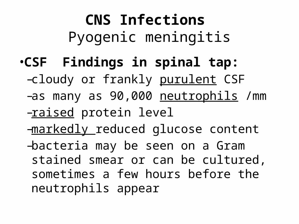
• CSF Findings in spinal tap: – cloudy or frankly purulent CSF– as many as 90,000 neutrophils /mm– raised protein level–markedly reduced glucose content–bacteria may be seen on a Gram stained
smear or can be cultured, sometimes a few hours before the neutrophils appear
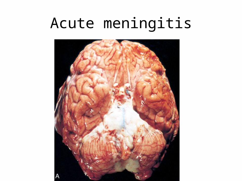
Acute meningitis
CNS Infections Meningitis Clinical Features
• Systemic non-specific signs of infection• Meningeal irritation signs and neurologic
impairment:– Headache, photophobia, irritability, clouding of
consciousness and neck stiffness• Untreated, pyogenic meningitis can be fatal• Effective antimicrobial agents markedly reduce
mortality associated with meningitis
CNS Infections Meningitis Complications
• Phlebitis may venous occlusion hemorrhagic infarction of the underlying brain
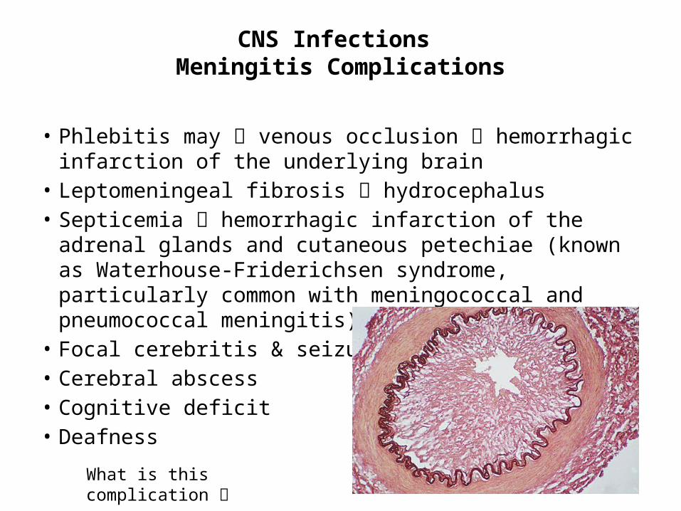
• Leptomeningeal fibrosis hydrocephalus• Septicemia hemorrhagic infarction of the adrenal glands
and cutaneous petechiae (known as Waterhouse-Friderichsen syndrome, particularly common with meningococcal and pneumococcal meningitis)
• Focal cerebritis & seizures• Cerebral abscess• Cognitive deficit• Deafness
What is this complication

CNS Infections Brain abscess
• Streptococci and staphylococci are the most common organisms identified in non-immunosuppressed populations
• Predisposing conditions: – Acute bacterial endocarditis (usually give multiple microabscesses)– Cyanotic congenital heart disease in which there is a right-to-left shunt – Loss of pulmonary filtration of organisms ( e.g, bronchiectasis)
• Most common on cerebral hemispheres
CNS Infections Brain abscess
• Morphologically, – Liquefactive necrosis– The surrounding brain is edematous , congested & contains
reactive astrocytes & perivascular inflammatory cells• Present clinically with progressive focal neurologic deficits in
addition to the general signs of raised intracranial pressure• The CSF
– Contain only scanty cells– ↑ protein– Normal level of glucose
• Complications of Brain abscess:– Herniation – Rupture of abscess into subarachnoid space or ventricle
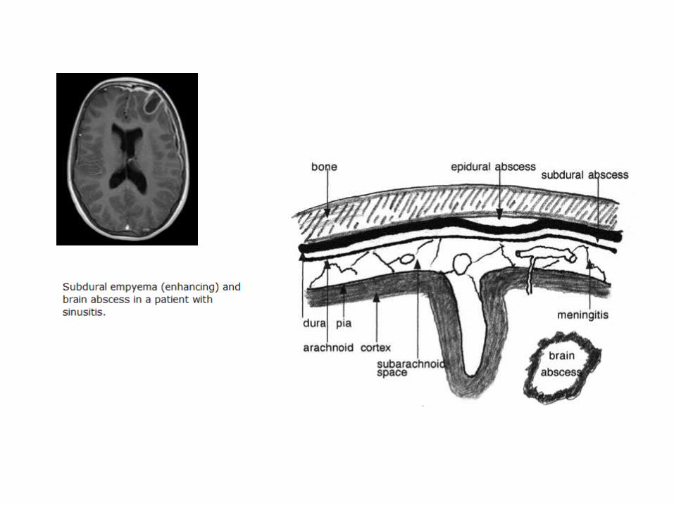
Epidural and Subdural Infections
• These spaces can be involved with bacterial or fungal infections, usually as a consequence of direct local spread
• Epidural abscess, commonly associated with osteomyelitis, arises from an adjacent focus of infection, such as sinusitis or a surgical procedure
• When the process occurs in the spinal epidural space, it may cause spinal cord compression and constitute a neurosurgical emergency
• Infections of the skull or air sinuses may also spread to the subdural space, producing subdural empyema. The underlying arachnoid and subarachnoid spaces are usually unaffected, but a large subdural empyema may produce a mass effect. In addition, thrombophlebitis may develop in the bridging veins that cross the subdural space, resulting in venous occlusion and infarction of the brain
• Symptoms include those referable to the source of the infection. Most patients are febrile, with headache and neck stiffness, and if untreated may develop focal neurologic signs, lethargy, and coma
• With treatment, including surgical drainage, resolution of the empyema occurs from the dural side; if resolution is complete, a thickened dura may be the only residual finding. With prompt treatment, complete recovery is usual

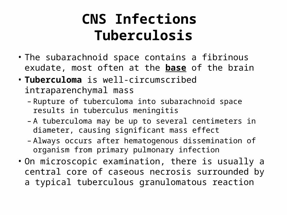
CNS Infections Tuberculosis
• The subarachnoid space contains a fibrinous exudate, most often at the base of the brain
• Tuberculoma is well-circumscribed intraparenchymal mass – Rupture of tuberculoma into subarachnoid space results in
tuberculus meningitis– A tuberculoma may be up to several centimeters in diameter,
causing significant mass effect– Always occurs after hematogenous dissemination of organism
from primary pulmonary infection• On microscopic examination, there is usually a central core
of caseous necrosis surrounded by a typical tuberculous granulomatous reaction
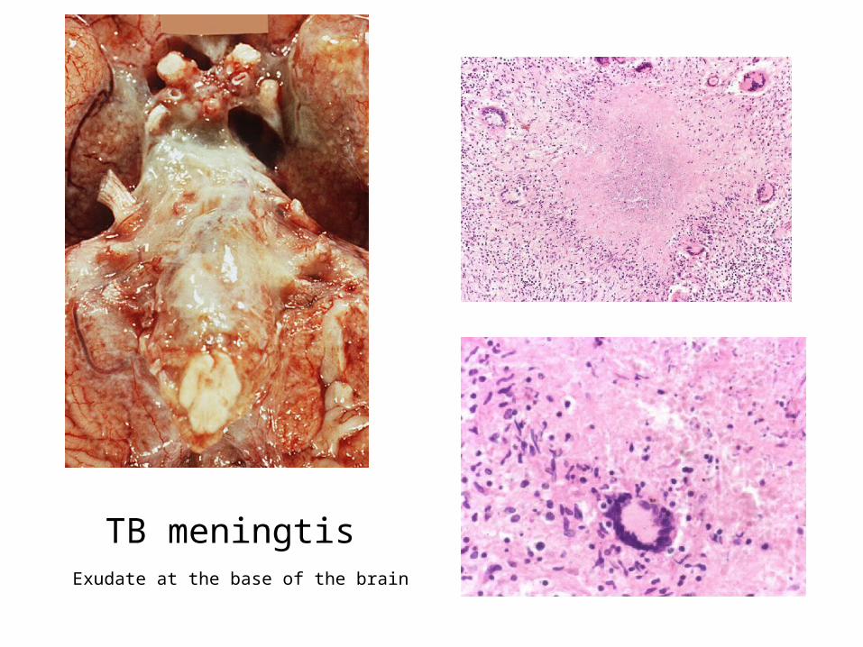
TB meningtis Exudate at the base of the brain
CNS Infections CSF in TB
• There is only a moderate increase in cellularity of the CSF (pleiocytosis) made up of mononuclear cells, or a mixture of polymorphonuclear and mononuclear cells
• The protein level is elevated, often strikingly so• The glucose content typically is moderately reduced
or normal

Aseptic Meningitis (Viral Meningitis)
• Aseptic meningitis is a misnomer• it is a clinical term for an illness comprising meningeal irritation, fever, and
alterations of consciousness of relatively acute onset without recognizable organisms
• The clinical course is less fulminant than in pyogenic meningitis, is usually self-limiting, and most often is treated symptomatically
• The CSF shows an increased number of lymphocytes (pleiocytosis), the protein elevation is only moderate, and glucose content is nearly always normal
• In approximately 70% of cases, a pathogen can eventually be identified, most commonly an enterovirus
• There are no distinctive macroscopic characteristics except for brain swelling, seen in only some instances
• On microscopic examination, there is either no recognizable abnormality or a mild to moderate infiltration of the leptomeninges with lymphocytes.
Homework
• Create a table of CSF findings in Meningitis, aseptic meningitis, TB meningitis, Brain abscess and multiple sclerosis!