Embed Size (px)
Citation preview
Research Article
IntroductionCell motility is essential for many aspects of metastasis; however,few molecular markers exist that can predict the migratory potentialof a tumor cell in vivo. Intravital multiphoton imaging in animalmodels can be used to characterize carcinoma and stromal cellbehavior within intact primary tumors in detail (Condeelis andSegall, 2003; Wang et al., 2007; Egeblad et al., 2008; Kedrin et al.,2008; Andresen et al., 2009; Perentes et al., 2009). Such imagingapproaches yield direct information at single-cell resolution andpermit quantification of cell motility, interactions between tumorand stromal cells, and direct observation of invasion, intravasationand extravasation. In mammary tumors, this technology was usedto describe the microenvironments in which tumor cells invade,migrate and intravasate, and revealed essential roles formacrophages in these events (reviewed in Condeelis and Segall,2003; Condeelis and Pollard, 2006; Yamaguchi et al., 2006; Kedrinet al., 2007). In particular, chemotaxis of tumor cells towardmacrophages is essential for invasion in mouse primary mammarytumors (Wyckoff et al., 2004; Goswami et al., 2005), whereaschemotaxis of tumor cells toward peri-vascular macrophages isessential for intravasation (Wyckoff et al., 2007).
Expression profiling of invasive tumor cells captured from theprimary tumor was used to obtain molecular information regardingthe pathways mediating carcinoma cell invasion and intravasation(Wyckoff et al., 2000a; Wang et al., 2004; Wang et al., 2007). The
‘invasion signature’ obtained from this profile revealed sets ofcoordinated expression changes associated with increased invasivepotential (Goswami et al., 2004; Wang et al., 2004; Wang et al.,2006; Wang et al., 2007; Goswami et al., 2009). Mena, a regulatorof actin polymerization and cell migration, is upregulated ininvasive tumor cells obtained from rat, mouse and human tumors(Di Modugno et al., 2006; Goswami et al., 2009; Robinson et al.,2009). Conservation of Mena upregulation in invasive tumor cellsacross species suggests that it plays a crucial role in metastaticprogression.
In patients, Mena expression correlates with metastatic risk:relatively high Mena expression has been observed in patientsamples from high-risk primary and metastatic breast tumors (DiModugno et al., 2006), as well as cervical, colorectal and pancreaticcancers compared with low-risk cases (Gurzu et al., 2008; Pino etal., 2008; Gurzu et al., 2009). Mena is also a component of amarker for metastatic risk called TMEM (tumor micro-environmentfor metastasis) (Robinson et al., 2009). TMEMs are identified byco-localization of Mena-positive tumor cells, macrophages andendothelial cells, and the TMEM score predicts risk independentlyof clinical subtype of cancer (Robinson et al., 2009). Therefore, thecontribution of Mena to metastasis is independent of clinicalsubtype.
These findings emphasize the importance of determining themechanism by which Mena and its isoforms differentially affect
SummaryWe have shown previously that distinct Mena isoforms are expressed in invasive and migratory tumor cells in vivo and that theinvasion isoform (MenaINV) potentiates carcinoma cell metastasis in murine models of breast cancer. However, the specific step ofmetastatic progression affected by this isoform and the effects on metastasis of the Mena11a isoform, expressed in primary tumor cells,are largely unknown. Here, we provide evidence that elevated MenaINV increases coordinated streaming motility, and enhancestransendothelial migration and intravasation of tumor cells. We demonstrate that promotion of these early stages of metastasis byMenaINV is dependent on a macrophage–tumor cell paracrine loop. Our studies also show that increased Mena11a expression correlateswith decreased expression of colony-stimulating factor 1 and a dramatically decreased ability to participate in paracrine-mediatedinvasion and intravasation. Our results illustrate the importance of paracrine-mediated cell streaming and intravasation on tumor celldissemination, and demonstrate that the relative abundance of MenaINV and Mena11a helps to regulate these key stages of metastaticprogression in breast cancer cells.
Key words: Breast cancer, Cell motility, Intravital imaging, Metastasis, Transendothelial migration
Accepted 18 February 2011Journal of Cell Science 124, 2120-2131 © 2011. Published by The Company of Biologists Ltddoi:10.1242/jcs.086231
Mena invasive (MenaINV) promotes multicellularstreaming motility and transendothelial migration in amouse model of breast cancerEvanthia T. Roussos1,*, Michele Balsamo2, Shannon K. Alford2, Jeffrey B. Wyckoff1,3, Bojana Gligorijevic1,Yarong Wang1, Maria Pozzuto4, Robert Stobezki5, Sumanta Goswami1,5, Jeffrey E. Segall1, Douglas A. Lauffenburger2, Anne R. Bresnick4, Frank B. Gertler2,* and John S. Condeelis1,3,*1Department of Anatomy and Structural Biology, Albert Einstein College of Medicine, Bronx, NY 10461, USA2David H. Koch Institute for Integrative Cancer Research, Massachusetts Institute of Technology, Cambridge, MA 02139, USA3Gruss Lipper Biophotonics Center, Albert Einstein College of Medicine, Bronx, NY 10461, USA4Department of Biochemistry, Albert Einstein College of Medicine, Bronx, NY 10461, USA5Department of Biology, Yeshiva University, New York, NY 10033, USA*Authors for correspondence ([email protected]; [email protected]; [email protected])
2120
Jour
nal o
f Cel
l Sci
ence
metastatic progression. Mena is a member of the Ena/VASP familyof proteins and binds actin to regulate the geometry and assemblyof filament networks through: (1) an anti-capping protein activity(Bear et al., 2002; Barzik et al., 2005; Hansen and Mullins, 2010)that involves binding to profilin and both G- and F-actin; (2) Menatetramerization, and (3) reduction in the density of actin-relatedproteins 2 and 3 (Arp2/3)-mediated branching (Gertler et al., 1996;Barzik et al., 2005; Ferron et al., 2007; Pasic et al., 2008; Bear andGertler, 2009; Hansen and Mullins, 2010). Alternative splicing forthe Mena gene has been reported: a 19 amino acid residue insertionjust after the EVH1 domain generates the Mena invasion isoform(MenaINV, formerly Mena+++) (Gertler et al., 1996; Philippar et al.,2008), whereas a 21 residue insertion in the EVH2 domaingenerates the Mena11a isoform (Di Modugno et al., 2007). Acomparison of the invasive and migratory tumor cells collected invivo, with primary tumor cells isolated from mouse, rat and humancell-line-derived mammary tumors, revealed that MenaINV
expression is upregulated and Mena11a is downregulated selectivelyin the invasive and migrating carcinoma cell population (Goswamiet al., 2009). The differential regulation of Mena isoforms acrossspecies suggests that these two isoforms have important roles ininvasion and metastasis.
In previous studies, we showed that expression of MenaINV in axenograft mouse mammary tumor promotes increased formation ofspontaneous lung metastases from orthotopic tumors and alters thesensitivity of tumor cells to epidermal growth factor (EGF) (Philipparet al., 2008). We undertook the current study to identify the step(s)in the metastatic cascade that are affected by MenaINV expressionand investigate the effect of expression of the second regulatedisoform, Mena11a, on metastatic progression. In particular, wedissected each step of metastatic progression to determine whichsteps are affected by expression of MenaINV that ultimately leads toenhancement of metastatic dissemination, and whether these samesteps are also affected by Mena11a expression in tumor cells.
We chose MTLn3 cells for our studies because they are wellcharacterized with respect to tumor cell invasion, migration andmetastasis (Levea et al., 2000; Sahai, 2005; Le Devedec et al.,2009; Le Devedec et al., 2010), tumor–stromal cell interactions(Sahai, 2005), TGF signaling in metastatic progression (Giampieriet al., 2009), and the role of Mena in breast cancer metastasis(Philippar et al., 2008; Goswami et al., 2009). MTLn3 cells arederived from the clonal selection of metastatic lung lesions fromrats with mammary tumors (Neri et al., 1982). These rat mammarytumors have been characterized as estrogen-independent and theymetastasize to the lymph nodes and lungs (Neri et al., 1982).Evaluation of vimentin and cytokeratins in MTLn3 mammarytumors, associated lymph nodes and lung metastases revealed thatMTLn3 tumor cells are comparable to a basal-like subtype ofbreast cancer (Lichtner et al., 1989).
ResultsMenaINV promotes coordinated cell migration in vivo in theform of streams of single cellsPreviously, we found that expression of Mena and MenaINV
increases in vivo cell motility, which we hypothesized contributesto the increased lung metastasis observed with these cells (Philipparet al., 2008). Different types of motility are thought to play diverseroles during tumor cell invasion (Wolf et al., 2003; Gaggioli et al.,2007; Ilina and Friedl, 2009; Friedl and Wolf, 2010), therefore wehypothesized that MenaINV expression promotes a type of motilitythat supports enhanced tumor cell invasion. To address this
2121Mena isoforms in migration and intravasation
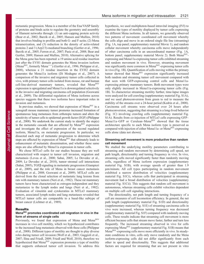
hypothesis, we used multiphoton-based intravital imaging (IVI) toexamine the types of motility displayed by tumor cells expressingthe different Mena isoforms. In all tumors, we generally observedtwo patterns of movement: coordinated cell movement wherebythe cells align and move in an ordered single file line (streaming)(Fig. 1A, top panel; supplementary material Movie 1), and randomcellular movement whereby carcinoma cells move independentlyof other carcinoma cells in an uncoordinated manner (Fig. 1A,bottom panel; supplementary material Movie 2). Both MenaINV-expressing and Mena11a-expressing tumor cells exhibited streamingand random movement in vivo. However, streaming movementwas significantly more common in MenaINV-expressing tumor cellsin vivo (Fig. 1B). Quantification of cells moving within the primarytumor showed that MenaINV expression significantly increasedboth random and streaming tumor cell movement compared withthat seen with GFP-expressing control cells and Mena11a-expressing primary mammary tumors. Both movement types wereonly slightly increased in Mena11a-expressing tumor cells (Fig.1B). To characterize streaming motility further, time-lapse imageswere analyzed for cell crawling (supplementary material Fig. S1A),and we used photoconversion from green to red to measure thestability of the streams over a 24-hour period (Kedrin et al., 2008).Carcinoma cell streams were observed over 24 hours afterphotoconversion, suggesting that streaming is a long-lived behavior(Fig. 1C) involving crawling cells (supplementary material Fig.S1A). Results from co-injection of MTLn3 cells expressing GFP–Mena11a–GFP or Cerulean–MenaINV showed that the tissuearchitecture specific to each Mena isoform type is preserved ascompared with injection of either Mena11a- or MenaINV-expressingcells alone (data not shown).
Streaming cell movement is more productive than randomcell movementWe studied the underlying motility parameters contributing tostreaming and random movement by determining cell speed, netpath length, directionality and turning frequency in vivo. In vivo,streaming cells moved significantly faster than randomly movingcells, regardless of Mena isoform expression (supplementarymaterial Fig. S1B), with average speeds of greater than 1.9m/minute. All cell types participating in random movementexhibited a narrow distribution of velocities (supplementarymaterial Fig. S1Ci), whereas cells that participated in streamingmovement had a broad distribution of velocities (supplementarymaterial Fig. S1Cii). This suggests that random cell movement isautonomous, whereas streaming cells exhibit velocities dependenton multiple cell–cell signaling interactions.
The directionality, net path length and turning frequency of acell are measures of cell locomotion efficiency. In general, the netpath length (supplementary material Fig. S1D) and directionality(supplementary material Fig. S1E) of streaming carcinoma cells invivo were increased, whereas turning frequency was decreased(supplementary material Fig. S1F) compared with randomly movingcells. These results indicate that streaming cell movement is moreefficient because cells that stream move faster, farther and turn lessfrequently. The increased streaming observed in vivo for cellsexpressing MenaINV (supplementary material Fig. S1B) means thatMenaINV-expressing cells move more efficiently in vivo. In steady-state conditions in vitro, cells only move randomly and the Menaisoform-expressing cells did not differ significantly from eachother in speed and directionality. This suggests that additionalfactors are required for streaming that are not present in vitro
Jour
nal o
f Cel
l Sci
ence

(supplementary material Fig. S2A,B). We investigated theseadditional in vivo factors and outline the results below.
In vivo invasion is enhanced by the expression of MenaINV
and suppressed by the expression of Mena11aTo determine whether the enhanced streaming exhibited byMenaINV-expressing carcinoma cells correlates with chemotaxis-dependent invasion, we used the in vivo invasion assay to evaluatethe ability of carcinoma cells to invade towards EGF in vivo(Wyckoff et al., 2000a). We showed previously that MTLn3 cellsexhibit a characteristic biphasic response to EGF whereby maximalchemotactic invasion is achieved in response to 25 nM EGF (Segallet al., 1996; Wyckoff et al., 2000a). Expression of MenaINV shiftsthis biphasic response, and maximal invasion is achieved inresponse to 1 nM EGF (Fig. 2A) (Philippar et al., 2008). This resultdemonstrates that sensitivity to EGF chemotaxis is increased invivo and is consistent with the increased sensitivity of MenaINV-expressing cells to EGF in vitro (supplementary material Fig.S3A,B) (Philippar et al., 2008). In vitro, MenaINV-expressing cellsexhibit protrusive activity in response to EGF concentrations aslow as 0.1 nM, whereas cells expressing GFP and Mena11a did not respond to stimulation with either 0.1 or 0.5 nM EGF
2122 Journal of Cell Science 124 (13)
(supplementary material Fig. S3A,B). Importantly, MenaINV
expression not only sensitizes tumor cells to EGF, but alsosignificantly increases the number of invasive cells collected withthe peak concentration of EGF, which indicates more efficient cellmigration (Fig. 2A). Mena11a-expressing tumors did not invadesignificantly above background levels in response to a broad rangeof EGF concentration (Fig. 2A). Thus, Mena11a and MenaINV
have opposite effects on chemotaxis-dependent invasion in vivo.
Expression of Mena isoforms alters paracrine loopsignaling with macrophages during in vivo invasionUsing the in vivo invasion assay, tumor cells have been observedto chemotax into needles containing either EGF or colony-stimulating factor 1 (CSF1) (Wyckoff et al., 2004; Patsialou et al.,2009). This can only occur if a paracrine signaling relay isestablished with macrophages because, in the absence ofmacrophages, the chemotatic signal cannot be transmitted overlong distances and few cells are collected (Wyckoff et al., 2004).Therefore, we looked at whether the tumor cell–macrophageparacrine loop is involved in the enhanced in vivo invasion ofMenaINV-expressing cells and the suppression of invasion inMena11a-expressing cells. Both macrophages and tumor cells enter
Fig. 1. MenaINV promotes coordinated carcinoma cell streaming withinthe primary tumor significantly more than other Mena isoforms.(A)Multiphoton microscopy images (20�) of MenaINV- and Mena11a-expressing mammary tumors in mice. Upper panels illustrate MTLn3–Cerulean–EGFP–MenaINV cells at different intervals in time, moving in astream; cells outlined in white follow the same path (direction indicatedwith white arrows in far left panel) as they move through the tumor. Lowerpanels illustrate MTLn3–Cerulean–EGFP–Mena11a cells at differentintervals of time, moving randomly; cells outlined in white are moving indifferent directions from each other (directions indicated with white arrowsin far left and middle panels). Scale bars: 25m. Green, Cerulean MTLn3cells expressing either EGFP–MenaINV or EGFP–Mena11a. Purple,collagen I second harmonic. Vector diagrams to the right illustratemovement patterns of streaming (top vector diagram) versus randomlymoving (bottom vector diagram) cells in the panels to the left. Vectordiagrams are representative of all cell types participating in eitherstreaming or random movement. (B)Average number of tumors cellsmoving randomly (white bars) or streaming (gray bars) per field quantifiedfrom IVI of primary mammary tumors derived from mammary glandinjection of cell types indicated; 30–50 fields analyzed per condition. Pvalues are indicated above bars. Error bars indicate s.e.m. (C)Multiphotonmicroscopy of MenaINV-expressing MTLn3 tumor cells coexpressingDendra2 moving coordinately in a cell stream. Images taken at 20� attime 0 (image taken immediately following Dendra2 photoconversion) and24 hours following Dendra2 photoconversion from green to red. Red arearesults from the same red photoconverted tumor cells in both images.White arrow, Denra2 photoconverted carcinoma cells in a stream. Green,Dendra2 in MTLn3–MenaINV cells. Scale bars: 50m.
Jour
nal o
f Cel
l Sci
ence

collection needles during in vivo invasion (Wyckoff et al., 2004),and typing of cells collected following in vivo invasion confirmedthe presence of both tumor cells and macrophages in collectionsfrom tumors expressing GFP, MenaINV and Mena11a (data notshown).
To assess the extent of paracrine signaling betweenmacrophages and MenaINV-expressing carcinoma cells during invivo invasion, we performed the in vivo invasion assay in thepresence of either 6.25 nM Erlotinib (Tarceva), an EGF receptor(EGFR) tyrosine kinase inhibitor, or a mouse CSF1 receptor-blocking antibody (-CSF1R) (Wyckoff et al., 2004). In vivoinvasion of both MenaINV- and GFP-expressing cells was reducedto background levels in assays with Erlotinib as compared with
2123Mena isoforms in migration and intravasation
invasion toward needles containing EGF+DMSO or EGF alone(Fig. 2B), demonstrating the requirement for EGFR-mediatedstimulation for invasion. Similarly, invasion of both MenaINV-and GFP-expressing cells decreased significantly with needlescontaining -CSF1R as compared with invasion toward needlescontaining EGF+DMSO or EGF+IgG antibodies (control IgG)(Fig. 2C), thus suggesting the necessity of EGF production andsignal propagation by macrophages. These results are consistentwith the requirement for co-migrating macrophages in tumor cellmigration, as shown in Fig. 3D and discussed below. Together,these results demonstrate the requirement for paracrine signalingbetween MenaINV-expressing tumor cells and macrophages invivo.
Fig. 2. MenaINV and Mena11a haveopposite effects on invasion in vivo and invitro. (A)In vivo invasion assay: EGF dose-response curve of cells collected fromprimary mammary tumors derived frommammary gland injection of indicated celltypes. Values represent averages of 15–25needles per xenograft model. (B)In vivoinvasion in the presence of Erlotinib. Cellswere collected from mammary tumorsderived from mammary gland injection ofeach cell type. Bars represent contents of15–25 needles per xenograft model. (C)Invivo invasion in the presence of CSF1R-blocking antibody (-CSF1R): Cellscollected from mammary tumors derivedfrom mammary gland injection of each celltype. Bars represent contents of 15–25needles per xenograft model. (D)Real timePCR of cell lines for expression of CSF1.Each bar represents n3 for threeindependent experiments. (E)In vitro 3Dinvasion assay. Proportion of GFP- andMena11a-expressing cells invading collagenin the absence (white bars) or presence(gray bars) of macrophages. Each barrepresents n3. P values are indicated abovebars. Error bars indicate s.e.m.
Jour
nal o
f Cel
l Sci
ence

Both CSF1 secretion and EGF binding to the EGFR bycarcinoma cells are essential components of the carcinoma cell–macrophage paracrine loop (Wyckoff et al., 2004; Patsialou et al.,2009). We used real time PCR to examine the relative mRNAexpression of CSF1 and EGFR in the Mena isoform-expressingcells lines in culture to determine whether changes in the expressionof these signaling molecules could contribute to the observeddifferences in EGF-dependent in vivo invasion (Wyckoff et al.,2004). Mena11a-expressing cells showed a fourfold decrease inCSF1 expression as compared with GFP-expressing cells, whereasCSF1 expression in Mena- and MenaINV-expressing cells wasunchanged (Fig. 2D). We previously reported no difference inEGFR expression in cells expressing either Mena or MenaINV ascompared with MTLn3 cells (Philippar et al., 2008). Cellsexpressing Mena11a also showed no statistical difference inexpression of EGFR compared with GFP- and MenaINV-expressingcells (supplementary material Fig. S4), indicating that alteredreceptor levels do not contribute to the altered EGF-dependentphenotypes observed in the different cell types. Thus, the inabilityof Mena11a-expressing cells to participate in macrophage-dependent invasion might arise in part due decreased CSF1expression along with a reduction in direct response EGF (Fig.2D).
2124 Journal of Cell Science 124 (13)
To determine whether suppression of chemotaxis-dependentinvasion of Mena11a-expressing tumor cells resulted fromdifferences in the ability of these tumor cells to co-migrate withmacrophages, we used a 3D invasion assay that measuresmacrophage-dependent co-migration of carcinoma cells withmacrophages in collagen (Goswami et al., 2005). Although additionof macrophages to GFP-expressing tumor cells significantlyincreased 3D invasion, addition of macrophages to Mena11a-expressing cells did not significantly increase invasion (Fig. 2E).This is consistent with the reduced response to EGF and thereduced CSF1 expression levels in Mena11a-expressing cells thatfall below the threshold needed to stimulate the pro-invasivemacrophage behavior (Fig. 2D).
MenaINV-expressing carcinoma cell streaming requires thepresence of macrophages and paracrine signalingBecause the enhanced invasion of MenaINV-expressing carcinomacells depends on paracrine loop signaling with macrophages, andthe paracrine loop has been shown to be required for tumor cellmigration in mammary tumors (Wyckoff et al., 2004), wehypothesized that the paracrine loop also drives carcinoma cellstreaming in vivo. Using IVI, we observed that multiple carcinomacells moved in streams among host cell shadows previously
Fig. 3. Macrophages co-migrate with carcinoma cells during coordinated cellmigration as part of the migratory stream. (A)Multiphoton microscopy ofcarcinoma cells (green cells outlined in white) moving coordinately in a stream withhost immune cells (black shadow outlined in orange). Images taken at 20� over 30minutes. Arrows point to host immune cells. Arrowheads point to carcinoma cells.Green, Cerulean MTLn3 cells expressing EGFP–MenaINV. Purple, collagen I secondharmonic. Scale bars: 25m. (B)Multiphoton microscopy of carcinoma cells (greencells outlined in white) and Texas Red dextran-labeled macrophages (red cellsoutlined in orange) moving in a stream. Green, Cerulean MTLn3 cells expressingEGFP–MenaINV. Purple, collagen I second harmonic. Red, Texas Red-labeleddextran. Images taken at 20� over 30 minutes. Scale bars: 25m.(C)Immunohistochemistry of EGFP–MenaINV primary tumor section stained forcarcinoma cells (pink) and macrophages (gray), imaged at 63�. Pink, EGFP–MenaINV within MTLn3–EGFP–MenaINV cells. Gray, F4/80 within macrophages.Green, nuclear counterstain. (D)Normalized number of cells streaming per fieldquantified from IVI of primary mammary tumors derived from mammary glandinjection of MTLn3–EGFP–MenaINV treated with the indicated reagents; 30–50fields analyzed per condition. P values indicated above bars. Error bars indicates.e.m.Jo
urna
l of C
ell S
cien
ce

identified as immune cells and macrophages (Wyckoff et al., 2000b;Wyckoff et al., 2007) (Fig. 3A; supplementary material Movie 3).Intravenous injection of Texas Red dextran during IVI indeedconfirmed the identity of the host cell shadows in tumor cellstreams as macrophages, because macrophages uniquely take updextran delivered intravenously into mammary tumors over thetime interval of these experiments (Wyckoff et al., 2007) (Fig. 3B;supplementary material Movie 4). Additionally,immunohistochemistry of fixed primary tumors identifiedmacrophages intercalated between carcinoma cells in cell streams(Fig. 3C).
To determine whether streaming required macrophages, micewere treated with clodronate liposomes 48 hours prior to IVI todecrease levels of functional macrophages (Hernandez et al., 2009).A 70% decrease in the number of Texas Red dextran-labeledmacrophages was observed in animals treated with clodronateliposomes compared with those treated with PBS liposomes(supplementary material Fig. S5). IVI of primary tumorsdemonstrated that there were 90% fewer streaming cells in micetreated with clodronate liposomes as compared with controls,confirming the involvement of the macrophage–tumor cell paracrineloop in streaming (Fig. 3D). To confirm the involvement of theparacrine loop in streaming, mice were treated with Erlotinib 2hours prior to IVI to block the EGFR on tumor cells (Zerbe et al.,2008) or with a CSF1R antibody 4 hours prior to IVI to block theCSF1R on macrophages (Wyckoff et al., 2007). IVI of primarytumors demonstrated that there were 65% fewer streaming cells inmice treated with Erlotinib and 80% fewer streaming cells in micetreated with -CSF1R as compared with tumors (Fig. 3D).
Expression of MenaINV in tumor cells leads to increasedtransendothelial migration and intravasationGiven that our data shows increased streaming (Fig. 1B) andinvasion (Fig. 2A) in MenaINV-expressing cells we hypothesizedthat expression of MenaINV might also enhance intravasation toincrease metastasis. Following intravenous injection of Texas Reddextran, IVI of MenaINV-expressing tumors showed that tumor cellstreaming was indeed directed toward blood vessels (Fig. 4A;supplementary material Movie 5). We then quantified theintravasation efficiency of tumors expressing MenaINV andMena11a using IVI of photoconverted carcinoma cells adjacent toblood vessels to determine the percentage of tumor cellsintravasating over a 24-hour period (Fig. 4B) (Kedrin et al., 2008).Quantification of the change in the photoconverted tumor area 24hours after photoconversion showed that 95% of Mena11a-expressing cells remained in the converted area as compared with75% of carcinoma cells expressing MenaINV (Fig. 4Bi,ii).Additionally, we evaluated the tumor cell blood burden to measureintravasation in vivo (Wyckoff et al., 2000b). Mice with MenaINV-expressing tumors had a fourfold increase in the number ofcarcinoma cells in circulation compared with mice with GFP- orMena11a-expressing tumors (Fig. 4C). Mena11a-expressingxenograft mice had similar numbers of circulating carcinoma cellsas compared with GFP-expressing xenograft mice (Fig. 4C).
Given that previous studies showed that interaction betweentumor cells and perivascular macrophages is required forintravasation (Wyckoff et al., 2007), and that our studies show thatenhanced MenaINV cell streaming and invasion are paracrine-dependent, we hypothesized that enhanced intravasation inMenaINV-expressing cells might also be paracrine-dependent. Toexamine the minimum requirements for macrophage-assisted
2125Mena isoforms in migration and intravasation
intravasation we used a subluminal-to-luminal transendothelialmigration (TEM) assay in which we could vary the presence ofmacrophages to determine their need for carcinoma cellintravasation (Fig. 4D). Interestingly, less than 0.5% of carcinomacells traversed the endothelium in the absence of macrophages,regardless of Mena isoform expression (Fig. 4E). Addition ofmacrophages did not enhance TEM for cells expressing GFP orMena11a (Fig. 4E). Remarkably, in the presence of macrophages,54% of MenaINV-expressing cells traversed the endothelium, a200-fold increase in TEM as compared with all other cell types(Fig. 4E).
To test the paracrine dependence of intravasation in vivo, weassessed the number of circulating tumor cells following functionalimpairment of macrophages achieved by treatment of mice withclodronate liposomes or CSF1R-blocking antibody, or impairmentof tumor cells by treatment with Erlotinib. Tumors formed frominjection of MenaINV-expressing cells showed a significant decreasein circulating tumor cells following treatment with clodronateliposomes, CSF1R blocking antibody and Erlotinib as comparedwith those treated with respective controls (Fig. 4F).
Expression of MenaINV, but not Mena11a, increasesintravasation, dissemination and lung metastasisTo determine the mechanistic consequence of enhancedtransendothelial migration and intravasation by MenaINV expressionor the suppression of invasion by Mena11a expression, weinvestigated the ability of these cells to extravasate, disseminateand metastasize to the lung. An experimental metastasis assay wasused as a measure of extravasation (Xue et al., 2006). Micro-metastases in the lungs were counted after intravenous injection ofGFP-, Mena11a- or MenaINV-expressing cells. The metastaticburden was similar for all cell lines (Fig. 5A).
Previous studies have shown that MTLn3 tumor cells forced toexpress MenaINV show a significant increase in the number ofmetastases formed (Philippar et al., 2008). Thus, we asked whetherdissemination of single tumor cells to the lung, a step precedinggrowth of macrometastases, was affected in xenograft mice derivedfrom injection of cells expressing MenaINV or Mena11a. Mice withMenaINV xenografts had significantly increased carcinoma celldissemination to the lungs compared with animals bearing eitherMena11a- or GFP-expressing tumors (Fig. 5B). Mice with Mena11axenografts had half as many cells in the lungs as mice bearingGFP-expressing tumors (Fig. 5B).
Given that MenaINV expression increases tumor celldissemination and that Mena11a expression decreases tumor celldissemination, we hypothesized that Mena isoform expressionwould similarly affect the final step in metastatic progression: theformation of spontaneous metastasis. Spontaneous metastases tothe lungs were scored in mice with mammary tumors at either 3 or4 weeks after mammary gland injection of GFP-, Mena11a- andMenaINV-expressing cells. Expression of MenaINV increased theincidence of metastasis compared with expression of GFP andMena11a, whereas expression of Mena11a decreased metastasesafter 3 weeks of tumor growth (Fig. 5C). However, after 4 weeksof tumor growth, all primary tumors resulted in detectablemetastases regardless of the Mena isoform expressed (Fig. 5C). Inaddition, MenaINV expression promoted metastatic spread to thelungs with little (at 3 weeks) or no (at 4 weeks) effect on primarytumor growth (supplementary material Fig. S6A,B) (Philippar etal., 2008) or cell growth in vitro (supplementary material Fig.S6C). Hence, differences in tumor metastasis occurring in tumors
Jour
nal o
f Cel
l Sci
ence

with different Mena isoform expression are not an indirectconsequence of tumor growth. These data indicate that the increasedincidence of spontaneous metastasis observed in MenaINV-expressing tumors is due to metastatic events occurring prior toextravasation.
DiscussionIncreased Mena expression is correlated with metastasis in breastcancer patients (Di Modugno et al., 2006). In particular, duringinvasion and migration of tumor cells, expression of MenaINV
increases whereas that of Mena11a decreases (Goswami et al.,2009). In this study, we have identified invasion, migration andintravasation as crucial steps of metastasis that are affected byexpression of MenaINV- and Mena11a-expressing tumor cells. Akey characteristic of MenaINV-expressing cells is their contributionto cell streaming and enhanced intravasation as a result of thedramatic increase in transendothelial migration. Another importantfinding is the effect of MenaINV expression on tumor cell sensitivity
2126 Journal of Cell Science 124 (13)
to macrophage-supplied EGF and the subsequent enhancement ofparacrine-mediated invasion. Our findings ultimately suggest thatthe EGF-dependent enhancement of invasion and intravasation inMenaINV-expressing tumor cells contributes to increased tumorcell dissemination and spontaneous metastasis to the lungs.
Conversely, we found that Mena11a-expressing cells do notshow dramatically increased streaming and fail to co-invade withmacrophages, which indicates a reduced paracrine signalinginteraction. The decrease in CSF1 expression in Mena11a-expressing cells contributes to impaired paracrine signaling andleads to the observed deficits that depend on this paracrine signalingloop in vivo, including streaming, invasion, transendothelialmigration, tumor cell dissemination and spontaneous metastasis tothe lungs.
During invasion, tumor cells are known to decrease theirexpression of Mena11a and begin producing the MenaINV isoform(Goswami et al., 2009). We have shown that Mena11a expressionis correlated with decreased EGF-induced in vivo invasion. We
Fig. 4. MenaINV cells promote macrophage-dependent transendothelial migration.(A)Multiphoton microscopy of MTLn3–EGFP–MenaINV cells moving towards a blood vessel within theprimary mammary tumor of a mouse over 30 minutes.Scale bars: 25m. Green, MTLn3–EGFP–MenaINV.Red, Texas Red dextran-labeled blood vessels. Whitearrow indicates direction of cell movement. Asteriskindicates location of blood vessel. See supplementarymaterial Movie 5. (Bi)Quantification of percentage ofDenra2 photoconverted tumor cells remaining in theconverted area located near a vessel at 0 and 24 hours.(Bii) Multiphoton microscopy of a primary tumor.Images taken at 20× over 30 minutes. Red area,Dendra2 photoconverted MenaINV tumor cells at 0 and24 hours. Green, Dendra2–MenaINV tumor cells. Whiteoutline, blood vessel. White bracket indicates areaevaluated at each time point. Scale bars: 50m.(C)Average number of single cells from 1 ml of bloodfrom mice with GFP, MenaINV and Mena11a mammaryxenografts; n10 animals per condition. (D)Cartoondepicting TEM assay. Pink cells, endothelial cells. Solidgray line, Matrigel. Dotted gray line, transwellmembrane. Red cells, macrophages (BAC1.2 cells).Green cells, carcinoma cells. (E)Quantification of TEMof each cell type in the absence or presence ofmacrophages (M); n3 experiments each done induplicate. (F)Average number of circulating carcinomacells in tumors derived from injection of MTLn3–EGFP–MenaINV cells following the indicated treatment;n10 experiments per condition. P values indicatedabove bars. Error bars indicate s.e.m.
Jour
nal o
f Cel
l Sci
ence

have also shown that MenaINV-expressing migratory carcinomacells are highly sensitive to EGF in their protrusion and chemotaxisactivities, leading to significantly enhanced in vivo invasion. Theseactivities can result in MenaINV-expressing cell migration towards,and association with, perivascular macrophages, resulting inenhanced transendothelial migration and intravasation.
In addition to decreased EGF-induced in vivo invasion ofMena11a-expressing cells, we also found that these cells expressless CSF1 mRNA. Data from patients suggests that CSF1 and itsreceptor play crucial roles during progression of breast cancer(Kacinski et al., 1991; Scholl et al., 1994) and that CSF1 and theCSF1R are coexpressed in >50% of breast tumors (Kacinski, 1997).Elevated circulating CSF1 was also suggested to be an indicator ofearly metastatic relapse in patients with breast cancer, independentof breast cancer subtype (Scholl et al., 1994; Tamimi et al., 2008;Beck et al., 2009). This suggests that lower levels of CSF1 inMena11a-expressing cells could lead to decreased metastaticprogression. The decreased invasion, intravasation anddissemination of Mena11a-expressing cells are consistent with thedecrease in expression of CSF1 and the reduced sensitivity to EGF,which would make these cells less likely to participate in a paracrinesignaling loop with macrophages.
2127Mena isoforms in migration and intravasation
A major finding of our study is that the expression of MenaINV
enhances a form of coordinated cell migration not previouslydescribed, where cell migration is spatially and temporallycoordinated between carcinoma cells that are not connected byjunctions. We call this newly described form of coordinated cellmigration ‘streaming’. Streaming differs from previously describedforms of coordinated cell migration, which require the stableretention of cell–cell junctions (Sahai, 2005), because streamingcells need not make contact and the velocities of migration are 10–100 times more rapid. Previous studies have shown that in vivoMTLn3 cells express CSF1 and EGFR, but do not produce CSF1Ror EGF, whereas macrophages express CSF1R and EGF but do notproduce CSF1 or EGFR (Goswami et al., 2005). Thereforecoordinated arms of the paracrine signaling pathways are active inboth cell types during invasion in vivo (Wyckoff et al., 2004). Inour study, we demonstrate that streaming requires paracrinechemotaxis between carcinoma cells and macrophages. The abilityof MenaINV-expressing cells to protrude and chemotax to 25- to50-fold lower concentrations of EGF than parental tumor cells, andto suppress cell turning in streams, undoubtedly contributes to theextraordinary coordination and maintenance of high velocitymigration as cell streams in vivo. We propose that the increasedsensitivity of MenaINV-expressing cells to EGF in the EGF–CSF1paracrine loop is responsible for the increase in streaming motility.This conclusion is supported by the inhibition of streamingfollowing blocking of the EGFR by Erlotinib, or of CSF1R by -CSF1R (Fig. 3D).
Invasive tumor cells from PyMT mice exhibit increased MenaINV
expression and decreased expression of Mena11a (Goswami et al.,2009). Interestingly, recent studies using intravital imaging ofmammary tumors in Mena-deficient PyMT mice have shownsignificantly decreased streaming motility of tumor cells, providingfurther evidence that Mena contributes to enhanced motility(Roussos et al., 2010). Finally, mammary tumors derived from thehuman breast cancer cell line, MDA-MB-231, have tumor cellsthat participate in macrophage–tumor cell paracrine-mediatedinvasion (Patsialou et al., 2009), and these invasive tumor cellshave also been shown to differentially upregulate MenaINV anddownregulate Mena11a (Goswami et al., 2009). Together, thesefindings suggest that paracrine-mediated carcinoma cell streamingis a generalized phenomenon that occurs in rat, mouse and humanmodels of breast cancer and is a consequence of the differentialregulation of the Mena isoforms.
In our study, the suppression of invasion and streaming by theinhibition of paracrine signaling between macrophages and tumorcells in vivo, and by decreasing macrophage function in vivo,demonstrates the crucial role of macrophages during coordinatedmigration of MenaINV-expressing cells (Wyckoff et al., 2007;Hernandez et al., 2009). We also demonstrate that macrophages areessential for transendothelial migration of MenaINV-expressingtumor cells. Our results are consistent with previous work showingthat paracrine signaling between tumor cells and macrophages, andthe presence of perivascular macrophages in the primary tumor,are required for invasion and intravasation, respectively (Wyckoffet al., 2004; Wyckoff et al., 2007). In particular, our results supportprevious work suggesting that MenaINV- but not Mena11a-expressing tumor cells specifically contribute to intravasation ofbreast cancer cells in humans by helping to assemble themacrophage-dependent intravasation compartment called TMEM(Robinson et al., 2009; Roussos et al., 2011).
Fig. 5. MenaINV enhances dissemination of tumor cells and spontaneousmetastasis to the lungs. (A)Experimental lung metastasis quantified afterintravenous injection of GFP-, Mena11a- or MenaINV-expressing cells showingno statistically significant difference; n10 animals per cell type. (B)Averagenumber of single tumor cells disseminated into the lungs of mice with GFP,Mena11a or MenaINV mammary xenografts; n10 animals per condition.(C)Number of animals with spontaneous lung metastases in mice with GFP,Mena11a and MenaINV mammary xenografts following primary tumor growth.P values indicated above bars. Error bars indicate s.e.m.
Jour
nal o
f Cel
l Sci
ence

2128 Journal of Cell Science 124 (13)
In vivo, we have shown that MenaINV-expressing cells invadetowards very low concentrations of EGF in macrophage-dependentparacrine chemotaxis. In vitro, low concentrations of EGF such asthat found in serum, lead to macrophage-independent 3D invasionof MenaINV-expressing cells (Philippar et al., 2008), whereascompletion of transendothelial migration requires EGF suppliedby macrophages (Wyckoff et al., 2004). The effects of MenaINV
expression on EGF-dependent processes lead to increased invasion,intravasation, dissemination and metastasis to the lungs. Thesedata suggest that drugs directed specifically to the inhibition ofMenaINV-dependent increased EGF sensitivity will disrupt theparacrine interactions with macrophages required for metastasis,and result in the inhibition of metastasis in mammary tumors.
In this regard, it will be important to understand how the Menaisoforms differ functionally. The INV exon is inserted just after theEVH1 domain, which is primarily responsible for the subcellularlocalization of Ena/VASP proteins and interactions with severalsignaling proteins such as Lamellipodin (Gertler et al., 1996;Urbanelli et al., 2006; Pula and Krause, 2008). It is thereforepossible that the INV exon might influence the function of MenaINV
by regulating its EVH1-mediated interactions (Niebuhr et al., 1997;Boeda et al., 2007). The 11a exon is inserted within the EVH2domain between the F-actin binding motif and the coiled-coiltetramerization site. F-actin binding is crucial for almost all knownEna/VASP functions, including localization to the tips oflamellipods and the ability to drive filopod and lamellipodformation and extension (Gertler et al., 1996; Loureiro et al., 2002;Applewhite et al., 2007). In vitro, F-actin binding is required forthe anti-capping activity of Ena/VASP and is disrupted byphosphorylation at nearby sites (Barzik et al., 2005), as is F-actinbundling. Because 11a is inserted in the analogous region of Mena,it will be interesting to determine whether the barbed end captureactivity is affected. Because the 11a insertion is phosphorylated(Di Modugno et al., 2006), it is possible that inclusion of the 11aexon provides a regulatory mechanism for Mena11a.
Future studies will be needed to investigate the molecular andbiochemical mechanisms of action of the MenaINV and Mena11aisoforms and their potential utility as a prognostic marker forpatient outcome, and as a therapeutic target for breast cancermetastasis.
Materials and MethodsCell linesWe used MTLn3 cells derived from metastatic lung lesions from rat mammaryadenocarcinoma derived following dietary administration of 7,12-dimethylbenz[]anthracene (tumor line 13762) (Neri et al., 1982). The in vivobiologic characteristics and metastatic potential of these cells have been determinedin previous studies and show that MTLn3 cells have a high metastatic potential (Neriet al., 1982; Welch et al., 1983). Western blots, immunofluorescence and fluorescence-activated cell sorting (FACS) confirm that MTLn3 cells express ErbB2, ErbB3(Levea et al., 2000; Xue et al., 2006; Kedrin et al., 2009) and ErbB4 (personalcommunication Sumanta Goswami, Yeshiva University, and Michele Balsamo, MIT,Cambridge, MA). Previous studies have also confirmed that EGFRs in MTLn3 cellsare fully active and homogenously distributed on the plasma membrane (Lichtner etal., 1992; Bailly et al., 2000). Dominant coexpression of vimentin and CK14 inMTLn3 cells suggests that these cells represent a myoepithelial or basal cell that haspartially dedifferentiated (Lichtner et al., 1991). Additionally, MTLn3 cells havebeen shown to have increased expression of EGFR as compared with the non-metastatic MTC clone derived from the same mouse model (Lichtner et al., 1995;Levea et al., 2000). Taken together, these data indicate that MTLn3 cells have aphenotype similar to basal-like tumors. MTLn3 cells were used in these experimentsbecause they are known to metastasize to the lung when injected into the mammarygland of SCID mice and thus are suitable for metastatic studies. Additionally, thesecells have been used to study metastasis by several laboratories, and Mena isoformsin particular (Wyckoff et al., 2000b; Wyckoff et al., 2000a; Sahai, 2005; Philippar etal., 2008; Le Devedec et al., 2010).
Molecular cloning, infection, FACS and cell cultureEGFP–Mena splice isoforms were subcloned into the retroviral vector packagingMurine stem cell virus–EGFP using standard techniques. MTLn3 cells were used forall of the experiments described. MTLn3–Cerulean EGFP–Mena splice isoformswere created using a lentiviral system pCCLsin.PPT.hPGK.Cerulean.pre (courtesyof Sanjeev Gupta, Albert Einstein College of Medicine). MTLn3-Dendra2 cells werecreated using a Dendra2 cloning vector C1 with G418 selection marker (courtesy ofVladislav Verkhusha, Albert Einstein College of Medicine). Transfection of Dendra2into MTLn3–EGFP–MenaINV and MTLn3–EGFP–Mena11a cells lines was doneusing Lipofectamine 2000 (Invitrogen). Retroviral packaging was performed aspreviously described (Bear et al., 2000). MTLn3–EGFP–MenaINV and MTLn3–EGFP–Mena11a cell lines were FACS sorted to a level of fourfold overexpressionof each fusion protein. Sorting of all other cells was done using FACS 72 hours aftertransfection. The 10% brightest population of infected cell lines were kept forculturing, and selection was maintained using 500 g/ml G418 geneticin (Invitrogen)when necessary. MTLn3 cells were cultured in -modified minimum essentialmedium (-MEM) supplemented with 5% fetal bovine serum (FBS) and 0.5%PenStrep (Invitrogen).
Animal model and assays for metastatic progressionIn vivo studies were performed in orthotopic tumors derived from injection ofMTLn3 rat adenocarcinoma cells into SCID mice. MTLn3 cells were engineered toexpress either EGFP (for controls) or Mena isoform EGFP-fusion proteins: EGFP–MenaINV (MenaINV) and EGFP–Mena11a (Mena11a) at roughly four times thenormal level, consistent with the level of spontaneous Mena upregulation in invasivetumor cells in vivo (Philippar et al., 2008; Goswami et al., 2009). SCID mice withtumors derived from injection of these cells into the mammary gland are referred toas GFP, MenaINV or Mena11a xenografts. Tumor cell blood burden was evaluatedfollowing 4 weeks of tumor growth as previously described (Wyckoff et al., 2000b).Briefly, blood was drawn from the right ventricle of anesthetized mice and cells wereplated in -MEM media. Following 7 days of cell culture, tumor cells were counted.All cells counted were GFP-positive, confirming their identity as tumor cells. Tofunctionally impair macrophages, mice were treated with 100 l of PBS (control) orclodronate (Cl2MDP)-containing liposomes per 10 g of weight via tail vein injection24 hours prior to collection of blood (Hernandez et al., 2009). Liposomes wereprepared as previously described using a clodronate concentration of 2.5 g/10 ml ofPBS (van Rooijen and van Kesteren-Hendrikx, 2003). Clodronate was a gift ofRoche Diagnostics (Mannheim, Germany). Phosphatidylcholine (Lipoid E PC) wasobtained from Lipoid (Ludwigshafen, Germany). Cholesterol was purchased fromSigma. Mice were treated with CSF1R blocking antibody or IgG antibody 4 hoursprior to collection of blood to block signaling between tumor cells and macrophages(Wyckoff et al., 2004). Single tumor cell dissemination to the lung was quantifiedex vivo using epifluorescence in ten random high power fields per set of mouselungs at 3 weeks after mammary gland injection of each cell line. Spontaneous lungmetastases (>2 mm) were evaluated ex vivo using epifluorescence at 3 and 4 weeksafter mammary gland injection of MenaINV- or Mena11a-expressing cells aspreviously described (Philippar et al., 2008). Experimental metastases were evaluatedex vivo 2 weeks after tail vein injection of 5�105 cells of each cell type as previouslydescribed (Wang et al., 2006). Lungs were examined using a 60� 1.2 NA waterimmersion correction lens on an inverted Olympus IX70 (Wyckoff et al., 2000b).For each experiment described above, 10–15 animals were used per Mena isoformcell type. All experiments involving animals were approved by the Einstein Institutefor Animal Studies.
In vivo invasion assay and in vitro 3D invasion assayThe in vivo invasion assay was performed in 5–10 mice per condition as previouslydescribed (Wyckoff et al., 2000a). We have previously measured the ranges of EGFand CSF1 concentrations required to initiate chemotaxis and migration in vivo(Wyckoff et al., 2004; Patsialou et al., 2009; Raja et al., 2010). The concentrationsshown to be effective and used in our study vary from 50% to 10% of the Kd of theseligands for their respective receptors. In addition, these are the concentrationsbelieved to be present in vivo (Byyny et al., 1974; Bartocci et al., 1986). Theparacrine loop was inhibited using 10 g of affinity-purified -mouse CSF1R-blocking antibody (-CSF1R; courtesy of Richard Stanley, Yeshiva University,Bronx, NY) or 6.25 nM Erlotinib (gift from OSI Pharmaceuticals, Melvile, NY),empirically determined by OSI Pharmaceuticals, dissolved in DMSO in needlescontaining 25 nM and 1 nM EGF for GFP- and MenaINV-expressing cells, respectively(Wyckoff et al., 2004); -rat IgG or DMSO were used as controls, respectively. Cellswere imaged on an Olympus IX70 inverted microscope with a 10� NA 0.30objective. For in vivo invasion experiments performed with EGFR (Erlotinib) andCSF1R inhibitors, we used 1 nM EGF in the needles inserted into MenaINV-expressingtumors and 25 nM EGF in the needles inserted into GFP-expressing tumors to allowmaximal tumor cell collection (350–600 cells collected per needle) (Philippar et al.,2008). In vitro 3D invasion assays were performed as previously described (Goswamiet al., 2005).
Intravital imagingIntravital multiphoton imaging was performed as described previously (Wang et al.,2002; Wyckoff et al., 2010) using a 20� 1.95 NA water immersion objective with
Jour
nal o
f Cel
l Sci
ence

correction lens. Time-lapse movies were analyzed for frequency of motility andtracking, and for measuring and quantifying of cell characteristics in 3D and throughtime using NIH ImageJ (Sahai et al., 2005) and custom software described elsewhere(Wyckoff et al., 2010). A cell movement event was defined as a translocation of >1cell diameter (25m) observed within a visual field that is defined in three dimensionsas 100 m by 512�512 pixels per minute. Streaming cells were quantified as thenumber of individual carcinoma cells in a field whose vector paths point in the samedirection (Fig. 1A). Random cells movements were quantified as the number ofindividual carcinoma cells in a field whose vector paths point in different directions(Fig. 1A). To confirm the elimination of macrophages in clodronate-treated mice,multiphoton microscopy of spleens removed from both clodronate- and PBSliposome-treated animals was performed as previously described (Hernandez et al.,2009). Mice were given intraperitoneal injection of 10 mg/kg body weight of vehicle(6% Captisol) or Erlotinib in 6% Captisol, 2 hours prior to IVI, or 2.5 g of CSF1R-blocking anitbody or IgG in PBS 4 hours prior to IVI. Tail vein injection of 200 l70 kDa Texas Red dextran was used to label blood vessels, which were immediatelyvisualized following injection. After 2 hours, macrophages were also labeled withdextran as previously described (Wyckoff et al., 2007).
Mammary imaging windows were placed over the mammary gland of mice 3weeks after injection of carcinoma cells (Kedrin et al., 2008). Photoconversion ofthe Dendra2 variant from green to red was done using a 405 nm UV laser on a LeicaTCS SP2 AOBS confocal microscope (Mannheim, Germany) equipped with a 20�glycerol objective (Kedrin et al., 2008). Z-stacks (~100 m deep) of the same fieldswere imaged at 0 and 24 hours after photoconversion in imaging sessions using amultiphoton microscope equipped with a MaiTai 15 W laser tuned to 1045 nm forred Dendra2 and a Tsunami 15 W laser tuned to 880 nm for green Dendra2 (Kedrinet al., 2008). Quantification of intravasation was done as previously described(Kedrin et al., 2008).
Real time PCRRNA was extracted from all cell lines at steady state, and quantitative real time PCRwas performed as previously described (Goswami et al., 2009). Primers designedagainst Rat EGFR and CSF1 were used to determine levels of expression of each.Primer sequences used for EGFR: rEGFR forward 5�-TCGTTGCCGACGCAGT-CACC-3�, rEGFR reverse 5�-TCCCTGAGGGTCGCATCCCG-3�. Primer sequencesused for CSF1: rCSF-1 forward 5�-GCTCGAGGGCAAGAAAAGTA-3�, rCSF-1reverse 5�-CCTGTGTCGGTCAAAGGAAT-3�.
Transendothelial migration assayTo prepare the endothelial monolayer, the underside of each transwell was coatedwith 50 l of 2.5 g/ml Matrigel (Invitrogen). Some 200,000 rat pulmonarymicrovascular endothelial cells (RLE) were plated in 50 l of DMEM + 10% FBS(medium 1) and incubated at 37°C for 4 hours. Transwells were flipped onto a 24-well plate containing 1 or 200 l of medium 1 in the lower or upper chambers,respectively. BAC1.2F5 macrophages and tumor cells were labeled with cell-trackerdyes. Then, 15,000 macrophages and 37,500 tumor cells were added to the upperchamber in 200 l of -MEM + 0.5% FBS (medium 2), and 200 l of -MEM +10% FBS + 3000 units CSF1 were added to the lower chamber. Following 18 hoursof transmigration, medium was removed from the top of the transwell and migratedcells were scraped from the bottom of the plate. Cells were fixed in 0.1%formaldehyde and analyzed using a Guava flow cytometer. Monolayers were testedfor permeability both before and after cell migration via quantification of fluorescence(Molecular Devices SpectraMax M5 plate reader) of 70 kDa rhodamine-dextran inthe media from both upper and lower transwell chambers added 24 hours prior.
ImmunohistochemistryPrimary tumors were fixed in 10% buffered formalin, and paraffin embedded.Sections of 10 m were cut and placed on slides for further staining.Immunohistochemistry was carried out using -GFP at 1:200 to identify tumor cellscontaining GFP-fusion proteins, and rat -F4/80 (courtesy of Jeffrey Pollard, AlbertEinstein College of Medicine) was used at 1:25 to identify macrophages. All stainingwas done using standard protocols, and eosin was used as a nuclear counterstain.Slides were analyzed and imaged using Zeiss AxioObserver.Z1 5� DIC1, EC Plan-Neofluar 10�/0.3 Ph1, EC Plan-Neofluar 20�/0.5 Ph2 M27, EC Plan-Neofluar40�/0.75 Ph2 M27, EC Plan-neofluar 63�/1.4 Oil and an AxioCamHR3.
Statistical analysisFor all experiments, statistical significances were determined using unpaired, two-tailed Student’s t-tests assuming equal variances and an alpha level of 0.05 unlessotherwise specified. Differences were considered significant for P<0.05. Forassessment of lung metastasis and circulating tumor cells, the non-parametric MannWhitney Wilcoxon rank sum test was used.
We would like to thank Diane Cox, Antonia Patsialou, and DaqianSun for stimulating discussion and helpful suggestions. We would alsolike to thank Richard Stanley (Albert Einstein College of Medicine,Yeshiva University, Bronx, NY) for his generous contribution ofCSF1R blocking antibody as well as OSI Pharmaceuticals (Melville,
2129Mena isoforms in migration and intravasation
NY) for their generous donation of Erlotinib. Many thanks to DavidEntenberg and Jenny Tadros for their technical support, Einsteinhistopathology and analytical imaging facilities and Koch InstituteMicroscopy core facility for their services. Grant support was providedby NIH-CA100324 (A.R.B., J.B.W., Y.W. S.G., J.E.S.), NIH-CA126511(B.G.), NIH-CA150344 (E.T.R., J.S.C.), NIH-CA77522 (J.E.S.),Ludwig Fund postdoctoral fellowship (M.B.), DoD CDMRP BCRPFellowship (BC087781) (S.K.A.), NIH-GM58801 and funds from theLudwig Center at MIT (F.B.G.), ICBP grant U54 CA112967 (D.A.L.,F.B.G.) and the Charles H. Revson Fellowship (B.G.). None of theauthors have any conflicts of interest. Data in this paper are from athesis to be submitted in partial fulfillment of the requirements for theDegree of Doctor of Philosophy in the Graduate Division of MedicalSciences, Albert Einstein College of Medicine, Yeshiva University.Deposited in PMC for release after 12 months.
Supplementary material available online athttp://jcs.biologists.org/cgi/content/full/124/13/2120/DC1
ReferencesAndresen, V., Alexander, S., Heupel, W. M., Hirschberg, M., Hoffman, R. M. and
Friedl, P. (2009). Infrared multiphoton microscopy: subcellular-resolved deep tissueimaging. Curr. Opin. Biotechnol. 20, 54-62.
Applewhite, D. A., Barzik, M., Kojima, S., Svitkina, T. M., Gertler, F. B. and Borisy,G. G. (2007). Ena/VASP proteins have an anti-capping independent function in filopodiaformation. Mol. Biol. Cell 18, 2579-2591.
Bailly, M., Wyckoff, J., Bouzahzah, B., Hammerman, R., Sylvestre, V., Cammer, M.,Pestell, R. and Segall, J. E. (2000). Epidermal growth factor receptor distributionduring chemotactic responses. Mol. Biol. Cell 11, 3873-3883.
Bartocci, A., Pollard, J. W. and Stanley, E. R. (1986). Regulation of colony-stimulatingfactor 1 during pregnancy. J. Exp. Med. 164, 956-961.
Barzik, M., Kotova, T. I., Higgs, H. N., Hazelwood, L., Hanein, D., Gertler, F. B. andSchafer, D. A. (2005). Ena/VASP proteins enhance actin polymerization in the presenceof barbed end capping proteins. J. Biol. Chem. 280, 28653-28662.
Bear, J. E. and Gertler, F. B. (2009). Ena/VASP: towards resolving a pointed controversyat the barbed end. J. Cell Sci. 122, 1947-1953.
Bear, J. E., Loureiro, J. J., Libova, I., Fassler, R., Wehland, J. and Gertler, F. B.(2000). Negative regulation of fibroblast motility by Ena/VASP proteins. Cell 101, 717-728.
Bear, J. E., Svitkina, T. M., Krause, M., Schafer, D. A., Loureiro, J. J., Strasser, G.A., Maly, I. V., Chaga, O. Y., Cooper, J. A., Borisy, G. G. et al. (2002). Antagonismbetween Ena/VASP proteins and actin filament capping regulates fibroblast motility.Cell 109, 509-521.
Beck, A. H., Espinosa, I., Edris, B., Li, R., Montgomery, K., Zhu, S., Varma, S.,Marinelli, R. J., van de Rijn, M. and West, R. B. (2009). The macrophage colony-stimulating factor 1 response signature in breast carcinoma. Clin. Cancer Res. 15, 778-787.
Boeda, B., Briggs, D. C., Higgins, T., Garvalov, B. K., Fadden, A. J., McDonald, N.Q. and Way, M. (2007). Tes, a specific Mena interacting partner, breaks the rules forEVH1 binding. Mol. Cell 28, 1071-1082.
Byyny, R. L., Orth, D. N., Cohen, S. and Doyne, E. S. (1974). Epidermal growth factor:effects of androgens and adrenergic agents. Endocrinology 95, 776-782.
Condeelis, J. and Pollard, J. W. (2006). Macrophages: obligate partners for tumor cellmigration, invasion, and metastasis. Cell 124, 263-266.
Condeelis, J. and Segall, J. E. (2003). Intravital imaging of cell movement in tumours.Nat. Rev. Cancer 3, 921-930.
Di Modugno, F., Mottolese, M., Di Benedetto, A., Conidi, A., Novelli, F., Perracchio,L., Venturo, I., Botti, C., Jager, E., Santoni, A. et al. (2006). The cytoskeletonregulatory protein hMena (ENAH) is overexpressed in human benign breast lesionswith high risk of transformation and human epidermal growth factor receptor-2-positive/hormonal receptor-negative tumors. Clin. Cancer Res. 12, 1470-1478.
Di Modugno, F., DeMonte, L., Balsamo, M., Bronzi, G., Nicotra, M. R., Alessio, M.,Jager, E., Condeelis, J. S., Santoni, A., Natali, P. G. et al. (2007). Molecular cloningof hMena (ENAH) and its splice variant hMena+11a: epidermal growth factor increasestheir expression and stimulates hMena+11a phosphorylation in breast cancer cell lines.Cancer Res. 67, 2657-2665.
Egeblad, M., Ewald, A. J., Askautrud, H. A., Truitt, M. L., Welm, B. E., Bainbridge,E., Peeters, G., Krummel, M. F. and Werb, Z. (2008). Visualizing stromal celldynamics in different tumor microenvironments by spinning disk confocal microscopy.Dis. Model. Mech. 1, 155-167; discussion 165.
Ferron, F., Rebowski, G., Lee, S. H. and Dominguez, R. (2007). Structural basis for therecruitment of profilin-actin complexes during filament elongation by Ena/VASP. EMBOJ. 26, 4597-4606.
Friedl, P. and Wolf, K. (2010). Plasticity of cell migration: a multiscale tuning model. J.Cell Biol. 188, 11-19.
Gaggioli, C., Hooper, S., Hidalgo-Carcedo, C., Grosse, R., Marshall, J. F., Harrington,K. and Sahai, E. (2007). Fibroblast-led collective invasion of carcinoma cells withdiffering roles for RhoGTPases in leading and following cells. Nat. Cell Biol. 9, 1392-1400.
Jour
nal o
f Cel
l Sci
ence

Gertler, F. B., Niebuhr, K., Reinhard, M., Wehland, J. and Soriano, P. (1996). Mena,a relative of VASP and Drosophila Enabled, is implicated in the control of microfilamentdynamics. Cell 87, 227-239.
Giampieri, S., Manning, C., Hooper, S., Jones, L., Hill, C. S. and Sahai, E. (2009).Localized and reversible TGFbeta signalling switches breast cancer cells from cohesiveto single cell motility. Nat. Cell Biol. 11, 1287-1296.
Goswami, S., Wang, W., Wyckoff, J. B. and Condeelis, J. S. (2004). Breast cancer cellsisolated by chemotaxis from primary tumors show increased survival and resistance tochemotherapy. Cancer Res. 64, 7664-7667.
Goswami, S., Sahai, E., Wyckoff, J. B., Cammer, M., Cox, D., Pixley, F. J., Stanley, E.R., Segall, J. E. and Condeelis, J. S. (2005). Macrophages promote the invasion ofbreast carcinoma cells via a colony-stimulating factor-1/epidermal growth factorparacrine loop. Cancer Res. 65, 5278-5283.
Goswami, S., Philippar, U., Sun, D., Patsialou, A., Avraham, J., Wang, W., Di Modugno,F., Nistico, P., Gertler, F. B. and Condeelis, J. S. (2009). Identification of invasionspecific splice variants of the cytoskeletal protein Mena present in mammary tumorcells during invasion in vivo. Clin. Exp. Metastasis 26, 153-159.
Gurzu, S., Jung, I., Prantner, I., Ember, I., Pavai, Z. and Mezei, T. (2008). Theexpression of cytoskeleton regulatory protein Mena in colorectal lesions. Rom. J.Morphol. Embryol. 49, 345-349.
Gurzu, S., Jung, I., Prantner, I., Chira, L. and Ember, I. (2009). Theimmunohistochemical aspects of protein Mena in cervical lesions. Rom. J. Morphol.Embryol. 50, 213-216.
Hansen, S. D. and Mullins, R. D. (2010). VASP is a processive actin polymerase thatrequires monomeric actin for barbed end association. J. Cell Biol. 191, 571-584.
Hernandez, L., Smirnova, T., Kedrin, D., Wyckoff, J., Zhu, L., Stanley, E. R., Cox,D., Muller, W. J., Pollard, J. W., Van Rooijen, N. et al. (2009). The EGF/CSF-1paracrine invasion loop can be triggered by heregulin beta1 and CXCL12. Cancer Res.69, 3221-3227.
Ilina, O. and Friedl, P. (2009). Mechanisms of collective cell migration at a glance. J.Cell Sci. 122, 3203-3208.
Kacinski, B. M. (1997). CSF-1 and its receptor in breast carcinomas and neoplasms of thefemale reproductive tract. Mol. Reprod. Dev. 46, 71-74.
Kacinski, B. M., Scata, K. A., Carter, D., Yee, L. D., Sapi, E., King, B. L., Chambers,S. K., Jones, M. A., Pirro, M. H., Stanley, E. R. et al. (1991). FMS (CSF-1 receptor)and CSF-1 transcripts and protein are expressed by human breast carcinomas in vivoand in vitro. Oncogene 6, 941-952.
Kedrin, D., van Rheenen, J., Hernandez, L., Condeelis, J. and Segall, J. E. (2007).Cell motility and cytoskeletal regulation in invasion and metastasis. J. Mammary GlandBiol. Neoplasia 12, 143-152.
Kedrin, D., Gligorijevic, B., Wyckoff, J., Verkhusha, V. V., Condeelis, J., Segall, J. E.and van Rheenen, J. (2008). Intravital imaging of metastatic behavior through amammary imaging window. Nat. Methods 5, 1019-1021.
Kedrin, D., Wyckoff, J., Boimel, P. J., Coniglio, S. J., Hynes, N. E., Arteaga, C. L. andSegall, J. E. (2009). ERBB1 and ERBB2 have distinct functions in tumor cell invasionand intravasation. Clin. Cancer Res. 15, 3733-3739.
Le Devedec, S. E., van Roosmalen, W., Maria, N., Grimbergen, M., Pont, C., Lalai,R. and van de Water, B. (2009). An improved model to study tumor cell autonomousmetastasis programs using MTLn3 cells and the Rag2(–/–) gammac (–/–) mouse. Clin.Exp. Metastasis 26, 673-684.
Le Devedec, S. E., Lalai, R., Pont, C., de Bont, H. and van de Water, B. (2010). Two-photon intravital multicolor imaging combined with inducible gene expression todistinguish metastatic behavior of breast cancer cells in vivo. Mol. Imaging Biol. 13,67-77.
Levea, C. M., McGary, C. T., Symons, J. R. and Mooney, R. A. (2000). PTP LARexpression compared to prognostic indices in metastatic and non-metastatic breastcancer. Breast Cancer Res. Treat. 64, 221-228.
Lichtner, R. B., Julian, J. A., Glasser, S. R. and Nicolson, G. L. (1989). Characterizationof cytokeratins expressed in metastatic rat mammary adenocarcinoma cells. CancerRes. 49, 104-111.
Lichtner, R. B., Julian, J. A., North, S. M., Glasser, S. R. and Nicolson, G. L. (1991).Coexpression of cytokeratins characteristic for myoepithelial and luminal cell lineagesin rat 13762NF mammary adenocarcinoma tumors and their spontaneous metastases.Cancer Res. 51, 5943-5950.
Lichtner, R. B., Wiedemuth, M., Kittmann, A., Ullrich, A., Schirrmacher, V. andKhazaie, K. (1992). Ligand-induced activation of epidermal growth factor receptor inintact rat mammary adenocarcinoma cells without detectable receptor phosphorylation.J. Biol. Chem. 267, 11872-11880.
Lichtner, R. B., Kaufmann, A. M., Kittmann, A., Rohde-Schulz, B., Walter, J.,Williams, L., Ullrich, A., Schirrmacher, V. and Khazaie, K. (1995). Ligand mediatedactivation of ectopic EGF receptor promotes matrix protein adhesion and lungcolonization of rat mammary adenocarcinoma cells. Oncogene 10, 1823-1832.
Loureiro, J. J., Rubinson, D. A., Bear, J. E., Baltus, G. A., Kwiatkowski, A. V. andGertler, F. B. (2002). Critical roles of phosphorylation and actin binding motifs, butnot the central proline-rich region, for Ena/vasodilator-stimulated phosphoprotein(VASP) function during cell migration. Mol. Biol. Cell 13, 2533-2546.
Neri, A., Welch, D., Kawaguchi, T. and Nicolson, G. L. (1982). Development andbiologic properties of malignant cell sublines and clones of a spontaneously metastasizingrat mammary adenocarcinoma. J. Natl. Cancer Inst. 68, 507-517.
Niebuhr, K., Ebel, F., Frank, R., Reinhard, M., Domann, E., Carl, U. D., Walter, U.,Gertler, F. B., Wehland, J. and Chakraborty, T. (1997). A novel proline-rich motifpresent in ActA of Listeria monocytogenes and cytoskeletal proteins is the ligand forthe EVH1 domain, a protein module present in the Ena/VASP family. EMBO J. 16,5433-5444.
2130 Journal of Cell Science 124 (13)
Pasic, L., Kotova, T. and Schafer, D. A. (2008). Ena/VASP proteins capture actin filamentbarbed ends. J. Biol. Chem. 283, 9814-9819.
Patsialou, A., Wyckoff, J., Wang, Y., Goswami, S., Stanley, E. R. and Condeelis, J. S.(2009). Invasion of human breast cancer cells in vivo requires both paracrine andautocrine loops involving the Colony-Stimulating Factor-1 receptor. Cancer Res. 69,9498-9506.
Perentes, J. Y., McKee, T. D., Ley, C. D., Mathiew, H., Dawson, M., Padera, T. P.,Munn, L. L., Jain, R. K. and Boucher, Y. (2009). In vivo imaging of extracellularmatrix remodeling by tumor-associated fibroblasts. Nat. Methods 6, 143-145.
Philippar, U., Roussos, E. T., Oser, M., Yamaguchi, H., Kim, H. D., Giampieri, S.,Wang, Y., Goswami, S., Wyckoff, J. B., Lauffenburger, D. A. et al. (2008). A Menainvasion isoform potentiates EGF-induced carcinoma cell invasion and metastasis. Dev.Cell 15, 813-828.
Pino, M. S., Balsamo, M., Di Modugno, F., Mottolese, M., Alessio, M., Melucci, E.,Milella, M., McConkey, D. J., Philippar, U., Gertler, F. B. et al. (2008). HumanMena+11a isoform serves as a marker of epithelial phenotype and sensitivity toepidermal growth factor receptor inhibition in human pancreatic cancer cell lines. Clin.Cancer Res. 14, 4943-4950.
Pula, G. and Krause, M. (2008). Role of Ena/VASP proteins in homeostasis and disease.Handb. Exp. Pharmacol. 186, 39-65.
Raja, W. K., Gligorijevic, B., Wyckoff, J., Condeelis, J. S. and Castracane, J. (2010).A new chemotaxis device for cell migration studies. Integr. Biol. (Camb.) 2, 696-706.
Robinson, B. D., Sica, G. L., Liu, Y. F., Rohan, T. E., Gertler, F. B., Condeelis, J. S.and Jones, J. G. (2009). Tumor microenvironment of metastasis in human breastcarcinoma: a potential prognostic marker linked to hematogenous dissemination. Clin.Cancer Res. 15, 2433-2441.
Roussos, E. T., Goswami, S., Balsamo, M., Wang, Y., Stobezki, R., Adler, E., Robinson,B. D., Jones, J. G., Gertler, F. B., Condeelis J. S. et al. (2011). Mena invasive(MenaINV) and Mena11a isoforms play distinct roles in breast cancer cell cohesion and association with TMEM. Clin. Exp. Metastasis [Epub ahead of print] doi:10.1007/s10585-011-9388-6.
Roussos, E. T., Wang, Y., Wyckoff, J. B., Sellers, R. S., Wang, W., Li, J., Pollard, J.W., Gertler, F. B. and Condeelis, J. S. (2010). Mena deficiency delays tumorprogression and decreases metastasis in polyoma middle-T transgenic mouse mammarytumors. Breast Cancer Res. 12, R101.
Sahai, E. (2005). Mechanisms of cancer cell invasion. Curr. Opin. Genet. Dev. 15, 87-96.Sahai, E., Wyckoff, J., Philippar, U., Segall, J. E., Gertler, F. and Condeelis, J. (2005).
Simultaneous imaging of GFP, CFP and collagen in tumors in vivo using multiphotonmicroscopy. BMC Biotechnol. 5, 14.
Scholl, S. M., Pallud, C., Beuvon, F., Hacene, K., Stanley, E. R., Rohrschneider, L.,Tang, R., Pouillart, P. and Lidereau, R. (1994). Anti-colony-stimulating factor-1antibody staining in primary breast adenocarcinomas correlates with markedinflammatory cell infiltrates and prognosis. J. Natl. Cancer Inst. 86, 120-126.
Segall, J. E., Tyerech, S., Boselli, L., Masseling, S., Helft, J., Chan, A., Jones, J. andCondeelis, J. (1996). EGF stimulates lamellipod extension in metastatic mammaryadenocarcinoma cells by an actin-dependent mechanism. Clin. Exp. Metastasis 14, 61-72.
Tamimi, R. M., Brugge, J. S., Freedman, M. L., Miron, A., Iglehart, J. D., Colditz, G.A. and Hankinson, S. E. (2008). Circulating colony stimulating factor-1 and breastcancer risk. Cancer Res. 68, 18-21.
Urbanelli, L., Massini, C., Emiliani, C., Orlacchio, A. and Bernardi, G. (2006).Characterization of human Enah gene. Biochim. Biophys. Acta 1759, 99-107.
van Rooijen, N. and van Kesteren-Hendrikx, E. (2003). “In vivo” depletion ofmacrophages by liposome-mediated “suicide”. Methods Enzymol. 373, 3-16.
Wang, W., Wyckoff, J. B., Frohlich, V. C., Oleynikov, Y., Huttelmaier, S., Zavadil, J.,Cermak, L., Bottinger, E. P., Singer, R. H., White, J. G. et al. (2002). Single cellbehavior in metastatic primary mammary tumors correlated with gene expressionpatterns revealed by molecular profiling. Cancer Res. 62, 6278-6288.
Wang, W., Goswami, S., Lapidus, K., Wells, A. L., Wyckoff, J. B., Sahai, E., Singer,R. H., Segall, J. E. and Condeelis, J. S. (2004). Identification and testing of a geneexpression signature of invasive carcinoma cells within primary mammary tumors.Cancer Res. 64, 8585-8594.
Wang, W., Mouneimne, G., Sidani, M., Wyckoff, J., Chen, X., Makris, A., Goswami,S., Bresnick, A. R. and Condeelis, J. S. (2006). The activity status of cofilin is directlyrelated to invasion, intravasation, and metastasis of mammary tumors. J. Cell Biol. 173,395-404.
Wang, W., Wyckoff, J. B., Goswami, S., Wang, Y., Sidani, M., Segall, J. E. andCondeelis, J. S. (2007). Coordinated regulation of pathways for enhanced cell motilityand chemotaxis is conserved in rat and mouse mammary tumors. Cancer Res. 67, 3505-3511.
Welch, D. R., Neri, A. and Nicolson, G. L. (1983). Comparison of ‘spontaneous’ and‘experimental’ metastasis using rat 13762 mammary adenocarcinoma metastatic cellclones. Invasion Metastasis 3, 65-80.
Wolf, K., Mazo, I., Leung, H., Engelke, K., von Andrian, U. H., Deryugina, E. I.,Strongin, A. Y., Brocker, E. B. and Friedl, P. (2003). Compensation mechanism intumor cell migration: mesenchymal-amoeboid transition after blocking of pericellularproteolysis. J. Cell Biol. 160, 267-277.
Wyckoff, J., Wang, W., Lin, E. Y., Wang, Y., Pixley, F., Stanley, E. R., Graf, T.,Pollard, J. W., Segall, J. and Condeelis, J. (2004). A paracrine loop between tumorcells and macrophages is required for tumor cell migration in mammary tumors. CancerRes. 64, 7022-7029.
Wyckoff, J., Gligoijevic, B., Entenberg, D., Segall, J. and Condeelis J. (2010). High-resolution multiphoton imaging of tumors in vivo. In Live Cell Imaging: A Laboratory
Jour
nal o
f Cel
l Sci
ence

2131Mena isoforms in migration and intravasation
Manual, 2nd edn (ed. R. D. Goldman, J. R. Swedlow and D. L. Spector), pp. 441-462.Cold Spring Harbor: Cold Spring Harbor Laboratory Press.
Wyckoff, J. B., Segall, J. E. and Condeelis, J. S. (2000a). The collection of the motilepopulation of cells from a living tumor. Cancer Res. 60, 5401-5404.
Wyckoff, J. B., Jones, J. G., Condeelis, J. S. and Segall, J. E. (2000b). A critical stepin metastasis: in vivo analysis of intravasation at the primary tumor. Cancer Res. 60,2504-2511.
Wyckoff, J. B., Wang, Y., Lin, E. Y., Li, J. F., Goswami, S., Stanley, E. R., Segall, J.E., Pollard, J. W. and Condeelis, J. (2007). Direct visualization of macrophage-assisted tumor cell intravasation in mammary tumors. Cancer Res. 67, 2649-2656.
Xue, C., Wyckoff, J., Liang, F., Sidani, M., Violini, S., Tsai, K. L., Zhang, Z. Y., Sahai,E., Condeelis, J. and Segall, J. E. (2006). Epidermal growth factor receptoroverexpression results in increased tumor cell motility in vivo coordinately withenhanced intravasation and metastasis. Cancer Res. 66, 192-197.
Yamaguchi, H., Pixley, F. and Condeelis, J. (2006). Invadopodia and podosomes intumor invasion. Eur. J. Cell Biol. 85, 213-218.
Zerbe, L. K., Dwyer-Nield, L. D., Fritz, J. M., Redente, E. F., Shroyer, R. J., Conklin,E., Kane, S., Tucker, C., Eckhardt, S. G., Gustafson, D. L. et al. (2008). Inhibitionby erlotinib of primary lung adenocarcinoma at an early stage in male mice. CancerChemother. Pharmacol. 62, 605-620.
Jour
nal o
f Cel
l Sci
ence