Embed Size (px)
Citation preview
BRAIN AND COGNITION 35, 85–109 (1997)ARTICLE NO. BR970929
Memory for Emotional Words Following UnilateralTemporal Lobectomy
Elizabeth A. Phelps,* Kevin S. LaBar,* and Dennis D. Spencer†
*Department of Psychology, Yale University; and †Section of Neurosurgery,Yale University School of Medicine
We recently reported that patients who had received unilateral temporal lobec-tomy, including the amygdala and hippocampus, show impaired acquisition in a fearconditioning task (LaBar, LeDoux, Spencer, & Phelps, 1995), indicating a deficit inemotional memory. In the present paper, we examined performance of these patientson two verbal, emotional memory tasks in an effort to determine the extent of thisdeficit. In Experiment 1, subjects were asked to recall emotional and non-emotionalwords. In Experiment 2, subjects were asked to recall neutral words which wereembedded in emotional and non-emotional sentence contexts. Both temporal lobec-tomy subjects and normal controls showed enhanced recall for emotional words(Experiment 1) and enhanced recall for neutral words embedded in emotional sen-tence contexts (Experiment 2). These results suggest that the deficit seen in emo-tional memory following unilateral temporal lobectomy is not a global deficit andmay be limited to specific circumstances where emotion influences memory perfor-mance. Several hypotheses concerning the discrepancy between the present studiesand the fear conditioning results (LaBar et al., 1995) are discussed. 1997 Academic
Press
Since the first reports of the famous amnesic patient, H.M., it has beenwidely accepted that the temporal lobe plays an important role in humanmemory (Scoville, 1968). The temporal lobe structure most often cited ascritical in mnemonic processing is the hippocampus (e.g., O’Keefe & Nadel,1978; Squire, 1987). Bilateral damage to the hippocampus in humans leadsto a severe memory impairment characterized by a deficit in declarative orexplicit memory (e.g., Schacter, 1988; Squire, 1986). Recent research with
This work was supported by McDonnell-Pew 90-34 and NIMH R29-MH50812 to E.A.P.and NIH MH10537 to K.S.L. The authors thank Dr. Kimberlee Sass for providing neuropsy-chological information concerning the patients, Dr. Mahzarin Banaji for assistance in the de-sign of Experiment 2, and Ohad Ziv for assistance in data collection.
Address reprint requests to Elizabeth A. Phelps, Ph.D., Department of Psychology, YaleUniversity, Box 208205, New Haven, CT 06520. Fax: (203) 432-7172. E-mail: [email protected].
850278-2626/97 $25.00
Copyright 1997 by Academic PressAll rights of reproduction in any form reserved.
86 PHELPS, LABAR, AND SPENCER
non-human animals has suggested another temporal lobe structure, the amyg-dala, may play a separate role in memory. The animal literature suggeststhat the role of the amygdala in memory may be limited to situations whereemotion influences memory performance (see LeDoux, 1992, for a review).Whether or not the amygdala has a similar function in humans has beendifficult to assess due to the scarcity of patients with bilateral amygdala dam-age who do not concurrently have hippocampal damage and the deficit indeclarative or explicit knowledge that follows.
In the present paper, we examine emotional memory in patients with uni-lateral damage to the medial temporal lobe. These patients have unilateraldamage to both the hippocampus and amygdala. It has been well documentedthat they often demonstrate mild forms of the type of memory deficit typicalof bilateral damage to the hippocampus (e.g., Milner, 1970). We have re-cently reported (LaBar, LeDoux, Spencer, & Phelps, 1995) that they alsodemonstrate a deficit in fear conditioning, the type of emotional memorydeficit that might be consistent with damage to the amygdala. Whether ornot this deficit in emotional memory following unilateral temporal lobectomyextends beyond fear conditioning is not known. Given this, examining emo-tional memory in these patients may provide clues to the unique effects ofunilateral amygdala damage and the role of the amygdala in human memory.
BACKGROUND: THE AMYGDALA AND EMOTION
Temporal lobe structures were first implicated in emotional processing inthe 1930s. Kluver and Bucy (1937) reported an emotional deficit called‘‘psychic blindness’’ following bilateral removal of the medial temporallobes. Monkeys who received these lesions were described as no longer ex-hibiting fear responses to previously threatening stimuli, demonstrating atendency to investigate the environment orally, and exhibiting hypersexual-ity. At about the same time, Papez (1937) proposed a circuit underlying emo-tional processing that included the hippocampus as one of the critical struc-tures.
These early reports, while implicating the importance of the temporal lobein emotional processing, did not suggest a particular role for the amygdala.Later studies, however, identified the amygdala as a critical component ofan emotion system. In a later reformulation of the circuit first proposed byPapez (1937), MacLean (1949) included the amygdala as one of the struc-tures involved. In a further examination of Kluver–Bucy syndrome in mon-keys, Weiskrantz (1956) was able to isolate the amygdala as the temporallobe structure whose damage leads to this emotional deficit. Thus, by thelate 1950s it was generally acknowledged that the amygdala was somehowinvolved in the processing of emotional stimuli.
The nature of the involvement of the amygdala in emotional processingwas further specified by several researchers who reported a deficit in fear
TEMPORAL LOBECTOMY AND EMOTION 87
conditioning following lesions to the amygdala (e.g., Kapp, Pascoe, & Bix-ler, 1984; LeDoux, 1990; Davis, 1992). Amygdala lesions have been foundto lead to deficits in conditioned bradycardia (Kapp et al., 1984), conditionedblood pressure increases (LeDoux, Farb, & Ruggerio, 1990), conditionedfreezing (LeDoux et al., 1990), and potentiation of startle (Davis, 1992).These studies suggest that the amygdala is necessary for assigning emotionalvalence to a neutral event. In a typical study, rats with and without amygdalalesions are exposed to a tone paired with a footshock (Phillips & LeDoux,1992). All the rats exhibit a ‘‘freezing’’ response when given a footshock.After a few pairings, rats without amygdala lesions exhibit the freezing re-sponse in the presence of the tone alone. Rats with amygdala lesions neverlearn to fear the tone itself, indicating an impairment in learning the emo-tional significance of the tone. Phillips and LeDoux (1992) have also exam-ined other temporal lobe structures and have shown that while the amygdalamay be involved in the acquisition of a fear response, the hippocampus playsa separate role and is necessary for conditioned responses to the learningcontext.
The research with non-human animals leads to the conclusion that theamygdala may play a unique role in the acquisition and expression of emo-tional responses, or at least fear, to neutral stimuli. The role of the amygdalain humans is less clear. The famous patient H.M. has a lesion very similarto the one originally described as leading to psychic blindness in monkeys.However, reports of H.M.’s emotional responses indicate that while he mayappear somewhat flat in emotional expressiveness, he certainly does not ex-hibit the lack of fear to threatening stimuli, oral tendencies, and hypersexual-ity that is typical of Kluver–Bucy syndrome (Scoville, 1968). There are afew reports of patients with amygdala damage who demonstrate symptomssimilar to that of psychic blindness, but these patients always have additionalcortical damage (see Aggleton, 1992, for a review). This suggests that theremay be some differences in the behavioral consequences of amygdala dam-age between humans and monkeys.
Other studies with humans, however, seem to confirm the role of theamygdala as part of an emotional processing system. There have been reportsof recording and stimulating the amygdala in human epileptic patients priorto surgery. Gloor, Oliver, Quensey, Andermann, and Horowitz (1982) foundthat stimulating the amygdala leads to a sensation of fear or anxiety andphysiological changes associated with fear. Halgren (1992) recorded fromthe amygdala during recall of emotional events and found an increase infiring in a subset of neurons. Both of these studies suggest that in humansthe amygdala plays a role in emotional responding.
There are several reports of patients who have received partial or completelesion of the amygdala, most often in an attempt to control behavioral prob-lems (e.g., aggression, schizophrenia; see Aggleton, 1992, for a review).These patients seem to demonstrate some emotional changes following
88 PHELPS, LABAR, AND SPENCER
amygdala lesions, but these are often difficult to quantify due to the pre-existing behavioral problems. In a case study, Lee et al. (1988) reported thatprior to amygdalectomy an overly aggressive patient showed abnormallyhigh Skin Conductance Responses (SCRs) to novel stimuli and did not showSCR habituation to repeated stimuli. Postoperatively, this patient showednormal SCR levels to novel stimuli and normal habituation. Although theaggressive behaviors in these studies are not well defined, these data seemto suggest that the amygdala in humans may be partially involved in emo-tional, or at least arousal, responses to stimuli.
There are a few reported cases of patients with a congenital disorder calledUrbach–Wiethe syndrome that leads to bilateral amygdala damage (Tranel &Hyman, 1990; Babinsky et al., 1993). While these patients may not be idealcase studies since it is likely their brains developed without functioningamygdalae, they still may provide useful clues to the function of the amyg-dala in humans. Performance on memory tasks with emotional componentshas been examined in a few of these patients. Bechara et al. (1995) examinedpatient SM-046 in a fear conditioning paradigm and reported that this patientdoes not exhibit normal fear conditioning to neutral visual and auditory stim-uli. This deficit in fear conditioning is evident despite explicit knowledge ofthe conditioning parameters. Memory for arousing vs. neutral portions of astory was examined in a second patient, B.P. Cahill, Babinsky, Markowitsch,and McGaugh (1995) presented B.P. and normal controls a story (picturesand narrative) in which the middle portion contained emotional events (anaccident and injuries). For normal subjects, recognition of the middle portionof the story was enhanced relative to early and late portions. B.P. failed toshow enhanced recall for the arousing, middle portion, but also showed im-paired recall of the neutral, late portion indicating a strong primacy effectrelative to controls. B.P.’s rating of the emotional content of the story wassimilar to controls.
It is also reported that one of these patients (SM-046), while not exhibitingany severe deficits in emotional expression, has a specific deficit in recogniz-ing fearful facial expressions (Adolphs, Tranel, Damasio, & Damasio, 1994).Young, Hellawell, Van De Wal, and Johnson (1996) report another patient,with bilateral amygdala damage due to epilepsy and surgery, who also showsa deficit in facial affect processing. In contrast, Hamann et al. (1996) didnot find a deficit in recognition of facial expressions in two encephaliticpatients with bilateral temporal lobe damage, including the amygdala. Theysuggest that the deficit may be limited to patients whose the amygdalae aredamaged early in development. Although some of the data is problematic,the evidence from patients with Urbach–Wiethe syndrome indicates that therole of the amygdala may be specific to responding to a subset of emotionalstimuli and/or emotional memory.
Finally, there is some pharmacological evidence that the human amygdalamight mediate enhanced memory during arousal. In a series of studies,
TEMPORAL LOBECTOMY AND EMOTION 89
McGaugh and colleagues (see McGaugh, Introini-Collison, Cahill, Kim, &Liang, 1992 for a review) have shown that in rats, activation of β-adrenergicreceptors in the amygdala leads to enhanced retention of an inhibitory avoid-ance response, and blocking the same receptors leads to impaired retention.Learning this inhibitory avoidance response is also impaired by lesions tothe amygdala. In a test of this neuromodulatory system in humans (Cahill,Prins, Weber, & McGaugh, 1994), subjects were given propranolol, a β-adrenergic receptor blocker, and asked to remember two stories (narrativeswith pictures); one that was emotional and arousing, and one that was neutral.Subjects who received a placebo showed enhanced retention for the arousingportions of the emotional story. Subjects who received propranolol did notshow this enhanced retention. This evidence is indirect, but nevertheless im-plies that the amygdala in humans is involved in memory for emotionalevents.
Although the research on amygdala lesions in humans is mostly limitedto a few patients, the results are somewhat consistent with the animal studiesin that they suggest the amygdala plays a role in emotional processing and,perhaps, may be primarily involved in emotional memory. The indirect phar-macological evidence (Cahill et al., 1994) further supports this interpretation.The questions that remain are: (1) to what extent can these findings be ex-tended to a larger population of subjects, and (2) does the function of theamygdala extend to other situations where emotion and memory interact inhumans? In the present paper we attempt to address these questions by study-ing patients who have received unilateral temporal lobectomies in an effortto control epilepsy. These patients provide an imperfect model in that theyonly have unilateral damage to the amygdala and they also have unilateraldamage to other temporal lobe structures, including the hippocampus. How-ever, research conducted with non-human animals suggests that the types ofmemory deficits found following damage to the hippocampus (declarative/relational memory) should differ from the memory deficits found followingamygdala lesions (fear conditioning—Phillips and LeDoux, 1992; see alsoZola-Morgan, Squire, & Amaral, 1989). There has been extensive researchcharacterizing the type of memory deficit seen following hippocampal dam-age in humans (e.g., Squire, 1986), which does not appear to extend to emo-tional tasks (Hamann, Stefanacci, & Squire, 1996; Johnson, Kim, & Risse,1985). Given our understanding of the type of deficit seen with hippocampaldamage in humans, examining emotional memory in these patients mightallow us to differentiate the mnemonic role of the amygdala.
MEMORY PERFORMANCE FOLLOWING UNILATERAL TEMPORALLOBECTOMY
Research on memory following unilateral temporal lobectomy has foundthat some of these patients may have a mild deficit in explicit/declarative
90 PHELPS, LABAR, AND SPENCER
memory, although not nearly as severe as the deficit observed following bi-lateral temporal lobe damage. It was first reported by Milner (1962, 1965)that patients with unilateral temporal lobe damage show subtle, material-specific memory deficits depending on the side of the lesion. Specifically,those with right temporal lobe damage (nonspeech dominant) were mildlyimpaired on tests of visual memory; while those with left temporal lobe dam-age (speech dominant) were mildly impaired on tests of verbal memory.Since these early reports, there have been several studies of memory perfor-mance in patients with unilateral temporal lobectomy and these generallyconfirm the results of Milner (e.g., Novelly et al., 1984; Jones-Gotman, 1986;Sass et al., 1992). Most of these studies use standard neuropsychologicaltests or similar tests that measure explicit memory performance. These arethe types of tests on which one would expect to see deficits following bilat-eral hippocampal damage.
There have been a few reports of implicit memory performance in tempo-ral lobectomy patients. Blaxton (1992) examined data-driven (i.e., word-fragment completion) and conceptual (i.e., general knowledge) priming inpatients with unilateral temporal lobe damage who demonstrated an explicitmemory impairment. She found that these patients were intact on data-drivenpriming tests as well as data-driven explicit tests (i.e., cued recall), but im-paired on conceptual priming tasks. A later study by Cermak, Verfaellie,and Chase (1995) suggested that the results obtained by Blaxton (1992) aredue to the same deficit observed following bilateral hippocampal damage orother lesions leading to human amnesia.
Another implicit memory task, eyeblink conditioning, has been examinedfollowing unilateral temporal lobectomy. Animal models of eyeblink condi-tioning have found that temporal lobe structures are not necessary in simpleeyeblink conditioning (see Lavond, Kim, & Thompson, 1993, for a review),but that the hippocampus seems to be necessary in more complex eyeblinkconditioning paradigms (e.g., Berger & Orr, 1983; Solomon, Vander Schaff,Thompson, Weiss, 1986). Gabrielli et al. (1995) have reported that eyeblinkconditioning is intact following bilateral hippocampal damage in humans.Consistent with these findings, Daum Channon, and Gray (1992) found thattemporal lobectomy patients were intact using a simple discrimination eye-blink paradigm. The patients acquired the conditioned response at the samerate as normal controls. However, when given a conditional discriminationparadigm in which the reinforcement status of the conditioned stimulus (CS)is only apparent when two cues (i.e., a light and tone) are combined, thesepatients were impaired (Daum, Channon, Polkey, & Gray, 1991). The tempo-ral lobe patients respond to the single CS, the tone, but do not inhibit theirresponse when the tone is preceded by a light indicating that the tone willnot be reinforced. This pattern of responding to a single cue, but not complexcues, is consistent with the type of deficit seen following hippocampal lesionsin animals (e.g., Cohen & Eichenbaum, 1993; Sutherland & Rudy, 1989).
TEMPORAL LOBECTOMY AND EMOTION 91
The studies on memory performance following unilateral temporal lobec-tomy seem to show subtle deficits in some patients, which is similar in typebut not severity to the deficit seen in human amnesia following bilateralhippocampal damage. The finding of intact eyeblink conditioning suggeststhat temporal lobectomy patients are not impaired on all conditioning tasks,but only the more complex paradigms. In contrast to nonemotional eyeblinkconditioning, one might expect a different pattern of results to emerge whenan emotional or fear conditioning paradigm is used. As outlined earlier, theanimal studies demonstrate that the amygdala may be a critical structure inthese fear conditioning tasks. To test this hypothesis, we conducted a seriesof studies to examine fear conditioning in temporal lobectomy patients withunilateral amygdala damage.
FEAR CONDITIONING FOLLOWING UNILATERAL TEMPORALLOBECTOMY
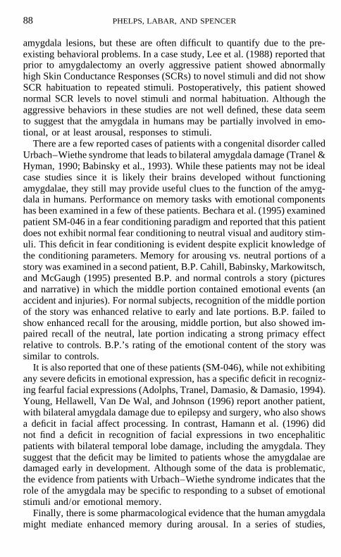
In a series of studies, LaBar et al. (1995) examined fear conditioning fol-lowing unilateral temporal lobectomy. In these studies, the unconditionedstimulus (US) was a series of short, loud white noise bursts and SCR wasmeasured as the unconditioned response (UR). In a simple discriminationparadigm, the reinforced conditioned stimulus (CS1) was a tone and theunreinforced stimulus (CS-) was a second tone. The study was conductedon 22 temporal lobectomy subjects (11 left, 11 right), 22 normal controls, and3 epileptic patients who had lesions in other brain areas following surgery tocontrol epilepsy.
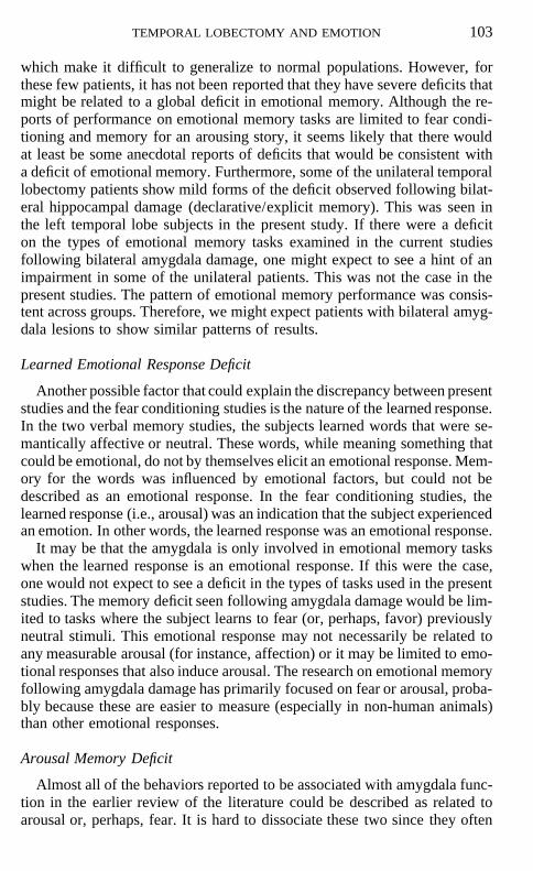
There was no difference in performance between patients who had re-ceived right or left temporal lobectomy so these subjects were combined.There was also no difference in level of SCR and habituation to the uncondi-tioned stimulus, suggesting that the temporal lobectomy subjects show nor-mal responses to arousing stimuli. As can be seen in Fig. 1, the epilepticand non-epileptic control groups showed an increase in SCR to the CS1,relative to the CS2, during acquisition. The temporal lobectomy subjectsshowed no increase in responding to the CS1 and demonstrated fairly flatresponses. In spite of the lack of a learned conditioned response, all but twoof the temporal lobe subjects were able to explicitly report that the CS1was followed by the US, while the CS2 was unreinforced.
A second study was conducted using a conditional discrimination para-digm similar to one used by Daum et al. (1991) in their eyeblink conditioningstudies. The CS was a single tone. If the tone was preceded by one coloredlight (e.g., red) it was reinforced (the CS1). If the tone was preceded bythe color-opponent light (e.g., green), it was not reinforced (CS2). With thisparadigm we found, like Daum et al., that the temporal lobectomy subjectswere impaired, in spite of the fact that they could explicitly report the correctstimulus relationships. However, our results differed from Daum et al.’s in

92 PHELPS, LABAR, AND SPENCER
FIG. 1. Simple discrimination conditioning in temporal lobectomy patients, nonepilepticcontrol subjects, and epileptic control subjects with other brain excisions. The data are differ-ence scores obtained by subtracting SCR on CS2 (unreinforced) trials from SCR on CS1(reinforced) trials. On all figures, error bars represent standard error.
the pattern of responding. In the Daum et al. study, the patients were impairedbecause they overgeneralized and showed an increased response to the CS2.In our study the patients were impaired because they failed to respond tothe CS1. This different pattern of results on the conditional discriminationstudy, combined with the different results in the simple discrimination study,implies that there may be different neural substrates underlying impairmentsseen in eyeblink and fear conditioning in unilateral temporal lobectomy pa-tients. Specifically, we propose that the deficit seen with fear conditioningis due to the unilateral amygdala damage, whereas the deficit seen in the morecomplex conditional discrimination eyeblink paradigm may be mediated bythe unilateral hippocampal damage.
Consistent with this interpretation is a study of fear conditioning followingunilateral amygdala lesions in rats (LaBar & LeDoux, 1996). Using the sameUS (loud white noise) as the human study described above, LaBar and Le-Doux (1996) found that unilateral damage to the amygdala, on both the rightand left, leads to an impairment in a conditioned freezing response. Theseresults provide additional support to the notion that it is the amygdala damagethat leads to the deficit in fear conditioning for the unilateral temporal lobec-tomy subjects.
This deficit seen in fear conditioning following unilateral temporal lobec-tomy is not surprising given the animal research (e.g., LeDoux, 1992) andthe impairment reported in the case study of patient SM-046 (Bechara etal., 1995). What is not known is whether this deficit in fear conditioning is
TEMPORAL LOBECTOMY AND EMOTION 93
representative of a deficit on all or most emotional memory tasks. Emotionalmemory, aside from fear conditioning, has not been studied in these patientsand fear conditioning is not what one would consider a typical emotionalmemory task in humans. If one is going to claim that the amygdala has aspecial role in memory tasks which are influenced by emotion, ideally thesepatients would show a deficit on several types of emotional memory tasks.In this paper we report two studies examining unilateral temporal lobectomypatients on verbal memory tasks where emotional factors influence memoryperformance. In Experiment 1, we examine memory for emotional wordsand in Experiment 2, we examine for memory neutral words embedded inemotional sentences. If the amygdala is involved in all types of emotionalmemory tasks, we might expect to see a deficit specifically in those situationswhere emotional factors influence memory performance.
EXPERIMENT 1
Method
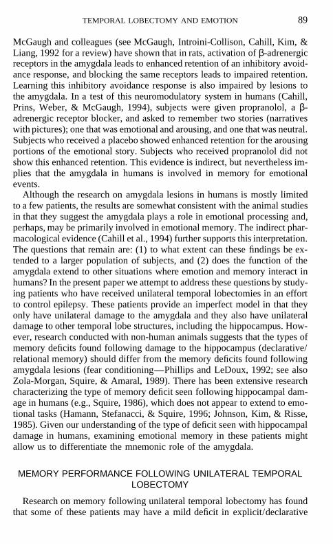
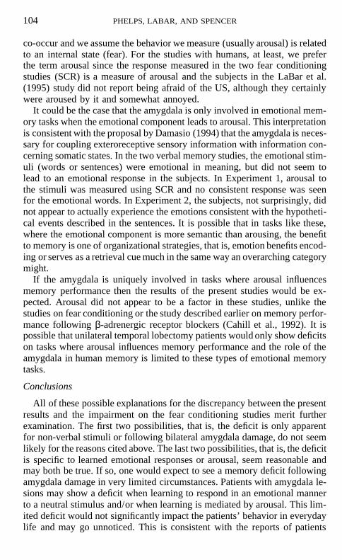
Subjects. Twenty-six subjects with medically refractory complex partial seizures of medialtemporal lobe origin were studied 2–6 years following unilateral anteromedial en bloc tempo-ral lobe resection (13 left, 13 right). The surgical procedure involved an approximate 3.5 cmresection of the anterior middle and inferior temporal gyri allowing access to the temporal horn.This is followed by dissection of the occipito-temporal fasciculus and subsequent removal of70–80% of the amygdala and all of the hippocampus, parahippocampus, and projection fibersto their posterior extent at the atrium of the lateral ventricle (Spencer, Spencer, Mattson, Wil-liamson, & Novelly, 1984). All patients received postoperative anatomical MRI scans con-firming the extent of surgical procedure. A representative scan in coronal and parasagittalsections is provided in Fig. 2. This procedure is highly successful in controlling seizure activityin the patients, with most patients reporting few, if any, seizures postoperatively. These patientstypically show some selective cognitive improvement following surgery due to reduction inseizure activity (Novelly et al., 1984), and most are gradually withdrawn from anticonvulsantmedication in accordance with postoperative neurological assessment. All but 4 subjects weretaking anticonvulsant medication at the time of testing. A subset of 23 patients (11 right, 12left) participated in Experiment 1.
Neuropsychological testing results, including the Wechsler Adult Intelligence Scale—Re-vised (WAIS-R) and the Russell adaptation of the Wechsler Memory Scale (WMS; Russell,1975), are presented in Table 1 along with data on other clinical variables. Neuropsychologicaldata was available for a subset of patients tested both 6 months preoperatively and 12 monthspostoperatively. Consistent with previous studies (e.g., Novelly et al., 1984), patients with left(speech dominant) medial temporal lobe epilepsy showed some impaired verbal memory incomparison to patients with right (nondominant) medial temporal lobe epilepsy on WMS sub-test scores (pre-operative: WMS delayed verbal, t(17) 5 2.80, p , .05; WMS immediateverbal, t(17) 5 2.45, p , .05; postoperative: WMS delayed verbal, t(12) 5 2.82, p , .05;WMS immediate verbal, t(12) 5 2.42, p , .05). Right and left medial temporal lobe epilepsypatients were not significantly different on any of the WAIS-R IQ scores or other subjectcharacteristics.
The control group consisted of 23 nonepileptic adult subjects matched for age and genderwithout a history of epilepsy or other neurological impairment. Control subjects were recruited

94 PHELPS, LABAR, AND SPENCER
FIG. 2. Representative postoperative T1-weighted MRI scans demonstrating the extent ofthe surgical procedure: Coronal and parasagittal sections.
TEMPORAL LOBECTOMY AND EMOTION 95
TABLE 1Demographic and Neuropsychological Profile of Patient Population
Seizure WAIS-R WMSEducation Onset (12 months (12 months
Group Age Gender (yrs) (age) post-op) post-op)
LTL 36 6 10 7M, 6F 13 6 2 9 6 10 99 6 14 F 11 6 2 IP(N 5 13) 102 6 13 P 8 6 3 DP
104 6 14 V 14 6 5 IV*7 6 6 DV*
RTL 38 6 9 3M, 10F 14 6 2 7 6 7 107 6 12 F 12 6 3 IP(N 5 13) 110 6 11 P 10 6 5 DP
104 6 14 V 20 6 5 IV16 6 6 DV
Note. All values represent means (6SD) unless otherwise indicated.* Statistically significant (LTL vs. RTL, p , .05).
through a local newspaper advertisement. All subjects provided informed consent and werepaid for their participation.
Materials. A list of 27 words, 9 each of three categories—positive (e.g., lucky), negative(e.g., victim) and neutral (e.g., stamp)—was used in this study (see Appendix 1). This listwas drawn from a master list of affective words generated by Kitayama (1989). The wordswere chosen so that all categories were similar in word frequency and length.
SCR was recorded bilaterally with Thought Technology skin conductance units (LafayetteInstruments, Lafayette IN), consisting of Ag-AgCl electrodes attached by velcro straps to themiddle phalanges of the subjects third and fourth digits. Lafayette instruments electrode gelwas used as electrolyte. The incoming analog signal was amplified and digitized by a LabMaster A/D converter controlled by ASYST software on an IBM AT computer. Skin conduc-tance was sampled at 100 Hz throughout a trial and was stored for off-line amplitude analysis.The minimum SCR resolution was .0144 µSiemen (µS).
Procedure. Subjects were told that they would see a list of words and were asked to rateeach word for affect on a scale of 1 (negative) to 5 (positive). Each word was presented ona computer screen for 4 sec during which time SCRs were recorded. Subjects were asked torespond with the affective rating only after the word disappeared from the screen. A new wordwas presented every 14 6 2 sec. Words were presented in a pseudorandom order such thatno more than 2 words of the same affect were presented in succession. After a delay of 1min, subjects were given a surprise recall test.
Results
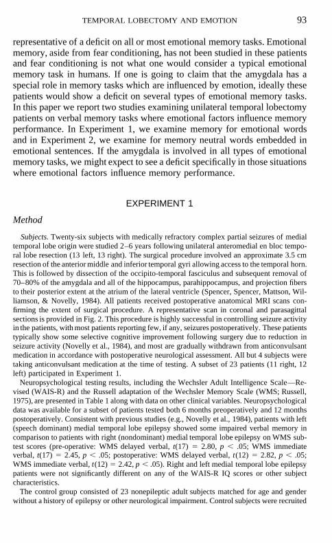
SCR. For each word presentation, the peak SCR amplitude in the 1- to 4-sec interval following stimulus onset was calculated. All SCR data werenormalized using a square root transformation. There was no difference inresponses for right and left hands for any subject group, so these responseswere combined. The results can be seen in Fig. 3.
A two-way ANOVA revealed a main effect for affect, F(2, 86) 5 3.76,p , .05, no effect for subject group and no affect 3 group interaction. Post-hoc tests did not reveal a significant effect of affect in any individual subject
96 PHELPS, LABAR, AND SPENCER
FIG. 3. Mean SCRs to negative, neutral, and positive words for normal controls (CTRL),left temporal lobectomy patients (LTL), and right temporal lobectomy patients (RTL).
group. The main effect is most likely due to the slightly greater respondingto the neutral words.
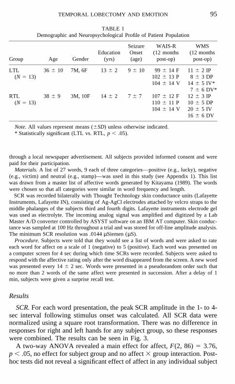
Recall. There was a marginally significant difference in overall numberof words recalled between subject groups, F(2, 43) 5 2.85, p , .07, dueprimarily to the poor recall of left temporal lobectomy subjects (see Fig. 4).
FIG. 4. Mean number of words recalled by normal controls, left temporal lobectomy patients,and right temporal lobectomy patients.
TEMPORAL LOBECTOMY AND EMOTION 97
FIG. 5. Mean number of negative, neutral, and positive words recalled for normal controls,left temporal lobectomy patients, and right temporal lobectomy patients.
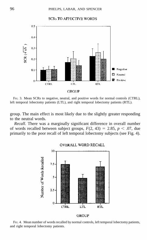
There was a significant main effect for affect, F(2, 86) 5 22.92, p , .001.Post-hoc Tukey HSD analysis revealed that all groups showed superior recallfor positive and negative words in comparison to neutral words (p , .05). Inaddition, positive words were recalled significantly more often than negativewords for the right temporal lobectomy subjects and normal controls (p ,.05). Left temporal lobectomy subjects showed a similar trend (see Fig. 5).
Discussion
The SCR results indicate that while the words are affective, they are notconsistently arousing for subjects. This is not particularly surprising giventhe types of emotional words used. These words are semantically emotional,but not surprising or shocking in any way.
The results on the recall test indicate that, consistent with previous studiesand the neuropsychological data for these subjects, the left temporal lobec-tomy subjects showed slightly worse overall levels of recall. In spite of thismild decrement, the pattern of performance was the same for all subjectgroups. All of the subjects recalled more of the affective words than theneutral words. Furthermore, all of the subjects showed slightly better recallfor positive than negative words. These results indicate that the temporallobectomy subjects show normal patterns of performance when recalling af-fective words. This result is in contrast to the emotional memory study onfear conditioning (LaBar et al., 1995) and suggests that patients with unilat-eral amygdala damage are not impaired on all memory tasks which are influ-enced by emotional factors.
98 PHELPS, LABAR, AND SPENCER
In the earlier review of the role of the amygdala in emotional memory itwas suggested that the amygdala does not seem to be necessary for the expe-rience of emotion in humans, but rather is necessary for assigning emotionalvalence to a previously neutral stimuli. It may be the case that we did not finda difference in mnemonic performance for emotional stimuli in the temporallobectomy subjects in Experiment 1 because the stimuli were emotionalwords, as opposed to neutral words whose memory is influenced by emo-tional factors. If the amygdala is uniquely involved in learning the emotionalsignificance of neutral events (as is the case in fear conditioning) then theresults of Experiment 1 might be expected. In Experiment 2, we attempt toaddress this issue using a paradigm introduced by Banaji (1986) in whichneutral words are embedded in emotional and neutral sentence contexts.
EXPERIMENT 2
Method
Subjects. There were 23 temporal lobectomy subjects (11 left, 12 right) drawn from thesame pool of subjects described in Experiment 1. There was an additional control group of5 epileptic subjects who received surgical excision of other brain areas (4 partial left occipitallobectomy, 1 anterior corpus callosotomy). There were 28 normal control subjects matchedfor age and gender with the epileptic patients. All subjects gave informed consent and werepaid for their participation.
Materials and procedure. Thirty nouns were selected from a list of affective word norms(Belleza, Greenwald, & Banaji, 1986). All words were selected to be neutral in affectivevalence. These words were randomly assigned to one of three sentence valences: positive,negative, or neutral (see Appendix 2). These assignments were constant across subjects. Eachword and its associated sentence valence were typed at the top of a half-sheet of paper. Subjectswere instructed to write a hypothetical, self-referent sentence using the word at the top of thepage. They were instructed that this sentence should reflect an experience of the assignedemotional valence (e.g., chair, negative—‘‘When I sat in the chair, it broke and I hurt myback’’). In this manner, neutral words were encoded in positive, negative, and neutral sentencecontexts. The presentation order was pseudorandomized for each subject and subjects wereallowed to take as much time as needed. After completing all 30 sentences, the subjects con-versed with the experimenter for 1 to 5 min and then were given a surprise recall test for theneutral words. After the recall test, subjects were asked to rate each sentence they wrote ona scale of 1 (negative) to 5 (positive) for emotional valence.
Results
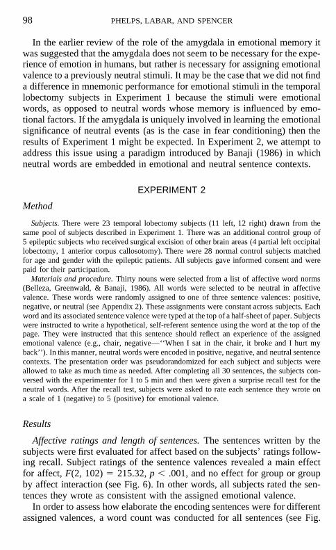
Affective ratings and length of sentences. The sentences written by thesubjects were first evaluated for affect based on the subjects’ ratings follow-ing recall. Subject ratings of the sentence valences revealed a main effectfor affect, F(2, 102) 5 215.32, p , .001, and no effect for group or groupby affect interaction (see Fig. 6). In other words, all subjects rated the sen-tences they wrote as consistent with the assigned emotional valence.
In order to assess how elaborate the encoding sentences were for differentassigned valences, a word count was conducted for all sentences (see Fig.
TEMPORAL LOBECTOMY AND EMOTION 99
FIG. 6. Mean sentence ratings using a scale from 1 (negative) to 5 (positive) for negative,neutral, and positive assigned valences for normal controls, left temporal lobectomy patients,right temporal lobectomy patients, and epileptic controls with other brain lesions (OL).
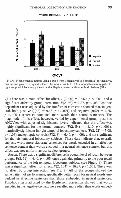
7). There was a main effect for affect, F(2, 96) 5 27.60, p , .001, and asignificant affect by group interaction, F(2, 96) 5 2.57, p , .05. Post-hocdependent t-tests adjusted by the Bonferroni correction showed that, in gen-eral, both positive (t(52) 5 9.16, p , .001) and negative (t(52) 5 6.76,p , .001) sentences contained more words than neutral sentences. Themagnitude of this effect, however, varied by experimental group: post-hocANOVAs with adjusted significance levels indicated that the effect washighly significant for the normal controls (F(2, 54) 5 44.10, p , .001);marginally significant in right temporal lobectomy subjects (F(2, 22) 5 5.08,p , .06) and epileptic controls (F(2, 8) 5 6.40, p , .09); and not significantfor the left temporal lobectomy subjects. These data indicate that, overall,subjects wrote more elaborate sentences for words encoded in an affectivesentence context than words encoded in a neutral sentence context, but thiseffect was not uniform across subject groups.
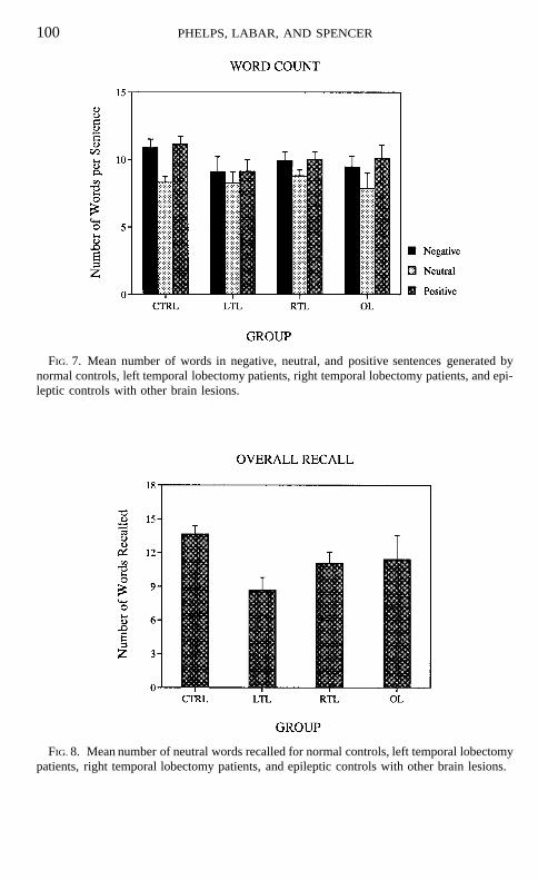
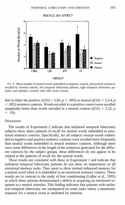
Recall. There was a significant difference in overall level of recall betweengroups, F(3, 52) 5 4.40, p , .05, once again due primarily to the poor recallperformance of the left temporal lobectomy subjects (see Figure 8). Therewas a significant effect for affect, F(2, 104) 5 16.27, p , .001. There wasno affect by group interaction (see Fig. 9). All of the groups showed thesame pattern of performance, specifically better recall for neutral words em-bedded in affective sentences than those embedded in neutral sentences.Post-hoc t tests adjusted by the Bonferroni correction showed that wordsencoded in the negative context were recalled more often than words embed-
100 PHELPS, LABAR, AND SPENCER
FIG. 7. Mean number of words in negative, neutral, and positive sentences generated bynormal controls, left temporal lobectomy patients, right temporal lobectomy patients, and epi-leptic controls with other brain lesions.
FIG. 8. Mean number of neutral words recalled for normal controls, left temporal lobectomypatients, right temporal lobectomy patients, and epileptic controls with other brain lesions.
TEMPORAL LOBECTOMY AND EMOTION 101
FIG. 9. Mean number of neutral words embedded in negative, neutral, and positive sentencesrecalled by normal controls, left temporal lobectomy patients, right temporal lobectomy pa-tients, and epileptic controls with other brain lesions.
ded in either the positive (t(55) 5 5.65, p , .005) or neutral (t(55) 5 3.3.4, p, .005) sentence contexts. Words encoded in a positive context were recalledmarginally better than words encoded in a neutral context (t(55) 5 2.22, p, .10).
Discussion
The results of Experiment 2 indicate that unilateral temporal lobectomysubjects show intact patterns of recall for neutral words embedded in emo-tional sentence contexts. Specifically, for all subjects neutral words embed-ded in negative and positive sentence contexts were recalled more frequentlythan neutral words embedded in neutral sentence contexts. Although therewere some differences in the length of the sentences generated for the differ-ent valences by the subject groups, these differences do not appear to berelated to the patterns of recall for the neutral words.
These results are consistent with those in Experiment 1 and indicate thatunilateral temporal lobectomy patients do not show an impairment on allemotional memory tasks. They seem to show normal enhanced memory fora neutral word when it is embedded in an emotional sentence context. Theseresults are in contrast to the study of fear conditioning (LaBar et al., 1995)in which these patients demonstrated a deficit in acquiring an emotional re-sponse to a neutral stimulus. This finding indicates that patients with unilat-eral temporal lobectomy are unimpaired on some tasks where a mnemonicresponse for a neutral event is mediated by emotion.

102 PHELPS, LABAR, AND SPENCER
GENERAL DISCUSSION
The results from these two studies indicate that unilateral temporal lobec-tomy patients demonstrate normal patterns of performance on some tests ofemotional memory. The earlier studies on fear conditioning (LaBar et al.,1995) show that these same patients are impaired on other emotional memorytasks. The present studies and the fear conditioning studies differ in manyways and it is not clear which factors may lead to the discrepant results.There are several possibilities as to what the critical differences betweenthese emotional memory tasks might be. Four possibilities, along with ourhypothesis concerning them, are discussed below.
Verbal vs. Visual or Auditory Memory
One difference between the present studies and the fear conditioning stud-ies is the type of stimuli. Both of the studies in the present are verbal memorystudies, while the CSs in the fear conditioning studies were auditory or visual(Bechara et al., 1995; LaBar et al., 1995). The left temporal lobectomy sub-jects in the present studies demonstrated a mild verbal memory deficit, how-ever this did not seem to influence the pattern of responding for emotionaland nonemotional words.
Although it is impossible to rule out that type of stimuli was a factor, itdoes not seem likely for a few reasons. First, there were no differences be-tween patterns of memory performance for right and left temporal lobectomysubjects in the present study or the fear conditioning study. One might expecta material specific deficit in emotional memory tasks to mirror that of non-emotional memory tasks. Second, the anatomy and physiology of the amyg-dala suggests that it is massively interconnected with the neocortex, receivinginput from all sensory modalities (Amaral, Price, Pitkanen, & Carmichael,1992). Given this, it would be surprising if the deficit in emotional memorywere specific to the visual or auditory domain.
Unilateral vs. Bilateral Amygdala Damage
It is possible that no deficit was detected in the current studies becausethe patients only had unilateral damage to the amygdala. The research on thehippocampus and memory might give credence to this interpretation sincebilateral lesions lead to a far more severe deficit than unilateral lesions (Mil-ner, 1970). In our earlier study (LaBar et al., 1995) we demonstrated a deficitin fear conditioning following unilateral lesions, but conditioning may be aspecial case. Daum et al. (1991) found an impairment on an eyeblink discrim-ination task, thought to be mediated by the hippocampus, in unilateral pa-tients who only show subtle deficits in explicit memory.
As stated earlier, there are very few individuals identified with bilateralamygdala damage without concurrent hippocampal damage and those thathave been described have either congenital disorders or behavioral problems
TEMPORAL LOBECTOMY AND EMOTION 103
which make it difficult to generalize to normal populations. However, forthese few patients, it has not been reported that they have severe deficits thatmight be related to a global deficit in emotional memory. Although the re-ports of performance on emotional memory tasks are limited to fear condi-tioning and memory for an arousing story, it seems likely that there wouldat least be some anecdotal reports of deficits that would be consistent witha deficit of emotional memory. Furthermore, some of the unilateral temporallobectomy patients show mild forms of the deficit observed following bilat-eral hippocampal damage (declarative/explicit memory). This was seen inthe left temporal lobe subjects in the present study. If there were a deficiton the types of emotional memory tasks examined in the current studiesfollowing bilateral amygdala damage, one might expect to see a hint of animpairment in some of the unilateral patients. This was not the case in thepresent studies. The pattern of emotional memory performance was consis-tent across groups. Therefore, we might expect patients with bilateral amyg-dala lesions to show similar patterns of results.
Learned Emotional Response Deficit
Another possible factor that could explain the discrepancy between presentstudies and the fear conditioning studies is the nature of the learned response.In the two verbal memory studies, the subjects learned words that were se-mantically affective or neutral. These words, while meaning something thatcould be emotional, do not by themselves elicit an emotional response. Mem-ory for the words was influenced by emotional factors, but could not bedescribed as an emotional response. In the fear conditioning studies, thelearned response (i.e., arousal) was an indication that the subject experiencedan emotion. In other words, the learned response was an emotional response.
It may be that the amygdala is only involved in emotional memory taskswhen the learned response is an emotional response. If this were the case,one would not expect to see a deficit in the types of tasks used in the presentstudies. The memory deficit seen following amygdala damage would be lim-ited to tasks where the subject learns to fear (or, perhaps, favor) previouslyneutral stimuli. This emotional response may not necessarily be related toany measurable arousal (for instance, affection) or it may be limited to emo-tional responses that also induce arousal. The research on emotional memoryfollowing amygdala damage has primarily focused on fear or arousal, proba-bly because these are easier to measure (especially in non-human animals)than other emotional responses.
Arousal Memory Deficit
Almost all of the behaviors reported to be associated with amygdala func-tion in the earlier review of the literature could be described as related toarousal or, perhaps, fear. It is hard to dissociate these two since they often
104 PHELPS, LABAR, AND SPENCER
co-occur and we assume the behavior we measure (usually arousal) is relatedto an internal state (fear). For the studies with humans, at least, we preferthe term arousal since the response measured in the two fear conditioningstudies (SCR) is a measure of arousal and the subjects in the LaBar et al.(1995) study did not report being afraid of the US, although they certainlywere aroused by it and somewhat annoyed.
It could be the case that the amygdala is only involved in emotional mem-ory tasks when the emotional component leads to arousal. This interpretationis consistent with the proposal by Damasio (1994) that the amygdala is neces-sary for coupling exteroreceptive sensory information with information con-cerning somatic states. In the two verbal memory studies, the emotional stim-uli (words or sentences) were emotional in meaning, but did not seem tolead to an emotional response in the subjects. In Experiment 1, arousal tothe stimuli was measured using SCR and no consistent response was seenfor the emotional words. In Experiment 2, the subjects, not surprisingly, didnot appear to actually experience the emotions consistent with the hypotheti-cal events described in the sentences. It is possible that in tasks like these,where the emotional component is more semantic than arousing, the benefitto memory is one of organizational strategies, that is, emotion benefits encod-ing or serves as a retrieval cue much in the same way an overarching categorymight.
If the amygdala is uniquely involved in tasks where arousal influencesmemory performance then the results of the present studies would be ex-pected. Arousal did not appear to be a factor in these studies, unlike thestudies on fear conditioning or the study described earlier on memory perfor-mance following β-adrenergic receptor blockers (Cahill et al., 1992). It ispossible that unilateral temporal lobectomy patients would only show deficitson tasks where arousal influences memory performance and the role of theamygdala in human memory is limited to these types of emotional memorytasks.
Conclusions
All of these possible explanations for the discrepancy between the presentresults and the impairment on the fear conditioning studies merit furtherexamination. The first two possibilities, that is, the deficit is only apparentfor non-verbal stimuli or following bilateral amygdala damage, do not seemlikely for the reasons cited above. The last two possibilities, that is, the deficitis specific to learned emotional responses or arousal, seem reasonable andmay both be true. If so, one would expect to see a memory deficit followingamygdala damage in very limited circumstances. Patients with amygdala le-sions may show a deficit when learning to respond in an emotional mannerto a neutral stimulus and/or when learning is mediated by arousal. This lim-ited deficit would not significantly impact the patients’ behavior in everydaylife and may go unnoticed. This is consistent with the reports of patients
TEMPORAL LOBECTOMY AND EMOTION 105
following unilateral temporal lobectomy as being relatively unimpaired (withthe exception of those that show a mild explicit memory deficit). Althoughthere is much more to be discovered about the deficit in emotional memoryfollowing unilateral temporal lobectomy, the finding of intact performancein the present studies indicates that these patients do not show a global deficiton all emotional memory tasks.
APPENDIX 1
Words from Experiment 1 by Emotional Valence
Positive
LuckyFunnyTrustTalentProudJokeComedySmileGlory
Neutral
StampSpareSwitchLocateHabitChairStoneBorderTrack
Negative
VictimErrorFalseDamageFaultWasteFoolCancerDevil
106 PHELPS, LABAR, AND SPENCER
APPENDIX 2
Neutral Words from Experiment 2 by Assigned Emotional Valence
Positive
PamphletCorkInkStoolDishRiddleBowlKeroseneMerchantHat
Neutral
TrumpetStatueGingerKeyFootPajamasThermometerClayRacketBag
Negative
TrunkDoorOfficeWagonScissorsMuseumMonkeyBoatBundleEngine
TEMPORAL LOBECTOMY AND EMOTION 107
REFERENCES
Adolphs, R., Tranel, D., Damasio, H., & Damasio, A. R. 1994. Impaired recognition of facialexpressions following bilateral damage to the human amygdala. Nature, 372, 669–672.
Adolphs, R., Tranel, D., Damasio, H., & Damasio, A. R. 1995. Fear and the human amygdala.The Journal of Neuroscience, 15, 5879–5891.
Aggleton, J. P. 1992. The functional effects of amygdala lesions in man: A comparison withfindings from monkeys. In J. P. Aggleton (Ed.), The amygdala: Neurobiological aspectsof emotion, memory, and mental dysfunction (pp. 485–503). New York: Wiley.
Amaral, D. G., Price, J. L., Pikanen, A., & Carmichael, S. T. 1992. Anatomical organizationof the primate amygdala complex. In J. P. Aggleton (Ed.), The amygdala: Neurobiologicalaspects of emotion, memory, and mental dysfunction. New York: Wiley.
Babinsky, R., Calabrese, P., Durwen, H. F., Markowitsch, H. J., Brechtelsbauer, D., Heuser,L., & Gehlen, W. 1993. The possible contribution of the amygdala to memory. BehavioralNeurology, 6, 167–170.
Banaji, M. R. 1986. Affect and memory: An experimental investigation. Dissertation AbstractsInternational, 47, 1325.
Bechara, A., Tranel, D., Damasio, H., Adolphs, R., Rockland, C., & Damasio, A. R. 1995.Double dissociation of conditioning and declarative knowledge relative to the amygdalaand hippocampus in humans. Science, 269, 1115–1118.
Belleza, F. S., Greenwald, A. G., & Banaji, M. R. 1986. Words high and low in pleasantnessas rated by male and female college students. Behavior Research Methods, Instruments &Computers, 18, 299–303.
Berger, T. W., & Orr, W. B. 1983. Hippocampectomy selectively disrupts discrimination rever-sal conditioning of the rabbit nicitating membrane response. Behavioral Brain Research,8, 49–68.
Blaxton, T. A. 1992. Dissociations among memory measures in memory-impaired subjects:Evidence for a processing account of memory. Memory and Cognition, 20, 549–562.
Cahill, L., Babinsky, R., Markowitsch, H. J., & McGaugh, J. L. 1995. The amygdala andemotional memory. Nature, 377, 295–296.
Cahill, L., Prins, B., Weber, M., & McGaugh, J. L. 1994. β-Adrenergic activation and memoryfor emotional events. Nature, 371, 702–704.
Cermak, L., Verfaellie, M., & Chase, K. A. 1995. Implicit and explicit memory in amnesia:An analysis of data-driven and conceptually driven processes. Neuropsychology, 9, 281–290.
Cohen, N. J., & Eichenbaum, H. 1993. Memory, amnesia, and the hippocampal system. Cam-bridge, MA: MIT Press.
Damasio, A. R. 1994. Descartes’ error: Emotion, reason, and the human brain. New York:Putnam.
Daum, I., Channon, S., & Gray, J. A. 1992. Classical conditioning after temporal lobe lesionsin man: Sparing of simple discrimination and extinction. Behavioral Brain Research, 52,159–165.
Daum, I., Channon, S., Polkey, C. E., & Gray, J. A. 1991. Classical conditioning after temporallobe lesions on man: Impairment in conditioned discrimination. Behavioral Neuroscience,105, 396–408.
Davis, M. 1992. The role of the amygdala in fear-potentiated startle: Implications for animalmodels of anxiety. Trends in Pharmacological Science, 13, 35–41.
Gabrielli, J. D. E., Carrillo, M. C., Cermak, L. S., McGlinchey-Berroth, R., Gluck, M. A., &Disterhoff, J. F. (1995). Intact delay-eyeblink classical conditioning in amnesia. Behav-ioral Neuroscience, 109, 819–827.
Gloor, P., Oliver, A., Quensey, L. F., Andermann, F., & Horowitz, M. S. 1982. The role ofthe limbic system in experiential phenomena of temporal lobe epilepsy. Annals of Neurol-ogy, 12, 129–143.
Halgren, E. 1992. Emotional neurophysiology of the amygdala within the context of human
108 PHELPS, LABAR, AND SPENCER
cognition. In J. P. Aggleton (Ed.), The amygdala: Neurobiological aspects of emotion,memory, and mental dysfunction. New York: Wiley.
Hamman, S., Stefanacci, L., & Squire, L. R. (1996, March). Emotional perception and memoryin amnesia. Poster presented at the 3rd annual meeting of the Cognitive NeuroscienceSociety, San Francisco.
Johnson, M. K., Kim, J. K., & Risse, G. 1985. Do alcoholic Korsakoff ’s syndrome patientsacquire affective reactions? Journal of Experimental Psychology: Learning, Memory, andCognition, 11, 22–36.
Jones-Gotman, M. 1986. Memory for designs: Hippocampal contribution. Neuropsychologia,24, 193–203.
Kapp, B. S., Pascoe, J. P., & Bixler, M. A. 1984. The amygdala: A neuroanatomical systemsapproach to its contributions to aversive conditioning. In N. Butters & L. R. Squire (Eds.),Neuropsychology of memory. New York: Guilford.
Kluver, H., & Bucy, P. C. 1937. ‘‘Psychic blindness’’ and other symptoms following bilateraltemporal lobectomy in rhesus monkeys. American Journal of Physiology, 119, 352–353.
LaBar, K. S., & LeDoux, J. E. (1996). Partial disruption of fear conditioning in rats withunilateral amygdala lesions: Correspondence with unilateral temporal lobectomy in hu-mans. Behavioral Neuroscience, 110, 991–997.
LaBar, K. S., LeDoux, J. E., Spencer, D. D., & Phelps, E. A. 1995. Impaired fear conditioningfollowing unilateral temporal lobectomy in humans. Journal of Neuroscience, 15, 6846–6855.
LeDoux, J. E. 1992. Emotion and the amygdala. In J. P. Aggleton (Ed.), The amygdala: Neuro-biological aspects of emotion, memory, and mental dysfunction. New York: Wiley.
LeDoux, J. E., Farb, C. R., & Ruggiero, D. A. 1990. Topographic organization of neurons inthe acoustic thalamus that project to the amygdala. Journal of Neuroscience, 10, 1043–1054.
Lavond, D. G., Kim, J. J., & Thompson, R. F. 1993. Mammalian brain substrates of aversiveclassical conditioning. Annual Review of Psychology, 44, 317–342.
Lee, G. P., Arena, J. G., Meador, K. J., Smith, J. R., Loring, D. W., & Flanigin, H. F. 1988.Changes in autonomic responsiveness following bilateral amygdalectomy in humans.Neuropsychiatry, Neuropsychology, and Behavioral Neurology, 1, 119–129.
McGaugh, J. L., Introini-Collison, I. B., Cahill, L., Kim, M., & Liang, K. C. 1992. Involvementof the amygdala in neuromodulatory influences on memory storage. In J. P. Aggleton(Ed.), The amygdala: Neurobiological aspects of emotion, memory, and mental dysfunc-tion. New York: Wiley.
MacLean, P. D. 1949. Psychosomatic disease and the visceral brain. Recent developmentsbearing on the Papez theory of emotion. Psychosomatic Medicine, 11, 338–353.
Milner, B. 1962. Laterality effects in audition. In V. B. Mountcastle (Ed.), Interhemisphericrelations and cerebral dominance. Baltimore: Johns Hopkins Press.
Milner, B. 1965. Visually guided maze learning in man: Effects of bilateral hippocampal,bilateral frontal, and unilateral cerebral lesions. Neuropsychologia, 3, 317–338.
Milner, B. 1970. Memory and the medial temporal regions of the brain. In K. H. Pribram &D. E. Broadbent (Eds.), Biological bases of memory. New York: Academic Press.
Novelly, R. A., Augustine, E. A., Mattson, R. H., Glaser, G. H., Williamson, P. D., Spencer,D. D., & Spencer, S. S. 1984. Selective memory improvement and impairment in temporallobectomy for epilepsy. Annals of Neurology, 15, 64–67.
Papez, J. W. 1937. A proposed mechanism of emotion. Archives of Neurology and Psychiatry,79, 217–224.
O’Keefe, J., & Nadel, L. 1978. The hippocampus as a cognitive map. Clarendon, Oxford,UK.
Phillips, R. G., & LeDoux, J. E. 1992. Differential contribution of amygdala and hippocampusto explicitly cued and contextual fear conditioning. Behavioral Neuroscience, 106, 274–285.

TEMPORAL LOBECTOMY AND EMOTION 109
Russell, E. W. 1975. A multiple scoring method for the assessment of complex memory func-tion. Journal of Consulting and Clinical Psychology, 43, 800–809.
Sass, K. J., Sass, A., Westerveld, M., Lenca, T., Rosewater, K. M., Novelly, R. A., Kim,J. H., & Spencer, D. D. 1992. Russell’s adaptation of the Weschler Memory Scale as anindex of hippocampal pathology. Journal of Epilepsy, 5, 24–30.
Schacter, D. L. 1988. On the relation between memory and consciousness: Dissociable interac-tions and conscious experience. In H. L. Roediger and F. I. M. Craik (Eds.), Varietiesof memory and consciousness: Essays in honor of Endel Tulving. Hillsdale, NJ: Erlbaum.
Scoville, W. B. 1968. Amnesia after bilateral medial temporal lobe excision: Introduction tocase H. M. Neuropsychologia, 6, 211–213.
Solomon, P. R., Vanser Schaff, E., Thompson, R. F., & Weisz, D. J. 1986. Hippocampus andtrace conditioning of the rabbits classically conditioned nictitating membrane response.Behavioral Neuroscience, 100, 729–744.
Spencer, D. D., Spencer, S. S., Mattson, R. H., Williamson, P. D., & Novelly, R. A. 1984.Access to the posterior medial temporal lobe structures in the surgical treatment of tempo-ral lobe epilepsy. Neurosurgery, 15, 667–671.
Spencer, D. D., & Spencer, S. S. 1985. Surgery for epilepsy. In L. Krantler (Ed.), Neurologicalclinics: Vol. 3. Advances in neurosurgery (pp. 313–330). Philadelphia: Saunders.
Sutherland, R. J., & Rudy, J. W. 1989. Configural association theory: The role of the hippocam-pal formation in learning, memory, and amnesia. Psychobiology, 17, 129–144.
Squire, L. R. 1986. Mechanisms of memory. Science, 232, 1612–1619.Squire, L. R. 1987. Memory and brain. New York: Oxford University Press.Tranel, D. & Hyman, B. T. 1990. Neuropsychological correlates of bilateral amygdala damage.
Archives of Neurology, 47, 349–355.Weiskrantz, L. 1956. Behavioral changes associated with ablation of the amygdaloid complex
in monkeys. Journal of Comparative Physiology, 49, 381–391.Young, A. W., Hellawell, D. J., Van De Wal, C., & Johnson, M. 1996. Facial expression
processing after amygdalotomy. Neuropsychologia, 34, 31–39.Zola-Morgan, S., Squire, L. R., & Amaral, D. G. 1989. Lesions of the amygdala that spare
adjacent cortical lesions do not impair or exacerbate the impairment following lesions ofthe hippocampal formation. Journal of Neuroscience, 9, 1922–1936.