Embed Size (px)
Citation preview
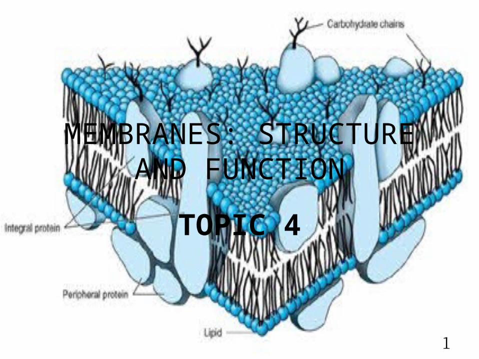
MEMBRANES: STRUCTURE AND FUNCTION
TOPIC 4
1
2
BIOMEDICAL IMPORTANCE• Plasma membranes- form closed compartments around cellular protoplasm to
separate one cell from another• Selective permeabilities- provided by channels and pumps for ions and
substrates• Receptor property• Exchange materials with extracellular environment – excocytosis and
endocytosis• Gap juctions• Cell-cell interactions • Transmembrane signaling• Form specialized compartments within cell – provide shape for mitoch, ER, golgi • Membrane localize enzymes-excitation response coupling- • Site for energy transduction• Changes in membrane structure?
3
Maintenance of a normal intra-&extracellular environment is fundamental to life
Life originated in aqueous environments- enzyme reactions, cellular and subcellular processes evolved to work in this milieu.
How this aqueous state is maintained?
4
Fluid distribution
2 large compartments that distribute water1. Intracellular fluid – ICF• 2/3 of body water• Provides the environment for the cell
to make, store and utilize energy to repair itself to replicate to perform special functions
5
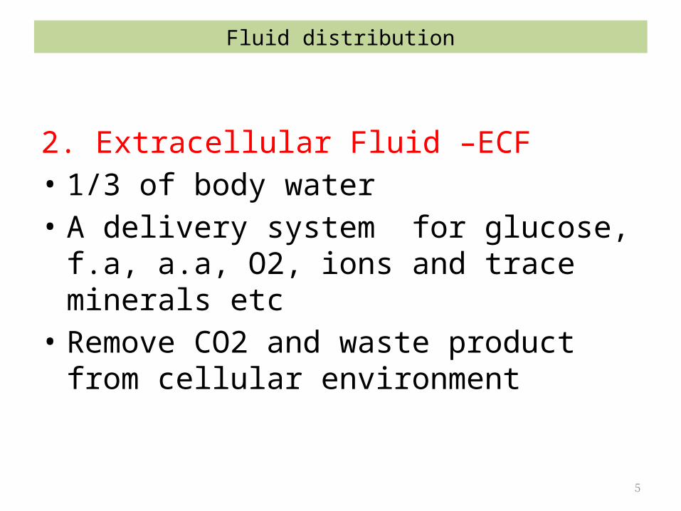
2. Extracellular Fluid –ECF• 1/3 of body water• A delivery system for glucose, f.a, a.a, O2, ions
and trace minerals etc• Remove CO2 and waste product from cellular
environment
Fluid distribution
6
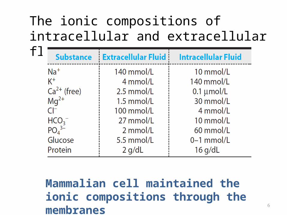
The ionic compositions of intracellular and extracellular fluids
Mammalian cell maintained the ionic compositions through the membranes
7
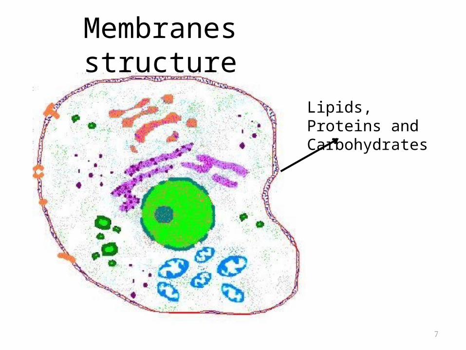
Membranes structure
Lipids, Proteins and Carbohydrates
8
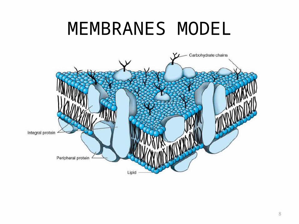
MEMBRANES MODEL
9
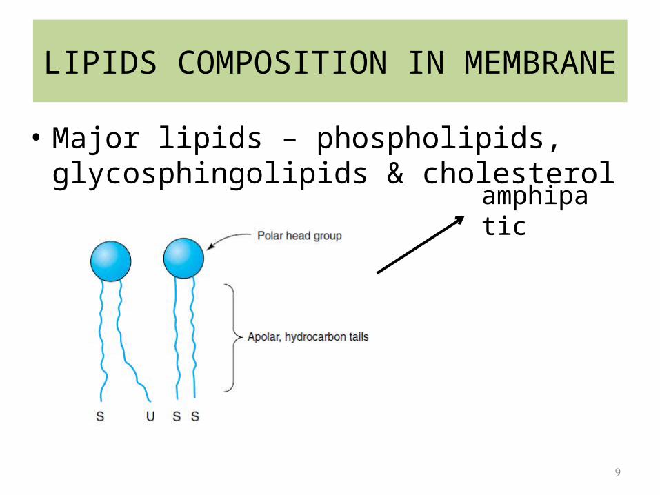
LIPIDS COMPOSITION IN MEMBRANE
• Major lipids – phospholipids, glycosphingolipids & cholesterol
amphipatic
10
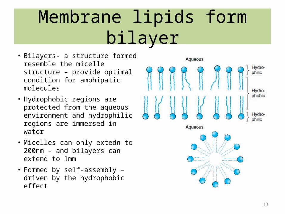
Membrane lipids form bilayer• Bilayers- a structure formed
resemble the micelle structure – provide optimal condition for amphipatic molecules
• Hydrophobic regions are protected from the aqueous environment and hydrophilic regions are immersed in water
• Micelles can only extedn to 200nm – and bilayers can extend to 1mm
• Formed by self-assembly – driven by the hydrophobic effect
11
Lipid bilayer
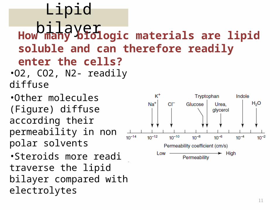
•O2, CO2, N2- readily diffuse•Other molecules (Figure) diffuse according their permeability in non polar solvents•Steroids more readily traverse the lipid bilayer compared with electrolytes
How many biologic materials are lipid soluble and can therefore readily enter the cells?
12
Lipid bilayer
• Membrane contain protein – form channels for the movement of ions and small molecules
• Serve as transporter for larger molecules• Different membrane consist of different composition
of protein• Include – enzymes, pumps and channels, structural
components, antigen (for MHC), and receptor for various molecules
• 2 types of proteins – integral and peripheral
How molecules that are non lipid-soluble cross the membrane?
13
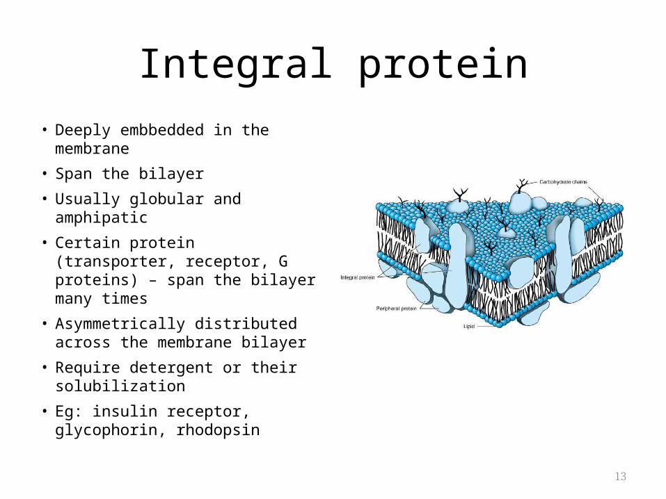
Integral protein• Deeply embbedded in the
membrane
• Span the bilayer
• Usually globular and amphipatic
• Certain protein (transporter, receptor, G proteins) – span the bilayer many times
• Asymmetrically distributed across the membrane bilayer
• Require detergent or their solubilization
• Eg: insulin receptor, glycophorin, rhodopsin
14
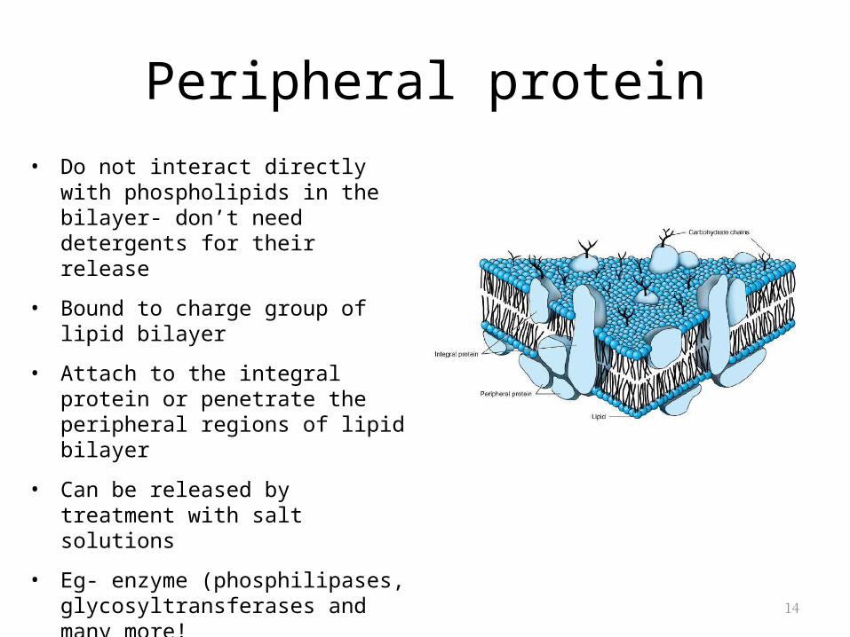
Peripheral protein• Do not interact directly with
phospholipids in the bilayer- don’t need detergents for their release
• Bound to charge group of lipid bilayer
• Attach to the integral protein or penetrate the peripheral regions of lipid bilayer
• Can be released by treatment with salt solutions
• Eg- enzyme (phosphilipases, glycosyltransferases and many more!
• Transportr of small hydrophobic molecules-glycolipid transfer protein, sterol carrier protein
15
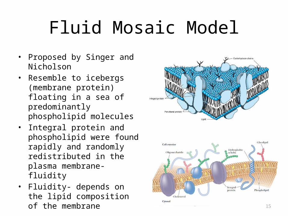
Fluid Mosaic Model• Proposed by Singer and
Nicholson• Resemble to icebergs (membrane
protein) floating in a sea of predominantly phospholipid molecules
• Integral protein and phospholipid were found rapidly and randomly redistributed in the plasma membrane-fluidity
• Fluidity- depends on the lipid composition of the membrane
16
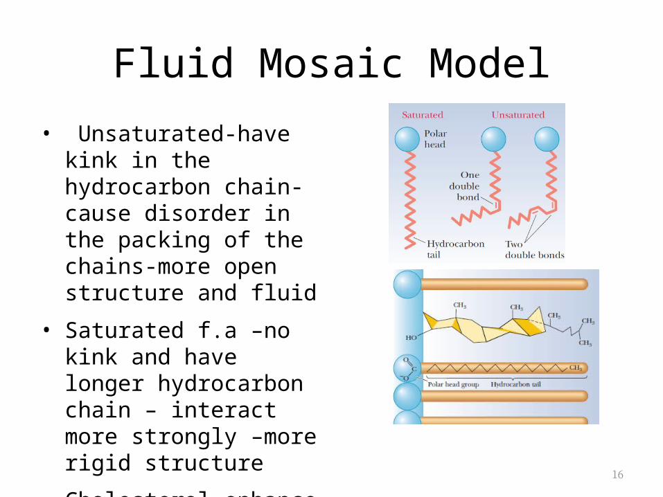
• Unsaturated-have kink in the hydrocarbon chain- cause disorder in the packing of the chains-more open structure and fluid
• Saturated f.a –no kink and have longer hydrocarbon chain – interact more strongly –more rigid structure
• Cholesterol-enhance order and rigidity
Fluid Mosaic Model
17
Effect of temperature
• Temperature increase- hydrophobic side chains undergo a transition from an ordered state (gel like) to a disordered state (fluid)
• Transition temperature• The longer and saturated the hydrocarbon
chains- the higher temperature needed to increase the fluditiy
18
Membrane fluidity
The fluidity of membrane affect its functions• Fluidity ↑- permeability to water and other small
hydrophilic molecule ↑-lateral mobility of integral protein ↑
• active site in hydrophilic region maybe affected• if protein involve in transportation, location changes cause
disruption in transportation
Eg. Insulin receptor↑ in unsaturated f.a cause ↑ in fluidity- alter the receptor so it binds more insulin (pls check)
19
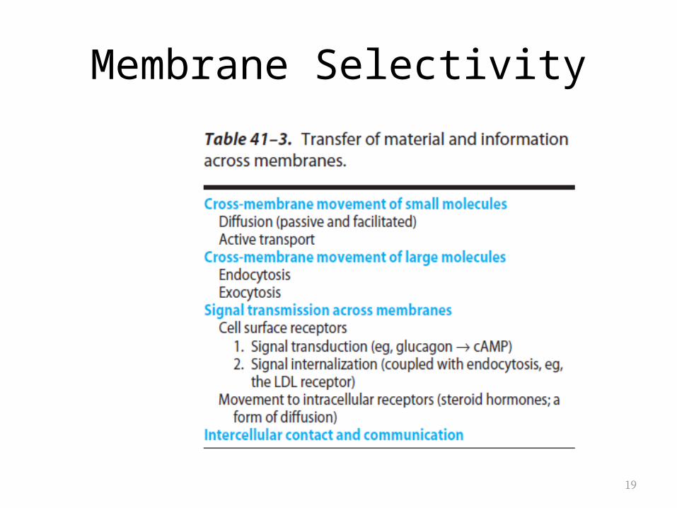
Membrane Selectivity
20
TYPE OF TRANSPORT MECHANISM
21
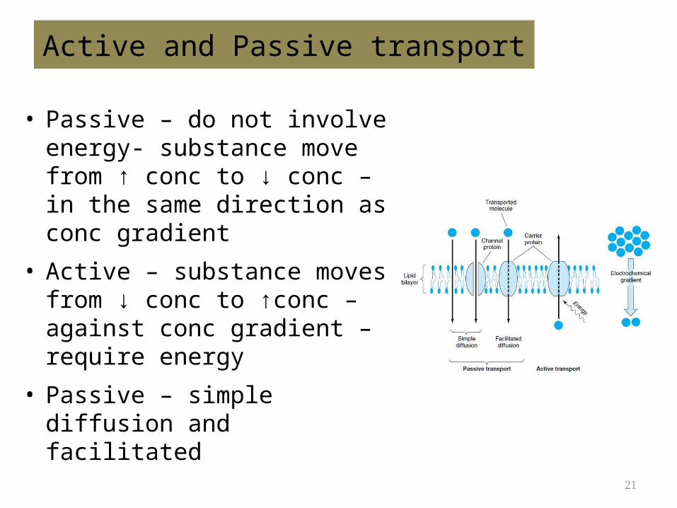
Active and Passive transport
• Passive – do not involve energy- substance move from ↑ conc to ↓ conc – in the same direction as conc gradient
• Active – substance moves from ↓ conc to ↑conc – against conc gradient – require energy
• Passive – simple diffusion and facilitated
22
Passive transport
Factors affect diffusion of a substance:1. Conc gradient across the memb2. The electrical potential across the memb – solutes move
toward the solution that has the opposite charge – inside of cell has a neg charge
3. The permeability coefficient of the substance for the memb4. The hydrostatic pressure gradient across the memb -
↑pressure will ↑the rate and force of the collision between the molecules and memb
5. Temperature -↑ temp will↑ the freq of collisions between external particles and the memb
23
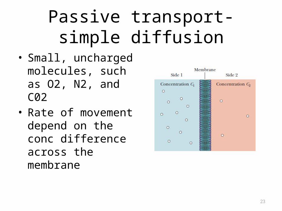
Passive transport- simple diffusion
• Small, uncharged molecules, such as O2, N2, and C02
• Rate of movement depend on the conc difference across the membrane
24
Active transport
• Identified by the presence of carrier protein• The need for an energy source to move
solutes against a gradient• Primary active transport-linked to hydrolysis of
energy – pumping water uphill -E.g sodium-potassium pump
• Secondary active transport –e.g- galactosidase permease in cell
25
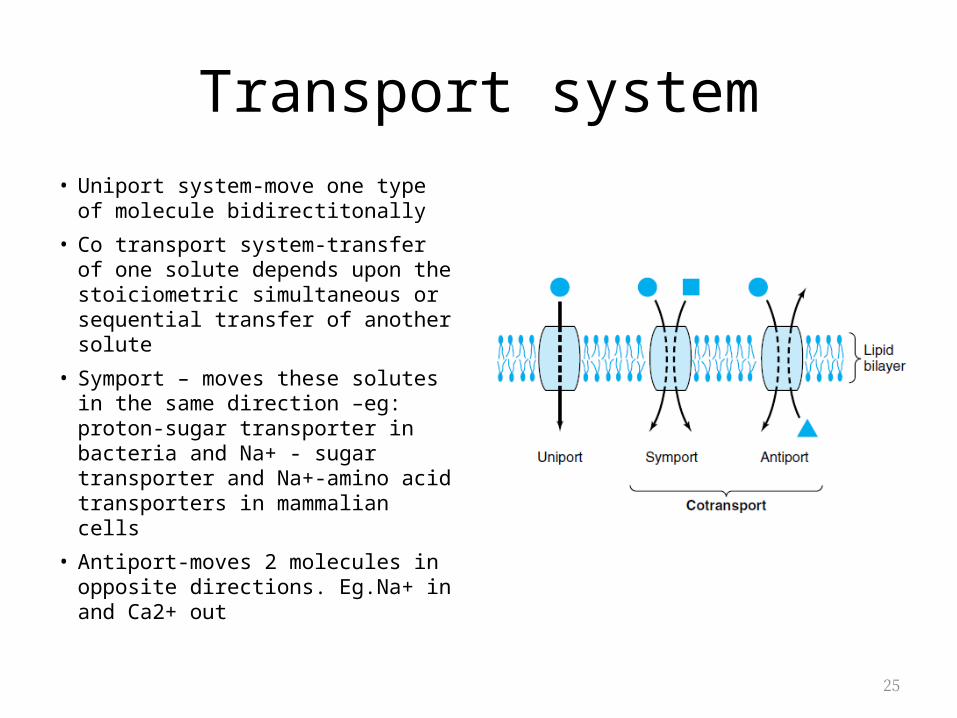
Transport system• Uniport system-move one type of
molecule bidirectitonally
• Co transport system-transfer of one solute depends upon the stoiciometric simultaneous or sequential transfer of another solute
• Symport – moves these solutes in the same direction –eg: proton-sugar transporter in bacteria and Na+ - sugar transporter and Na+-amino acid transporters in mammalian cells
• Antiport-moves 2 molecules in opposite directions. Eg.Na+ in and Ca2+ out
26
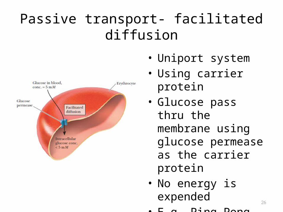
Passive transport- facilitated diffusion
• Uniport system• Using carrier protein• Glucose pass thru the
membrane using glucose permease as the carrier protein
• No energy is expended• E.g. Ping-Pong
mechanism
27
Passive transport- facilitated diffusion
• Hormones regulate facilitated diffusion by changing the number of transporters available
• Insulin increases glucose transport from intracellular reservoir
• Glucocorticoid hormones-enhance transport of aa into liver
• Growth hormon-increase amino acid transport in all cells
28
Transport System
1. Ion channels2. Ionophores3. Water channels (Aquaporins)4. Gap Junction
29
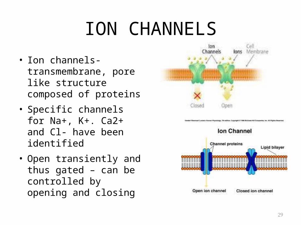
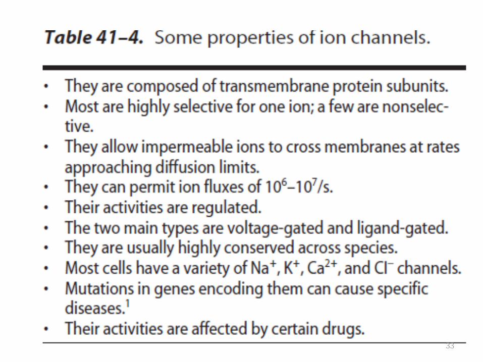
ION CHANNELS
• Ion channels- transmembrane, pore like structure composed of proteins
• Specific channels for Na+, K+. Ca2+ and Cl- have been identified
• Open transiently and thus gated – can be controlled by opening and closing
30
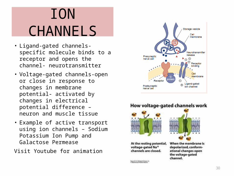
• Ligand-gated channels- specific molecule binds to a receptor and opens the channel- neurotransmitter
• Voltage-gated channels-open or close in response to changes in membrane potential- activated by changes in electrical potential difference – neuron and muscle tissue
• Example of active transport using ion channels – Sodium Potassium Ion Pump and Galactose Permease
Visit Youtube for animation
ION CHANNELS
31
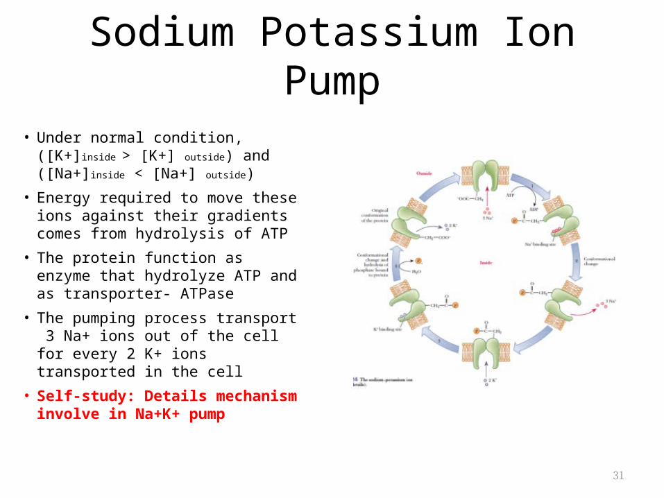
Sodium Potassium Ion Pump
• Under normal condition, ([K+]inside > [K+] outside) and ([Na+]inside < [Na+] outside)
• Energy required to move these ions against their gradients comes from hydrolysis of ATP
• The protein function as enzyme that hydrolyze ATP and as transporter- ATPase
• The pumping process transport 3 Na+ ions out of the cell for every 2 K+ ions transported in the cell
• Self-study: Details mechanism involve in Na+K+ pump
32
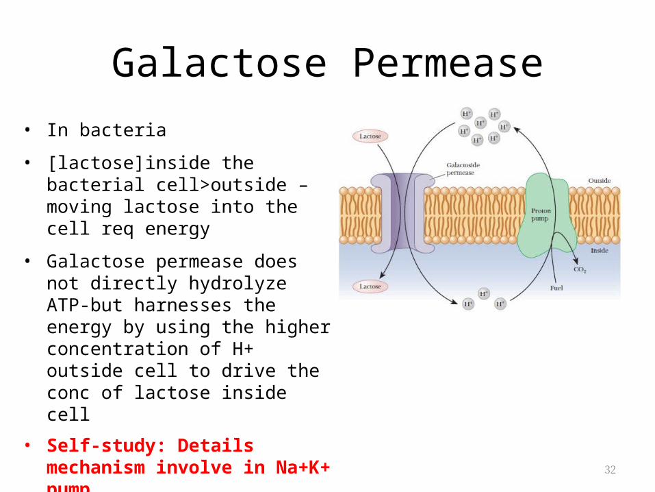
Galactose Permease• In bacteria
• [lactose]inside the bacterial cell>outside – moving lactose into the cell req energy
• Galactose permease does not directly hydrolyze ATP-but harnesses the energy by using the higher concentration of H+ outside cell to drive the conc of lactose inside cell
• Self-study: Details mechanism involve in Na+K+ pump
33
34
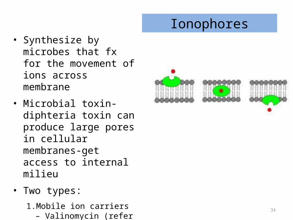
Ionophores• Synthesize by microbes that
fx for the movement of ions across membrane
• Microbial toxin-diphteria toxin can produce large pores in cellular membranes-get access to internal milieu
• Two types:
1. Mobile ion carriers – Valinomycin (refer uncouplers of oxidative phosphorilation)
2. Channel formers - gramicidin
35
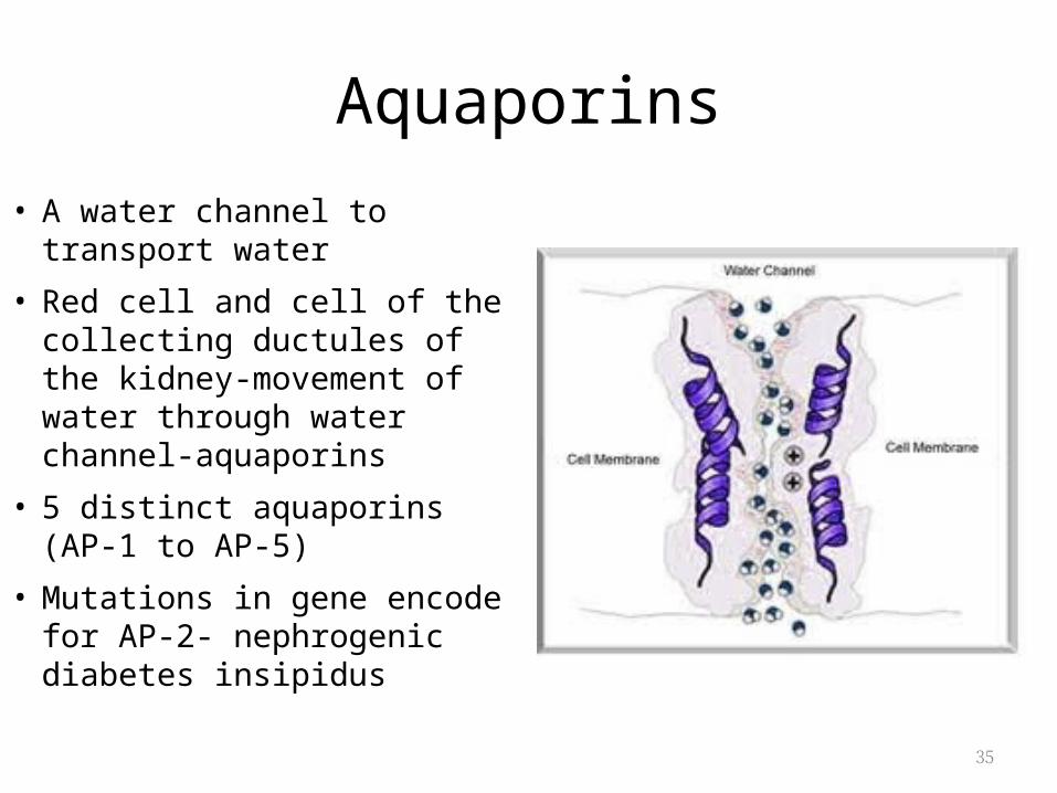
Aquaporins
• A water channel to transport water
• Red cell and cell of the collecting ductules of the kidney-movement of water through water channel-aquaporins
• 5 distinct aquaporins (AP-1 to AP-5)
• Mutations in gene encode for AP-2- nephrogenic diabetes insipidus
36
Transmembrane signalling
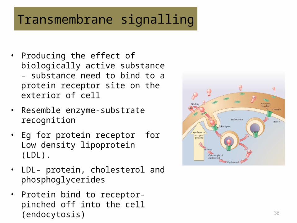
• Producing the effect of biologically active substance – substance need to bind to a protein receptor site on the exterior of cell
• Resemble enzyme-substrate recognition
• Eg for protein receptor for Low density lipoprotein (LDL).
• LDL- protein, cholesterol and phosphoglycerides
• Protein bind to receptor- pinched off into the cell (endocytosis)
• Cholesterol is used in the cell
• The receptor is recycled back to the surface of cell
37
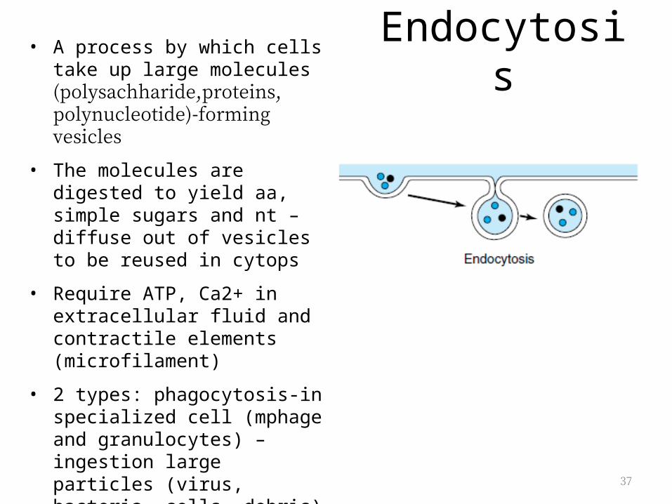
Endocytosis• A process by which cells take up large molecules (polysachharide,proteins, polynucleotide)-forming vesicles
• The molecules are digested to yield aa, simple sugars and nt – diffuse out of vesicles to be reused in cytops
• Require ATP, Ca2+ in extracellular fluid and contractile elements (microfilament)
• 2 types: phagocytosis-in specialized cell (mphage and granulocytes) – ingestion large particles (virus, bacteria, cells, debris)
• Pinocytosis – fluid phase and absorptive pinocytosis
38
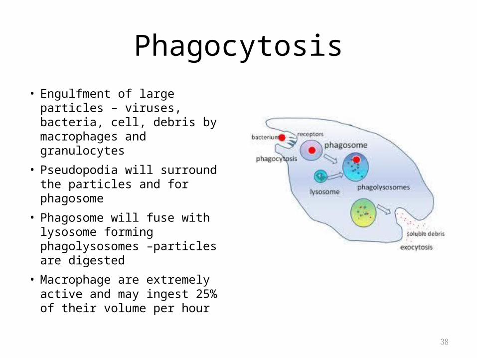
Phagocytosis• Engulfment of large particles –
viruses, bacteria, cell, debris by macrophages and granulocytes
• Pseudopodia will surround the particles and for phagosome
• Phagosome will fuse with lysosome forming phagolysosomes –particles are digested
• Macrophage are extremely active and may ingest 25% of their volume per hour
39
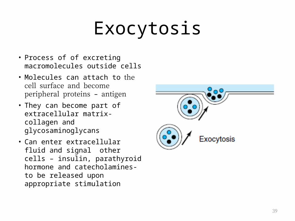
Exocytosis• Process of of excreting
macromolecules outside cells
• Molecules can attach to the cell surface and become peripheral proteins – antigen
• They can become part of extracellular matrix- collagen and glycosaminoglycans
• Can enter extracellular fluid and signal other cells – insulin, parathyroid hormone and catecholamines-to be released upon appropriate stimulation
40
Pinocytosis
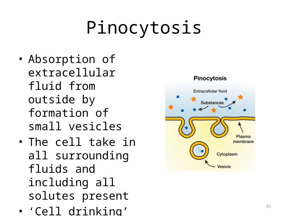
• Absorption of extracellular fluid from outside by formation of small vesicles
• The cell take in all surrounding fluids and including all solutes present
• ‘Cell drinking’
41
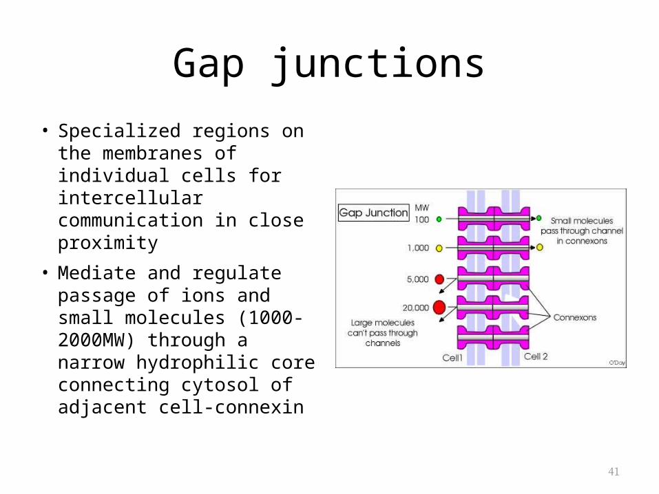
Gap junctions
• Specialized regions on the membranes of individual cells for intercellular communication in close proximity
• Mediate and regulate passage of ions and small molecules (1000-2000MW) through a narrow hydrophilic core connecting cytosol of adjacent cell-connexin
42
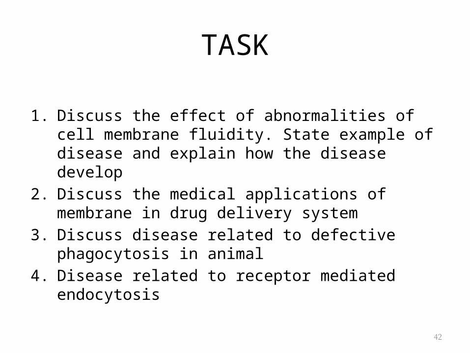
TASK
1. Discuss the effect of abnormalities of cell membrane fluidity. State example of disease and explain how the disease develop
2. Discuss the medical applications of membrane in drug delivery system
3. Discuss disease related to defective phagocytosis in animal4. Disease related to receptor mediated endocytosis