Embed Size (px)
Citation preview
Membranes and TransportCh. 6; 6.1-6.5
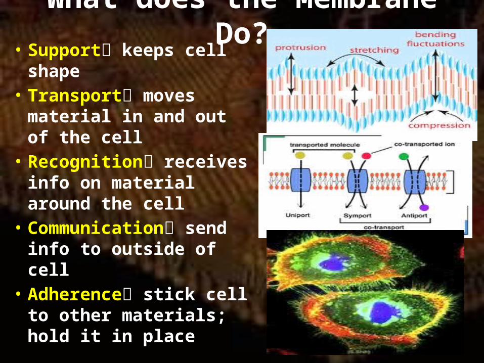
What does the Membrane Do?• Support keeps cell
shape• Transport moves
material in and out of the cell
• Recognition receives info on material around the cell
• Communication send info to outside of cell
• Adherence stick cell to other materials; hold it in place

Membrane Structure• What are the parts?1) Phospholipids hydrophilic and hydrophobic ends; 2 layers2) Sterols non-polar rings and polar alcohol groups; cholesterol 3) Embedded proteins do all the jobs of the membrane4) Glycolipids and glycoproteins
protective coat or communication
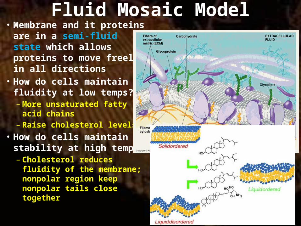
Fluid Mosaic Model• Membrane and it proteins
are in a semi-fluid state which allows proteins to move freely in all directions
• How do cells maintain fluidity at low temps?– More unsaturated fatty acid
chains– Raise cholesterol levels
• How do cells maintain stability at high temps?– Cholesterol reduces fluidity
of the membrane; nonpolar region keep nonpolar tails close together
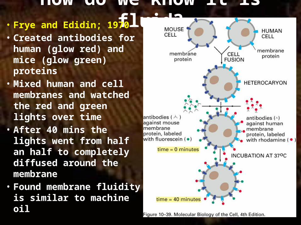
How do we know it is fluid?• Frye and Edidin; 1970• Created antibodies for
human (glow red) and mice (glow green) proteins
• Mixed human and cell membranes and watched the red and green lights over time
• After 40 mins the lights went from half an half to completely diffused around the membrane
• Found membrane fluidity is similar to machine oil
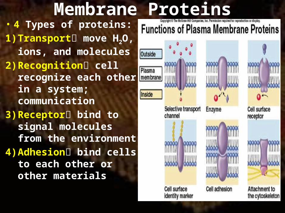
Membrane Proteins• 4 Types of proteins:1) Transport move H2O,
ions, and molecules2) Recognition cell
recognize each other in a system; communication
3) Receptor bind to signal molecules from the environment
4) Adhesion bind cells to each other or other materials
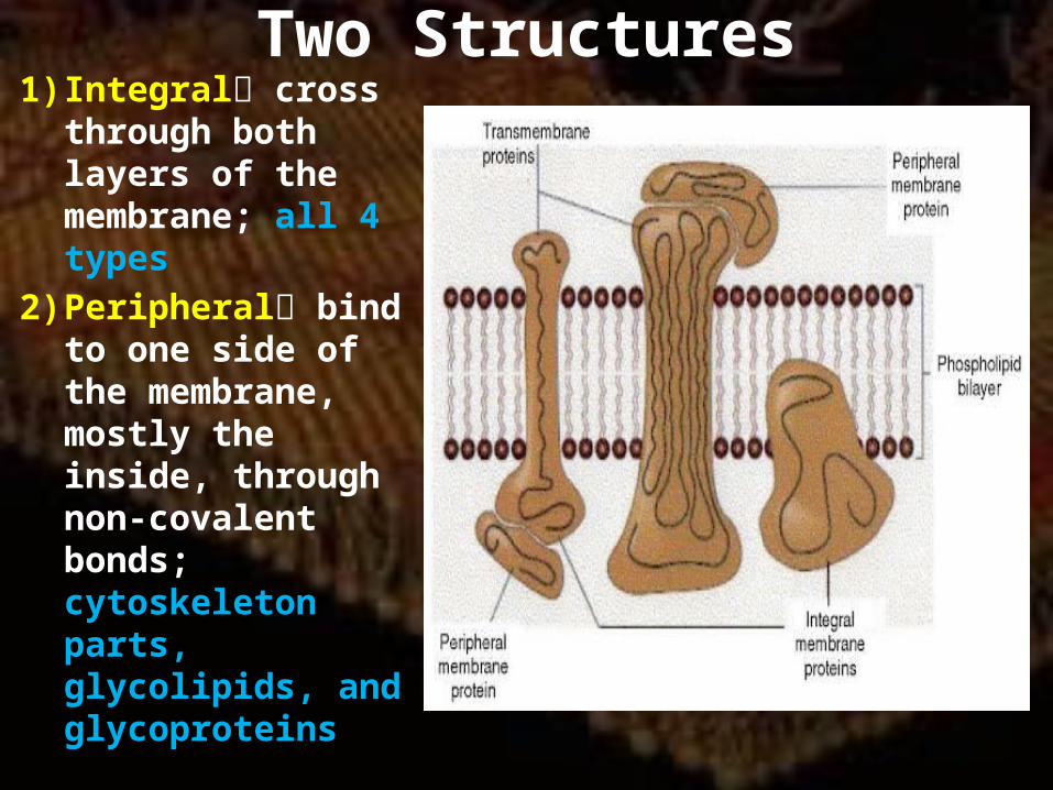
Two Structures1) Integral cross
through both layers of the membrane; all 4 types
2) Peripheral bind to one side of the membrane, mostly the inside, through non-covalent bonds; cytoskeleton parts, glycolipids, and glycoproteins
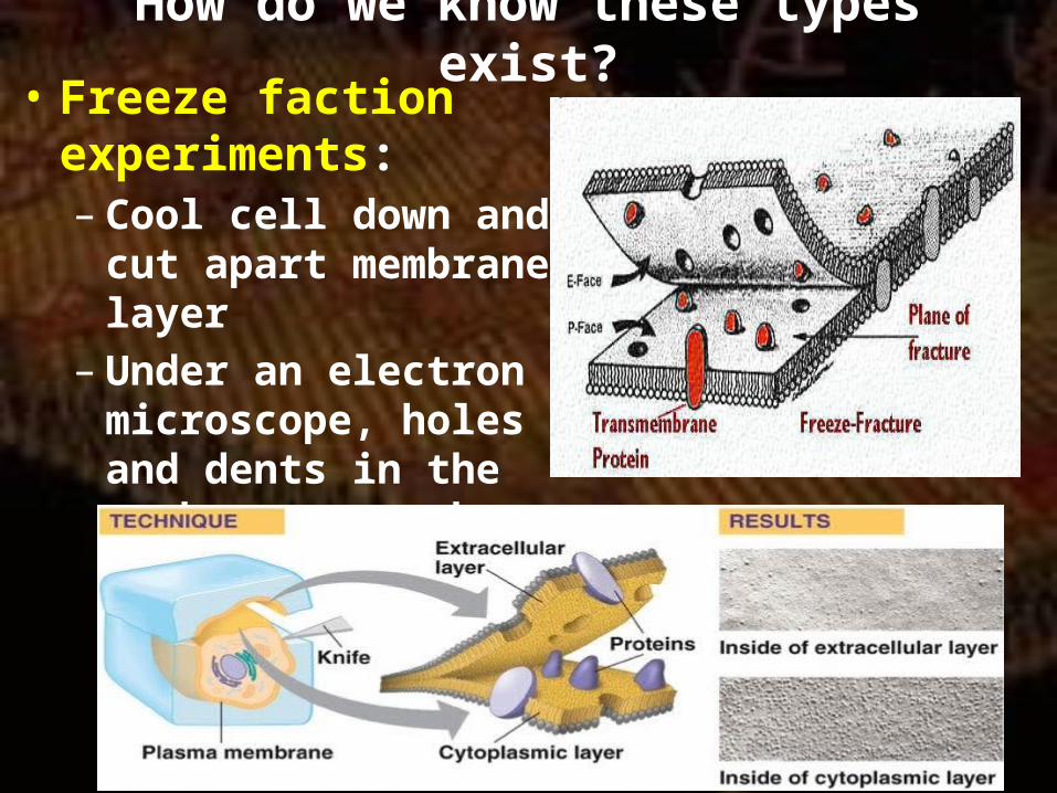
How do we know these types exist?• Freeze faction
experiments:– Cool cell down and cut
apart membrane layer– Under an electron
microscope, holes and dents in the membranes match like puzzle pieces
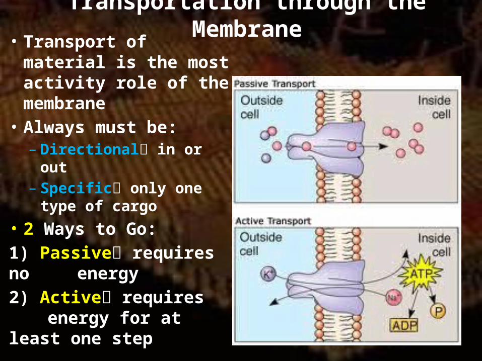
Transportation through the Membrane• Transport of material is
the most activity role of the membrane
• Always must be:– Directional in or out– Specific only one type
of cargo• 2 Ways to Go:1) Passive requires no
energy2) Active requires
energy for at least one step
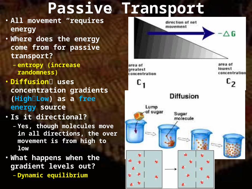
Passive Transport• All movement “requires”
energy• Where does the energy come
from for passive transport?– entropy (increase
randomness)• Diffusion uses concentration
gradients (HighLow) as a free energy source
• Is it directional?– Yes, though molecules move in
all directions, the over movement is from high to low
• What happens when the gradient levels out?– Dynamic equilibrium
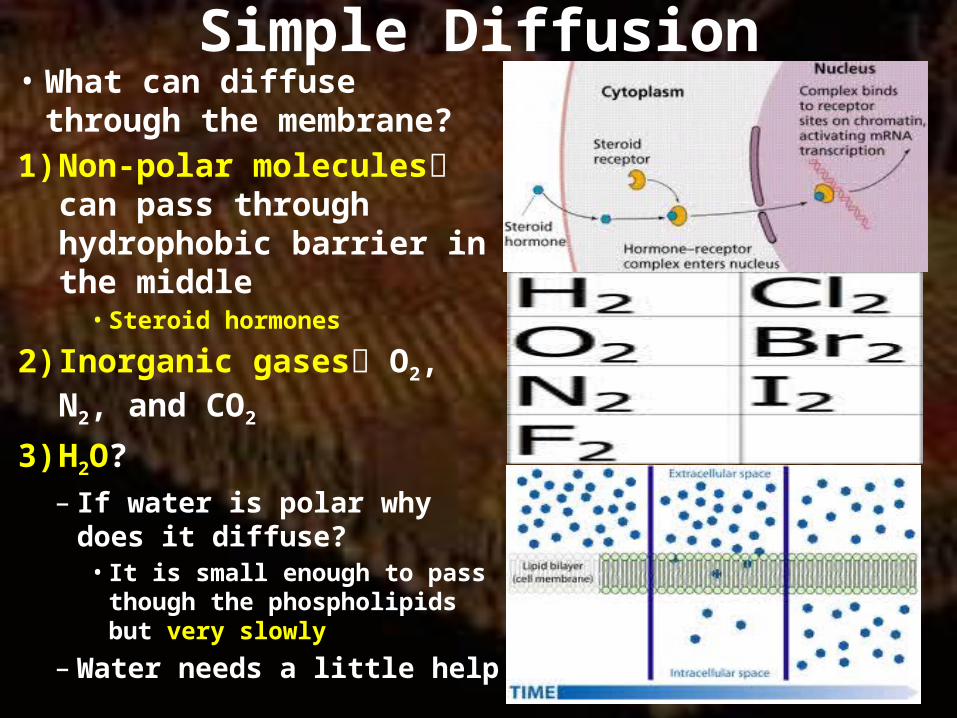
Simple Diffusion• What can diffuse through the
membrane?1) Non-polar molecules can
pass through hydrophobic barrier in the middle• Steroid hormones
2) Inorganic gases O2, N2, and CO2
3) H2O?– If water is polar why does it
diffuse?• It is small enough to pass though
the phospholipids but very slowly
– Water needs a little help
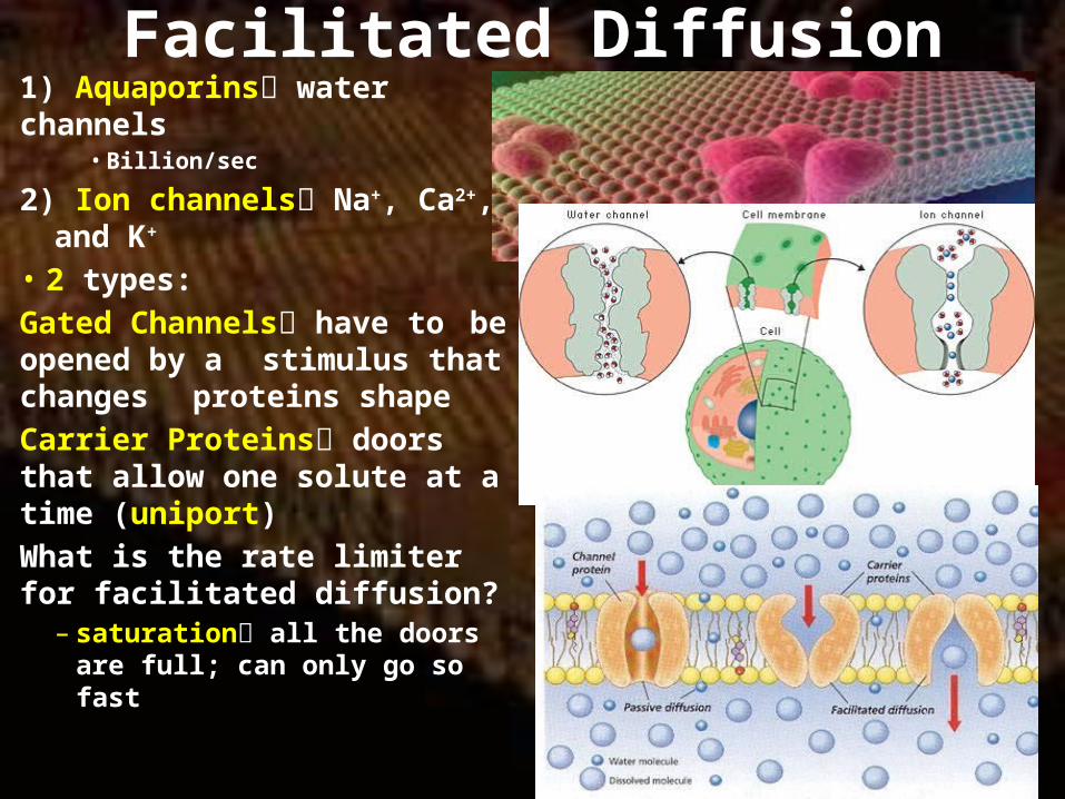
Facilitated Diffusion1) Aquaporins water channels
• Billion/sec
2) Ion channels Na+, Ca2+, and K+
• 2 types:Gated Channels have to be opened by a stimulus that changes proteins shapeCarrier Proteins doors that allow one solute at a time (uniport)What is the rate limiter for facilitated diffusion?
– saturation all the doors are full; can only go so fast

Osmosis• Movement of H2O is controlled
by the osmotic pressure of the solutions in and outside the cell
• The greater the difference in non-diffusing solutes the stronger the pressure
• 3 solution types:1) Hypotonic low concentration
outside the cell; water flows into the cell (turgor pressure)
2) Hypertonic high concentration outside the cell; water flows out of the cell (plasmolysis)
3) Isotonic equal concentrations

Active Transport• Any transportation that
requires energy• 3 main functions:1) Brining important nutrients
into the cell2) Removing waster from the
cell3) Maintaining concentrations
of ions across the membraneWhat ions are most important?
– H+, Na+, K+, and Ca2+
– Ions create membrane potential electrical potential difference between sides of membranes

Primary Active Transport• Transport protein is same
one hydrolyzing ATP• H+ Pumps:
– Connects to ATP synthase enzymes to power ATP production
– What other membrane would need H+ regulation?• Lysosome; need low pH
• Ca2+ Pumps:– Ca2+ concentration is high
inside vesicles– Release of Ca2+ can regulate
muscle contractions, microtubule assembly, and even secretions from the cell

Primary Active Transport• Na+/K+ Pumps:– Essential for all animal
cells and the nerve system of complex organisms
– 1 ATP 3 Na+ out and 2 K+ in
– Generates membrane potentials from -20mV to – 200mV
• Electrochemical gradient concentration gradient produces both a movement of chemicals and an electrical charge
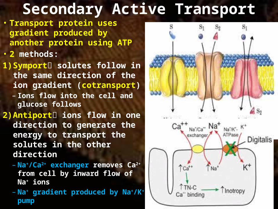
Secondary Active Transport• Transport protein uses gradient
produced by another protein using ATP
• 2 methods:1) Symport solutes follow in the
same direction of the ion gradient (cotransport)– Ions flow into the cell and glucose
follows
2) Antiport ions flow in one direction to generate the energy to transport the solutes in the other direction– Na+/Ca2+ exchanger removes Ca2+
from cell by inward flow of Na+ ions– Na+ gradient produced by Na+/K+
pump
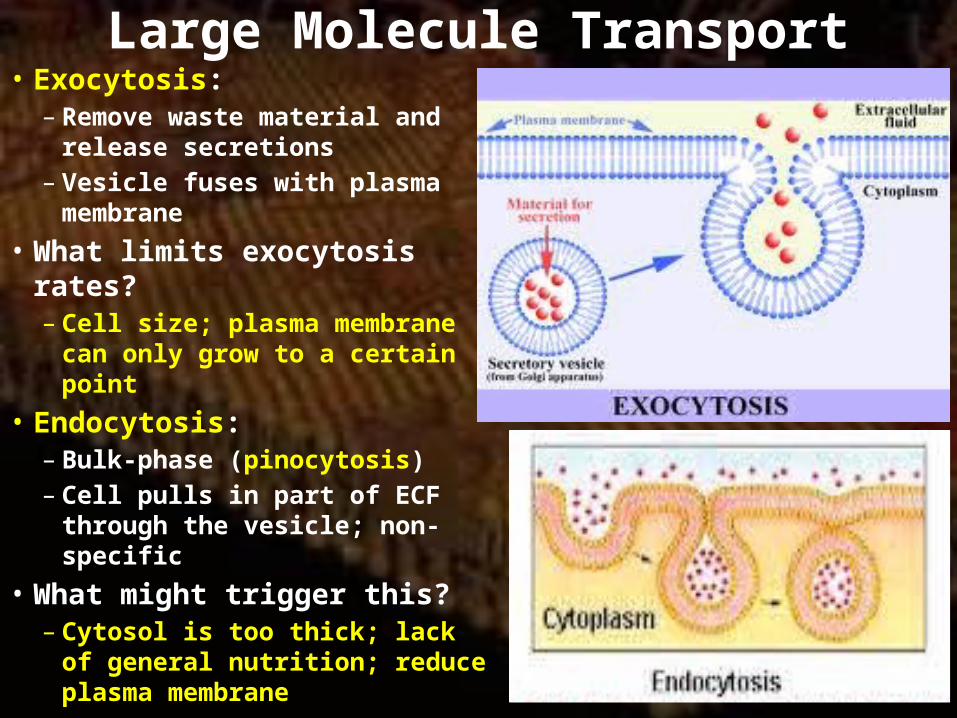
Large Molecule Transport• Exocytosis:
– Remove waste material and release secretions
– Vesicle fuses with plasma membrane
• What limits exocytosis rates?– Cell size; plasma membrane can
only grow to a certain point• Endocytosis:
– Bulk-phase (pinocytosis)– Cell pulls in part of ECF through
the vesicle; non-specific• What might trigger this?
– Cytosol is too thick; lack of general nutrition; reduce plasma membrane
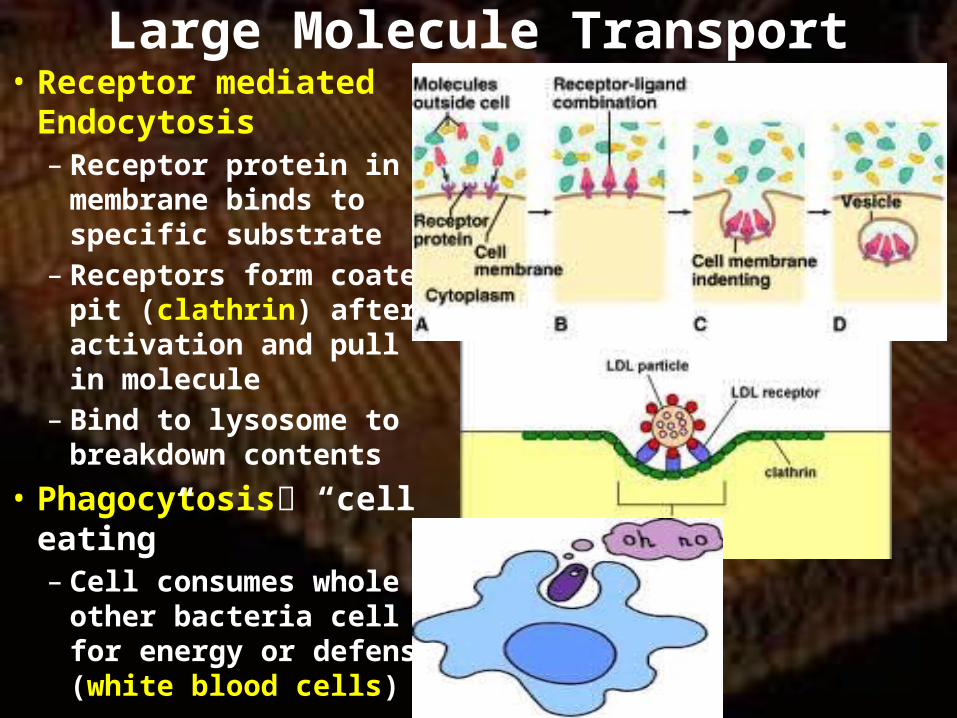
Large Molecule Transport• Receptor mediated
Endocytosis– Receptor protein in
membrane binds to specific substrate
– Receptors form coated pit (clathrin) after activation and pull in molecule
– Bind to lysosome to breakdown contents
• Phagocytosis “cell eating”– Cell consumes whole other
bacteria cell for energy or defense (white blood cells)
Homework• Read Ch. 7• Ch. 6 vocabulary• Test your Knowledge for Ch. 6 and do the
“Express your Opinion” and “Interpret the Data” in your notebook.