Embed Size (px)
Citation preview

MOL #66126
1
Membrane Transport and Intracellular Sequestration of Novel
Thiosemicarbazone Chelators for the Treatment of Cancer
Angelica M. Merlot, Namfon Pantarat, David B. Lovejoy, Danuta S. Kalinowski‡ and
Des R. Richardson‡*
Department of Pathology and Bosch Institute, University of Sydney, Sydney, NSW 2006
Australia.
‡D.S.K. and D.R.R. contributed equally to this publication as senior authors.
Molecular Pharmacology Fast Forward. Published on July 6, 2010 as doi:10.1124/mol.110.066126
Copyright 2010 by the American Society for Pharmacology and Experimental Therapeutics.
This article has not been copyedited and formatted. The final version may differ from this version.Molecular Pharmacology Fast Forward. Published on July 6, 2010 as DOI: 10.1124/mol.110.066126
at ASPE
T Journals on July 15, 2020
molpharm
.aspetjournals.orgD
ownloaded from

MOL #66126
2
Running title: Transport mechanisms of novel thiosemicarbazones.
*Corresponding author: D.R. Richardson, Department of Pathology, Blackburn Building
(D06), The University of Sydney, Sydney, New South Wales, 2006, Australia; Phone: +61-2-
9036-6548. Fax: +61-2-9351-3429. E-mail: [email protected]
Text Pages: 32
Tables: 0
Figures: 7
References: 38
Abstract: 248 words
Introduction: 725 words
Discussion: 1500 words
Abbreviations: 2,4-DNP, 2,4-dinitrophenol; BpT, 2-benzoylpyridine thiosemicarbazone;
Bp4eT, 2-benzoylpyridine 4-ethyl-3-thiosemicarbazone; DFO, desferrioxamine; DMEM +
GLU, Dulbecco’s modified Eagle’s medium with glucose; DMEM – GLU, Dulbecco’s
modified Eagle’s medium without glucose; Dp44mT, di-2-pyridylketone 4,4-dimethyl-3-
thiosemicarbazone; HUVECs, human umbilical vein endothelial cells; MES, 4-morpholine
ethanesulfonic acid; NaCN, sodium cyanide; NaF, sodium fluoride; NaN3, sodium azide; PIH,
pyridoxal isonicotinoyl hydrazone; ROS, reactive oxygen species; RR, ribonucleotide
reductase; Tf, transferrin; TfR1, transferrin receptor 1.
This article has not been copyedited and formatted. The final version may differ from this version.Molecular Pharmacology Fast Forward. Published on July 6, 2010 as DOI: 10.1124/mol.110.066126
at ASPE
T Journals on July 15, 2020
molpharm
.aspetjournals.orgD
ownloaded from

MOL #66126
3
Abstract
Iron is a critical nutrient for DNA synthesis and cellular proliferation. Targeting iron in cancer
cells using specific chelators is a potential new strategy for the development of novel anti-
cancer agents. One such chelator, 2-benzoylpyridine 4-ethyl-3-thiosemicarbazone (Bp4eT),
possesses potent and selective anti-cancer activity (Kalinowski, D.S. et al., (2007) J. Med.
Chem. 50:3716-3729). To elucidate the mechanisms of its potent anti-tumor activity, Bp4eT
was labeled with 14C. Its efficacy was then compared to the 14C-labeled iron chelator,
pyridoxal isonicotinoyl hydrazone (PIH), which exhibits low anti-cancer activity. The ability
of these ligands to permeate the cell membrane and their cellular retention was examined
under various conditions using SK-N-MC neuroepithelioma cells. The rate of 14C-PIH uptake
into cells was significantly (p<0.001) lower than that of 14C-Bp4eT at 37oC, indicating that
the increased hydrophilicity of 14C-PIH reduced membrane permeability. In contrast, the
efflux of 14C-PIH was significantly (p<0.05) higher than that of 14C-Bp4eT, leading to
increased cellular retention of 14C-Bp4eT. Additionally, the uptake and release of the 14C-
chelators was not reduced by metabolic inhibitors, indicating these processes were energy-
independent. No significant differences were evident in the uptake of 14C-Bp4eT at 37oC or
4oC, demonstrating a temperature-independent mechanism. Furthermore, adjusting the pH of
the culture medium to model the tumor microenvironment did not affect 14C-Bp4eT
membrane transport. It can be concluded that 14C-Bp4eT more effectively permeated the cell
membrane and evaded rapid efflux in contrast to 14C-PIH. This property, in part, accounts for
the more potent anti-cancer activity of Bp4eT relative to PIH.
This article has not been copyedited and formatted. The final version may differ from this version.Molecular Pharmacology Fast Forward. Published on July 6, 2010 as DOI: 10.1124/mol.110.066126
at ASPE
T Journals on July 15, 2020
molpharm
.aspetjournals.orgD
ownloaded from

MOL #66126
4
Introduction
Cancer remains one of the main underlying causes of morbidity and mortality (Curado et al.,
2007). Current treatments are far from optimal, having undesirable side-effects due to their
non-selective nature (Grahame-Smith and Aronson, 2006). Thus, new improved
chemotherapeutic strategies are urgently required.
Iron is an essential co-factor for the catalytic activity of many enzymes including
ribonucleotide reductase (RR), which catalyzes the rate-limiting step in DNA synthesis
(Kolberg et al., 2004). As neoplastic cells are generally more metabolically active than their
normal counterparts, they require larger amounts of iron (Pahl and Horwitz, 2005). In fact,
cancer cells express high levels of transferrin receptor 1 (TfR1), which is responsible for iron
uptake from the iron transport protein, transferrin (Tf) (Richardson and Baker, 1990; Trinder
and Baker, 2003). High expression of TfR1 has been demonstrated to correlate with tumor
growth and metastasis in animal models (Cavanaugh et al., 1999). In addition, cancer cells
have been shown to express greater levels of RR in comparison to normal cells (Elford et al.,
1970). Considering this, cancer cells could be expected to be more sensitive to iron-
deprivation than normal cells (Le and Richardson, 2002).
In the search for more effective anti-cancer agents, iron chelators have emerged as a novel
class of chemotherapeutics worthy of investigation. The chelator, desferrioxamine (DFO;
Figure 1A), is classically used for the treatment of iron overload disease, but has also been
assessed for anti-cancer activity (Kalinowski and Richardson, 2005). However, its poor
membrane permeability due to its relative hydrophilicity, short half-life and relatively low
anti-proliferative activity, has resulted in mixed outcomes in clinical trials (Kalinowski and
Richardson, 2005). This has led to studies examining iron chelators of greater lipophilicity.
This article has not been copyedited and formatted. The final version may differ from this version.Molecular Pharmacology Fast Forward. Published on July 6, 2010 as DOI: 10.1124/mol.110.066126
at ASPE
T Journals on July 15, 2020
molpharm
.aspetjournals.orgD
ownloaded from

MOL #66126
5
In comparison to hydrophilic DFO, the relatively hydrophobic iron chelator, pyridoxal
isonicotinoyl hydrazone (PIH; Figure 1B) is able to bind FeIII with high selectivity, inducing
the mobilization of intracellular iron from a number of cell types (Huang and Ponka, 1983;
Richardson, 1997; Richardson and Ponka, 1994). Interestingly, although PIH can effectively
mobilize iron from cells, it appears to target iron pools that are not necessary for proliferation,
demonstrating poor anti-proliferative activity in cell culture (IC50: 75 µM) (Richardson et al.,
1995).
A series of structure-activity relationship studies of the PIH analogues over the last 10 years
has assessed both anti-proliferative efficacy and iron chelation activity (Becker et al., 2003;
Richardson et al., 1995; Whitnall et al., 2006; Yuan et al., 2004). This work has led to the
development of the 2-benzoylpyridine thiosemicarbazone (BpT) chelators, which demonstrate
potent and selective anti-cancer activity (Kalinowski et al., 2007). These ligands have the
ability to sequester cellular iron, as well as the capacity to form iron complexes that are
redox-active (Kalinowski et al., 2007). Hence, the BpT analogues are able to cause iron
mobilization and generate cytotoxic reactive oxygen species (ROS), providing a novel
approach for the inhibition of cancer cell growth (Kalinowski et al., 2007).
Although the BpT series possess anti-proliferative activity in SK-N-MC neuroepithelioma
cells (IC50: 0.002-0.005 µM; Kalinowski et al., 2007), they show 1,250-3,000-fold less
efficacy for inhibiting the proliferation of normal MRC-5 fibroblasts (IC50 > 6.25 μM)
(Kalinowski et al., 2007). Moreover, BpT chelators are effective in vivo, selectively inhibiting
the growth of human tumor xenografts in nude mice (Yu, Y. and Richardson, D.R.,
unpublished results). Within the BpT series, 2-benzoylpyridine 4-ethyl-3-thiosemicarbazone
(Bp4eT; Figure 1C), showed marked anti-proliferative activity with an IC50 of 0.002 µM and
This article has not been copyedited and formatted. The final version may differ from this version.Molecular Pharmacology Fast Forward. Published on July 6, 2010 as DOI: 10.1124/mol.110.066126
at ASPE
T Journals on July 15, 2020
molpharm
.aspetjournals.orgD
ownloaded from

MOL #66126
6
was a clear leading candidate for further studies (Kalinowski et al., 2007).
While Bp4eT possessed potent and selective anti-cancer activity, the exact mechanisms
behind these important properties remain unclear. Thus, the aim of the current study was to
investigate the mechanisms of action of the potent anti-proliferative activity of Bp4eT in
comparison to PIH, which exhibits poor anti-proliferative efficacy. In order to do this, 14C-
labeled chelators were employed. Since chemotherapeutic cytotoxicity depends on a variety
of factors, including drug transport across membranes and retention within cells (Gottesman
et al., 2002), both the cellular uptake and release of 14C-chelators were examined. In this
investigation, the effects of temperature, metabolic inhibitors, pH and extracellular protein on
the cellular retention of the chelators were measured. In addition, the cellular uptake of 14C-
chelators was analyzed in a variety of cultured neoplastic and normal cell types. It can be
concluded that Bp4eT has efficient membrane transport and retention within tumor cells in
comparison to PIH, resulting in greater drug delivery and maximal cytotoxicity.
This article has not been copyedited and formatted. The final version may differ from this version.Molecular Pharmacology Fast Forward. Published on July 6, 2010 as DOI: 10.1124/mol.110.066126
at ASPE
T Journals on July 15, 2020
molpharm
.aspetjournals.orgD
ownloaded from

MOL #66126
7
Materials and Methods
14C-Chelators
14C-PIH and 14C-Bp4eT were synthesized by The Institute of Isotopes Ltd (Budapest,
Hungary) incorporating the 14C label at the imine carbon which represents a highly stable site
for incorporating the isotope. The final purity of both compounds was determined by HPLC
using UV detection. The certificate of analysis indicated the purity of 14C-PIH and 14C-Bp4eT
were 98.5% and 100%, respectively, with a final specific radioactivity of 75 µCi/mg. All
compounds were prepared as their hydrochloride salts to maximize solubility. Chelators were
dissolved in dimethyl sulfoxide (DMSO, Sigma-Aldrich, St. Louis, MO, USA) as 10 mM
stock solutions and diluted in complete medium so that the final [DMSO] < 0.5% (v/v).
Cell Culture
The human SK-N-MC neuroepithelioma cell line (American Type Culture Collection
(ATCC), Manassas, VA) was grown as previously described (Richardson et al., 1995). Briefly,
the cells were grown in minimal essential medium (MEM; Invitrogen, Victoria, Australia)
with 10% (v/v) fetal calf serum (Sigma-Aldrich, St. Louis, USA) and supplemented with the
following additions from Gibco (Victoria, Australia): 1% (v/v) sodium pyruvate, 1% (v/v)
non-essential amino acids, 100 U/mL penicillin, 100 µg/mL streptomycin, 2 mM glutamine
and 0.28 ng/mL fungizone. The cells were then incubated at 37oC in a humidified atmosphere
of 5% CO2/95% air in a water-jacketed incubator (Thermo Scientific, Forma Series II,
Marietta, OH, USA). The SK-N-MC cell line was chosen for the majority of these
investigations as its iron metabolism and the effect of a variety of chelators on this cell type is
well characterized (Richardson and Ponka, 1994; Richardson et al., 1995).
The human SK-Mel-28 melanoma, DMS-53 lung carcinoma, MCF-7 breast cancer and MRC-
This article has not been copyedited and formatted. The final version may differ from this version.Molecular Pharmacology Fast Forward. Published on July 6, 2010 as DOI: 10.1124/mol.110.066126
at ASPE
T Journals on July 15, 2020
molpharm
.aspetjournals.orgD
ownloaded from

MOL #66126
8
5 fibroblast cell lines were also obtained from the ATCC. These cell lines were cultured
similarly to that of the SK-N-MC cell line described above. Human umbilical vein endothelial
cells (HUVECs) were kindly donated by Mr. Pat Pisansarakit (Heart Research Institute,
Sydney, Australia). HUVECs were cultured in M199 media (SAFC Biosciences, St Louis,
MO, USA) with the standard supplementation described above for MEM alone and 0.5%
vascular endothelial growth factor (Starrate, Bethungra, Australia).
General 14C-Chelator Cellular Uptake Procedure
The 14C-chelator uptake into cells was examined using the SK-N-MC cell line by
implementing standard protocols (Huang and Ponka, 1983; Richardson, 1997). A cell
suspension of SK-N-MC cells at 1 x 10⁶ cells/mL was added to tissue culture plates and
incubated for 24 h at 37oC to produce a confluent monolayer. In experiments designed to
examine 14C-chelator uptake, the cells were incubated with complete media containing 14C-
Bp4eT (5-250 µM), 14C-PIH (5-250 µM) or no chelator (control) at 37oC for 120 min.
Subsequent to these studies, a 14C-chelator concentration of 25 µM was selected as it provided
appropriate labeling efficiency of cells that led to a highly sensitive assay. In experiments
measuring the effect of temperature on 14C-chelator uptake, the cells were incubated in media
containing 14C-chelator at 4oC or 37oC for up to 120 min.
Studies examining the influence of the Warburg effect (Warburg, 1956) on 14C-chelator uptake
utilized pH-adjusted complete media. This media was altered to a final pH of 5.5 or 6.5 using
MES (25 mM; Sigma-Aldrich) and to a pH of 7.4 and 8.0 (control media) using HEPES (25
mM; Sigma-Aldrich).
During all incubations with chelators, cells remained viable as judged by cellular morphology,
This article has not been copyedited and formatted. The final version may differ from this version.Molecular Pharmacology Fast Forward. Published on July 6, 2010 as DOI: 10.1124/mol.110.066126
at ASPE
T Journals on July 15, 2020
molpharm
.aspetjournals.orgD
ownloaded from

MOL #66126
9
adherence to the culture substratum and the exclusion of trypan blue. This was determined
using a Countess™ Automated Cell Counter (Invitrogen, Carlsbad, CA, USA). At the
conclusion of the incubation period, the media was aspirated and the cells were washed 4
times on ice with ice-cold phosphate-buffered saline (PBS) to remove extracellular 14C-
chelator. Then PBS (1 mL) was added to each plate and the cells were removed from the
substratum using a plastic spatula. This cell suspension was placed into counting tubes to
quantitate 14C-chelator uptake into cells. Scintillation fluid (2.5 mL; Perkin-Elmer,
Melbourne, Victoria, Australia) was added to the cell suspension and the radioactivity of each
sample, including backgrounds and 14C-chelator standards, were counted using a Micro-Beta
Counter (Perkin Elmer, Waltham, MA, USA). Results were expressed as molecules of
chelator/cell as a function of chelator concentration or incubation time.
In some experiments, 14C-chelator uptake in a variety of normal and cancer cell types was
compared and calculated as a function of the surface area of the cells. This was performed as
the relative size of the cells varied markedly. The surface area (µm2) of each cell type was
determined using Image J 1.42 software (National Institutes of Health, USA) analysis of
phase-contrast images taken with a Zeiss Axio Observer.Z1 microscope equipped with an
AxioCam camera and AxioVision Release 4.7 Software (Zeiss, Munich, Germany). The
average cell surface area was calculated after measuring 100 cells/cell type.
The Effect of Metabolic Inhibitors on 14C-Chelator Uptake
The effect of metabolic inhibitors on the uptake of 14C-chelators was studied using five well
characterized inhibitors (Sigma-Aldrich), including: sodium azide (NaN3; 30 mM),
oligomycin (30 µM), sodium cyanide (NaCN; 5 mM), sodium fluoride (NaF, 15 mM) and 2,4-
dinitrophenol (2,4-DNP, 2 mM). In brief, SK-N-MC neuroepithelioma cells were pre-
This article has not been copyedited and formatted. The final version may differ from this version.Molecular Pharmacology Fast Forward. Published on July 6, 2010 as DOI: 10.1124/mol.110.066126
at ASPE
T Journals on July 15, 2020
molpharm
.aspetjournals.orgD
ownloaded from

MOL #66126
10
incubated with inhibitors or media alone for 30 min at 37oC. The media was removed and
replaced with media containing 14C-PIH (25 µM) or 14C-Bp4eT (25 µM) in the presence or
absence of inhibitors and the cells were subsequently incubated for 60 min at 37oC. The
remainder of the experiment was conducted using the general uptake procedure described
above. During these studies, Dulbecco’s modified Eagle’s medium without glucose (DMEM -
GLU; Invitrogen) was used to aid the ATP-depletion induced by the metabolic inhibitors
(Richardson, 1997). Results were expressed as a percentage of the control, namely DMEM +
GLU.
General 14C-Chelator Cellular Efflux Procedure
The release of the 14C-chelators from pre-labeled SK-N-MC cells was performed using well
established techniques (Huang and Ponka, 1983; Richardson, 1997; Richardson et al., 1995).
Briefly, SK-N-MC cells were pre-labeled with either 14C-Bp4eT (25 µM), 14C-PIH (25 µM),
or media alone for 120 min at 37oC. The cells were then placed on ice, the media aspirated
and the cell monolayer washed four times with ice-cold PBS. For experiments examining the
effect of temperature on 14C-chelators release, complete media (1 mL; 4oC or 37oC) was
added to each plate and the cells were incubated at 4oC or 37oC for up to 180 min. There was
no significant decrease in viability throughout these incubations as measured by the Trypan
blue exclusion assay. Experiments conducted to analyze the effect of pH on 14C-chelator
efflux were incubated at 37oC for up to 120 min in pH-adjusted media (pH 5.5, 6.5, 7.4 or
8.0), as described for uptake experiments above. In some studies, the effect of the
extracellular proteins, Tf and albumin, on 14C-chelator release was assessed. In these
experiments, cells were incubated at 37oC in MEM without FCS (control) or in this medium
containing Tf (5 or 40 mg/mL) or albumin (5 or 40 mg/mL).
This article has not been copyedited and formatted. The final version may differ from this version.Molecular Pharmacology Fast Forward. Published on July 6, 2010 as DOI: 10.1124/mol.110.066126
at ASPE
T Journals on July 15, 2020
molpharm
.aspetjournals.orgD
ownloaded from

MOL #66126
11
At the end of each incubation period, the cells were placed on ice and the overlying media
was placed into counting tubes to estimate the level of extracellular 14C-chelator. Then PBS (1
mL) was added to the cells which were subsequently scraped from the plates using a plastic
spatula. This suspension was placed into counting tubes to represent the fraction of
intracellular 14C-chelator. Scintillation fluid (2.5 mL) was added to each sample as well as the
backgrounds and standards and the tubes were counted using a Micro-Beta Counter.
The Effect of Metabolic Inhibitors on 14C-Chelator Efflux
SK-N-MC cells were pre-incubated with 14C-PIH (25 µM) or 14C-Bp4eT (25 µM) in DMEM
+ GLU for 90 min at 37oC. The cells were then placed on ice and the metabolic inhibitors
described previously or media alone were added and incubated for 30 min at 37oC. Following
four washes with ice-cold PBS, the plates were re-incubated in DMEM - GLU for 5, 15, 30,
and 60 min at 37oC in media in the presence or absence of inhibitors. The remainder of the
experiment was completed in accordance with the general efflux methods described above.
It should be noted that the inhibitor, 2,4-DNP, was not used in efflux studies due to problems
associated with its intense color that led to quenching and thus problems associated with
quantification of 14C by β-counting. On the other hand, in uptake experiments, the inhibitors
were washed off cells prior to β-counting, and thus, quenching did not affect the counting
efficiency of 2,4-DNP-treated samples.
Cellular ATP Determination
Parallel ATP determinations were performed simultaneously in uptake and efflux experiments
involving the use of metabolic inhibitors in order to confirm their inhibitory effects on energy
metabolism. ATP levels were quantitatively analyzed using an ATP bioluminescence assay kit
This article has not been copyedited and formatted. The final version may differ from this version.Molecular Pharmacology Fast Forward. Published on July 6, 2010 as DOI: 10.1124/mol.110.066126
at ASPE
T Journals on July 15, 2020
molpharm
.aspetjournals.orgD
ownloaded from

MOL #66126
12
(Sigma-Aldrich) following the manufacturer’s instructions. Throughout the experiment, assay
samples were kept on ice. Briefly, to lyse cells, the samples underwent three freeze-thaw
cycles and plates were then scraped in distilled water (140 µL). The samples were then
centrifuged at 4oC for 45 min at 14,000 rpm. The ATP assay mix (100 µL) was added to a 96-
well plate, mixed and allowed to stand 3 min at room temperature to remove any endogenous
ATP. The sample supernatant (100 µL) was then added to the ATP assay mix, shaken and the
fluorescence was measured on a microplate reader (560 nm). A new standard curve was
generated with each assay using the ATP standard supplied.
Statistical Analysis
Results are expressed as mean ± S.E.M. Statistical significance was determined using the
Student’s t-test, one-way ANOVA or two-way ANOVA with replication. Results were
considered statistically significant when p < 0.05.
This article has not been copyedited and formatted. The final version may differ from this version.Molecular Pharmacology Fast Forward. Published on July 6, 2010 as DOI: 10.1124/mol.110.066126
at ASPE
T Journals on July 15, 2020
molpharm
.aspetjournals.orgD
ownloaded from

MOL #66126
13
Results
Rate of 14C-Chelator Uptake
The BpT series of ligands have shown high anti-proliferative activity and selectivity against
cancer cells (Kalinowski et al., 2007). To further understand their mechanism of action the
current studies have assessed the ability of one of the most potent members of this series of
compounds, namely Bp4eT, to permeate cancer cell membranes. For comparison to Bp4eT,
we have assessed the uptake of the chelator, PIH, which shows some structural similarity
(Figure 1), but demonstrates poor anti-proliferative activity (Richardson et al., 1995). By
determining the cellular uptake and subsequent release of these ligands, information relevant
to their marked differences in anti-proliferative activity should be obtained.
In initial studies, the uptake of 14C-Bp4eT and 14C-PIH were measured after a 120 min
incubation at 37oC to evaluate the rate of entry of the ligands into SK-N-MC
neuroepithelioma cells (Fig. 2A). The uptake of both chelators increased as a linear function
of chelator concentration (r2 = 0.99) in the range of 5-250 µM without any evidence of
saturation. Higher concentrations of chelator were not used due to the potential toxic effects
on cells, leading to spurious results and also because of limitations in the solubility of these
relatively hydrophobic compounds. Although both 14C-chelators possessed linear uptake
kinetics, the rate of 14C-PIH uptake (1.45 x 10⁶ ± 0.96 molecules of chelator/cell/min) was
significantly (p<0.001) lower than that of 14C-Bp4eT (3.02 x 10⁶ ± 0.3 molecules of
chelator/cell/min). These results demonstrated that 14C-Bp4eT had an increased rate of entry
into SK-N-MC cells than 14C-PIH (Fig. 2A).
The Effect of Temperature on 14C-Chelator Uptake
The effect of temperature and time on 14C-Bp4eT and 14C-PIH uptake was investigated in
This article has not been copyedited and formatted. The final version may differ from this version.Molecular Pharmacology Fast Forward. Published on July 6, 2010 as DOI: 10.1124/mol.110.066126
at ASPE
T Journals on July 15, 2020
molpharm
.aspetjournals.orgD
ownloaded from

MOL #66126
14
order to determine whether these processes could potentially be carrier-mediated. At 37oC and
4oC, a biphasic mode of uptake as a function of time was evident for 14C-Bp4eT and 14C-PIH
(Fig. 2B). The uptake of 14C-Bp4eT was markedly and significantly (p<0.001) greater than
14C-PIH at all time points at both 37oC and 4oC. The cellular uptake of 14C-Bp4eT increased
rapidly with time and reached a plateau within 30 min of incubation at both 4oC and 37oC.
Between 5 to 15 min of incubation, the cellular uptake of 14C-Bp4eT was significantly
(p<0.01) greater at 37oC than at 4oC (Fig. 2B). Additionally, at 37oC, 14C-Bp4eT uptake
peaked at the 15 min time point suggesting that the initial rate of uptake of the chelator may
have exceeded the rate of simultaneous efflux from the cell. However, the remainder of the
time points (30-120 min) demonstrated no significant (p>0.05) difference in 14C-Bp4eT
uptake between 4oC and 37oC. Therefore, after 30 min, it appeared the rate of simultaneous
uptake and efflux equilibrated to an extent similar to that observed at 4oC (Fig. 2B).
In contrast to 14C-Bp4eT uptake, the uptake of 14C-PIH was significantly (p<0.01) lower in
cells incubated at 4oC than at 37oC (Fig. 2B), demonstrating a dependence on temperature and
a different mechanism of uptake to that found with 14C-Bp4eT.
The Effect of Temperature on 14C-Chelator Efflux
The release of 14C-Bp4eT and 14C-PIH from SK-N-MC cells was also examined as a function
of time at 4oC and 37oC (Fig. 2C) to investigate its dependence on temperature. In these
experiments, cells were labeled for 120 min with the 14C-chelators, washed and then
reincubated for up to 180 min at 4oC or 37oC. After the 180 min reincubation, 57 ± 3% of 14C-
Bp4eT was released at 37oC. In comparison, significantly (p<0.01) lower levels of 14C-Bp4eT
were released at 4oC (20 ± 2%) after 180 min (Fig. 2C). Thus, 14C-Bp4eT release from cells
was temperature-dependent in contrast to its uptake (Fig. 2B).
This article has not been copyedited and formatted. The final version may differ from this version.Molecular Pharmacology Fast Forward. Published on July 6, 2010 as DOI: 10.1124/mol.110.066126
at ASPE
T Journals on July 15, 2020
molpharm
.aspetjournals.orgD
ownloaded from

MOL #66126
15
Within 15 min of reincubation at 37oC, the release of 14C-PIH was significantly (p<0.05)
greater than that of 14C-Bp4eT, with 80 ± 2% of 14C-PIH released from cells after 180 min
(Fig. 2C). Additionally, the percentage of cellular 14C-PIH released was significantly
(p<0.001) lower at 4oC (17 ± 2% at 180 min) when compared to 37oC (80 ± 2% at 180 min).
In fact, the release of 14C-PIH at 4oC was similar to 14C-Bp4eT at 4oC (Fig. 2C). Therefore,
the release of intracellular 14C-PIH was also found to be temperature-dependent, as identified
for its uptake (Fig. 2B).
Considering the greater intracellular concentrations of 14C-Bp4eT within cells (Fig. 2B) and
its lower release relative to 14C-PIH (Fig. 2C), it is clear that 14C-Bp4eT was becoming
sequestered within cells (Fig. 2D). In fact, after a 180 min reincubation at 37oC, 6.4 ± 1.2 x
107 molecules/cell of Bp4eT were found. This was equal to 39 ± 7% of the total 14C-Bp4eT
initially found intracellularly when the reincubation began. In contrast, 32-fold less of 14C-
PIH remained associated within the cell after a 180 min reincubation at 37oC (i.e., 0.2 ± 0.01
x 107 molecules/cell; Fig. 2D). This represented 12 ± 0.2% of the 14C-PIH found initially
within the cell at the start of the reincubation. Hence, far more 14C-Bp4eT remained
sequestered within the cell relative to 14C-PIH.
The Effect of Metabolic Inhibitors on 14C-Chelator Uptake
Considering the results described above, experiments were then performed to determine
whether chelator uptake was energy-dependent (Fig. 3A and B). The effect of five well
characterized metabolic inhibitors, namely NaN3, oligomycin, NaCN, 2,4-DNP and NaF
(Henderson and Zevely, 1984; Qian and Morgan, 1991; Richardson, 1997; Svec, 1985), on
14C-Bp4eT and 14C-PIH uptake into SK-N-MC cells was investigated at 37oC. In addition, in
This article has not been copyedited and formatted. The final version may differ from this version.Molecular Pharmacology Fast Forward. Published on July 6, 2010 as DOI: 10.1124/mol.110.066126
at ASPE
T Journals on July 15, 2020
molpharm
.aspetjournals.orgD
ownloaded from

MOL #66126
16
these studies, ATP assays were performed in parallel to 14C-ligand uptake experiments to
assess the effects of the inhibitors on energy metabolism (Fig. 3C and D). Studies compared
the effects on 14C-ligand uptake in DMEM ± GLU (Fig. 3). Furthermore, in all of these
experiments, the metabolic inhibitors were added to DMEM - GLU to ensure ATP-depletion
of cells (Richardson, 1997).
Although the metabolic inhibitors, NaN3, oligomycin, NaCN, 2,4-DNP and NaF significantly
(p<0.01) reduced cellular ATP levels in comparison to control medium (namely DMEM +
GLU; Fig. 3C), they did not significantly (p>0.05) decrease 14C-Bp4eT uptake (Fig. 3A). In
fact, 2,4-DNP caused a significant (p<0.01) increase in intracellular 14C-Bp4eT in comparison
to control medium (Fig. 3A). The increased uptake of 14C-Bp4eT in the presence of 2,4-DNP
could be consistent with non-specific adsorption of the 14C-labeled ligand to the cell due to
membrane damage, as observed for other molecules (Baker et al., 1992). In contrast to 2,4-
DNP, the inhibitors NaN3, oligomycin, NaCN and NaF had no significant (p>0.05) effect on
14C-Bp4eT uptake compared to the control (DMEM + GLU). Overall, all five metabolic
inhibitors had no inhibitory effects on 14C-Bp4eT uptake, suggesting that 14C-Bp4eT uptake is
an energy-independent process.
In addition, no significant (p>0.05) inhibitory effect of the metabolic poisons was evident on
the uptake of 14C-PIH by cells as compared to the controls (Fig. 3B). This was despite the
effect of these inhibitors to significantly (p<0.01) reduce ATP levels relative to the control
(DMEM + GLU; Fig. 3D). Both 2,4-DNP and NaF increased 14C-PIH uptake relative to the
control (Fig. 3B). Again, this may be due to membrane damage induced by the inhibitor,
leading to increased non-specific adsorption of 14C-PIH. In summary, these results suggest
that energy-independent mechanisms may also be responsible for the uptake of 14C-PIH.
This article has not been copyedited and formatted. The final version may differ from this version.Molecular Pharmacology Fast Forward. Published on July 6, 2010 as DOI: 10.1124/mol.110.066126
at ASPE
T Journals on July 15, 2020
molpharm
.aspetjournals.orgD
ownloaded from

MOL #66126
17
The Effect of Metabolic Inhibitors on 14C-Chelator Efflux
Further studies then examined the effect of metabolic inhibitors on the cellular release of 14C-
Bp4eT and 14C-PIH to determine whether this was an energy-dependent process (Fig. 4A and
B). Again, cellular ATP levels were assessed simultaneously in order to confirm the inhibitory
effects of the metabolic inhibitors (Fig. 4C and D). Although inhibitor-treated cells possessed
significantly (p<0.001) lower levels of ATP than control medium (DMEM ± GLU; Fig. 4C
and D), no significant (p>0.05) effect was observed on the cellular release of 14C-Bp4eT and
14C-PIH (Fig. 4A and B). These results indicated that the cellular release of 14C-Bp4eT and
14C-PIH occurred through an energy-independent mechanism, involving the passive diffusion
of both these ligands across the cell membrane.
The Effect of pH on 14C-Chelator Uptake
A common hallmark of malignant neoplasms is the Warburg effect, in which the tumor
possesses a slightly acidic (pH 6.92-7.24) microenvironment (Helmlinger et al., 1997;
Warburg, 1956). Since the thiosemicarbazone classes of chelators are polyprotic (Richardson
et al., 2006; Richardson et al., 1990), their net charge is dependent upon the pH of the
solution. This can markedly alter membrane permeability and thus it was important to
understand the potential influence the Warburg effect has on the cellular uptake of 14C-Bp4eT
and 14C-PIH. Considering this, the pH of the medium was altered using a variety of buffers
(see Materials and Methods) so that the final pH values were equal to 5.5, 6.5, 7.4 and 8.0
(Fig. 5A-D). These experiments demonstrated that the initial cellular uptake of 14C-Bp4eT
was significantly (p<0.05) lower at pH 5.5 and 6.5 than at pH 7.4 between 5-30 min at 37oC
(Fig. 5A). However, for the remainder of the incubation up to 120 min at 37oC, there was no
significant (p>0.05) difference in uptake compared to pH 7.4 (Fig. 5A). At pH 8.0, the uptake
This article has not been copyedited and formatted. The final version may differ from this version.Molecular Pharmacology Fast Forward. Published on July 6, 2010 as DOI: 10.1124/mol.110.066126
at ASPE
T Journals on July 15, 2020
molpharm
.aspetjournals.orgD
ownloaded from

MOL #66126
18
of 14C-Bp4eT was not significantly different to that found at pH 7.4. In summary, after a 2 h
incubation, intracellular levels of 14C-Bp4eT were not altered by the pH range examined.
In contrast to the results found for 14C-Bp4eT, the cellular uptake of 14C-PIH demonstrated
considerable dependence on pH (Fig. 5B). At time points of 30 min or greater, significantly
(p<0.01) higher levels of intracellular 14C-PIH were evident at pH 5.5 and pH 6.5 compared to
control (pH 7.4). Interestingly, the cellular uptake of 14C-PIH was greatest at pH 5.5 and this
was found to be approximately double that found at pH 7.4 (Fig. 5B). Conversely, no
significant (p>0.05) difference in 14C-PIH uptake was evident at pH 8.0 in comparison to pH
7.4. Therefore, the cellular uptake of 14C-PIH was enhanced under slightly acidic conditions.
The Effect of pH on 14C-Chelator Efflux
Additional experiments assessed the effect of extracellular pH on the cellular release of 14C-
Bp4eT and 14C-PIH in order to analyze the influence the Warburg effect may have on tumor
drug retention in vivo (Fig. 5C and D). The cellular release of 14C-Bp4eT and 14C-PIH was not
significantly (p>0.05) altered by extracellular pH (pH 5.5, 6.5 and 8.0) in comparison to the
control (pH 7.4).
The Effect of Proteins on 14C-Chelator Efflux
In general, tumors possess leaky, immature and tortuous blood vessels (Carmeliet and Jain,
2000), resulting in high levels of protein within the tumor interstitium (Greish, 2007).
Furthermore, interactions of drugs with proteins may also affect the bioavailability of a drug
(van der Veldt et al., 2008). Thus, the cellular release of 14C-Bp4eT and 14C-PIH was
performed in the presence and absence of proteins, namely human Tf and albumin, at
concentrations of 5 or 40 mg/mL (Fig. 6A and B) to determine if the protein
This article has not been copyedited and formatted. The final version may differ from this version.Molecular Pharmacology Fast Forward. Published on July 6, 2010 as DOI: 10.1124/mol.110.066126
at ASPE
T Journals on July 15, 2020
molpharm
.aspetjournals.orgD
ownloaded from

MOL #66126
19
microenvironment of tumors affects chelator retention. The cellular release of 14C-Bp4eT was
significantly (p<0.05) lower in protein-free media (control) in comparison to media
containing Tf (5 or 40 mg/mL) or albumin (5 or 40 mg/mL; Fig. 6A). However, no significant
(p>0.05) difference in 14C-Bp4eT release was evident among the varying concentrations of
albumin and Tf analyzed, suggesting that the release of the ligand from cells was independent
of the specific protein type and concentration. These results indicate that the proteins could
act as an extracellular sink to facilitate the release of 14C-Bp4eT from cells. In contrast, no
significant (p>0.05) difference in the release of 14C-PIH was observed in the presence or
absence of proteins (Fig. 6B).
14C-Chelator Uptake in Normal and Cancer Cells
Our previous studies have shown that Bp4eT markedly inhibits the growth of cancer cells
relative to normal cells both in vitro (Kalinowski et al., 2007) and in vivo (Yu, Y. and
Richardson, D.R. unpublished data). Moreover, it is essential that clinically used
chemotherapeutic agents preferentially target cancer cells as opposed to normal tissue to
minimize side effects (Grahame-Smith and Aronson, 2006). Thus, the following experiments
were designed to examine the differences in the uptake of 14C-Bp4eT and 14C-PIH between
cancer (SK-N-MC neuroepithelioma, MCF-7 breast cancer and SK-Mel-28 melanoma cells)
and normal cells (MRC-5 fibroblasts and endothelial cells [HUVECs]). These studies were
performed using confluent (100%) and sub-confluent (50%) plates of cells to examine the
effect of the degree of confluency on cellular uptake. Results were expressed as molecules of
chelator/total surface area per cell. This was necessary as the surface area of different cell
types varied markedly and this allowed the accurate quantification of the uptake of the
chelators for comparison between cells.
This article has not been copyedited and formatted. The final version may differ from this version.Molecular Pharmacology Fast Forward. Published on July 6, 2010 as DOI: 10.1124/mol.110.066126
at ASPE
T Journals on July 15, 2020
molpharm
.aspetjournals.orgD
ownloaded from
MOL #66126
20
Under confluent conditions, MCF-7 breast cancer cells possessed significantly (p<0.05)
higher intracellular levels of 14C-Bp4eT in contrast to the normal MRC-5 and HUVEC cell
types (Fig. 7A). Additionally, the SK-N-MC cell line also showed significantly (p<0.05)
increased intracellular levels of 14C-Bp4eT compared to MRC-5 cells, although no significant
(p>0.05) differences were evident in comparison to HUVEC cells. Furthermore, there was no
significant (p>0.05) increase in the cellular uptake of 14C-Bp4eT in SK-Mel-28 melanoma
cells relative to normal cell types (Fig. 7A). Interestingly, the uptake of 14C-Bp4eT differed in
sub-confluent cells and this may be due to the adherence of the ligand to the plastic
substratum of the culture plates under sub-confluent conditions (Fig. 7B). Under these
conditions, similar levels of 14C-Bp4eT uptake were observed in SK-N-MC and SK-Mel-28
cells compared to normal cell lines (Fig. 7B). In contrast, significantly (p<0.05) higher
intracellular levels of 14C-Bp4eT were present in MCF-7 cells relative to that in normal cells.
Collectively, these results demonstrate that generally there is no clear difference in the uptake
of 14C-Bp4eT between normal and neoplastic cells. However, uptake of this ligand by MCF-7
breast cancer cells was greater than that found for the normal and also other neoplastic cells.
No significant increase in 14C-PIH uptake by cancer cell lines relative to normal cells was
observed under confluent conditions (Fig. 7C). However, the uptake of 14C-PIH was markedly
and significantly (p<0.001) greater by HUVECs than any other neoplastic or normal cell type
(Fig. 7C). In addition, under sub-confluent conditions, 14C-PIH uptake into normal MRC-5
cells was significantly (p<0.05) lower than all other cancer and normal cells (Fig. 7D),
demonstrating that 14C-PIH showed no selective uptake under sub-confluent conditions.
This article has not been copyedited and formatted. The final version may differ from this version.Molecular Pharmacology Fast Forward. Published on July 6, 2010 as DOI: 10.1124/mol.110.066126
at ASPE
T Journals on July 15, 2020
molpharm
.aspetjournals.orgD
ownloaded from

MOL #66126
21
Discussion
The toxicity of anti-cancer drugs as well as tumor resistance remain major problems in
oncology (Grahame-Smith and Aronson, 2006). Common causes for resistance and toxicity to
chemotherapeutic agents include insufficient drug delivery to the tumor, inadequate drug
uptake and/or rapid efflux (Gottesman et al., 2002). Consequently, the current studies were
undertaken to examine the membrane transport of the potential anti-tumor agent, Bp4eT.
Bp4eT Rapidly Enters Cells via a Metabolic Energy-Independent Mechanism Consistent
with Diffusion
As part of this study, the uptake of 14C-Bp4eT, which possesses marked anti-cancer activity
(Kalinowski et al., 2007), was compared to 14C-PIH, a ligand with low anti-proliferative
activity (Richardson et al., 1995). The rate of 14C-Bp4eT uptake by cells was markedly greater
than that of 14C-PIH (Fig. 2A), illustrating that 14C-Bp4eT can more easily and rapidly
permeate cells. This can be attributed to the lipophilic nature of Bp4eT (log P 4.02;
Kalinowski et al., 2007), in contrast to the relatively more hydrophilic ligand, PIH (log P -
2.11) (Richardson et al., 1995). Numerous studies have shown that hydrophilic chelators
possess poor anti-proliferative activity, while compounds with relatively high lipophilicity
have potent anti-proliferative effects (Hodges et al., 2004; Richardson et al., 1995).
Consequently, the higher intracellular levels of 14C-Bp4eT, owing to its greater membrane
permeability, may be partly responsible for its potent anti-cancer activity (Kalinowski et al.,
2007). In contrast, the relatively hydrophilic 14C-PIH led to lower membrane permeability,
correlating with its poor anti-proliferative activity (Richardson et al., 1995).
Since some transporters can influence cellular drug retention, such as the multi-drug
resistance associated protein and P-glycoprotein (Gottesman et al., 2002), it was important to
This article has not been copyedited and formatted. The final version may differ from this version.Molecular Pharmacology Fast Forward. Published on July 6, 2010 as DOI: 10.1124/mol.110.066126
at ASPE
T Journals on July 15, 2020
molpharm
.aspetjournals.orgD
ownloaded from

MOL #66126
22
examine the mechanisms involved in membrane transport of 14C-Bp4eT and 14C-PIH. The
current results indicate that Bp4eT permeates cells via passive diffusion. The following three
observations support this conclusion. First, results obtained from 14C-chelator uptake studies
as a function of ligand concentration (Fig. 2A) demonstrated that uptake occurred by zero-
order kinetics and remained unsaturated throughout the broad concentration range employed.
Second, 14C-Bp4eT uptake by cells was not reduced despite depleting ATP stores using
metabolic inhibitors (Fig. 3A, C). The third piece of evidence demonstrating that 14C-Bp4eT
permeates membranes through a passive process can be deduced by comparing its uptake at
37oC and 4oC. For the duration of the steady state (Fig. 2B), there was no significant
difference in intracellular 14C-Bp4eT at 37oC and 4oC, demonstrating temperature-
independent uptake. Collectively, Bp4eT permeate cells via passive diffusion, rather than
active transport.
Bp4eT and PIH are Released from Cells by a Temperature-Dependent Mechanism, but
Hydrophobic Bp4eT is Sequestered in Cells Relative to PIH
Although it is important for adequate quantities of drug to permeate cells, it is also essential
they evade efflux to allow sufficient quantities to be retained to cause cytotoxicity (Gottesman
et al., 2002). Therefore, to understand why Bp4eT was a more potent anti-cancer drug than
PIH, 14C-chelator efflux was investigated. In these studies, Bp4eT and PIH demonstrated a
significant reduction in 14C-chelator release at 4oC than 37oC (Fig. 2C). These findings are
consistent with a temperature-dependent release mechanism and may result from: (1) a
change in membrane fluidity that occurs upon decreasing temperature affecting drug transport
(Zimmer and Schirmer, 1974); or (2) a decrease in the efficacy of an energy-dependent
transporter. To clarify this, additional efflux experiments performed with metabolic inhibitors
(Fig. 4A and B) showed no difference in the release of intracellular 14C-chelator compared to
This article has not been copyedited and formatted. The final version may differ from this version.Molecular Pharmacology Fast Forward. Published on July 6, 2010 as DOI: 10.1124/mol.110.066126
at ASPE
T Journals on July 15, 2020
molpharm
.aspetjournals.orgD
ownloaded from
MOL #66126
23
the control, despite a marked decrease in ATP. This indicated the mechanism of ligand release
was energy-independent, but temperature-dependent. Hence, Bp4eT transport occurs via
temperature-dependent passive diffusion, where membrane fluidity can affect membrane
transport.
Importantly, the percentage of 14C-PIH released from cells was significantly higher than that
of 14C-Bp4eT at 37°C (Fig. 2C). These results together with the rapid uptake of 14C-Bp4eT
(Fig. 2B), resulted in 32-fold higher levels of 14C-Bp4eT relative to 14C-PIH within cells (Fig.
2D). The marked permeability and increased retention of 14C-Bp4eT may be partly
responsible for its potent anti-cancer effects, resulting in increased intracellular ROS and cell
death (Kalinowski and Richardson, 2005; Kalinowski et al., 2007). This is facilitated by the
difference in the iron-binding site of Bp4eT (N,N,S) relative to PIH (O,N,O) (Figure 1), which
leads to the ability of Bp4eT to bind metals and redox cycle to generate ROS (Kalinowski et
al., 2007).
The Warburg Effect Does Not Influence Bp4eT Uptake or Release by Tumor Cells
Tumors often contain a slightly acidic microenvironment due to enhanced glycolysis, which
has been referred to as the Warburg effect (Warburg, 1956). Considering the chelators used
are polyprotic compounds, their charge is influenced by pH, which affects membrane
permeability (Richardson et al., 2006; Richardson et al., 1990). Thus, the extracellular pH was
modified to model variations within the tumor microenvironment that might influence
chelator uptake and efflux. The results demonstrated that alterations in the extracellular pH to
those encountered in tumors (pH 6.9-7.2; Helminger et al., 1997) had little effect on 14C-
chelator efflux (Fig. 5C and D). Interestingly, greater intracellular uptake of 14C-PIH was
evident under acidic conditions (pH 5.5 and 6.5) in comparison to the control (pH 7.4) (Fig.
This article has not been copyedited and formatted. The final version may differ from this version.Molecular Pharmacology Fast Forward. Published on July 6, 2010 as DOI: 10.1124/mol.110.066126
at ASPE
T Journals on July 15, 2020
molpharm
.aspetjournals.orgD
ownloaded from

MOL #66126
24
5B). These results suggest a greater amount of 14C-PIH was present in its neutral form at a pH
of 5.5-6.5, allowing higher permeability than at pH 7.4. This concept is consistent with the
species distribution of PIH as a function of pH, where its neutral species predominates at pH
6 (Doungdee, 1995; Richardson et al., 1990).
In contrast, alterations in pH had no significant effect on 14C-Bp4eT uptake (Fig. 5A). These
data indicate 14C-Bp4eT is present largely as a neutral ligand between pH 5.5-8.0, resulting in
facile passage across the cell membrane. This suggestion is in accordance with the predicted
protonation constants of Bp4eT based on the structure of very similar DpT ligands,
particularly Dp4eT (Richardson et al., 2006). This latter compound predominantly exists in its
neutral form in the pH range of 4-11 (Richardson et al., 2006). Accordingly, although the
tumor milieu varies with respect to pH in accordance with the Warburg effect (Warburg,
1956), it is unlikely it would influence Bp4eT uptake.
Extracellular Protein Increases Efflux of Bp4eT, but not PIH
Another factor in tumors which could affect the retention of these compounds is the quantity
of protein in the interstitial fluid. Tumors possess more permeable blood vessels (Carmeliet
and Jain, 2000), which result in higher levels of proteins within the tumor interstitium
(Greish, 2007; Maeda, 2001). Hence, studies were performed in the presence of common
proteins present in the interstitial fluid, namely Tf and albumin, to determine whether the
protein microenvironment of tumors affects the cellular retention of the 14C-labeled ligands.
These investigations demonstrated 14C-Bp4eT release was increased in the presence of
proteins compared to protein-free media and was independent of the protein type (Fig. 6A).
Since lipophilic ligands have an affinity for proteins, these macromolecules may act as an
extracellular “sink”, increasing release of intracellular 14C-Bp4eT (Buss et al., 2002; Buss et
This article has not been copyedited and formatted. The final version may differ from this version.Molecular Pharmacology Fast Forward. Published on July 6, 2010 as DOI: 10.1124/mol.110.066126
at ASPE
T Journals on July 15, 2020
molpharm
.aspetjournals.orgD
ownloaded from

MOL #66126
25
al., 2003). It is also notable that low levels of extracellular protein (5 mg/mL) resulted in
similar efflux of 14C-Bp4eT as high concentrations of protein (40 mg/mL). Thus, the higher
protein levels within tumor interstitial fluid due to increased permeability of tumor
vasculature (Carmeliet and Jain, 2000), will probably not promote 14C-Bp4eT release to any
greater extent than in normal tissues. Conversely to Bp4eT, the presence of protein in medium
had no marked effect on 14C-PIH efflux at either protein concentration examined (Fig. 6B).
These observations could be interpreted to suggest the added proteins preferentially bind the
hydrophobic ligand, Bp4eT, to a greater extent than the more hydrophilic chelator, PIH.
All Cancer Cell Lines did not Preferentially Accumulate Higher Intracellular Levels of
Bp4eT Relative to Normal Cells
Effective chemotherapy requires compounds to preferentially target cancer cells over normal
cells to minimize systemic toxicity (Grahame-Smith and Aronson, 2006). Previous studies
demonstrated that Bp4eT has selective anti-proliferative activity in vitro (Kalinowski et al.,
2007) and in vivo (Yu, Y. and Richardson, D.R. unpublished results). Furthermore, closely
related compounds of the DpT class showed selective anti-tumor activity in vivo (Whitnall et
al., 2006). Therefore, 14C-chelator uptake was compared between cancer and normal cells.
Although increased 14C-Bp4eT uptake was evident in MCF-7 cancer cells in comparison to
normal cells, this difference was not apparent in all tumor cell types (Fig. 7A and B). Thus,
other factors may be responsible for the selective anti-cancer activity of Bp4eT. These could
include the greater sensitivity of cancer cells to iron-deprivation, as well as their effect on
other iron-dependent targets, including: RR, the metastasis suppressor NDRG1, cyclin D1 etc
(Le and Richardson, 2004; Yu et al., 2007).
In conclusion, 14C-Bp4eT and 14C-PIH uptake occurs through passive diffusion. Interestingly,
This article has not been copyedited and formatted. The final version may differ from this version.Molecular Pharmacology Fast Forward. Published on July 6, 2010 as DOI: 10.1124/mol.110.066126
at ASPE
T Journals on July 15, 2020
molpharm
.aspetjournals.orgD
ownloaded from

MOL #66126
26
these results reveal the lipophilic chelator, 14C-Bp4eT, can rapidly permeate cells, reaching
higher intracellular levels than the more hydrophilic ligand, 14C-PIH. In addition, 14C-Bp4eT
was retained within cells to a greater extent than 14C-PIH. Thus, 14C-Bp4eT easily enters
cancer cells and sufficient levels of the ligand or its redox-active iron complex are retained to
facilitate cytotoxicity. In contrast, 14C-PIH was efficiently released which probably
contributes to its low anti-proliferative effects. These studies also demonstrate that 14C-Bp4eT
membrane transport was not adversely affected by pH. Hence, it is unlikely the mildly acidic
tumor microenvironment would hinder Bp4eT uptake, enabling maximum efficacy.
Acknowledgments
We kindly thank Dr Katie Dixon, Dr Christopher Austin and Dr Helena Mangs for critical
evaluation of the manuscript prior to submission.
This article has not been copyedited and formatted. The final version may differ from this version.Molecular Pharmacology Fast Forward. Published on July 6, 2010 as DOI: 10.1124/mol.110.066126
at ASPE
T Journals on July 15, 2020
molpharm
.aspetjournals.orgD
ownloaded from
MOL #66126
27
References
Baker E, Richardson D, Gross S and Ponka P (1992) Evaluation of the iron chelation potential
of hydrazones of pyridoxal, salicylaldehyde and 2-hydroxy-1-naphthylaldehyde using
the hepatocyte in culture. Hepatology 15: 492-501.
Becker EM, Lovejoy DB, Greer JM, Watts R and Richardson DR (2003) Identification of the
di-pyridyl ketone isonicotinoyl hydrazone (PKIH) analogues as potent iron chelators
and anti-tumour agents. Br J Pharmacol 138: 819-830.
Buss JL, Arduini E and Ponka P (2002) Mobilization of intracellular iron by analogs of
pyridoxal isonicotinoyl hydrazone (PIH) is determined by the membrane permeability
of the iron-chelator complexes. Biochem Pharmacol 64: 1689-1701.
Buss JL, Arduini E, Shephard KC and Ponka P (2003) Lipophilicity of analogs of pyridoxal
isonicotinoyl hydrazone (PIH) determines the efflux of iron complexes and toxicity in
K562 cells. Biochem Pharmacol 65: 349-360.
Carmeliet P and Jain RK (2000) Angiogenesis in cancer and other diseases. Nature 407: 249-
257.
Cavanaugh PG, Jia L, Zou Y and Nicolson GL (1999) Transferrin receptor overexpression
enhances transferrin responsiveness and the metastatic growth of a rat mammary
adenocarcinoma cell line. Breast Cancer Res Treat 56: 203-217.
Curado MP, Edwards B, Shin HR, Ferlay J, Heanue M, Boyle P and Storm H (2007) Cancer
Incidence in Five Continents, Volume IX. IARC Scientific Publication.
Doungdee P, Sarel, S., Wongvisetsirikul, N. and Avramovici-Grisaru, S. (1995) Iron Chelators
of the Pyridoxal 2-Pyridyl Hydrazone Class. Part 4. pKa Values of the Chelators and
their Relevance to Biological Properties. J Chem Soc Perkin Trans 2.
Elford HL, Freese M, Passamani E and Morris HP (1970) Ribonucleotide reductase and cell
proliferation. I. Variations of ribonucleotide reductase activity with tumor growth rate
This article has not been copyedited and formatted. The final version may differ from this version.Molecular Pharmacology Fast Forward. Published on July 6, 2010 as DOI: 10.1124/mol.110.066126
at ASPE
T Journals on July 15, 2020
molpharm
.aspetjournals.orgD
ownloaded from

MOL #66126
28
in a series of rat hepatomas. J Biol Chem 245: 5228-5233.
Gottesman MM, Fojo T and Bates SE (2002) Multidrug resistance in cancer: role of ATP-
dependent transporters. Nat Rev Cancer 2: 48-58.
Grahame-Smith DG and Aronson JK (2006) Clinical Pharmacology and Drug Therapy.
Oxford University Press, New York.
Greish K (2007) Enhanced permeability and retention of macromolecular drugs in solid
tumors: a royal gate for targeted anticancer nanomedicines. J Drug Target 15: 457-
464.
Helmlinger G, Yuan F, Dellian M and Jain RK (1997) Interstitial pH and pO2 gradients in
solid tumors in vivo: high-resolution measurements reveal a lack of correlation. Nat
Med 3: 177-182.
Henderson GB and Zevely EM (1984) Transport routes utilized by L1210 cells for the influx
and efflux of methotrexate. J Biol Chem 259: 1526-1531.
Hodges YK, Antholine WE and Horwitz LD (2004) Effect on ribonucleotide reductase of
novel lipophilic iron chelators: the desferri-exochelins. Biochem Biophys Res Commun
315: 595-598.
Huang AR and Ponka P (1983) A study of the mechanism of action of pyridoxal isonicotinoyl
hydrazone at the cellular level using reticulocytes loaded with non-heme 59Fe.
Biochim Biophys Acta 757: 306-315.
Kalinowski DS and Richardson DR (2005) The evolution of iron chelators for the treatment
of iron overload disease and cancer. Pharmacol Rev 57: 547-583.
Kalinowski DS, Yu Y, Sharpe PC, Islam M, Liao YT, Lovejoy DB, Kumar N, Bernhardt PV
and Richardson DR (2007) Design, synthesis, and characterization of novel iron
chelators: structure-activity relationships of the 2-benzoylpyridine thiosemicarbazone
series and their 3-nitrobenzoyl analogues as potent antitumor agents. J Med Chem 50:
This article has not been copyedited and formatted. The final version may differ from this version.Molecular Pharmacology Fast Forward. Published on July 6, 2010 as DOI: 10.1124/mol.110.066126
at ASPE
T Journals on July 15, 2020
molpharm
.aspetjournals.orgD
ownloaded from
MOL #66126
29
3716-3729.
Kolberg M, Strand KR, Graff P and Andersson KK (2004) Structure, function, and
mechanism of ribonucleotide reductases. Biochim Biophys Acta 1699: 1-34.
Le NT and Richardson DR (2002) The role of iron in cell cycle progression and the
proliferation of neoplastic cells. Biochim Biophys Acta 1603: 31-46.
Le NT and Richardson DR (2004) Iron chelators with high antiproliferative activity up-
regulate the expression of a growth inhibitory and metastasis suppressor gene: a link
between iron metabolism and proliferation. Blood 104: 2967-2975.
Maeda H (2001) The enhanced permeability and retention (EPR) effect in tumor vasculature:
the key role of tumor-selective macromolecular drug targeting. Adv Enzyme Regul 41:
189-207.
Pahl PM and Horwitz LD (2005) Cell permeable iron chelators as potential cancer
chemotherapeutic agents. Cancer Invest 23: 683-691.
Qian ZM and Morgan EH (1991) Effect of metabolic inhibitors on uptake of non-transferrin-
bound iron by reticulocytes. Biochim Biophys Acta 1073: 456-462.
Richardson DR (1997) Mobilization of iron from neoplastic cells by some iron chelators is an
energy-dependent process. Biochim Biophys Acta 1320: 45-57.
Richardson DR and Baker E (1990) The uptake of iron and transferrin by the human
malignant melanoma cell. Biochim Biophys Acta 1053: 1-12.
Richardson DR and Ponka P (1994) The iron metabolism of the human neuroblastoma cell:
lack of relationship between the efficacy of iron chelation and the inhibition of DNA
synthesis. J Lab Clin Med 124: 660-671.
Richardson DR, Sharpe PC, Lovejoy DB, Senaratne D, Kalinowski DS, Islam M and
Bernhardt PV (2006) Dipyridyl thiosemicarbazone chelators with potent and selective
antitumor activity form iron complexes with redox activity. J Med Chem 49: 6510-
This article has not been copyedited and formatted. The final version may differ from this version.Molecular Pharmacology Fast Forward. Published on July 6, 2010 as DOI: 10.1124/mol.110.066126
at ASPE
T Journals on July 15, 2020
molpharm
.aspetjournals.orgD
ownloaded from

MOL #66126
30
6521.
Richardson DR, Tran EH and Ponka P (1995) The potential of iron chelators of the pyridoxal
isonicotinoyl hydrazone class as effective antiproliferative agents. Blood 86: 4295-
4306.
Richardson DR, Vitolo LM, Hefter GT, May PM, Clare BW, Webb J and Wilairat P (1990)
Iron Chelators of the Pyridoxal Isonicotinoyl Hydrazone Class. Part I. Ionisation
Characteristics of the Ligands and their Relevance to Biological Properties.
Inorganica Chimica Acta 170: 165-170.
Svec F (1985) Relationship between intact cell ATP levels and glucocorticoid receptor-
binding capacity in the AtT-20 cell. Biochim Biophys Acta 847: 147-154.
Trinder D and Baker E (2003) Transferrin receptor 2: a new molecule in iron metabolism. Int
J Biochem Cell Biol 35: 292-296.
van der Veldt AA, Luurtsema G, Lubberink M, Lammertsma AA and Hendrikse NH (2008)
Individualized treatment planning in oncology: role of PET and radiolabelled
anticancer drugs in predicting tumour resistance. Curr Pharm Des 14: 2914-2931.
Warburg O (1956) On the origin of cancer cells. Science 123: 309-314.
Whitnall M, Howard J, Ponka P and Richardson DR (2006) A class of iron chelators with a
wide spectrum of potent antitumor activity that overcomes resistance to
chemotherapeutics. Proc Natl Acad Sci U S A 103: 14901-14906.
Yu Y, Kovacevic Z and Richardson DR (2007) Tuning cell cycle regulation with an iron key.
Cell Cycle 6: 1982-1994.
Yuan J, Lovejoy DB and Richardson DR (2004) Novel di-2-pyridyl-derived iron chelators
with marked and selective antitumor activity: in vitro and in vivo assessment. Blood
104: 1450-1458.
Zimmer G and Schirmer H (1974) Viscosity changes of erythrocyte membrane and membrane
This article has not been copyedited and formatted. The final version may differ from this version.Molecular Pharmacology Fast Forward. Published on July 6, 2010 as DOI: 10.1124/mol.110.066126
at ASPE
T Journals on July 15, 2020
molpharm
.aspetjournals.orgD
ownloaded from

MOL #66126
31
lipids at transition temperature. Biochim Biophys Acta 345: 314-320.
This article has not been copyedited and formatted. The final version may differ from this version.Molecular Pharmacology Fast Forward. Published on July 6, 2010 as DOI: 10.1124/mol.110.066126
at ASPE
T Journals on July 15, 2020
molpharm
.aspetjournals.orgD
ownloaded from
MOL #66126
32
Footnotes
This work was supported by a National Health and Medical Research Council of Australia
Senior Principal Research Fellowship and project grant [Grant 570952]; the Australian
Research Council [Discovery Grant DP0773027]; and by Cancer Institute New South Wales
Early Career Development Fellowships [07/ECF/1-19, 08/ECF/1-30].
Corresponding author: D.R. Richardson, Department of Pathology, Blackburn Building
(D06), The University of Sydney, Sydney, New South Wales, 2006, Australia; Phone: +61-2-
9036-6548. Fax: +61-2-9351-3429. E-mail: [email protected]
This article has not been copyedited and formatted. The final version may differ from this version.Molecular Pharmacology Fast Forward. Published on July 6, 2010 as DOI: 10.1124/mol.110.066126
at ASPE
T Journals on July 15, 2020
molpharm
.aspetjournals.orgD
ownloaded from

MOL #66126
33
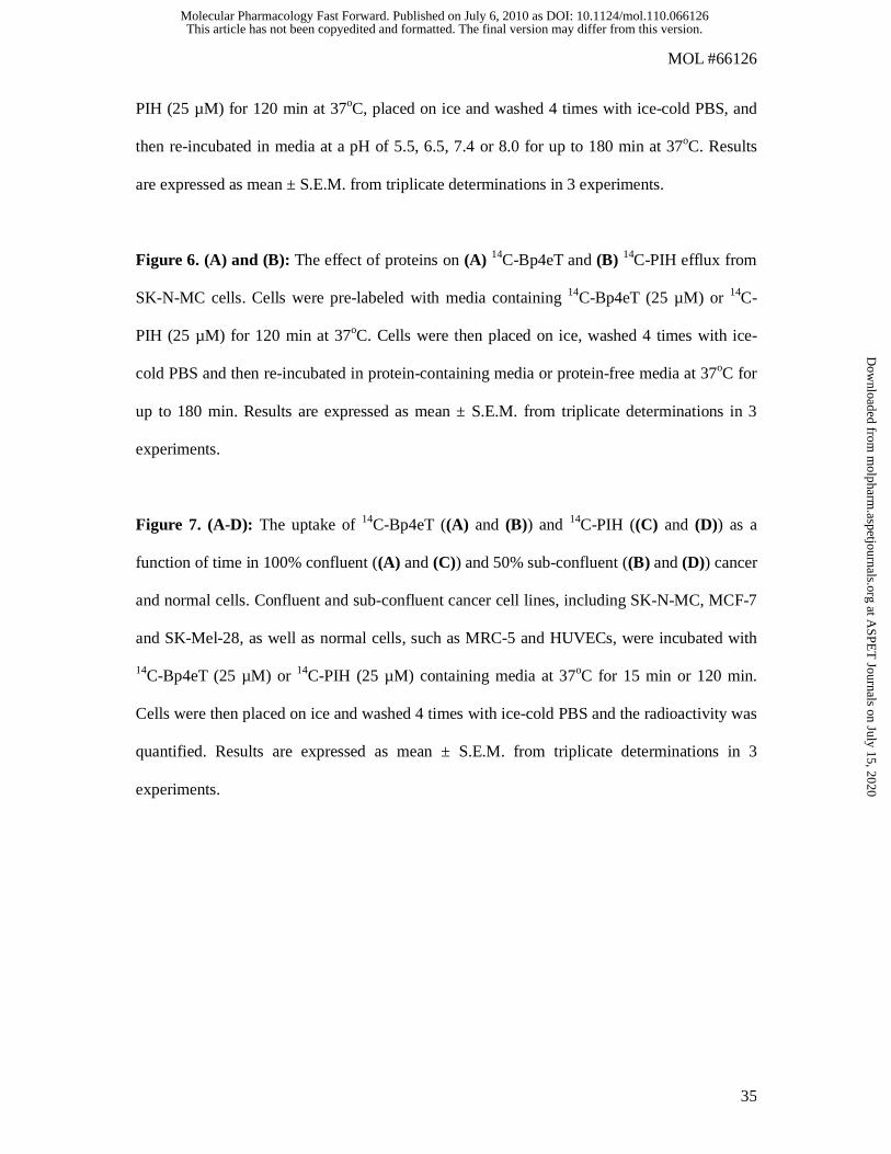
Figure Legend
Figure 1. Chemical structure of the iron chelators: (A) desferrioxamine (DFO), (B) pyridoxal
isonicotinoyl hydrazone (PIH) and (C) 2-benzoylpyridine 4-ethyl-3-thiosemicarbazone
(Bp4eT).
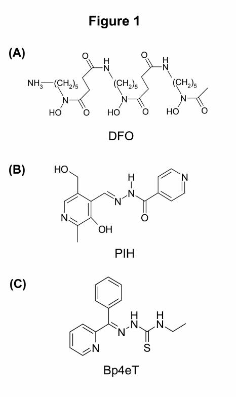
Figure 2. (A): The rate of 14C-Bp4eT and 14C-PIH uptake by SK-N-MC cells. The cells were
incubated in media containing 14C-Bp4eT or 14C-PIH at 5-250 µM for 120 min at 37oC. The
cells were then placed on ice, washed 4 times using ice-cold PBS and radioactivity was
quantified. (B): The effect of temperature and time on 14C-chelator uptake by SK-N-MC cells.
Cells were incubated with media containing 14C-Bp4eT (25 µM) or 14C-PIH (25 µM) at 37oC
or 4oC for up to 120 min. The remainder of the experiment was performed as for (A). (C) and
(D): The effect of temperature and time on 14C-chelator efflux. Cells were pre-labeled with
14C-Bp4eT (25 µM) or 14C-PIH (25 µM) for 120 min at 37oC. Cells were then placed on ice
and washed 4 times with ice-cold PBS and re-incubated in media for up to 180 min at 37oC or
4oC. Results are expressed as mean ± S.E.M. from triplicate determinations of 3 experiments.
Figure 3. (A) and (B): The effect of metabolic inhibitors on (A) 14C-Bp4eT and (B) 14C-PIH
uptake by SK-N-MC cells after 60 min at 37oC. Cells were pre-incubated with inhibitors:
NaN3 (30 mM), NaF (15 mM), 2,4-DNP (2 mM), oligomycin (30 µM) or NaCN (5 mM), or
media alone (DMEM ± glucose [GLU]) for 30 min at 37oC. The media was removed and the
cells then incubated with media containing 14C-PIH (25 µM) or 14C-Bp4eT (25 µM) with or
with out inhibitors for 60 min at 37oC. The cells were then placed on ice and washed 4 times
with ice-cold PBS and the radioactivity was quantified. Results are expressed as mean ±
S.E.M. from triplicate determinations of 6 experiments. * vs DMEM ± GLU, p<0.05; **
DMEM ± GLU, p<0.01; using one-way ANOVA. (C) and (D): The effect of metabolic
This article has not been copyedited and formatted. The final version may differ from this version.Molecular Pharmacology Fast Forward. Published on July 6, 2010 as DOI: 10.1124/mol.110.066126
at ASPE
T Journals on July 15, 2020
molpharm
.aspetjournals.orgD
ownloaded from

MOL #66126
34
inhibitors on ATP levels in (C) 14C-Bp4eT and (D) 14C-PIH uptake experiments that were
performed in (A) and (B), respectively. Results are mean ± S.E.M. from triplicate
determinations of 2 experiments and are expressed as a % of the control (DMEM + GLU). *
vs DMEM ± GLU, p<0.05; ** DMEM ± GLU, p<0.01; using one-way ANOVA.
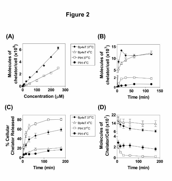
Figure 4. (A) and (B): The effect of metabolic inhibitors and time on (A) 14C-Bp4eT and (B)
14C-PIH efflux from SK-N-MC cells. Cells were pre-labeled with media containing 14C-
Bp4eT (25 µM) or 14C-PIH (25 µM) for 90 min at 37oC. The cells were then placed on ice and
metabolic inhibitors, namely NaN3 (30 mM), NaF (15 mM), oligomycin (30 µM) or NaCN (5
mM), were added to appropriate plates and the cells incubated for a further 30 min at 37oC.
After this incubation, the cells were placed on ice, washed 4 times with ice-cold PBS and then
reincubated for up to 60 min at 37oC in media with or without inhibitors. Results are
expressed as mean ± S.E.M. calculated from triplicates of 4 experiments. Statistical
significance was determined using two-way ANOVA with replication. (C) and (D): The effect
of metabolic inhibitors on ATP levels in (C) 14C-Bp4eT and (D) 14C-PIH efflux experiments
that were performed in (A) and (B), respectively. Results are mean ± S.E.M. of triplicates in a
typical experiment of 3 experiments performed and are expressed as a % of the control
(DMEM + GLU).
Figure 5. The effect of altering the extracellular pH on (A) 14C-Bp4eT and (B) 14C-PIH
uptake into SK-N-MC cells. Cells were incubated with 14C-Bp4eT (25 µM) or 14C-PIH (25
µM) in media at a pH of 5.5, 6.5, 7.4 or 8.0 for up to 120 min at 37oC. The cells were then
placed on ice, washed 4 times using ice-cold PBS and the radioactivity was quantified. The
effect of altering the extracellular pH on (C) 14C-Bp4eT and (D) 14C-PIH efflux from SK-N-
MC cells. Cells were pre-labeled with media (pH 7.4) containing 14C-Bp4eT (25 µM) or 14C-
This article has not been copyedited and formatted. The final version may differ from this version.Molecular Pharmacology Fast Forward. Published on July 6, 2010 as DOI: 10.1124/mol.110.066126
at ASPE
T Journals on July 15, 2020
molpharm
.aspetjournals.orgD
ownloaded from
MOL #66126
35
PIH (25 µM) for 120 min at 37oC, placed on ice and washed 4 times with ice-cold PBS, and
then re-incubated in media at a pH of 5.5, 6.5, 7.4 or 8.0 for up to 180 min at 37oC. Results
are expressed as mean ± S.E.M. from triplicate determinations in 3 experiments.
Figure 6. (A) and (B): The effect of proteins on (A) 14C-Bp4eT and (B) 14C-PIH efflux from
SK-N-MC cells. Cells were pre-labeled with media containing 14C-Bp4eT (25 µM) or 14C-
PIH (25 µM) for 120 min at 37oC. Cells were then placed on ice, washed 4 times with ice-
cold PBS and then re-incubated in protein-containing media or protein-free media at 37oC for
up to 180 min. Results are expressed as mean ± S.E.M. from triplicate determinations in 3
experiments.
Figure 7. (A-D): The uptake of 14C-Bp4eT ((A) and (B)) and 14C-PIH ((C) and (D)) as a
function of time in 100% confluent ((A) and (C)) and 50% sub-confluent ((B) and (D)) cancer
and normal cells. Confluent and sub-confluent cancer cell lines, including SK-N-MC, MCF-7
and SK-Mel-28, as well as normal cells, such as MRC-5 and HUVECs, were incubated with
14C-Bp4eT (25 µM) or 14C-PIH (25 µM) containing media at 37oC for 15 min or 120 min.
Cells were then placed on ice and washed 4 times with ice-cold PBS and the radioactivity was
quantified. Results are expressed as mean ± S.E.M. from triplicate determinations in 3
experiments.
This article has not been copyedited and formatted. The final version may differ from this version.Molecular Pharmacology Fast Forward. Published on July 6, 2010 as DOI: 10.1124/mol.110.066126
at ASPE
T Journals on July 15, 2020
molpharm
.aspetjournals.orgD
ownloaded from
This article has not been copyedited and formatted. The final version may differ from this version.Molecular Pharmacology Fast Forward. Published on July 6, 2010 as DOI: 10.1124/mol.110.066126
at ASPE
T Journals on July 15, 2020
molpharm
.aspetjournals.orgD
ownloaded from
This article has not been copyedited and formatted. The final version may differ from this version.Molecular Pharmacology Fast Forward. Published on July 6, 2010 as DOI: 10.1124/mol.110.066126
at ASPE
T Journals on July 15, 2020
molpharm
.aspetjournals.orgD
ownloaded from

This article has not been copyedited and formatted. The final version may differ from this version.Molecular Pharmacology Fast Forward. Published on July 6, 2010 as DOI: 10.1124/mol.110.066126
at ASPE
T Journals on July 15, 2020
molpharm
.aspetjournals.orgD
ownloaded from
This article has not been copyedited and formatted. The final version may differ from this version.Molecular Pharmacology Fast Forward. Published on July 6, 2010 as DOI: 10.1124/mol.110.066126
at ASPE
T Journals on July 15, 2020
molpharm
.aspetjournals.orgD
ownloaded from

This article has not been copyedited and formatted. The final version may differ from this version.Molecular Pharmacology Fast Forward. Published on July 6, 2010 as DOI: 10.1124/mol.110.066126
at ASPE
T Journals on July 15, 2020
molpharm
.aspetjournals.orgD
ownloaded from

This article has not been copyedited and formatted. The final version may differ from this version.Molecular Pharmacology Fast Forward. Published on July 6, 2010 as DOI: 10.1124/mol.110.066126
at ASPE
T Journals on July 15, 2020
molpharm
.aspetjournals.orgD
ownloaded from

This article has not been copyedited and formatted. The final version may differ from this version.Molecular Pharmacology Fast Forward. Published on July 6, 2010 as DOI: 10.1124/mol.110.066126
at ASPE
T Journals on July 15, 2020
molpharm
.aspetjournals.orgD
ownloaded from