Embed Size (px)
Citation preview
REVIEW
Membrane trafficking in neuronal maintenance and degeneration
Dong Wang • Chih-Chiang Chan • Smita Cherry •
P. Robin Hiesinger
Received: 6 August 2012 / Revised: 13 October 2012 / Accepted: 15 October 2012 / Published online: 8 November 2012
� The Author(s) 2012. This article is published with open access at Springerlink.com
Abstract Defects in membrane trafficking and degrada-
tion are hallmarks of most, and maybe all, neurodegenerative
disorders. Such defects typically result in the accumulation
of undegraded proteins due to aberrant endosomal sorting,
lysosomal degradation, or autophagy. The genetic or envi-
ronmental cause of a specific disease may directly affect
these membrane trafficking processes. Alternatively, chan-
ges in intracellular sorting and degradation can occur as
cellular responses of degenerating neurons to unrelated pri-
mary defects such as insoluble protein aggregates or other
neurotoxic insults. Importantly, altered membrane traffick-
ing may contribute to the pathogenesis or indeed protect the
neuron. The observation of dramatic changes to membrane
trafficking thus comes with the challenging need to distin-
guish pathological from protective alterations. Here, we will
review our current knowledge about the protective and
destructive roles of membrane trafficking in neuronal
maintenance and degeneration. In particular, we will first
focus on the question of what type of membrane trafficking
keeps healthy neurons alive in the first place. Next, we will
discuss what alterations of membrane trafficking are known
to occur in Alzheimer’s disease and other tauopathies, Par-
kinson’s disease, polyQ diseases, peripheral neuropathies,
and lysosomal storage disorders. Combining the mainte-
nance and degeneration viewpoints may yield insight into
how to distinguish when membrane trafficking functions
protectively or contributes to degeneration.
Keywords Autophagy � Endosome � Lysosome �Huntington � Alzheimer � Parkinson
Membrane trafficking and maintenance: what keeps
the healthy neuron alive?
Neurons are extraordinarily polarized cells. Both axonal
and dendritic branches represent challenges especially with
regard to membrane trafficking for both neuronal function
and maintenance. Synaptic vesicles outnumber any other
membrane compartment on the presynaptic site. Even
though neurotransmitter release is an extensively studied
process, we still understand little about the generation,
sorting, and maintenance of synaptic vesicles [1]. In par-
ticular, it is not clear how dysfunctional vesicle proteins or
complete vesicles are recognized, sorted, and degraded. In
addition, we know a lot about specialized membrane traf-
ficking on the postsynaptic site, especially from pioneering
studies on the recycling of neurotransmitter receptors [2,
3], and yet, similar to the presynaptic site, the sorting,
quality control, and degradation of the underlying traf-
ficking compartments are largely uncharacterized. Failure
to provide adequate quality control and degradation of pre-
or post-synaptic trafficking compartments leads to the
accumulation of dysfunctional intracellular machinery [4,
5]. Accumulations of dysfunctional membrane compart-
ments may be toxic to the neuron, for example if acidified
compartments start leaking protons or activated proteases.
Alternatively, the neuron may execute a cellular reaction to
D. Wang � C.-C. Chan � S. Cherry � P. R. Hiesinger (&)
Department of Physiology, University of Texas Southwestern
Medical Center, Dallas, TX 75390-9040, USA
e-mail: [email protected]
P. R. Hiesinger
Green Center for Systems Biology, University of Texas
Southwestern Medical Center, Dallas, TX 75390-9040, USA
Present Address:
C.-C. Chan
Institute of Physiology, National Taiwan University,
Taipei, Taiwan
Cell. Mol. Life Sci. (2013) 70:2919–2934
DOI 10.1007/s00018-012-1201-4 Cellular and Molecular Life Sciences
123
aberrant accumulations that may itself become the cause of
synaptic dysfunction, degradation, or cell death. Hence, a
decrease in normal degradative capacity may lead to
degeneration either because of inherent toxicity of accu-
mulating cargo or because of a toxic cellular reaction to
such accumulations (Fig. 1). Importantly, both the accu-
mulating compartments and the cellular response may also,
at least initially, serve protective roles. Hence, neither the
observation of aberrant intracellular compartments nor a
cellular clearance response can straightforwardly be inter-
preted as either toxic or protective. As we will see below,
the same considerations need to be discussed in the context
of defective normal maintenance and neurotoxic protein
accumulations associated with neurodegenerative diseases
(Fig. 1). We therefore think that the study of machinery
employed by wild-type neurons to maintain healthy syn-
apses is likely to reveal fundamental insights into common
features of neurodegenerative disorders that are character-
ized by intracellular accumulations. Towards this goal, we
will first briefly highlight the neuronal maintenance roles of
the three known endomembrane sorting and degradation
mechanisms: autophagy, ubiquitous endolysosomal degra-
dation and neuron-specific ‘sort-and-degrade’ [4].
Autophagy
Autophagy is a conserved intracellular degradation pathway
that clears proteins and organelles from the cytoplasm and
makes resources available to the cell in response to starvation.
Autophagy is classified into three major types: chaperone-
mediated autophagy (CMA), microautophagy, and macro-
autophagy [6]. Macroautophagy mediates bulk degradation of
cytoplasmic components including organelles [7]. The
importance of membrane trafficking machinery that keeps
neurons healthy is highlighted by seminal studies on the role
of macroautophagy (hereafter referred to as autophagy) in
adult neurons. Loss of autophagy in neurons of otherwise
wild-type mice (through loss of atg5 or atg7) leads to adult-
onset degeneration [8, 9]. These finding suggests that low
levels of autophagy, even though not readily detectable by
microscopy in wild-type neurons, are required for neuronal
maintenance in healthy neurons [10]. Neurons are long-lived
postmitotic cells, in which dysfunctional proteins and dam-
aged organelles cannot be transferred to daughter cells, which
is often argued to make them more sensitive to accumulation
of undegraded cargo [11]. In addition to the maintenance role,
autophagy is a known cellular response to intracellular cargo
overload. As such, autophagy can be considered a salvage
mechanism to remove excess intracellular debris. The deg-
radation of aggregated proteins in the cytosol by autophagy is
well characterized [12, 13]. At low levels, increased autoph-
agy acts protectively. Mild induction of autophagy can confer
partial neuroprotection [14, 15]. However, the salvage
mechanism has an emergency exit: increased autophagy
above a certain threshold is a cell death mechanism. Hence,
the same autophagic cellular reaction can function protec-
tively and turn into a cell death mechanism in response to
increasing cargo load [7, 16, 17]. Consequently, upregulation
of the autophagic maintenance mechanism may not be a
generally applicable solution to combat cargo overload in
neuronal health and disease (Fig. 1) [18–20].
Ubiquitous endolysosomal degradation
Endocytosed material traffics through transport vesicles to
early endosomes which mature into multivesicular bodies
(MVBs) or late endosomes and finally fuse with ER/Golgi-
derived vesicles that contain degradative machinery to form
lysosomes (Fig. 2). Phagocytosis and autophagy provide
alternative entry points for larger molecules and organelles.
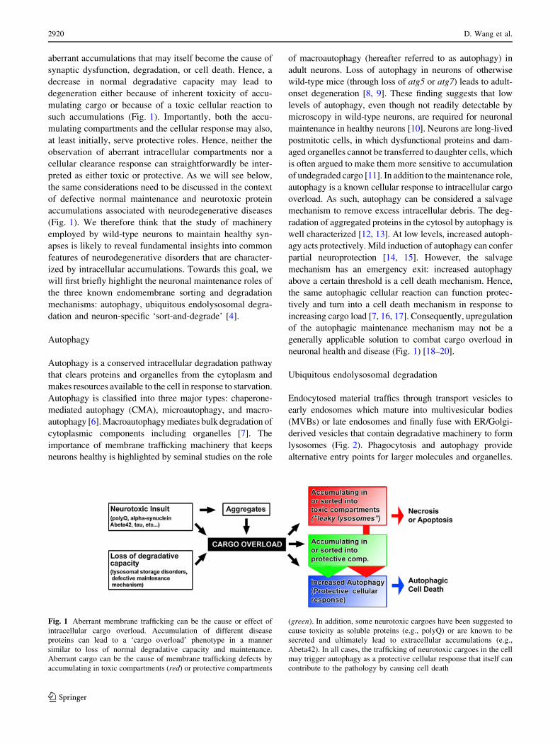
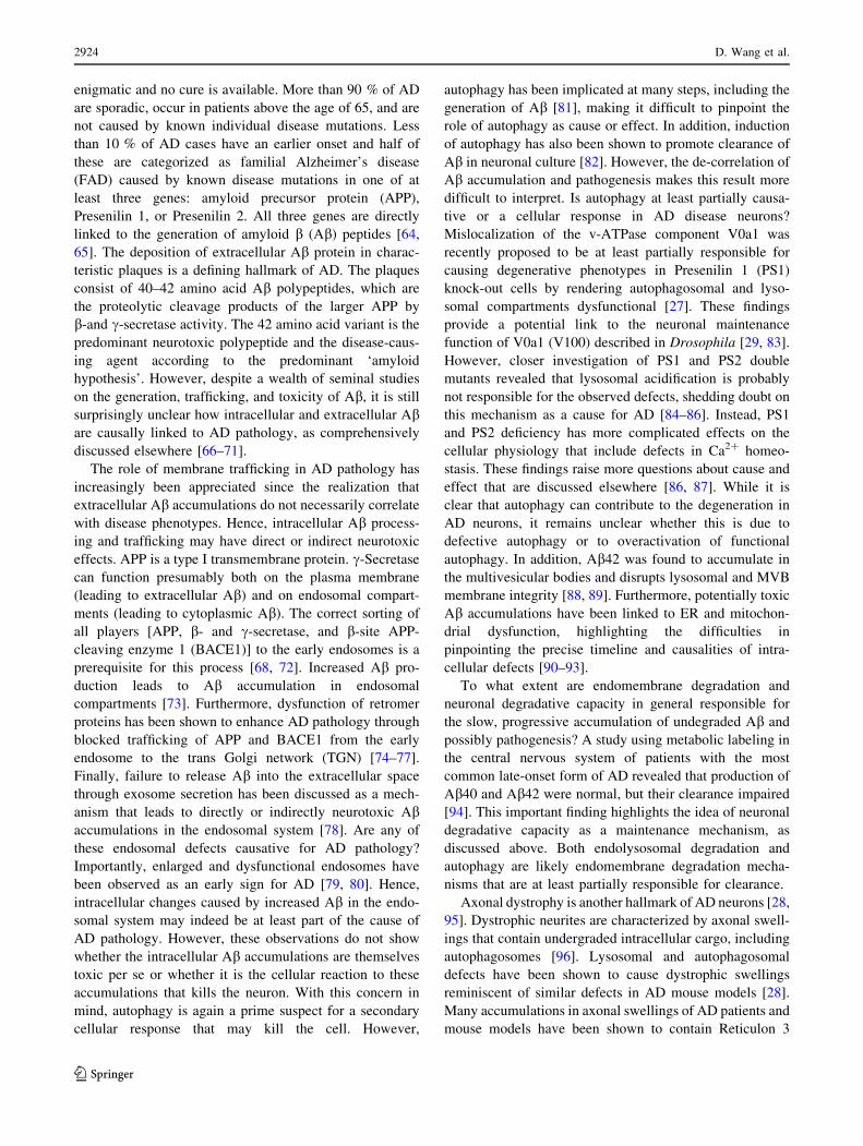
Fig. 1 Aberrant membrane trafficking can be the cause or effect of
intracellular cargo overload. Accumulation of different disease
proteins can lead to a ‘cargo overload’ phenotype in a manner
similar to loss of normal degradative capacity and maintenance.
Aberrant cargo can be the cause of membrane trafficking defects by
accumulating in toxic compartments (red) or protective compartments
(green). In addition, some neurotoxic cargoes have been suggested to
cause toxicity as soluble proteins (e.g., polyQ) or are known to be
secreted and ultimately lead to extracellular accumulations (e.g.,
Abeta42). In all cases, the trafficking of neurotoxic cargoes in the cell
may trigger autophagy as a protective cellular response that itself can
contribute to the pathology by causing cell death
2920 D. Wang et al.
123
Late endosomes, lysosomes, and autophagosomes are the
primary organelles for endomembrane degradation. Muta-
tions in proteins that affect late endosomal or lysosomal
function are often initially viable but cause intracellular
membrane accumulations [21]. In many cases, these accu-
mulations (or a cellular response to them) ultimately cause
neuronal cell death. Consequently, such mutants have been
utilized to model neurodegeneration and lysosomal storage
disorders [22–24].
An increasing pH gradient in the endolysosomal path-
way is required for intracellular trafficking [25, 26]. The
maturation of endosomes into lysosomes is marked by the
progressive acidification of the compartment, to ultimately
allow for the activation of acidification-activated proteases
in lysosomes. Impaired endocytic trafficking by disrupting
the pH gradient or mutations in the cargo-carrier proteins
can cause neurodegeneration [27–29]. Although endolys-
osomal degradation occurs ubiquitously, dysfunctional
degradation firstly causes problems in tissues in which the
substrate turnover is high. As discussed below, this may be
a reason why two-thirds of lysosomal storage disorders
affect the central nervous system and cause progressive
cognitive and motor decline [21]. The accumulation of
undegraded substrates can be the primary cause that exerts
toxic effects on other cellular functions. Conversely, the
undegraded aggregates may be the result of an independent
dysfunctional membrane trafficking process (Fig. 1).
Neuronal ‘sort-and-degrade’
Autophagy and endolysosomal degradation are ubiquitous
mechanisms thought to be required for the function and
maintenance of all cells. We have recently identified a
neuron-specific degradation pathway [29]. Mutations in
v0a1 (v100 in Drosophila) lead to neuron-specific degra-
dation defects and are, to our knowledge, the first
mutations in a neuron-specific regulator of membrane
trafficking shown to cause neurodegeneration. Loss of v100
causes intracellular sorting and degradation defects down-
stream of endocytosis [29, 30]. Similarly, mutations in the
synaptic vesicle SNARE neuronal Synaptobrevin (n-syb)
cause intracellular sorting and degradation defects that lead
to slow adult-onset degeneration in Drosophila [31]. Both
v0a1 and n-syb are neuron-specific membrane trafficking
proteins that predominantly function at synapses [4]. It is
interesting to note that loss of neuronal degradative
capacity in these mutants may cause a similar ‘cargo
overload’ problem in neurons as the accumulation of dis-
ease proteins due to increased expression, misfolding, or
aggregation (Fig. 1). In both cases, autophagy is initiated
as a cellular response—with both a protective and cell
death potential as discussed above. It is not clear whether
the v100- and n-syb-dependent neuronal ‘sort-and-degrade’
mechanism has a specificity for synaptic cargo. Alterna-
tively, v100 and n-syb may simply increase general
neuronal degradative capacity predominantly at synapses.
Both v100 and n-syb have close homologs (v0a2-4 and
cellubrevin) that exert very similar functions in other cell
types.
The idea of a degradation mechanism with specificity
for synaptic cargo is supported by the knowledge that
synapses contain numerous specializations of membrane
trafficking. Both v100 and n-syb function on synaptic
vesicles and are required for normal neurotransmitter
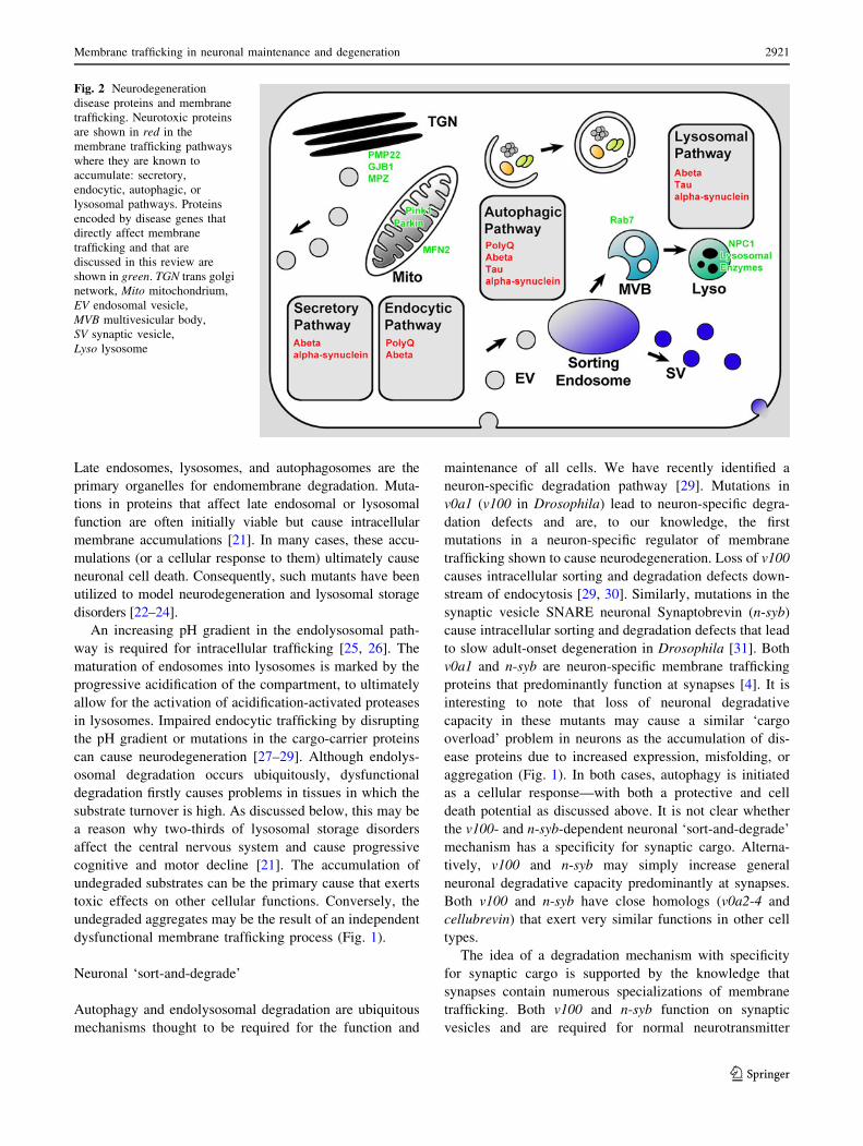
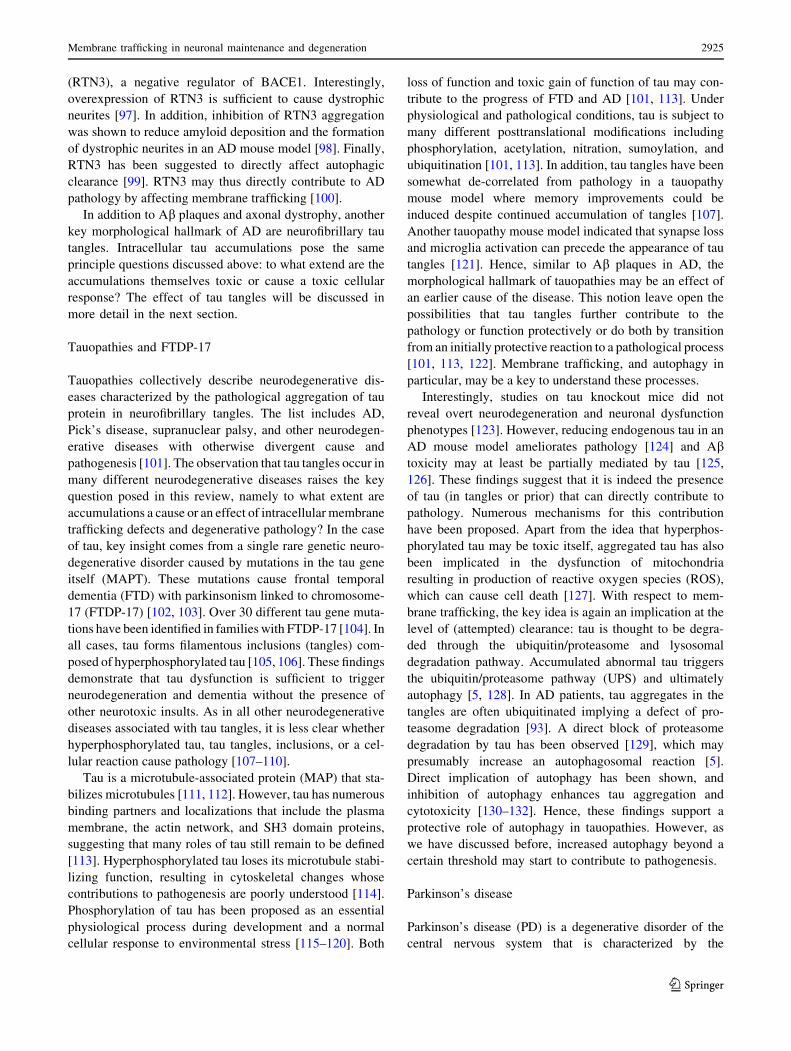
Fig. 2 Neurodegeneration
disease proteins and membrane
trafficking. Neurotoxic proteins
are shown in red in the
membrane trafficking pathways
where they are known to
accumulate: secretory,
endocytic, autophagic, or
lysosomal pathways. Proteins
encoded by disease genes that
directly affect membrane
trafficking and that are
discussed in this review are
shown in green. TGN trans golgi
network, Mito mitochondrium,
EV endosomal vesicle,
MVB multivesicular body,
SV synaptic vesicle,
Lyso lysosome
Membrane trafficking in neuronal maintenance and degeneration 2921
123
release, suggesting a molecular link between the synaptic
vesicle cycle and synaptic endolysosomal ‘sort-and-
degrade’. A similar link has recently been identified in the
skywalker mutant in Drosophila. skywalker encodes a
rabGAP that functions at the intersection of synaptic ves-
icle recycling, sorting, and degradation [32]. The recent
discovery of many novel synaptic endosomal Rab GTPases
further suggests the existence of more neuronal membrane
trafficking machinery required for neuronal maintenance
[33, 34].
Membrane trafficking and neurodegeneration: what
kills the degenerating neuron?
In the following sections, we will discuss known mem-
brane trafficking defects for several prominent
neurodegenerative diseases. For all these diseases aberrant
membrane trafficking has been observed and linked to
neuronal degeneration. We will focus on the basic ques-
tions raised by our review of neuronal maintenance
mechanisms. What are the causal relationships between the
observed defects in membrane trafficking and pathology?
When do they represent primary defects or cellular
responses? In addition, we will focus on the idea of neu-
ronal degradative capacity. How far do intracellular
accumulations in the endomembrane system cause a ‘cargo
overload’ situation similar to the loss of degradative
maintenance mechanisms (Fig. 1)? With these questions in
mind, we will review which intracellular accumulations are
known in different neurodegenerative diseases, what is
known about their inherent toxicity, and the cellular
membrane trafficking reaction to these accumulations
(Fig. 2).
PolyQ diseases
PolyQ disorders are caused by mutations that lead to the
expansion of a polyglutamine (PolyQ) stretch in known
disease proteins. These disorders include Huntington’s
disease (HD), several types of spinocerebellar ataxia
(SCA), and spinobulbar muscle atrophy (SBMA). The
onset and severity of these disorders positively correlate to
at least some extent with the length of the CAG repeat
coding for glutamine [35–39]. Intracellular PolyQ protein
aggregates formed by the mutant proteins are the hallmark
of the diseases. In all polyQ diseases, correlations between
aggregate formation and disease phenotypes are well
established. The length of the polyQ stretch directly affects
the propensity of the mutant protein to aggregate, which in
turn correlates to some degree with the severity of disease
phenotypes. Thus, the causal relationship between polyQ
protein and phenotype seems deceivingly clear. In what
way can the idea of polyQ as the cause of disease
phenotypes be misleading? First, the correlation of the
observed polyQ inclusions in different parts of patient
brains do not necessarily correlate with where the degen-
eration occurs; in fact, some neurons that seem to evade
degeneration exhibit most polyQ inclusions [40]. These
and other data have been interpreted to argue that polyQ
inclusions may represent protective cellular responses—a
common theme across disorders characterized by unde-
graded protein accumulations. Second, a direct causal
relationship between polyQ length, aggregation, and tox-
icity could still hold if the aggregates were not toxic
themselves, but instead the result of a cellular response to
the aggregates or pre-aggregate variants of polyQ proteins.
The ubiquitin/proteasome system (UPS) and autophagy are
the major clearance mechanisms and cellular responses to
aberrant intracellular accumulations. In addition, a defect
or overload of the UPS can trigger a further increase in
autophagy [13, 41], which itself can be sufficient to cause
cell death. In such a scenario, the polyQ protein remains
the primary cause of a chain of event that results in
degeneration, but the actual cause of pathogenesis may be
the cellular response. The importance of this difference can
be highlighted by a comparison to allergic reactions (see
‘bee sting analogy’, Fig. 3). An allergen can be the cause
of severe pathology and even cell death in a dosage-
dependent manner; however, the actual disease mechanism
is an (over-) reaction of a cellular response. Reducing this
cellular (and normally protective) response is where ther-
apeutic intervention is required and successful. This
analogy of course cannot explain polyQ-induced pathology
in its entirety. However, it is currently unclear to what
extent aberrant protein aggregates and the inclusion for-
mation through endomembrane system responses cause
degeneration (Fig. 3). This is not only the case for polyQ
aggregates, but several intracellular cargos that are con-
sidered neurotoxic based on their occurence in the diseases
discussed in this review. We will highlight commonalities
of polyQ diseases mainly based on examples from polyQ-
Huntingtin and polyQ-Sca1. Specifics on the different
polyQ diseases are reviewed in detail elsewhere [36, 42,
43].
PolyQ proteins have been observed to accumulate in both
the nucleus and cytoplasm where they directly or indirectly
affect a plethora of cellular functions. Potential primary
effects of PolyQ proteins include disrupted transcription,
mitochondrial function, Ca2? homeostasis, axonal transport,
and the UPS and autophagosomal clearance systems [38].
Membrane trafficking is directly or indirectly affected in
every single one of these potential targets of polyQ toxicity,
which may be best studied for Huntingtin-polyQ. HD is the
most common form of polyQ disorders and is caused by
mutant variants of the huntingtin (htt) gene with elongated
glutamine repeats over 36–40 residues [44]. Wild-type htt
2922 D. Wang et al.
123
protein is ubiquitous and mostly cytosolic [36]. An N-ter-
minal fragment containing the polyQ stretch is produced by
the proteolysis of mutant htt, associates with membranes,
and may be sufficient to cause pathology [45]. Loss of htt in
knock-out mice leads to embryonic lethality [46–48]. A
polyQ-extended variant of htt can rescue the embryonic
lethality, suggesting that the wild-type function and polyQ
disease mechanism may be different [49]. Htt-polyQ over-
expression has been shown to both increase synaptic function
at the mouse neuromuscular junction [50] and reduce syn-
aptic function at the Drosophila larval neuromuscular
junction [51]. Synaptic defects can be partially rescued by
overexpression of Rab11, a recycling endosome protein, in
Drosophila [51, 52]. Taken together, extensive work on Htt-
polyQ indicates a pathological role of the polyQ oligopeptide
with commonalities in all polyQ diseases. However, it is
difficult to pinpoint a direct effect of polyQ on membrane
trafficking and many of the observed changes are likely the
indirect results of multiple changes of the cellular physiol-
ogy. In addition, RNA-based toxicity of the CAG repeat
containing mRNA encoding the polyQ stretch has been
shown in Drosophila and is discussed elsewhere [53, 54].
Both the UPS or autophagic clearance systems can be
triggered by intracellular accumulations in several polyQ
diseases, including SBMA [55], HD [12], and Sca1 [13].
Upregulation of chaperones like Hsp70 are common in
polyQ diseases [56–59]. It is less clear how far the UPS or
autophagy are negatively affected by polyQ proteins prior
to or after aggregation. Although inclusions in HD include
ubiquitinated proteins and proteasome components, the
UPS is not significantly impaired in mouse models for HD
or Sca7 [60, 61]. In contrast, macroautophagy may be
directly impaired by Htt-polyQ [11, 62]. Pharmacological
upregulation of macroautophagy has been shown to be
effective in reducing neuronal aggregates and slowing the
progression of neurological symptoms in fly and mouse
models of HD [63]. However, in some cases, defective
autophagy is deduced from the observation of massively
increased autophagy, including intermediates of auto-
phagosome maturation. We have recently observed such an
increased autophagy phenotype in Drosophila neurons
mutant for a neuron-specific endomembrane degradation
mechanism [4, 31]. These neurons clearly exhibit endo-
membrane trafficking, intracellular degradation defects,
and dramatically increased autophagy with many auto-
phagosome intermediates that are not normally observed.
However, measurement of autophagosome acidification
and activity of autophagy proteases (Cathepsins) revealed
that autophagy is functional in these neurons at least to a
very late step [31]. This finding may serve as a cautionary
note that the observation of aberrant autophagy may not
necessarily be the result of a defect in autophagy but can
also result from a dramatic upregulation of fully functional
autophagy as a cellular response to a neurotoxic insult. As
discussed above, this cellular response may initially be
protective but can become a cell death mechanism as a
function of levels. This common theme is observed in all
neurodegenerative diseases discussed here, including Alz-
heimer’s disease and other tauopathies, Parkinson’s
disease, peripheral neuropathies and lysosomal storage
disorders.
Alzheimer’s disease
Alzheimer’s disease (AD) is the most common neurode-
generative disorder. The cause (or causes) of AD remain
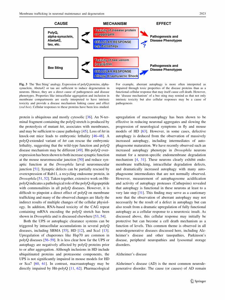
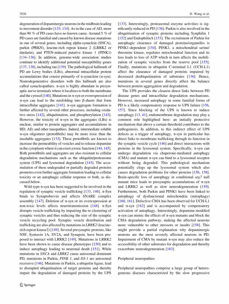
Fig. 3 The ‘Bee Sting’ analogy. Expression of polyQ proteins, alpha-
synuclein, Abeta42 or tau are sufficient to induce degeneration in
neurons. Hence, they are a direct cause of pathogenesis and disease
phenotypes. Properties like intracellular aggregation and inclusion in
membrane compartments are easily interpreted to have intrinsic
toxicity and provide a disease mechanism linking cause and effect
(red box). Cellular responses to these proteins have been less studied.
For example, aberrant autophagy is more often interpreted as
impaired through toxic properties of the disease proteins than as a
functional cellular response that may itself cause cell death. However,
the ‘disease mechanism’ of a bee sting may remind us that not only
intrinsic toxicity but also cellular responses may be a cause of
pathogenesis
Membrane trafficking in neuronal maintenance and degeneration 2923
123
enigmatic and no cure is available. More than 90 % of AD
are sporadic, occur in patients above the age of 65, and are
not caused by known individual disease mutations. Less
than 10 % of AD cases have an earlier onset and half of
these are categorized as familial Alzheimer’s disease
(FAD) caused by known disease mutations in one of at
least three genes: amyloid precursor protein (APP),
Presenilin 1, or Presenilin 2. All three genes are directly
linked to the generation of amyloid b (Ab) peptides [64,
65]. The deposition of extracellular Ab protein in charac-
teristic plaques is a defining hallmark of AD. The plaques
consist of 40–42 amino acid Ab polypeptides, which are
the proteolytic cleavage products of the larger APP by
b-and c-secretase activity. The 42 amino acid variant is the
predominant neurotoxic polypeptide and the disease-caus-
ing agent according to the predominant ‘amyloid
hypothesis’. However, despite a wealth of seminal studies
on the generation, trafficking, and toxicity of Ab, it is still
surprisingly unclear how intracellular and extracellular Abare causally linked to AD pathology, as comprehensively
discussed elsewhere [66–71].
The role of membrane trafficking in AD pathology has
increasingly been appreciated since the realization that
extracellular Ab accumulations do not necessarily correlate
with disease phenotypes. Hence, intracellular Ab process-
ing and trafficking may have direct or indirect neurotoxic
effects. APP is a type I transmembrane protein. c-Secretase
can function presumably both on the plasma membrane
(leading to extracellular Ab) and on endosomal compart-
ments (leading to cytoplasmic Ab). The correct sorting of
all players [APP, b- and c-secretase, and b-site APP-
cleaving enzyme 1 (BACE1)] to the early endosomes is a
prerequisite for this process [68, 72]. Increased Ab pro-
duction leads to Ab accumulation in endosomal
compartments [73]. Furthermore, dysfunction of retromer
proteins has been shown to enhance AD pathology through
blocked trafficking of APP and BACE1 from the early
endosome to the trans Golgi network (TGN) [74–77].
Finally, failure to release Ab into the extracellular space
through exosome secretion has been discussed as a mech-
anism that leads to directly or indirectly neurotoxic Abaccumulations in the endosomal system [78]. Are any of
these endosomal defects causative for AD pathology?
Importantly, enlarged and dysfunctional endosomes have
been observed as an early sign for AD [79, 80]. Hence,
intracellular changes caused by increased Ab in the endo-
somal system may indeed be at least part of the cause of
AD pathology. However, these observations do not show
whether the intracellular Ab accumulations are themselves
toxic per se or whether it is the cellular reaction to these
accumulations that kills the neuron. With this concern in
mind, autophagy is again a prime suspect for a secondary
cellular response that may kill the cell. However,
autophagy has been implicated at many steps, including the
generation of Ab [81], making it difficult to pinpoint the
role of autophagy as cause or effect. In addition, induction
of autophagy has also been shown to promote clearance of
Ab in neuronal culture [82]. However, the de-correlation of
Ab accumulation and pathogenesis makes this result more
difficult to interpret. Is autophagy at least partially causa-
tive or a cellular response in AD disease neurons?
Mislocalization of the v-ATPase component V0a1 was
recently proposed to be at least partially responsible for
causing degenerative phenotypes in Presenilin 1 (PS1)
knock-out cells by rendering autophagosomal and lyso-
somal compartments dysfunctional [27]. These findings
provide a potential link to the neuronal maintenance
function of V0a1 (V100) described in Drosophila [29, 83].
However, closer investigation of PS1 and PS2 double
mutants revealed that lysosomal acidification is probably
not responsible for the observed defects, shedding doubt on
this mechanism as a cause for AD [84–86]. Instead, PS1
and PS2 deficiency has more complicated effects on the
cellular physiology that include defects in Ca2? homeo-
stasis. These findings raise more questions about cause and
effect that are discussed elsewhere [86, 87]. While it is
clear that autophagy can contribute to the degeneration in
AD neurons, it remains unclear whether this is due to
defective autophagy or to overactivation of functional
autophagy. In addition, Ab42 was found to accumulate in
the multivesicular bodies and disrupts lysosomal and MVB
membrane integrity [88, 89]. Furthermore, potentially toxic
Ab accumulations have been linked to ER and mitochon-
drial dysfunction, highlighting the difficulties in
pinpointing the precise timeline and causalities of intra-
cellular defects [90–93].
To what extent are endomembrane degradation and
neuronal degradative capacity in general responsible for
the slow, progressive accumulation of undegraded Ab and
possibly pathogenesis? A study using metabolic labeling in
the central nervous system of patients with the most
common late-onset form of AD revealed that production of
Ab40 and Ab42 were normal, but their clearance impaired
[94]. This important finding highlights the idea of neuronal
degradative capacity as a maintenance mechanism, as
discussed above. Both endolysosomal degradation and
autophagy are likely endomembrane degradation mecha-
nisms that are at least partially responsible for clearance.
Axonal dystrophy is another hallmark of AD neurons [28,
95]. Dystrophic neurites are characterized by axonal swell-
ings that contain undergraded intracellular cargo, including
autophagosomes [96]. Lysosomal and autophagosomal
defects have been shown to cause dystrophic swellings
reminiscent of similar defects in AD mouse models [28].
Many accumulations in axonal swellings of AD patients and
mouse models have been shown to contain Reticulon 3
2924 D. Wang et al.
123
(RTN3), a negative regulator of BACE1. Interestingly,
overexpression of RTN3 is sufficient to cause dystrophic
neurites [97]. In addition, inhibition of RTN3 aggregation
was shown to reduce amyloid deposition and the formation
of dystrophic neurites in an AD mouse model [98]. Finally,
RTN3 has been suggested to directly affect autophagic
clearance [99]. RTN3 may thus directly contribute to AD
pathology by affecting membrane trafficking [100].
In addition to Ab plaques and axonal dystrophy, another
key morphological hallmark of AD are neurofibrillary tau
tangles. Intracellular tau accumulations pose the same
principle questions discussed above: to what extend are the
accumulations themselves toxic or cause a toxic cellular
response? The effect of tau tangles will be discussed in
more detail in the next section.
Tauopathies and FTDP-17
Tauopathies collectively describe neurodegenerative dis-
eases characterized by the pathological aggregation of tau
protein in neurofibrillary tangles. The list includes AD,
Pick’s disease, supranuclear palsy, and other neurodegen-
erative diseases with otherwise divergent cause and
pathogenesis [101]. The observation that tau tangles occur in
many different neurodegenerative diseases raises the key
question posed in this review, namely to what extent are
accumulations a cause or an effect of intracellular membrane
trafficking defects and degenerative pathology? In the case
of tau, key insight comes from a single rare genetic neuro-
degenerative disorder caused by mutations in the tau gene
itself (MAPT). These mutations cause frontal temporal
dementia (FTD) with parkinsonism linked to chromosome-
17 (FTDP-17) [102, 103]. Over 30 different tau gene muta-
tions have been identified in families with FTDP-17 [104]. In
all cases, tau forms filamentous inclusions (tangles) com-
posed of hyperphosphorylated tau [105, 106]. These findings
demonstrate that tau dysfunction is sufficient to trigger
neurodegeneration and dementia without the presence of
other neurotoxic insults. As in all other neurodegenerative
diseases associated with tau tangles, it is less clear whether
hyperphosphorylated tau, tau tangles, inclusions, or a cel-
lular reaction cause pathology [107–110].
Tau is a microtubule-associated protein (MAP) that sta-
bilizes microtubules [111, 112]. However, tau has numerous
binding partners and localizations that include the plasma
membrane, the actin network, and SH3 domain proteins,
suggesting that many roles of tau still remain to be defined
[113]. Hyperphosphorylated tau loses its microtubule stabi-
lizing function, resulting in cytoskeletal changes whose
contributions to pathogenesis are poorly understood [114].
Phosphorylation of tau has been proposed as an essential
physiological process during development and a normal
cellular response to environmental stress [115–120]. Both
loss of function and toxic gain of function of tau may con-
tribute to the progress of FTD and AD [101, 113]. Under
physiological and pathological conditions, tau is subject to
many different posttranslational modifications including
phosphorylation, acetylation, nitration, sumoylation, and
ubiquitination [101, 113]. In addition, tau tangles have been
somewhat de-correlated from pathology in a tauopathy
mouse model where memory improvements could be
induced despite continued accumulation of tangles [107].
Another tauopathy mouse model indicated that synapse loss
and microglia activation can precede the appearance of tau
tangles [121]. Hence, similar to Ab plaques in AD, the
morphological hallmark of tauopathies may be an effect of
an earlier cause of the disease. This notion leave open the
possibilities that tau tangles further contribute to the
pathology or function protectively or do both by transition
from an initially protective reaction to a pathological process
[101, 113, 122]. Membrane trafficking, and autophagy in
particular, may be a key to understand these processes.
Interestingly, studies on tau knockout mice did not
reveal overt neurodegeneration and neuronal dysfunction
phenotypes [123]. However, reducing endogenous tau in an
AD mouse model ameliorates pathology [124] and Abtoxicity may at least be partially mediated by tau [125,
126]. These findings suggest that it is indeed the presence
of tau (in tangles or prior) that can directly contribute to
pathology. Numerous mechanisms for this contribution
have been proposed. Apart from the idea that hyperphos-
phorylated tau may be toxic itself, aggregated tau has also
been implicated in the dysfunction of mitochondria
resulting in production of reactive oxygen species (ROS),
which can cause cell death [127]. With respect to mem-
brane trafficking, the key idea is again an implication at the
level of (attempted) clearance: tau is thought to be degra-
ded through the ubiquitin/proteasome and lysosomal
degradation pathway. Accumulated abnormal tau triggers
the ubiquitin/proteasome pathway (UPS) and ultimately
autophagy [5, 128]. In AD patients, tau aggregates in the
tangles are often ubiquitinated implying a defect of pro-
teasome degradation [93]. A direct block of proteasome
degradation by tau has been observed [129], which may
presumably increase an autophagosomal reaction [5].
Direct implication of autophagy has been shown, and
inhibition of autophagy enhances tau aggregation and
cytotoxicity [130–132]. Hence, these findings support a
protective role of autophagy in tauopathies. However, as
we have discussed before, increased autophagy beyond a
certain threshold may start to contribute to pathogenesis.
Parkinson’s disease
Parkinson’s disease (PD) is a degenerative disorder of the
central nervous system that is characterized by the
Membrane trafficking in neuronal maintenance and degeneration 2925
123
degeneration of dopaminergic neurons in the midbrain leading
to movement disorder [133, 134]. As in the case of AD, more
than 90 % of PD cases have no known cause. Around 5 % of
PD cases are familial and caused by known disease mutations
in one of several genes, including alpha-synuclein (SNCA),
parkin (PRKN), leucine-rich repeat kinase 2 (LRRK2 or
dardarin), and PTEN-induced putative kinase 1 (PINK1)
[134–136]. In addition, genome-wide association studies
continue to identify additional potential susceptibility genes
[137, 138], including tau [139]. The pathological hallmark of
PD are Lewy bodies (LBs), abnormal intracellular protein
accumulations that consist primarily of a-synuclein (a-syn).
Neurodegenerative disorders with this hallmark are also
called synucleopathies. a-syn is highly abundant in presyn-
aptic nerve terminals where it localizes to both the membrane
and the cytosol [140]. Mutations in a-syn or overexpression of
a-syn can lead to the misfolding into b-sheets that form
intracellular aggregates [141]. a-syn aggregate formation is
further affected by several other processes, including oxida-
tive stress [142], ubiquitination, and phosphorylation [143].
However, the toxicity of a-syn in the aggregates (LBs) is
unclear, similar to protein aggregates and accumulations in
HD, AD, and other tauopathies. Indeed, intermediate soluble
a-syn oligomers (protofibrils) may be more toxic than the
insoluble aggregates [133]. These protofibrils are known to
increase the permeability of vesicles and to release dopamine
in the cytoplasm where it can exert a toxic function [144, 145].
Both protofibrils and aggregates are also resistant to cellular
degradation mechanisms such as the ubiquitin/proteasome
system (UPS) and lysosomal degradation [143]. The accu-
mulation of these undegraded or partially degraded proteins
promotes even further aggregate formation leading to cellular
toxicity or an autophagic cellular response or both, as dis-
cussed below.
Wild-type a-syn has been suggested to be involved in the
regulation of synaptic vesicle trafficking [133, 146]. a-Syn
binds to Synaptobrevin and affects SNARE complex
assembly [147]. Deletion of a-syn or its overexpression at
non-toxic levels affects neurotransmission [148]. a-Syn
disrupts vesicle trafficking by impairing the re-clustering of
synaptic vesicles and thus reducing the size of the synaptic
vesicle recycling pool. Synaptic vesicle distribution and
trafficking are also affected by mutations in LRRK2 (leucine-
rich repeat kinase2) [149]. Several presynaptic proteins, like
NSF, Syntaxin 1A, SV2A, and Synapsin, have been pro-
posed to interact with LRRK2 [149]. Mutations in LRRK2
have been shown to cause disease phenotypes [150] and to
induce autophagy leading to neuronal death [151]. While
mutations in SNCA and LRRK2 cause autosomal dominant
PD, mutations in Parkin, PINK 1, and DJ-1 are autosomal
recessive [146]. Mutations in Parkin, a ubiquitin ligase, lead
to disrupted ubiquitination of target proteins and thereby
impair the degradation of damaged proteins by the UPS
[133]. Interestingly, proteasomal enzyme activities is sig-
nificantly reduced in PD [136]. Parkin is also involved in the
ubiquitination of synaptic proteins including Synphilin 1
[152] and EndophilinA [153]. The recruitment of Parkin for
autophagic clearance of damaged proteins/organelles is
PINK1-dependent [154]. PINK1, a mitochondrial serine/
threonine kinase, regulates mitochondrial function and its
loss leads to loss of ATP which in turn affects the mobili-
zation of synaptic vesicles from the reserve pool [155].
Finally, mutations in ubiquitin C-terminal L1 (UCH-L1)
affect the clearance of damaged proteins impaired by
decreased deubiquitination of substrates [136]. Hence,
mutations in several genes directly affect the balance
between protein aggregation and degradation.
The UPS provides the clearest direct links between PD
disease genes and intracellular degradation mechanisms.
However, increased autophagy in some familial forms of
PD is a likely compensatory response to UPS failure [156,
157]. Since blocking of the UPS is known to induce
autophagy [13, 41], endomembrane degradation may play a
common role highlighted here: an initially protective
mechanism that above a certain threshold contributes to the
pathogenesis. In addition, to this indirect effect of UPS
defects as a trigger of autophagy, a-syn in particular has
direct links to membrane trafficking through its function in
the synaptic vesicle cycle [146] and direct interactions with
proteins in the lysosomal system. Specifically, a-syn can
undergo degradation via chaperone-mediated autophagy
(CMA) and mutant a-syn can bind to a lysosomal receptor
without being degraded. This pathological mechanism
potentially clogs up the lysosomal system which also
causes degradation problems for other proteins [156, 158].
Brain-specific loss of autophagy in conditional atg7 null
mutant mice leads to presynaptic accumulations of a-syn
and LRRK2 as well as slow neurodegeneration [159].
Furthermore, both Parkin and PINK1 have been linked to
autophagy of dysfunctional mitochondria (mitophagy)
[160, 161]. Defective CMA has been observed for UCH-L1
and a-syn [162] and is accompanied by compensatory
activation of autophagy. Interestingly, dopamine-modified
a-syn can mimic the effects of a-syn mutants and block the
CMA degradation pathway, making the affected neurons
more vulnerable to other stressors or insults [158]. This
might provide a partial explanation why dopaminergic
neurons are the most severely affected neurons in PD.
Impairment of CMA by mutant a-syn may also reduce the
accessibility of other substrates for degradation and thereby
exacerbate neurodegeneration [163].
Peripheral neuropathies
Peripheral neuropathies comprise a large group of hetero-
geneous diseases characterized by the slow progressive
2926 D. Wang et al.
123
degeneration of neurons of the peripheral nervous system,
muscle atrophy, and motor and sensory impairment of the
limbs. Among the hereditary motor and sensory neuropa-
thies (HMSN), Charcot–Marie–Tooth (CMT) disease is the
most common form and is associated with mutations in
more than 45 genes involved in Schwann cells homeostasis
and neuronal function. Type 1 (demyelinating CMT)
affects the myelin sheath whereas type 2 (axonal CMT) is
characterized by decreased motor action potentials caused
by abnormalities in the axon. Although intracellular accu-
mulations are not regarded as disease hallmarks, defects in
intracellular trafficking are a common feature in most, and
maybe all, peripheral neuropathies.
Many CMT-causing mutations lead to defects in various
steps of intracellular trafficking including vesicle budding,
cytoskeletal transport, protein degradation, and mitochon-
drial axonal transport [164]. Mutations in four genes
account for over 90 % of all CMT molecular diagnoses,
including peripheral myelin protein 22 (PMP22), gap
junction b-1 (GJB1), myelin protein zero (MPZ), and
mitofusion 2 (MFN2) [165]. The most common cause of
CMT type 1 are mutations in PMP22, an integral mem-
brane glycoprotein that is primarily expressed in Schwann
cells. Mutations in PMP22 cause hypomyelination [166].
While the wild-type PMP22 protein resides in the plasma
membrane, the mutant forms are not able to target to the
plasma membrane and accumulate in the ER and Golgi
[167]. Misfolded PMP22 may cause a ‘cargo overload’
phenotype with cellular responses discussed here for all
degenerative diseases [168]. In further similarity to other
diseases, the accumulations containing mutant PMP22
form ubiquitinylated cytoplasmic inclusions that have been
suggested to play a cytoprotective role [169, 170].
Mutations in GJB1 also cause myelination defects. The
GJB1 protein is a transmembrane protein that assembles to
form gap-junction channels that facilitate the transport of
ions and small molecules across the myelin sheath. Muta-
tions in GJB1 lead to dysfunctional gap junctions that
disrupt communication between Schwann cells and neu-
rons [171]. GJB1 mutants are often retained in either the
ER or Golgi, where they potentially affect intracellular
trafficking and trigger cellular clearance responses [172–
174]. Similar trafficking defects are observed for the major
integral membrane protein of peripheral nerve myelin,
MPZ. Some disease-associated mutant forms of MPZ
proteins can be retained in the ER or Golgi, while other
MPZ mutants can be incorporated into myelin and disrupt
the myelin structure by dominant–negative interactions
between mutant and wild-type MPZ [175].
The leading cause of the axonal CMT (type 2) are
mutations in MFN2, a gene encoding a mitochondrial
membrane protein that mediates mitochondrial fusion and
the tethering of ER and mitochondrial membranes [176,
177]. Mutations in MFN2 not only lead to disruption in
mitochondrial fusion but also interfere with axonal trans-
port of mitochondria [178]. In neurons, it is particularly
important for the mitochondria to be distributed far away to
the axons and dendrites. This could be the reason why
defects in mitochondrial fusion affect neurons prior to
other cells. Both loss of MFN2 and expression of CMT-
associated MFN2 mutants lead to slow movement of
mitochondria along microtubules [178, 179]. In a rare form
of CMT, type 2 mutations are found in the late endosomal
rab GTPase rab7 that may alter its functional properties
and thereby endolysosomal degradation directly [180, 181].
Finally, the fact that neurons are post-mitotic cells with a
long life span is the most commonly evoked argument to
explain the particular neuronal susceptibility to mutations
in ubiquitous genes that predominantly affect neurons.
Lysosomal storage diseases
Lysosomal storage diseases (LSDs) result from the mal-
function of lysosomal degradation. They comprise a class
of more than 50 different, rare, inherited, and monogenic
disorders caused by loss-of-function mutations in genes
that encode proteins directly or indirectly linked to lyso-
somal function [21]. LSDs are particularly interesting in
the context of this review, because they are clearly defined
by a primary membrane trafficking and degradation defect.
A further particularity of LSDs is that they typically affect
a ubiquitous lysosomal function, yet nervous system
defects are often the first and most prominent symptoms.
Most LSDs do not affect early brain development or neu-
ronal function during early developmental stages. All LSDs
are progressive. Accumulation of metabolic substrates of a
specific enzyme or protein typically lead to aberrant stor-
age compartments. The defective proteins encoded by the
mutant LSD genes can be categorized into three classes:
lysosomal enzymes, non-enzymatic lysosomal proteins,
and proteins involved in lysosomal biogenesis. Defects in
any of these proteins can lead to accumulations that
directly or indirectly affect numerous cellular processes
from Ca2? homeostasis to vesicle exocytosis [21]. In
Pompe disease, for example, failure to digest glycogen
leads to progressive accumulation of glycogen in the
lysosomes and the cytoplasm. The accumulations results in
lysosomal expansion in many tissues, and leads to cardiac
failure and skeletal myopathy [182]. In Tay–Sachs disease
(GM2 gangliosidosis), dysfunctional b-hexosaminidase A,
a lysosomal enzyme to degrade ganglioside GM2, results in
accumulation of GM2 in expanded lysosomal structures in
the brain, and ultimately leads to neuronal death. However,
the accumulated component is not always the substrate of
the defective enzyme. For example, the late infantile form
of neuronal ceroid lipofuscinosis (LINCL) is characterized
Membrane trafficking in neuronal maintenance and degeneration 2927
123
by progressive and extensive neuronal death. It is caused
by deficiencies in the lysosomal protease tripeptidyl pep-
tidase I (TPP1), which has broad substrate specificity. Yet,
the major constituent of the storage material is the subunit
c of the mitochondrial ATP synthase [183]. Whether sub-
unit c accumulation in LINCL is a primary effect of TPP1
mutations remains to be determined.
Although most LSDs exhibit accumulation of undigested
metabolites in the lysosomes, not all storage is lysosomal.
Mucopolysaccharidosis IIIB (MPS IIIB) disease is charac-
terized by defects in a-N-acetylglucosaminidase, a
lysosomal enzyme essential in degrading heparin sulfate.
However, in the neurons of a mouse model of MPS IIIB, the
altered structures for storage are the Golgi bodies, indicat-
ing that the problems may come from defects in vesicle
trafficking from the Golgi to the lysosome [184]. This
finding also indicates that LSD mutations can affect the
intracellular trafficking of proteins upstream or downstream
of lysosomes. Finally, dysfunctional proteins responsible
for the biogenesis of or trafficking to lysosomes may also
cause LSDs. In action myoclonus–renal failure syndrome
(AMRF), the causative mutation is the lysosomal integral
membrane protein type 2 (LIMP-2). LIMP-2 mediates the
transport of b-glucocerebrosidase (b-Glc) from the ER to
the lysosomes. Mutations in LIMP-2 not only result in
reduced lysosomal b-Glc activity but also lead to accumu-
lation of unidentified storage materials in a certain
compartment in the brain, and ultimately cause progressive
myoclonus epilepsy and ataxia [185]. All these cases imply
‘cargo overload’ or ‘traffic jam’ ideas during pathogenesis.
Indeed, the defects in some LSDs are not attributable to
abnormal catabolism of the substrates, but rather a defect in
intracellular trafficking. For example, mucolipidosis type
IV (MLIV) is characterized by the accumulation of enlarged
endolysosomal compartments and impaired lysosomal
exocytosis [186]. In Niemann–Pick type C1 (NPC1), the
trafficking between late endosomes and lysosomes is dis-
rupted and cholesterol accumulates in the membranes of
these compartments [187]. Overexpression of Rab7 or Rab9
increases late endocytic trafficking and restores the lipid
trafficking defects in NPC1 mutant cells [188]. Hence,
dysfunctional late endosomal trafficking may be at least
partially responsible for the neurodegeneration caused in
some LSDs. In further similarity to other neurodegenerative
diseases discussed here, the common cellular response is
autophagy. However, it has also been shown for several
LSDs that autophagy may be impaired at the level of
autophagic vacuole maturation or autophagosomal–lyso-
somal fusion [189–191]. As noted above, there is a
possibility that increased functional autophagy leads to a
build-up of autophagosomal structures that can easily be
misinterpreted as defects in autophagy. Defects in the
maturation of autophagosomal structures are likely to lead
to further activation of autophagy as a feedback response
[192, 193].
Conclusion
In this review, we compared the roles of membrane traf-
ficking in maintenance mechanism and several major
neurodegenerative diseases. The maintenance mechanisms
considered here include autophagy, ubiquitous and neuron-
specific endolysosomal degradation. The neurodegenera-
tive disorders highlighted for their direct or indirect
dysfunctions of membrane trafficking include PolyQ dis-
eases like Huntington’s disease and spinocerebellar ataxias,
Alzheimer’s disease and other tauopathies, Parkinson’s
disease, sensory neuropathies and lysosomal storage dis-
orders. Remarkably, loss of any of the maintenance
mechanisms can cause cellular defects that share the fol-
lowing common features with every single one of these
degenerative disorders:
1. Few or no early defects, but progressive deterioration.
2. Defects are more pronounced or occur exclusively in
the nervous system even if ubiquitous mechanisms are
affected.
3. Increased cargo (over-)load inside the cells.
4. Endomembrane degradation, most prominently
autophagy, is either directly affected or a cellular
response—or both.
5. It is unclear when accumulations and inclusions are
protective or toxic.
We conclude that a better understanding of membrane
sorting and degradation in healthy neurons may hold a key
to understanding some remarkably similar features under-
lying numerous neurodegenerative diseases.
Acknowledgments We thank all members of the Hiesinger labo-
ratory for discussion. This work was supported by grants from the
National Institutes of Health (RO1EY018884), the Welch Foundation
(I-1657) and the Cancer Prevention Research Institute of Texas to
Dr. Michael Buszczak and P.R.H. (RP100516). P.R.H is a Eugene
McDermott Scholar in Biomedical Research.
Open Access This article is distributed under the terms of the
Creative Commons Attribution License which permits any use, dis-
tribution, and reproduction in any medium, provided the original
author(s) and the source are credited.
References
1. Sudhof TC, Rizo J (2011) Synaptic vesicle exocytosis. Cold
Spring Harb Perspect Biol 3:a005637
2. Elias GM, Nicoll RA (2007) Synaptic trafficking of glutamate
receptors by MAGUK scaffolding proteins. Trends Cell Biol
17:343–352
2928 D. Wang et al.
123
3. Newpher TM, Ehlers MD (2008) Glutamate receptor dynamics
in dendritic microdomains. Neuron 58:472–497
4. Wang D, Hiesinger PR (2012) Autophagy, neuron-specific
degradation and neurodegeneration. Autophagy 8:711–713
5. Wong E, Cuervo AM (2010) Integration of clearance mecha-
nisms: the proteasome and autophagy. Cold Spring Harb
Perspect Biol 2:a006734
6. Banerjee R, Beal MF, Thomas B (2010) Autophagy in neuro-
degenerative disorders: pathogenic roles and therapeutic
implications. Trends Neurosci 33:541–549
7. Yang Z, Klionsky DJ (2010) Eaten alive: a history of macro-
autophagy. Nat Cell Biol 12:814–822
8. Hara T, Nakamura K, Matsui M, Yamamoto A, Nakahara Y,
Suzuki-Migishima R, Yokoyama M, Mishima K, Saito I, Okano
H et al (2006) Suppression of basal autophagy in neural cells
causes neurodegenerative disease in mice. Nature 441:885–889
9. Komatsu M, Waguri S, Chiba T, Murata S, Iwata J, Tanida I,
Ueno T, Koike M, Uchiyama Y, Kominami E et al (2006) Loss
of autophagy in the central nervous system causes neurode-
generation in mice. Nature 441:880–884
10. Boland B, Kumar A, Lee S, Platt FM, Wegiel J, Yu WH, Nixon
RA (2008) Autophagy induction and autophagosome clearance
in neurons: relationship to autophagic pathology in Alzheimer’s
disease. J Neurosci 28:6926–6937
11. Wong E, Cuervo AM (2010) Autophagy gone awry in neuro-
degenerative diseases. Nat Neurosci 13:805–811
12. Ravikumar B, Duden R, Rubinsztein DC (2002) Aggregate-
prone proteins with polyglutamine and polyalanine expansions
are degraded by autophagy. Hum Mol Genet 11:1107–1117
13. Iwata A, Christianson JC, Bucci M, Ellerby LM, Nukina N,
Forno LS, Kopito RR (2005) Increased susceptibility of cyto-
plasmic over nuclear polyglutamine aggregates to autophagic
degradation. Proc Natl Acad Sci USA 102:13135–13140
14. Wang T, Lao U, Edgar BA (2009) TOR-mediated autophagy
regulates cell death in Drosophila neurodegenerative disease.
J Cell Biol 186:703–711
15. Mizushima N, Levine B, Cuervo AM, Klionsky DJ (2008)
Autophagy fights disease through cellular self-digestion. Nature
451:1069–1075
16. Galluzzi L, Vitale I, Abrams JM, Alnemri ES, Baehrecke EH,
Blagosklonny MV, Dawson TM, Dawson VL, El-Deiry WS,
Fulda S et al (2012) Molecular definitions of cell death sub-
routines: recommendations of the Nomenclature Committee on
Cell Death 2012. Cell Death Differ 19:107–120
17. Baehrecke EH (2005) Autophagy: dual roles in life and death?
Nat Rev Mol Cell Biol 6:505–510
18. Samara C, Syntichaki P, Tavernarakis N (2008) Autophagy is
required for necrotic cell death in Caenorhabditis elegans. Cell
Death Differ 15:105–112
19. Uchiyama Y, Koike M, Shibata M, Sasaki M (2009) Autophagic
neuron death. Methods Enzymol 453:33–51
20. Cherra SJ 3rd, Chu CT (2008) Autophagy in neuroprotection
and neurodegeneration: a question of balance. Future Neurol
3:309–323
21. Schultz ML, Tecedor L, Chang M, Davidson BL (2011) Clari-
fying lysosomal storage diseases. Trends Neurosci 34:401–410
22. Dermaut B, Norga KK, Kania A, Verstreken P, Pan H, Zhou Y,
Callaerts P, Bellen HJ (2005) Aberrant lysosomal carbohydrate
storage accompanies endocytic defects and neurodegeneration in
Drosophila benchwarmer. J Cell Biol 170:127–139
23. Chinchore Y, Mitra A, Dolph PJ (2009) Accumulation of
rhodopsin in late endosomes triggers photoreceptor cell degen-
eration. PLoS Genet 5:e1000377
24. Akbar MA, Ray S, Kramer H (2009) The SM protein Car/
Vps33A regulates SNARE-mediated trafficking to lysosomes
and lysosome-related organelles. Mol Biol Cell 20:1705–1714
25. Nishi T, Forgac M (2002) The vacuolar (H?)-ATPases–nature’s
most versatile proton pumps. Nat Rev Mol Cell Biol 3:94–
103
26. Sobota JA, Back N, Eipper BA, Mains RE (2009) Inhibitors of
the V0 subunit of the vacuolar H?-ATPase prevent segregation
of lysosomal- and secretory-pathway proteins. J Cell Sci
122:3542–3553
27. Lee JH, Yu WH, Kumar A, Lee S, Mohan PS, Peterhoff CM,
Wolfe DM, Martinez-Vicente M, Massey AC, Sovak G et al
(2010) Lysosomal proteolysis and autophagy require presenilin
1 and are disrupted by Alzheimer-related PS1 mutations. Cell
141:1146–1158
28. Lee S, Sato Y, Nixon RA (2011) Lysosomal proteolysis inhi-
bition selectively disrupts axonal transport of degradative
organelles and causes an Alzheimer’s-like axonal dystrophy.
J Neurosci 31:7817–7830
29. Williamson WR, Wang D, Haberman AS, Hiesinger PR (2010)
A dual function of V0-ATPase a1 provides an endolysosomal
degradation mechanism in Drosophila melanogaster photore-
ceptors. J Cell Biol 189:885–899
30. Williamson WR, Yang T, Terman JR, Hiesinger PR (2010)
Guidance receptor degradation is required for neuronal con-
nectivity in the Drosophila nervous system. PLoS Biol
8:e1000553
31. Haberman A, Williamson WR, Epstein D, Wang D, Rina S,
Meinertzhagen IA, Hiesinger PR (2012) The synaptic vesicle
SNARE neuronal Synaptobrevin promotes endolysosomal deg-
radation and prevents neurodegeneration. J Cell Biol
196:261–276
32. Uytterhoeven V, Kuenen S, Kasprowicz J, Miskiewicz K,
Verstreken P (2011) Loss of skywalker reveals synaptic endo-
somes as sorting stations for synaptic vesicle proteins. Cell
145:117–132
33. Chan CC, Scoggin S, Wang D, Cherry S, Dembo T, Greenberg
B, Jin EJ, Kuey C, Lopez A, Mehta SQ et al (2011) Systematic
discovery of Rab GTPases with synaptic functions in Dro-
sophila. Curr Biol 21:1704–1715
34. Jin EJ, Chan CC, Agi E, Cherry S, Hanacik E, Buszczak M,
Hiesinger PR (2012) Similarities of Drosophila rab GTPases
based on expression profiling: completion and analysis of the
rab-Gal4 kit. PLoS One 7:e40912
35. The Huntington’s Disease Collaborative Research Group (1993)
A novel gene containing a trinucleotide repeat that is expanded
and unstable on Huntington’s disease chromosomes. Cell
72:971–983
36. Gil JM, Rego AC (2008) Mechanisms of neurodegeneration in
Huntington’s disease. Eur J Neurosci 27:2803–2820
37. Zoghbi HY, Orr HT (2000) Glutamine repeats and neurode-
generation. Annu Rev Neurosci 23:217–247
38. Takahashi T, Katada S, Onodera O (2010) Polyglutamine dis-
eases: where does toxicity come from? What is toxicity? Where
are we going? J Mol Cell Biol 2:180–191
39. Orr HT, Zoghbi HY (2007) Trinucleotide repeat disorders. Annu
Rev Neurosci 30:575–621
40. Kuemmerle S, Gutekunst CA, Klein AM, Li XJ, Li SH, Beal
MF, Hersch SM, Ferrante RJ (1999) Huntington aggregates may
not predict neuronal death in Huntington’s disease. Ann Neurol
46:842–849
41. Pandey UB, Nie Z, Batlevi Y, McCray BA, Ritson GP, Nedelsky
NB, Schwartz SL, DiProspero NA, Knight MA, Schuldiner O
et al (2007) HDAC6 rescues neurodegeneration and provides an
essential link between autophagy and the UPS. Nature
447:859–863
42. Zoghbi HY, Orr HT (2009) Pathogenic mechanisms of a poly-
glutamine-mediated neurodegenerative disease, spinocerebellar
ataxia type 1. J Biol Chem 284:7425–7429
Membrane trafficking in neuronal maintenance and degeneration 2929
123
43. Orr HT (2012) Cell biology of spinocerebellar ataxia. J Cell Biol
197:167–177
44. Orr HT (2001) Beyond the Qs in the polyglutamine diseases.
Genes Dev 15:925–932
45. Kim YJ, Yi Y, Sapp E, Wang Y, Cuiffo B, Kegel KB, Qin ZH,
Aronin N, DiFiglia M (2001) Caspase 3-cleaved N-terminal
fragments of wild-type and mutant huntingtin are present in
normal and Huntington’s disease brains, associate with mem-
branes, and undergo calpain-dependent proteolysis. Proc Natl
Acad Sci USA 98:12784–12789
46. Duyao MP, Auerbach AB, Ryan A, Persichetti F, Barnes GT,
McNeil SM, Ge P, Vonsattel JP, Gusella JF, Joyner AL et al
(1995) Inactivation of the mouse Huntington’s disease gene
homolog Hdh. Science 269:407–410
47. Zeitlin S, Liu JP, Chapman DL, Papaioannou VE, Efstratiadis A
(1995) Increased apoptosis and early embryonic lethality in
mice nullizygous for the Huntington’s disease gene homologue.
Nat Genet 11:155–163
48. Nasir J, Floresco SB, O’Kusky JR, Diewert VM, Richman JM,
Zeisler J, Borowski A, Marth JD, Phillips AG, Hayden MR
(1995) Targeted disruption of the Huntington’s disease gene
results in embryonic lethality and behavioral and morphological
changes in heterozygotes. Cell 81:811–823
49. Leavitt BR, Guttman JA, Hodgson JG, Kimel GH, Singaraja R,
Vogl AW, Hayden MR (2001) Wild-type huntingtin reduces the
cellular toxicity of mutant huntingtin in vivo. Am J Hum Genet
68:313–324
50. Rozas JL, Gomez-Sanchez L, Tomas-Zapico C, Lucas JJ, Fer-
nandez-Chacon R (2011) Increased neurotransmitter release at
the neuromuscular junction in a mouse model of polyglutamine
disease. J Neurosci 31:1106–1113
51. Steinert JR, Campesan S, Richards P, Kyriacou CP, Forsythe ID,
Giorgini F (2012) Rab11 rescues synaptic dysfunction and
behavioural deficits in a Drosophila model of Huntington’s
disease. Hum Mol Genet 21:2912–2922
52. Richards P, Didszun C, Campesan S, Simpson A, Horley B,
Young KW, Glynn P, Cain K, Kyriacou CP, Giorgini F et al
(2011) Dendritic spine loss and neurodegeneration is rescued by
Rab11 in models of Huntington’s disease. Cell Death Differ
18:191–200
53. Li LB, Yu Z, Teng X, Bonini NM (2008) RNA toxicity is a
component of ataxin-3 degeneration in Drosophila. Nature
453:1107–1111
54. Shieh SY, Bonini NM (2011) Genes and pathways affected by
CAG-repeat RNA-based toxicity in Drosophila. Hum Mol Genet
20:4810–4821
55. Rusmini P, Bolzoni E, Crippa V, Onesto E, Sau D, Galbiati M,
Piccolella M, Poletti A (2010) Proteasomal and autophagic
degradative activities in spinal and bulbar muscular atrophy.
Neurobiol Dis 40:361–369
56. Zhai RG, Zhang F, Hiesinger PR, Cao Y, Haueter CM, Bellen
HJ (2008) NAD synthase NMNAT acts as a chaperone to protect
against neurodegeneration. Nature 452:887–891
57. Walter GM, Smith MC, Wisen S, Basrur V, Elenitoba-Johnson
KS, Duennwald ML, Kumar A, Gestwicki JE (2011) Ordered
assembly of heat shock proteins, Hsp26, Hsp70, Hsp90, and
Hsp104, on expanded polyglutamine fragments revealed by
chemical probes. J Biol Chem 286:40486–40493
58. Turturici G, Sconzo G, Geraci F (2011) Hsp70 and its molecular
role in nervous system diseases. Biochem Res Int 2011:618127
59. Li XJ, Li S (2011) Proteasomal dysfunction in aging and Hun-
tington disease. Neurobiol Dis 43:4–8
60. Bowman AB, Yoo SY, Dantuma NP, Zoghbi HY (2005) Neu-
ronal dysfunction in a polyglutamine disease model occurs in
the absence of ubiquitin-proteasome system impairment and
inversely correlates with the degree of nuclear inclusion for-
mation. Hum Mol Genet 14:679–691
61. Diaz-Hernandez M, Hernandez F, Martin-Aparicio E, Gomez-
Ramos P, Moran MA, Castano JG, Ferrer I, Avila J, Lucas JJ
(2003) Neuronal induction of the immunoproteasome in Hun-
tington’s disease. J Neurosci 23:11653–11661
62. Martinez-Vicente M, Talloczy Z, Wong E, Tang G, Koga H,
Kaushik S, de Vries R, Arias E, Harris S, Sulzer D et al (2010)
Cargo recognition failure is responsible for inefficient autophagy
in Huntington’s disease. Nat Neurosci 13:567–576
63. Ravikumar B, Vacher C, Berger Z, Davies JE, Luo S, Oroz LG,
Scaravilli F, Easton DF, Duden R, O’Kane CJ et al (2004)
Inhibition of mTOR induces autophagy and reduces toxicity of
polyglutamine expansions in fly and mouse models of Hun-
tington disease. Nat Genet 36:585–595
64. Price DL, Tanzi RE, Borchelt DR, Sisodia SS (1998) Alzhei-
mer’s disease: genetic studies and transgenic models. Annu Rev
Genet 32:461–493
65. Bertram L, Tanzi RE (2008) Thirty years of Alzheimer’s disease
genetics: the implications of systematic meta-analyses. Nat Rev
Neurosci 9:768–778
66. Annaert W, De Strooper B (2002) A cell biological perspective
on Alzheimer’s disease. Annu Rev Cell Dev Biol 18:25–51
67. O’Brien RJ, Wong PC (2011) Amyloid precursor protein pro-
cessing and Alzheimer’s disease. Annu Rev Neurosci
34:185–204
68. Rajendran L, Annaert W (2012) Membrane trafficking pathways
in Alzheimer’s disease. Traffic 13:759–770
69. Benilova I, Karran E, De Strooper B (2012) The toxic Abeta
oligomer and Alzheimer’s disease: an emperor in need of
clothes. Nat Neurosci 15:349–357
70. Haass C, Selkoe DJ (2007) Soluble protein oligomers in neu-
rodegeneration: lessons from the Alzheimer’s amyloid beta-
peptide. Nat Rev Mol Cell Biol 8:101–112
71. Nixon RA, Yang DS, Lee JH (2008) Neurodegenerative lyso-
somal disorders: a continuum from development to late age.
Autophagy 4:590–599
72. Chia PZ, Gleeson PA (2011) Intracellular trafficking of the beta-
secretase and processing of amyloid precursor protein. IUBMB
Life 63:721–729
73. Rajendran L, Knobloch M, Geiger KD, Dienel S, Nitsch R,
Simons K, Konietzko U (2007) Increased Abeta production
leads to intracellular accumulation of Abeta in flotillin-1-posi-
tive endosomes. Neurodegener Dis 4:164–170
74. Small SA, Kent K, Pierce A, Leung C, Kang MS, Okada H,
Honig L, Vonsattel JP, Kim TW (2005) Model-guided micro-
array implicates the retromer complex in Alzheimer’s disease.
Ann Neurol 58:909–919
75. He X, Li F, Chang WP, Tang J (2005) GGA proteins mediate the
recycling pathway of memapsin 2 (BACE). J Biol Chem
280:11696–11703
76. Wen L, Tang FL, Hong Y, Luo SW, Wang CL, He W, Shen C,
Jung JU, Xiong F, Lee DH et al (2011) VPS35 haploinsuffi-ciency increases Alzheimer’s disease neuropathology. J Cell
Biol 195:765–779
77. Sullivan CP, Jay AG, Stack EC, Pakaluk M, Wadlinger E, Fine
RE, Wells JM, Morin PJ (2011) Retromer disruption promotes
amyloidogenic APP processing. Neurobiol Dis 43:338–345
78. Rajendran L, Honsho M, Zahn TR, Keller P, Geiger KD,
Verkade P, Simons K (2006) Alzheimer’s disease beta-amyloid
peptides are released in association with exosomes. Proc Natl
Acad Sci USA 103:11172–11177
79. Ginsberg SD, Alldred MJ, Counts SE, Cataldo AM, Neve RL,
Jiang Y, Wuu J, Chao MV, Mufson EJ, Nixon RA et al (2010)
Microarray analysis of hippocampal CA1 neurons implicates
2930 D. Wang et al.
123
early endosomal dysfunction during Alzheimer’s disease pro-
gression. Biol Psychiatry 68:885–893
80. Ginsberg SD, Mufson EJ, Counts SE, Wuu J, Alldred MJ, Nixon
RA, Che S (2010) Regional selectivity of rab5 and rab7 protein
upregulation in mild cognitive impairment and Alzheimer’s
disease. J Alzheimers Dis 22:631–639
81. Yu WH, Cuervo AM, Kumar A, Peterhoff CM, Schmidt SD, Lee
JH, Mohan PS, Mercken M, Farmery MR, Tjernberg LO et al
(2005) Macroautophagy—a novel Beta-amyloid peptide-gener-
ating pathway activated in Alzheimer’s disease. J Cell Biol
171:87–98
82. Tian Y, Bustos V, Flajolet M, Greengard P (2011) A small-
molecule enhancer of autophagy decreases levels of Abeta and
APP-CTF via Atg5-dependent autophagy pathway. FASEB J
25:1934–1942
83. Williamson WR, Hiesinger PR (2010) On the role of v-ATPase
V0a1-dependent degradation in Alzheimer Disease. Commun
Integr Biol 3:604–607
84. Zhang X, Garbett K, Veeraraghavalu K, Wilburn B, Gilmore R,
Mirnics K, Sisodia SS (2012) A role for presenilins in autophagy
revisited: normal acidification of lysosomes in cells lacking
PSEN1 and PSEN2. J Neurosci 32:8633–8648
85. Coen K, Flannagan RS, Baron S, Carraro-Lacroix LR, Wang D,
Vermeire W, Michiels C, Munck S, Baert V, Sugita S et al
(2012) Lysosomal calcium homeostasis defects, not proton
pump defects, cause endo-lysosomal dysfunction in PSEN-
deficient cells. J Cell Biol 198:23–35
86. Bezprozvanny I (2012) Presenilins: a novel link between intra-
cellular calcium signaling and lysosomal function? J Cell Biol
198:7–10
87. Supnet C, Bezprozvanny I (2011) Presenilins function in ER
calcium leak and Alzheimer’s disease pathogenesis. Cell Cal-
cium 50:303–309
88. Pastorino L, Sun A, Lu PJ, Zhou XZ, Balastik M, Finn G, Wulf
G, Lim J, Li SH, Li X et al (2006) The prolyl isomerase Pin1
regulates amyloid precursor protein processing and amyloid-
beta production. Nature 440:528–534
89. Yang AJ, Chandswangbhuvana D, Margol L, Glabe CG (1998)
Loss of endosomal/lysosomal membrane impermeability is an
early event in amyloid Abeta1-42 pathogenesis. J Neurosci Res
52:691–698
90. Umeda T, Tomiyama T, Sakama N, Tanaka S, Lambert MP,
Klein WL, Mori H (2011) Intraneuronal amyloid beta oligomers
cause cell death via endoplasmic reticulum stress, endosomal/
lysosomal leakage, and mitochondrial dysfunction in vivo.
J Neurosci Res 89:1031–1042
91. Caspersen C, Wang N, Yao J, Sosunov A, Chen X, Lustbader
JW, Xu HW, Stern D, McKhann G, Yan SD (2005) Mito-
chondrial Abeta: a potential focal point for neuronal
metabolic dysfunction in Alzheimer’s disease. FASEB J
19:2040–2041
92. Lustbader JW, Cirilli M, Lin C, Xu HW, Takuma K, Wang N,
Caspersen C, Chen X, Pollak S, Chaney M et al (2004) ABAD
directly links Abeta to mitochondrial toxicity in Alzheimer’s
disease. Science 304:448–452
93. Selkoe DJ (2011) Alzheimer’s disease. Cold Spring Harb Per-
spect Biol 3:a004457
94. Mawuenyega KG, Sigurdson W, Ovod V, Munsell L, Kasten T,
Morris JC, Yarasheski KE, Bateman RJ (2010) Decreased
clearance of CNS beta-amyloid in Alzheimer’s disease. Science
330:1774
95. Morris JC, Price JL (2001) Pathologic correlates of nonde-
mented aging, mild cognitive impairment, and early-stage
Alzheimer’s disease. J Mol Neurosci 17:101–118
96. Nixon RA (2007) Autophagy, amyloidogenesis and Alzheimer
disease. J Cell Sci 120:4081–4091
97. Hu X, Shi Q, Zhou X, He W, Yi H, Yin X, Gearing M, Levey A,
Yan R (2007) Transgenic mice overexpressing reticulon 3
develop neuritic abnormalities. EMBO J 26:2755–2767
98. Shi Q, Prior M, He W, Tang X, Hu X, Yan R (2009) Reduced
amyloid deposition in mice overexpressing RTN3 is adversely
affected by preformed dystrophic neurites. J Neurosci
29:9163–9173
99. Chen R, Jin R, Wu L, Ye X, Yang Y, Luo K, Wang W, Wu D,
Huang L, Huang T et al (2011) Reticulon 3 attenuates the
clearance of cytosolic prion aggregates via inhibiting autophagy.
Autophagy 7:205–216
100. Yan R, Shi Q, Hu X, Zhou X (2006) Reticulon proteins:
emerging players in neurodegenerative diseases. Cell Mol Life
Sci 63:877–889
101. Ballatore C, Lee VM, Trojanowski JQ (2007) Tau-mediated
neurodegeneration in Alzheimer’s disease and related disorders.
Nat Rev Neurosci 8:663–672
102. Hutton M, Lendon CL, Rizzu P, Baker M, Froelich S, Houlden
H, Pickering-Brown S, Chakraverty S, Isaacs A, Grover A et al
(1998) Association of missense and 50-splice-site mutations in
tau with the inherited dementia FTDP-17. Nature 393:702–705
103. Spillantini MG, Murrell JR, Goedert M, Farlow MR, Klug A,
Ghetti B (1998) Mutation in the tau gene in familial multiple
system tauopathy with presenile dementia. Proc Natl Acad Sci
USA 95:7737–7741
104. Goedert M, Jakes R (2005) Mutations causing neurodegenera-
tive tauopathies. Biochim Biophys Acta 1739:240–250
105. Gotz J, Probst A, Spillantini MG, Schafer T, Jakes R, Burki K,
Goedert M (1995) Somatodendritic localization and hyper-
phosphorylation of tau protein in transgenic mice expressing the
longest human brain tau isoform. EMBO J 14:1304–1313
106. Alonso AC, Grundke-Iqbal I, Iqbal K (1996) Alzheimer’s dis-
ease hyperphosphorylated tau sequesters normal tau into tangles
of filaments and disassembles microtubules. Nat Med 2:783–787
107. Santacruz K, Lewis J, Spires T, Paulson J, Kotilinek L, In-
gelsson M, Guimaraes A, DeTure M, Ramsden M, McGowan E
et al (2005) Tau suppression in a neurodegenerative mouse
model improves memory function. Science 309:476–481
108. Morsch R, Simon W, Coleman PD (1999) Neurons may live for
decades with neurofibrillary tangles. J Neuropathol Exp Neurol
58:188–197
109. Cash AD, Aliev G, Siedlak SL, Nunomura A, Fujioka H, Zhu X,
Raina AK, Vinters HV, Tabaton M, Johnson AB et al (2003)
Microtubule reduction in Alzheimer’s disease and aging is
independent of tau filament formation. Am J Pathol
162:1623–1627
110. Wittmann CW, Wszolek MF, Shulman JM, Salvaterra PM,
Lewis J, Hutton M, Feany MB (2001) Tauopathy in Drosophila:
neurodegeneration without neurofibrillary tangles. Science
293:711–714
111. Weingarten MD, Lockwood AH, Hwo SY, Kirschner MW
(1975) A protein factor essential for microtubule assembly. Proc
Natl Acad Sci USA 72:1858–1862
112. Cleveland DW, Hwo SY, Kirschner MW (1977) Purification of
tau, a microtubule-associated protein that induces assembly of
microtubules from purified tubulin. J Mol Biol 116:207–225
113. Morris M, Maeda S, Vossel K, Mucke L (2011) The many faces
of tau. Neuron 70:410–426
114. Iqbal K, Liu F, Gong CX, Grundke-Iqbal I (2010) Tau in Alz-
heimer disease and related tauopathies. Curr Alzheimer Res
7:656–664
115. Su B, Wang X, Drew KL, Perry G, Smith MA, Zhu X (2008)
Physiological regulation of tau phosphorylation during hiber-
nation. J Neurochem 105:2098–2108
116. Planel E, Richter KE, Nolan CE, Finley JE, Liu L, Wen Y,
Krishnamurthy P, Herman M, Wang L, Schachter JB et al
Membrane trafficking in neuronal maintenance and degeneration 2931
123
(2007) Anesthesia leads to tau hyperphosphorylation through
inhibition of phosphatase activity by hypothermia. J Neurosci
27:3090–3097
117. Arendt T, Stieler J, Strijkstra AM, Hut RA, Rudiger J, Van der
Zee EA, Harkany T, Holzer M, Hartig W (2003) Reversible
paired helical filament-like phosphorylation of tau is an adaptive
process associated with neuronal plasticity in hibernating ani-
mals. J Neurosci 23:6972–6981
118. Miao Y, Chen J, Zhang Q, Sun A (2010) Deletion of tau
attenuates heat shock-induced injury in cultured cortical neu-
rons. J Neurosci Res 88:102–110
119. Burkhart KK, Beard DC, Lehman RA, Billingsley ML (1998)
Alterations in tau phosphorylation in rat and human neocortical
brain slices following hypoxia and glucose deprivation. Exp
Neurol 154:464–472
120. Yu Y, Run X, Liang Z, Li Y, Liu F, Liu Y, Iqbal K, Grundke-
Iqbal I, Gong CX (2009) Developmental regulation of tau
phosphorylation, tau kinases, and tau phosphatases. J Neuro-
chem 108:1480–1494
121. Yoshiyama Y, Higuchi M, Zhang B, Huang SM, Iwata N, Saido
TC, Maeda J, Suhara T, Trojanowski JQ, Lee VM (2007)
Synapse loss and microglial activation precede tangles in a
P301S tauopathy mouse model. Neuron 53:337–351
122. Andorfer C, Acker CM, Kress Y, Hof PR, Duff K, Davies P
(2005) Cell-cycle reentry and cell death in transgenic mice
expressing nonmutant human tau isoforms. J Neurosci
25:5446–5454
123. Dawson HN, Ferreira A, Eyster MV, Ghoshal N, Binder LI,
Vitek MP (2001) Inhibition of neuronal maturation in primary
hippocampal neurons from tau deficient mice. J Cell Sci
114:1179–1187
124. Roberson ED, Scearce-Levie K, Palop JJ, Yan F, Cheng IH, Wu
T, Gerstein H, Yu GQ, Mucke L (2007) Reducing endogenous
tau ameliorates amyloid beta-induced deficits in an Alzheimer’s
disease mouse model. Science 316:750–754
125. Ittner LM, Ke YD, Delerue F, Bi M, Gladbach A, van Eersel
J, Wolfing H, Chieng BC, Christie MJ, Napier IA et al
(2010) Dendritic function of tau mediates amyloid-beta
toxicity in Alzheimer’s disease mouse models. Cell 142:387–
397
126. Roberson ED, Halabisky B, Yoo JW, Yao J, Chin J, Yan F, Wu
T, Hamto P, Devidze N, Yu GQ et al (2011) Amyloid-beta/Fyn-
induced synaptic, network, and cognitive impairments depend
on tau levels in multiple mouse models of Alzheimer’s disease.
J Neurosci 31:700–711
127. Ittner A, Ke YD, van Eersel J, Gladbach A, Gotz J, Ittner LM
(2011) Brief update on different roles of tau in neurodegenera-
tion. IUBMB Life 63:495–502
128. Loewen CA, Feany MB (2010) The unfolded protein response
protects from tau neurotoxicity in vivo. PLoS One 5:e13084
129. Keck S, Nitsch R, Grune T, Ullrich O (2003) Proteasome
inhibition by paired helical filament-tau in brains of patients
with Alzheimer’s disease. J Neurochem 85:115–122
130. Wang Y, Martinez-Vicente M, Kruger U, Kaushik S, Wong E,
Mandelkow EM, Cuervo AM, Mandelkow E (2009) Tau frag-
mentation, aggregation and clearance: the dual role of lysosomal
processing. Hum Mol Genet 18:4153–4170
131. Rodriguez-Navarro JA, Rodriguez L, Casarejos MJ, Solano RM,
Gomez A, Perucho J, Cuervo AM, Garcia de Yebenes J, Mena
MA (2010) Trehalose ameliorates dopaminergic and tau
pathology in parkin deleted/tau overexpressing mice through
autophagy activation. Neurobiol Dis 39:423–438
132. Ambegaokar SS, Jackson GR (2011) Functional genomic screen
and network analysis reveal novel modifiers of tauopathy dis-
sociated from tau phosphorylation. Hum Mol Genet
20:4947–4977
133. Lotharius J, Brundin P (2002) Pathogenesis of Parkinson’s dis-
ease: dopamine, vesicles and alpha-synuclein. Nat Rev Neurosci
3:932–942
134. Nuytemans K, Theuns J, Cruts M, Van Broeckhoven C (2010)
Genetic etiology of Parkinson disease associated with mutations
in the SNCA, PARK2, PINK1, PARK7, and LRRK2 genes: a
mutation update. Hum Mutat 31:763–780
135. Cookson MR, Bandmann O (2010) Parkinson’s disease: insights
from pathways. Hum Mol Genet 19:R21–R27
136. Olanow CW, McNaught K (2011) Parkinson’s disease, proteins,
and prions: milestones. Mov Disord 26:1056–1071
137. Lesage S, Brice A (2009) Parkinson’s disease: from monogenic
forms to genetic susceptibility factors. Hum Mol Genet 18:R48–
R59
138. Hardy J (2010) Genetic analysis of pathways to Parkinson dis-
ease. Neuron 68:201–206
139. Wray S, Lewis PA (2010) A tangled web—tau and sporadic
Parkinson’s disease. Front Psychiatry 1:150
140. Wood-Kaczmar A, Gandhi S, Wood NW (2006) Understanding
the molecular causes of Parkinson’s disease. Trends Mol Med
12:521–528
141. Conway KA, Lee SJ, Rochet JC, Ding TT, Williamson RE,
Lansbury PT Jr (2000) Acceleration of oligomerization, not fi-
brillization, is a shared property of both alpha-synuclein
mutations linked to early-onset Parkinson’s disease: implica-
tions for pathogenesis and therapy. Proc Natl Acad Sci USA
97:571–576
142. Hashimoto M, Hsu LJ, Xia Y, Takeda A, Sisk A, Sundsmo M,
Masliah E (1999) Oxidative stress induces amyloid-like aggre-
gate formation of NACP/alpha-synuclein in vitro. NeuroReport
10:717–721
143. Corti O, Lesage S, Brice A (2011) What genetics tells us about
the causes and mechanisms of Parkinson’s disease. Physiol Rev
91:1161–1218
144. Volles MJ, Lee SJ, Rochet JC, Shtilerman MD, Ding TT, Kessler
JC, Lansbury PT Jr (2001) Vesicle permeabilization by proto-
fibrillar alpha-synuclein: implications for the pathogenesis and
treatment of Parkinson’s disease. Biochemistry 40:7812–7819
145. Lashuel HA, Petre BM, Wall J, Simon M, Nowak RJ, Walz T,
Lansbury PT Jr (2002) Alpha-synuclein, especially the Parkin-
son’s disease-associated mutants, forms pore-like annular and
tubular protofibrils. J Mol Biol 322:1089–1102
146. Esposito G, Ana Clara F, Verstreken P (2012) Synaptic vesicle
trafficking and Parkinson’s disease. Dev Neurobiol 72:134–144
147. Burre J, Sharma M, Tsetsenis T, Buchman V, Etherton MR,
Sudhof TC (2010) Alpha-synuclein promotes SNARE-complex
assembly in vivo and in vitro. Science 329:1663–1667
148. Nemani VM, Lu W, Berge V, Nakamura K, Onoa B, Lee MK,
Chaudhry FA, Nicoll RA, Edwards RH (2010) Increased
expression of alpha-synuclein reduces neurotransmitter release
by inhibiting synaptic vesicle reclustering after endocytosis.
Neuron 65:66–79
149. Piccoli G, Condliffe SB, Bauer M, Giesert F, Boldt K, De Astis
S, Meixner A, Sarioglu H, Vogt-Weisenhorn DM, Wurst W et al
(2011) LRRK2 controls synaptic vesicle storage and mobiliza-
tion within the recycling pool. J Neurosci 31:2225–2237
150. Lee BD, Shin JH, VanKampen J, Petrucelli L, West AB, Ko HS,
Lee YI, Maguire-Zeiss KA, Bowers WJ, Federoff HJ et al
(2010) Inhibitors of leucine-rich repeat kinase-2 protect against
models of Parkinson’s disease. Nat Med 16:998–1000
151. Plowey ED, Chu CT (2011) Synaptic dysfunction in genetic
models of Parkinson’s disease: a role for autophagy? Neurobiol
Dis 43:60–67
152. Chung KK, Zhang Y, Lim KL, Tanaka Y, Huang H, Gao J, Ross
CA, Dawson VL, Dawson TM (2001) Parkin ubiquitinates the
alpha-synuclein-interacting protein, synphilin-1: implications
2932 D. Wang et al.
123
for Lewy-body formation in Parkinson disease. Nat Med
7:1144–1150
153. Trempe JF, Chen CX, Grenier K, Camacho EM, Kozlov G,
McPherson PS, Gehring K, Fon EA (2009) SH3 domains from a
subset of BAR proteins define a Ubl-binding domain and
implicate parkin in synaptic ubiquitination. Mol Cell 36:1034–
1047
154. de Castro IP, Martins LM, Loh SH (2011) Mitochondrial quality
control and Parkinson’s disease: a pathway unfolds. Mol Neu-
robiol 43:80–86
155. Morais VA, Verstreken P, Roethig A, Smet J, Snellinx A,
Vanbrabant M, Haddad D, Frezza C, Mandemakers W, Vogt-
Weisenhorn D et al (2009) Parkinson’s disease mutations in
PINK1 result in decreased Complex I activity and deficient
synaptic function. EMBO Mol Med 1:99–111
156. Cuervo AM, Stefanis L, Fredenburg R, Lansbury PT, Sulzer D
(2004) Impaired degradation of mutant alpha-synuclein by
chaperone-mediated autophagy. Science 305:1292–1295
157. Stefanis L, Larsen KE, Rideout HJ, Sulzer D, Greene LA (2001)
Expression of A53T mutant but not wild-type alpha-synuclein in
PC12 cells induces alterations of the ubiquitin-dependent deg-
radation system, loss of dopamine release, and autophagic cell
death. J Neurosci 21:9549–9560
158. Martinez-Vicente M, Talloczy Z, Kaushik S, Massey AC,
Mazzulli J, Mosharov EV, Hodara R, Fredenburg R, Wu DC,
Follenzi A et al (2008) Dopamine-modified alpha-synuclein
blocks chaperone-mediated autophagy. J Clin Invest
118:777–788
159. Friedman LG, Lachenmayer ML, Wang J, He L, Poulose SM,
Komatsu M, Holstein GR, Yue Z (2012) Disrupted autophagy
leads to dopaminergic axon and dendrite degeneration and
promotes presynaptic accumulation of alpha-synuclein and
LRRK2 in the brain. J Neurosci 32:7585–7593
160. Narendra D, Tanaka A, Suen DF, Youle RJ (2008) Parkin is
recruited selectively to impaired mitochondria and promotes
their autophagy. J Cell Biol 183:795–803
161. Geisler S, Holmstrom KM, Skujat D, Fiesel FC, Rothfuss OC,
Kahle PJ, Springer W (2010) PINK1/Parkin-mediated mito-
phagy is dependent on VDAC1 and p62/SQSTM1. Nat Cell Biol
12:119–131
162. Kabuta T, Wada K (2008) Insights into links between familial
and sporadic Parkinson’s disease: physical relationship between
UCH-L1 variants and chaperone-mediated autophagy. Autoph-
agy 4:827–829
163. Yang Q, She H, Gearing M, Colla E, Lee M, Shacka JJ, Mao Z
(2009) Regulation of neuronal survival factor MEF2D by
chaperone-mediated autophagy. Science 323:124–127
164. Bucci C, Bakke O, Progida C (2012) Charcot-Marie-Tooth
disease and intracellular traffic. Prog Neurobiol (in press)
165. Murphy SM, Laura M, Fawcett K, Pandraud A, Liu YT,
Davidson GL, Rossor AM, Polke JM, Castleman V, Manji H
et al (2012) Charcot-Marie-Tooth disease: frequency of genetic
subtypes and guidelines for genetic testing. J Neurol Neurosurg
Psychiatry 83:706–710
166. Young P, Suter U (2001) Disease mechanisms and potential
therapeutic strategies in Charcot-Marie-Tooth disease. Brain
Res Brain Res Rev 36:213–221
167. D’Urso D, Prior R, Greiner-Petter R, Gabreels-Festen AA,
Muller HW (1998) Overloaded endoplasmic reticulum-Golgi
compartments, a possible pathomechanism of peripheral neur-
opathies caused by mutations of the peripheral myelin protein
PMP22. J Neurosci 18:731–740
168. Colby J, Nicholson R, Dickson KM, Orfali W, Naef R, Suter U,
Snipes GJ (2000) PMP22 carrying the trembler or trembler-J
mutation is intracellularly retained in myelinating Schwann
cells. Neurobiol Dis 7:561–573
169. Isaacs AM, Jeans A, Oliver PL, Vizor L, Brown SD, Hunter AJ,
Davies KE (2002) Identification of a new Pmp22 mouse mutant
and trafficking analysis of a Pmp22 allelic series suggesting that
protein aggregates may be protective in Pmp22-associated
peripheral neuropathy. Mol Cell Neurosci 21:114–125
170. Notterpek L, Ryan MC, Tobler AR, Shooter EM (1999) PMP22
accumulation in aggresomes: implications for CMT1A pathol-
ogy. Neurobiol Dis 6:450–460
171. Abrams CK, Freidin M, Bukauskas F, Dobrenis K, Bargiello
TA, Verselis VK, Bennett MV, Chen L, Sahenk Z (2003)
Pathogenesis of X-linked Charcot-Marie-Tooth disease: differ-
ential effects of two mutations in connexin 32. J Neurosci
23:10548–10558
172. Castro C, Gomez-Hernandez JM, Silander K, Barrio LC (1999)
Altered formation of hemichannels and gap junction channels
caused by C-terminal connexin-32 mutations. J Neurosci
19:3752–3760
173. Matsuyama W, Nakagawa M, Moritoyo T, Takashima H,
Umehara F, Hirata K, Suehara M, Osame M (2001) Phenotypes
of X-linked Charcot-Marie-Tooth disease and altered trafficking
of mutant connexin 32 (GJB1). J Hum Genet 46:307–313
174. Kleopa KA, Scherer SS (2006) Molecular genetics of X-linked
Charcot-Marie-Tooth disease. Neuromolecular Med 8:107–122
175. Konde V, Eichberg J (2006) Myelin protein zero: mutations in
the cytoplasmic domain interfere with its cellular trafficking.
J Neurosci Res 83:957–964
176. de Brito OM, Scorrano L (2008) Mitofusin 2 tethers endoplas-
mic reticulum to mitochondria. Nature 456:605–610
177. Koshiba T, Detmer SA, Kaiser JT, Chen H, McCaffery JM,
Chan DC (2004) Structural basis of mitochondrial tethering by
mitofusin complexes. Science 305:858–862
178. Baloh RH, Schmidt RE, Pestronk A, Milbrandt J (2007) Altered
axonal mitochondrial transport in the pathogenesis of Charcot-
Marie-Tooth disease from mitofusin 2 mutations. J Neurosci
27:422–430
179. Misko A, Jiang S, Wegorzewska I, Milbrandt J, Baloh RH
(2010) Mitofusin 2 is necessary for transport of axonal mito-
chondria and interacts with the Miro/Milton complex.
J Neurosci 30:4232–4240
180. Verhoeven K, De Jonghe P, Coen K, Verpoorten N, Auer-
Grumbach M, Kwon JM, FitzPatrick D, Schmedding E, De
Vriendt E, Jacobs A et al (2003) Mutations in the small GTP-ase
late endosomal protein RAB7 cause Charcot-Marie-Tooth type
2B neuropathy. Am J Hum Genet 72:722–727
181. Spinosa MR, Progida C, De Luca A, Colucci AM, Alifano P,
Bucci C (2008) Functional characterization of Rab7 mutant
proteins associated with Charcot-Marie-Tooth type 2B disease.
J Neurosci 28:1640–1648
182. Desnuelle C, Salviati L (2011) Challenges in diagnosis and treat-
ment of late-onset Pompe disease. Curr Opin Neurol 24:443–448
183. Palmer DN, Fearnley IM, Walker JE, Hall NA, Lake BD, Wolfe
LS, Haltia M, Martinus RD, Jolly RD (1992) Mitochondrial
ATP synthase subunit c storage in the ceroid-lipofuscinoses
(Batten disease). Am J Med Genet 42:561–567
184. Vitry S, Bruyere J, Hocquemiller M, Bigou S, Ausseil J, Colle
MA, Prevost MC, Heard JM (2010) Storage vesicles in neuronsare related to Golgi complex alterations in mucopolysacchari-
dosis IIIB. Am J Pathol 177:2984–2999
185. Blanz J, Groth J, Zachos C, Wehling C, Saftig P, Schwake M
(2010) Disease-causing mutations within the lysosomal integral
membrane protein type 2 (LIMP-2) reveal the nature of binding to
its ligand beta-glucocerebrosidase. Hum Mol Genet 19:563–572
186. LaPlante JM, Sun M, Falardeau J, Dai D, Brown EM, Sla-
ugenhaupt SA, Vassilev PM (2006) Lysosomal exocytosis is
impaired in mucolipidosis type IV. Mol Genet Metab
89:339–348
Membrane trafficking in neuronal maintenance and degeneration 2933
123
187. Carstea ED, Morris JA, Coleman KG, Loftus SK, Zhang D,
Cummings C, Gu J, Rosenfeld MA, Pavan WJ, Krizman DB
et al (1997) Niemann-Pick C1 disease gene: homology to
mediators of cholesterol homeostasis. Science 277:228–231
188. Choudhury A, Dominguez M, Puri V, Sharma DK, Narita K,
Wheatley CL, Marks DL, Pagano RE (2002) Rab proteins
mediate Golgi transport of caveola-internalized glyco-
sphingolipids and correct lipid trafficking in Niemann-Pick C
cells. J Clin Invest 109:1541–1550
189. Jennings JJ Jr, Zhu JH, Rbaibi Y, Luo X, Chu CT, Kiselyov K
(2006) Mitochondrial aberrations in mucolipidosis type IV.
J Biol Chem 281:39041–39050
190. Pacheco CD, Kunkel R, Lieberman AP (2007) Autophagy in
Niemann-Pick C disease is dependent upon Beclin-1 and
responsive to lipid trafficking defects. Hum Mol Genet
16:1495–1503
191. Settembre C, Fraldi A, Jahreiss L, Spampanato C, Venturi C,
Medina D, de Pablo R, Tacchetti C, Rubinsztein DC, Ballabio A
(2008) A block of autophagy in lysosomal storage disorders.
Hum Mol Genet 17:119–129
192. Cao Y, Espinola JA, Fossale E, Massey AC, Cuervo AM,
MacDonald ME, Cotman SL (2006) Autophagy is disrupted in a
knock-in mouse model of juvenile neuronal ceroid lipofusci-
nosis. J Biol Chem 281:20483–20493
193. Fukuda T, Roberts A, Ahearn M, Zaal K, Ralston E, Plotz PH,
Raben N (2006) Autophagy and lysosomes in Pompe disease.
Autophagy 2:318–320
2934 D. Wang et al.
123