Embed Size (px)
DESCRIPTION
The pineal product melatonin has remarkableantioxidant properties. It is secreted during darkness andplays a key role in various physiological responsesincluding regulation of circadian rhythms, sleep homeostasis,retinal neuromodulation, and vasomotor responses.It scavenges hydroxyl, carbonate, and various organicradicals as well as a number of reactive nitrogen species.Melatonin also enhances the antioxidant potential of thecell by stimulating the synthesis of antioxidant enzymesincluding superoxide dismutase, glutathione peroxidase,and glutathione reductase, and by augmenting glutathionelevels. Melatonin preserves mitochondrial homeostasis,reduces free radical generation and protects mitochondrialATP synthesis by stimulating Complexes I and IV activities. The decline in melatonin production in agedindividuals has been suggested as one of the primarycontributing factors for the development of age-associatedneurodegenerative diseases. The efficacy of melatonin inpreventing oxidative damage in either cultured neuronalcells or in the brains of animals treated with various neurotoxicagents, suggests that melatonin has a potentialtherapeutic value as a neuroprotective drug in treatment ofAlzheimer’s disease (AD), Parkinson’s disease (PD),amyotrophic lateral sclerosis (ALS), Huntington’s disease(HD), stroke, and brain trauma. Therapeutic trials withmelatonin indicate that it has a potential therapeutic valueas a neuroprotective drug in treatment of AD, ALS, andHD. In the case of other neurological conditions, like PD, the evidence is less compelling. Melatonin’s efficacy incombating free radical damage in the brain suggests that itcan be a valuable therapeutic agent in the treatment ofcerebral edema following traumatic brain injury or stroke.Clinical trials employing melatonin doses in the range of50–100 mg/day are warranted before its relative merits as aneuroprotective agent is definitively established.
Citation preview

REVIEW
Melatonin Antioxidative Defense: Therapeutical Implicationsfor Aging and Neurodegenerative Processes
Seithikurippu R. Pandi-Perumal • Ahmed S. BaHammam •
Gregory M. Brown • D. Warren Spence • Vijay K. Bharti •
Charanjit Kaur • Rudiger Hardeland • Daniel P. Cardinali
Received: 17 April 2012 / Revised: 12 June 2012 / Accepted: 13 June 2012 / Published online: 28 June 2012
� Springer Science+Business Media, LLC 2012
Abstract The pineal product melatonin has remarkable
antioxidant properties. It is secreted during darkness and
plays a key role in various physiological responses
including regulation of circadian rhythms, sleep homeo-
stasis, retinal neuromodulation, and vasomotor responses.
It scavenges hydroxyl, carbonate, and various organic
radicals as well as a number of reactive nitrogen species.
Melatonin also enhances the antioxidant potential of the
cell by stimulating the synthesis of antioxidant enzymes
including superoxide dismutase, glutathione peroxidase,
and glutathione reductase, and by augmenting glutathione
levels. Melatonin preserves mitochondrial homeostasis,
reduces free radical generation and protects mitochondrial
ATP synthesis by stimulating Complexes I and IV
activities. The decline in melatonin production in aged
individuals has been suggested as one of the primary
contributing factors for the development of age-associated
neurodegenerative diseases. The efficacy of melatonin in
preventing oxidative damage in either cultured neuronal
cells or in the brains of animals treated with various neu-
rotoxic agents, suggests that melatonin has a potential
therapeutic value as a neuroprotective drug in treatment of
Alzheimer’s disease (AD), Parkinson’s disease (PD),
amyotrophic lateral sclerosis (ALS), Huntington’s disease
(HD), stroke, and brain trauma. Therapeutic trials with
melatonin indicate that it has a potential therapeutic value
as a neuroprotective drug in treatment of AD, ALS, and
HD. In the case of other neurological conditions, like PD,
S. R. Pandi-Perumal
Somnogen Canada Inc, College Street, Toronto, ON M6H 1C5,
Canada
S. R. Pandi-Perumal � A. S. BaHammam
Sleep Disorders Center, College of Medicine, King Saud
University, Box 225503, Riyadh 11324, Saudi Arabia
G. M. Brown
Department of Psychiatry, University of Toronto, Toronto,
ON, Canada
G. M. Brown
Centre for Addiction and Mental Health, 250 College St.,
Toronto, ON M5T 1R8, Canada
D. W. Spence
323 Brock Ave, Toronto, ON, Canada
V. K. Bharti
Nutrition and Toxicology Laboratory, Defence Institute of High
Altitude Research (DIHAR), Defence Research and
Development Organization (DRDO), Ministry of Defence,
C/o- 56 APO, Leh 194101, India
C. Kaur
Department of Anatomy, Yong Loo Lin School of Medicine,
National University of Singapore, Singapore 117597, Singapore
R. Hardeland
Institut fur Zoologie und Anthropologie, Universitat Gottingen,
37073 Gottingen, Germany
D. P. Cardinali (&)
Departmento de Docencia e Investigacion, Facultad de Ciencias
Medicas, Pontificia Universidad Catolica Argentina, 1107
Buenos Aires, Argentina
e-mail: [email protected]
123
Neurotox Res (2013) 23:267–300
DOI 10.1007/s12640-012-9337-4

the evidence is less compelling. Melatonin’s efficacy in
combating free radical damage in the brain suggests that it
can be a valuable therapeutic agent in the treatment of
cerebral edema following traumatic brain injury or stroke.
Clinical trials employing melatonin doses in the range of
50–100 mg/day are warranted before its relative merits as a
neuroprotective agent is definitively established.
Keywords Melatonin Mitochondria � Free radicals �Oxidative stress � Aging � Parkinson’s disease �Alzheimer’s disease � Huntington’s disease � Amyotrophic
lateral sclerosis � Stroke
Abbreviations
3xTg-AD Triple-Tg mouse model of Alzheimer’s
disease
6-OHDA 6-Hydroxydopamine
AANAT Arylalkylamine N-acetyltransferase
AD Alzheimer’s disease
AFMK N1-acetyl-N2-formyl-5-methoxykynuramine
AMK N1-acetyl-5-methoxykynuramine
ALS Amyotrophic lateral sclerosis
apoE4 Apolipoprotein E4
APP Amyloid protein precursor
ASMT Acetylserotonin O-methyltransferase
AVP Arginine vasopressin
Ab Amyloid beta
BBB Blood brain barrier
Bcl-2 B cell lymphoma proto-oncogene protein
c3OHM Cyclic 3-hydroxymelatonin
CaM Calmodulin
CSF Cerebrospinal fluid
DA Dopamine
ETC Electron transport chain
GABA c-Aminobutyric acid
GH Growth hormone
GPx Glutathione peroxidase
GR Glutathione reductase
GSH Glutathione
GSK-3 Glycogen synthase kinase 3
HD Huntington’s disease
HIOMT Hydroxyindole-O-methyl transferase
IL-1b Interleukin-1bIL-R1 Interleukin-1 receptor 1
iNOS Inducible nitric oxide synthase
KA Kainic acid
MAO Monoamine oxidase
MAP Microtubule-associated protein
MCI Mild cognitive impairment
mHtt Mutated huntingtin gene
MPTP 1-Methyl-4-phenyl-1,2,3,6 tetrahydropyridine
MT1 Melatonin receptor 1
MT2 Melatonin receptor 2
mtNOS Mitochondrial nitric oxide synthase
mtPTP Mitochondrial permeability transition pore
NMDA N-methyl-D-aspartate
nNOS Neuronal nitric oxide synthase
NOS Nitric oxide synthase
PD Parkinson’s disease
PP Protein phosphatase
PS1 Presenilin 1
QR2 Quinone reductase
RBD Rapid eye movement-associated sleep
behavior disorder
RNS Reactive nitrogen species
ROS Reactive oxygen species
SCN Suprachiasmatic nuclei
SOD Superoxide dismutase
Tg Transgenic
TNF-R1 Tumor necrosis factor receptor 1
TNF-a Tumor necrosis factor-aVEGF Vascular endothelial growth factor
VIP Vasoactive intestinal polypeptide
Basic Melatonin Physiology
Melatonin (N-acetyl-5-methoxytryptamine) is a ubiquitous
substance secreted by the pineal gland of all mammals,
including man. Additionally, its presence has been con-
firmed in many plants (Dubbels et al. 1995), Chinese herbs
(Chen et al. 2003), and unicellular organisms (Balzer and
Hardeland 1991; Hardeland et al. 1995). Melatonin par-
ticipates in diverse functions of the body including sleep
and circadian rhythm regulation, immunoregulation and
may have anti-cancer actions (Pandi-Perumal et al. 2006).
Melatonin is a potent free radical scavenger and regulator
of redox-active enzymes (for a recent review see Galano
et al. 2011).
Besides the pineal gland or related structures, such as
the retina, a quite a number of different organs or cells have
the capability to synthesize melatonin. These include the
gastrointestinal tract, bone marrow, leukocytes, membra-
nous cochlea, Harderian gland, and, perhaps, also skin and
other brain areas (Hardeland 2008, 2009a; Hardeland and
Poeggeler 2007, 2008; Jimenez-Jorge et al. 2007; Tan et al.
2003). From these other sites of formation, melatonin is
either poorly released or only in response to specific
stimuli, e.g., as a post-prandial surge from the gastroin-
testinal tract (Bubenik 2002; Hardeland and Pandi-Perumal
2005; Huether et al. 1992; Huether 1993, 1994). Relative to
the amounts present in the pineal gland and the circulation,
the quantities of melatonin in extrapineal tissues are by no
means negligible (Bubenik 2002; Huether 1993).
Melatonin is synthesized from serotonin through two
enzymatic steps. A first step is the N-acetylation by
268 Neurotox Res (2013) 23:267–300
123

arylalkylamine N-acetyltransferase (AANAT) to yield
N-acetylserotonin. The physiological regulation of AANAT,
with its sharp increase in activity at night and very rapid
decrease at light onset, has received considerable attention as
the major regulatory phenomenon controlling the onset and
offset of melatonin synthesis (Klein 2007). The second step
in melatonin synthesis is the transfer of a methyl group from
S-adenosylmethionine to the 5-hydroxy group of N-ace-
tylserotonin to yield melatonin. This reaction is catalyzed by
the enzyme hydroxyindole-O-methyl transferase (HIOMT),
more recently called acetylserotonin O-methyltransferase
(ASMT) in human genetic databases. Although the day/night
changes of HIOMT are less prominent (Cardinali and Pevet
1998; Claustrat et al. 2005), it is now known to be responsible
for the amplitude of peak levels of melatonin reached during
darkness (Liu and Borjigin 2005; Ribelayga et al. 2000).
Other details of melatonin biosynthetic pathway are dis-
cussed in the legend of Fig. 1.
Environmental lighting, acting through the eye in adult
mammals and in part directly on the pineal in lower ver-
tebrates and birds, has profound effects on rhythms in pineal
melatonin biosynthesis. Exposure of animals to light at
night rapidly depresses pineal melatonin synthesis. Based
on denervation or nerve stimulation studies, a simple model
of pineal regulation was envisioned, comprising two pre-
mises: (i) the neural route for environmental lighting control
of melatonin secretion is the neuronal circuit ‘‘retina–reti-
nohypothalamic tract–suprachiasmatic nuclei (SCN)–peri-
ventricular hypothalamus–intermediolateral column of the
thoracic cord gray–superior cervical ganglion–internal
carotid nerves–pineal gland’’; (ii) norepinephrine released
from sympathetic terminals at night activates postsynaptic
b-adrenoceptors coupled to the adenylate cyclase-cAMP
system, with a contribution of a1B-adrenergic activation of
phospholipase Cb that leads to rises in Ca2?, protein kinase
C, and calmodulin (CaM) kinases (Cardinali 1983; Cardi-
nali and Pevet 1998; Maronde and Stehle 2007). These
processes jointly stimulate melatonin synthesis and release.
The additional presence of central peptidergic pinealopetal
pathways indicates that regulation of melatonin biosynthe-
sis is more complex and multifactorial than commonly
inferred (Cardinali 1983; Cardinali and Pevet 1998; Moller
and Baeres 2002; Mukda et al. 2009; Simonneaux and
Ribelayga 2003).
Once formed melatonin is not stored within the pineal
gland but diffuses out into capillary blood and cerebro-
spinal fluid (CSF) (Tan et al. 2010). The delicate connec-
tive tissue capsule of the pineal gland does not prevent
diffusion of melatonin into CSF. Inasmuch as melatonin
arrives early in CSF at the 3rd ventricle as compared to the
lateral ventricles. As melatonin passes through all biolog-
ical membranes with ease, brain tissue may have higher
melatonin levels than other tissues in the body (Tan et al.
2010). Indeed melatonin levels in the CSF entering the 3rd
ventricle from the pineal recess have been found to be 5–10
times higher than simultaneous blood levels (Tricoire et al.
2002). However, in most parts of the ventricular system
and in the spinal canal, melatonin concentrations are much
lower. Whether melatonin is taken up by the brain tissue
and/or rapidly metabolized, is unknown, since determina-
tions of melatonin levels in the CNS and brain areas have
yielded highly divergent results (Hardeland 2010b).
Melatonin is involved in the control of various physio-
logical functions in the body such as seasonal reproduction
(Dardente 2012; Reiter 1980), sleep regulation (Cardinali
NH
NH
O
CH3OCH3
NH
NH
O
CH3OH
NH
NH2OH
NH
NH2OH
COOH
NH
NH2
COOH
Tryptophan
5-Hydroxytryptophan
Serotonin
N-Acetylserotonin
Melatonin
Tryptophan 5-hydroxylase*
Aromatic amino acid decarboxylase
Arylalkylamine N-acetyltransferase (AANAT)
Hydroxyindole O-methyltransferase (HIOMT)**
Primary rate-limiting step
Secondary rate-limiting step, only relevant to peak levels
Strong circadian control, in humans by phosphorylation (PKA, PKC) and stabilization of pAANAT by 14-3-3 protein
Weak circadian control
Photic shutoff mechanism by dephosphorylation of non-stabilized enzyme and its proteasomal degradation
Fig. 1 Biosynthetic pathway of melatonin in the pineal gland.
* In various non-mammalian species, tryptophan 5-hydroxylase can
act as a rate-limiting enzyme. This may be also discussed for some
extrapineal sources of melatonin. At some extrapineal sites, AANAT
seems to be replaced by other, less specific N-acetyltransferases
(NATs). In rodents, the circadian increase of AANAT activity is
instead caused by strong upregulation of gene expression. The
complex of a pAANAT dimer with 14-3-3 proteins is only moderately
stable. Upon its dissociation, pAANAT is readily dephosphorylated.
The photic shutoff is initiated by decreases in cAMP and Ca2?. In
humans, this leads to a lack of re-phosphorylation of AANAT
subunits, in rodents to decreases pCREB (cAMP/Ca2? response
element-binding protein)-dependent AANAT transcription. ** Alter-
nate name (also denomination of gene): acetylserotonin methyltrans-
ferase (ASMT). Again, the possibility of O-methylation by other, less
specific O-methyltransferases has been discussed for some extrapineal
sites
Neurotox Res (2013) 23:267–300 269
123
et al. 2012; Monti and Cardinali 2000; Wurtman and
Zhdanova 1995), immune function (Carrillo-Vico et al.
2006; Esquifino et al. 2004; Radogna et al. 2010), inhibi-
tion of tumor growth (Blask et al. 2011; Mediavilla et al.
2010), blood pressure regulation (Domınguez-Rodrıguez
et al. 2010; Scheer et al. 2004), retinal physiology (Guido
et al. 2010; Rosenstein et al. 2010; Scher et al. 2003),
control of circadian rhythms (Dawson and Armstrong
1996), modulation of human mood and behavior (Brown
et al. 2010), and free radical scavenging (Galano et al.
2011; Hardeland et al. 2011).
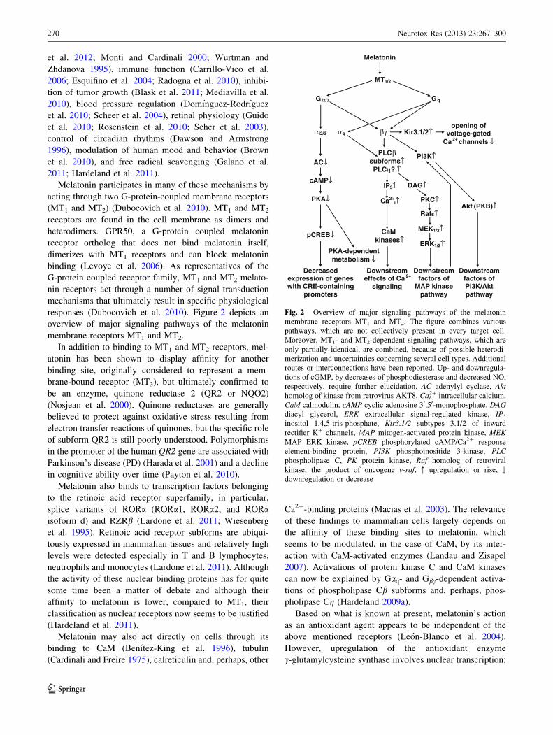
Melatonin participates in many of these mechanisms by
acting through two G-protein-coupled membrane receptors
(MT1 and MT2) (Dubocovich et al. 2010). MT1 and MT2
receptors are found in the cell membrane as dimers and
heterodimers. GPR50, a G-protein coupled melatonin
receptor ortholog that does not bind melatonin itself,
dimerizes with MT1 receptors and can block melatonin
binding (Levoye et al. 2006). As representatives of the
G-protein coupled receptor family, MT1 and MT2 melato-
nin receptors act through a number of signal transduction
mechanisms that ultimately result in specific physiological
responses (Dubocovich et al. 2010). Figure 2 depicts an
overview of major signaling pathways of the melatonin
membrane receptors MT1 and MT2.
In addition to binding to MT1 and MT2 receptors, mel-
atonin has been shown to display affinity for another
binding site, originally considered to represent a mem-
brane-bound receptor (MT3), but ultimately confirmed to
be an enzyme, quinone reductase 2 (QR2 or NQO2)
(Nosjean et al. 2000). Quinone reductases are generally
believed to protect against oxidative stress resulting from
electron transfer reactions of quinones, but the specific role
of subform QR2 is still poorly understood. Polymorphisms
in the promoter of the human QR2 gene are associated with
Parkinson’s disease (PD) (Harada et al. 2001) and a decline
in cognitive ability over time (Payton et al. 2010).
Melatonin also binds to transcription factors belonging
to the retinoic acid receptor superfamily, in particular,
splice variants of RORa (RORa1, RORa2, and RORaisoform d) and RZRb (Lardone et al. 2011; Wiesenberg
et al. 1995). Retinoic acid receptor subforms are ubiqui-
tously expressed in mammalian tissues and relatively high
levels were detected especially in T and B lymphocytes,
neutrophils and monocytes (Lardone et al. 2011). Although
the activity of these nuclear binding proteins has for quite
some time been a matter of debate and although their
affinity to melatonin is lower, compared to MT1, their
classification as nuclear receptors now seems to be justified
(Hardeland et al. 2011).
Melatonin may also act directly on cells through its
binding to CaM (Benıtez-King et al. 1996), tubulin
(Cardinali and Freire 1975), calreticulin and, perhaps, other
Ca2?-binding proteins (Macias et al. 2003). The relevance
of these findings to mammalian cells largely depends on
the affinity of these binding sites to melatonin, which
seems to be modulated, in the case of CaM, by its inter-
action with CaM-activated enzymes (Landau and Zisapel
2007). Activations of protein kinase C and CaM kinases
can now be explained by Gaq- and Gbc-dependent activa-
tions of phospholipase Cb subforms and, perhaps, phos-
pholipase Cg (Hardeland 2009a).
Based on what is known at present, melatonin’s action
as an antioxidant agent appears to be independent of the
above mentioned receptors (Leon-Blanco et al. 2004).
However, upregulation of the antioxidant enzyme
c-glutamylcysteine synthase involves nuclear transcription;
Melatonin
MT1/2
G i2/3 Gq
ERK1/2↑↑
α i2/3 αq βγ
AC↓
cAMP↓
PKA↓
pCREB↓
PLCβsubforms↑PLCη? ↑
PKC↑
DAG↑IP3↑
Ca2+i↑
MEK1/2↑
Rafs↑
CaM kinases↑
Downstream factors of
MAP kinase pathway
PI3K↑
Downstream effects of Ca 2+
signaling
opening of voltage-gated
Ca 2+ channels ↓Kir3.1/2↑
Akt (PKB)↑
Downstream factors of PI3K/Akt pathway
Decreased expression of genes with CRE-containing
promoters
PKA-dependent metabolism ↓
Fig. 2 Overview of major signaling pathways of the melatonin
membrane receptors MT1 and MT2. The figure combines various
pathways, which are not collectively present in every target cell.
Moreover, MT1- and MT2-dependent signaling pathways, which are
only partially identical, are combined, because of possible heterodi-
merization and uncertainties concerning several cell types. Additional
routes or interconnections have been reported. Up- and downregula-
tions of cGMP, by decreases of phosphodiesterase and decreased NO,
respectively, require further elucidation. AC adenylyl cyclase, Akthomolog of kinase from retrovirus AKT8, Cai
2? intracellular calcium,
CaM calmodulin, cAMP cyclic adenosine 30,50-monophosphate, DAGdiacyl glycerol, ERK extracellular signal-regulated kinase, IP3
inositol 1,4,5-tris-phosphate, Kir3.1/2 subtypes 3.1/2 of inward
rectifier K? channels, MAP mitogen-activated protein kinase, MEKMAP ERK kinase, pCREB phosphorylated cAMP/Ca2? response
element-binding protein, PI3K phosphoinositide 3-kinase, PLCphospholipase C, PK protein kinase, Raf homolog of retroviral
kinase, the product of oncogene v-raf, : upregulation or rise, ;downregulation or decrease
270 Neurotox Res (2013) 23:267–300
123
electrophoretic mobility shift assay analysis has shown
melatonin-dependent increases in DNA binding of AP-1
and RZR/RORa (Urata et al. 1999), indicating the
involvement of a nuclear receptor. In addition findings on
protection of liver and heart by the MT1/MT2-selective
melatonergic agonist ramelteon under conditions leading to
oxidative stress points to the inference that these membrane
receptors may have antioxidant activity, mainly because
ramelteon has no substantial radical scavenging properties
(Mathes et al. 2008).
Melatonin Effects on Mitochondria
Free Radical Generation
The synthesis of ATP via the mitochondrial respiratory
chain is the result of a proton potential generated by
the electron transport chain (ETC) (for rev. see Acuna-
Castroviejo et al. 2011). Although ideally all the oxygen
should be reduced to water via a 4-electron reduction
reaction driven by Complex IV, under normal conditions a
certain, but relevant percentage of oxygen can be reduced
by dissipating single electrons yielding free radicals.
Approximately 3–5 % of oxygen is converted to reactive
oxygen species (ROS).
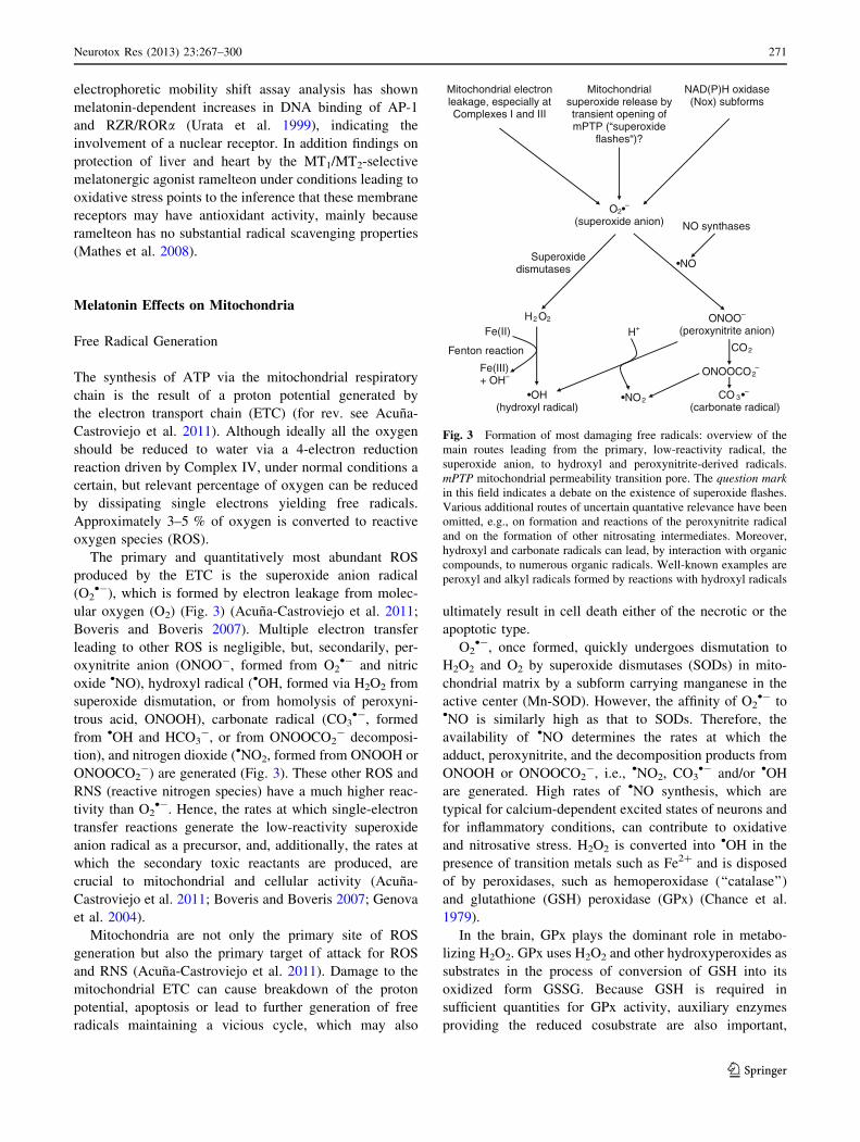
The primary and quantitatively most abundant ROS
produced by the ETC is the superoxide anion radical
(O2•-), which is formed by electron leakage from molec-
ular oxygen (O2) (Fig. 3) (Acuna-Castroviejo et al. 2011;
Boveris and Boveris 2007). Multiple electron transfer
leading to other ROS is negligible, but, secondarily, per-
oxynitrite anion (ONOO-, formed from O2•- and nitric
oxide •NO), hydroxyl radical (•OH, formed via H2O2 from
superoxide dismutation, or from homolysis of peroxyni-
trous acid, ONOOH), carbonate radical (CO3•-, formed
from •OH and HCO3-, or from ONOOCO2
- decomposi-
tion), and nitrogen dioxide (•NO2, formed from ONOOH or
ONOOCO2-) are generated (Fig. 3). These other ROS and
RNS (reactive nitrogen species) have a much higher reac-
tivity than O2•-. Hence, the rates at which single-electron
transfer reactions generate the low-reactivity superoxide
anion radical as a precursor, and, additionally, the rates at
which the secondary toxic reactants are produced, are
crucial to mitochondrial and cellular activity (Acuna-
Castroviejo et al. 2011; Boveris and Boveris 2007; Genova
et al. 2004).
Mitochondria are not only the primary site of ROS
generation but also the primary target of attack for ROS
and RNS (Acuna-Castroviejo et al. 2011). Damage to the
mitochondrial ETC can cause breakdown of the proton
potential, apoptosis or lead to further generation of free
radicals maintaining a vicious cycle, which may also
ultimately result in cell death either of the necrotic or the
apoptotic type.
O2•-, once formed, quickly undergoes dismutation to
H2O2 and O2 by superoxide dismutases (SODs) in mito-
chondrial matrix by a subform carrying manganese in the
active center (Mn-SOD). However, the affinity of O2•- to
•NO is similarly high as that to SODs. Therefore, the
availability of •NO determines the rates at which the
adduct, peroxynitrite, and the decomposition products from
ONOOH or ONOOCO2-, i.e., •NO2, CO3
•- and/or •OH
are generated. High rates of •NO synthesis, which are
typical for calcium-dependent excited states of neurons and
for inflammatory conditions, can contribute to oxidative
and nitrosative stress. H2O2 is converted into •OH in the
presence of transition metals such as Fe2? and is disposed
of by peroxidases, such as hemoperoxidase (‘‘catalase’’)
and glutathione (GSH) peroxidase (GPx) (Chance et al.
1979).
In the brain, GPx plays the dominant role in metabo-
lizing H2O2. GPx uses H2O2 and other hydroxyperoxides as
substrates in the process of conversion of GSH into its
oxidized form GSSG. Because GSH is required in
sufficient quantities for GPx activity, auxiliary enzymes
providing the reduced cosubstrate are also important,
Mitochondrial electron leakage, especially at Complexes I and III
Mitochondrial superoxide release by transient opening of mPTP (“superoxide
flashes“)?
NAD(P)H oxidase (Nox) subforms
O2•–
(superoxide anion)
H2 O2
Superoxide dismutases
Fe(II)
Fe(III) + OH–
Fenton reaction
•OH(hydroxyl radical)
•NO
NO synthases
ONOO–
(peroxynitrite anion) H+
•NO2
CO2
ONOOCO2–
CO 3•–
(carbonate radical)
Fig. 3 Formation of most damaging free radicals: overview of the
main routes leading from the primary, low-reactivity radical, the
superoxide anion, to hydroxyl and peroxynitrite-derived radicals.
mPTP mitochondrial permeability transition pore. The question markin this field indicates a debate on the existence of superoxide flashes.
Various additional routes of uncertain quantative relevance have been
omitted, e.g., on formation and reactions of the peroxynitrite radical
and on the formation of other nitrosating intermediates. Moreover,
hydroxyl and carbonate radicals can lead, by interaction with organic
compounds, to numerous organic radicals. Well-known examples are
peroxyl and alkyl radicals formed by reactions with hydroxyl radicals
Neurotox Res (2013) 23:267–300 271
123

including GSH reductase (GR) and glucose-6-phosphate
dehydrogenase (which synthesize reducing equivalents for
the action of GR) as well as the rate-limiting enzyme of
GSH formation, c-glutamylcysteine synthase (Ghanta and
Chattopadhyay 2011). All these enzymes are upregulated
by melatonin, either through direct or indirect mechanisms
(Hardeland 2005).
Among other sources of ROS, formation of H2O2, O2•-,
and OCl- by microglia or invading myeloperoxidase-
expressing leukocytes becomes relevant in inflammatory
situations. This is not restricted to acute brain inflamma-
tion, but also plays a role in some neurodegenerative dis-
eases (Cunningham 2011). Alzheimer’s disease (AD) is an
example of an atypical, lingering form of inflammation, in
which some classical hallmarks such as neutrophil infil-
tration and edema are usually absent, whereas other char-
acteristics including acute-phase proteins and cytokines are
clearly demonstrable (Cunningham 2011). In the brain,
inflammatory ROS production is intertwined with second-
ary excitotoxicity and RNS formation that is, e.g.,
demonstrable by enhanced protein nitration (for discussion
see Hardeland 2009b). Various flavin enzymes also gen-
erate ROS, usually H2O2, sometimes accompanied by O2•-
production. In dopaminergic neurons, the oxidation of
dopamine (DA) by monoamine oxidase (MAO) generates
hydrogen peroxide. The increased destruction of DA by
MAO has been postulated to be the reason for degeneration
of dopaminergic neurons causing PD (Olanow 1990).
The free radical •NO is produced by several subforms of
nitric oxide synthase (NOS) (Pacher et al. 2007). Since•NO is a gaseous compound, it can cross membranes with
ease and, therefore, it enters mitochondria, regardless of its
neuronal, glial or vascular origin. •NO strongly interferes
with components of the respiratory chain, in particular
cytochrome c oxidase (Pacher et al. 2007). Moreover, its
metabolite ONOO-and radicals derived from this can
damage proteins of the respiratory complexes. The exis-
tence of a separate mitochondrial NOS (mtNOS) has been
described but many of the published data are debatable
(Chuang 2010). In the adult mouse brain, no mtNOS was
demonstrated under carefully controlled conditions (Lacza
et al. 2004). •NO entering neuronal mitochondria and
peroxynitrite formed there by combination with O2•- from
electron leakage do not only interfere with the respiratory
chain, at already moderate concentrations, but, at elevated
levels, lead to free radical-mediated chain reactions that
destroy protein, lipid, and DNA molecules.
Melatonin and Mitochondrial Homeostasis
Several studies have suggested that the neuroprotective role
of melatonin in aging and in many neurodegenerative con-
ditions such as AD and PD can be due to its direct antioxidant
role in mitochondrial homeostasis (Acuna-Castroviejo et al.
2011; Srinivasan et al. 2011). When acutely added in vitro,
melatonin diminished the increase in respiration caused by
addition of Krebs’ cycle substrates and ADP to mitochon-
drial preparations (Reyes Toso et al. 2003). This finding
demonstrates melatonin’s ability to interact with the ETC
(Hardeland 2005), but the possibility of pharmacological
effects of electron interception by elevated melatonin levels
has to be considered, which may partially block the elec-
tron flux by alternate electron acceptance. Melatonin does
not affect basal mitochondrial respiration (Reyes Toso et al.
2003).
In one study the daily administration of physiological
amounts of melatonin to senescence-accelerated or senes-
cence-resistant mice over a period of 5 months was
reported to increase state three respiration and respiratory
control indexes in liver mitochondria (Okatani et al. 2002).
These results were taken as evidence of melatonin’s ability
to normalize cellular respiration, to attenuate oxidant for-
mation and, thus, to protect cells against oxidative damage.
Although mild uncoupling is usually expected to reduce
electron leakage, additionally observed rises in dinitrophe-
nol-dependent uncoupled respiration may reflect melatonin-
induced changes in respiratory capacity. A long-term
improvement of mitochondrial function in vivo can be
explained in different ways, either in terms of interactions
with the electron flux or by induction of antioxidant
enzymes which prevent damage to the respiratory chain
(Acuna-Castroviejo et al. 2011). Moreover, increases in gene
expression of components of Complexes I and IV were
observed, which may have also contributed to normalized
respiration.
A low-affinity mitochondrial binding site for melatonin
(IC50 = 0.8 lM) has been associated with an inhibition of
the mitochondrial permeability transition pore (mtPTP) and
reported to mediate an anti-apoptotic action of melatonin at
pharmacological concentrations (Andrabi et al. 2004).
Under physiological conditions, this binding site would be
only relevant if melatonin accumulated in sufficiently high
concentrations, a fact suggested by the intramitochondrial
concentrations of melatonin observed in some experiments
(Venegas et al. 2012). Indeed melatonin, which possesses
both hydrophilic and lipophilic properties, crosses cell
membranes with ease and is capable of concentrating within
subcellular compartments (Menendez-Pelaez and Reiter
1993) including mitochondria (Martin et al. 2000). Melato-
nin at millimolar concentrations has been reported to stabi-
lize the fluidity of mitochondrial inner membranes (Garcıa
et al. 1999).
The binding of melatonin to mitochondrial membranes
was first reported in studies using 2-[125I]-iodomelatonin
(Yuan and Pang 1991). Administration of melatonin
increases the activities of mitochondrial respiratory
272 Neurotox Res (2013) 23:267–300
123

Complexes I and IV in a time-dependent manner in brain and
liver tissues (Martin et al. 2002). It has been suggested that
melatonin interacts with complexes of the ETC and may
donate electrons and, as an oxidized intermediate, may also
accept electrons, thereby increasing the electron flow. This
property, which is not shared by most antioxidants previ-
ously investigated (with the exception of ubiquinone), can
enable melatonin both to participate in electron transport and
to act as an antioxidant (Acuna-Castroviejo et al. 2011).
The phenomenon of electron donation by melatonin and
electron acceptance by the melatonyl cation radical was used
as the basis of a model proposing that melatonin could act at
low, quasi-catalytic concentrations to reduce electron leak-
age (Hardeland 2005). Similar effects were predicted for
the melatonin metabolite N1-acetyl-5-methoxykynuramine
(AMK), with regard to its single-electron transfer reactions,
reactivity and amphiphilicity (Hardeland 2005). In fact,
mitochondrial protection by AMK was confirmed by the
findings of Acuna-Castroviejo et al. (2002). However, mel-
atonin-mediated modulation of electron flux can be
explained by other mechanisms, including direct flux control
at Complex I, prevention of ROS- and RNS-induced inter-
ruptions of electron transport, and de novo synthesis of
respirasomal proteins (Acuna-Castroviejo et al. 2011).
Melatonin increases the mitochondrial GSH pool and
reduces hydroxyperoxide levels. The latter effect can be
regarded as the sum of indirect antioxidant effects via GSH
levels and of reduction of electron leakage. By safe-
guarding electron flow through the transport chain mela-
tonin increases ATP production (Acuna-Castroviejo et al.
2011). Melatonin prevents membrane lipid peroxidation
and mitochondrial and nuclear DNA oxidation induced by
enhanced oxidative stress and has been shown to be anti-
apoptotic in several experimental model systems (Sainz
et al. 2003).
Melatonin administration has been found to effectively
counteract oxidative mitochondrial DNA damage and to
restore the mitochondrial respiratory control system, by
preventing the decrease in Complexes I and IV activities
(Zhang et al. 2009). Since reduced Complex I activity is
associated with and is a sign of enhanced electron leak-
age, the resulting increase in oxidative stress is suffi-
cient to induce apoptosis. Melatonin’s action in restoring
Complex I activity back to normal levels thus assumes
significance for overall health in its prevention of age-
associated degenerative changes (Acuna-Castroviejo et al.
2011).
Melatonin and Neuronal Damage
Many acute conditions, e.g., hypoxia, stroke, physi-
cal trauma, hypoglycemia, drug neurotoxicity, viruses,
radiation or noxious stimuli, are sufficient to produce
neuronal damage. Similar mechanisms are also likely to be
involved in neurodegenerative disorders, a group of
chronic and progressive diseases that are characterized by
selective and symmetric losses of neurons in motor, sen-
sory, or cognitive systems. Clinically relevant examples of
these disorders are AD, PD, amyotrophic lateral sclerosis
(ALS), and Huntington’s disease (HD) (Aliev et al. 2011;
Green et al. 2011).
Although the origin of neurodegenerative diseases has
remained mostly undefined, three major and frequently
interrelated processes, i.e., glutamate excitotoxicity, free
radical-mediated damage and mitochondrial dysfunction,
have been identified as common pathophysiological
mechanisms leading to neuronal death (Reiter 1998).
Because melatonin is a direct and indirect antioxidant it has
been proposed as a neuroprotective agent (Reiter et al.
2010). Melatonin’s neuroprotective properties, as well as
regulatory effects on circadian disturbances, validate mel-
atonin’s benefits as a therapeutic substance in the symp-
tomatic treatment of neurodegenerative diseases. Moreover,
melatonin exerts anti-excitatory, and at sufficient dosage,
sedating effects (Caumo et al. 2009; Golombek et al. 1996)
so that a second neuroprotective mode of action may exist
involving the c-aminobutyric acid (GABA)-ergic system as
a mediator. This view is supported by studies indicating that
melatonin protects neurons from the toxicity of the amy-
loid-b (Ab) peptide (the main neurotoxin involved in AD)
via activation of GABA receptors (Louzada et al. 2004).
Melatonin also has anti-excitotoxic actions. Early stud-
ies in this regard employed kainate, an agonist of iono-
tropic glutamate receptors, and gave support to the
hypothesis that melatonin prevents neuronal death induced
by glutamate (Giusti et al. 1996b; Manev et al. 1996). It has
also been reported that administration of melatonin reduces
the injury of hippocampal CA1 neurons caused by transient
forebrain ischemia (Cho et al. 1997; Kilic et al. 1999) or
high glucocorticoid doses (Furio et al. 2008). Following a
hypoxic injury in rats, melatonin administration reduced
glutamate levels and structural damage caused by hypoxia
to neurons, axons, and dendrites in the brainstem sug-
gesting that it is capable of ameliorating excitotoxic dam-
age (Kaur et al. 2011). A further demonstration that
melatonin deficiency can potentiate neuronal damage is the
finding that more severe brain damage and neurodegener-
ation occurs after stroke or excitotoxic seizures in mela-
tonin-deficient rats (Manev et al. 1996). Pineal melatonin
concentration decreased after hypoxic injuries in rats (Kaur
et al. 2007).
Melatonin has often demonstrated superiority to vita-
mins C and E in protection against oxidative damage and in
scavenging free radicals (Galano et al. 2011). Additionally,
melatonin potentiates effects by other antioxidants, such as
Neurotox Res (2013) 23:267–300 273
123
vitamin C, Trolox (a water soluble vitamin E analog) and
NADH. The antioxidative efficiency of melatonin is high
because the metabolites formed by free radical scavenging
also act as free radical scavengers. This holds, in particular,
for cyclic 3-hydroxymelatonin, N1-acetyl-N2-formyl-5-
methoxykynuramine (AFMK) (Tan et al. 2003) and, with
highest potency, AMK (Behrends et al. 2004; Silva et al.
2004). Thus, the interaction of melatonin with free radicals
initiates an antioxidant cascade, which may allow the
elimination of, in the extreme, up to ten oxidizing free
radicals.
Nevertheless, these effects are not sufficient for
explaining melatonin’s protective potency. Various sec-
ondary antioxidant effects have been described, which are
based on upregulation of antioxidant and downregulation
of prooxidant enzymes. In several studies some of these
effects were highly variable, depending on the tissue and
species involved, such as induction of SODs and catalase
(for details see Hardeland 2005). Other effects were con-
sistently observed; in particular, this holds for GPx and for
GR, presumably in response to GPx-dependent increases in
GSSG, the oxidized form of GSH. Notably, enzyme
inductions were more pronounced in the CNS of avian and
other non-mammalian species.
Melatonin contributes to maintain normal GSH levels
(Subramanian et al. 2007) by stimulating GSH biosynthesis
via c-glutamylcysteine synthase and glucose-6-phosphate
dehydrogenase (Kilanczyk and Bryszewska 2003; Rodrı-
guez et al. 2004). Melatonin treatment enhanced brain GSH
levels which were depressed following a hypoxic injury of
developing rats (Kaur et al. 2010).
Antioxidative signaling is of particular significance
because blood concentrations of melatonin are low, even at
night, as compared to other antioxidants such as vitamin C or
GSH (Galano et al. 2011). Although melatonin levels are
several times higher in certain body fluids and tissues than in
blood this may not be sufficient to fully explain the protective
effects observed. Nevertheless, the much higher melatonin
concentrations, e.g., in the bile (Tan et al. 1999), gastroin-
testinal tract (Bubenik 2002; Kvetnoy et al. 2002), or bone
marrow (Conti et al. 2000) should be of significance.
According to Reiter and Tan (2003) melatonin concentra-
tions in the blood should not be taken as an index to judge its
concentration in other body fluids and intracellular com-
partments of the cell. Inasmuch as many cells of the body
have the potential to synthesize melatonin, local melatonin
production could increase under high free radical generating
conditions. Assuming these conditions are met, melatonin
could fulfill all the requirements for designation as an
effective physiological antioxidant (Galano et al. 2011).
In addition to stroke, the efficacy of melatonin in
inhibiting oxidative damage has also been tested in a
variety of neurological disease models where free radicals
have been implicated as being at least partial causal agents
of the condition. Thus, melatonin has been shown to reduce
Ab protein toxicity in AD animal models (Dragicevic et al.
2011; Matsubara et al. 2003; Olcese et al. 2009; Pappolla
et al. 1997), to reduce oxidative damage in several models
of PD (Acuna-Castroviejo et al. 1997; Chuang and Chen
2004; Dabbeni-Sala et al. 2001; Jin et al. 1998; Saravanan
et al. 2007; Singhal et al. 2011), to protect against gluta-
mate excitotoxicity (Das et al. 2010; Giusti et al. 1996a)
and to lower neural damage due to cadmium toxicity
(Jimenez-Ortega et al. 2011; Poliandri et al. 2006), d-am-
inolevulinic acid toxicity (porphyria) (Carneiro and Reiter
1998; Onuki et al. 2005; Princ et al. 1997), hyperbaric
hyperoxia (Pablos et al. 1997; Shaikh et al. 1997), brain
trauma (Beni et al. 2004; Kabadi and Maher 2010; Tsai
et al. 2011), c radiation (Erol et al. 2004; Shirazi et al.
2011; Taysi et al. 2008), focal ischemia (Kilic et al. 2011;
Koh 2012; Lee et al. 2004; Tai et al. 2011), and a variety of
neural toxins (Reiter et al. 2010).
The various types of toxicities listed above can result in
cell death by necrosis or apoptosis. Apoptotic neuronal
death requires RNA and protein synthesis, and depletion of
trophic factors. Apoptosis also involves single-strand
breaks of DNA. Neurotrophic factors have been found to
rescue neurons from this type of death (Green et al. 2011).
They may act via cellular anti-apoptotic components, such
as the B cell lymphoma proto-oncogene protein (Bcl-2).
The neuroprotective function of Bcl-2 is particularly well
demonstrated in naturally occurring or experimentally
induced neuronal death, which can be prevented by over-
expression of Bcl-2 (Chuang 2010; Green et al. 2011). In
the highly complex context of different roles of members
of the Bcl family, Bcl-2 is capable of blocking the apop-
totic pathway by preventing the formation of a functional
mtPTP and, thus, the release of the mitochondrial enzyme
cytochrome c, which represents the final and no-return
signal of the apoptotic program (Green et al. 2011;
Khandelwal et al. 2011; Kluck et al. 1997). Studies in vitro
indicate that melatonin enhances expression of Bcl-2 and
prevents apoptosis (Jiao et al. 2004; Koh 2011; Radogna
et al. 2010; Wang 2009). More recent studies have gone
beyond the functions of Bcl. Melatonin was shown to
directly inhibit the opening of the mtPTP, thereby rescuing
cells (Jou 2011; Peng et al. 2012). In addition to its anti-
oxidant actions, melatonin directly diminished mtPTP
currents, with an IC50 of 0.8 lM (Andrabi et al. 2004), a
concentration which seems physiologically possible if
mitochondrial accumulation of melatonin occurs.
Besides glutamate toxicity and oxidative stress, inflam-
mation has been reported as another mechanism leading to
damage/death of neurons in several brain pathologies
including hypoxic-ischemic injuries. Activated microglia
and endothelial cells under such conditions release
274 Neurotox Res (2013) 23:267–300
123

proinflammatory cytokines such as tumor necrosis factor-a(TNF-a) and interleukin-1b (IL-1b), which cause damage
to neurons (Floden et al. 2005). TNF-a has been shown to
mediate neuronal death through binding with its receptor
TNF-R1 which has an intracellular death domain and its
activation results in mitochondrial dysfunction, oxidative
damage and silencing of survival signals (Nakazawa et al.
2006). IL-1b damages neurons by binding to its receptor
IL-R1 and by initiating mechanisms such as excitotoxicity
and increased inducible NOS (iNOS) production through
IL-R1 signaling. Melatonin is known to exert anti-inflam-
matory actions and has been shown to reverse the inflam-
matory response in several brain pathologies by
suppressing the production of inflammatory cytokines.
Experimental studies have shown that melatonin is neuro-
protective in ischemia/reperfusion injury as it inhibits the
inflammatory response (Pei and Cheung 2004). Melatonin
treatment has also been demonstrated to reduce the levels
of proinflammatory cytokines TNF-a and IL-1b induced by
Ab in rat brain (Rosales-Corral et al. 2003) and to suppress
the release of these cytokines by microglia (Kaur et al.,
unpublished data).
Therefore, data have accumulated indicating that mela-
tonin may curtail all major processes in neuronal damage,
i.e., glutamate excitotoxicity, free radical-mediated injury,
neuroinflammation and apoptosis. In addition, melatonin,
in acting as an endocrine arm of the circadian clock, pro-
motes the restorative phases of sleep, a situation associated
with neurotrophic effects. Thus melatonin may function as
a unique chronobiotic–cytoprotective agent (Cardinali and
Scacchi 2010).
Neurodegenerative diseases have become a major health
problem and a growing recognition exists that efforts to
prevent these diseases must be undertaken by both gov-
ernmental and non-governmental organizations. Regular
intake of antioxidants by the elderly has been recom-
mended for prevention of age-associated neurodegenera-
tive diseases, although the efficacy of this treatment is still
discussed (Hausman et al. 2011). In this context, the pineal
product melatonin may have major significance since one
of the features of advancing age is the gradual decrease in
the endogenous synthesis of this important antioxidant
(Bubenik and Konturek 2011).
Melatonin and Aging
In humans pineal melatonin production is higher in
younger age groups (18–54 years) as compared to older
individuals (Karasek and Reiter 2002; Sack et al. 1986;
Skene and Swaab 2003). The highest peak secretion of
melatonin is found in children at 3–5 years of age (Cavallo
1993). With some exceptions (Fourtillan et al. 2001;
Zeitzer et al. 1999) the decline of melatonin production
with age has been consistently reported (Brown et al. 1979;
Dori et al. 1994; Ferrari et al. 2008; Girotti et al. 2000;
Iguchi et al. 1982; Lieverse et al. 2011; Luboshitzky et al.
2001; Mazzoccoli et al. 2010b; Mishima et al. 2000, 2001;
Rosen et al. 2009; Siegrist et al. 2001; Waldhauser and
Steger 1986).
Age-associated changes in the day/night rhythm of
melatonin production have been found, with phase
advances being encountered more frequently in the elderly
as compared to young women (Skene and Swaab 2003). It
has also been shown that SCN function declines with age,
particularly in patients with aging-associated neurodegen-
erative disorders, a major cause of dementia and other poor
health condition in the elderly population (Pandi-Perumal
et al. 2002; Wu and Swaab 2007). As shown in non-human
primates, in addition to the age-associated attenuation of
hormone levels and reduction of humoral circadian sig-
naling, there are also significant age-related changes in
intracrine processing enzymes and hormone receptors
which may further affect the functional efficacy of these
hormones (Urbanski and Sorwell 2011). In addition to
degenerative processes in the SCN or its input and output
pathways, pineal calcification can be another reason for
age-related declines in melatonin secretion (Mahlberg et al.
2009).
The decline in melatonin production and altered mela-
tonin rhythms can be major contributing factors to the
increased levels of oxidative stress and the associated
degenerative changes that are seen in the elderly. Never-
theless, individuals of the same chronological age can
exhibit dissimilar degrees of senescence-associated func-
tional impairment, differences which may be attributable to
the well documented inter-individual variations in mela-
tonin levels (Bergiannaki et al. 1995; Ferrari et al. 2008;
Grof et al. 1985; Travis et al. 2003). Variations in the
degenerative changes of cells and tissues have been
attributed to variations in melatonin production, changes
which are more often determined by the physiological age
of an individual rather than his chronological age (Barzilai
et al. 2010). Recently it has been shown that there are
genetic variations in the enzyme ASMT (HIOMT), a
metabolic step that determines the amount of melatonin
produced. These variations have been linked to autism
spectrum disorder (Jonsson et al. 2010; Melke et al. 2008),
recurrent depression (Galecki et al. 2010), and intellectual
disability (Pagan et al. 2011).
In terms of aging, the immunostimulatory actions of
melatonin are of particularly high relevance (Cardinali
et al. 2008; Guerrero and Reiter 2002; Mazzoccoli et al.
2010a). Although the importance of the age-dependent
decreases in melatonin production as contributors to
immunosenescence is not yet fully established, this issue
Neurotox Res (2013) 23:267–300 275
123
merits further research attention. Age-associated changes
in the average melatonin concentration, as well as in the
amplitude of its circadian rhythm, can have profound
effects on the entire circadian system. These changes can
be expected to produce a host of multiple, pleiotropic
effects and, presumably, dysfunctions that could be effi-
ciently antagonized by melatonin (Cardinali et al. 2008).
Melatonin in AD
AD is an age-associated neurodegenerative disease that is
characterized by progressive loss of cognitive function,
loss of memory, and other neurobehavioral manifestations.
In spite of the number of studies undertaken, its etiology
remains an enigma. Many mechanisms such as genetic
factors, chronic inflammation associated with cytokine
release, oxidative stress, and trace element neurotox-
icity have been suggested as possible underlying causes
(Ehrnhoefer et al. 2011; Jucker and Walker 2011). The
pathological manifestations of AD include amyloid plaques
and neurofibrillary tangles. The free Ab molecule is Fen-
ton-reactive due to bound copper and, therefore, leads to
cell death through induction of oxidative stress. Addition-
ally, Ab initiates flavoenzyme-dependent increases in
intracellular H2O2 and lipid peroxides, which also promote
free radical generation.
AD is related to mitochondrial dysfunction and can now
be viewed as having characteristics reminiscent of other
mitochondrial diseases associated with pathological oxi-
dant formation (Swerdlow 2011). Attenuation or preven-
tion by administration of suitable antioxidants should be
the basis for a strategic treatment of AD. Though vitamins
E and C have been used for treatment of patients (Devore
et al. 2010), the neurohormone melatonin has assumed a
significant role in view of the fact that it has been shown to
be an effective antioxidant in a number of transgenic
mouse models of AD, as discussed below. In one of these
models, 4-month-old transgenic mice exhibited increases in
levels of brain thiobarbituric acid-reactive substances, and
reductions in SOD and GSH content, changes that were
attenuated by administration of melatonin (Feng et al.
2006). Moreover, melatonin has been shown to exhibit
antifibrillogenic activities, even when fibrillogenesis was
enhanced by apolipoprotein E4 (apoE4), effects which
were not seen to this extent when antioxidant vitamins
were applied (Poeggeler et al. 2001).
Experimentally it has been shown that suppression of
serum melatonin levels in rats through exposure to constant
illumination results in a number of AD-like behavioral
effects and neurochemical changes (Ling et al. 2009).
These included spatial memory deficits, tau hyperphosph-
orylation at multiple sites, activation of glycogen synthase
kinase-3 and protein kinase A, as well as suppression of
protein phosphatase-1. An increased expression of endo-
plasmic reticulum stress-related proteins including BiP/
GRP78 and CHOP/GADD153 was also taken as evidence
of prominent oxidative damage and organelle lesions. All
these impairments were partially attenuated by the simul-
taneous administration of melatonin (Ling et al. 2009).
Endogenous melatonin deficiency can, therefore, contribute
to the disease progression, and the beneficial effects of
replacement therapy may well be warranted in AD.
Melatonin prevents the death of neuroblastoma cells
exposed to Ab polypeptide (Pappolla et al. 1997, 1999,
2000). Using murine neuroblastoma cells (N2a) (Pappolla
et al. 1997) demonstrated that co-incubation of neuroblas-
toma cells with Ab polypeptide and melatonin significantly
reduced several features of apoptosis such as cellular
shrinkage or formation of membrane blebs. Melatonin also
reduced the levels of lipid peroxidation in the cultured
neuroblastoma cells by scavenging free radicals released by
Ab (He et al. 2010). The neurofibrillary tangles of AD
patients are composed of abnormally bundled cytoskeletal
fibers, due to hyperphosphorylation of tau, a microtubule-
associated protein, and of neurofilament H/M subunits,
processes which lead to misfolding and accumulation of
these proteins, along with disruption of microtubules. Thus,
inhibition or reversal of hyperphosphorylation may be
effective in preventing tauopathies.
Okadaic acid, a potent protein phosphatase inhibitor,
induced cell death in neuroblastoma cells, accompanied by
a striking decrease in mitochondrial metabolic activity
(Benıtez-King et al. 2003). Okadaic acid induces physio-
logical and biochemical changes similar to those seen in
AD. It increased the levels of 4-hydroxynonenal and
decreased antioxidant enzyme activities in cultured neu-
ronal cells (Perez et al. 2002). Either melatonin or vitamin
C administration prevented the effects of okadaic acid in
NIE 115 cells (Benıtez-King et al. 2003). While both
vitamin C and melatonin reduced free radical damage, the
reduction was greater with melatonin. Although vitamin C
failed to increase the levels of glutathione S-transferase and
GR, melatonin increased the levels of both enzymes sig-
nificantly. Melatonin also inhibited the phosphorylation
and accumulation of neurofilaments.
Similar results were obtained, in neuroblastoma N2a
cells, with calyculin A, an inhibitor of protein phosphatases
2A and 1, but in this study an additional activation of
glycogen synthase kinase 3 (GSK-3, a redox-controlled
enzyme involved in various cell regulatory mechanisms)
was observed (Li et al. 2005). Apart from hyperphosph-
orylation and lethal oxidative stress, including decreases in
SOD, melatonin also reversed GSK-3 activation, thus
supporting the conclusion that the methoxyindole action
was not only based on its antioxidant properties, but also on
276 Neurotox Res (2013) 23:267–300
123
interference with protein phosphorylation, especially by
stress kinases.
Inhibition of protein phosphatase (PP)-2A and PP-1 by
calyculin A-induced AD-like hyperphosphorylation of tau
and spatial memory retention impairment. The adminis-
tration of melatonin before injection of calyculin A could
prevent calyculin A-induced synaptophysin loss, memory
retention deficits, as well as hyperphosphorylation of tau
and neurofilaments. Furthermore, melatonin partially
reversed the phosphorylation of the catalytic subunit of PP-
2A at Tyrosine 307 (Y307), a crucial site negatively reg-
ulating the activity of PP-2A, and reduced the levels of
malondialdehyde, a marker of oxidative stress, induced by
calyculin A (Yang et al. 2011). Melatonin’s antioxidant
action may be of additional importance with regard to
inflammatory responses of chronically activated microglia
in AD (Kitazawa et al. 2004). For a recent review on the
effect of melatonin on several neurological diseases with
inflammatory components, see Esposito and Cuzzocrea
(2010).
In several experiments transgenic (Tg) murine models of
AD have been used to assess a possible therapeutical ben-
eficial effect of melatonin (Bedrosian et al. 2011; Dragic-
evic et al. 2011; Feng et al. 2006; Feng and Zhang 2004;
Garcıa et al. 2010; Garcıa-Mesa et al. 2012; Matsubara et al.
2003; Olcese et al. 2009; Spuch et al. 2010). The admin-
istration of melatonin partially inhibited the expected time-
dependent elevation of Ab, reduced abnormal nitration of
proteins, and increased survival in the treated amyloid
protein precursor (APP) Tg mice (Matsubara et al. 2003).
Melatonin was found to be effective in inhibiting Abdeposition in APP 695 Tg mice, a model in which senile
plaques appear in the cortex as early as 8 months of age
(Feng et al. 2006; Feng and Zhang 2004). These mice
display behavioral impairments and memory deficits, defi-
ciencies that were alleviated following long-term adminis-
tration of melatonin at a daily dose of 10 mg/kg. The
treatment also reduced the number of apoptotic neurons
(Feng et al. 2006; Feng and Zhang 2004). AAP Tg mice
were also used to explore a ‘‘sundowning’’-like behavior
and the efficacy of melatonin to treat it (Bedrosian et al.
2011). A temporal pattern of anxiety-like behavior emerged
with elevated locomotor activity relative to adult mice near
the end of the dark phase which was refractory to melatonin
treatment (Bedrosian et al. 2011).
In other experiments APP ? presenilin 1 (PS1) double
Tg mice were used. In one of these experiments APP/PS1
Tg mice receiving melatonin from 2 to 2.5 months of age
to their killing at age 7.5 months exhibited reduced cog-
nitive impairments. Decreased Ab deposition and inflam-
matory cytokines in hippocampus and entorhinal cortex
were also found. Cortical mRNA expression of three
antioxidant enzymes (SOD-1, GPx, catalase) was also
significantly reduced (Olcese et al. 2009). In a similar
group of animals treated for 1 month with melatonin the
analysis of isolated brain mitochondria indicated that
melatonin treatment decreased mitochondrial Ab levels by
two- to fourfold in different brain regions (Dragicevic et al.
2011). This was accompanied by a near complete restora-
tion of mitochondrial respiratory rates, membrane poten-
tial, and ATP levels in isolated mitochondria. In APP-
expressing neuroblastoma cells in culture, mitochondrial
function was restored by melatonin or by the structurally
related compounds indole-3-propionic acid or AFMK, an
effect blocked by melatonin receptor antagonists indicating
melatonin receptor signaling is required for the full effect
(Dragicevic et al. 2011). APP/PS1 Tg mice were also used
to assess the efficacy of a tacrine-melatonin hybrid to
inhibit amyloid-induced cell death and amyloid burden in
brain parenchyma. The efficacy of the tacrine-melatonin
hybrid to reduce Ab toxicity suggested the possibility for a
new potential therapeutic strategy in AD (Spuch et al.
2010).
In APPP Tg 2576 mice fed with aluminum lactate
melatonin co-administration prevented the prooxidant
effect of the toxic in the hippocampus (Garcıa et al. 2010)
but not its behavioral effects (Garcıa et al. 2009). The tri-
ple-Tg mouse model of AD (3xTg-AD) is the only model
to exhibit both Ab and tau pathology that is characteristic
of the human form (Sterniczuk et al. 2010). Melatonin was
highly effective against the immunosenescence and cog-
nitive loss that 3xTg-AD mice show (Garcıa-Mesa et al.
2012).
From all these studies it is clear that melatonin treatment
gives neuroprotection against oxidative injury by main-
taining the survival of both neuronal cells and glial cells.
Studies carried out on cultured neuroblastoma cells in
transgenic models of AD reveal that melatonin can atten-
uate the oxidative damage induced by Ab. Overall, they
explain why clinical studies on melatonin efficacy at the
early stages of AD showed significant regression of
disease.
Melatonin Secretion in AD Patients
Circulating melatonin levels are lower in AD patients than
in age-matched controls (Ferrari et al. 2000; Liu et al.
1999; Mishima et al. 1999; Ohashi et al. 1999; Skene et al.
1990; Uchida et al. 1996). Decreased CSF melatonin levels
observed in AD patients have been linked to a reduced
pineal melatonin synthesis rather than to dilution of the
CSF (Tan et al. 2010). It is interesting to note that CSF
melatonin levels are decreased even in the preclinical
stages when the patients have no cognitive impairment
(Braak stages I–II), thus suggesting that reduced melatonin
Neurotox Res (2013) 23:267–300 277
123
levels may be an early marker for the very first stages of
the disease (Wu et al. 2003; Zhou et al. 2003). The
decreased nocturnal melatonin levels with loss of melato-
nin diurnal rhythmicity may be the consequences of SCN
clock gene dysfunction with altered noradrenergic regula-
tion and depletion of the melatonin precursor serotonin by
increased MAO A activity (Wu et al. 2006b). According to
one study (Skene and Swaab 2003), reduced light trans-
mission through the ocular lens or a defective retina-reti-
nohypothalamic tract-SCN pathway are possible causes of
disturbed circadian rhythmicity in AD patients.
Treatment of AD Patients with Melatonin
There are two reasons why it is quite convenient the use of
melatonin or melatonin analogs in AD patients. AD
patients show a greater breakdown of the circadian sleep/
wake cycle compared to similarly aged, non demented
controls. Demented patients spend their nights in a state of
frequent restlessness and their days in a state of frequent
sleepiness. These sleep/wake disturbances become
increasingly more marked with progression of the disease
(Zhong et al. 2011). Hence, replacement of melatonin
levels in brain can be highly convenient in these patients.
On the other hand, the bulk of information on the neuro-
protective properties of melatonin derived from experi-
mental studies turns highly desirable to employ
pharmacological doses in AD patients with the aim of
arresting or slowing disease’s progression.
In AD patients with disturbed sleep/wake rhythms there
is a greater degree of irregularities in melatonin secretion
than in patients who do not exhibit such disturbances
(Mishima et al. 1999). Impairments in melatonin secretion
are also related to both age and severity of mental dys-
function. Other factors that are related to mental impair-
ment are suppressed levels of nocturnal growth hormone
(GH) and increases in both the mean levels and nadir
values of plasma cortisol (Magri et al. 2004; Mazzoccoli
et al. 2010b). Interestingly, melatonin augments GH
secretion as well as prevents in animal models of gluco-
corticoid-induced neuronal loss in the hippocampus (Furio
et al. 2008).
In several studies loss or damage of neurons in the
hypothalamic SCN and other parts of the circadian timing
system have been implicated in the circadian disturbances
of demented patients (Hu et al. 2009; Skene and Swaab
2003; Swabb et al. 1985; van Someren 2000). The SCN of
AD patients have tangles (Stopa et al. 1999). Wu et al.
(2007) have shown by immunocytochemistry that both
MT1-expressing neurons and arginine vasopressin (AVP)/
vasoactive intestinal polypeptide (VIP)-expressing neurons
in the SCN are strongly diminished in the advanced
neuropathological stages of AD.
A chronobiological approach using melatonin, bright
light therapy, restricted time in bed and diurnal activity has
been proposed as a therapeutic alternative for the man-
agement of sleep/wake disorders in AD patients. The aim
of these therapies is to improve sleep and diurnal activity
and consequently to increase the quality of life in patients.
The safety of melatonin treatment is high, with very few or
no adverse effects. In the case of bright light therapy,
however, there is a very significant risk of retinal damage
from repeated exposure to the high intensities of visible
light (Beatty et al. 2000; Hall and Gale 2002; Wu et al.
2006a).
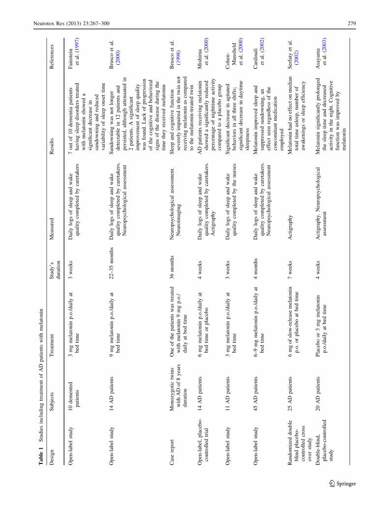
Table 1 summarizes the clinical studies in AD patients
published so far. An initial, preliminary examination of the
sleep-promoting action of melatonin (3 mg p.o. for
21 days) was carried out in a small non-homogenous group
of elderly patients with primary insomnia and with
insomnia associated with dementia or depression. The
investigation found that 7 out of 10 dementia patients who
had sleep disorders and who were treated with melatonin
(3 mg p.o. at bed time) showed decreased sundowning and
reduced variability of sleep onset time (Fainstein et al.
1997). In another study, 10 individuals with mild cognitive
impairment (MCI) were given 6 mg of melatonin before
bedtime. Improvement was found in sleep, mood, and
memory (Jean-Louis et al. 1998b).
Another study in this line of investigation evaluated the
effect on AD patients with sleep disorders and sundowning
agitation of daily administration of 6–9 mg melatonin for
longer periods of time (2–3 years). The retrospective
account of 14 AD patients after the extended period of
therapy with melatonin indicated that all experienced
improvements in sleep quality (Brusco et al. 2000). Sun-
downing, diagnosed clinically in all patients examined, was
no longer detectable in 12 of them, with attenuated
symptoms in the other 2 patients. Another significant
observation in this study was the arrest of the cognitive and
amnesic alterations expected in comparable populations of
patients not receiving melatonin. This should be contrasted
with the significant deterioration of clinical conditions of
the disease found in patients after 1–3 years of progression
of the disease. Further support for the hypothesis that
melatonin could be useful to slow cognitive decay in AD
was provided by a case report study which included two
79-year-old male monozygotic twins diagnosed 8 years
earlier. One of them was treated with melatonin whereas
his brother did not receive melatonin treatment (Brusco
et al. 1998).
The potential efficacy of melatonin for treatment of AD
patients is also supported by other studies. Mishima et al.
(2000) administered a 6-mg dose of melatonin for 4 weeks
to 7 inpatients with AD who exhibited an irregular
sleep-waking cycle. Melatonin significantly reduced the
278 Neurotox Res (2013) 23:267–300
123
Ta
ble
1S
tud
ies
incl
ud
ing
trea
tmen
to
fA
Dp
atie
nts
wit
hm
elat
on
in
Des
ign
Su
bje
cts
Tre
atm
ent
Stu
dy
’s
du
rati
on
Mea
sure
dR
esu
lts
Ref
eren
ces
Op
en-l
abel
stu
dy
10
dem
ente
d
pat
ien
ts
3m
gm
elat
on
inp
.o./
dai
lyat
bed
tim
e
3w
eek
sD
aily
log
so
fsl
eep
and
wak
e
qu
alit
yco
mp
lete
db
yca
reta
ker
s
7o
ut
of
10
dem
enti
ap
atie
nts
hav
ing
slee
pd
iso
rder
str
eate
d
wit
hm
elat
on
insh
ow
eda
sig
nifi
can
td
ecre
ase
in
sun
do
wn
ing
and
red
uce
d
var
iab
ilit
yo
fsl
eep
on
set
tim
e
Fai
nst
ein
etal
.(1
99
7)
Op
en-l
abel
stu
dy
14
AD
pat
ien
ts9
mg
mel
ato
nin
p.o
./d
aily
at
bed
tim
e
22
–3
5m
on
ths
Dai
lylo
gs
of
slee
pan
dw
ake
qu
alit
yco
mp
lete
db
yca
reta
ker
s.
Neu
rop
sych
olo
gic
alas
sess
men
t
Su
nd
ow
nin
gw
asn
ot
lon
ger
det
ecta
ble
in1
2p
atie
nts
and
per
sist
ed,
alth
ou
gh
atte
nu
ated
in
2p
atie
nts
.A
sig
nifi
can
t
imp
rov
emen
to
fsl
eep
qu
alit
y
was
fou
nd
.L
ack
of
pro
gre
ssio
n
of
the
cog
nit
ive
and
beh
avio
ral
sig
ns
of
the
dis
ease
du
rin
gth
e
tim
eth
eyre
ceiv
edm
elat
on
in
Bru
sco
etal
.
(20
00
)
Cas
ere
po
rtM
on
ozy
go
tic
twin
s
wit
hA
Do
f8
yea
rs
du
rati
on
On
eo
fth
ep
atie
nts
was
trea
ted
wit
hm
elat
on
in9
mg
p.o
./
dai
lyat
bed
tim
e
36
mo
nth
sN
euro
psy
cho
log
ical
asse
ssm
ent.
Neu
roim
agin
g
Sle
epan
dco
gn
itiv
efu
nct
ion
sev
erel
yim
pai
red
inth
etw
inn
ot
rece
ivin
gm
elat
on
inas
com
par
ed
toth
em
elat
on
in-t
reat
edtw
in
Bru
sco
etal
.
(19
98
)
Op
en-l
abel
,p
lace
bo
-
con
tro
lled
tria
l
14
AD
pat
ien
ts6
mg
mel
ato
nin
p.o
./d
aily
at
bed
tim
eo
rp
lace
bo
4w
eek
sD
aily
log
so
fsl
eep
and
wak
e
qu
alit
yco
mp
lete
db
yca
reta
ker
s.
Act
igra
ph
y
AD
pat
ien
tsre
ceiv
ing
mel
ato
nin
sho
wed
asi
gn
ifica
ntl
yre
du
ced
per
cen
tag
eo
fn
igh
ttim
eac
tiv
ity
com
par
edto
ap
lace
bo
gro
up
Mis
him
a
etal
.(2
00
0)
Op
en-l
abel
stu
dy
11
AD
pat
ien
ts3
mg
mel
ato
nin
p.o
./d
aily
at
bed
tim
e
3w
eek
sD
aily
log
so
fsl
eep
and
wak
e
qu
alit
yco
mp
lete
db
yth
en
urs
es
Sig
nifi
can
td
ecre
ase
inag
itat
ed
beh
avio
rsin
all
thre
esh
ifts
;
sig
nifi
can
td
ecre
ase
ind
ayti
me
slee
pin
ess
Co
hen
-
Man
sfiel
d
etal
.(2
00
0)
Op
en-l
abel
stu
dy
45
AD
pat
ien
ts6
–9
mg
mel
ato
nin
p.o
./d
aily
at
bed
tim
e
4m
on
ths
Dai
lylo
gs
of
slee
pan
dw
ake
qu
alit
yco
mp
lete
db
yca
reta
ker
s.
Neu
rop
sych
olo
gic
alas
sess
men
t
Mel
ato
nin
imp
rov
edsl
eep
and
sup
pre
ssed
sun
do
wn
ing
,an
effe
ctse
enre
gar
dle
sso
fth
e
con
com
itan
tm
edic
atio
n
emp
loy
ed
Car
din
ali
etal
.(2
00
2)
Ran
do
miz
edd
ou
ble
bli
nd
pla
ceb
o-
con
tro
lled
cro
ss
ov
erst
ud
y
25
AD
pat
ien
ts6
mg
of
slo
w-r
elea
sem
elat
on
in
p.o
.o
rp
lace
bo
atb
edti
me
7w
eek
sA
ctig
rap
hy
Mel
ato
nin
had
no
effe
cto
nm
edia
n
tota
lti
me
asle
ep,
nu
mb
ero
f
awak
enin
gs
or
slee
pef
fici
ency
Ser
faty
etal
.
(20
02
)
Do
ub
le-b
lin
d,
pla
ceb
o-c
on
tro
lled
stu
dy
20
AD
pat
ien
tsP
lace
bo
or
3m
gm
elat
on
in
p.o
./d
aily
atb
edti
me
4w
eek
sA
ctig
rap
hy
.N
euro
psy
cho
log
ical
asse
ssm
ent
Mel
ato
nin
sig
nifi
can
tly
pro
lon
ged
the
slee
pti
me
and
dec
reas
ed
acti
vit
yin
the
nig
ht.
Co
gn
itiv
e
fun
ctio
nw
asim
pro
ved
by
mel
ato
nin
Asa
yam
a
etal
.(2
00
3)
Neurotox Res (2013) 23:267–300 279
123

Ta
ble
1co
nti
nu
ed
Des
ign
Su
bje
cts
Tre
atm
ent
Stu
dy
’s
du
rati
on
Mea
sure
dR
esu
lts
Ref
eren
ces
Ran
do
miz
ed,
pla
ceb
o-c
on
tro
lled
clin
ical
tria
l
15
7A
Dp
atie
nts
2.5
-mg
slo
w-r
elea
sem
elat
on
in,
or
10
-mg
mel
ato
nin
or
pla
ceb
oat
bed
tim
e
2m
on
ths
Act
igra
ph
y.
Car
egiv
erra
tin
gs
of
slee
pq
ual
ity
No
nsi
gn
ifica
nt
tren
ds
for
incr
ease
dn
oct
urn
alto
tal
slee
p
tim
ean
dd
ecre
ased
wak
eaf
ter
slee
po
nse
tw
ere
ob
serv
edin
the
mel
ato
nin
gro
up
sre
lati
ve
to
pla
ceb
o.
On
sub
ject
ive
mea
sure
s,
care
giv
erra
tin
gs
of
slee
pq
ual
ity
sho
wed
asi
gn
ifica
nt
imp
rov
emen
tin
the
2.5
-mg
sust
ain
ed-r
elea
sem
elat
on
in
gro
up
rela
tiv
eto
pla
ceb
o
Sin
ger
etal
.
(20
03
)
Op
en-l
abel
stu
dy
7A
Dp
atie
nts
3m
gm
elat
on
inp
.o./
dai
lyat
bed
tim
e
3w
eek
sA
ctig
rap
hy
.N
euro
psy
cho
log
ical
asse
ssm
ent
Co
mp
lete
rem
issi
on
of
day
/nig
ht
rhy
thm
dis
turb
ance
so
r
sun
do
wn
ing
was
seen
in4
pat
ien
ts,w
ith
par
tial
rem
issi
on
in
oth
er2
Mah
lber
g
etal
.(2
00
4)
Ran
do
miz
ed,
pla
ceb
o-c
on
tro
lled
stu
dy
17
AD
pat
ien
ts3
mg
mel
ato
nin
p.o
./d
aily
at
bed
tim
e(7
pat
ien
ts).
Pla
ceb
o
(10
pat
ien
ts)
2w
eek
sA
ctig
rap
hy
.N
euro
psy
cho
log
ical
asse
ssm
ent
Inm
elat
on
in-t
reat
edg
rou
p,
auto
gra
ph
icn
oct
urn
alac
tiv
ity
and
agit
atio
nsh
ow
edsi
gn
ifica
nt
red
uct
ion
sco
mp
ared
tob
asel
ine
Mah
lber
gan
d
Wal
ther
(20
07
)
Ran
do
miz
ed,
pla
ceb
o-c
on
tro
lled
stu
dy
50
AD
pat
ien
tsM
orn
ing
lig
ht
exp
osu
re(2
,50
0
lux
,1
h)
and
5m
gm
elat
on
in
(n=
16
)o
rp
lace
bo
(n=
17
)
inth
eev
enin
g.
Co
ntr
ol
sub
ject
s(n
=1
7)
rece
ived
usu
alin
do
or
lig
ht
(15
0–
20
0
lux
)
10
wee
ks
Nig
htt
ime
slee
pv
aria
ble
s,d
ay
slee
pti
me,
day
acti
vit
y,
day
:
nig
ht
slee
pra
tio
,an
dre
st-a
ctiv
ity
par
amet
ers
wer
ed
eter
min
ed
usi
ng
acti
gra
ph
y
Lig
ht
trea
tmen
tal
on
ed
idn
ot
imp
rov
en
igh
ttim
esl
eep
,
day
tim
ew
ake,
or
rest
-act
ivit
y
rhy
thm
.L
igh
ttr
eatm
ent
plu
s
mel
ato
nin
incr
ease
dd
ayti
me
wak
eti
me
and
acti
vit
yle
vel
san
d
stre
ng
then
edth
ere
st-a
ctiv
ity
rhy
thm
Do
wli
ng
etal
.(2
00
8)
Cas
ere
po
rt6
8-y
ear-
old
man
wit
hA
Dw
ho
dev
elo
ped
rap
id
eye
mo
vem
ent
(RE
M)
slee
p
beh
avio
rd
iso
rder
5–
10
mg
mel
ato
nin
p.o
./d
aily
atb
edti
me
20
mo
nth
sP
oly
som
no
gra
ph
yM
elat
on
inw
asef
fect
ive
to
sup
pre
ssR
EM
slee
pb
ehav
ior
dis
ord
er
An
der
son
etal
.(2
00
8)
Ran
do
miz
ed,
pla
ceb
o-c
on
tro
lled
stu
dy
41
AD
pat
ien
tsM
elat
on
in(8
.5m
gim
med
iate
rele
ase
and
1.5
mg
sust
ain
ed
rele
ase)
(N=
24
)o
rp
lace
bo
(N=
17
)ad
min
iste
red
at
10
:00
P.M
.
10
day
sA
ctig
rap
hy
Th
ere
wer
en
osi
gn
ifica
nt
effe
cts
of
mel
ato
nin
,co
mp
ared
wit
h
pla
ceb
o,
on
slee
p,
circ
adia
n
rhy
thm
s,o
rag
itat
ion
Geh
rman
etal
.(2
00
9)
280 Neurotox Res (2013) 23:267–300
123
percentage of nighttime activity compared to placebo.
Cohen-Mansfield et al. (2000) reported the efficacy of
melatonin (3 mg/day at bed time) for improving sleep and
alleviating sundowning in 11 elderly AD patients. Analysis
revealed a significant decrease in agitated behavior and a
significant decrease in daytime sleepiness. A remarkable
effect of a 3-mg dose of melatonin to reduce sundowning
was reported in an open study using actigraphy to define
sleep patterns in 7 AD patients (Mahlberg et al. 2004). In
another study including 45 AD patients having sleep dis-
turbances and treated for 4 months with 6–9 mg melatonin/
day, a positive effect of melatonin was documented when
used as an adjuvant medication to treat cognitive or
behavioral consequences of AD (Cardinali et al. 2002). In a
double blind study 3 mg of melatonin given for 4 weeks
significantly prolonged actigraphically evaluated sleep
time, decreased activity at night, and improved cognitive
function in AD type of dementia (Asayama et al. 2003). In
a multicenter, randomized, placebo-controlled clinical trial
of two dose formulations of oral melatonin, 157 subjects
with AD and nighttime sleep disturbance were randomly
assigned to 1 of 3 treatment groups: placebo, 2.5-mg slow-
release melatonin, or 10-mg melatonin given daily for
2 months (Singer et al. 2003). When sleep was defined by
actigraphy, trends for increased nocturnal total sleep time
and a decreased number of awakenings after sleep onset in
the melatonin groups were observed. On subjective mea-
sures, caregiver ratings of sleep quality showed significant
improvements in the 2.5-mg sustained-release melatonin
group relative to placebo (Singer et al. 2003).
In a recent publication, published data concerning mel-
atonin treatment in AD were analyzed (Cardinali et al.
2010). As summarized in Table 1, seven reports (5 open-
label studies, 2 case reports) (N = 89 patients) supported a
possible efficacy of melatonin: sleep quality improved and
in patients with AD sundowning was reduced and the
progression of cognitive decay was slowed. In six double
blind, randomized placebo-controlled trials 210 AD
patients were examined. Sleep was objectively measured
by wrist actigraphy (N = 5) and additionally neuropsy-
chological assessment and sleep quality were subjectively
evaluated (N = 6). In four studies (N = 143) sleep quality
increased, sundowning decreased significantly and cogni-
tive performance improved whereas there was absence of
effects in two studies (N = 67) (Cardinali et al. 2010).
In another paper de Jonghe et al. (2010) performed a
systematic search of studies published between 1985 and
April 2009 on melatonin and sundowning in AD patients.
All papers on melatonin treatment in dementia were
retrieved. The effects of melatonin on circadian rhythm
disturbances were scored by means of scoring sundowning/
agitated behavior, sleep quality, and daytime functioning.
A total of nine papers, including four randomized
controlled trials (n = 243), and five case series (n = 87)
were reviewed. Two of the randomized controlled trials
found a significant improvement in sundowning/agitated
behavior. All five case series found an improvement. The
results on sleep quality and daytime functioning were
inconclusive.
Therefore, the question whether melatonin has any value
in preventing or treating AD, affecting disease progression
of the neuropathology and the driving mechanisms,
remains unanswered. One of the problems with AD
patients in fully developed pathology is the heterogeneity
of the group examined. Moreover, a reduced hippocampal
expression of MT2 melatonin receptors has been reported
in AD patients, heralding a possible lack of response to
exogenous melatonin (Savaskan et al. 2005). In addition,
the diminution of MT1 receptors in the circadian apparatus
at late stages the disease may explain why melatonin
treatment is less effective at this stage (Wu and Swaab
2007).
Thus, early initiation of treatment can be decisive for
therapeutic success (Quinn et al. 2005). MCI is an eti-
ologically heterogeneous syndrome characterized by
cognitive impairment shown by objective measures
adjusted for age and education in advance of dementia
(Gauthier et al. 2006). Some of these patients develop
AD. As shown in Table 2, five double blind, randomized
placebo-controlled trials and one open-label retrospective
study (N = 651) consistently show that the administra-
tion of daily evening melatonin improves sleep quality
and cognitive performance in MCI (Cardinali et al. 2010;
Furio et al. 2007; Jean-Louis et al. 1998b; Peck et al.
2004; Riemersma-van der Lek et al. 2008). Therefore,
melatonin treatment could be very effective at early
stages of the disease.
In any event, double-blind multicenter studies are nee-
ded to further explore and investigate the potential and
usefulness of melatonin as an antidementia drug. Its
apparent usefulness in symptomatic treatment, enhancing
sleep quality, reducing sundowning, etc., even in a pro-
gressed state, further underlines the need for such decisive
studies.
The mechanisms accounting for the therapeutic effect of
melatonin in AD patients and MCI remain unknown.
Melatonin treatment mainly promotes slow wave sleep in
the elderly (Monti et al. 1999), and can be beneficial in AD
by augmenting the restorative phases of sleep, including
the augmented secretion of GH and neurotrophins. Mela-
tonin was found to protect against the circadian changes
produced by b-amyloid25–35 microinjection in SCN of
golden hamsters (Furio et al. 2002). Demonstration of the
utility of melatonin treatment in correcting the biochemical
pathology of AD was established in the Tg murine models,
as discussed above.
Neurotox Res (2013) 23:267–300 281
123

Ta
ble
2S
tud
ies
incl
ud
ing
trea
tmen
to
fM
CI
pat
ien
tsw
ith
mel
ato
nin
Des
ign
Su
bje
cts
Tre
atm
ent
Stu
dy
’s
du
rati
on
Mea
sure
dR
esu
lts
Ref
eren
ces
Do
ub
le-b
lin
d,
pla
ceb
o-
con
tro
lled
,
cro
sso
ver
stu
dy
10
pat
ien
tsw
ith
MC
I
6m
gm
elat
on
inp
.o./
dai
ly
atb
edti
me
10
day
sA
ctig
rap
hy
.N
euro
psy
cho
log
ical
asse
ssm
ent
Mel
ato
nin
enh
ance
dth
ere
st-a
ctiv
ity
rhy
thm
and
imp
rov
edsl
eep
qu
alit
y.
To
tal
slee
pti
me
un
affe
cted
.T
he
abil
ity
tore
mem
ber
pre
vio
usl
y
lear
ned
item
sim
pro
ved
alo
ng
wit
ha
sig
nifi
can
tre
du
ctio
nin
dep
ress
ed
mo
od
Jean
-Lo
uis
etal
.
(19
98
a)
Do
ub
le-b
lin
d,
pla
ceb
o-c
on
tro
lled
pil
ot
stu
dy
26
ind
ivid
ual
sw
ith
age-
rela
ted
MC
I
1m
gm
elat
on
inp
.o.
or
pla
ceb
oat
bed
tim
e
4w
eek
sS
leep
qu
esti
on
nai
rean
da
bat
tery
of
cog
nit
ive
test
sat
bas
elin
ean
dat
4w
eek
s
Mel
ato
nin
adm
inis
trat
ion
imp
rov
ed
rep
ort
edm
orn
ing
‘‘re
sted
nes
s’’
and
slee
pla
ten
cyaf
ter
no
ctu
rnal
awak
enin
g.
Ital
soim
pro
ved
sco
res
on
the
Cal
ifo
rnia
Ver
bal
Lea
rnin
g
Tes
t-in
terf
eren
cesu
bte
st
Pec
ket
al.
(20
04
)
Op
en-l
abel
,
retr
osp
ecti
ve
stu
dy
50
MC
Io
utp
atie
nts
25
had
rece
ived
dai
ly
3–
9m
go
fa
fast
-rel
ease
mel
ato
nin
pre
par
atio
np
.o.
atb
edti
me.
Mel
ato
nin
was
giv
enin
add
itio
nto
the
stan
dar
dm
edic
atio
n
9–
18
mo
nth
sD
aily
log
so
fsl
eep
and
wak
eq
ual
ity
.
Init
ial
and
fin
aln
euro
psy
cho
log
ical
asse
ssm
ent
Pat
ien
tstr
eate
dw
ith
mel
ato
nin
sho
wed
sig
nifi
can
tly
bet
ter
per
form
ance
inn
euro
psy
cho
log
ical
asse
ssm
ent.
Ab
no
rmal
lyh
igh
Bec
k
Dep
ress
ion
Inv
ento
rysc
ore
s
dec
reas
edin
mel
ato
nin
-tre
ated
pat
ien
ts,
con
com
itan
tly
wit
han
imp
rov
emen
tin
wak
efu
lnes
san
d
slee
pq
ual
ity
Fu
rio
etal
.
(20
07
)
Ran
do
miz
ed,
do
ub
le
bli
nd
,p
lace
bo
-
con
tro
lled
stu
dy
35
4in
div
idu
als
wit
h
age-
rela
ted
MC
I
Pro
lon
ged
rele
ase
mel
ato
nin
(Cir
cad
in,
2m
g)
or
pla
ceb
o,
2h
bef
ore
bed
tim
e
3w
eek
sL
eed
sS
leep
Ev
alu
atio
nan
dP
itts
bu
rgh
Sle
epQ
ues
tio
nn
aire
s,C
lin
ical
Glo
bal
Imp
rov
emen
tsc
ale
sco
rean
d
qu
alit
yo
fli
fe
PR
-mel
ato
nin
resu
lted
insi
gn
ifica
nt
and
clin
ical
lym
ean
ing
ful
imp
rov
emen
tsin
slee
pq
ual
ity
,
mo
rnin
gal
ertn
ess,
slee
po
nse
t
late
ncy
and
qu
alit
yo
fli
fe
Wad
eet
al.
(20
07
)
Lo
ng
-ter
m,
do
ub
le-
bli
nd
,p
lace
bo
-
con
tro
lled
,2
92
fact
ori
al
ran
do
miz
edst
ud
y
18
9in
div
idu
als
wit
h
age-
rela
ted
cog
nit
ive
dec
ay
Lo
ng
-ter
md
aily
trea
tmen
t
wit
hw
ho
le-d
ayb
rig
ht
(1,0
00
lux
)o
rd
im(3
00
lux
)li
gh
t.E
ven
ing
mel
ato
nin
(2.5
mg
)o
r
pla
ceb
oad
min
istr
atio
n
1–
3.5
yea
rsS
tan
dar
diz
edsc
ales
for
cog
nit
ive
and
no
nco
gn
itiv
esy
mp
tom
s,li
mit
atio
ns
of
acti
vit
ies
of
dai
lyli
vin
g,
and
adv
erse
effe
cts
asse
ssed
ever
y
6m
on
ths
Lig
ht
atte
nu
ated
cog
nit
ive
det
erio
rati
on
and
amel
iora
ted
dep
ress
ive
sym
pto
ms.
Mel
ato
nin
sho
rten
edsl
eep
on
set
late
ncy
and
incr
ease
dsl
eep
du
rati
on
bu
t
adv
erse
lyaf
fect
edsc
ore
sfo
r
dep
ress
ion
.T
he
com
bin
edtr
eatm
ent
of
bri
gh
tli
gh
tp
lus
mel
ato
nin
sho
wed
the
bes
tef
fect
s
Rie
mer
sma-
van
der
Lek
etal
.
(20
08
)
Pro
spec
tiv
e,
ran
do
miz
ed,
do
ub
le-b
lin
d,
pla
ceb
o-
con
tro
lled
,st
ud
y
22
ind
ivid
ual
sw
ith
age-
rela
ted
cog
nit
ive
dec
ay
Par
tici
pan
tsre
ceiv
ed
2m
on
ths
of
mel
ato
nin
(5m
gp
.o./
day
)an
d
2m
on
ths
of
pla
ceb
o
2m
on
ths
Sle
epd
iso
rder
sw
ere
eval
uat
edw
ith
the
No
rth
sid
eH
osp
ital
Sle
ep
Med
icin
eIn
stit
ute
(NH
SM
I)te
st.
Beh
avio
ral
dis
ord
ers
wer
eev
alu
ated
wit
hth
eY
esav
age
Ger
iatr
ic
Dep
ress
ion
Sca
lean
dG
old
ber
g
An
xie
tyS
cale
Mel
ato
nin
trea
tmen
tsi
gn
ifica
ntl
y
imp
rov
edsl
eep
qu
alit
ysc
ore
s.
Dep
ress
ion
also
imp
rov
ed
sig
nifi
can
tly
afte
rm
elat
on
in
adm
inis
trat
ion
Gar
zon
etal
.
(20
09
)
282 Neurotox Res (2013) 23:267–300
123
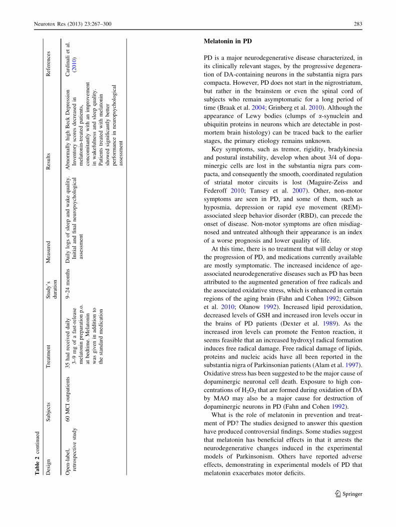
Melatonin in PD
PD is a major neurodegenerative disease characterized, in
its clinically relevant stages, by the progressive degenera-
tion of DA-containing neurons in the substantia nigra pars
compacta. However, PD does not start in the nigrostriatum,
but rather in the brainstem or even the spinal cord of
subjects who remain asymptomatic for a long period of
time (Braak et al. 2004; Grinberg et al. 2010). Although the
appearance of Lewy bodies (clumps of a-synuclein and
ubiquitin proteins in neurons which are detectable in post-
mortem brain histology) can be traced back to the earlier
stages, the primary etiology remains unknown.
Key symptoms, such as tremor, rigidity, bradykinesia
and postural instability, develop when about 3/4 of dopa-
minergic cells are lost in the substantia nigra pars com-
pacta, and consequently the smooth, coordinated regulation
of striatal motor circuits is lost (Maguire-Zeiss and
Federoff 2010; Tansey et al. 2007). Other, non-motor
symptoms are seen in PD, and some of them, such as
hyposmia, depression or rapid eye movement (REM)-
associated sleep behavior disorder (RBD), can precede the
onset of disease. Non-motor symptoms are often misdiag-
nosed and untreated although their appearance is an index
of a worse prognosis and lower quality of life.
At this time, there is no treatment that will delay or stop
the progression of PD, and medications currently available
are mostly symptomatic. The increased incidence of age-
associated neurodegenerative diseases such as PD has been
attributed to the augmented generation of free radicals and
the associated oxidative stress, which is enhanced in certain
regions of the aging brain (Fahn and Cohen 1992; Gibson
et al. 2010; Olanow 1992). Increased lipid peroxidation,
decreased levels of GSH and increased iron levels occur in
the brains of PD patients (Dexter et al. 1989). As the
increased iron levels can promote the Fenton reaction, it
seems feasible that an increased hydroxyl radical formation
induces free radical damage. Free radical damage of lipids,
proteins and nucleic acids have all been reported in the
substantia nigra of Parkinsonian patients (Alam et al. 1997).
Oxidative stress has been suggested to be the major cause of
dopaminergic neuronal cell death. Exposure to high con-
centrations of H2O2 that are formed during oxidation of DA
by MAO may also be a major cause for destruction of
dopaminergic neurons in PD (Fahn and Cohen 1992).
What is the role of melatonin in prevention and treat-
ment of PD? The studies designed to answer this question
have produced controversial findings. Some studies suggest
that melatonin has beneficial effects in that it arrests the
neurodegenerative changes induced in the experimental
models of Parkinsonism. Others have reported adverse
effects, demonstrating in experimental models of PD that
melatonin exacerbates motor deficits.Ta
ble
2co
nti
nu
ed
Des
ign
Su
bje
cts
Tre
atm
ent
Stu
dy
’s
du
rati
on
Mea
sure
dR
esu
lts
Ref
eren
ces
Op
en-l
abel
,
retr
osp
ecti
ve
stu
dy
60
MC
Io
utp
atie
nts
35
had
rece
ived
dai
ly
3–
9m
go
fa
fast
-rel
ease
mel
ato
nin
pre
par
atio
np
.o.
atb
edti
me.
Mel
ato
nin
was
giv
enin
add
itio
nto
the
stan
dar
dm
edic
atio
n
9–
24
mo
nth
sD
aily
log
so
fsl
eep
and
wak
eq
ual
ity
.
Init
ial
and
fin
aln
euro
psy
cho
log
ical
asse
ssm
ent
Ab
no
rmal
lyh
igh
Bec
kD
epre
ssio
n
Inv
ento
rysc
ore
sd
ecre
ased
in
mel
ato
nin
-tre
ated
pat
ien
ts,
con
com
itan
tly
wit
han
imp
rov
emen
t
inw
akef
uln
ess
and
slee
pq
ual
ity
.
Pat
ien
tstr
eate
dw
ith
mel
ato
nin
sho
wed
sig
nifi
can
tly
bet
ter
per
form
ance
inn
euro
psy
cho
log
ical
asse
ssm
ent
Car
din
ali
etal
.
(20
10
)
Neurotox Res (2013) 23:267–300 283
123
Acuna-Castroviejo et al. (1997) used the neurotoxin
1-methyl-4-phenyl-1,2,3,6 tetrahydropyridine (MPTP)
model of PD in rats to show that melatonin could counteract
MPTP-induced lipid peroxidation in striatum, hippocampal,
and midbrain regions. Mayo et al. (1998) showed that when
added to incubation medium containing the dopaminergic
neurotoxin 6-hydroxydopamine (6-OHDA), melatonin sig-
nificantly prevented the increased lipid peroxidation which
normally would have occurred in cultured DA PC 12 cells.
Melatonin also increased the levels of antioxidant enzymes
(Mayo et al. 1998). Additionally, melatonin reduced pyra-
midal cell loss in the hippocampus, a cellular area which
undergoes degeneration in the brains of PD patients, and
which presumably causes memory deficits in patients.
Thomas and Mohanakumar (2004) similarly demonstrated
in vitro and ex vivo models, as well as in an in vivo MPTP
rodent model, that melatonin had potent hydroxyl radical
scavenger activity in the mouse striatum and in isolated
mitochondria. In addition to these primary effects the
investigators also found secondary increases in SOD
activity.
The attenuation of MPTP-induced superoxide formation
indicates an additional neuroprotective mechanism by
melatonin. Intra-median forebrain bundle infusion of a
ferrous-ascorbate-DA hydroxyl radical generating system,
which causes significant depletion of striatal DA, could
be significantly attenuated by melatonin administration
(Borah and Mohanakumar 2009). In another study, Antolin
et al. (2002) used the MPTP model and found that mela-
tonin was effective in preventing neuronal cell death in the
nigrostriatal pathway as indicated by the number of pre-
served DA cells, of tyrosine hydroxylase levels, and other
ultrastructural features. These findings thus demonstrated
that melatonin clearly prevents nigral dopaminergic cell
death induced by chronic treatment with MPTP.
MPTP elicits its neurotoxic effects by increasing the
amount of •NO derived from iNOS. This action mainly
affects DA neurons while •NO derived from neuronal NOS
(nNOS) has a damaging effect on dopaminergic fibers and
terminals in the striatum. A future therapy for PD may
require agents that inhibit the degenerative effects of iNOS
in the substantia nigra pars compacta (Zhang et al. 2000).
Since melatonin can effectively downregulate iNOS and
prevent •NO formation in the brain (Cuzzocrea et al. 1997;
Escames et al. 2004) it should be regarded as a drug of
potential choice for arresting the neuronal degeneration
associated with PD.
MPTP, through its metabolite 1-methyl-4-phenylpyrid-
inium, causes direct inhibition of Complex I of the mito-
chondrial ETC. Such an inhibition of Complex I has been
reported in the substantia nigra of patients suffering from
PD. By increasing Complexes I and IV activities of the
mitochondrial ETC, melatonin may exert a beneficial effect
on MPTP-induced lesions (Acuna-Castroviejo et al. 2011).
Melatonin also stimulates the gene expression of three
antioxidant enzymes Cu/Zn-SOD, Mn-SOD, and GPx in
cultured dopaminergic cells (Mayo et al. 1998).
It has been said that an abnormal assembly of the
cytoskeleton is involved in the pathogenesis of neurode-
generative diseases. Lewy bodies, which are considered to
be cytopathologic markers of Parkinsonism, comprise
abnormal arrangements of tubulin, ubiquitin, microtubule-
associated protein (MAP) 1 and MAP 2 (Vekrellis et al.
2011). Melatonin is very effective in promoting cytoskel-
etal rearrangements and thus may have a potential thera-
peutic value in the treatment of neurodegenerative diseases
including Parkinsonism (Benıtez-King et al. 2004).
However, other studies do not support the hypothesis
that melatonin is of therapeutic benefit in Parkinsonism.
For instance, reduction of melatonin by pinealectomy, or
by exposure of rats to bright light, have been found to
enhance recovery from experimental Parkinsonism in rats,
i.e., spontaneous remission of symptoms following
6-OHDA or MPTP have been observed, whereas melatonin
administration aggravated them (Willis and Armstrong
1999). The mechanism by which melatonin exacerbated
motor impairments appeared to be similar to that seen
caused by excess DA production and release, processes
which occur in degenerating indoleamine and catechol-
amine containing neurons in experimental PD. Tapias et al.
(2010), using a rotenone model of PD in rats, found that
melatonin administration led to striatal catecholamine
depletion, striatal terminal loss, and nigral DA cell loss,
thus aggravating the disease.
Based on this, Willis and Armstrong (1999) suggested
that a possible treatment for PD could involve the reduction
of melatonin production or the pharmacological blockade
of melatonin action on its receptors. Indeed, reduction of
rigidity and bradykinesia through inhibition of melatonin
synthesis (through the strategic application of bright light)
has been found in pilot studies to reduce the incidence
of insomnia as well as fragmented sleep (Willis and
Armstrong 1999). These observations thus contradict
published reports of melatonin’s beneficial effects on Par-
kinsonian symptoms, and the use of melatonin as an
adjunct therapy to either halt progressive degeneration or
for providing symptomatic relief in PD patients has been
questioned (Willis and Robertson 2004). In a study in rats
using the 6-OHDA model of Parkinsonism, the adminis-
tration of the melatonin receptor antagonist ML-23 totally
abolished 6-OHDA-induced mortality, thus suggesting that
blockade of melatonin’s functions or decreasing its bio-
availability may offer a novel approach for the treatment of
Parkinsonism (Willis and Robertson 2004).
A possible hampering role of melatonin in PD is also
suggested by epidemiological studies. Because of the lower
284 Neurotox Res (2013) 23:267–300
123

rates of cancer mortality/incidence in patients with PD,
speculations about risk or preventive factors common to
both diseases, including life-style factors (such as smoking)
and genetic susceptibility have been entertained (Rod et al.
2010). Longer intervals of working night shifts is associ-
ated with reduced melatonin levels and reduced risk of PD
among whereas longer hours of sleep appear to increase
their risk (Schernhammer et al. 2006). While lower mela-
tonin concentrations may predict a higher cancer risk
(Stevens et al. 2011), there is also some evidence that they
may be associated with a lower risk of PD.
The study of melatonin secretion in PD has revealed
some interesting findings. A phase advance in nocturnal
melatonin levels in levodopa-treated Parkinsonian patients
was noted but this was not observed in untreated patients
when compared to control subjects (Fertl et al. 1993).
Similarly a phase advance of about 2 h in plasma melato-
nin secretion was seen in PD patients receiving dopami-
nergic treatment when compared to untreated patients
(Bordet et al. 2003). An increase in daytime melatonin
secretion was also noted in levodopa-treated patients and
was suggested to be an adaptive response to neurodegen-
eration that could play a neuroprotective role through an
antioxidant effect (Bordet et al. 2003).
The occurrence of motor fluctuations in PD was related
to fluctuations in serum melatonin levels, a finding that was
attributed to interactions of monoamines with melatonin in
the striatal complex (Escames et al. 1996). Melatonin may
exert direct motor effects through its interactions with DA
and serotonin. Changes in levodopa-related motor com-
plications may be related to changes in melatonin secretion
pattern. Levodopa-related motor complications occur in
nearly half of the patients with PD on completion of first
5 years of treatment (Koller 1996).
The hypothesis that melatonin has an inhibitory motor
effect which is probably involved in wearing-off episode
(i.e., the progressively shorter intervals during which
symptoms remain adequately controlled as if the effects of
medication would start to ‘‘wear off’’) has been supported
by some therapeutic studies. Stimulation of globus pallidus
inhibited an increase in daytime plasma melatonin levels
seen in PD patients as compared to healthy subjects (Catala
et al. 1997) and was also reported to improve motor
symptoms and complications in patients with PD (Olanow
et al. 2000).
The finding that a reduced expression of melatonin MT1
and MT2 receptors occurs in amygdala and substantia nigra
in patients with PD (Adi et al. 2010) suggests the
involvement of the melatonergic system in the abnormal
sleep mechanisms seen in PD. Indeed, melatonin has been
used for treating sleep problems, insomnia, and daytime
sleepiness in PD patients. The published data are summa-
rized in Table 3.
In a study undertaken on 40 patients (11 women, 29
men; range 43–76 years) melatonin was administered for a
treatment period of 2 weeks, in doses ranging from 5 mg to
50 mg/day (Dowling et al. 2005). To avoid the possibility
of producing a circadian shift melatonin was administered
30 min before bedtime (circadian shifts can occur if
administered melatonin is administered at any other time).
All subjects were taking stable doses of antiparkinsonian
medications during the course of the study. Relative to
placebo, treatment with 50 mg of melatonin significantly
increased night time sleep, as revealed by actigraphy. As
compared to 50 mg or placebo, administration of 5 mg of
melatonin was associated with significant improvement of
sleep in the subjective reports. The study also found that
the high dose of melatonin (50 mg) was well tolerated
(Dowling et al. 2005).
In another study 18 PD patients were randomized after
performing a basal polysomnography to receive melatonin
(3 mg) or placebo 1 h before bedtime for 4 weeks
(Medeiros et al. 2007). Subjective sleep quality was
assessed by the Pittsburgh Sleep Quality Index and daytime
somnolence by the Epworth Sleepiness Scale. All measures
were repeated at the end of treatment. On initial assess-
ment, 14 patients (70 %) showed poor quality sleep and
eight (40 %) excessive diurnal somnolence. Increased
sleep latency (50 %), REM sleep without atonia (66 %),
and reduced sleep efficiency (72 %) were found in poly-
somnography (PSG). Sleep fragmentation tended to be
more severe in patients on lower doses of levodopa
although melatonin significantly improved subjective
quality of sleep. The objective abnormalities remained
unchanged. Motor dysfunction was not improved by the
use of melatonin (Medeiros et al. 2007).
Administration of melatonin 3–12 mg at bedtime has
been shown to be effective in the treatment of RBD
(Table 3). This benefit has been reported in one case report
(Kunz and Bes 1997), two open-label prospective case
series of patients with RBD (Kunz and Bes 1999; Takeuchi
et al. 2001), and two retrospective case series (Anderson
and Shneerson 2009; Boeve et al. 2003).Taken together,
these reports include a total of 38 patients. Thirty-one were
noted to experience improvement with melatonin, two
more experienced transient improvements and one seemed
to worsen. Follow-up as far as 25 months was reported.
PSG showed statistically significant decreases in number of
R epochs without atonia and in movement time in R. This
contrasted with the persistence of tonic muscle tone in R
sleep seen with patients treated with clonazepam. Because
of these data a recent clinical consensus recommended
melatonin use in RBD at Level B, i.e., ‘‘assessment sup-
ported by sparse high grade data or a substantial amount of
low-grade data and/or clinical consensus by the task force’’
(Aurora et al. 2010).
Neurotox Res (2013) 23:267–300 285
123
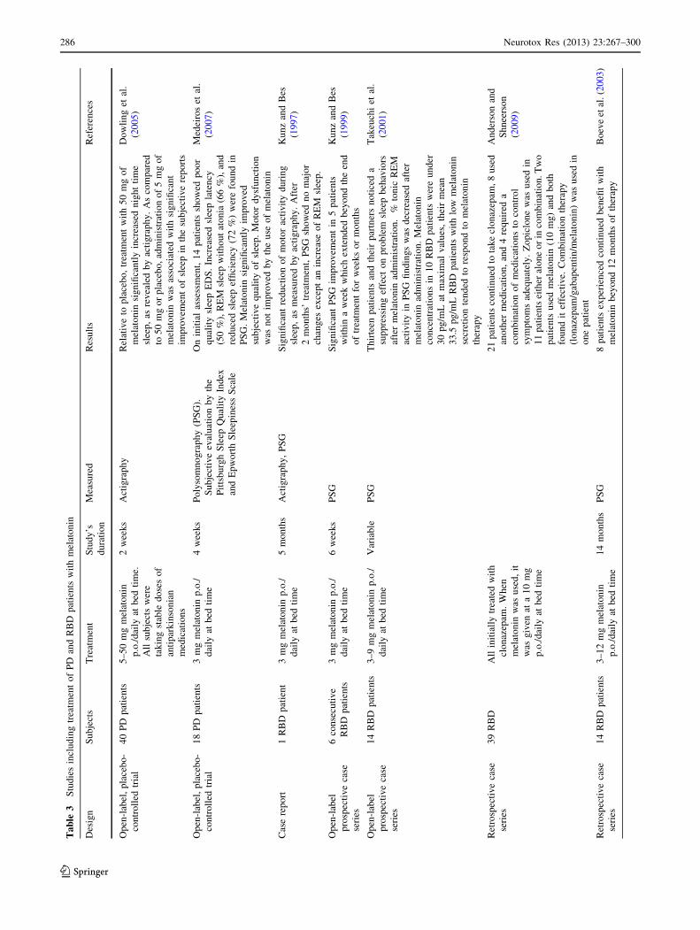
Ta
ble
3S
tud
ies
incl
ud
ing
trea
tmen
to
fP
Dan
dR
BD
pat
ien
tsw
ith
mel
ato
nin
Des
ign
Su
bje
cts
Tre
atm
ent
Stu
dy
’s
du
rati
on
Mea
sure
dR
esu
lts
Ref
eren
ces
Op
en-l
abel
,p
lace
bo
-
con
tro
lled
tria
l
40
PD
pat
ien
ts5
–5
0m
gm
elat
on
in
p.o
./d
aily
atb
edti
me.
All
sub
ject
sw
ere
tak
ing
stab
led
ose
so
f
anti
par
kin
son
ian
med
icat
ion
s
2w
eek
sA
ctig
rap
hy
Rel
ativ
eto
pla
ceb
o,
trea
tmen
tw
ith
50
mg
of
mel
ato
nin
sig
nifi
can
tly
incr
ease
dn
igh
tti
me
slee
p,
asre
vea
led
by
acti
gra
ph
y.
As
com
par
ed
to5
0m
go
rp
lace
bo
,ad
min
istr
atio
no
f5
mg
of
mel
ato
nin
was
asso
ciat
edw
ith
sig
nifi
can
t
imp
rov
emen
to
fsl
eep
inth
esu
bje
ctiv
ere
po
rts
Do
wli
ng
etal
.
(20
05
)
Op
en-l
abel
,p
lace
bo
-
con
tro
lled
tria
l
18
PD
pat
ien
ts3
mg
mel
ato
nin
p.o
./
dai
lyat
bed
tim
e
4w
eek
sP
oly
som
no
gra
ph
y(P
SG
).
Su
bje
ctiv
eev
alu
atio
nb
yth
e
Pit
tsb
urg
hS
leep
Qu
alit
yIn
dex
and
Ep
wo
rth
Sle
epin
ess
Sca
le
On
init
ial
asse
ssm
ent,
14
pat
ien
tssh
ow
edp
oo
r
qu
alit
ysl
eep
ED
S.
Incr
ease
dsl
eep
late
ncy
(50
%),
RE
Msl
eep
wit
ho
ut
ato
nia
(66
%),
and
red
uce
dsl
eep
effi
cien
cy(7
2%
)w
ere
fou
nd
in
PS
G.
Mel
ato
nin
sig
nifi
can
tly
imp
rov
ed
sub
ject
ive
qu
alit
yo
fsl
eep
.M
oto
rd
ysf
un
ctio
n
was
no
tim
pro
ved
by
the
use
of
mel
ato
nin
Med
eiro
set
al.
(20
07
)
Cas
ere
po
rt1
RB
Dp
atie
nt
3m
gm
elat
on
inp
.o./
dai
lyat
bed
tim
e
5m
on
ths
Act
igra
ph
y,
PS
GS
ign
ifica
nt
red
uct
ion
of
mo
tor
acti
vit
yd
uri
ng
slee
p,
asm
easu
red
by
acti
gra
ph
y.
Aft
er
2m
on
ths’
trea
tmen
t,P
SG
sho
wed
no
maj
or
chan
ges
exce
pt
anin
crea
seo
fR
EM
slee
p.
Ku
nz
and
Bes
(19
97
)
Op
en-l
abel
pro
spec
tiv
eca
se
seri
es
6co
nse
cuti
ve
RB
Dp
atie
nts
3m
gm
elat
on
inp
.o./
dai
lyat
bed
tim
e
6w
eek
sP
SG
Sig
nifi
can
tP
SG
imp
rov
emen
tin
5p
atie
nts
wit
hin
aw
eek
wh
ich
exte
nd
edb
eyo
nd
the
end
of
trea
tmen
tfo
rw
eek
so
rm
on
ths
Ku
nz
and
Bes
(19
99
)
Op
en-l
abel
pro
spec
tiv
eca
se
seri
es
14
RB
Dp
atie
nts
3–
9m
gm
elat
on
inp
.o./
dai
lyat
bed
tim
e
Var
iab
leP
SG
Th
irte
enp
atie
nts
and
thei
rp
artn
ers
no
tice
da
sup
pre
ssin
gef
fect
on
pro
ble
msl
eep
beh
avio
rs
afte
rm
elat
on
inad
min
istr
atio
n.
%to
nic
RE
M
acti
vit
yin
PS
Gfi
nd
ing
sw
asd
ecre
ased
afte
r
mel
ato
nin
adm
inis
trat
ion
.M
elat
on
in
con
cen
trat
ion
sin
10
RB
Dp
atie
nts
wer
eu
nd
er
30
pg
/mL
atm
axim
alv
alu
es,
thei
rm
ean
33
.5p
g/m
LR
BD
pat
ien
tsw
ith
low
mel
ato
nin
secr
etio
nte
nd
edto
resp
on
dto
mel
ato
nin
ther
apy
Tak
euch
iet
al.
(20
01
)
Ret
rosp
ecti
ve
case
seri
es
39
RB
DA
llin
itia
lly
trea
ted
wit
h
clo
naz
epam
.W
hen
mel
ato
nin
was
use
d,
it
was
giv
enat
a1
0m
g
p.o
./d
aily
atb
edti
me
21
pat
ien
tsco
nti
nu
edto
tak
ecl
on
azep
am,
8u
sed
ano
ther
med
icat
ion
,an
d4
req
uir
eda
com
bin
atio
no
fm
edic
atio
ns
toco
ntr
ol
sym
pto
ms
adeq
uat
ely
.Z
op
iclo
ne
was
use
din
11
pat
ien
tsei
ther
alo
ne
or
inco
mb
inat
ion
.T
wo
pat
ien
tsu
sed
mel
ato
nin
(10
mg
)an
db
oth
fou
nd
itef
fect
ive.
Co
mb
inat
ion
ther
apy
(lo
naz
epam
/gab
apen
tin
/mel
ato
nin
)w
asu
sed
in
on
ep
atie
nt
An
der
son
and
Sh
nee
rso
n
(20
09
)
Ret
rosp
ecti
ve
case
seri
es
14
RB
Dp
atie
nts
3–
12
mg
mel
ato
nin
p.o
./d
aily
atb
edti
me
14
mo
nth
sP
SG
8p
atie
nts
exp
erie
nce
dco
nti
nu
edb
enefi
tw
ith
mel
ato
nin
bey
on
d1
2m
on
ths
of
ther
apy
Bo
eve
etal
.(2
00
3)
286 Neurotox Res (2013) 23:267–300
123
As mentioned above, not all the clinical studies are in
the vein to support a beneficial role of melatonin in PD.
Exposure to light of 1,000–1,500 lux intensity for 1–1.5 h,
1 h prior to bedtime for 2–5 weeks has been found to
improve the bradykinesia and rigidity observed in 12 PD
patients (Willis and Turner 2007). A reduction in agitation
and psychiatric side effects were also reported in this study.
The authors suggested that activation of the circadian
system by antagonizing melatonin secretion with bright
light has a therapeutic value for treating the symptoms of
PD (Willis 2008).
It must be noted, however, that bright light has been
employed in a number of studies for treating depressive
symptoms (Rosenthal et al. 1984) on the basis that it could
reset the phase of abnormal circadian rhythms seen in
depressed patients (Lewy et al. 1984). Indeed, although
evening bright light exposure produces a momentary sup-
pression of melatonin, it actually causes a rebound increase in
melatonin secretion late in the night (Beck-Friis et al. 1985).
Hence, the bright light exposure may ultimately facilitate
melatonin secretion rather than suppress it. The conclusion
that PD is a ‘‘melatonin hyperplasia’’ disorder (Willis 2008)
awaits further demonstration. Indeed, the decrease in the
amplitude of circulating melatonin rhythm (Bordet et al. 2003)
and the reduced melatonin receptor expression in the sub-
stantia nigra reported in PD patients (Adi et al. 2010) indicate a
weakened melatonin signaling in PD.
Bright light effects may be indicative of circadian
changes in PD. This is supported by the reduced expression
of the circadian clock gene Bmal1 in leukocytes (Cai et al.
2010b), although effects on peripheral oscillators do not
necessarily allow conclusions on changes in the hypotha-
lamic master clock. The recent finding that the mouse
striatal DA receptors D1R and D2R are under circadian
control (Cai et al. 2010a), can be seen as an interesting
facet in this context, although circadian variations in
receptor expression are by no means exceptional features.
Melatonin in HD
Sleep disturbances are very prevalent in HD patients and
can substantially impair their quality of life. They coexist
with abnormal melatonin secretion as shown by examining
the 24 h secretion profiles in early stage HD patients and
normal controls (Aziz et al. 2009). Although mean diurnal
melatonin levels were not different between the two
groups, the timing of the evening rise in melatonin levels
was significantly delayed by about 90 min in HD patients.
Moreover, diurnal melatonin levels strongly correlated
with both motor and functional impairment, indicating that
melatonin levels in HD may progressively decline with
advancing disease (Alders et al. 2009; Aziz et al. 2009).
Mitochondrial dysfunction occurs in HD (Chen 2011).
However, although current evidence from genetic models of
HD indicated the link between mutation of the huntingtin
gene (mHtt) and mitochondrial dysfunction, impairment of
ETC appears to be a late secondary event in disease’s
evolution (Oliveira 2010). Upstream events include defec-
tive mitochondrial calcium handling and impaired ATP
production. Also, transcription abnormalities affecting
mitochondria composition, reduced mitochondria traffick-
ing to synapses, and direct interference with mitochondrial
structures enriched in striatal neurons, are possible mech-
anisms by which mHtt amplifies striatal vulnerability
(Oliveira 2010).
An animal model was developed by using 3-nitroprop-
ionic acid, an inhibitor of mitochondrial Complex II. In this
model, that replicated the neurochemical, histological and
clinical features of HD, melatonin administration was
reported to defer the clinical signs of the disease (Tunez
et al. 2004). The data confirmed an early observation in rat
brain homogenate treated with quinolinic acid (Southgate
and Daya 1999).
Recently, a detailed examination of the neuroprotective
properties of melatonin in the genetic model of HD was
published (Wang et al. 2011). Melatonin delayed disease
onset and prolongs lifespan in the R6/2 transgenic mouse
HD model. In addition, melatonin MT1 receptor levels
decrease in cultured striatal cells, mouse brain, and human
striatum associated with mHtt-mediated toxicity, receptor
depletion becoming greater as the disease progresses.
Further, MT1 receptor knockdown made cells more vul-
nerable to cell death, whereas MT1 receptor overexpression
increases resistance to cell death (Wang et al. 2011). The
administration of melatonin counteracted the MT1 receptor
depletion attributable to mHtt in vitro and in vivo. The
authors concluded that functional MT1 receptor depletion
contributed to cell death in the genetic model of HD (Wang
et al. 2011). Melatonin had little or no inhibitory effect on
huntingtin fibrillogenesis (Heiser et al. 2000).
Melatonin in ALS
ALS is a neurodegenerative disease in which motoneurons
in the anterior horn of the spinal cord and motor neurons of
cerebral cortex gradually degenerate. Dysfunction and
premature death of these neurons causes spasticity,
hyperreflexia, muscular atrophy, and generalized paralysis
(Blackhall 2012). The disorder produces muscle weakness
and muscular atrophy as both the upper and lower moto-
neurons deteriorate. In contrast, mental function remains
normal. Respiratory failure is the common cause of death,
occurring within 2–5 years after diagnosis. Most ALS
cases occur sporadic. Among those individuals with the
Neurotox Res (2013) 23:267–300 287
123

familial form, roughly 20 % have a mutation in the gene
for the antioxidant enzyme SOD (Synofzik et al. 2012).
Different pathological mechanisms have been suggested
to contribute to cell death in ALS, independent of the
underlying molecular/genetic defect. These include
impaired axonal transport, mitochondrial dysfunction,
neurofilament disorganization, protein aggregation, and
impaired proteasome function. Also excitotoxic mecha-
nisms contribute to ALS. Indeed, the only therapeutic drug
with a marginal effect on patient survival is riluzole, an
antiexcitotoxin (Cifra et al. 2012).
Since there is no known cure for ALS a number of
strategies have been tested. Melatonin is one of them.
However, a limited number of studies are available. Given
that oxidative stress has been associated with ALS,
Weishaupt et al. (2006) examined the ability of melatonin to
attenuate neural damage in a genetic mouse model of ALS
(SOD1G93A-transgenic mouse) and in 31 patients with
sporadic ALS. In the mouse model, orally administered
melatonin delayed disease progression and extended sur-
vival. In this report, disease progression was defined as the
time span between the onset of hind limb tremor and pre-
mature death; this was delayed by 25 % in the melatonin-
treated mice. In the ALS patients, melatonin (5 mg/kg) was
applied nightly as a suppository; duration of treatment
varied among the patients and ranged from 2 to 24 months.
Melatonin was well tolerated with some patients reporting
improved sleep quality (Weishaupt et al. 2006). Circulating
protein carbonyls were reduced in the blood after more than
4 months of treatment compared to levels before treatment
onset. Therefore, combining melatonin with the conven-
tional drugs used for treatment, due to their presumed
synergistic actions, may improve the outcome of ALS
patients (Weishaupt et al. 2006). Additional trials with
melatonin, alone and in combination with other drugs, are
needed to clarify the potential benefit of melatonin in sub-
jects with ALS.
Melatonin and Brain Trauma
The neuropathological changes observed during neuronal
injury, trauma, or stroke, have all been ascribed to an
enhancement of oxidative stress which in turn induces lipid
peroxidation, protein and DNA oxidation (Hall et al. 2010;
Hall 2011; Lin and Lee 2009). When administered to rats
kainic acid (KA), an agonist of the ionotropic glutamate
receptor, causes excessive release of glutamate and con-
sequently produces severe brain damage via N-methyl-D-
aspartate (NMDA) receptor activation. The KA model of
excitoxicity is used as a model for studying oxidative stress
during neuronal degeneration induced by brain trauma.
KA ionotropic glutamate receptor activation results in
increased Ca2? influx via NMDA-controlled channels
causing •NO production through activation of nNOS.
Melatonin inhibits either glutamate- or NMDA-induced
excitation (Tan et al. 1998). Melatonin iontophores have
been found to counteract excitation induced by tris(2-car-
boxyethyl)phosphine hydrochloride, showing that the
redox site of the NMDA receptor may be target of mela-
tonin action (Escames et al. 2004). Both in vivo and in vitro
studies have shown that melatonin prevents the neurode-
generation induced by kainate. Melatonin greatly reduces
kainate induced oxidative damage in homogenates of
cerebral cortex, cerebellum, hippocampus, hypothalamus,
and striatum (Melchiorri et al. 1996).
Oxidative apoptotic cell death induced directly by glu-
tamate in cultured hippocampal cells (HT-22) and hippo-
campal brain cells have been found to be reduced after
melatonin administration (Lezoualc’h et al. 1996). MT2
receptors are present in the human hippocampus and pre-
sumably are involved in the mediation of these effects of
melatonin (Savaskan et al. 2005). Bouslama et al. (2007)
reported that melatonin administration could prevent
learning disorders in brain-lesioned newborn mice. Injec-
tions of the glutamate analog ibotenate into the brains of
5-day-old mice were used to produce excitotoxic lesions
resembling those of white-matter lesions that are seen in
hypoxic preterm newborns. Behavioral conditioning and
learning responses were also abolished by the ibotenate
treatment, the administration of melatonin protecting
against the development of these deficits. Melatonin also
reduced ibotenate-induced lesions in white matter as con-
firmed by histological analysis (Bouslama et al. 2007). A
recent study has shown that melatonin treatment reduced
the apoptosis of oligodendrocytes, axon degeneration and
vascular endothelial damage in the white matter of neonatal
rats following a hypoxic injury (Kaur et al. 2010).
The neuroprotective effect of melatonin in cerebral
edema has also been investigated. Edema plays a major
contributing role in neurotrauma that follows traumatic
brain injury (Fraser 2011). Trauma causes neuronal death
by abnormal production of ROS such as superoxide radical,
hydroxyl radical, and hydrogen peroxide leading to exces-
sive lipid peroxidation. Pinealectomy or melatonin defi-
ciency has been found to aggravate brain damage caused by
stroke (Manev et al. 1996) or focal ischemia (Kilic et al.
1999). In a study of vasogenic brain edema caused by cold-
induced lesions in rats, Gorgulu et al. (2001) evaluated the
effect of melatonin in the development of edema and the
destruction of blood brain barrier (BBB) that become
manifest 24 h after injury. Melatonin was found to be
effective in decreasing brain edema and restoring BBB
permeability at the penumbra zone of cold injury, and
in reducing the infarct area (Kabadi and Maher 2010).
Ultrastructural studies showed that melatonin treatment
288 Neurotox Res (2013) 23:267–300
123
decreased nuclear and mitochondrial damage, axonal and
myelin changes. Recently evidence that this effect was
independent on membrane melatonin receptors was pro-
vided in MT1/MT2 knockout mice (Kilic et al. 2011).
The pathophysiology of traumatic cerebral injury and
cerebral edema has been attributed to excessive •NO
release (Wada et al. 1998), a process which can induce the
formation of peroxynitrite anions by reacting with super-
oxide anions. Melatonin prevents the toxic effects of per-
oxynitrite both by inhibiting •NO synthesis as well as by
scavenging peroxynitrite (Cuzzocrea et al. 1997; Lin and
Lee 2009; Mesenge et al. 1998). In a study of children with
epilepsy under carbamazepine therapy, administration of
melatonin (6–9 mg/day for 14 days) increased the activity
of the antioxidant enzyme GPx in plasma, thus suggesting
that melatonin exerts antioxidant activity in these patients
(Gupta et al. 2004).
Besides •NO, vascular endothelial growth factor
(VEGF) has been suggested as a factor underlying dis-
ruption of the BBB and subsequent edema formation (Nag
et al. 2011). Following a hypoxic injury, disruption of the
BBB was evidenced in many parts of the brain by an
increased permeability of the blood vessels which resulted
in swelling of the astrocyte processes in close association
with the blood vessels. This was attributed to increased
VEGF and •NO and production following the hypoxic
injury (Kaur et al. 2006). The swelling of astrocytes or their
processes was reduced significantly following melatonin
administration. The concomitant reduction in VEGF pro-
duction was thought to be responsible for a decrease in
edema formation through decreased vascular permeability
and reduced astrocytic swelling (Kaur et al. 2006).
A number of animal models are being used to investi-
gate the effects of melatonin on brain injury caused by
ischemia followed by reperfusion (Lin and Lee 2009). One
of these models is the Mongolian gerbil which has an
incomplete circle of Willis and in which bilateral ligation
of the carotid arteries totally interrupts the blood supply to
the forebrain. By using this model it was demonstrated that
a 10-min ischemic period caused by occlusion of carotid
arteries and followed by 5 min reperfusion resulted in
significant increases in nitrite/nitrate levels an effect pre-
vented by the administration of melatonin prior to occlu-
sion inhibited •NO production (Guerrero et al. 1997). In
another study melatonin reduced the cortical and striatal
infarct volume by 60 and 30 %, respectively ((Borlongan
et al. 2000). In addition to reducing infarct volume, mel-
atonin also decreased DNA double and single-strand breaks
and enhanced cell viability in the penumbral area of the
infarct (Cheung 2003). In a meta-analysis of 14 studies
involving 432 animals examined in different models of
focal cerebral ischemia, melatonin was found equally
effective in permanent or temporary ischemia, thus
suggesting that melatonin should be considered as a can-
didate neuroprotective drug for the treatment of human
stroke (Macleod et al. 2005). Indeed, a cross-sectional
matched case–control analysis indicated an impaired noc-
turnal melatonin secretion in the acute phase of ischemic
stroke in humans (Atanassova et al. 2009).
Melatonin Versus Melatonin Analogs
As melatonin exhibits both hypnotic and chronobiotic
properties, it has been used for treatment of age-related
insomnia as well as of other primary and secondary
insomnia. A recent consensus of the British Association for
Psychopharmacology on evidence-based treatment of
insomnia, parasomnia and circadian rhythm sleep disorders
concluded that melatonin is the first choice treatment when
a hypnotic is indicated in patients over 55 years (Wilson
et al. 2010). Melatonin has also been successfully used for
treatment of sleep problems related to perturbations of the
circadian time keeping system like those caused by jet-lag,
shift-work disorder, or delayed sleep phase syndrome
(Arendt et al. 1997; Srinivasan et al. 2010).
Since melatonin has a short half-life (less than 30 min)
its efficacy in promoting and maintaining sleep has not
been uniform in the studies undertaken so far. Thus the
need for the development of prolonged release preparations
of melatonin or of melatonin agonists with a longer dura-
tion of action on sleep regulatory structures in the brain
arose (Turek and Gillette 2004). Slow-release forms of
melatonin (e.g., Circadin�, a 2-mg preparation developed
by Neurim, Tel Aviv, Israel, and approved by the European
Medicines Agency in 2007) and the melatonin analogs
ramelteon, agomelatine, tasimelteon and TK-301 are
examples of this strategy.
Ramelteon (Rozerem�, Takeda Pharmaceuticals, Japan)
is a melatonergic hypnotic analog approved by the FDA for
treatment of insomnia in 2005. It is a selective agonist for
MT1/MT2 receptors without significant affinity for other
receptor sites (Miyamoto 2009). In vitro binding studies
have shown that ramelteon affinity for MT1 and MT2
receptors is 3–16 times higher than that of melatonin.
Agomelatine (Valdoxan�, Servier, France) is a recently
introduced melatonergic antidepressant, acts on both MT1
and MT2 melatonergic receptors with a similar affinity to
that of melatonin (IC50 1.3 9 10-10 and 4.7 9 10-10 M,
respectively) and also acts as an antagonist to 5-HT2C
receptors at three orders of magnitude greater concentra-
tion (IC50 2.7 9 10-7 M) (Millan et al. 2003). Agomela-
tine has been licensed by EMEA for treatment of major
depressive disorder at doses of 25–50 mg/day.
Tasimelteon (VES-162) is a MT1/MT2 agonist devel-
oped by Vanda Pharmaceuticals that completed phase III
Neurotox Res (2013) 23:267–300 289
123

trial in 2010. In animal studies, tasimelteon exhibited the
circadian phase shifting properties of melatonin. In clinical
studies involving healthy human subjects, tasimelteon was
administered at doses of 10–100 mg/day (Rajaratnam et al.
2009). The FDA granted tasimelteon orphan drug desig-
nation status for blind individuals without light perception
with non-24-h sleep–wake disorder in 2010.
TIK-301 (formerly LY-156,735) has been in a phase II
clinical trial in the USA since 2002. Originally it was
developed by Eli Lilly and Company and called LY-
156,735. In 2007 Tikvah Pharmaceuticals took over the
development and named it TIK-301. It is a chlorinated
derivative of melatonin with MT1/MT2 agonist activity and
5HT2C antagonist activity. TIK-301 pharmacokinetics,
pharmacodynamics and safety have been examined in a
placebo-controlled study using 20–100 mg/day doses in
healthy volunteers (Mulchahey et al. 2004). The FDA
granted TIK-301 orphan drug designation in 2004, to use as
a treatment for circadian rhythm sleep disorder in blind
individuals without light perception and individuals with
tardive dyskinesia.
As shown by the binding affinities, half-life and relative
potencies of the different melatonin agonists it is clear that
studies using 2–5 mg melatonin/day are probably unsuit-
able to give appropriate comparison with the effect of
ramelteon, agomelatine, tasimelteon or TIK-301, which in
addition to being generally more potent than the native
molecule are employed in considerably higher amounts
(Cardinali et al. 2012).
Several other compounds are being investigated with
relatively more selective activity on melatonin receptor
subtypes, therefore heralding an interesting future era for
melatonin agonist research (Hardeland 2010a; Spadoni
et al. 2011).
Conclusions
Oxidative stress has been implicated in the pathogenesis of
a number of neurodegenerative diseases such as AD, PD,
HD, and ALS, and of pathologic neurological conditions
such as stroke and brain trauma. A high oxygen con-
sumption rate coupled with a low anti-antioxidant potential
of the brain are the main causes for enhanced oxidative
stress of the brain in aged individuals.
Melatonin is an effective antioxidant both in vitro and in
vivo in experimental models of neurodegenerative dis-
eases. Melatonin has been shown to arrest neuropatho-
logical changes not only by scavenging free radicals but
also by increasing the antioxidant enzymes and by atten-
uating free radical formation by neuronal, astrocyte, and
microglial cells. At both physiological and pharmacologi-
cal concentrations, melatonin increases gene expression
and the activities of antioxidant enzymes, e.g. GPx, GR,
and SODs. Melatonin also increases the efficiency of
mitochondrial ETC by enhancing Complex I and IV
activity in brain tissues. Through all of these mechanisms
melatonin prevents nuclear and mitochondrial DNA
cleavage and apoptosis induced by neurotoxic agents such
as Ab, kainate or quinolinic acid. Melatonin also inhibits
NMDA-induced neuronal excitation by inhibiting nNOS
and by redox site modulation.
The efficacy of melatonin in preventing oxidative
damage in either cultured neuronal cells or in the brains of
animals treated with various neurotoxic agents, suggests
that melatonin has a potential therapeutic value as a neu-
roprotective drug in treatment of AD, HD, ALS, and brain
trauma. In the case of other neurological conditions, like
PD, the evidence is less compelling. Inasmuch as melato-
nin deficiency seems to be one of the major causes of the
development of neurodegenerative diseases, intake of
melatonin or its synthetic analogs (selected according to
their chronobiotic and/or antioxidant actions) can be rec-
ommended not only for arresting the progress of this dis-
ease but also for preventing its occurrence as well.
It must be noted that the doses of melatonin employed in
humans may be unnecessarily low when one takes into
consideration the binding affinities, half-life and relative
potencies of the different melatonin agonists on the market.
In addition to being generally more potent than the native
molecule these analogs are employed in considerably
higher amounts (Cardinali et al. 2011). Licensed doses of
ramelteon vary from 8 to 32 mg/day while agomelatine has
been licensed for treatment of major depressive disorder at
doses of 25–50 mg/day. In clinical studies involving
healthy human subjects, tasimelteon was administered at
doses of 10–100 mg/day (Rajaratnam et al. 2009) and TIK-
301 pharmacokinetics, pharmacodynamics and safety have
been examined in a placebo-controlled study using
20–100 mg/day (Mulchahey et al. 2004).
Melatonin has a high safety profile and it is usually
remarkably well tolerated. In some studies melatonin has
been administered at large doses to patients (Chahbouni
et al. 2010; Voordouw et al. 1992; Waldhauser et al. 1984;
Weishaupt et al. 2006). Therefore, further studies
employing melatonin doses in the 100 mg/day are needed
to clarify its potential therapeutical implications in humans.
From animal studies it is clear that a number of preventive
effects of melatonin, like those in neurodegenerative dis-
orders, need high doses of melatonin to become apparent.
If one expects melatonin to be an effective neuroprotector
it is likely that the low doses of melatonin employed so far
(2–5 mg/day) are not very beneficial.
Disclosures S.R. Pandi-Perumal is a stockholder and the President
and Chief Executive Officer of Somnogen Canada Inc., a Canadian
290 Neurotox Res (2013) 23:267–300
123
Corporation. He declares that he has no competing interests that
might be perceived to influence the content of this article. All
remaining authors declare that they have no proprietary, financial,
professional, nor any other personal interest of any nature or kind in
any product or services and/or company that could be construed or
considered to be a potential conflict of interest that might have
influenced the views expressed in this manuscript.
References
Acuna-Castroviejo D, Coto-Montes A, Gaia MM, Ortiz GG, Reiter RJ
(1997) Melatonin is protective against MPTP-induced striatal
and hippocampal lesions. Life Sci 60:L23–L29
Acuna-Castroviejo CD, Escames G, Carazo A, Leon J, Khaldy H,
Reiter RJ (2002) Melatonin, mitochondrial homeostasis and
mitochondrial-related diseases. Curr Top Med Chem 2:133–151
Acuna-Castroviejo CD, Lopez LC, Escames G, Lopez A, Garcıa JA,
Reiter RJ (2011) Melatonin-mitochondria interplay in health and
disease. Curr Top Med Chem 11:221–240
Adi N, Mash DC, Ali Y, Singer C, Shehadeh L, Papapetropoulos S
(2010) Melatonin MT1 and MT2 receptor expression in Parkin-
son’s disease. Med Sci Monit 16:61–67
Alam ZI, Jenner A, Daniel SE, Lees AJ, Cairns N, Marsden CD,
Jenner P, Halliwell B (1997) Oxidative DNA damage in the
parkinsonian brain: an apparent selective increase in 8-hydroxy-
guanine levels in substantia nigra. J Neurochem 69:1196–1203
Alders J, Smits M, Kremer B, Naarding P (2009) The role of
melatonin in sleep disturbances in end-stage Huntington’s
disease. J Neuropsychiatry Clin Neurosci 21:226–227
Aliev G, Li Y, Palacios HH, Obrenovich ME (2011) Oxidative stress
induced mitochondrial DNA deletion as a hallmark for the drug
development in the context of the cerebrovascular diseases.
Recent Pat Cardiovasc Drug Discov 6:222–241
Anderson KN, Shneerson JM (2009) Drug treatment of REM sleep
behavior disorder: the use of drug therapies other than clona-
zepam. J Clin Sleep Med 5:235–239
Anderson KN, Jamieson S, Graham AJ, Shneerson JM (2008) REM
sleep behaviour disorder treated with melatonin in a patient with
Alzheimer’s disease. Clin Neurol Neurosurg 110:492–495
Andrabi SA, Sayeed I, Siemen D, Wolf G, Horn TF (2004) Direct
inhibition of the mitochondrial permeability transition pore: a
possible mechanism responsible for anti-apoptotic effects of
melatonin. FASEB J 18:869–871
Antolin I, Mayo JC, Sainz RM, del Brio ML, Herrera F, Martin V,
Rodrıguez C (2002) Protective effect of melatonin in a chronic
experimental model of Parkinson’s disease. Brain Res
943:163–173
Arendt J, Skene DJ, Middleton B, Lockley SW, Deacon S (1997)
Efficacy of melatonin treatment in jet lag, shift work, and
blindness. J Biol Rhythms 12:604–617
Asayama K, Yamadera H, Ito T, Suzuki H, Kudo Y, Endo S (2003)
Double blind study of melatonin effects on the sleep-wake
rhythm, cognitive and non-cognitive functions in Alzheimer type
dementia. J Nippon Med Sch 70:334–341
Atanassova PA, Terzieva DD, Dimitrov BD (2009) Impaired
nocturnal melatonin in acute phase of ischaemic stroke: cross-
sectional matched case-control analysis. J Neuroendocrinol
21:657–663
Aurora RN, Zak RS, Maganti RK, Auerbach SH, Casey KR,
Chowdhuri S, Karippot A, Ramar K, Kristo DA, Morgenthaler
TI (2010) Best practice guide for the treatment of REM sleep
behavior disorder (RBD). J Clin Sleep Med 6:85–95
Aziz NA, Pijl H, Frolich M, Schroder-van der Elst JP, van der BC,
Roelfsema F, Roos RA (2009) Delayed onset of the diurnal
melatonin rise in patients with Huntington’s disease. J Neurol
256:1961–1965
Balzer I, Hardeland R (1991) Photoperiodism and effects of
indoleamines in a unicellular alga, Gonyaulax polyedra. Science
253:795–797
Barzilai N, Gabriely I, Atzmon G, Suh Y, Rothenberg D, Bergman A
(2010) Genetic studies reveal the role of the endocrine and
metabolic systems in aging. J Clin Endocrinol Metab 95:
4493–4500
Beatty S, Koh H, Phil M, Henson D, Boulton M (2000) The role of
oxidative stress in the pathogenesis of age-related macular
degeneration. Surv Ophthalmol 45:115–134
Beck-Friis J, Kjellman BF, Aperia B, Unden F, von Rosen D,
Ljunggren JG, Wetterberg L (1985) Serum melatonin in relation
to clinical variables in patients with major depressive disorder
and a hypothesis of a low melatonin syndrome. Acta Psychiatr
Scand 71:319–330
Bedrosian TA, Herring KL, Weil ZM, Nelson RJ (2011) Altered
temporal patterns of anxiety in aged and amyloid precursor
protein (APP) transgenic mice. Proc Natl Acad Sci USA
108:11686–11691
Behrends A, Hardeland R, Ness H, Grube S, Poeggeler B, Haldar C
(2004) Photocatalytic actions of the pesticide metabolite 2-hy-
droxyquinoxaline: destruction of antioxidant vitamins and bio-
genic amines—implications of organic redox cycling. Redox
Rep 9:279–288
Beni SM, Kohen R, Reiter RJ, Tan DX, Shohami E (2004) Melatonin-
induced neuroprotection after closed head injury is associated
with increased brain antioxidants and attenuated late-phase
activation of NF-kappaB and AP-1. FASEB J 18:149–151
Benıtez-King G, Rios A, Martinez A, Anton-Tay F (1996) In vitro
inhibition of Ca2?/calmodulin-dependent kinase II activity by
melatonin. Biochim Biophys Acta 1290:191–196
Benıtez-King G, Tunez I, Bellon A, Ortiz GG, Anton-Tay F (2003)
Melatonin prevents cytoskeletal alterations and oxidative stress
induced by okadaic acid in N1E-115 cells. Exp Neurol
182:151–159
Benıtez-King G, Ramirez-Rodrıguez G, Ortiz L, Meza I (2004) The
neuronal cytoskeleton as a potential therapeutical target in
neurodegenerative diseases and schizophrenia. Curr Drug Tar-
gets CNS Neurol Disord 3:515–533
Bergiannaki JD, Soldatos CR, Paparrigopoulos TJ, Syrengelas M,
Stefanis CN (1995) Low and high melatonin excretors among
healthy individuals. J Pineal Res 18:159–164
Blackhall LJ (2012) Amyotrophic lateral sclerosis and palliative care:
where we are, and the road ahead. Muscle Nerve 45:311–318
Blask DE, Hill SM, Dauchy RT, Xiang S, Yuan L, Duplessis T, Mao
L, Dauchy E, Sauer LA (2011) Circadian regulation of
molecular, dietary, and metabolic signaling mechanisms of
human breast cancer growth by the nocturnal melatonin signal
and the consequences of its disruption by light at night. J Pineal
Res 51:259–269
Boeve BF, Silber MH, Ferman TJ (2003) Melatonin for treatment of
REM sleep behavior disorder in neurologic disorders: results in
14 patients. Sleep Med 4:281–284
Borah A, Mohanakumar KP (2009) Melatonin inhibits 6-hydroxy-
dopamine production in the brain to protect against experimental
parkinsonism in rodents. J Pineal Res 47:293–300
Bordet R, Devos D, Brique S, Touitou Y, Guieu JD, Libersa C, Destee
A (2003) Study of circadian melatonin secretion pattern at
different stages of Parkinson’s disease. Clin Neuropharmacol
26:65–72
Borlongan CV, Yamamoto M, Takei N, Kumazaki M, Ungsuparkorn
C, Hida H, Sanberg PR, Nishino H (2000) Glial cell survival is
enhanced during melatonin-induced neuroprotection against
cerebral ischemia. FASEB J 14:1307–1317
Neurotox Res (2013) 23:267–300 291
123
Bouslama M, Renaud J, Olivier P, Fontaine RH, Matrot B, Gressens
P, Gallego J (2007) Melatonin prevents learning disorders in
brain-lesioned newborn mice. Neuroscience 150:712–719
Boveris DL, Boveris A (2007) Oxygen delivery to the tissues and
mitochondrial respiration. Front Biosci 12:1014–1023
Braak H, Ghebremedhin E, Rub U, Bratzke H, Del Tredici K (2004)
Stages in the development of Parkinson’s disease-related
pathology. Cell Tissue Res 318:121–134
Brown GM, Young SN, Gauthier S, Tsui H, Grota LJ (1979)
Melatonin in human cerebrospinal fluid in daytime; its origin and
variation with age. Life Sci 25:929–936
Brown GM, Cardinali DP, Pandi-Perumal SR (2010) Melatonin and
mental illness. In: Pandi-Perumal SR, Kramer M (eds) Sleep and
Mental Illness. Cambridge University Press, Cambridge,
pp 119–129
Brusco LI, Marquez M, Cardinali DP (1998) Monozygotic twins with
Alzheimer’s disease treated with melatonin: case report. J Pineal
Res 25:260–263
Brusco LI, Marquez M, Cardinali DP (2000) Melatonin treatment
stabilizes chronobiologic and cognitive symptoms in Alzhei-
mer’s disease. Neuroendocrinol Lett 21:39–42
Bubenik GA (2002) Gastrointestinal melatonin: localization, function,
and clinical relevance. Dig Dis Sci 47:2336–2348
Bubenik GA, Konturek SJ (2011) Melatonin and aging: prospects for
human treatment. J Physiol Pharmacol 62:13–19
Cai Y, Ding H, Li N, Chai Y, Zhang Y, Chan P (2010a) Oscillation
development for neurotransmitter-related genes in the mouse
striatum. NeuroReport 21:79–83
Cai Y, Liu S, Sothern RB, Xu S, Chan P (2010b) Expression of clock
genes Per1 and Bmal1 in total leukocytes in health and
Parkinson’s disease. Eur J Neurol 17:550–554
Cardinali DP (1983) Molecular mechanisms of neuroendocrine
integration in the central nervous system: an approach through
the study of the pineal gland and its innervating sympathetic
pathway. Psychoneuroendocrinology 8:3–30
Cardinali DP, Freire F (1975) Melatonin effects on brain. Interaction
with microtubule protein, inhibition of fast axoplasmic flow and
induction of crystaloid and tubular formations in the hypothal-
amus. Mol Cell Endocrinol 2:317–330
Cardinali DP, Pevet P (1998) Basic aspects of melatonin action. Sleep
Med Rev 2:175–190
Cardinali DP, Scacchi PA (2010) Chronophysiology of melatonin:
therapeutical implications. Open Neuroendocrinol J 3:72–84
Cardinali DP, Brusco LI, Liberczuk C, Furio AM (2002) The use of
melatonin in Alzheimer’s disease. Neuroendocrinol Lett 23(sup-
pl. 1):20–23
Cardinali DP, Esquifino AI, Srinivasan V, Pandi-Perumal SR (2008)
Melatonin and the immune system in aging. NeuroImmuno-
Modulation 15:272–278
Cardinali DP, Furio AM, Brusco LI (2010) Clinical aspects of
melatonin intervention in Alzheimer’s disease progression. Curr
Neuropharmacol 8:218–227
Cardinali DP, Cano P, Jimenez-Ortega V, Esquifino AI (2011)
Melatonin and the metabolic syndrome: physiopathologic and
therapeutical implications. Neuroendocrinology 93:133–142
Cardinali DP, Srinivasan V, Brzezinski A, Brown GM (2012)
Melatonin and its analogs in insomnia and depression. J Pineal
Res 52:365–375
Carneiro RC, Reiter RJ (1998) Melatonin protects against lipid
peroxidation induced by delta-aminolevulinic acid in rat cere-
bellum, cortex and hippocampus. Neuroscience 82:293–299
Carrillo-Vico A, Reiter RJ, Lardone PJ, Herrera JL, Fernandez-
Montesinos R, Guerrero JM, Pozo D (2006) The modulatory role
of melatonin on immune responsiveness. Curr Opin Investig
Drugs 7:423–431
Catala MD, Canete-Nicolas C, Iradi A, Tarazona PJ, Tormos JM,
Pascual-Leone A (1997) Melatonin levels in Parkinson’s disease:
drug therapy versus electrical stimulation of the internal globus
pallidus. Exp Gerontol 32:553–558
Caumo W, Levandovski R, Hidalgo MP (2009) Preoperative anxio-
lytic effect of melatonin and clonidine on postoperative pain and
morphine consumption in patients undergoing abdominal hys-
terectomy: a double-blind, randomized, placebo-controlled
study. J Pain 10:100–108
Cavallo A (1993) Melatonin and human puberty: current perspectives.
J Pineal Res 15:115–121
Chahbouni M, Escames G, Venegas C, Sevilla B, Garcıa JA, Lopez
LC, Munoz-Hoyos A, Molina-Carballo A, Acuna-Castroviejo D
(2010) Melatonin treatment normalizes plasma pro-inflammatory
cytokines and nitrosative/oxidative stress in patients suffering
from Duchenne muscular dystrophy. J Pineal Res 48:282–289
Chance B, Sies H, Boveris A (1979) Hydroperoxide metabolism in
mammalian organs. Physiol Rev 59:527–605
Chen CM (2011) Mitochondrial dysfunction, metabolic deficits, and
increased oxidative stress in Huntington’s disease. Chang Gung
Med J 34:135–152
Chen G, Huo Y, Tan DX, Liang Z, Zhang W, Zhang Y (2003)
Melatonin in Chinese medicinal herbs. Life Sci 73:19–26
Cheung RT (2003) The utility of melatonin in reducing cerebral
damage resulting from ischemia and reperfusion. J Pineal Res
34:153–160
Cho S, Joh TH, Baik HH, Dibinis C, Volpe BT (1997) Melatonin
administration protects CA1 hippocampal neurons after transient
forebrain ischemia in rats. Brain Res 755:335–338
Chuang YC (2010) Mitochondrial dysfunction and oxidative stress in
seizure-induced neuronal cell death. Acta Neurol Taiwan
19:3–15
Chuang JI, Chen TH (2004) Effect of melatonin on temporal changes
of reactive oxygen species and glutathione after MPP? treatment
in human astrocytoma U373MG cells. J Pineal Res 36:117–125
Cifra A, Mazzone GL, Nistri A (2012) Riluzole: What It Does to
Spinal and Brainstem Neurons and How It Does It. Neurosci-
entist. http://dx.doi.org/10.1177/1073858412444932
Claustrat B, Brun J, Chazot G (2005) The basic physiology and
pathophysiology of melatonin. Sleep Med Rev 9:11–24
Cohen-Mansfield J, Garfinkel D, Lipson S (2000) Melatonin for
treatment of sundowning in elderly persons with dementia—a
preliminary study. Arch Gerontol Geriatr 31:65–76
Conti A, Conconi S, Hertens E, Skwarlo-Sonta K, Markowska M,
Maestroni JM (2000) Evidence for melatonin synthesis in mouse
and human bone marrow cells. J Pineal Res 28:193–202
Cunningham C (2011) Systemic inflammation and delirium: impor-
tant co-factors in the progression of dementia. Biochem Soc
Trans 39:945–953
Cuzzocrea S, Zingarelli B, Gilad E, Hake P, Salzman AL, Szabo C
(1997) Protective effect of melatonin in carrageenan-induced
models of local inflammation: relationship to its inhibitory effect
on nitric oxide production and its peroxynitrite scavenging
activity. J Pineal Res 23:106–116
Dabbeni-Sala F, Di Santo S, Franceschini D, Skaper SD, Giusti P
(2001) Melatonin protects against 6-OHDA-induced neurotox-
icity in rats: a role for mitochondrial complex I activity. FASEB
J 15:164–170
Dardente H (2012) Melatonin-dependent timing of seasonal repro-
duction by the pars tuberalis: pivotal roles for long daylengths
and thyroid hormones. J Neuroendocrinol 24:249–266
Das A, McDowell M, Pava MJ, Smith JA, Reiter RJ, Woodward JJ,
Varma AK, Ray SK, Banik NL (2010) The inhibition of
apoptosis by melatonin in VSC4.1 motoneurons exposed to
oxidative stress, glutamate excitotoxicity, or TNF-alpha toxicity
292 Neurotox Res (2013) 23:267–300
123
involves membrane melatonin receptors. J Pineal Res
48:157–169
Dawson D, Armstrong SM (1996) Chronobiotics—drugs that shift
rhythms. Pharmacol Ther 69:15–36
de Jonghe A, Korevaar JC, van Munster BC, de Rooij SE (2010)
Effectiveness of melatonin treatment on circadian rhythm
disturbances in dementia. Are there implications for delirium?
A systematic review. Int J Geriatr Psychiatry 25:1201–1208
Devore EE, Grodstein F, van Rooij FJ, Hofman A, Stampfer MJ,
Witteman JC, Breteler MM (2010) Dietary antioxidants and
long-term risk of dementia. Arch Neurol 67:819–825
Dexter DT, Carter CJ, Wells FR, Javoy-Agid F, Agid Y, Lees A,
Jenner P, Marsden CD (1989) Basal lipid peroxidation in
substantia nigra is increased in Parkinson’s disease. J Neurochem
52:381–389
Domınguez-Rodrıguez A, Abreu-Gonzalez P, Sanchez–Sanchez JJ,
Kaski JC, Reiter RJ (2010) Melatonin and circadian biology in
human cardiovascular disease. J Pineal Res 49:14–22
Dori D, Casale G, Solerte SB, Fioravanti M, Migliorati G, Cuzzoni G,
Ferrari E (1994) Chrono-neuroendocrinological aspects of
physiological aging and senile dementia. Chronobiologia
21:121–126
Dowling GA, Mastick J, Colling E, Carter JH, Singer CM, Aminoff
MJ (2005) Melatonin for sleep disturbances in Parkinson’s
disease. Sleep Med 6:459–466
Dowling GA, Burr RL, Van Someren EJ, Hubbard EM, Luxenberg
JS, Mastick J, Cooper BA (2008) Melatonin and bright-light
treatment for rest-activity disruption in institutionalized patients
with Alzheimer’s disease. J Am Geriatr Soc 56:239–246
Dragicevic N, Copes N, O’Neal-Moffitt G, Jin J, Buzzeo R, Mamcarz
M, Tan J, Cao C, Olcese JM, Arendash GW, Bradshaw PC
(2011) Melatonin treatment restores mitochondrial function in
Alzheimer’s mice: a mitochondrial protective role of melatonin
membrane receptor signaling. J Pineal Res 51:75–86
Dubbels R, Reiter RJ, Klenke E, Goebel A, Schnakenberg E, Ehlers
C, Schiwara HW, Schloot W (1995) Melatonin in edible plants
identified by radioimmunoassay and by high performance liquid
chromatography-mass spectrometry. J Pineal Res 18:28–31
Dubocovich ML, Delagrange P, Krause DN, Sugden D, Cardinali DP,
Olcese J (2010) International Union of Basic and Clinical
Pharmacology. LXXV. Nomenclature, classification, and phar-
macology of G protein-coupled melatonin receptors. Pharmacol
Rev 62:343–380
Ehrnhoefer DE, Wong BK, Hayden MR (2011) Convergent patho-
genic pathways in Alzheimer’s and Huntington’s diseases:
shared targets for drug development. Nat Rev Drug Discov
10:853–867
Erol FS, Topsakal C, Ozveren MF, Kaplan M, Ilhan N, Ozercan IH,
Yildiz OG (2004) Protective effects of melatonin and vitamin E
in brain damage due to gamma radiation: an experimental study.
Neurosurg Rev 27:65–69
Escames G, Acuna-Castroviejo CD, Vives F (1996) Melatonin–
dopamine interaction in the striatal projection area of sensori-
motor cortex in the rat. NeuroReport 7:597–600
Escames G, Leon J, Lopez LC, Acuna-Castroviejo D (2004)
Mechanisms of N-methyl-D-aspartate receptor inhibition by
melatonin in the rat striatum. J Neuroendocrinol 16:929–935
Esposito E, Cuzzocrea S (2010) Antiinflammatory activity of
melatonin in central nervous system. Curr Neuropharmacol
8:228–242
Esquifino AI, Pandi-Perumal SR, Cardinali DP (2004) Circadian
organization of the immune response: a role for melatonin. Clin
Applied Immunol Rev 4:423–433
Fahn S, Cohen G (1992) The oxidant stress hypothesis in Parkinson’s
disease: evidence supporting it. Ann Neurol 32:804–812
Fainstein I, Bonetto A, Brusco LI, Cardinali DP (1997) Effects of
melatonin in elderly patients with sleep disturbance. A pilot
study. Curr Ther Res 58:990–1000
Feng Z, Zhang JT (2004) Melatonin reduces amyloid beta-induced
apoptosis in pheochromocytoma (PC12) cells. J Pineal Res
37:257–266
Feng Z, Qin C, Chang Y, Zhang JT (2006) Early melatonin
supplementation alleviates oxidative stress in a transgenic mouse
model of Alzheimer’s disease. Free Radic Biol Med 40:101–109
Ferrari E, Fioravanti M, Magri F, Solerte SB (2000) Variability of
interactions between neuroendocrine and immunological func-
tions in physiological aging and dementia of the Alzheimer’s
type. Ann N Y Acad Sci 917:582–596
Ferrari E, Cravello L, Falvo F, Barili L, Solerte SB, Fioravanti M,
Magri F (2008) Neuroendocrine features in extreme longevity.
Exp Gerontol 43:88–94
Fertl E, Auff E, Doppelbauer A, Waldhauser F (1993) Circadian
secretion pattern of melatonin in de novo parkinsonian patients:
evidence for phase-shifting properties of L-dopa. J Neural
Transm Park Dis Dement Sect 5:227–234
Floden AM, Li S, Combs CK (2005) Beta-amyloid-stimulated
microglia induce neuron death via synergistic stimulation of
tumor necrosis factor alpha and NMDA receptors. J Neurosci
25:2566–2575
Fourtillan JB, Brisson AM, Fourtillan M, Ingrand I, Decourt JP,
Girault J (2001) Melatonin secretion occurs at a constant rate in
both young and older men and women. Am J Physiol Endocrinol
Metab 280:E11–E22
Fraser PA (2011) The role of free radical generation in increasing
cerebrovascular permeability. Free Radic Biol Med 51:967–977
Furio AM, Cutrera RA, Castillo TV, Perez LS, Riccio P, Caccuri RL,
Brusco LL, Cardinali DP (2002) Effect of melatonin on changes
in locomotor activity rhythm of Syrian hamsters injected with
beta amyloid peptide 25–35 in the suprachiasmatic nuclei. Cell
Mol Neurobiol 22:699–709
Furio AM, Brusco LI, Cardinali DP (2007) Possible therapeutic value
of melatonin in mild cognitive impairment: a retrospective study.
J Pineal Res 43:404–409
Furio AM, Fontao R, Falco N, Ruiz JI, Caccuri RL, Cardinali DP
(2008) Neuroprotective effect of melatonin on glucocorticoid
toxicity in the rat hippocampus. Open Physiol J 1:23–27
Galano A, Tan DX, Reiter RJ (2011) Melatonin as a natural ally
against oxidative stress: a physicochemical examination. J Pineal
Res 51:1–16
Galecki P, Szemraj J, Bartosz G, Bienkiewicz M, Galecka E,
Florkowski A, Lewinski A, Karbownik-Lewinska M (2010)
Single-nucleotide polymorphisms and mRNA expression for
melatonin synthesis rate-limiting enzyme in recurrent depressive
disorder. J Pineal Res 48:311–317
Garcıa JJ, Reiter RJ, Pie J, Ortiz GG, Cabrera J, Sainz RM, Acuna-
Castroviejo D (1999) Role of pinoline and melatonin in
stabilizing hepatic microsomal membranes against oxidative
stress. J Bioenerg Biomembr 31:609–616
Garcıa T, Ribes D, Colomina MT, Cabre M, Domingo JL, Gomez M
(2009) Evaluation of the protective role of melatonin on the
behavioral effects of aluminum in a mouse model of Alzheimer’s
disease. Toxicology 265:49–55
Garcıa T, Esparza JL, Nogues MR, Romeu M, Domingo JL, Gomez
M (2010) Oxidative stress status and RNA expression in
hippocampus of an animal model of Alzheimer’s disease after
chronic exposure to aluminum. Hippocampus 20:218–225
Garcıa-Mesa Y, Gimenez-Llort L, Lopez LC, Venegas C, Cristofol R,
Escames G, Acuna-Castroviejo D, Sanfeliu C (2012) Melatonin
plus physical exercise are highly neuroprotective in the 3xTg-
AD mouse. Neurobiol Aging 33:1124–1129
Neurotox Res (2013) 23:267–300 293
123

Garzon C, Guerrero JM, Aramburu O, Guzman T (2009) Effect of
melatonin administration on sleep, behavioral disorders and
hypnotic drug discontinuation in the elderly: a randomized,
double-blind, placebo-controlled study. Aging Clin Exp Res
21:38–42
Gauthier S, Reisberg B, Zaudig M, Petersen RC, Ritchie K, Broich K,
Belleville S, Brodaty H, Bennett D, Chertkow H, Cummings JL,
de Leon M, Feldman H, Ganguli M, Hampel H, Scheltens P,
Tierney MC, Whitehouse P, Winblad B (2006) Mild cognitive
impairment. Lancet 367:1262–1270
Gehrman PR, Connor DJ, Martin JL, Shochat T, Corey-Bloom J,
Ancoli-Israel S (2009) Melatonin fails to improve sleep or
agitation in double-blind randomized placebo-controlled trial of
institutionalized patients with Alzheimer disease. Am J Geriatr
Psychiatry 17:166–169
Genova ML, Pich MM, Bernacchia A, Bianchi C, Biondi A, Bovina
C, Falasca AI, Formiggini G, Castelli GP, Lenaz G (2004) The
mitochondrial production of reactive oxygen species in relation
to aging and pathology. Ann N Y Acad Sci 1011:86–100
Ghanta S, Chattopadhyay S (2011) Glutathione as a signaling
molecule: another challenge to pathogens. Plant Signal Behav
6:783–788
Gibson GE, Starkov A, Blass JP, Ratan RR, Beal MF (2010) Cause
and consequence: mitochondrial dysfunction initiates and prop-
agates neuronal dysfunction, neuronal death and behavioral
abnormalities in age-associated neurodegenerative diseases.
Biochim Biophys Acta 1802:122–134
Girotti L, Lago M, Ianovsky O, Carbajales J, Elizari MV, Brusco LI,
Cardinali DP (2000) Low urinary 6-sulphatoxymelatonin levels
in patients with coronary artery disease. J Pineal Res 29:138–142
Giusti P, Franceschini D, Petrone M, Manev H, Floreani M (1996a)
In vitro and in vivo protection against kainate-induced excito-
toxicity by melatonin. J Pineal Res 20:226–231
Giusti P, Lipartiti M, Franceschini D, Schiavo N, Floreani M, Manev
H (1996b) Neuroprotection by melatonin from kainate-induced
excitotoxicity in rats. FASEB J 10:891–896
Golombek DA, Pevet P, Cardinali DP (1996) Melatonin effects on
behavior: possible mediation by the central GABAergic system.
Neurosci Biobehav Rev 20:403–412
Gorgulu A, Palaoglu S, Ismailoglu O, Tuncel M, Surucu MT, Erbil M,
Kilinc K (2001) Effect of melatonin on cerebral edema in rats.
Neurosurgery 49:1434–1441
Green DR, Galluzzi L, Kroemer G (2011) Mitochondria and the
autophagy-inflammation-cell death axis in organismal aging.
Science 333:1109–1112
Grinberg LT, Rueb U, Alho AT, Heinsen H (2010) Brainstem
pathology and non-motor symptoms in PD. J Neurol Sci
289:81–88
Grof E, Grof P, Brown GM, Arato M, Lane J (1985) Investigations of
melatonin secretion in man. Prog Neuropsychopharmacol Biol
Psychiatry 9:609–612
Guerrero JM, Reiter RJ (2002) Melatonin-immune system relation-
ships. Curr Top Med Chem 2:167–179
Guerrero JM, Reiter RJ, Ortiz GG, Pablos MI, Sewerynek E, Chuang
JI (1997) Melatonin prevents increases in neural nitric oxide and
cyclic GMP production after transient brain ischemia and
reperfusion in the Mongolian gerbil (Meriones unguiculatus).
J Pineal Res 23:24–31
Guido ME, Garbarino-Pico E, Contin MA, Valdez DJ, Nieto PS,
Verra DM, Acosta-Rodrıguez VA, de Zavalia N, Rosenstein RE
(2010) Inner retinal circadian clocks and non-visual photore-
ceptors: novel players in the circadian system. Prog Neurobiol
92:484–504
Gupta M, Gupta YK, Agarwal S, Aneja S, Kalaivani M, Kohli K
(2004) Effects of add-on melatonin administration on antioxidant
enzymes in children with epilepsy taking carbamazepine
monotherapy: a randomized, double-blind, placebo-controlled
trial. Epilepsia 45:1636–1639
Hall ED (2011) Antioxidant therapies for acute spinal cord injury.
Neurotherapeutics 8:152–167
Hall NF, Gale CR (2002) Prevention of age related macular
degeneration. BMJ 325:1–2
Hall ED, Vaishnav RA, Mustafa AG (2010) Antioxidant therapies for
traumatic brain injury. Neurotherapeutics 7:51–61
Harada S, Fujii C, Hayashi A, Ohkoshi N (2001) An association
between idiopathic Parkinson’s disease and polymorphisms of
phase II detoxification enzymes: glutathione S-transferase M1
and quinone oxidoreductase 1 and 2. Biochem Biophys Res
Commun 288:887–892
Hardeland R (2005) Antioxidative protection by melatonin: multi-
plicity of mechanisms from radical detoxification to radical
avoidance. Endocrine 27:119–130
Hardeland R (2008) Melatonin, hormone of darkness and more—
occurrence, control mechanisms, actions and bioactive metabo-
lites. Cell Mol Life Sci 65:2001–2018
Hardeland R (2009a) Melatonin: signaling mechanisms of a pleio-
tropic agent. BioFactors 35:183–192
Hardeland R (2009b) Neuroprotection by radical avoidance: search
for suitable agents. Molecules 14:5054–5102
Hardeland R (2010a) Investigational melatonin receptor agonists.
Expert Opin Investig Drugs 19:747–764
Hardeland R (2010b) Melatonin metabolism in the central nervous
system. Curr Neuropharmacol 8:168–181
Hardeland R, Pandi-Perumal SR (2005) Melatonin, a potent agent in
antioxidative defense: actions as a natural food constituent,
gastrointestinal factor, drug and prodrug. Nutr Metab (Lond)
2:22
Hardeland R, Poeggeler B (2007) Actions of melatonin, its structural and
functional analogs in the central nervous system and the significance
of metabolism. Cent Nerv Syst Agents Med Chem 7:303
Hardeland R, Poeggeler B (2008) Melatonin beyond its classical
functions. Open Physiol J 1:1–23
Hardeland R, Balzer I, Poeggeler B, Fuhrberg B, Uria H, Behrmann
G, Wolf R, Meyer TJ, Reiter RJ (1995) On the primary functions
of melatonin in evolution: mediation of photoperiodic signals in
a unicell, photooxidation, and scavenging of free radicals.
J Pineal Res 18:104–111
Hardeland R, Cardinali DP, Srinivasan V, Spence DW, Brown GM,
Pandi-Perumal SR (2011) Melatonin-A pleiotropic, orchestrating
regulator molecule. Prog Neurobiol 93:350–384
Hausman DB, Fischer JG, Johnson MA (2011) Nutrition in
centenarians. Maturitas 68:203–209
He H, Dong W, Huang F (2010) Anti-amyloidogenic and anti-
apoptotic role of melatonin in Alzheimer disease. Curr Neuro-
pharmacol 8:211–217
Heiser V, Scherzinger E, Boeddrich A, Nordhoff E, Lurz R,
Schugardt N, Lehrach H, Wanker EE (2000) Inhibition of
huntingtin fibrillogenesis by specific antibodies and small
molecules: implications for Huntington’s disease therapy. Proc
Natl Acad Sci USA 97:6739–6744
Hu K, Van Someren EJ, Shea SA, Scheer FA (2009) Reduction of
scale invariance of activity fluctuations with aging and Alzhei-
mer’s disease: involvement of the circadian pacemaker. Proc
Natl Acad Sci USA 106:2490–2494
Huether G (1993) The contribution of extrapineal sites of melatonin
synthesis to circulating melatonin levels in higher vertebrates.
Experientia 49:665–670
Huether G (1994) Melatonin synthesis in the gastrointestinal tract and
the impact of nutritional factors on circulating melatonin. Ann N
Y Acad Sci 719:146–158
Huether G, Poeggeler B, Reimer A, George A (1992) Effect of
tryptophan administration on circulating melatonin levels in
294 Neurotox Res (2013) 23:267–300
123

chicks and rats: evidence for stimulation of melatonin synthesis
and release in the gastrointestinal tract. Life Sci 51:945–953
Iguchi H, Kato KI, Ibayashi H (1982) Melatonin serum levels and
metabolic clearance rate in patients with liver cirrhosis. J Clin
Endocrinol Metab 54:1025–1027
Jean-Louis G, von Gizycki H, Zizi F (1998a) Melatonin effects on
sleep, mood, and cognition in elderly with mild cognitive
impairment. J Pineal Res 25:177–183
Jean-Louis G, Zizi F, von Gizycki H, Taub H (1998b) Effects of
melatonin in two individuals with Alzheimer’s disease. Percept
Mot Skills 87:331–339
Jiao S, Wu MM, Hu CL, Zhang ZH, Mei YA (2004) Melatonin
receptor agonist 2-iodomelatonin prevents apoptosis of cerebel-
lar granule neurons via K(?) current inhibition. J Pineal Res
36:109–116
Jimenez-Jorge S, Guerrero JM, Jimenez-Caliani AJ, Naranjo MC,
Lardone PJ, Carrillo-Vico A, Osuna C, Molinero P (2007)
Evidence for melatonin synthesis in the rat brain during
development. J Pineal Res 42:240–246
Jimenez-Ortega V, Cano P, Scacchi PA, Cardinali DP, Esquifino AI
(2011) Cadmium-induced disruption in 24-h expression of clock
and redox enzyme genes in rat medial basal hypothalamus.
Prevention by melatonin. Front Neurol 2:13
Jin BK, Shin DY, Jeong MY, Gwag MR, Baik HW, Yoon KS, Cho
YH, Joo WS, Kim YS, Baik HH (1998) Melatonin protects nigral
dopaminergic neurons from 1-methyl-4-phenylpyridinium
(MPP?) neurotoxicity in rats. Neurosci Lett 245:61–64
Jonsson L, Ljunggren E, Bremer A, Pedersen C, Landen M,
Thuresson K, Giacobini M, Melke J (2010) Mutation screening
of melatonin-related genes in patients with autism spectrum
disorders. BMC Med Genomics 3:10
Jou MJ (2011) Melatonin preserves the transient mitochondrial
permeability transition for protection during mitochondrial Ca2?
stress in astrocyte. J Pineal Res 50:427–435
Jucker M, Walker LC (2011) Pathogenic protein seeding in Alzhei-
mer disease and other neurodegenerative disorders. Ann Neurol
70:532–540
Kabadi SV, Maher TJ (2010) Posttreatment with uridine and
melatonin following traumatic brain injury reduces edema in
various brain regions in rats. Ann N Y Acad Sci 1199:105–113
Karasek M, Reiter RJ (2002) Melatonin and aging. Neuroendocrinol
Lett 23(Suppl 1):14–16
Kaur C, Sivakumar V, Zhang Y, Ling EA (2006) Hypoxia-induced
astrocytic reaction and increased vascular permeability in the rat
cerebellum. Glia 54:826–839
Kaur C, Sivakumar V, Ling EA (2007) Expression of transferrin
receptors in the pineal gland of postnatal and adult rats and its
alteration in hypoxia and melatonin treatment. Glia 55:263–273
Kaur C, Sivakumar V, Ling EA (2010) Melatonin protects periven-
tricular white matter from damage due to hypoxia. J Pineal Res
48:185–193
Kaur C, Viswanathan S, Ling EA (2011) Hypoxia-induced cellular
and vascular changes in the nucleus tractus solitarius and
ventrolateral medulla. J Neuropathol Exp Neurol 70:201–217
Khandelwal PJ, Herman AM, Moussa CE (2011) Inflammation in the
early stages of neurodegenerative pathology. J Neuroimmunol
238:1–11
Kilanczyk E, Bryszewska M (2003) The effect of melatonin on
antioxidant enzymes in human diabetic skin fibroblasts. Cell Mol
Biol Lett 8:333–336
Kilic E, Ozdemir YG, Bolay H, Kelestimur H, Dalkara T (1999)
Pinealectomy aggravates and melatonin administration attenu-
ates brain damage in focal ischemia. J Cereb Blood Flow Metab
19:511–516
Kilic U, Yilmaz B, Ugur M, Yuksel A, Reiter RJ, Hermann DM, Kilic
E (2011) Evidence that membrane-bound G protein-coupled
melatonin receptors MT1 and MT2 are not involved in the
neuroprotective effects of melatonin in focal cerebral ischemia.
J Pineal Res 52:228–235
Kitazawa M, Yamasaki TR, Laferla FM (2004) Microglia as a
potential bridge between the amyloid {beta}-peptide and tau.
Ann N Y Acad Sci 1035:85–103
Klein DC (2007) Arylalkylamine N-acetyltransferase: ‘‘the Time-
zyme’’. J Biol Chem 282:4233–4237
Kluck RM, Bossy-Wetzel E, Green DR, Newmeyer DD (1997) The
release of cytochrome c from mitochondria: a primary site for
Bcl-2 regulation of apoptosis. Science 275:1132–1136
Koh PO (2011) Melatonin prevents down-regulation of astrocytic
phosphoprotein PEA-15 in ischemic brain injury. J Pineal Res
51:381–386
Koh PO (2012) Melatonin attenuates decrease of protein phosphatase
2A subunit B in ischemic brain injury. J Pineal Res 52:57–61
Koller WC (1996) Management of motor fluctuations in Parkinson’s
disease. Eur Neurol 36(Suppl 1):43–48
Kunz D, Bes F (1997) Melatonin effects in a patient with severe REM
sleep behavior disorder: case report and theoretical consider-
ations. Neuropsychobiology 36:211–214
Kunz D, Bes F (1999) Melatonin as a therapy in REM sleep behavior
disorder patients: an open-labeled pilot study on the possible
influence of melatonin on REM-sleep regulation. Mov Disord
14:507–511
Kvetnoy IM, Ingel IE, Kvetnaia TV, Malinovskaya NK, Rapoport SI,
Raikhlin NT, Trofimov AV, Yuzhakov VV (2002) Gastrointes-
tinal melatonin: cellular identification and biological role. Neuro
Endocrinol Lett 23:121–132
Lacza Z, Horn TF, Snipes JA, Zhang J, Roychowdhury S, Horvath
EM, Figueroa JP, Kollai M, Szabo C, Busija DW (2004) Lack of
mitochondrial nitric oxide production in the mouse brain.
J Neurochem 90:942–951
Landau M, Zisapel N (2007) The low affinity binding of melatonin to
calmodulin: use of computational methods to explain its
physiological relevance. In: Cardinali DP, Pandi-Perumal SR
(eds) Melatonin - From Molecules to Therapy. Nova Science,
New York, pp 69–79
Lardone PJ, Guerrero JM, Fernandez-Santos JM, Rubio A, Martin-
Lacave I, Carrillo-Vico A (2011) Melatonin synthesized by T
lymphocytes as a ligand of the retinoic acid-related orphan
receptor. J Pineal Res 51:454–462
Lee EJ, Wu TS, Lee MY, Chen TY, Tsai YY, Chuang JI, Chang GL
(2004) Delayed treatment with melatonin enhances electrophys-
iological recovery following transient focal cerebral ischemia in
rats. J Pineal Res 36:33–42
Leon-Blanco MM, Guerrero JM, Reiter RJ, Pozo D (2004) RNA
expression of human telomerase subunits TR and TERT is
differentially affected by melatonin receptor agonists in the
MCF-7 tumor cell line. Cancer Lett 216:73–80
Levoye A, Dam J, Ayoub MA, Guillaume JL, Couturier C,
Delagrange P, Jockers R (2006) The orphan GPR50 receptor
specifically inhibits MT1 melatonin receptor function through
heterodimerization. EMBO J 25:3012–3023
Lewy AJ, Sack RA, Singer CL (1984) Assessment and treatment of
chronobiologic disorders using plasma melatonin levels and
bright light exposure: the clock-gate model and the phase
response curve. Psychopharmacol Bull 20:561–565
Lezoualc’h F, Skutella T, Widmann M, Behl C (1996) Melatonin
prevents oxidative stress-induced cell death in hippocampal
cells. NeuroReport 7:2071–2077
Li XC, Wang ZF, Zhang JX, Wang Q, Wang JZ (2005) Effect of
melatonin on calyculin A-induced tau hyperphosphorylation. Eur
J Pharmacol 510:25–30
Lieverse R, Van Someren EJ, Nielen MM, Uitdehaag BM, Smit JH,
Hoogendijk WJ (2011) Bright light treatment in elderly patients
Neurotox Res (2013) 23:267–300 295
123

with nonseasonal major depressive disorder: a randomized
placebo-controlled trial. Arch Gen Psychiatry 68:61–70
Lin HW, Lee EJ (2009) Effects of melatonin in experimental stroke
models in acute, sub-acute, and chronic stages. Neuropsychiatr
Dis Treat 5:157–162
Ling ZQ, Tian Q, Wang L, Fu ZQ, Wang XC, Wang Q, Wang JZ
(2009) Constant illumination induces Alzheimer-like damages
with endoplasmic reticulum involvement and the protection of
melatonin. J Alzheimers Dis 16:287–300
Liu T, Borjigin J (2005) N-acetyltransferase is not the rate-limiting
enzyme of melatonin synthesis at night. J Pineal Res 39:91–96
Liu RY, Zhou JN, Van Heerikhuize J, Hofman MA, Swaab DF (1999)
Decreased melatonin levels in postmortem cerebrospinal fluid in
relation to aging, Alzheimer’s disease, and apolipoprotein E-e4/4 genotype. J Clin Endocrinol Metab 84:323–327
Louzada PR, Paula Lima AC, Mendonca-Silva DL, Noel F, De Mello
FG, Ferreira ST (2004) Taurine prevents the neurotoxicity of
beta-amyloid and glutamate receptor agonists: activation of
GABA receptors and possible implications for Alzheimer’s
disease and other neurological disorders. FASEB J 18:511–518
Luboshitzky R, Shen-Orr Z, Tzischichinsky O, Maldonado M, Herer
P, Lavie P (2001) Actigraphic sleep–wake patterns and urinary
6-sulfatoxymelatonin excretion in patients with Alzheimer’s
disease. Chronobiol Int 18:513–524
Macias M, Escames G, Leon J, Coto A, Sbihi Y, Osuna A, Acuna-
Castroviejo D (2003) Calreticulin–melatonin. An unexpected
relationship. Eur J Biochem 270:832–840
Macleod MR, O’Collins T, Horky LL, Howells DW, Donnan GA
(2005) Systematic review and meta-analysis of the efficacy of
melatonin in experimental stroke. J Pineal Res 38:35–41
Magri F, Sarra S, Cinchetti W, Guazzoni V, Fioravanti M, Cravello L,
Ferrari E (2004) Qualitative and quantitative changes of
melatonin levels in physiological and pathological aging and in
centenarians. J Pineal Res 36:256–261
Maguire-Zeiss KA, Federoff HJ (2010) Future directions for immune
modulation in neurodegenerative disorders: focus on Parkinson’s
disease. J Neural Transm 117:1019–1025
Mahlberg R, Walther S (2007) Actigraphy in agitated patients with
dementia: monitoring treatment outcomes. Z Gerontol Geriatr
40:178–184
Mahlberg R, Kunz D, Sutej I, Kuhl KP, Hellweg R (2004) Melatonin
treatment of day–night rhythm disturbances and sundowning in
Alzheimer disease: an open-label pilot study using actigraphy.
J Clin Psychopharmacol 24:456–459
Mahlberg R, Kienast T, Hadel S, Heidenreich JO, Schmitz S, Kunz D
(2009) Degree of pineal calcification (DOC) is associated with
polysomnographic sleep measures in primary insomnia patients.
Sleep Med 10:439–445
Manev H, Uz T, Kharlamov A, Joo JY (1996) Increased brain damage
after stroke or excitotoxic seizures in melatonin-deficient rats.
FASEB J 10:1546–1551
Maronde E, Stehle JH (2007) The mammalian pineal gland: known
facts, unknown facets. Trends Endocrinol Metab 18:142–149
Martin M, Macias M, Escames G, Reiter RJ, Agapito MT, Ortiz GG,
Acuna-Castroviejo D (2000) Melatonin-induced increased activ-
ity of the respiratory chain complexes I and IV can prevent
mitochondrial damage induced by ruthenium red in vivo.
J Pineal Res 28:242–248
Martin M, Macias M, Leon J, Escames G, Khaldy H, Acuna-
Castroviejo D (2002) Melatonin increases the activity of the
oxidative phosphorylation enzymes and the production of ATP
in rat brain and liver mitochondria. Int J Biochem Cell Biol
34:348–357
Mathes AM, Kubulus D, Weiler J, Bentley A, Waibel L, Wolf B,
Bauer I, Rensing H (2008) Melatonin receptors mediate
improvements of liver function but not of hepatic perfusion
and integrity after hemorrhagic shock in rats. Crit Care Med
36:24–29
Matsubara E, Bryant-Thomas T, Pacheco QJ, Henry TL, Poeggeler B,
Herbert D, Cruz-Sanchez F, Chyan YJ, Smith MA, Perry G,
Shoji M, Abe K, Leone A, Grundke-Ikbal I, Wilson GL, Ghiso J,
Williams C, Refolo LM, Pappolla MA, Chain DG, Neria E
(2003) Melatonin increases survival and inhibits oxidative and
amyloid pathology in a transgenic model of Alzheimer’s disease.
J Neurochem 85:1101–1108
Mayo JC, Sainz RM, Uria H, Antolin I, Esteban MM, Rodrıguez C
(1998) Melatonin prevents apoptosis induced by 6-hydroxydop-
amine in neuronal cells: implications for Parkinson’s disease.
J Pineal Res 24:179–192
Mazzoccoli G, De Cata A, Carughi S, Greco A, Inglese M, Perfetto F,
Tarquini R (2010a) A possible mechanism for altered immune
response in the elderly. In Vivo (Athens, Greece) 24:471–487
Mazzoccoli G, Vendemiale G, Inglese M, De Cata A, Piepoli A,
Pazienza V, Muscarella LA, Tarquini R (2010b) Neuroendocrine
axes function in healthy aging: Evaluation of predictive and
manipulable blood serum indexes. Biomed Pharmacother.
http://dx.doi.org/10.1016/j.biopha.2010.09.001
Medeiros CA, Carvalhedo de Bruin PF, Lopes LA, Magalhaes MC, de
Lourdes SM, Sales dBV (2007) Effect of exogenous melatonin
on sleep and motor dysfunction in Parkinson’s disease: a
randomized, double blind, placebo-controlled study. J Neurol
254:459–464
Mediavilla MD, Sanchez-Barcelo EJ, Tan DX, Manchester L, Reiter
RJ (2010) Basic mechanisms involved in the anti-cancer effects
of melatonin. Curr Med Chem 17:4462–4481
Melchiorri D, Reiter RJ, Chen LD, Sewerynek E, Nistico G (1996)
Melatonin affords protection against kainate-induced in vitro
lipid peroxidation in brain. Eur J Pharmacol 305:239–242
Melke J, Goubran BH, Chaste P, Betancur C, Nygren G, Anckarsater
H, Rastam M, Stahlberg O, Gillberg IC, Delorme R, Chabane N,Mouren-Simeoni MC, Fauchereau F, Durand CM, Chevalier F,
Drouot X, Collet C, Launay JM, Leboyer M, Gillberg C,
Bourgeron T (2008) Abnormal melatonin synthesis in autism
spectrum disorders. Mol Psychiatry 13:90–98
Menendez-Pelaez A, Reiter RJ (1993) Distribution of melatonin in
mammalian tissues: the relative importance of nuclear versus
cytosolic localization. J Pineal Res 15:59–69
Mesenge C, Margaill I, Verrecchia C, Allix M, Boulu RG, Plotkine M
(1998) Protective effect of melatonin in a model of traumatic
brain injury in mice. J Pineal Res 25:41–46
Millan MJ, Gobert A, Lejeune F, Dekeyne A, Newman-Tancredi A,
Pasteau V, Rivet JM, Cussac D (2003) The novel melatonin
agonist agomelatine (S20098) is an antagonist at 5-hydroxytryp-
tamine2C receptors, blockade of which enhances the activity of
frontocortical dopaminergic and adrenergic pathways. J Pharma-
col Exp Ther 306:954–964
Mishima K, Tozawa T, Satoh K, Matsumoto Y, Hishikawa Y, Okawa
M (1999) Melatonin secretion rhythm disorders in patients with
senile dementia of Alzheimer’s type with disturbed sleep-
waking. Biol Psychiatry 45:417–421
Mishima K, Okawa M, Hozumi S, Hishikawa Y (2000) Supplemen-
tary administration of artificial bright light and melatonin as
potent treatment for disorganized circadian rest-activity and
dysfunctional autonomic and neuroendocrine systems in institu-
tionalized demented elderly persons. Chronobiol Int 17:419–432
Mishima K, Okawa M, Shimizu T, Hishikawa Y (2001) Diminished
melatonin secretion in the elderly caused by insufficient
environmental illumination. J Clin Endocrinol Metab
86:129–134
Miyamoto M (2009) Pharmacology of ramelteon, a selective MT1/
MT2 receptor agonist: a novel therapeutic drug for sleep
disorders. CNS Neurosci Ther 15:32–51
296 Neurotox Res (2013) 23:267–300
123

Moller M, Baeres FM (2002) The anatomy and innervation of the
mammalian pineal gland. Cell Tissue Res 309:139–150
Monti JM, Cardinali DP (2000) A critical assessment of the melatonin
effect on sleep in humans. Biol Signals Recept 9:328–339
Monti JM, Alvarino F, Cardinali DP, Savio I, Pintos A (1999)
Polysomnographic study of the effect of melatonin on sleep in
elderly patients with chronic primary insomnia. Arch Gerontol
Geriatr 28:85–98
Mukda S, Moller M, Ebadi M, Govitrapong P (2009) The modulatory
effect of substance P on rat pineal norepinephrine release and
melatonin secretion. Neurosci Lett 461:258–261
Mulchahey JJ, Goldwater DR, Zemlan FP (2004) A single blind,
placebo controlled, across groups dose escalation study of the
safety, tolerability, pharmacokinetics and pharmacodynamics of
the melatonin analog beta-methyl 6 chloromelatonin. Life Sci
75:1843–1856
Nag S, Kapadia A, Stewart DJ (2011) Review: molecular pathogen-
esis of blood-brain barrier breakdown in acute brain injury.
Neuropathol Appl Neurobiol 37:3–23
Nakazawa T, Nakazawa C, Matsubara A, Noda K, Hisatomi T, She H,
Michaud N, Hafezi-Moghadam A, Miller JW, Benowitz LI
(2006) Tumor necrosis factor-alpha mediates oligodendrocyte
death and delayed retinal ganglion cell loss in a mouse model of
glaucoma. J Neurosci 26:12633–12641
Nosjean O, Ferro M, Coge F, Beauverger P, Henlin JM, Lefoulon F,
Fauchere JL, Delagrange P, Canet E, Boutin JA (2000)
Identification of the melatonin-binding site MT3 as the quinone
reductase 2. J Biol Chem 275:31311–31317
Ohashi Y, Okamoto N, Uchida K, Iyo M, Mori N, Morita Y (1999)
Daily rhythm of serum melatonin levels and effect of light
exposure in patients with dementia of the Alzheimer’s type. Biol
Psychiatry 45:1646–1652
Okatani Y, Wakatsuki A, Reiter RJ, Miyahara Y (2002) Hepatic
mitochondrial dysfunction in senescence-accelerated mice: cor-
rection by long-term, orally administered physiological levels of
melatonin. J Pineal Res 33:127–133
Olanow CW (1990) Oxidation reactions in Parkinson’s disease.
Neurology 40(Suppl):32–37
Olanow CW (1992) An introduction to the free radical hypothesis in
Parkinson’s disease. Ann Neurol 32(Suppl):S2–S9
Olanow CW, Brin MF, Obeso JA (2000) The role of deep brain
stimulation as a surgical treatment for Parkinson’s disease.
Neurology 55:S60–S66
Olcese JM, Cao C, Mori T, Mamcarz MB, Maxwell A, Runfeldt MJ,
Wang L, Zhang C, Lin X, Zhang G, Arendash GW (2009)
Protection against cognitive deficits and markers of neurode-
generation by long-term oral administration of melatonin in a
transgenic model of Alzheimer disease. J Pineal Res 47:82–96
Oliveira JM (2010) Nature and cause of mitochondrial dysfunction in
Huntington’s disease: focusing on huntingtin and the striatum.
J Neurochem 114:1–12
Onuki J, Almeida EA, Medeiros MH, Di Mascio P (2005) Inhibition
of 5-aminolevulinic acid-induced DNA damage by melatonin,
N1-acetyl-N2-formyl-5-methoxykynuramine, quercetin or resve-
ratrol. J Pineal Res 38:107–115
Pablos MI, Reiter RJ, Chuang JI, Ortiz GG, Guerrero JM, Sewerynek
E, Agapito MT, Melchiorri D, Lawrence R, Deneke SM (1997)
Acutely administered melatonin reduces oxidative damage in
lung and brain induced by hyperbaric oxygen. J Appl Physiol
83:354–358
Pacher P, Beckman JS, Liaudet L (2007) Nitric oxide and peroxyni-
trite in health and disease. Physiol Rev 87:315–424
Pagan C, Goubran-Botros H, Poirier K, Dumaine A, Jamain S,
Moreno S, de Brouwer A, Van Esch H, Delorme R, Launay JM,
Tzschach A, Kalscheuer VM, Lacombe D, Briault S, Laumon-
nier F, Raynaud M, van Bon BW, Willemsen MH, Leboyer M,
Chelly J, Bourgeron T (2011) Mutation screening of ASMT, the
last enzyme of the melatonin pathway, in a large sample of
patients with Intellectual Disability. BMC Med Genet 12:17
Pandi-Perumal SR, Seils LK, Kayumov L, Ralph MR, Lowe A,
Moller H, Swaab DF (2002) Senescence, sleep, and circadian
rhythms. Ageing Res Rev 1:559–604
Pandi-Perumal SR, Srinivasan V, Maestroni GJM, Cardinali DP,
Poeggeler B, Hardeland R (2006) Melatonin: nature’s most
versatile biological signal? FEBS J 273(13):2813–2838
Pappolla MA, Sos M, Omar RA, Bick RJ, Hickson-Bick DL, Reiter
RJ, Efthimiopoulos S, Robakis NK (1997) Melatonin prevents
death of neuroblastoma cells exposed to the Alzheimer amyloid
peptide. J Neurosci 17:1683–1690
Pappolla MA, Chyan YJ, Poeggeler B, Bozner P, Ghiso J, LeDoux
SP, Wilson GL (1999) Alzheimer beta protein mediated
oxidative damage of mitochondrial DNA: prevention by mela-
tonin. J Pineal Res 27:226–229
Pappolla MA, Chyan YJ, Poeggeler B, Frangione B, Wilson G, Ghiso
J, Reiter RJ (2000) An assessment of the antioxidant and the
antiamyloidogenic properties of melatonin: implications for
Alzheimer’s disease. J Neural Transm 107:203–231
Payton A, Miyajima F, Ollier W, Rabbitt P, Pickles A, Weiss V,
Pendleton N, Horan M (2010) Investigation of a functional
quinine oxidoreductase (NQO2) polymorphism and cognitive
decline. Neurobiol Aging 31:351–352
Peck JS, LeGoff DB, Ahmed I, Goebert D (2004) Cognitive effects of
exogenous melatonin administration in elderly persons: a pilot
study. Am J Geriatr Psychiatry 12:432–436
Pei Z, Cheung RT (2004) Pretreatment with melatonin exerts anti-
inflammatory effects against ischemia/reperfusion injury in a rat
middle cerebral artery occlusion stroke model. J Pineal Res
37:85–91
Peng TI, Hsiao CW, Reiter RJ, Tanaka M, Lai YK, Jou MJ (2012)
mtDNA T8993G mutation-induced mitochondrial complex V
inhibition augments cardiolipin-dependent alterations in mito-
chondrial dynamics during oxidative, Ca2?, and lipid insults in
NARP cybrids: a potential therapeutic target for melatonin.
J Pineal Res 52:93–106
Perez M, Hernandez F, Gomez-Ramos A, Smith M, Perry G, Avila J
(2002) Formation of aberrant phosphotau fibrillar polymers in
neural cultured cells. Eur J Biochem 269:1484–1489
Poeggeler B, Miravalle L, Zagorski MG, Wisniewski T, Chyan YJ,
Zhang Y, Shao H, Bryant-Thomas T, Vidal R, Frangione B,
Ghiso J, Pappolla MA (2001) Melatonin reverses the profibrill-
ogenic activity of apolipoprotein E4 on the Alzheimer amyloid
Ab peptide. Biochemistry 40:14995–15001
Poliandri AH, Esquifino AI, Cano P, Jimenez V, Lafuente A,
Cardinali DP, Duvilanski BH (2006) In vivo protective effect of
melatonin on cadmium-induced changes in redox balance and
gene expression in rat hypothalamus and anterior pituitary.
J Pineal Res 41:238–246
Princ FG, Juknat AA, Maxit AG, Cardalda C, Batlle A (1997)
Melatonin’s antioxidant protection against delta-aminolevulinic
acid-induced oxidative damage in rat cerebellum. J Pineal Res
23:40–46
Quinn J, Kulhanek D, Nowlin J, Jones R, Pratico D, Rokach J,
Stackman R (2005) Chronic melatonin therapy fails to alter
amyloid burden or oxidative damage in old Tg2576 mice:
implications for clinical trials. Brain Res 1037:209–213
Radogna F, Diederich M, Ghibelli L (2010) Melatonin: a pleiotropic
molecule regulating inflammation. Biochem Pharmacol
80:1844–1852
Rajaratnam SM, Polymeropoulos MH, Fisher DM, Roth T, Scott C,
Birznieks G, Klerman EB (2009) Melatonin agonist tasimelteon
(VEC-162) for transient insomnia after sleep-time shift: two
randomised controlled multicentre trials. Lancet 373:482–491
Neurotox Res (2013) 23:267–300 297
123
Reiter RJ (1980) The pineal and its hormones in the control of
reproduction in mammals. Endocr Rev 1:109–131
Reiter RJ (1998) Oxidative damage in the central nervous system:
protection by melatonin. Prog Neurobiol 56:359–384
Reiter RJ, Tan DX (2003) What constitutes a physiological concen-
tration of melatonin? J Pineal Res 34:79–80
Reiter RJ, Manchester LC, Tan DX (2010) Neurotoxins: free radical
mechanisms and melatonin protection. Curr Neuropharmacol
8:194–210
Reyes Toso C, Ricci C, de Mignone IR, Reyes P, Linares LM,
Albornoz LE, Cardinali DP, Zaninovich AA (2003) In vitro
effect of melatonin on oxygen consumption in liver mitochon-
dria of rats. Neuroendocrinol Lett 24:341–344
Ribelayga C, Pevet P, Simonneaux V (2000) HIOMT drives the
photoperiodic changes in the amplitude of the melatonin peak of
the Siberian hamster. Am J Physiol Regul Integr Comp Physiol
278:R1339–R1345
Riemersma-van der Lek RF, Swaab DF, Twisk J, Hol EM,
Hoogendijk WJ, van Someren EJ (2008) Effect of bright light
and melatonin on cognitive and noncognitive function in elderly
residents of group care facilities: a randomized controlled trial.
JAMA 299:2642–2655
Rod NH, Hansen J, Schernhammer E, Ritz B (2010) Major life events
and risk of Parkinson’s disease. Mov Disord 25:1639–1645
Rodrıguez C, Mayo JC, Sainz RM, Antolin I, Herrera F, Martin V,
Reiter RJ (2004) Regulation of antioxidant enzymes: a signif-
icant role for melatonin. J Pineal Res 36:1–9
Rosales-Corral S, Tan DX, Reiter RJ, Valdivia-Velazquez M,
Martinez-Barboza G, Acosta-Martinez JP, Ortiz GG (2003)
Orally administered melatonin reduces oxidative stress and
proinflammatory cytokines induced by amyloid-beta peptide in
rat brain: a comparative, in vivo study versus vitamin C and E.
J Pineal Res 35:80–84
Rosen R, Hu DN, Perez V, Tai K, Yu GP, Chen M, Tone P,
McCormick SA, Walsh J (2009) Urinary 6-sulfatoxymelatonin
level in age-related macular degeneration patients. Mol Vis
15:1673–1679
Rosenstein RE, Pandi-Perumal SR, Srinivasan V, Spence DW, Brown
GM, Cardinali DP (2010) Melatonin as a therapeutic tool in
ophthalmology: implication for glaucoma and uveitis. J Pineal
Res 49:1–13
Rosenthal NE, Sack DA, Gillin JC, Lewy AJ, Goodwin FK,
Davenport Y, Mueller PS, Newsome DA, Wehr TA (1984)
Seasonal affective disorder. A description of the syndrome and
preliminary findings with light therapy. Arch Gen Psychiatry
41:72–80
Sack RL, Lewy AJ, Erb DL, Vollmer WM, Singer CM (1986) Human
melatonin production decreases with age. J Pineal Res 3:379–388
Sainz RM, Mayo JC, Rodrıguez C, Tan DX, Lopez-Burillo S, Reiter
RJ (2003) Melatonin and cell death: differential actions on
apoptosis in normal and cancer cells. Cell Mol Life Sci
60:1407–1426
Saravanan KS, Sindhu KM, Mohanakumar KP (2007) Melatonin
protects against rotenone-induced oxidative stress in a hemipar-
kinsonian rat model. J Pineal Res 42:247–253
Savaskan E, Ayoub MA, Ravid R, Angeloni D, Fraschini F, Meier F,
Eckert A, Muller-Spahn F, Jockers R (2005) Reduced hippo-
campal MT2 melatonin receptor expression in Alzheimer’s
disease. J Pineal Res 38:10–16
Scheer FA, Van Montfrans GA, Van Someren EJ, Mairuhu G, Buijs
RM (2004) Daily nighttime melatonin reduces blood pressure in
male patients with essential hypertension. Hypertension
43:192–197
Scher J, Wankiewicz E, Brown GM, Fujieda H (2003) AII amacrine
cells express the MT1 melatonin receptor in human and macaque
retina. Exp Eye Res 77:375–382
Schernhammer E, Chen H, Ritz B (2006) Circulating melatonin
levels: possible link between Parkinson’s disease and cancer
risk? Cancer Causes Control 17:577–582
Serfaty M, Kennell-Webb S, Warner J, Blizard R, Raven P (2002)
Double blind randomised placebo controlled trial of low dose
melatonin for sleep disorders in dementia. Int J Geriatr
Psychiatry 17:1120–1127
Shaikh AY, Xu J, Wu Y, He L, Hsu CY (1997) Melatonin protects
bovine cerebral endothelial cells from hyperoxia-induced DNA
damage and death. Neurosci Lett 229:193–197
Shirazi A, Mihandoost E, Mohseni M, Ghazi-Khansari M, Rabie MS
(2011) Radio-protective effects of melatonin against irradiation-
induced oxidative damage in rat peripheral blood. Phys Med.
http://dx.doi.org/10.1016/j.ejmp.2011.11.007
Siegrist C, Benedetti C, Orlando A, Beltran JM, Tuchscherr L,
Noseda CM, Brusco LI, Cardinali DP (2001) Lack of changes in
serum prolactin, FSH, TSH, and estradiol after melatonin
treatment in doses that improve sleep and reduce benzodiazepine
consumption in sleep-disturbed, middle-aged, and elderly
patients. J Pineal Res 30:34–42
Silva SO, Rodrigues MR, Carvalho SR, Catalani LH, Campa A,
Ximenes VF (2004) Oxidation of melatonin and its catabolites,
N1-acetyl-N2-formyl-5-methoxykynuramine and N1-acetyl-5-
methoxykynuramine, by activated leukocytes. J Pineal Res
37:171–175
Simonneaux V, Ribelayga C (2003) Generation of the melatonin
endocrine message in mammals: a review of the complex
regulation of melatonin synthesis by norepinephrine, peptides,
and other pineal transmitters. Pharmacol Rev 55:325–395
Singer C, Tractenberg RE, Kaye J, Schafer K, Gamst A, Grundman
M, Thomas R, Thal LJ (2003) A multicenter, placebo-controlled
trial of melatonin for sleep disturbance in Alzheimer’s disease.
Sleep 26:893–901
Singhal NK, Srivastava G, Patel DK, Jain SK, Singh MP (2011)
Melatonin or silymarin reduces maneb- and paraquat-induced
Parkinson’s disease phenotype in the mouse. J Pineal Res
50:97–109
Skene DJ, Swaab DF (2003) Melatonin rhythmicity: effect of age and
Alzheimer’s disease. Exp Gerontol 38:199–206
Skene DJ, Vivien-Roels B, Sparks DL, Hunsaker JC, Pevet P, Ravid
D, Swaab DF (1990) Daily variation in the concentration of
melatonin and 5-methoxytryptophol in the human pineal gland:
effect of age and Alzheimer’s disease. Brain Res 528:170–174
Southgate G, Daya S (1999) Melatonin reduces quinolinic acid-
induced lipid peroxidation in rat brain homogenate. Metab BrainDis 14:165–171
Spadoni G, Bedini A, Rivara S, Mor M (2011) Melatonin receptor
agonists: new options for insomnia and depression treatment.
CNS Neurosci Ther 17:733–741
Spuch C, Antequera D, Isabel Fernandez-Bachiller M, Isabel
Rodrıguez-Franco M, Carro E (2010) A new tacrine-melatonin
hybrid reduces amyloid burden and behavioral deficits in a
mouse model of Alzheimer’s disease. Neurotox Res 17:421–431
Srinivasan V, Singh J, Pandi-Perumal SR, Brown GM, Spence DW,
Cardinali DP (2010) Jet lag, circadian rhythm sleep disturbances,
and depression: the role of melatonin and its analogs. Adv Ther
27:796–813
Srinivasan V, Spence DW, Pandi-Perumal SR, Brown GM (2011)
Cardinali DP (2011) Melatonin in mitochondrial dysfunction and
related disorders. Int J Alzheimer0s Dis vol. doi:
10.4061/2011/741974
Sterniczuk R, Dyck RH, Laferla FM, Antle MC (2010) Character-
ization of the 3xTg-AD mouse model of Alzheimer’s disease:
part 1. Circadian changes. Brain Res 1348:139–148
Stevens RG, Hansen J, Costa G, Haus E, Kauppinen T, Aronson KJ,
Castano-Vinyals G, Davis S, Frings-Dresen MH, Fritschi L,
298 Neurotox Res (2013) 23:267–300
123
Kogevinas M, Kogi K, Lie JA, Lowden A, Peplonska B, Pesch
B, Pukkala E, Schernhammer E, Travis RC, Vermeulen R,
Zheng T, Cogliano V, Straif K (2011) Considerations of
circadian impact for defining ‘shift work’ in cancer studies:
IARC Working Group Report. Occup Environ Med 68:154–162
Stopa EG, Volicer L, Kuo-Leblanc V, Harper D, Lathi D, Tate B,
Satlin A (1999) Pathologic evaluation of the human suprachias-
matic nucleus in severe dementia. J Neuropathol Exp Neurol
58:29–39
Subramanian P, Mirunalini S, Pandi-Perumal SR, Trakht I, Cardinali
DP (2007) Melatonin treatment improves the antioxidant status
and decreases lipid content in brain and liver of rats. Eur J
Pharmacol 571:116–119
Swabb DF, Fliers E, Partiman TS (1985) The suprachiasmatic nucleus
of the human brain in relation to sex, age and senile dementia.
Brain Res 342:37–44
Swerdlow RH (2011) Brain aging, Alzheimer’s disease, and mito-
chondria. Biochim Biophys Acta 1812:1630–1639
Synofzik M, Ronchi D, Keskin I, Basak AN, Wilhelm C, Gobbi C,
Birve A, Biskup S, Zecca C, Fernandez-Santiago R, Kaugesaar
T, Schols L, Marklund SL, Andersen PM (2012) Mutant
superoxide dismutase-1 indistinguishable from wild type causes
ALS. Hum Mol Genet. http://dx.doi.org/10.1093/hmg/dds188
Tai SH, Hung YC, Lee EJ, Lee AC, Chen TY, Shen CC, Chen HY,
Lee MY, Huang SY, Wu TS (2011) Melatonin protects against
transient focal cerebral ischemia in both reproductively active
and estrogen-deficient female rats: the impact of circulating
estrogen on its hormetic dose-response. J Pineal Res 50:292–303
Takeuchi N, Uchimura N, Hashizume Y, Mukai M, Etoh Y,
Yamamoto K, Kotorii T, Ohshima H, Ohshima M, Maeda H
(2001) Melatonin therapy for REM sleep behavior disorder.
Psychiatry Clin Neurosci 55:267–269
Tan DX, Manchester LC, Reiter RJ, Plummer BF, Hardies LJ,
Weintraub ST, Vijayalaxmi Shepherd AM (1998) A novel
melatonin metabolite, cyclic 3-hydroxymelatonin: a biomarker
of in vivo hydroxyl radical generation. Biochem Biophys Res
Commun 253:614–620
Tan D, Manchester LC, Reiter RJ, Qi W, Hanes MA, Farley NJ
(1999) High physiological levels of melatonin in the bile of
mammals. Life Sci 65:2523–2529
Tan DX, Manchester LC, Hardeland R, Lopez-Burillo S, Mayo JC,
Sainz RM, Reiter RJ (2003) Melatonin: a hormone, a tissue
factor, an autocoid, a paracoid, and an antioxidant vitamin.
J Pineal Res 34:75–78
Tan DX, Manchester LC, Sanchez-Barcelo E, Mediavilla MD, Reiter
RJ (2010) Significance of high levels of endogenous melatonin
in mammalian cerebrospinal fluid and in the central nervous
system. Curr Neuropharmacol 8:162–167
Tansey MG, McCoy MK, Frank-Cannon TC (2007) Neuroinflamma-
tory mechanisms in Parkinson’s disease: potential environmental
triggers, pathways, and targets for early therapeutic intervention.
Exp Neurol 208:1–25
Tapias V, Cannon JR, Greenamyre JT (2010) Melatonin treatment
potentiates neurodegeneration in a rat rotenone Parkinson’s
disease model. J Neurosci Res 88:420–427
Taysi S, Memisogullari R, Koc M, Yazici AT, Aslankurt M,
Gumustekin K, Al B, Ozabacigil F, Yilmaz A, Tahsin OH
(2008) Melatonin reduces oxidative stress in the rat lens due to
radiation-induced oxidative injury. Int J Radiat Biol 84:803–808
Thomas B, Mohanakumar KP (2004) Melatonin protects against
oxidative stress caused by 1-methyl-4-phenyl-1,2,3,6-tetrahy-
dropyridine in the mouse nigrostriatum. J Pineal Res 36:25–32
Travis RC, Allen NE, Peeters PH, van Noord PA, Key TJ (2003)
Reproducibility over 5 years of measurements of 6-sul-
phatoxymelatonin in urine samples from postmenopausal
women. Cancer Epidemiol Biomarkers Prev 12:806–808
Tricoire H, Locatelli A, Chemineau P, Malpaux B (2002) Melatonin
enters the cerebrospinal fluid through the pineal recess. Endo-
crinology 143:84–90
Tsai MC, Chen WJ, Tsai MS, Ching CH, Chuang JI (2011) Melatonin
attenuates brain contusion-induced oxidative insult, inactivation
of signal transducers and activators of transcription 1, and
upregulation of suppressor of cytokine signaling-3 in rats.
J Pineal Res 51:233–245
Tunez I, Montilla P, Del Carmen MM, Feijoo M, Salcedo M (2004)
Protective effect of melatonin on 3-nitropropionic acid-induced
oxidative stress in synaptosomes in an animal model of
Huntington’s disease. J Pineal Res 37:252–256
Turek FW, Gillette MU (2004) Melatonin, sleep, and circadian
rhythms: rationale for development of specific melatonin ago-
nists. Sleep Med 5:523–532
Uchida K, Okamoto N, Ohara K, Morita Y (1996) Daily rhythm of
serum melatonin in patients with dementia of the degenerate
type. Brain Res 717:154–159
Urata Y, Honma S, Goto S, Todoroki S, Iida T, Cho S, Honma K,
Kondo T (1999) Melatonin induces gamma-glutamylcysteine
synthetase mediated by activator protein-1 in human vascular
endothelial cells. Free Radic Biol Med 27:838–847
Urbanski HF, Sorwell KG (2011) Age-related changes in neuroen-
docrine rhythmic function in the rhesus macaque. Age (Dordr ).
http://dx.doi.org/10.1007/s11357-011-9352-z
van Someren EJ (2000) Circadian and sleep disturbances in the
elderly. Exp Gerontol 35:1229–1237
Vekrellis K, Xilouri M, Emmanouilidou E, Rideout HJ, Stefanis L
(2011) Pathological roles of alpha-synuclein in neurological
disorders. Lancet Neurol 10:1015–1025
Venegas C, Garcıa JA, Escames G, Ortiz F, Lopez A, Doerrier C,
Garcıa-Corzo L, Lopez LC, Reiter RJ, Acuna-Castroviejo D
(2012) Extrapineal melatonin: analysis of its subcellular distri-
bution and daily fluctuations. J Pineal Res 52:217–227
Voordouw BC, Euser R, Verdonk RE, Alberda BT, de Jong FH,
Drogendijk AC, Fauser BC, Cohen M (1992) Melatonin and
melatonin–progestin combinations alter pituitary-ovarian func-
tion in women and can inhibit ovulation. J Clin Endocrinol
Metab 74:108–117
Wada K, Chatzipanteli K, Busto R, Dietrich WD (1998) Role of nitric
oxide in traumatic brain injury in the rat. J Neurosurg
89:807–818
Wade AG, Ford I, Crawford G, McMahon AD, Nir T, Laudon M,
Zisapel N (2007) Efficacy of prolonged release melatonin in
insomnia patients aged 55–80 years: quality of sleep and next-
day alertness outcomes. Curr Med Res Opin 23:2597–2605
Waldhauser F, Steger H (1986) Changes in melatonin secretion with
age and pubescence. J Neural Transm Suppl 21:183–197
Waldhauser F, Waldhauser M, Lieberman HR, Deng MH, Lynch HJ,
Wurtman RJ (1984) Bioavailability of oral melatonin in humans.
Neuroendocrinology 39:307–313
Wang X (2009) The antiapoptotic activity of melatonin in neurode-
generative diseases. CNS Neurosci Ther 15:345–357
Wang X, Sirianni A, Pei Z, Cormier K, Smith K, Jiang J, Zhou S,
Wang H, Zhao R, Yano H, Kim JE, Li W, Kristal BS, Ferrante
RJ, Friedlander RM (2011) The melatonin MT1 receptor axis
modulates mutant Huntingtin-mediated toxicity. J Neurosci
31:14496–14507
Weishaupt JH, Bartels C, Polking E, Dietrich J, Rohde G, Poeggeler
B, Mertens N, Sperling S, Bohn M, Huther G, Schneider A, Bach
A, Siren AL, Hardeland R, Bahr M, Nave KA, Ehrenreich H
(2006) Reduced oxidative damage in ALS by high-dose enteral
melatonin treatment. J Pineal Res 41:313–323
Wiesenberg I, Missbach M, Kahlen JP, Schrader M, Carlberg C
(1995) Transcriptional activation of the nuclear receptor RZR
alpha by the pineal gland hormone melatonin and identification
Neurotox Res (2013) 23:267–300 299
123
of CGP 52608 as a synthetic ligand. Nucleic Acids Res
23:327–333
Willis GL (2008) Parkinson’s disease as a neuroendocrine disorder of
circadian function: dopamine-melatonin imbalance and the
visual system in the genesis and progression of the degenerative
process. Rev Neurosci 19:245–316
Willis GL, Armstrong SM (1999) A therapeutic role for melatonin
antagonism in experimental models of Parkinson’s disease.
Physiol Behav 66:785–795
Willis GL, Robertson AD (2004) Recovery of experimental Parkin-
son’s disease with the melatonin analogues ML-23 and S-20928
in a chronic, bilateral 6-OHDA model: a new mechanism
involving antagonism of the melatonin receptor. Pharmacol
Biochem Behav 79:413–429
Willis GL, Turner EJ (2007) Primary and secondary features of
Parkinson’s disease improve with strategic exposure to bright
light: a case series study. Chronobiol Int 24:521–537
Wilson SJ, Nutt DJ, Alford C, Argyropoulos SV, Baldwin DS,
Bateson AN, Britton TC, Crowe C, Dijk DJ, Espie CA, Gringras
P, Hajak G, Idzikowski C, Krystal AD, Nash JR, Selsick H,
Sharpley AL, Wade AG (2010) British Association for Psycho-
pharmacology consensus statement on evidence-based treatment
of insomnia, parasomnias and circadian rhythm disorders.
J Psychopharmacol 24:1577–1601
Wu YH, Swaab DF (2007) Disturbance and strategies for reactivation
of the circadian rhythm system in aging and Alzheimer’s disease.
Sleep Med 8:623–636
Wu YH, Feenstra MG, Zhou JN, Liu RY, Torano JS, Van Kan HJ,
Fischer DF, Ravid R, Swaab DF (2003) Molecular changes
underlying reduced pineal melatonin levels in Alzheimer
disease: alterations in preclinical and clinical stages. J Clin
Endocrinol Metab 88:5898–5906
Wu J, Seregard S, Algvere PV (2006a) Photochemical damage of the
retina. Surv Ophthalmol 51:461–481
Wu YH, Zhou JN, Balesar R, Unmehopa U, Bao A, Jockers R, Van
Heerikhuize J, Swaab DF (2006b) Distribution of MT1
melatonin receptor immunoreactivity in the human hypothala-
mus and pituitary gland: colocalization of MT1 with vasopressin,
oxytocin, and corticotropin-releasing hormone. J Comp Neurol
499:897–910
Wu YH, Zhou JN, Van Heerikhuize J, Jockers R, Swaab DF (2007)
Decreased MT1 melatonin receptor expression in the suprach-
iasmatic nucleus in aging and Alzheimer’s disease. Neurobiol
Aging 28:1239–1247
Wurtman RJ, Zhdanova I (1995) Improvement of sleep quality by
melatonin. Lancet 346:1491
Yang X, Yang Y, Fu Z, Li Y, Feng J, Luo J, Zhang Q, Wang Q, Tian
Q (2011) Melatonin ameliorates Alzheimer-like pathological
changes and spatial memory retention impairment induced by
calyculin A. J Psychopharmacol 25:1118–1125
Yuan H, Pang SF (1991) [125I]Iodomelatonin-binding sites in the
pigeon brain: binding characteristics, regional distribution and
diurnal variation. J Endocrinol 128:475–482
Zeitzer JM, Daniels JE, Duffy JF, Klerman EB, Shanahan TL, Dijk
DJ, Czeisler CA (1999) Do plasma melatonin concentrations
decline with age? Am J Med 107:432–436
Zhang Y, Dawson VL, Dawson TM (2000) Oxidative stress and
genetics in the pathogenesis of Parkinson’s disease. Neurobiol
Dis 7:240–250
Zhang Q, Ding H, Li W, Fan Z, Sun A, Luo J, Ke ZJ (2009)
Senescence accelerated mouse strain is sensitive to neurodegen-
eration induced by mild impairment of oxidative metabolism.
Brain Res 1264:111–118
Zhong G, Naismith SL, Rogers NL, Lewis SJ (2011) Sleep-wake
disturbances in common neurodegenerative diseases: a closer
look at selected aspects of the neural circuitry. J Neurol Sci
307:9–14
Zhou JN, Liu RY, Kamphorst W, Hofman MA, Swaab DF (2003)
Early neuropathological Alzheimer’s changes in aged individu-
als are accompanied by decreased cerebrospinal fluid melatonin
levels. J Pineal Res 35:125–130
300 Neurotox Res (2013) 23:267–300
123