Embed Size (px)
Citation preview
Metastatic MelanomaZaid Obaida
Case- 74 y.o man with hx of prostate cancer, pulmonary nodule and melanoma of
the right 4th toe presented with new right sided inguinal lymphadenopathy.- Melanoma history:
- Diagnosed in 2016 with biopsy followed by amputation. Pathology at that time showed malignant melanoma with lymph/vascular and perineural invasion.
- Margins were negative. Patient did not have sentinel lymph node biopsy at UVA since he established care elsewhere.
- T4bNx (atleast stage IIB)- Social History:
- Worked as a carpenter and had exposure to asbestos and Radon- Prior smoker (quit 5 years ago)
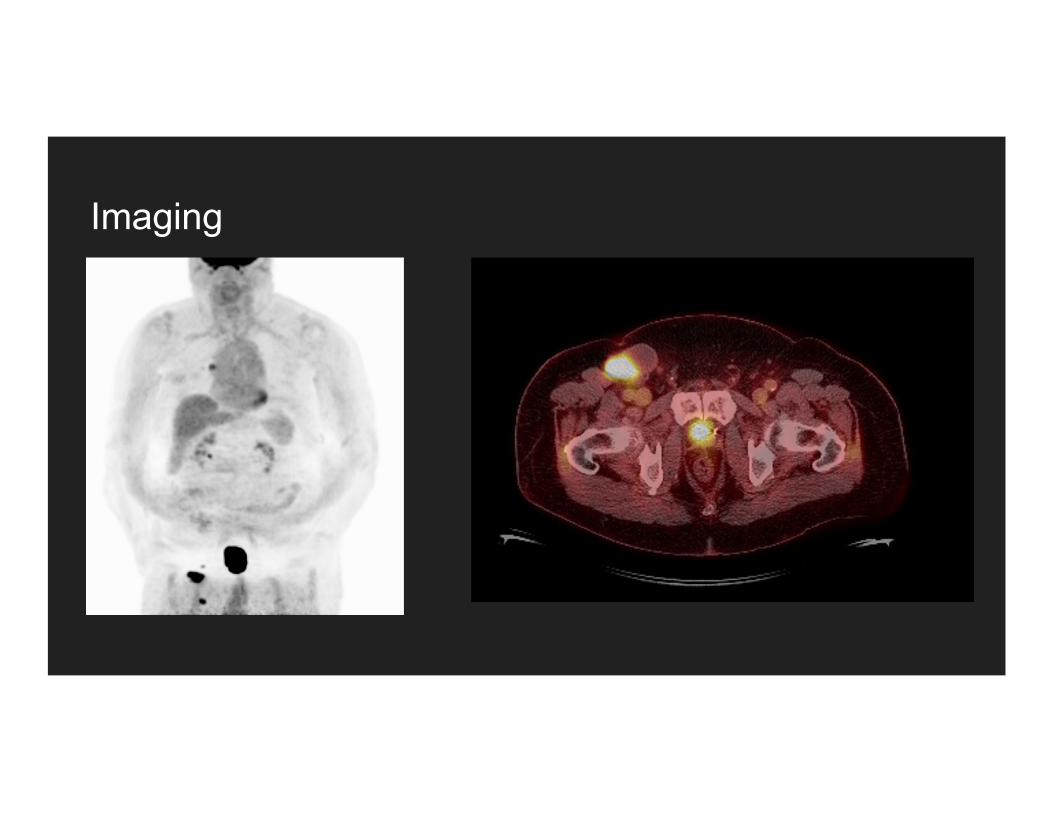
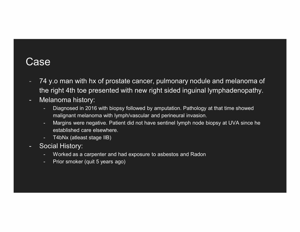
Imaging
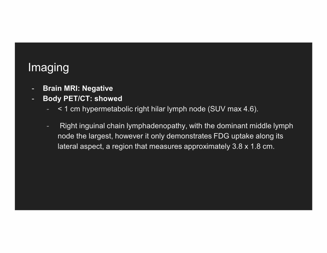
Imaging- Brain MRI: Negative- Body PET/CT: showed
- < 1 cm hypermetabolic right hilar lymph node (SUV max 4.6).
- Right inguinal chain lymphadenopathy, with the dominant middle lymph node the largest, however it only demonstrates FDG uptake along its lateral aspect, a region that measures approximately 3.8 x 1.8 cm.
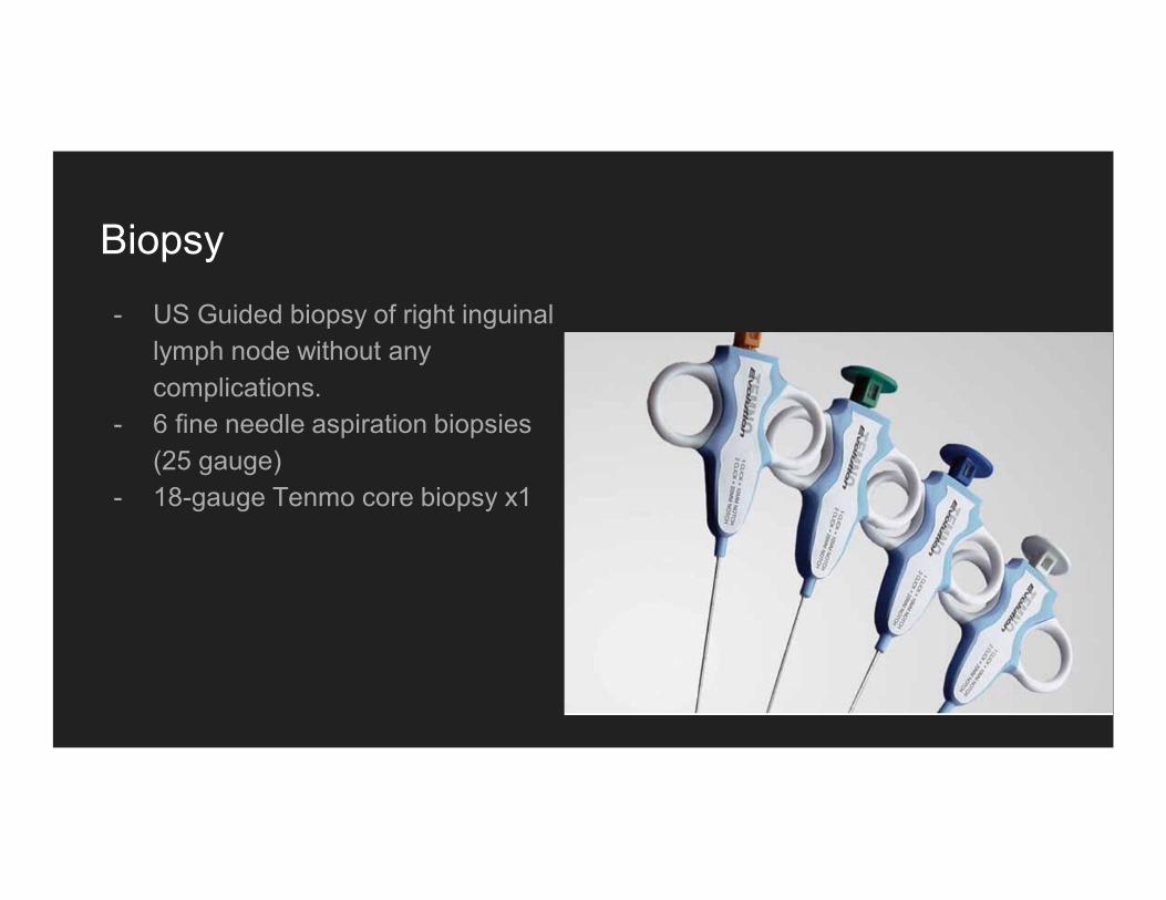
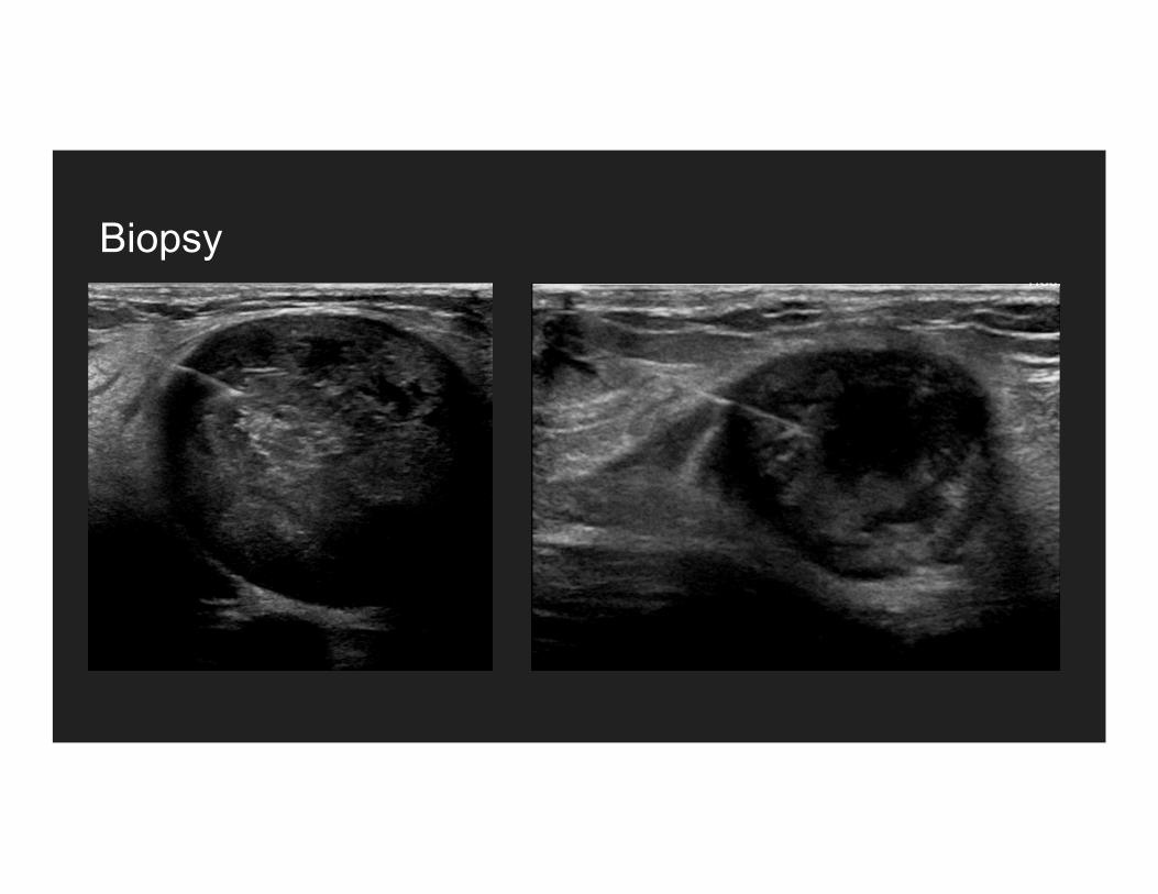
Biopsy- US Guided biopsy of right inguinal
lymph node without any complications.
- 6 fine needle aspiration biopsies (25 gauge)
- 18-gauge Tenmo core biopsy x1
Biopsy
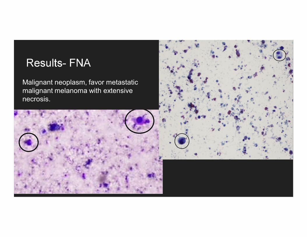
Results- FNAMalignant neoplasm, favor metastatic malignant melanoma with extensive necrosis.
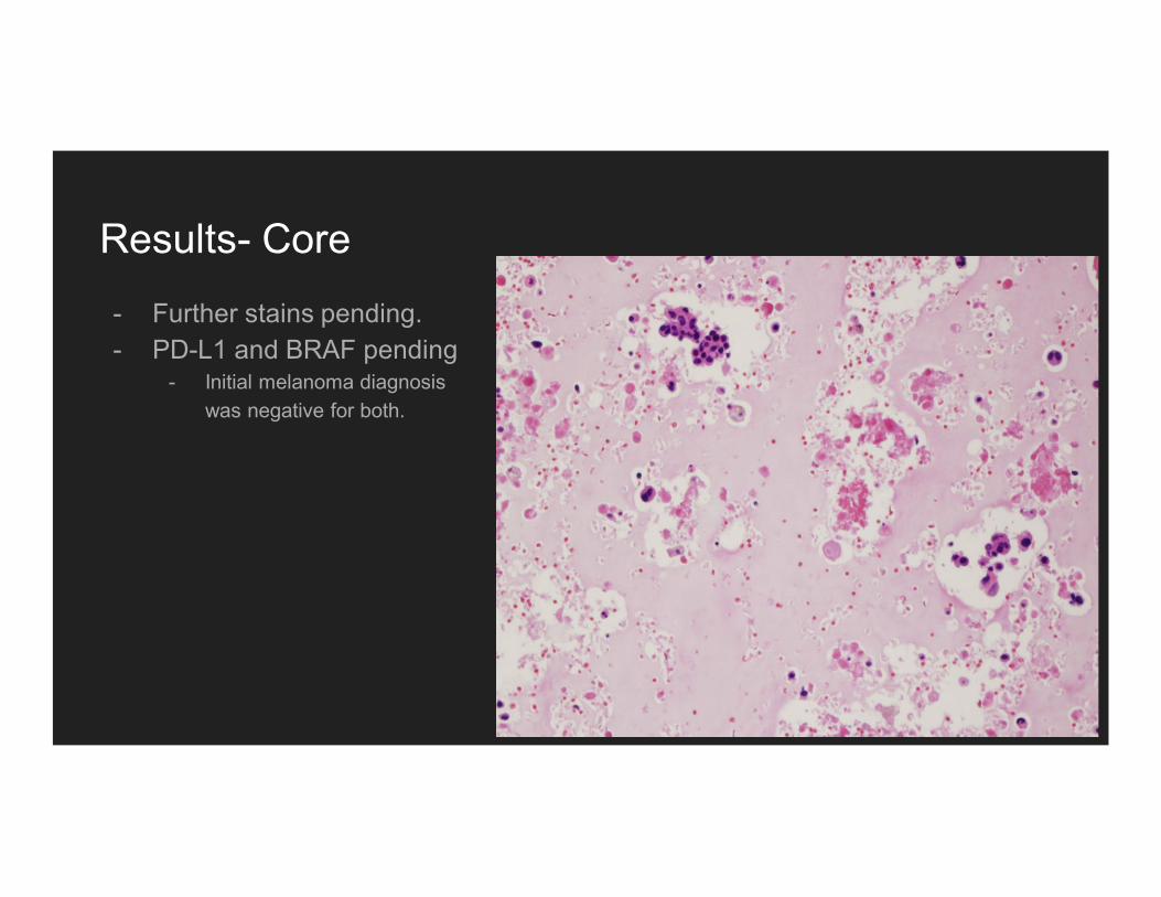
Results- Core- Further stains pending. - PD-L1 and BRAF pending
- Initial melanoma diagnosis was negative for both.
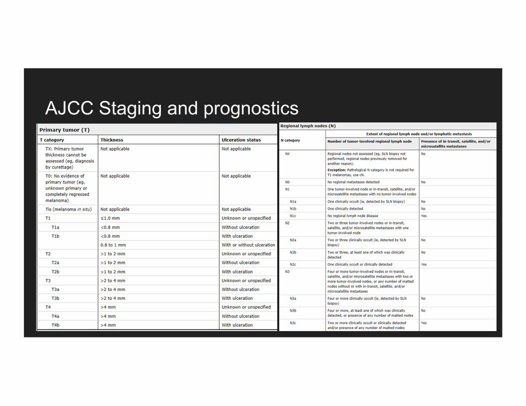
AJCC Staging and prognostics
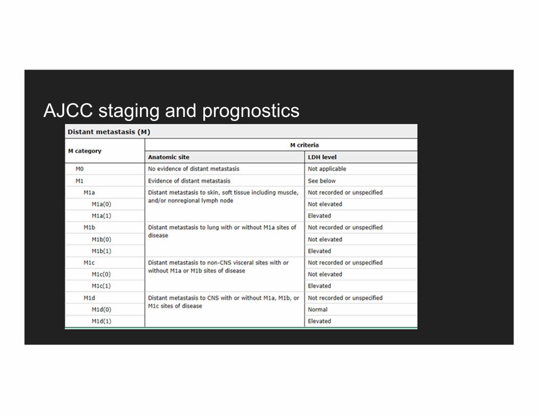
AJCC staging and prognostics
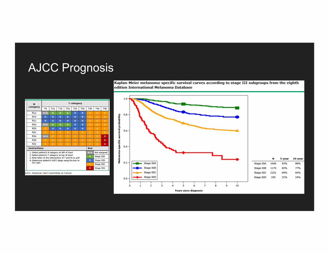
AJCC Prognosis
Closing- Follow up
- Pending on Path results.- Will need prior lung CT to be compared to current.- Possible lung bx. - Node resection vs systemic therapy pending on lung nodule.
- The End.
References1.Patient EMR Chart Review2.Patient PACS Imaging Review3. Melanoma staging: Evidence-based changes in the American Joint Committee on Cancer eighth edition cancer staging manual Gershenwald, J. E., Scolyer, R. A., Hess, K. R., Sondak, V. K, etc. A Cancer Journal for Clinicians, 67: 472-492. doi:10.3322/caac.21409.4. Pathologyoutlines.com/topic/skintumormelanocyticsuperficialspreading.html