Embed Size (px)
Citation preview
CLINICAL MICROBIOLOGY REVIEWS, Oct. 2010, p. 884–928 Vol. 23, No. 40893-8512/10/$12.00 doi:10.1128/CMR.00019-10Copyright © 2010, American Society for Microbiology. All Rights Reserved.
Melanized Fungi in Human DiseaseSanjay G. Revankar1* and Deanna A. Sutton2
Department of Medicine, Division of Infectious Diseases, Wayne State University, Detroit, Michigan,1 andFungus Testing Laboratory, University of Texas Health Science Center, San Antonio, Texas2
INTRODUCTION .......................................................................................................................................................885ECOLOGY ...................................................................................................................................................................886CLASSIFICATION, TAXONOMY, AND NOMENCLATURE .............................................................................886IDENTIFICATION OF ETIOLOGIC AGENTS.....................................................................................................887
Phenotypic Identification .......................................................................................................................................890Macroscopic morphology ...................................................................................................................................890Microscopic morphology and pleomorphism ..................................................................................................890Physiologic features ............................................................................................................................................892
Molecular Characterization...................................................................................................................................892ANAMORPHIC HYPHOMYCETE GENERA ........................................................................................................893
Capnodiales ...............................................................................................................................................................893Hortaea..................................................................................................................................................................893Cladosporium ........................................................................................................................................................893
Dothideales ................................................................................................................................................................893Aureobasidium ......................................................................................................................................................893Hormonema...........................................................................................................................................................893
Pleosporales ...............................................................................................................................................................893Alternaria ..............................................................................................................................................................893Bipolaris ................................................................................................................................................................893Curvularia .............................................................................................................................................................893Exserohilum ..........................................................................................................................................................893
Chaetothyriales..........................................................................................................................................................894Exophiala ..............................................................................................................................................................894Cladophialophora..................................................................................................................................................895Fonsecaea ..............................................................................................................................................................895Ochroconis ............................................................................................................................................................896Phialophora...........................................................................................................................................................896Rhinocladiella .......................................................................................................................................................896Veronaea................................................................................................................................................................897
Microascales ..............................................................................................................................................................897Scedosporium ........................................................................................................................................................897Scopulariopsis .......................................................................................................................................................897
Sordariales ................................................................................................................................................................897Madurella ..............................................................................................................................................................897Myceliophthora......................................................................................................................................................897Acrophialophora....................................................................................................................................................897
Calosphaeriales .........................................................................................................................................................897Phialemonium .......................................................................................................................................................897Phaeoacremonium ................................................................................................................................................898Pleurostomophora .................................................................................................................................................898
Coniochaetales...........................................................................................................................................................898Lecythophora.........................................................................................................................................................898
Ophiostomatales ........................................................................................................................................................898Sporothrix..............................................................................................................................................................898
ANAMORPHIC COELOMYCETE GENERA.........................................................................................................898Pleosporales ...............................................................................................................................................................898
Phoma and Phoma-like pycnidial coelomycetes ..............................................................................................898Botryosphaeriales ......................................................................................................................................................899
Lasiodiplodia.........................................................................................................................................................899Macrophomina ......................................................................................................................................................899Neoscytalidium ......................................................................................................................................................899
* Corresponding author. Mailing address: Harper University Hos-pital, 3990 John R. St., 5 Hudson, Detroit, MI 48201. Phone: (313)745-8599. Fax: (313) 993-0302. E-mail: [email protected].
884
Dow
nloa
ded
from
http
s://j
ourn
als.
asm
.org
/jour
nal/c
mr
on 1
8 N
ovem
ber
2021
by
211.
21.2
54.1
51.
Sordariales ................................................................................................................................................................899Phomopsis .............................................................................................................................................................899
TELEOMORPHIC GENERA....................................................................................................................................899Sordariales ................................................................................................................................................................899
Chaetomium and Achaetomium ..........................................................................................................................899Pleosporales ...............................................................................................................................................................899
Leptosphaeria ........................................................................................................................................................899Microascales ..............................................................................................................................................................899
Microascus ............................................................................................................................................................899Pseudallescheria ....................................................................................................................................................900
PATHOGENESIS........................................................................................................................................................900Role of Melanin.......................................................................................................................................................900Other Putative Virulence Factors.........................................................................................................................901
DIAGNOSIS ................................................................................................................................................................901Initial Specimen Processing ..................................................................................................................................901Direct Microscopy...................................................................................................................................................902Histopathology and Special Stains.......................................................................................................................902Isolation Procedures and Culture ........................................................................................................................903Radiology..................................................................................................................................................................904Antigen Testing and Serology ...............................................................................................................................904Molecular Diagnostics............................................................................................................................................904
IN VITRO ANTIFUNGAL SUSCEPTIBILITY........................................................................................................904Polyenes....................................................................................................................................................................904
Amphotericin B ...................................................................................................................................................904Natamycin ............................................................................................................................................................904
Azoles........................................................................................................................................................................905Itraconazole .........................................................................................................................................................905Voriconazole.........................................................................................................................................................905Posaconazole........................................................................................................................................................905Investigational azoles .........................................................................................................................................905
Flucytosine ...............................................................................................................................................................906Allylamines...............................................................................................................................................................906Echinocandins .........................................................................................................................................................906Other Agents............................................................................................................................................................906Antifungal Combinations.......................................................................................................................................906
ANIMAL MODELS OF INFECTION......................................................................................................................906CLINICAL SYNDROMES AND THEIR MANAGEMENT ..................................................................................907
Eumycetoma.............................................................................................................................................................907Chromoblastomycosis.............................................................................................................................................907Phaeohyphomycosis ................................................................................................................................................908
Allergic disease....................................................................................................................................................908(i) Allergic fungal sinusitis............................................................................................................................908(ii) ABPM.........................................................................................................................................................908
Superficial infections..........................................................................................................................................909(i) Onychomycosis...........................................................................................................................................909(ii) Tinea nigra................................................................................................................................................909
Deep local infections ..........................................................................................................................................909(i) Subcutaneous lesions ................................................................................................................................909(ii) Keratitis.....................................................................................................................................................909(iii) Bone and joint infections.......................................................................................................................910(iv) Peritonitis .................................................................................................................................................910(v) Miscellaneous infections..........................................................................................................................910
Pulmonary infection ...........................................................................................................................................910Central nervous system infection .....................................................................................................................911Disseminated infection.......................................................................................................................................911
CONCLUSIONS .........................................................................................................................................................912REFERENCES ............................................................................................................................................................912
INTRODUCTION
Melanin is a ubiquitous compound found in many microbesand animals. Its functions are varied but are based on theunique molecular characteristics of its structure, which make itan extremely stable molecule, resistant to a variety of destruc-tive physicochemical processes (83, 109, 324). In recent years
its pathogenic role in fungi has become well described (123, 292,375, 460, 546). This review will focus on fungi that are consideredto be melanized as a primary feature, particularly with regard totheir phenotypic appearance (macroscopic and microscopic mor-phologies) and appearance in tissue (histology).
The terms used to describe these fungi have evolved over the
VOL. 23, 2010 MELANIZED FUNGI IN HUMAN DISEASE 885
Dow
nloa
ded
from
http
s://j
ourn
als.
asm
.org
/jour
nal/c
mr
on 1
8 N
ovem
ber
2021
by
211.
21.2
54.1
51.
past several decades. As Sporothrix schenckii was one of theearliest melanized fungi described, “sporotrichoid” was oftenused to describe similar fungi, though currently it has beenreplaced by other, more useful terms. “Phaeoid,” “phaeo-spo-rotrichose,” and “dematiaceous” have also been mentioned inthe literature (574). “Phaeo” comes from the Greek meaning“dark” and has been commonly used, particularly when de-scribing infections due to these fungi as “phaeohyphomycosis,”i.e., infection caused by dark-walled fungi, as suggested byAjello et al. (12, 630). It has been suggested that the term“dematiaceous” is not appropriate given its etymologic deriva-tion from the Greek “deme,” meaning bundle, though it hasbecome fairly entrenched in medical mycological literature andwill likely persist in nomenclature (574). The term “melanized”has become more utilized recently, given its specific meaning.For the purposes of this review, however, the terms “dematia-ceous,” “melanized,” “dark,” and “phaeoid” are used inter-changeably to denote fungal elements containing melanin.
The presence of melanin alone is probably not a useful crite-rion for inclusion in this group of clinically important fungi, asmelanin has been demonstrated in practically all “nondematia-ceous” clinical fungi examined in the literature, including His-toplasma capsulatum, Paracoccidioides brasiliensis, Aspergillusspp., and even Candida albicans (293, 521, 548, 599, 757). Onemight contrast the fungi discussed here as heavily melanized,with brown-pigmented hyphae in tissue often discernible with-out the use of any staining procedure. At present, no quanti-tative measure of melanin is available to distinguish dematia-ceous from other fungi. In addition, Sporothrix schenckii, witha yeast form in tissue, is well known and well described (177)as the agent of a unique clinical entity, sporotrichosis, and onlyissues regarding its mycology will be discussed here.
Melanized fungi are common in the environment (see Ecol-ogy below) and are often isolated in the microbiology lab,where they may be considered contaminants. Indeed, only 10%of dematiaceous lab isolates are likely to have clinical signifi-cance (72, 607). Clinical disease due to these fungi is uncom-mon, with one estimate from a large metropolitan area of onecase/million persons/year (617). Despite their rarity in clinicalpractice, melanized fungi have become increasingly recognizedas important pathogens, particularly in immunocompromisedpatients, though individuals with apparently “normal” immunesystems have also been reported to have invasive, often fatalinfections (627, 628).
The clinical syndromes caused by these fungi are differenti-ated based on histologic findings into eumycetoma, chromo-blastomycosis, and phaeohyphomycosis. Eumycetoma is a deeptissue infection, usually of the lower extremities, characterizedby the presence of mycotic granules (572). It is associated witha relatively small group of fungi. Chromoblastomycosis iscaused primarily by a few species of fungi that produce char-acteristic sclerotic bodies in tissue and is usually seen in trop-ical areas (501, 572). Phaeohyphomycosis is a term generallyreserved for the remainder of clinical syndromes caused bymelanized fungi (623, 630). For the purposes of this review,these will be arbitrarily divided into allergic disease, superficialand deep local infections, pulmonary disease, central nervoussystem (CNS) infection, and disseminated disease. We do notaim to review every publication regarding melanized fungi, butrather we seek to provide a broad, yet in-depth overview of the
field as it currently stands, recognizing that it will continue toevolve and expand with our increasing knowledge of and ex-perience with these clinically important fungi.
ECOLOGY
Melanized or dematiaceous fungi as defined above are fre-quently considered ubiquitous saprobes inhabiting living anddead plant material and, for the most part, residing in the soil.We now know, however, that these generalized assumptionsare incorrect for the group as a whole, as several etiologicagents occupy specific ecological niches or microenvironments,and the knowledge of their natural ecology contributes to ourunderstanding of their opportunistic/pathogenic potential(175, 177, 606, 779). It has been suggested (175) that our use ofthe term “dematiaceous” be restricted to those ubiquitous,mostly plant-associated hyphomycetous fungi with brown hy-phae (220, 221), such as Alternaria, Bipolaris, Curvularia, andExserohilum in the order Pleosporales. The natural ecology ofmelanized fungi in several other orders is more restricted (187).For example, fungi in the order Calosphaeriales belong mostly towoody-plant- or wood-inhabiting genera such as Phaeoacremo-nium, Phialemonium, and Pleurostomophora, whereas some gen-era in the order Chaetothyriales, such as Exophiala, may havespecific microenvironments and are characterized as “micro-extremophiles.” The ability of some species in this genus, suchas E. xenobiotica, to grow in high concentrations of xenobiotics(606) such as xylene, toluene, or creosote-treated utility poles,as well as to cause human disease, is truly remarkable. Speciesin the genus Exophiala and related genera are frequently re-ferred to as the “black yeast-like fungi” and are so namedbecause of their ability to produce budding, yeast-like cells atsome point in their life cycle as well as dark hyphae. Theecology of Pseudallescheria and Scedosporium was also recentlyinvestigated by examining the occurrence of these species innatural and human-dominated environments (393). Thesefindings demonstrated increasing environmental recovery withincreasing human habitation and a concomitant elevation innitrogen concentrations. Another genus defined by its resi-dence in a particular environmental niche is the halophilicgenus Hortaea in the order Capnodiales. Hortaea werneckii, theagent of tinea nigra, is found in subtropical saltwater habitatsand is manifested by its opportunistic adherence to the dead,salty keratin layers of the human hand (87). Thus, while severalgenera are considered “ubiquitous,” many prefer well-definedmicroenvironments which, for some genera, predispose themto causing disease where similar conditions exist in the host.
In addition, there are species that appear to be geographi-cally restricted, such as Rhinocladiella mackenziei, which hasbeen seen primarily in patients from the Middle East (726).While Scedosporium prolificans has been reported from manylocations, most clinical cases originate from Australia andSpain, for unclear reasons (76, 336). This may be due to envi-ronmental features that preferentially support specific fungalspecies.
CLASSIFICATION, TAXONOMY, AND NOMENCLATURE
Classification of fungi is simply their assignment into definedcategories. A classification system is composed of hierarchical
886 REVANKAR AND SUTTON CLIN. MICROBIOL. REV.
Dow
nloa
ded
from
http
s://j
ourn
als.
asm
.org
/jour
nal/c
mr
on 1
8 N
ovem
ber
2021
by
211.
21.2
54.1
51.

groups which may be further subdivided to indicate degrees ofrelationships. The basic unit of classification is the species,although there is currently no universally acceptable definitionfor this unit. Taxonomy is the arrangement of these fungi intoa classification. With multilocus sequencing providing classifi-cation insights unavailable to the phenotypic systematists(those who study the relationships and classification of organ-isms and the processes by which they have evolved), new phy-logenetic classification schemes have emerged. Taylor et al.have provided an excellent treatment of the phylogenetic con-cepts underlying the definition of species in fungi (737). Theabbreviated classification scheme to the ordinal level for asco-mycetous melanized fungi covered in this review is based uponthe most recent work of Hibbett et al. (340) and the Myconet“Outline of Ascomycotya” (463).Kingdom: Fungi
Phylum: AscomycotaSubphylum: Pezizomycotina
Class: DothideomycetesOrders: Capnodiales, Dothideales, Pleosporales,
BotryosphaerialesClass: Eurotiomycetes
Order: ChaetothyrialesClass: Sordariomycetes
Orders: Microascales, Sordariales, Calosphaeriales,Ophiostomatales
As seen above and in Table 1, clinically significant melanizedfungi span several ascomycetous orders in the kingdom Fungi.
Nomenclature refers to assigning formal scientific names. Thisprocess is regulated by the International Code of Botanical No-menclature (ICBN) (http://www.bgbm.org/iapt/nomenclature/code/default.htm) to facilitate a stable naming system and toavoid and reject names which are in error or are ambiguous(789). Lack of adherence to these requisites often invalidates ataxon name and results in multiple names for the same organ-ism. Other reasons for name changes include the placement offungi into new genera as determined by phylogenetic studies,which frequently occurs within this heterogeneous group offungi. When this occurs, the species epithet is retained, but itmay require modification in keeping with the rules of Latingrammar. An example of recent changes for melanized fungiinclude the movement of Phialophora richardsiae to Pleurosto-mophora richardsiae and of Phialophora parasitica to Phaeo-acremonium parasiticum. Discovery of a previously unrecog-nized teleomorph (sexual or meiotic state) may also precipitatea name change. A recent example is found in the discovery ofthe teleomorph for Scedosporium apiospermum, which was in-correctly thought to be Pseudallescheria boydii. We now knowthrough the work of Gilgado et al. that the teleomorph for S.apiospermum is the heterothallic ascomycete Pseudallescheriaapiosperma, as evidenced by the production of cleistothecia(round sexual structures containing asci and ascospores) andascospores (the sexual reproductive propagules) between com-patible mating strains of S. apiospermum (283). As the teleo-morph name takes precedence over the anamorph (asexual,mitotic) name, the correct binomial would be the sexual state.Whether this name would be adopted by clinicians in everydayusage remains problematic.
Also confusing for clinicians and laboratorians alike is the
naming convention that permits the use of more than onename for the same fungus. This is allowed when a particularform of the fungus is the one more commonly seen in thelaboratory. Fungi recovered in culture commonly display onlyan anamorphic state. They may be either heterothallic isolateswith no known teleomorph or homothallic strains failing toproduce their sexual state in vitro. A few clinically significanthomothallic melanized fungi do form both anamorphs andteleomorphs in culture. In this situation, as mentioned above,the teleomorph name takes precedence over the anamorphname, e.g., Pseudallescheria boydii rather than Scedosporiumboydii and Microascus cinereus rather than Scopulariopsis cine-rea. Additionally, in some genera, such as Pseudallescheria, twoseparate anamorphs which are distinctively different micro-scopically may be produced, and these are referred to assynanamorphs (another asexual form of the same fungus).Some homothallic strains, however, lack anamorphs and areknown only by the name of the sexual state. Examples wouldinclude members of the genus Chaetomium. The advent ofsequencing characterization has provided the tools necessaryto reevaluate the evolutionary relationships of these blackmolds, and today multilocus molecular phylogenetic studiesare clearly redefining previously described entities, uncoveringnew species and varieties, and correlating these with theirnatural habitats.
IDENTIFICATION OF ETIOLOGIC AGENTS
Over 150 species and 70 genera of dematiaceous fungi havebeen implicated in human and animal disease (Table 2). As thenumber of patients immunocompromised as a result of dis-eases and medical therapy increases, additional species arebeing reported as causes of human disease, expanding an al-ready long list of potential pathogens. Identification of mela-nized etiologic agents known to cause human or animal diseasehas traditionally been based upon phenotypic features of theisolate observed in culture (175, 177, 220, 221). This practicecontinues to be the mainstay of fungal identification in mostroutine settings. More recently, molecular techniques em-ployed for classification purposes and those provided by re-search facilities have provided additional tools for the charac-terization of these molds. Extensive sequencing for somegenera has illustrated the concept of “species complexes,” orthe inclusion of several separate species into what was formerlyreferred to as a single species. This has been clearly demon-strated in the genera Exophiala (825), Scedosporium (281–283),and Phaeoacremonium (525). The “splitting” of these speciesinto separate taxa has of necessity changed our reporting prac-tices. As an example, laboratories previously comfortable withdiscriminating only between Exophiala (Wangiella) dermatitidisand E. jeanselmei are now aware of several other clinicallysignificant species that are not easily separated by phenotypicfeatures alone (177, 184, 825) and that E. jeanselmei is in factone of the less frequent agents of disease. Therefore, speciesother than E. dermatitidis are best reported as an Exophiala sp.,not E. dermatitidis, unless sequencing has provided a speciesidentification. These “new and improved” reporting tech-niques, however, must be communicated to clinicians in a man-ner consistent with their understanding of current organismterminology and the associated mycoses.
VOL. 23, 2010 MELANIZED FUNGI IN HUMAN DISEASE 887
Dow
nloa
ded
from
http
s://j
ourn
als.
asm
.org
/jour
nal/c
mr
on 1
8 N
ovem
ber
2021
by
211.
21.2
54.1
51.

TA
BL
E1.
Salie
ntfe
atur
esof
sele
cted
clin
ical
lysi
gnifi
cant
dem
atia
ceou
sfu
ngia
Gen
usty
peO
rder
Gen
usor
spec
ies
Salie
ntph
enot
ypic
and/
ordi
agno
stic
feat
ures
b
Ana
mor
phic
(ase
xual
)hy
phom
ycet
e(c
onid
iaC
apno
dial
esH
orta
eaw
erne
ckii
Col
onie
sol
ivac
eous
tobl
ack,
muc
oid
toye
ast-
like,
rest
rict
ed;b
road
hyph
ae,w
ide
anne
llate
dzo
nes
prod
uce
pale
brow
n1-
tom
ultic
elle
dan
nello
coni
dia
born
efr
ee)
Cla
dosp
oriu
msp
p.C
olon
ies
oliv
aceo
usto
blac
k,ve
lvet
y;co
nidi
opho
res
sim
ple
orbr
anch
ed,w
ithor
with
out
node
sor
swel
lings
;ra
moc
onid
ia(“
shie
ldce
lls”)
give
rise
tobr
anch
ing
chai
nsof
frag
ile,d
ark,
mos
tly1-
or2-
celle
dco
nidi
aw
ithpr
omin
ent
atta
chm
ent
scar
es(h
ila)
Dot
hide
ales
Aur
eoba
sidi
umpu
llula
nsC
olon
ies
ofA
.pul
lula
nsva
r.pu
llula
nsm
ucoi
d,cr
eam
topi
nkin
itial
lyan
dla
ter
brow
nto
blac
k,w
hile
thos
eof
A.
pullu
lans
var.
mel
anig
enum
blac
kat
the
outs
et;h
yalin
ebl
asto
coni
dia
born
esy
nchr
onou
sly
from
hyal
ine
hyph
ae;d
ark,
thic
k-w
alle
dch
lam
ydos
pore
s;D
NA
sequ
enci
ngne
cess
ary
for
relia
ble
diffe
rent
iatio
nof
A.p
ullu
lans
and
H.
dem
atio
ides
Hor
mon
ema
dem
atio
ides
Col
onie
ssi
mila
rto
thos
ese
enin
A.p
ullu
lans
;hya
line
blas
toco
nidi
apr
oduc
edas
ynch
rono
usly
bype
rcur
rent
prol
ifera
tion
from
hyal
ine
and
dark
hyph
aeP
leos
pora
les
Alte
rnar
iasp
p.C
olon
ies
woo
lly,p
ale
tool
ivac
eous
tobl
ack,
with
rapi
dgr
owth
;lar
ge,d
ark,
euse
ptat
ec ,mur
iform
coni
dia
inch
ains
;A.
infe
ctor
iaco
nidi
am
aybe
spar
sean
dha
velo
ngap
ical
beak
sse
rvin
gas
seco
ndar
yco
nidi
ogen
ous
cells
Bip
olar
issp
p.C
olon
ies
woo
lly,g
ray
tobl
ack,
with
rapi
dgr
owth
,bip
olar
germ
inat
ion,
geni
cula
teco
nidi
opho
res,
flatt
ened
hilu
m;B
.sp
icife
raha
s3
dist
osep
tad
and
4ce
lls,w
hile
B.h
awai
iens
isha
spr
edom
inat
ely
5di
stos
epta
and
6ce
llsC
urvu
laria
spp.
Col
onie
sw
oolly
,gra
yto
blac
k,w
ithra
pid
grow
th;g
enic
ulat
eco
nidi
opho
res;
coni
dia
euse
ptat
ean
dcu
rved
(pro
noun
ced
tosu
btle
)du
eto
swol
len
mid
dle
cell
whi
chis
dark
erin
C.l
unat
a;C
.lun
ata
var.
aeria
prod
uces
larg
est
rom
avi
sibl
ew
ithth
ena
ked
eye
Exs
eroh
ilum
spp.
Col
onie
sw
oolly
,gra
yto
blac
k,w
ithra
pid
grow
th;g
enic
ulat
eco
nidi
opho
res,
trun
cate
prot
rudi
nghi
lum
;E.r
ostr
atum
has
7-9
dist
osep
ta;8
-10
cells
;pro
min
ent
dark
basa
land
dist
alse
pta;
E.l
ongi
rost
ratu
mha
slo
nger
coni
dia
with
cent
ral
curv
atur
e;E
.mcg
inni
siih
assu
btle
war
typr
ojec
tions
onco
nidi
aC
haet
othy
riale
sE
xoph
iala
spp.
Col
onie
sm
ucoi
din
itial
ly,l
ater
mor
efil
amen
tous
;con
idio
geno
usce
llspr
edom
inat
ely
anne
llidi
c;ph
ialid
esso
met
imes
pres
ent;
anne
llate
dbl
ack
yeas
tsy
nana
mor
phof
ten
pres
ent;
man
ysp
ecie
sve
rysi
mila
rm
icro
scop
ical
ly;n
itrat
epo
sitiv
e;D
NA
sequ
enci
ngof
ITS
regi
onfa
cilit
ates
spec
ies
iden
tifica
tion;
max
imum
tem
pva
ries
;mos
tfr
eque
ntly
seen
clin
ical
spec
ies
incl
ude
E.x
enob
iotic
a,E
.olig
ospe
rma,
E.l
ecan
ii-co
rni,
and
E.p
haeo
mur
iform
isE
xoph
iala
derm
atiti
dis
Col
onie
sbl
ack,
muc
oid;
nitr
ate
nega
tive;
grow
that
40°C
;bla
ckye
ast
E.e
xoph
iala
esy
nana
mor
phpr
esen
t;m
ost
com
mon
clin
ical
Exo
phia
lasp
ecie
s;ac
cura
tely
iden
tified
byph
enot
ypic
feat
ures
;obs
olet
ena
me
Wan
giel
lade
rmat
itidi
sC
lado
phia
loph
ora
spp.
Col
onie
sbl
ack,
velv
ety;
grow
thra
tes
and
tem
pera
ture
sva
ryfo
rin
divi
dual
spec
ies;
mic
rosc
opic
ally
sim
ilar
toC
lado
spor
ium
spp.
but
lack
coni
diop
hore
s,“s
hiel
dce
lls,”
and
prom
inen
thi
la;c
onid
iaar
eno
nfra
gile
(rem
ain
inta
ctin
chai
ns);
neur
otro
pic
spec
ies
incl
ude
C.b
antia
naan
dC
.mod
esta
;oth
erhu
man
path
ogen
icsp
ecie
sin
clud
eC
.arx
ii,C
.bo
ppii,
C.c
arrio
nii,
C.d
evrie
sii,
C.e
mm
onsi
i,C
.myc
etom
atis
,C.s
amoe
nsis
,and
C.s
atur
nica
Fon
seca
easp
p.C
olon
ies
oliv
aceo
usto
blac
k,ve
lvet
y;co
nidi
afo
rmed
from
swol
len
dent
icle
sgi
ving
rise
tose
cond
ary
and
tert
iary
coni
dia
inch
ains
ofup
tofo
urco
nidi
a;co
nidi
aal
sofo
rmed
onsy
mpo
dial
coni
diop
hore
slik
ein
Rhi
nocl
adie
llaan
doc
casi
onal
lyfr
omdi
scre
teph
ialid
eslik
ein
Phi
alop
hora
;F
.ped
roso
ian
agen
tof
chro
mob
last
omyc
osis
,F.m
onop
hora
anag
ent
ofbo
thch
rom
obla
stom
ycos
isan
dce
rebr
alph
aeoh
ypho
myc
osis
Och
roco
nis
gallo
pava
Col
onie
sar
ebr
owni
sh,v
elve
ty,w
itha
mar
oon
diffu
sing
pigm
ent;
2-ce
lled,
clav
ate
coni
dia
born
efr
omde
ntic
les;
grow
that
45°C
;no
grow
thon
med
iaco
ntai
ning
cycl
ohex
imid
e;ne
urot
ropi
c;ob
sole
tena
mes
Dac
tyla
riaga
llopa
va,D
.co
nstr
icta
var.
gallo
pava
Phi
alop
hora
spp.
Col
onie
sol
ivac
eous
tobl
ack,
velv
ety;
thre
esp
ecie
sar
ecl
inic
ally
sign
ifica
nt;P
.ver
ruco
saha
sda
rk,f
unne
l-sha
ped
colla
rett
es;P
.am
eric
ana
has
deep
,vas
e-sh
aped
colla
rett
es;t
hesl
ow-g
row
ing
P.e
urop
aea
has
very
shor
tco
llare
ttes
Rhi
nocl
adie
llasp
p.C
olon
ies
oliv
aceo
usto
blac
k,ve
lvet
y;lo
ng,e
rect
,bro
wn,
unbr
anch
edsy
mpo
dial
coni
diop
hore
s;1-
celle
dpa
leel
lipso
idal
coni
dia
born
eon
crow
ded
dent
icle
s;an
Exo
phia
laye
ast
syna
nam
orph
may
bepr
esen
t;R
.mac
kenz
ieii
sa
neur
otro
pic
spec
ies
inth
ege
nus
with
rela
tivel
yfe
wco
nidi
ape
rfe
rtile
part
ofth
ege
nicu
late
coni
diop
hore
;con
idia
1-ce
lled,
pale
brow
n,el
lipso
idal
with
apr
omin
ent
trun
cate
hilu
m;p
oor
grow
that
25°C
,goo
dgr
owth
at40
°C;o
bsol
ete
nam
eR
amic
hlor
idiu
mm
acke
nzie
i;ot
her
path
ogen
icsp
ecie
sin
clud
eR
.aqu
aspe
rsa
and
R.s
imili
sV
eron
aea
botr
yosa
Col
onie
sgr
ayto
blac
kish
-bro
wn,
woo
lly;l
ong,
brow
nco
nidi
opho
res;
pale
brow
n,2-
celle
dco
nidi
aw
itha
roun
ded
apex
and
trun
cate
base
born
efr
omcl
osel
ysp
aced
inte
rcal
ary
coni
diog
enou
sce
llsM
icro
asca
les
Sced
ospo
rium
spp.
Col
onie
spa
leto
yello
wis
h-gr
ayto
dark
ergr
ay,s
ome
with
oran
gere
vers
e,w
oolly
;con
idio
geno
usce
llsan
nelli
dic;
som
esp
ecie
spr
oduc
ea
Pse
udal
lesc
heria
tele
omor
phan
da
Gra
phiu
msy
nana
mor
ph;s
imila
rhu
man
path
ogen
icsp
ecie
sin
the
Pse
udal
lesc
heria
boyd
iisp
ecie
sco
mpl
exas
defin
edby
rece
ntm
olec
ular
stud
ies
incl
ude
S.ap
iosp
erm
um,S
.boy
dii,
S.au
rant
iacu
m,a
ndS.
deho
ogii;
S.pr
olifi
cans
(obs
olet
ena
me
S.in
flatu
m)
poss
essi
ngin
flate
dan
nelli
dic
coni
diog
enou
sce
lls,i
sun
rela
ted
tom
embe
rsof
the
P.b
oydi
ispe
cies
com
plex
Scop
ular
iops
issp
p.C
olon
ies
gray
tool
ivac
eous
-bro
wn,
woo
lly;c
onid
ioge
nous
cells
anne
llidi
c;se
vera
lver
ysi
mila
rda
rksp
ecie
sar
ean
amor
phs
ofva
riou
sM
icro
ascu
ssp
p.
888 REVANKAR AND SUTTON CLIN. MICROBIOL. REV.
Dow
nloa
ded
from
http
s://j
ourn
als.
asm
.org
/jour
nal/c
mr
on 1
8 N
ovem
ber
2021
by
211.
21.2
54.1
51.
Sord
aria
les
Mad
urel
lam
ycet
omat
isC
olon
ies
very
slow
grow
ing
and
ofte
nhe
aped
;dar
kbr
own
tobl
ack;
diffu
sibl
ebr
own
pigm
ent;
unlik
eM
.gris
ea,M
.m
ycet
omat
isgr
ows
at40
°Can
dfa
ilsto
assi
mila
tesu
cros
e;pr
ecis
eid
entifi
catio
nfa
cilit
ated
byIT
Sse
quen
cing
Myc
elio
phth
ora
ther
mop
hila
Col
onie
slig
htbr
own,
pow
dery
;ill-
defin
edm
argi
n;co
nidi
abo
rne
from
ampu
llifo
rmsw
ellin
gsar
ehy
alin
ean
dsm
ooth
initi
ally
beco
min
gro
ugh
and
brow
nat
mat
urity
;gro
wth
at48
°CA
crop
hial
opho
rafu
sisp
ora
Col
onie
sce
ntra
llyda
rkfr
ont
and
reve
rse;
unbr
anch
ed,e
rect
,bro
wn,
echi
nula
teco
nidi
opho
res
are
anch
ored
bya
foot
cell;
chai
nsof
coni
dia
with
fine
orco
arse
spir
als
prod
uced
from
apex
ofbr
own
coni
diop
hore
san
din
flate
dph
ialid
eson
hyal
ine
hyph
ae;g
row
that
40°C
Cal
osph
aeria
les
Phi
alem
oniu
msp
p.C
olon
ies
buff
togr
ayto
yello
w;c
onid
ioge
nous
cells
phia
lides
and
adel
ophi
alid
es(r
educ
edph
ialid
esla
ckin
ga
basa
lse
ptum
);P
.obo
vatu
mha
sob
ovat
eco
nidi
aan
da
gree
ndi
ffusi
ngpi
gmen
t;sp
orod
ochi
a-pr
oduc
ing
isol
ates
ofP
.cu
rvat
umha
vebe
enre
port
edP
haeo
acre
mon
ium
spp.
Col
onie
sra
nge
from
buff
topa
leye
llow
topa
leor
dark
pink
tova
riou
ssh
ades
ofbr
own;
hyph
aebr
own;
coni
diop
hore
sof
ten
have
smal
lwar
ts(e
xuda
tes)
;thr
eedi
stin
ctty
pes
ofph
ialid
esm
aybe
pres
ent
(typ
esI,
II,a
ndII
I);p
olyp
hial
ides
may
also
bepr
esen
t;1-
celle
dco
nidi
aag
greg
ate
atap
ices
ofph
ialid
esan
dar
eco
mm
only
reni
form
(kid
ney
shap
ed)
toal
lant
oid
(sau
sage
shap
ed);
hum
anpa
thog
enic
spec
ies
that
grow
at40
°Cin
clud
eP
.par
asiti
cum
(obs
olet
ena
me
Phi
alop
hora
para
sitic
a),P
.rub
rigen
um,P
.alv
esii,
P.a
mst
elod
amen
se,P
.kra
jede
nii,
P.t
ardi
cres
cens
,and
P.v
enez
uele
nse
Ple
uros
tom
opho
rasp
p.C
olon
ies
ofP
.ric
hard
siae
(obs
olet
ena
me
Phi
alop
hora
rich
ards
iae)
dark
brow
n,ve
lvet
y;ph
ialid
esw
ithpr
omin
ent
flari
ngco
llare
ttes
bear
glob
ose,
brow
nco
nidi
aw
hile
phia
lides
with
indi
stin
ctco
llare
ttes
bear
pale
alla
ntoi
dto
cylin
dric
alco
nidi
a;co
loni
esof
P.r
epen
s(o
bsol
ete
nam
eP
hial
opho
rare
pens
)pa
lebr
own,
phia
lides
lack
flari
ngco
llare
ttes
,and
coni
dia
are
pale
,alla
ntoi
dto
cylin
dric
alC
onio
chae
tale
sL
ecyt
hoph
ora
spp.
Col
onie
sm
oist
,sal
mon
toor
ange
;con
idio
geno
usce
llspr
imar
ilyad
elop
hial
ides
;con
idia
aggr
egat
eat
apic
esof
coni
diog
enou
sce
lls;L
.mut
abili
sdi
stin
guis
hed
from
L.h
offm
anni
iby
dark
chla
myd
ocon
idia
Oph
iost
omat
ales
Spor
othr
ixsp
p.C
olon
ies
initi
ally
crea
m-c
olor
ed,m
oist
,with
afin
ely
wri
nkle
dsu
rfac
e,be
com
ing
brow
nish
-gra
yish
with
the
prod
uctio
nof
dark
sess
ileco
nidi
a;hy
alin
e,bu
ddin
gci
gar-
shap
edye
ast
cells
pres
ent
inho
stan
dat
35°C
;S.s
chen
ckii
isa
spec
ies
com
plex
asde
term
ined
byca
lmod
ulin
sequ
enci
ng;h
uman
path
ogen
icsp
ecie
sin
clud
eS.
sche
ncki
i(se
ssile
coni
dia
tria
ngul
arto
oval
);S.
glob
osa
(ses
sile
coni
dia
glob
ose,
nogr
owth
at37
°C);
S.br
asili
ensi
s(s
essi
leco
nidi
asu
bglo
bose
,ge
ogra
phic
ally
rest
rict
edto
Bra
zil)
;S.lu
riei(
dark
sess
ileco
nidi
aab
sent
)
Ana
mor
phic
(ase
xual
)co
elom
ycet
e(c
onid
iabo
rne
with
inen
clos
ed
Ple
ospo
rale
sP
hom
a,P
leur
opho
ma,
Ple
urop
hom
opsi
ssp
p.C
olon
ies
pale
tolig
htbr
owni
sh-g
ray
toda
rker
gray
,woo
lly;p
ycni
dia
brow
nto
blac
k;co
nidi
asm
all(
4-6
�m
),ob
long
,so
met
imes
slig
htly
curv
ed,h
yalin
e,of
ten
gutt
ulat
e(c
onta
inin
gsm
alld
ropl
ets)
;spe
cies
are
very
sim
ilar
and
best
diffe
rent
iate
dby
ITS
sequ
enci
ngor
sem
ienc
lose
dst
ruct
ures
;org
anis
ms
trea
ted
here
have
Con
ioth
yriu
m,
Par
acon
ioth
yriu
m,
Mic
rosp
haer
opsi
ssp
p.
Col
onie
spa
legr
ayto
gray
ish-
brow
nto
brow
nish
-bla
ck,s
ome
prod
ucin
gda
rkdi
ffusi
ble
pigm
ents
into
the
agar
,woo
lly;
pycn
idia
brow
nto
blac
k;co
nidi
am
ostly
oblo
ngof
vari
ous
size
s,pa
lebr
own
toda
rk;s
peci
esar
eve
rysi
mila
ran
dbe
stdi
ffere
ntia
ted
byIT
Sse
quen
cing
pycn
idia
lco
nidi
omat
a;P
yren
ocha
eta
spp.
Col
onie
sol
ivac
eous
togr
ay-b
lack
,res
tric
ted,
velv
ety;
pycn
idia
brow
nto
blac
kw
ithse
tae
surr
ound
ing
the
ostio
le;
coni
dia
1-ce
lled,
hyal
ine
freq
uent
lyac
quir
edby
trau
mat
icim
plan
tatio
n)
Bot
ryos
phae
riale
sL
asio
dipl
odia
theo
brom
aeC
olon
ies
gray
ish-
blac
k,w
oolly
;pyc
nidi
aos
tiola
te,s
omet
imes
with
seta
e;co
nidi
ogen
ous
cells
anne
llidi
c;la
rge
coni
dia
20-3
0by
10-1
5�m
,ini
tially
asep
tate
and
hyal
ine;
1se
ptat
e,da
rk,l
ongi
tudi
nally
stri
ate
atm
atur
ity;o
bsol
ete
nam
eB
otry
odip
lodi
ath
eobr
omae
Neo
scyt
alid
ium
dim
idia
tum
Col
onie
sw
oolly
,bla
ck,w
ithra
pid
grow
th,fi
lling
plat
ew
ithin
afe
wda
ys;a
not
herw
ise
sim
ilar
hyal
ine
vari
ant
isal
sore
ferr
edto
asN
.dim
idia
tum
;1-
and
2-ce
lled,
dark
orhy
alin
ear
thro
coni
dia
not
sepa
rate
dby
disj
unct
orce
lls;t
hin
hyal
ine
and
wid
e(1
0-12
�m
)da
rkor
hyal
ine
hyph
ae;m
ultil
ocul
arpy
cnid
ialc
oelo
myc
etou
ssy
nana
mor
phre
quir
esse
vera
lwee
ksto
mat
ure
onpl
ant-
base
dm
edia
and
prod
uces
vers
icol
ored
coni
dia
(mid
dle
cell
dark
er);
nolo
nger
refe
rred
toas
Nat
tras
sia
man
gife
rae
asth
isor
gani
smis
anun
rela
ted
frui
tpa
thog
enno
wkn
own
asN
eofu
sico
ccum
man
gife
rae
Mac
roph
omin
aph
aseo
lina
Col
onie
sgr
ay,w
oolly
,with
ada
rkdi
ffusi
ngpi
gmen
tan
dsm
allb
lack
dots
repr
esen
ting
imm
atur
e/m
atur
esc
lero
tia;
pycn
idia
and
coni
dia
usua
llyno
tfo
rmed
incu
lture
;ide
ntifi
catio
nby
sequ
enci
ngSo
rdar
iale
sP
hom
opsi
ssp
p.C
olon
ies
pale
tolig
htbr
own
orgr
ay,w
oolly
;pyc
nidi
abr
own
tobl
ack,
may
bem
ultil
ocul
ar;c
onid
iaof
two
type
s,al
pha
coni
dia
ellip
soid
alw
hile
beta
coni
dia
long
,fila
men
tous
,cur
ved
Tel
eom
orph
ic(s
exua
l)(p
rodu
ceas
com
ata,
asci
,and
asco
spor
esin
cultu
re)
Sord
aria
les
Cha
etom
ium
spp.
Col
onie
sol
ivac
eous
togr
ayis
h-br
own,
woo
lly;a
scom
ata
peri
thec
ial(
open
ing
atto
p)an
dco
vere
dw
ithse
tae
(hai
rs);
larg
e,re
ddis
h-br
own
ellip
tical
asco
spor
es;C
.glo
bosu
m,s
etae
coile
d,as
cosp
ores
subg
lobo
se,g
row
that
35°C
,no
grow
that
42°;
C.a
trob
runn
eum
,neu
rotr
opic
,set
aem
ostly
stra
ight
,asc
ospo
res
narr
owly
fuso
idal
,gro
wth
at42
°C;C
.pe
rluci
dum
,neu
rotr
opic
,ver
ysi
mila
rto
C.a
trob
runn
eum
inco
lony
mor
phol
ogy
seta
e,an
das
cosp
ore
size
;gro
wth
at42
°C
Con
tinue
don
follo
win
gpa
ge
VOL. 23, 2010 MELANIZED FUNGI IN HUMAN DISEASE 889
Dow
nloa
ded
from
http
s://j
ourn
als.
asm
.org
/jour
nal/c
mr
on 1
8 N
ovem
ber
2021
by
211.
21.2
54.1
51.

Phenotypic Identification
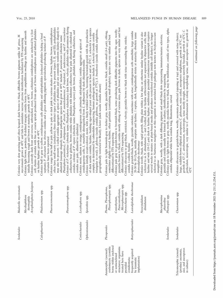
The level to which black molds can or should be identified inthe routine laboratory may depend on several factors, suchas the genus of the organism recovered, whether or not anepidemiologic investigation is warranted, and/or the level ofidentification required for appropriate patient management.The phenotypic identification of black molds is based primarilyupon their macroscopic morphology (color, growth rate, andgrowth characteristics on standardized media), their micro-scopic morphology (hyphae, conidiogenous cells [specializedcells that produce the conidia], conidia [asexual reproductivepropagules], etc.), and a limited number of physiologic features(primarily cycloheximide tolerance, nitrate assimilation, ureahydrolysis, and growth at various salt concentrations). Onlygenus-level identification may be possible or required for gen-era with several similar, closely related species, such as de-scribed above for Exophiala. In some other genera, certainspecies are clearly associated with a particular type of mycosis,and a combination of morphologic features, temperature, andphysiology can provide a species-level identification. This is thecase for the agent of cerebral phaeohyphomycosis, Cladophia-lophora bantiana.
Macroscopic morphology. The medium (see “Isolation Pro-cedures and Culture” in Diagnosis below) is an importantconsideration in the identification of melanized fungi. The useof a medium that promotes growth most consistently matchingthe original description of the organism is preferred, and thistypically is a plant-based medium. The most commonly used ispotato dextrose agar (PDA) or variations thereof. It providescolony colors that are close to those originally described, and itis usually adequate for conidiation. Other plant-based mediainclude malt extract agar, V-8 juice agar, cereal agar, carnationleaf agar, cornmeal dextrose agar, and others. A more com-plete list of media and reagents may be found in the Manual ofClinical Microbiology, 9th ed. (441), and in the Atlas of ClinicalFungi, 2nd ed. (175). Phaeoid molds vary considerably in theircolony colors. Although this characteristic is highly dependentupon environmental conditions, it is one that can be useful inthe initial separation of genera/species. While most species arevarious shades of pale gray to dark gray to black, others may bebrown or very pale or may turn darker only with the productionof certain structures. Others may be some shade of purple ordistinguished by diffusible pigments. Etiologic agents which aretypically brown on PDA include Ochroconis gallopava, Pleuro-stomophora richardsiae, Pleurostomophora repens, some Phaeo-acremonium species, Wallemia sebi, Myceliophthora ther-mophila, and Veronaea botryosa. The “pale list” includes fungiwhich seldom turn dark, such as Phialemonium species. Lecy-thophora mutabilis remains lightly colored until the productionof dark chlamydospores. Ochroconis gallopava exudes a wine-red pigment into the agar (more pronounced on Sabourauddextrose agar [SDA]), and several Phaeoacremonium speciesexhibit purple to lavender colonies.
Microscopic morphology and pleomorphism. Variable mi-croscopic morphology in the same fungus, also referred to aspleomorphism or pleoanamorphism, is another feature usefulin the phenotypic identification of black molds. Some fungimay display more than one form, such as yeast-like growthinitially and more filamentous growth subsequently. This is
TA
BL
E1—
Con
tinue
d
Gen
usty
peO
rder
Gen
usor
spec
ies
Salie
ntph
enot
ypic
and/
ordi
agno
stic
feat
ures
b
Ach
aeto
miu
mst
rum
ariu
mC
olon
ies
pale
tolig
htbr
own
with
redd
ish-
brow
ndi
ffusi
ngpi
gmen
taf
ter
2w
eeks
,woo
lly;a
scom
ata
peri
thec
ialw
ithlo
ng,s
light
lycu
rved
seta
e;as
cosp
ores
hyal
ine
toda
rk,1
3-17
.5by
8.5-
11�
m,f
usoi
dal;
neur
otro
pic
spec
ies
with
grow
that
40°C
Ple
ospo
rale
sL
epto
spha
eria
spp.
Col
onie
sda
rk,v
elve
tyto
slig
htly
woo
lly,s
low
-gro
win
g;as
com
ata
clei
stot
heci
al(n
oop
enin
g);a
scos
pore
shy
alin
e,m
ostly
with
4-6
sept
a;L
.sen
egal
ensi
san
dL
.tho
mpk
insi
idis
tingu
ishe
dby
asco
spor
efe
atur
esM
icro
asca
les
Mic
roas
cus
spp.
Col
onie
sin
itial
lyw
hite
togr
ayto
brow
nish
;asc
omat
ape
rith
ecia
l,de
velo
ping
asbl
ack
dots
inco
ncen
tric
ring
son
the
agar
,and
may
exud
ea
cirr
hus
ofre
das
cosp
ores
atm
atur
ity;s
peci
estr
eate
dhe
reha
veve
rysi
mila
r,da
rkSc
opul
ario
psis
anam
orph
s;M
.cin
ereu
s,sh
ort
peri
thec
ialn
ecks
and
oran
ge-s
egm
ent-
shap
edas
cosp
ores
;M.c
irros
us,
long
erpe
rith
ecia
lnec
ksw
ithhe
art-
shap
edas
cosp
ores
;M.t
rigon
ospo
rus,
long
erpe
rith
ecia
lnec
ksw
ithtr
iang
ular
asco
spor
esP
seud
alle
sche
riasp
p.C
olon
ies
pale
toye
llow
ish
gray
togr
ayto
brow
nish
,woo
lly;a
scom
ata
clei
stot
heci
al;S
cedo
spor
ium
and
Gra
phiu
man
amor
phs
pres
ent;
hum
anpa
thog
enic
spec
ies
asde
fined
byre
cent
mol
ecul
arst
udie
sar
eP
seud
alle
sche
riabo
ydii
(ana
mor
phSc
edos
poriu
mbo
ydii)
,Pse
udal
lesc
heria
apio
sper
ma
(ana
mor
phSc
edos
poriu
map
iosp
erm
um,h
eter
otha
llic,
does
not
form
ate
leom
orph
incu
lture
, D-r
ibos
ene
gativ
e),P
.elli
psoi
dea
aA
dapt
edfr
omT
able
14-1
from
refe
renc
e72
4w
ithpe
rmis
sion
.Thi
slis
tis
not
alli
nclu
sive
.Pic
ture
sof
allo
rgan
ism
sar
eav
aila
ble
atdo
ctor
fung
us.o
rgor
onth
eC
D-R
OM
ofth
eA
tlas
ofC
linic
alF
ungi
,pilo
tve
rsio
nof
3rd
ed.(
174)
.b
On
pota
tofla
keag
arat
25°C
.c
Tru
ese
pta
cont
inuo
usw
ithou
ter
wal
l.d
Pseu
dose
pta
whe
reon
lyin
ner
wal
lsar
ein
volv
ed.
890 REVANKAR AND SUTTON CLIN. MICROBIOL. REV.
Dow
nloa
ded
from
http
s://j
ourn
als.
asm
.org
/jour
nal/c
mr
on 1
8 N
ovem
ber
2021
by
211.
21.2
54.1
51.

TABLE 2. Melanized fungi in human diseasea
Genus Species
Achaetomiumd....................................................................A. strumariumAcrophialophorab ...............................................................A. fusisporaAlternariab...........................................................................A. alternata, A. chlamydospora, A. dianthicola, A. infectoria, A. longipes, A. tenuissimaAnthopsisb ...........................................................................A. deltoideaArniumd ..............................................................................A. leporinumArthriniumb .........................................................................A. phaeospermumAscotrichad..........................................................................A. chartarumAureobasidiumb ..................................................................A. pullulansBipolarisb.............................................................................B. australiensis, B. hawaiiensis, B. papendorfii, B. spiciferaBotryomycesb.......................................................................B. caespitosusChaetomiumd .....................................................................C. atrobrunneum, C. funicola, C. globosum, C. murorum, C. perlucidumCladophialophorab .............................................................C. arxii, C. bantiana, C. boppii, C. carrionii, C. devriesii, C. emmonsii, C. modesta, C. mycetomatis, C. saturnica,
C. samoensisCladorrhinumb....................................................................C. bulbillosumCladosporiumb....................................................................C. cladosporioides, C. herbarum, C. oxysporum, C. sphaerospermumColletotrichumc...................................................................C. coccodes, C. crassipes, C. dematium, C. gloeosporioides, C. graminicolaConiothyriumb ....................................................................C. fuckeliiCorynesporab.......................................................................C. cassiicolaCurvulariab .........................................................................C. brachyspora, C. clavata, C. geniculata, C. inequalis, C. lunata, C. pallescens, C. senegalensis, C. verruculosaCyphellophorab ...................................................................C. laciniata, C. pluriseptataDichotomophthorab............................................................D. portulacaeDichotomophthoropsisb......................................................D. nymphaearumDissitimurusb.......................................................................D. exedrusDrechslerab..........................................................................D. biseptataExophialab ..........................................................................E. asiatica, E. attenuata, E. bergeri, E. castellanii, E. dermatitidis, E. jeanselmei, E. lecanii-corni, E. moniliae, E.
oligosperma, E. phaeomuriformis, E. pisciphila, E. salmonis, E. spinifera, E. xenobioticaExserohilumb.......................................................................E. longirostratum, E. mcginnisii, E. rostratumFonsecaeab..........................................................................F. monophora, F. pedrosoiHormonemab ......................................................................H. dematioidesHortaeaeb ............................................................................H. werneckiiLasiodiplodiac.....................................................................L. theobromaeLecythophorab ....................................................................L. hoffmannii, L. mutabilisLeptosphaeriad....................................................................L. senegalensis, L. thompkinsiiMacrophominac ..................................................................M. phaseolinaMadurellab ..........................................................................M. grisea, M. mycetomatisMicroascusd ........................................................................M. cinereus, M. cirrosus, M. trigonosporusMonilliellab .........................................................................M. suaveolensMicrosphaeropsisc...............................................................M. arundinis, M. olivaceaMyceliophthorab..................................................................M. thermophilaMycocentrosporab ...............................................................M. acerinaMycoleptodiscusb ................................................................M. indicusNeoscytalidiumc ..................................................................N. dimidiatumNeotestudinad .....................................................................N. rosatiiNigrosporab .........................................................................N. sphaericaOchrocladosporiumb ..........................................................O. elatumOchroconisb ........................................................................O. gallopava, O. humicola, O. tshawytschaeOidiodendronb ....................................................................O. cerealisPhaeoacremoniumb ............................................................P. alvesii, P. amstelodamense, P. griseorubrum, P. krajdenii, P. parasiticum, P. rubrigenum, P. sphinctrophorum,
P. tardicrescens, P. venezuelensePhaeosclerab .......................................................................P. dematioidesPhaeotrichoconisb...............................................................P. crotalariaePhialemoniumb ...................................................................P. curvatum, P. obovatumPhialophorab .......................................................................P. americana, P. bubakii, P. europaea, P. reptans, P. verrucosaPhomab ...............................................................................P. cruris-hominis, P. dennisii var. oculo-hominis, P. eupyrena, P. glomerata, P. herbarum, P. minutella,
P. minutispora, P. sorghinaPiedraiad .............................................................................P. hortaePleurophomac .....................................................................P. cavaPleurophomopsisc ...............................................................P. lignicolaPleurostomophorab .............................................................P. repens, P. richardsiaePseudochaetosphaeronemab ..............................................P. larensePseudomicrodochiumb .......................................................P. suttoniiPyrenochaetab .....................................................................P. mackinnonii, P. romeroi, P. unguis-hominisRhinocladiellab ...................................................................R. aquaspersa, R. basitona, R. mackenziei, R. similisSarcinomycesb.....................................................................S. phaeomuriformisScedosporiuma ....................................................................S. prolificansScopulariopsisb ...................................................................S. asperula, S. brumptii, S. fuscaSphaeropsisc ........................................................................S. subglobosaStenellab ..............................................................................S. araguataTaeniolellab .........................................................................T. stillbosporaTetraploab ...........................................................................T. aristataThermomycesb ....................................................................T. lanuginosusUlocladiumb........................................................................U. chartarumVeronaeab............................................................................V. botryosa
a Some doubtful cases have been omitted; the list may not be all inclusive. Some genera that are outside the taxonomic orders discussed in the text but that containmelanized structures are included. Adapted from reference 493 with permission of the publisher.
b Anamorphic hyphomycete.c Anamorphic coelomycete.d Teleomorphic ascomycete.
VOL. 23, 2010 MELANIZED FUNGI IN HUMAN DISEASE 891
Dow
nloa
ded
from
http
s://j
ourn
als.
asm
.org
/jour
nal/c
mr
on 1
8 N
ovem
ber
2021
by
211.
21.2
54.1
51.

common in the black yeasts such as Exophiala and relatedgenera. Pleoanamorphism may also be exhibited by differenttypes of anamorphic structures (synanamorphs), such as theGraphium state in Pseudallescheria or variably shaped conidiain Pleurostomophora richardsiae (Fig. 1). Identification of ho-mothallic ascomycetes is typically based on the type of asco-mata produced (primarily cleistothecia [round, closed struc-tures containing asci and ascospores] or perithecia [round topear-shaped structures with an opening or ostiole containingasci and ascospores], as in Pseudallescheria or Chaetomium/Achaetomium/Microascus, respectively) and differences in as-cospore morphology. Ascospores may be of various sizes,shapes, colors, and ornamentations. The bulk of clinical blackmolds, however, are heterothallic ascomycetes. These mito-sporic fungi are identified mostly by their methods of conidio-genesis and the morphology of their conidia. The majority ofmitosporic isolates are hyphomycetes with their conidia bornefree in the aerial mycelium. Also seen are coelomycetes, whoseconidia are borne within asexual structures known as conidi-omata. The methods of conidiogenesis (blastic [blown-out �blastoconidia, as seen in many genera] or thallic [formed frompreexisting hyphae � arthroconidia, as in Neoscytalidium]), thetypes of conidiogenous cells (primarily annellidic [Scedospo-rium, Scopulariopsis, and Hortaea] or phialidic [many genera]),and the morphology of the conidia are taken in aggregate toform the basis for a morphologic identification. Annelloconidiaare formed from percurrent, indeterminate conidiogenouscells that produce rings or annellations and become longer andnarrower with the production of conidia, while phialoconidiaare formed from conidiogenous cells with collarettes that maybe quite distinct or subtle, and the conidiogenous cell remainsthe same size and shape with conidial production. It should bepointed out, however, that these morphologic features used toidentify anamorphic species lacking teleomorphs are strictlyphenotypic and do not define their phylogenetic placementwithin the order (157).
Physiologic features. Physiologic characteristics may also as-sist in separation of various genera/species. However, onlythose that are available in routine laboratories are widely em-ployed. The ability or inability of isolates to grow on mediacontaining cycloheximide (referred to as cycloheximide toler-ance), nitrate assimilation, urease activity, and salt tolerance,particularly for halophilic strains, are all useful adjuncts to themorphologic examination. Larger reference labs and researchfacilities also may use a battery of carbon assimilation profiles.Temperature tolerance is also useful in segregating potentialpathogens. Those that fail to grow at 35°C are more likely to berecovered from superficial sites, while those capable of growthat this temperature have the potential for more invasive humandisease. Several clinically significant dematiaceous molds arethermotolerant to thermophilic, with maximum growth tem-peratures to 45°C and beyond. A partial listing of these poten-tially neurotropic species includes E. dermatitidis, O. gallopava,C. bantiana, C. modesta, C. emmonsii, R. mackenziei, Acrophia-lophora fusispora, Fonsecaea monophora, and some aggressiveAchaetomium and Chaetomium species.
Molecular Characterization
Molecular characterization of fungi is a mature disciplinein the molecular systematics arena, with multilocus datasets,extensive taxon sampling, and rigorous analytical methodsbeing the norm (340). Its use in the clinical laboratory,however, is mostly restricted to epidemiologic studies and toidentification of unusual/uncommon or difficult-to-identifyisolates. Molecular identification of most species relies onsequencing of ribosomal genes and comparison with pub-lished databases, notably those in GenBank; however, over10% of these deposits may be erroneous (176). Private da-tabases are also sometimes utilized for particular genera;however, these are difficult to access and may also containincorrect deposits. Also, various methods and genes or generegions such as the internal transcribed spacer regions ITS1and ITS2, the D1/D2 domains, �-tubulin, actin, calmodulin,manganese superoxide dismutase, ATPase subunit 6, chitinsynthase, mitochondrial small-subunit (SSU) rRNA, trans-lation elongation factor 1�, and others are utilized, so thatinterlaboratory standardization of sequencing is lacking.Several International Society for Human and Animal My-cology (ISHAM) working groups are addressing standard-ization of fungal sequencing (58) as is the Clinical andLaboratory Standards Institute (CLSI) (146). Genera forwhich substantial sequencing data are available and forwhich species distinction appears to be satisfactory includethose known as black yeasts, i.e., Exophiala and relatedgenera (ITS) (825), Sporothrix species (calmodulin) (480),Phaeoacremonium species (�-tubulin and actin) (525), andPseudallescheria/Scedosporium species (ribosomal DNA[rDNA] gene cluster, �-tubulin, calmodulin, and translationelongation factor 1�) (330). Molecular characterizationshould always be evaluated in light of phenotypic features,and sequence data for uncommon and/or potentially newspecies should be compared with those for ex-type strains.
FIG. 1. Conidiogenous cells of Pleurostomophora richardsiae, dem-onstrating prominent flaring collarettes as well as the two types ofconidia (oval and globose) produced by this species. (Unless otherwisenoted, in this and subsequent figures light microscopy photomicro-graphs of conidiogenous cells and/or conidia were taken from slideculture preparations grown on potato flakes agar for 7 days at 25°C.)
892 REVANKAR AND SUTTON CLIN. MICROBIOL. REV.
Dow
nloa
ded
from
http
s://j
ourn
als.
asm
.org
/jour
nal/c
mr
on 1
8 N
ovem
ber
2021
by
211.
21.2
54.1
51.
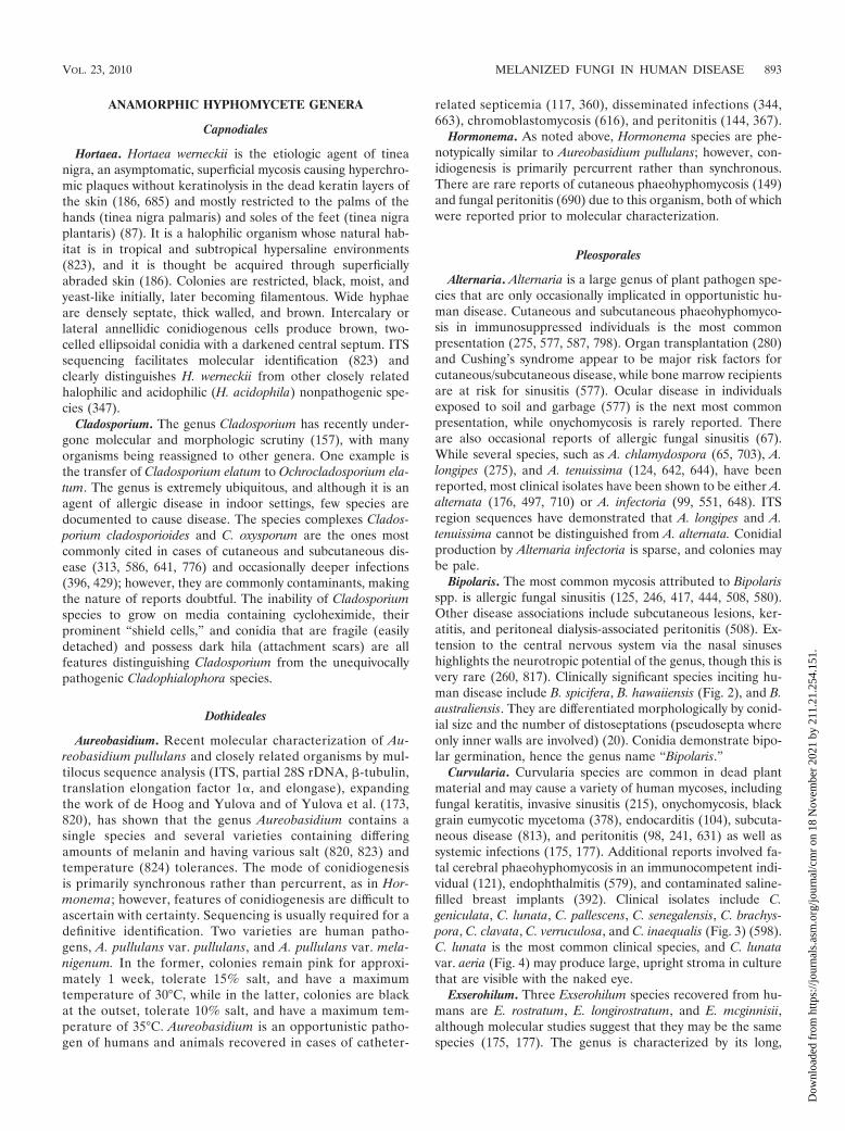
ANAMORPHIC HYPHOMYCETE GENERA
Capnodiales
Hortaea. Hortaea werneckii is the etiologic agent of tineanigra, an asymptomatic, superficial mycosis causing hyperchro-mic plaques without keratinolysis in the dead keratin layers ofthe skin (186, 685) and mostly restricted to the palms of thehands (tinea nigra palmaris) and soles of the feet (tinea nigraplantaris) (87). It is a halophilic organism whose natural hab-itat is in tropical and subtropical hypersaline environments(823), and it is thought be acquired through superficiallyabraded skin (186). Colonies are restricted, black, moist, andyeast-like initially, later becoming filamentous. Wide hyphaeare densely septate, thick walled, and brown. Intercalary orlateral annellidic conidiogenous cells produce brown, two-celled ellipsoidal conidia with a darkened central septum. ITSsequencing facilitates molecular identification (823) andclearly distinguishes H. werneckii from other closely relatedhalophilic and acidophilic (H. acidophila) nonpathogenic spe-cies (347).
Cladosporium. The genus Cladosporium has recently under-gone molecular and morphologic scrutiny (157), with manyorganisms being reassigned to other genera. One example isthe transfer of Cladosporium elatum to Ochrocladosporium ela-tum. The genus is extremely ubiquitous, and although it is anagent of allergic disease in indoor settings, few species aredocumented to cause disease. The species complexes Clados-porium cladosporioides and C. oxysporum are the ones mostcommonly cited in cases of cutaneous and subcutaneous dis-ease (313, 586, 641, 776) and occasionally deeper infections(396, 429); however, they are commonly contaminants, makingthe nature of reports doubtful. The inability of Cladosporiumspecies to grow on media containing cycloheximide, theirprominent “shield cells,” and conidia that are fragile (easilydetached) and possess dark hila (attachment scars) are allfeatures distinguishing Cladosporium from the unequivocallypathogenic Cladophialophora species.
Dothideales
Aureobasidium. Recent molecular characterization of Au-reobasidium pullulans and closely related organisms by mul-tilocus sequence analysis (ITS, partial 28S rDNA, �-tubulin,translation elongation factor 1�, and elongase), expandingthe work of de Hoog and Yulova and of Yulova et al. (173,820), has shown that the genus Aureobasidium contains asingle species and several varieties containing differingamounts of melanin and having various salt (820, 823) andtemperature (824) tolerances. The mode of conidiogenesisis primarily synchronous rather than percurrent, as in Hor-monema; however, features of conidiogenesis are difficult toascertain with certainty. Sequencing is usually required for adefinitive identification. Two varieties are human patho-gens, A. pullulans var. pullulans, and A. pullulans var. mela-nigenum. In the former, colonies remain pink for approxi-mately 1 week, tolerate 15% salt, and have a maximumtemperature of 30°C, while in the latter, colonies are blackat the outset, tolerate 10% salt, and have a maximum tem-perature of 35°C. Aureobasidium is an opportunistic patho-gen of humans and animals recovered in cases of catheter-
related septicemia (117, 360), disseminated infections (344,663), chromoblastomycosis (616), and peritonitis (144, 367).
Hormonema. As noted above, Hormonema species are phe-notypically similar to Aureobasidium pullulans; however, con-idiogenesis is primarily percurrent rather than synchronous.There are rare reports of cutaneous phaeohyphomycosis (149)and fungal peritonitis (690) due to this organism, both of whichwere reported prior to molecular characterization.
Pleosporales
Alternaria. Alternaria is a large genus of plant pathogen spe-cies that are only occasionally implicated in opportunistic hu-man disease. Cutaneous and subcutaneous phaeohyphomyco-sis in immunosuppressed individuals is the most commonpresentation (275, 577, 587, 798). Organ transplantation (280)and Cushing’s syndrome appear to be major risk factors forcutaneous/subcutaneous disease, while bone marrow recipientsare at risk for sinusitis (577). Ocular disease in individualsexposed to soil and garbage (577) is the next most commonpresentation, while onychomycosis is rarely reported. Thereare also occasional reports of allergic fungal sinusitis (67).While several species, such as A. chlamydospora (65, 703), A.longipes (275), and A. tenuissima (124, 642, 644), have beenreported, most clinical isolates have been shown to be either A.alternata (176, 497, 710) or A. infectoria (99, 551, 648). ITSregion sequences have demonstrated that A. longipes and A.tenuissima cannot be distinguished from A. alternata. Conidialproduction by Alternaria infectoria is sparse, and colonies maybe pale.
Bipolaris. The most common mycosis attributed to Bipolarisspp. is allergic fungal sinusitis (125, 246, 417, 444, 508, 580).Other disease associations include subcutaneous lesions, ker-atitis, and peritoneal dialysis-associated peritonitis (508). Ex-tension to the central nervous system via the nasal sinuseshighlights the neurotropic potential of the genus, though this isvery rare (260, 817). Clinically significant species inciting hu-man disease include B. spicifera, B. hawaiiensis (Fig. 2), and B.australiensis. They are differentiated morphologically by conid-ial size and the number of distoseptations (pseudosepta whereonly inner walls are involved) (20). Conidia demonstrate bipo-lar germination, hence the genus name “Bipolaris.”
Curvularia. Curvularia species are common in dead plantmaterial and may cause a variety of human mycoses, includingfungal keratitis, invasive sinusitis (215), onychomycosis, blackgrain eumycotic mycetoma (378), endocarditis (104), subcuta-neous disease (813), and peritonitis (98, 241, 631) as well assystemic infections (175, 177). Additional reports involved fa-tal cerebral phaeohyphomycosis in an immunocompetent indi-vidual (121), endophthalmitis (579), and contaminated saline-filled breast implants (392). Clinical isolates include C.geniculata, C. lunata, C. pallescens, C. senegalensis, C. brachys-pora, C. clavata, C. verruculosa, and C. inaequalis (Fig. 3) (598).C. lunata is the most common clinical species, and C. lunatavar. aeria (Fig. 4) may produce large, upright stroma in culturethat are visible with the naked eye.
Exserohilum. Three Exserohilum species recovered from hu-mans are E. rostratum, E. longirostratum, and E. mcginnisii,although molecular studies suggest that they may be the samespecies (175, 177). The genus is characterized by its long,
VOL. 23, 2010 MELANIZED FUNGI IN HUMAN DISEASE 893
Dow
nloa
ded
from
http
s://j
ourn
als.
asm
.org
/jour
nal/c
mr
on 1
8 N
ovem
ber
2021
by
211.
21.2
54.1
51.

multidistoseptate conidia and a protruding hilum. E. rostratumexhibits darkened basal and distal septa, and E. longirostratumhas conidia that are noticeably longer and centrally curved(Fig. 5), while E. mcginnisii has conidia with warty projectionson their outer walls. Not all authorities agree that E. rostratum
and E. longirostratum are separate species. Species are oppor-tunistic and are etiologic agents of sinusitis (566), which mayextend to the central nervous system (46), and keratitis (488),as well as cutaneous and subcutaneous mycoses (359, 508, 580).A fatal disseminated case was reported in a patient with aplas-tic anemia (37).
Chaetothyriales
Exophiala. Species in the genus Exophiala are frequentlyreferred to as “black yeasts” due to the ability of several spe-cies to form a budding yeast-like synanamorph as well as hy-phal forms. Colonies are olivaceous-black with a black reverseand are initially moist or yeast-like, later becoming velvety atmaturity. Asexual replication is by annellidic conidiogenouscells, and conidia are formed in clusters both from intercalary
FIG. 2. Conidia of Bipolaris hawaiiensis, demonstrating mostly fivedistosepta and six cells being borne from a geniculate conidiophore/conidiogeous cell.
FIG. 3. Conidia of Curvularia inaequalis with mostly five septa andsix cells borne from a geniculate conidiogenous cells.
FIG. 4. Conidia of Curvularia lunata var. aeria borne from a genic-ulate conidiogenous cell. Note that the middle cell is slightly enlarged,and septa are eusepta (true septa continuous with the outer wall).
FIG. 5. Multidistoseptate conidia of Exserohilum longirostratum,demonstrating a prominent basal septum (true septum) and a protrud-ing hilum.
894 REVANKAR AND SUTTON CLIN. MICROBIOL. REV.
Dow
nloa
ded
from
http
s://j
ourn
als.
asm
.org
/jour
nal/c
mr
on 1
8 N
ovem
ber
2021
by
211.
21.2
54.1
51.
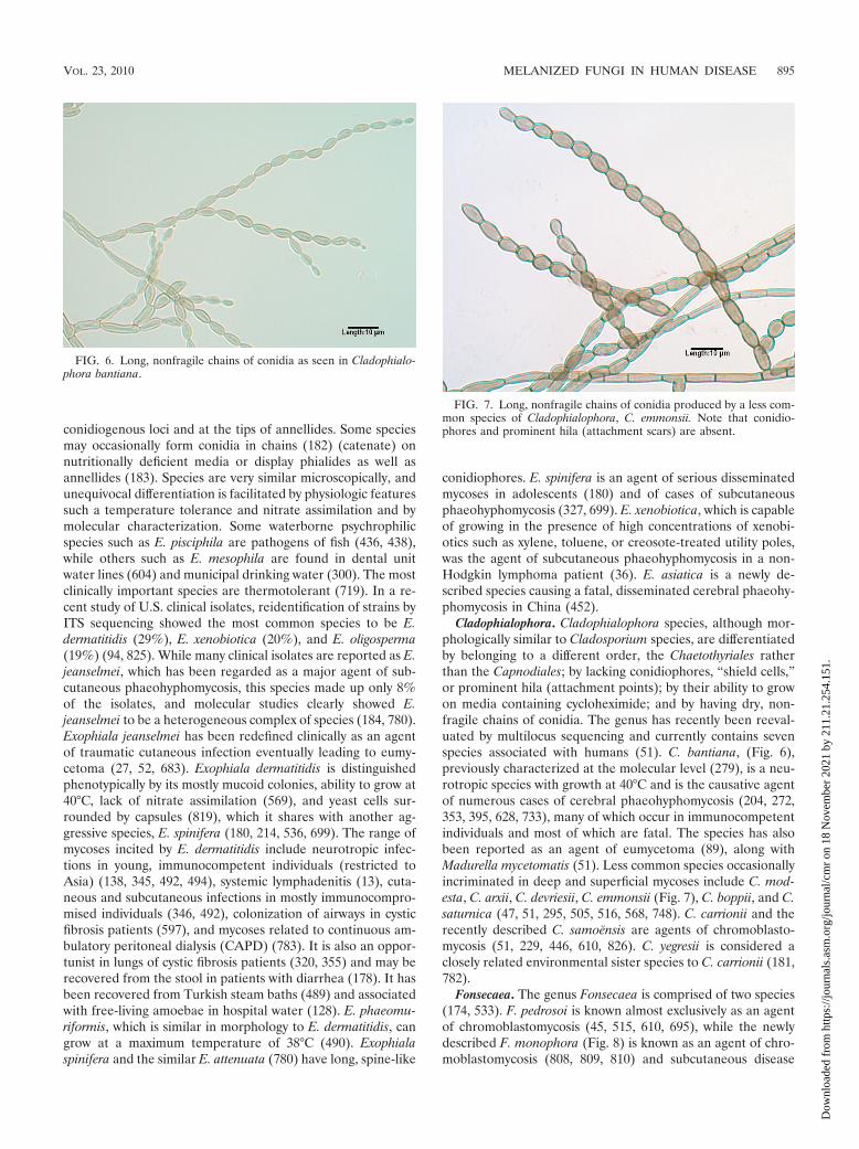
conidiogenous loci and at the tips of annellides. Some speciesmay occasionally form conidia in chains (182) (catenate) onnutritionally deficient media or display phialides as well asannellides (183). Species are very similar microscopically, andunequivocal differentiation is facilitated by physiologic featuressuch a temperature tolerance and nitrate assimilation and bymolecular characterization. Some waterborne psychrophilicspecies such as E. pisciphila are pathogens of fish (436, 438),while others such as E. mesophila are found in dental unitwater lines (604) and municipal drinking water (300). The mostclinically important species are thermotolerant (719). In a re-cent study of U.S. clinical isolates, reidentification of strains byITS sequencing showed the most common species to be E.dermatitidis (29%), E. xenobiotica (20%), and E. oligosperma(19%) (94, 825). While many clinical isolates are reported as E.jeanselmei, which has been regarded as a major agent of sub-cutaneous phaeohyphomycosis, this species made up only 8%of the isolates, and molecular studies clearly showed E.jeanselmei to be a heterogeneous complex of species (184, 780).Exophiala jeanselmei has been redefined clinically as an agentof traumatic cutaneous infection eventually leading to eumy-cetoma (27, 52, 683). Exophiala dermatitidis is distinguishedphenotypically by its mostly mucoid colonies, ability to grow at40°C, lack of nitrate assimilation (569), and yeast cells sur-rounded by capsules (819), which it shares with another ag-gressive species, E. spinifera (180, 214, 536, 699). The range ofmycoses incited by E. dermatitidis include neurotropic infec-tions in young, immunocompetent individuals (restricted toAsia) (138, 345, 492, 494), systemic lymphadenitis (13), cuta-neous and subcutaneous infections in mostly immunocompro-mised individuals (346, 492), colonization of airways in cysticfibrosis patients (597), and mycoses related to continuous am-bulatory peritoneal dialysis (CAPD) (783). It is also an oppor-tunist in lungs of cystic fibrosis patients (320, 355) and may berecovered from the stool in patients with diarrhea (178). It hasbeen recovered from Turkish steam baths (489) and associatedwith free-living amoebae in hospital water (128). E. phaeomu-riformis, which is similar in morphology to E. dermatitidis, cangrow at a maximum temperature of 38°C (490). Exophialaspinifera and the similar E. attenuata (780) have long, spine-like
conidiophores. E. spinifera is an agent of serious disseminatedmycoses in adolescents (180) and of cases of subcutaneousphaeohyphomycosis (327, 699). E. xenobiotica, which is capableof growing in the presence of high concentrations of xenobi-otics such as xylene, toluene, or creosote-treated utility poles,was the agent of subcutaneous phaeohyphomycosis in a non-Hodgkin lymphoma patient (36). E. asiatica is a newly de-scribed species causing a fatal, disseminated cerebral phaeohy-phomycosis in China (452).
Cladophialophora. Cladophialophora species, although mor-phologically similar to Cladosporium species, are differentiatedby belonging to a different order, the Chaetothyriales ratherthan the Capnodiales; by lacking conidiophores, “shield cells,”or prominent hila (attachment points); by their ability to growon media containing cycloheximide; and by having dry, non-fragile chains of conidia. The genus has recently been reeval-uated by multilocus sequencing and currently contains sevenspecies associated with humans (51). C. bantiana, (Fig. 6),previously characterized at the molecular level (279), is a neu-rotropic species with growth at 40°C and is the causative agentof numerous cases of cerebral phaeohyphomycosis (204, 272,353, 395, 628, 733), many of which occur in immunocompetentindividuals and most of which are fatal. The species has alsobeen reported as an agent of eumycetoma (89), along withMadurella mycetomatis (51). Less common species occasionallyincriminated in deep and superficial mycoses include C. mod-esta, C. arxii, C. devriesii, C. emmonsii (Fig. 7), C. boppii, and C.saturnica (47, 51, 295, 505, 516, 568, 748). C. carrionii and therecently described C. samoensis are agents of chromoblasto-mycosis (51, 229, 446, 610, 826). C. yegresii is considered aclosely related environmental sister species to C. carrionii (181,782).
Fonsecaea. The genus Fonsecaea is comprised of two species(174, 533). F. pedrosoi is known almost exclusively as an agentof chromoblastomycosis (45, 515, 610, 695), while the newlydescribed F. monophora (Fig. 8) is known as an agent of chro-moblastomycosis (808, 809, 810) and subcutaneous disease
FIG. 6. Long, nonfragile chains of conidia as seen in Cladophialo-phora bantiana.
FIG. 7. Long, nonfragile chains of conidia produced by a less com-mon species of Cladophialophora, C. emmonsii. Note that conidio-phores and prominent hila (attachment scars) are absent.
VOL. 23, 2010 MELANIZED FUNGI IN HUMAN DISEASE 895
Dow
nloa
ded
from
http
s://j
ourn
als.
asm
.org
/jour
nal/c
mr
on 1
8 N
ovem
ber
2021
by
211.
21.2
54.1
51.

and, more recently, cerebral phaeohyphomycosis (721, 733).Prior reported cases of central nervous system and/or otherdeep tissue infections (520, 545, 701) should most likely beattributed to F. monophora. A murine model of disseminatedinfection with F. monophora was recently reported (113). Bothspecies form conidia from swollen denticles which give rise tosecondary and tertiary conidia in short chains of up to fourconidia. Conidia may also be formed from sympodial conidio-phores, as in Rhinocladiella, and in balls from discrete phialideswith collarettes, as in Phialophora. Molecular characterizationis required for unequivocal differentiation.
Ochroconis. O. gallopava was initially observed to cause cen-tral nervous system disease in poultry (354). It has subse-quently been shown to be an etiologic agent of neurotropicinfections in immunocompromised humans (692) as well aspulmonary infections in immunocompetent hosts (348, 554).O. gallopava has colonies that are brownish rather than gray orolivaceous, produces a maroon diffusing pigment more pro-nounced on SDA than on PDA, grows at 40°C, fails to grow onmedia containing cycloheximide, and displays clavate, two-celled, hyaline conidia borne on long denticles (Fig. 9).
Phialophora. Some human pathogens with phialidic conidio-genesis previously assigned to Phialophora (263) have beenmoved to other genera, namely, Phaeoacremonium (525) andPleurostomophora (777), leaving only those species that arefilamentous throughout their life cycle. Both P. verrucosa andP. americana produce their conidia from phialides with con-spicuous darkened collarettes; these are funnel shaped andvase shaped in P. verrucosa (Fig. 10) and P. americana (Fig.11), respectively. Sequencing has demonstrated a close relat-edness, suggesting that the species may be synonymous (185,811). P. verrucosa is primarily an agent of chromoblastomycosis(257, 770), although other reported infections include endo-carditis, keratitis, and osteomyelitis (209, 760). A recently de-scribed species implicated in superficial infections, P. europaea,has very short collarettes (179).
Rhinocladiella. Four species of Rhinocladiella are knownagents of human disease. R. mackenziei (formerly Ramichlo-ridium mackenziei and also thought to be synonymous withRamichloridium obovoideum, which is now unrelated in thegenus Pleurothecium as P. obovoideum) (43) is a frequentlyfatal neurotropic organism previously thought to be restrictedto individuals residing in or immigrating from Middle Easterncountries (114, 394, 726). It has now been reported as theetiologic agent of a brain abscess in a man from India, an areawhere it is not endemic, who reported no travel outside thecountry (48). R. aquaspersa is an occasional agent of chromo-blastomycosis (39, 589, 693). R. basitona was recovered fromsubcutaneous lesions in a man from Japan (43). R. similis (184)appears to be the agent reported under the name R. atrovirensin cases of mycetoma (535) and cerebral phaeohyphomycosisin an AIDS patient (193).
FIG. 8. Conidial formation in Fonsecaea monophora. Conidia areformed from swollen denticles which give rise to secondary and tertiaryconidia in chains of up to four conidia. The same type of conidiogen-esis occurs in F. pedrosoi.
FIG. 9. Two-celled, clavate (club-shaped) conidia of Ochroconisgallopava borne on long, thin denticles.
FIG. 10. Dark, funnel-shaped collarettes at the tips of the conid-iogenous cells (phialides) in Phialophora verrucosa. Also note the oval-shaped conidia.
896 REVANKAR AND SUTTON CLIN. MICROBIOL. REV.
Dow
nloa
ded
from
http
s://j
ourn
als.
asm
.org
/jour
nal/c
mr
on 1
8 N
ovem
ber
2021
by
211.
21.2
54.1
51.
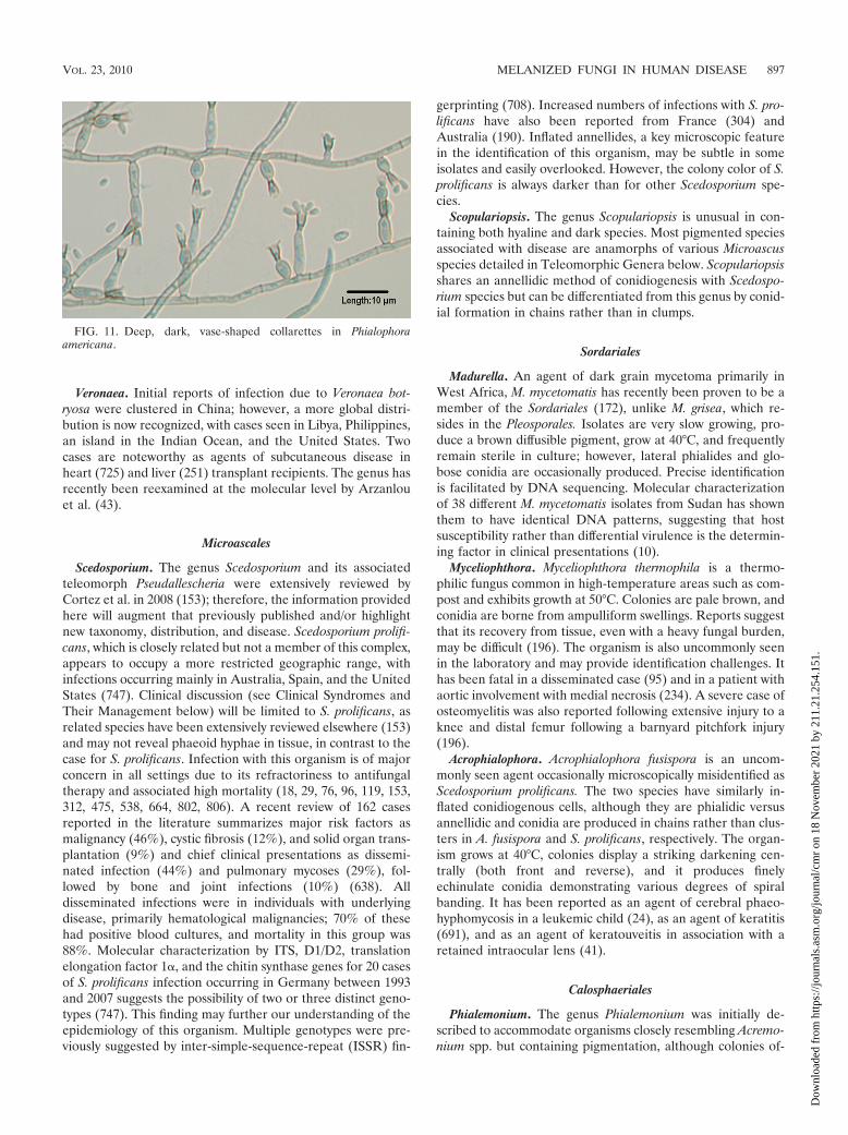
Veronaea. Initial reports of infection due to Veronaea bot-ryosa were clustered in China; however, a more global distri-bution is now recognized, with cases seen in Libya, Philippines,an island in the Indian Ocean, and the United States. Twocases are noteworthy as agents of subcutaneous disease inheart (725) and liver (251) transplant recipients. The genus hasrecently been reexamined at the molecular level by Arzanlouet al. (43).
Microascales
Scedosporium. The genus Scedosporium and its associatedteleomorph Pseudallescheria were extensively reviewed byCortez et al. in 2008 (153); therefore, the information providedhere will augment that previously published and/or highlightnew taxonomy, distribution, and disease. Scedosporium prolifi-cans, which is closely related but not a member of this complex,appears to occupy a more restricted geographic range, withinfections occurring mainly in Australia, Spain, and the UnitedStates (747). Clinical discussion (see Clinical Syndromes andTheir Management below) will be limited to S. prolificans, asrelated species have been extensively reviewed elsewhere (153)and may not reveal phaeoid hyphae in tissue, in contrast to thecase for S. prolificans. Infection with this organism is of majorconcern in all settings due to its refractoriness to antifungaltherapy and associated high mortality (18, 29, 76, 96, 119, 153,312, 475, 538, 664, 802, 806). A recent review of 162 casesreported in the literature summarizes major risk factors asmalignancy (46%), cystic fibrosis (12%), and solid organ trans-plantation (9%) and chief clinical presentations as dissemi-nated infection (44%) and pulmonary mycoses (29%), fol-lowed by bone and joint infections (10%) (638). Alldisseminated infections were in individuals with underlyingdisease, primarily hematological malignancies; 70% of thesehad positive blood cultures, and mortality in this group was88%. Molecular characterization by ITS, D1/D2, translationelongation factor 1�, and the chitin synthase genes for 20 casesof S. prolificans infection occurring in Germany between 1993and 2007 suggests the possibility of two or three distinct geno-types (747). This finding may further our understanding of theepidemiology of this organism. Multiple genotypes were pre-viously suggested by inter-simple-sequence-repeat (ISSR) fin-
gerprinting (708). Increased numbers of infections with S. pro-lificans have also been reported from France (304) andAustralia (190). Inflated annellides, a key microscopic featurein the identification of this organism, may be subtle in someisolates and easily overlooked. However, the colony color of S.prolificans is always darker than for other Scedosporium spe-cies.
Scopulariopsis. The genus Scopulariopsis is unusual in con-taining both hyaline and dark species. Most pigmented speciesassociated with disease are anamorphs of various Microascusspecies detailed in Teleomorphic Genera below. Scopulariopsisshares an annellidic method of conidiogenesis with Scedospo-rium species but can be differentiated from this genus by conid-ial formation in chains rather than in clumps.
Sordariales
Madurella. An agent of dark grain mycetoma primarily inWest Africa, M. mycetomatis has recently been proven to be amember of the Sordariales (172), unlike M. grisea, which re-sides in the Pleosporales. Isolates are very slow growing, pro-duce a brown diffusible pigment, grow at 40°C, and frequentlyremain sterile in culture; however, lateral phialides and glo-bose conidia are occasionally produced. Precise identificationis facilitated by DNA sequencing. Molecular characterizationof 38 different M. mycetomatis isolates from Sudan has shownthem to have identical DNA patterns, suggesting that hostsusceptibility rather than differential virulence is the determin-ing factor in clinical presentations (10).
Myceliophthora. Myceliophthora thermophila is a thermo-philic fungus common in high-temperature areas such as com-post and exhibits growth at 50°C. Colonies are pale brown, andconidia are borne from ampulliform swellings. Reports suggestthat its recovery from tissue, even with a heavy fungal burden,may be difficult (196). The organism is also uncommonly seenin the laboratory and may provide identification challenges. Ithas been fatal in a disseminated case (95) and in a patient withaortic involvement with medial necrosis (234). A severe case ofosteomyelitis was also reported following extensive injury to aknee and distal femur following a barnyard pitchfork injury(196).
Acrophialophora. Acrophialophora fusispora is an uncom-monly seen agent occasionally microscopically misidentified asScedosporium prolificans. The two species have similarly in-flated conidiogenous cells, although they are phialidic versusannellidic and conidia are produced in chains rather than clus-ters in A. fusispora and S. prolificans, respectively. The organ-ism grows at 40°C, colonies display a striking darkening cen-trally (both front and reverse), and it produces finelyechinulate conidia demonstrating various degrees of spiralbanding. It has been reported as an agent of cerebral phaeo-hyphomycosis in a leukemic child (24), as an agent of keratitis(691), and as an agent of keratouveitis in association with aretained intraocular lens (41).
Calosphaeriales
Phialemonium. The genus Phialemonium was initially de-scribed to accommodate organisms closely resembling Acremo-nium spp. but containing pigmentation, although colonies of-
FIG. 11. Deep, dark, vase-shaped collarettes in Phialophoraamericana.
VOL. 23, 2010 MELANIZED FUNGI IN HUMAN DISEASE 897
Dow
nloa
ded
from
http
s://j
ourn
als.
asm
.org
/jour
nal/c
mr
on 1
8 N
ovem
ber
2021
by
211.
21.2
54.1
51.

ten remain pale (264). Colonies are typically moist to slightlyfilamentous, and conidiogenous cells are a mixture of medium-length phialides and adelophialides (short phialides lacking abasal septum). The genus currently contains two species ofclinical interest, P. obovatum and P. curvatum. P. obovatumproduces a green, diffusible pigment; has obovate conidia (likean upside-down egg); and has been reported as an agent offatal endocarditis in a neonate (273). P. curvatum isolatesrange from cream to yellowish to pale brownish and haveallantoid (curved) conidia. Infections attributed to P. curvatuminclude cutaneous and subcutaneous disease, disseminated in-fection, endophthalmitis, peritonitis, arthritis, and fungemia(167, 264, 308, 793). Also reported are cases of hemodialysis-associated endovascular infections (608) and endocarditis(561), with some cases linked to intracavernous penile injec-tions in men frequenting impotence clinics (717). Several re-cent cases have demonstrated sporodochial formation in P.curvatum, a feature not previously seen in this species (167,608, 793). Rivero et al. have recently reviewed published Phi-alemonium cases (634).
Phaeoacremonium. The genus Phaeoacremonium initially ac-commodated isolates with features similar to those seen inboth Acremonium and Phialophora (159). It differs from theformer by having pigmented hyphae and conidiophores andfrom the latter by having indistinct collarettes and warty con-idiogenous cells. A recent morphologic and molecular charac-terization of the genus using �-tubulin sequences (525) hasmore clearly defined the genus and provided differential fea-tures for clinically significant species. Human pathogens in-clude P. parasiticum (obsolete Phialophora parasitica) (Fig. 12)(335), P. alvesii (567), P. amstelodamense, P. griseorubrum, P.krajdenii (525), P. rubrigenum (491), P. tardicrescens, and P.venezuelense (309, 525). Infections caused by P. parasiticuminclude subcutaneous abscesses (245), thorn-induced arthritis(651), and disseminated infection (54). Colony colors mayrange from yellowish brown to orange-brown to brown to lav-ender.
Pleurostomophora. Clinically significant species in the mostlywood-inhabiting genus Pleurostomophora include P. richardsiae(obsolete Phialophora richardsiae) and P. repens (obsolete Phi-alophora repens), and individuals acquiring these mycoses arecommonly immunocompromised (369, 601, 815). Species areanamorphs of the genus Pleurostoma. P. richardsiae is charac-terized microscopically by distinctive flaring collarettes (Fig. 1)and both globose and oval conidia. The colonies of both spe-cies tend to be brown rather than gray or olivaceous. Humaninfections include subcutaneous cases (311) and bone disease(761).
Coniochaetales
Lecythophora. Two Lecythophora species, L. mutabilis and L.hoffmannii, are of clinical significance. Both produce orange,moist colonies initially, with central darkening in L. mutabilisas pigmented chlamydospores are produced. Organisms areagents of endophthalmitis (677), sinusitis (485), and prostheticvalve endocarditis (207). Recent large-subunit rDNA sequenc-ing confirms the association of Lecythophora species with te-leomorphs in the genus Coniochaeta (792) in the order Conio-chaetales (361).
Ophiostomatales
Sporothrix. Sporotrichosis occurs worldwide, with the pri-mary agent of disease being Sporothrix schenckii. The disease iscommonly acquired by implantation of the fungus from varioustypes of woody/plant material. Lymphocutaneous lesions arethe norm; however, pulmonary disease and disseminated in-fections may occur in patients with underlying diseases (177).As a dimorphic fungus, it exhibits cigar-shaped yeasts in tissueand at 35°C and filamentous growth in culture. Only the sessileconidia borne along the sides of the hyphae are melanized. Ina recent study characterizing the genus by calmodulin sequenc-ing (480) and critically reviewing morphologic/physiologic fea-tures, these sessile conidia were shown to vary according tospecies within the S. schenckii species complex (479). They areelongate to triangular in S. schenckii and globose to subglobosein S. brasiliensis and S. globosa.
ANAMORPHIC COELOMYCETE GENERA
Pleosporales
Phoma and Phoma-like pycnidial coelomycetes. Several gen-era of morphologically similar pycnidial coelomycetes are oc-casionally recovered in cases of human subcutaneous disease(307, 585, 704), endophthalmitis (685), and deep tissue infec-tion (411); however, their documentation and reporting asetiologic agents is limited by a lack of adequate identification(727). They include Phoma, Pleurophoma, Pleurophomopsis,and Pyrenochaeta species, with small, hyaline, typically one-celled conidia, and Coniothyrium (411, 704), Paraconiothyrium(773), and Microsphaeropsis species (307, 585, 685), with palebrown to dark, one-celled conidia (Fig. 13). The morphologicfeatures of species within several sections in the genus Phomahave been detailed by Boerema et al. (84). Species in thesesimilar genera are best differentiated by ITS sequencing.
FIG. 12. Melanized hyphae, demonstrating warts (bottom), longrobust phialides, and allantoid (curved) conidia of Phaeoacremoniumparasiticum.
898 REVANKAR AND SUTTON CLIN. MICROBIOL. REV.
Dow
nloa
ded
from
http
s://j
ourn
als.
asm
.org
/jour
nal/c
mr
on 1
8 N
ovem
ber
2021
by
211.
21.2
54.1
51.
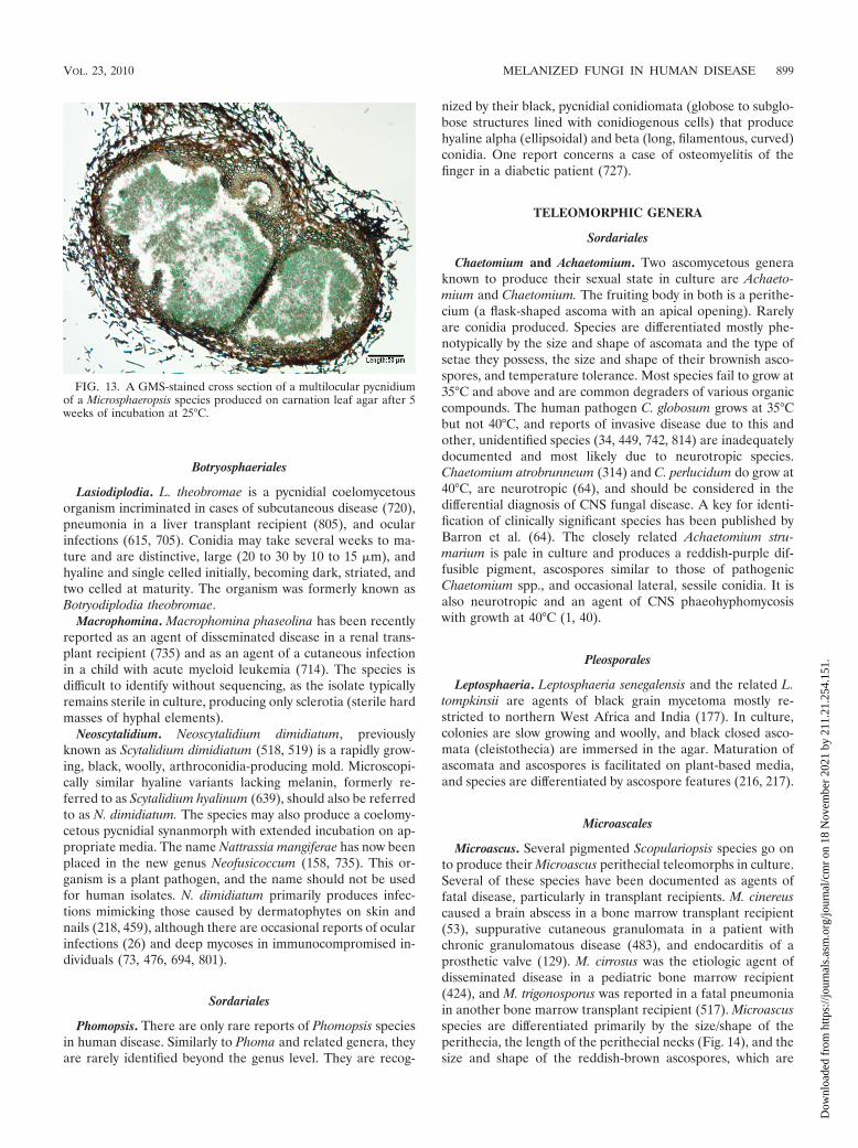
Botryosphaeriales
Lasiodiplodia. L. theobromae is a pycnidial coelomycetousorganism incriminated in cases of subcutaneous disease (720),pneumonia in a liver transplant recipient (805), and ocularinfections (615, 705). Conidia may take several weeks to ma-ture and are distinctive, large (20 to 30 by 10 to 15 �m), andhyaline and single celled initially, becoming dark, striated, andtwo celled at maturity. The organism was formerly known asBotryodiplodia theobromae.
Macrophomina. Macrophomina phaseolina has been recentlyreported as an agent of disseminated disease in a renal trans-plant recipient (735) and as an agent of a cutaneous infectionin a child with acute myeloid leukemia (714). The species isdifficult to identify without sequencing, as the isolate typicallyremains sterile in culture, producing only sclerotia (sterile hardmasses of hyphal elements).
Neoscytalidium. Neoscytalidium dimidiatum, previouslyknown as Scytalidium dimidiatum (518, 519) is a rapidly grow-ing, black, woolly, arthroconidia-producing mold. Microscopi-cally similar hyaline variants lacking melanin, formerly re-ferred to as Scytalidium hyalinum (639), should also be referredto as N. dimidiatum. The species may also produce a coelomy-cetous pycnidial synanmorph with extended incubation on ap-propriate media. The name Nattrassia mangiferae has now beenplaced in the new genus Neofusicoccum (158, 735). This or-ganism is a plant pathogen, and the name should not be usedfor human isolates. N. dimidiatum primarily produces infec-tions mimicking those caused by dermatophytes on skin andnails (218, 459), although there are occasional reports of ocularinfections (26) and deep mycoses in immunocompromised in-dividuals (73, 476, 694, 801).
Sordariales
Phomopsis. There are only rare reports of Phomopsis speciesin human disease. Similarly to Phoma and related genera, theyare rarely identified beyond the genus level. They are recog-
nized by their black, pycnidial conidiomata (globose to subglo-bose structures lined with conidiogenous cells) that producehyaline alpha (ellipsoidal) and beta (long, filamentous, curved)conidia. One report concerns a case of osteomyelitis of thefinger in a diabetic patient (727).
TELEOMORPHIC GENERA
Sordariales
Chaetomium and Achaetomium. Two ascomycetous generaknown to produce their sexual state in culture are Achaeto-mium and Chaetomium. The fruiting body in both is a perithe-cium (a flask-shaped ascoma with an apical opening). Rarelyare conidia produced. Species are differentiated mostly phe-notypically by the size and shape of ascomata and the type ofsetae they possess, the size and shape of their brownish asco-spores, and temperature tolerance. Most species fail to grow at35°C and above and are common degraders of various organiccompounds. The human pathogen C. globosum grows at 35°Cbut not 40°C, and reports of invasive disease due to this andother, unidentified species (34, 449, 742, 814) are inadequatelydocumented and most likely due to neurotropic species.Chaetomium atrobrunneum (314) and C. perlucidum do grow at40°C, are neurotropic (64), and should be considered in thedifferential diagnosis of CNS fungal disease. A key for identi-fication of clinically significant species has been published byBarron et al. (64). The closely related Achaetomium stru-marium is pale in culture and produces a reddish-purple dif-fusible pigment, ascospores similar to those of pathogenicChaetomium spp., and occasional lateral, sessile conidia. It isalso neurotropic and an agent of CNS phaeohyphomycosiswith growth at 40°C (1, 40).
Pleosporales
Leptosphaeria. Leptosphaeria senegalensis and the related L.tompkinsii are agents of black grain mycetoma mostly re-stricted to northern West Africa and India (177). In culture,colonies are slow growing and woolly, and black closed asco-mata (cleistothecia) are immersed in the agar. Maturation ofascomata and ascospores is facilitated on plant-based media,and species are differentiated by ascospore features (216, 217).
Microascales
Microascus. Several pigmented Scopulariopsis species go onto produce their Microascus perithecial teleomorphs in culture.Several of these species have been documented as agents offatal disease, particularly in transplant recipients. M. cinereuscaused a brain abscess in a bone marrow transplant recipient(53), suppurative cutaneous granulomata in a patient withchronic granulomatous disease (483), and endocarditis of aprosthetic valve (129). M. cirrosus was the etiologic agent ofdisseminated disease in a pediatric bone marrow recipient(424), and M. trigonosporus was reported in a fatal pneumoniain another bone marrow transplant recipient (517). Microascusspecies are differentiated primarily by the size/shape of theperithecia, the length of the perithecial necks (Fig. 14), and thesize and shape of the reddish-brown ascospores, which are
FIG. 13. A GMS-stained cross section of a multilocular pycnidiumof a Microsphaeropsis species produced on carnation leaf agar after 5weeks of incubation at 25°C.
VOL. 23, 2010 MELANIZED FUNGI IN HUMAN DISEASE 899
Dow
nloa
ded
from
http
s://j
ourn
als.
asm
.org
/jour
nal/c
mr
on 1
8 N
ovem
ber
2021
by
211.
21.2
54.1
51.

orange section shaped in M. cinereus, heart shaped in M. cir-rosus (Fig. 15), and triangular in M. trigonosporus (Fig. 16).
Pseudallescheria. As discussed above for the anamorphic ge-nus Scedosporium, Cortez et al. extensively reviewed Pseudall-escheria/Scedosporium in 2008 (153), and so only subsequenttaxonomic changes will be discussed here. The human patho-genic species as defined by recent molecular studies are asfollows: Pseudallescheria boydii (anamorph Scedosporium boy-dii), Pseudallescheria apiosperma (anamorph Scedosporium ap-iospermum, heterothallic, not forming its teleomorph in cul-ture, and D-ribose negative), and Pseudallescheria ellipsoidea(281–283). Other species of clinical interest in the P. boydiispecies complex include S. aurantiacum (190, 281) and S. de-hoogii (282).
PATHOGENESIS
Surveys of outdoor air for fungal spores routinely show de-matiaceous fungi (687). This suggests that all individuals areexposed, though few develop disease. Exposure is primarilyfrom inhalation or minor trauma, which is frequently not evennoticed by the patient. Relatively little is known regarding thepathogenic mechanisms by which melanized fungi cause dis-ease, particularly in immunocompetent individuals.
Role of Melanin
One of the likely virulence factors is the presence of melaninin the cell wall, which is common to all dematiaceous fungi,though relatively few species have been studied (439, 522, 573,666). Melanin in fungi is derived primarily from either dihy-droxyphenylalanine (L-DOPA) or dihydroxynaphthalene(DHN) (437). Dematiaceous fungi contain only DHN melanin;L-DOPA melanin has not been described to our knowledge(122, 274, 439, 766). It is generally localized to the cell wall,though the exact mechanism of its production is poorly under-stood. In the species F. pedrosoi, melanin is produced in mela-nosomes associated with Fe2� and Ca2� and then transportedto the cell wall (253). Melanin is extremely resistant to a varietyof physicochemical agents, including free radical compounds,toxic metals, desiccation, and even ionizing radiation (165, 249,331, 795). A species of Chaetomium was isolated from grassthat had been frozen in a glacier for over 5,000 years (331).
Considerable work has been done to elucidate the virulencepotentials of several fungi (dematiaceous and nondematia-ceous) that contain melanin, notably Aspergillus fumigatus,Cryptococcus neoformans, E. dermatiditis, and S. prolificans(123, 134, 201, 203, 205, 284, 428, 549, 573, 653, 671, 730, 756).There are multiple proposed mechanisms by which melaninmay act as a virulence factor (109, 324, 375). It may confer aprotective advantage by scavenging free radicals and hypochlo-rite that are produced by phagocytic cells in their oxidative
FIG. 14. Perithecium of Microascus trigonosporus formed on potatoflake agar after 3 weeks of incubation at 25°C. Note ascospores beingreleased from the ostiole in the neck of the perithecium.
FIG. 15. Heart-shaped ascospores of Microascus cirrosus producedon potato flake agar after 3 weeks of incubation at 25°C.
FIG. 16. Triangular ascospores of Microascus trigonosporus pro-duced on potato flake agar after 3 weeks of incubation at 25°C.
900 REVANKAR AND SUTTON CLIN. MICROBIOL. REV.
Dow
nloa
ded
from
http
s://j
ourn
als.
asm
.org
/jour
nal/c
mr
on 1
8 N
ovem
ber
2021
by
211.
21.2
54.1
51.

burst and that would normally kill most organisms (376, 671).In addition, melanin may bind to hydrolytic enzymes, therebypreventing their action on the plasma membrane, and to anti-fungal drugs, preventing their action (370, 375, 547, 768).There is also evidence that certain melanized fungi are lesssusceptible to phagocytosis and killing by neutrophils and mac-rophages (334, 584). These multiple functions may help explainthe pathogenic potential of some dematiaceous fungi, even inimmunocompetent hosts. Specifically, in E. dermatitidis, dis-ruption of melanin production leads to markedly reduced vir-ulence in animal models and restriction of hyphal growth (103,201, 203, 428). However, hyphae of S. prolificans were found tobe more susceptible to damage from neutrophils than A. fu-migatus (284). Melanin has also been shown to reduce thesusceptibility of M. mycetomatis to ketoconazole and itracon-azole by binding these drugs (766). Though only a minority ofdematiaceous fungi have been studied, it is likely that melaninplays a critical role in pathogenesis for clinically importantspecies.
Other Putative Virulence Factors
It is interesting to note that most allergic disease and eosino-philia is caused by three genera, Alternaria, Bipolaris, and Cur-vularia (622). The virulence factors in these fungi that areresponsible for eliciting allergic reactions are not well under-stood, though Alternaria was found to stimulate the degranu-lation of eosinophils, possibly due to an aspartic protease(372, 496). These organisms are very common in the envi-ronment, so exposure is practically universal, though theincidence of allergic disease is relatively low, suggesting thathost factors may play a role. A study by Schubert et al. foundthat HLA-DQB1*03 was associated with allergic fungal si-nusitis (676). Further studies are needed to better delineatethe importance of virulence factors other than melanin.
DIAGNOSIS
The timely and accurate diagnosis of fungal infections bymelanized fungi consists of a multifaceted approach. With theexponential increase in immunocompromised individuals, par-ticularly those in tertiary care cancer centers (72), this becomesimperative to prevent potentially fatal outcomes. Standardconventional diagnostic procedures include direct microscopy,histopathological stains to document tissue invasion, radio-graphic and computerized tomography (CT) findings, and iso-lation procedures to recover the fungus and identify the etio-logic agent. The clinical presentation and diagnostic findingssegregate these infections into the major categories of eumy-cetoma, chromoblastomycosis, and phaeohyphomycosis. Phae-ohyphomycosis maybe further delineated depending uponwhether infections are superficial or deep, by their anatomiclocation, and by the host’s response. The microscopic featuresseen in phaeohyphomycosis, however, are similar regardless ofthe anatomic site. The confusion surrounding the placement ofmembers of the Sporothrix schenckii and Pseudallescheria boydiispecies complexes within the dark molds is related to theirtissue presentation as yeast cells and hyaline hyphae, respec-tively. As the term phaeohyphomycosis is commonly used todescribe fungi with dark hyphae in tissue, these organisms
would be excluded; however, both produce melanized conidiain culture. As a thorough review of infections caused by Pseu-dallescheria/Scedosporium species has recently been published(153), this paper will concentrate on taxonomic changes andnew species documented as etiologic agents subsequent to thatreview.
Initial Specimen Processing
The appropriate specimen collection, transport, and pro-cessing procedures are important considerations in the dem-onstration of melanized fungi in tissue and their recovery inculture. The most useful diagnostic specimens are those col-lected at the source of infection; however, specimens periph-eral to the site of infection, such as blood cultures in hemat-ogenously disseminated disease, may also be diagnostic in theabsence of focal manifestations or when foci are not easilyaccessible. Appropriate specimens for the recovery of fungi aredetailed elsewhere in several reference works (723). Specimenscommonly obtained for recovery of melanized fungi includetissue biopsy specimens, aspirates, and body fluids. Surgicallyobtained specimens should always be cultured as well as pro-cessed for histopathology, and the inoculum should be finelysliced or minced rather than ground (as in the case for recoveryof H. capsulatum). Gross examination may occasionally revealevidence of melanized fungi as well (Fig. 17). Small volumes ofsterile body fluids may also be concentrated by syringe filtra-tion (0.2 �m). Several blood culture systems are available, andthe maximum amount of blood recommended should alwaysbe used. Swab cultures from superficial sites are usually notrepresentative of the disease process, frequently contain indig-enous contaminating mycobiota, and should generally beavoided. Grains or granules should also be washed severaltimes in antimicrobial-containing saline to avoid bacterialovergrowth (504). Also compromising etiologic agent recoveryis a delay in specimen transport. Optimally, most specimens
FIG. 17. Bipolaris spicifera colonies in stomach mucosa of patientwith disseminated disease (autopsy). (Reproduced from reference 624[original Fig. 15-6A] with kind permission of Springer Science andBusiness Media.)
VOL. 23, 2010 MELANIZED FUNGI IN HUMAN DISEASE 901
Dow
nloa
ded
from
http
s://j
ourn
als.
asm
.org
/jour
nal/c
mr
on 1
8 N
ovem
ber
2021
by
211.
21.2
54.1
51.
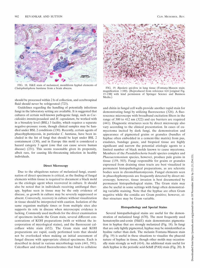
should be processed within 2 h of collection, and cerebrospinalfluid should never be refrigerated (723).
Guidelines regarding the handling of potentially infectiousfungi in the laboratory setting are available. It is suggested thatcultures of certain well-known pathogenic fungi, such as Coc-cidioides immitis/posadasii and H. capsulatum, be worked within a biosafety level (BSL) 3 facility, which requires a separatenegative-pressure room, though clinical samples may be han-dled under BSL 2 conditions (130). Recently, certain agents ofphaeohyphomycosis, in particular C. bantiana, have been in-cluded in the list of fungi that should be kept under BSL 2containment (130), and in Europe this mold is considered ahazard category 3 agent (one that can cause severe humandisease) (231). This seems reasonable given its propensity,albeit rare, for causing life-threatening infection in healthyindividuals.
Direct Microscopy
Due to the ubiquitous nature of melanized fungi, exami-nation of direct specimens is critical, as the finding of fungalelements within tissue is required to document a black moldas the etiologic agent when recovered in culture. It shouldalso be noted that in individuals receiving antifungal ther-apy, hyphae seen in tissue may be the only evidence ofdisease, as growth in culture may be severely suppressed orabsent. Conversely, recovery in culture without visualizationin tissue should be interpreted with caution. Isolation of thesame organism multiple times or from multiple sites alsosupports its role in disease when microscopic evidence islacking. Commonly used methods for the direct examinationof specimens include the Gram stain, several different con-centrations of KOH preparations (with or without the in-corporation of mycological stains), and the fluorescent cal-cofluor white stain (652). The Gram stain and KOHpreparations are rapid, easily performed tests that shouldnot be overlooked when making an initial assessment offungal disease with appropriate clinical specimens. Each isdescribed in detail in various microbiology texts (441, 591).Calcofluor and related fluorochromes that bind to cellulose
and chitin in fungal cell walls provide another rapid stain fordemonstrating fungi by utilizing fluorescence (326). A fluo-rescence microscope with broadband excitation filters in therange of 300 to 412 nm (322) and eye barriers are required(441). Diagnostic structures seen by direct microscopy alsovary according to the clinical presentation. In cases of eu-mycetoma incited by dark fungi, the demonstration andappearance of pigmented grains or granules (bundles ofhyphae often embedded in a cement-like matrix) from pus,exudates, bandage gauze, and biopsied tissue are highlysignificant and narrow the potential etiologic agents to alimited number of black molds known to cause mycetoma.Members of the Pseudallescheria boydii species complex andPhaeoacremonium species, however, produce pale grains intissue (159, 503). Fungi responsible for grains or granulesexpressed from draining sinus tracts are best visualized inpermanent histopathological preparations, as are scleroticbodies seen in chromoblastomycosis. Fungal elements seenin phaeohyphomycosis are frequently detected by direct mi-croscopy; however, tissue invasion is best documented bypermanent histopathological stains. The Gram stain mayalso be useful in some settings with fungi often demonstrat-ing variable staining. Note that the hyphae are often Gramnegative while the conidia are Gram positive; however, ei-ther structure may be Gram variable.
Histopathology and Special Stains
Several histopathological stains are useful for the demon-stration of melanized fungi (670). The most frequently usedhematoxylin-and-eosin (H&E) stain demonstrates pigmenta-tion in hyphae that are strongly melanized (Fig. 18). In fungithat are only lightly pigmented, hyphae may be misidentified ashyaline rather than dark. The melanin Fontana-Masson stain(Fig. 19) is useful in these situations to visualize the phaeoidnature of hyphae in tissue, though other molds may occasion-ally stain strongly as well (414). An additional stain useful fordark hyphae is the periodic acid-Schiff (PAS) stain (Fig. 20). It
FIG. 18. H&E stain of melanized, moniliform hyphal elements ofCladophialophora bantiana from a brain abscess. FIG. 19. Bipolaris spicifera in lung tissue (Fontana-Masson stain;
magnification �100). (Reproduced from reference 624 [original Fig.15-13B] with kind permission of Springer Science and BusinessMedia.)
902 REVANKAR AND SUTTON CLIN. MICROBIOL. REV.
Dow
nloa
ded
from
http
s://j
ourn
als.
asm
.org
/jour
nal/c
mr
on 1
8 N
ovem
ber
2021
by
211.
21.2
54.1
51.
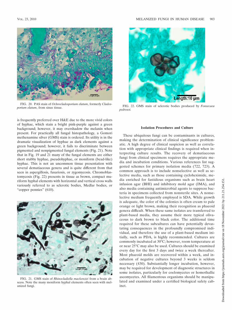
is frequently preferred over H&E due to the more vivid colorsof hyphae, which stain a bright pink-purple against a greenbackground; however, it may overshadow the melanin whenpresent. For practically all fungal histopathology, a Gomorimethenamine silver (GMS) stain is ordered. Its utility is in thedramatic visualization of hyphae as dark elements against agreen background; however, it fails to discriminate betweenpigmented and nonpigmented fungal elements (Fig. 21). Notethat in Fig. 19 and 21 many of the fungal elements are eithershort stubby hyphae, pseudohyphae, or moniform (bead-like)hyphae. This is not an uncommon tissue presentation withseveral dematiaceous genera and is quite different from thatseen in aspergillosis, fusariosis, or zygomycosis. Chromoblas-tomycosis (Fig. 22) presents in tissue as brown, compact mu-riform hyphal elements with horizontal and vertical cross wallsvariously referred to as sclerotic bodies, Medlar bodies, or“copper pennies” (610).
Isolation Procedures and Culture
These ubiquitous fungi can be contaminants in cultures,making the determination of clinical significance problem-atic. A high degree of clinical suspicion as well as correla-tion with appropriate clinical findings is required when in-terpreting culture results. The recovery of dematiaceousfungi from clinical specimens requires the appropriate me-dia and incubation conditions. Various references list sug-gested schemes for primary isolation media (722, 723). Acommon approach is to include nonselective as well as se-lective media, such as those containing cycloheximide, me-dia enriched for fastidious organisms such as brain heartinfusion agar (BHI) and inhibitory mold agar (IMA), andalso media containing antimicrobial agents to suppress bac-teria in specimens collected from nonsterile sites. A nonse-lective medium frequently employed is SDA. While growthis adequate, the color of the colonies is often cream to paleorange or light brown, making their recognition as phaeoidgenera difficult. When these same isolates are transferred toplant-based media, they assume their more typical oliva-ceous to dark brown to black color. The additional timerequired for these subcultures can have potentially devas-tating consequences in the profoundly compromised indi-vidual, and therefore the use of a plant-based medium ini-tially, such as PDA, is highly recommended. Cultures arecommonly incubated at 30°C; however, room temperature ator near 25°C may also be used. Cultures should be examinedevery day for the first 3 days and twice a week thereafter.Most phaeoid molds are recovered within a week, and in-cubation of negative cultures beyond 3 weeks is seldomnecessary (430). Substantially longer incubation, however,may be required for development of diagnostic structures insome isolates, particularly for coelomycetes or homothallicascomycetes. All filamentous organisms should be manipu-lated and examined under a certified biological safety cab-inet.
FIG. 20. PAS stain of Ochrocladosporium elatum, formerly Clados-porium elatum, from sinus tissue.
FIG. 21. GMS stain of Rhinocladiella mackenziei from a brain ab-scess. Note the many moniform hyphal elements often seen with mel-anized fungi.
FIG. 22. GMS stain of sclerotic bodies produced by Fonsecaeapedrosoi.
VOL. 23, 2010 MELANIZED FUNGI IN HUMAN DISEASE 903
Dow
nloa
ded
from
http
s://j
ourn
als.
asm
.org
/jour
nal/c
mr
on 1
8 N
ovem
ber
2021
by
211.
21.2
54.1
51.
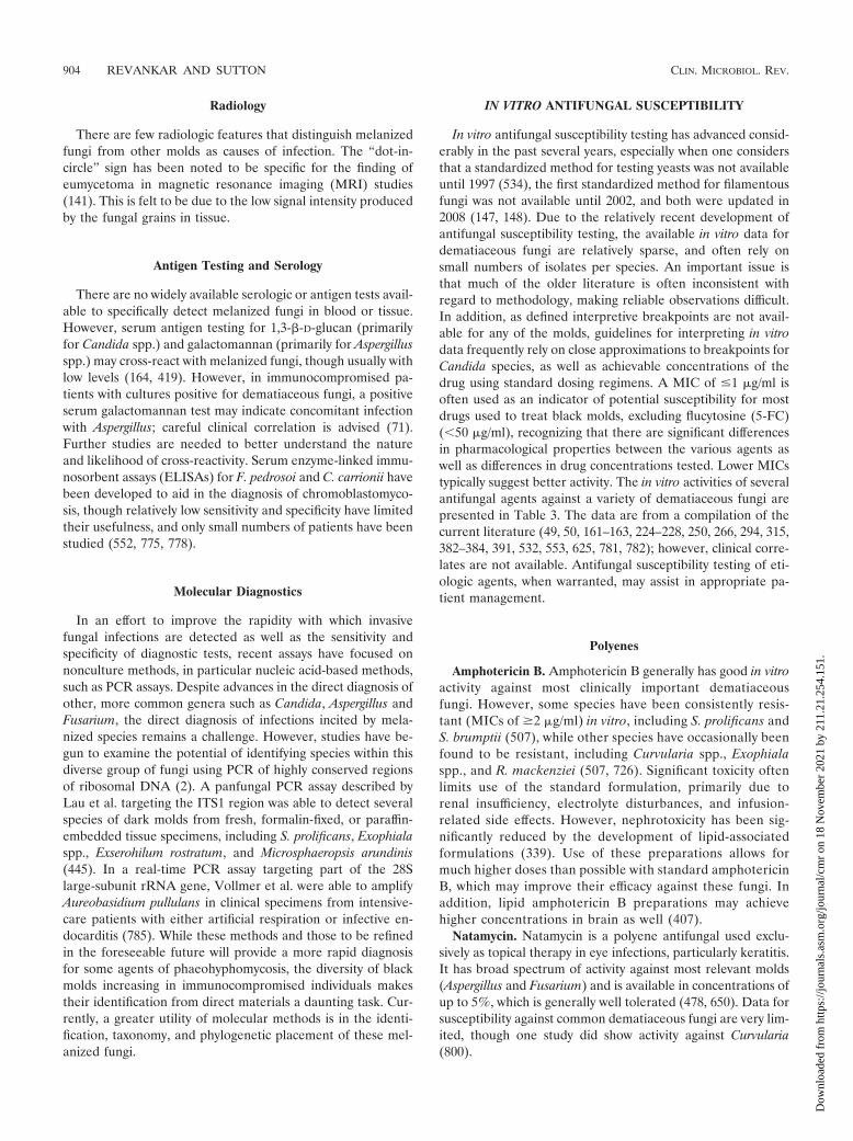
Radiology
There are few radiologic features that distinguish melanizedfungi from other molds as causes of infection. The “dot-in-circle” sign has been noted to be specific for the finding ofeumycetoma in magnetic resonance imaging (MRI) studies(141). This is felt to be due to the low signal intensity producedby the fungal grains in tissue.
Antigen Testing and Serology
There are no widely available serologic or antigen tests avail-able to specifically detect melanized fungi in blood or tissue.However, serum antigen testing for 1,3-�-D-glucan (primarilyfor Candida spp.) and galactomannan (primarily for Aspergillusspp.) may cross-react with melanized fungi, though usually withlow levels (164, 419). However, in immunocompromised pa-tients with cultures positive for dematiaceous fungi, a positiveserum galactomannan test may indicate concomitant infectionwith Aspergillus; careful clinical correlation is advised (71).Further studies are needed to better understand the natureand likelihood of cross-reactivity. Serum enzyme-linked immu-nosorbent assays (ELISAs) for F. pedrosoi and C. carrionii havebeen developed to aid in the diagnosis of chromoblastomyco-sis, though relatively low sensitivity and specificity have limitedtheir usefulness, and only small numbers of patients have beenstudied (552, 775, 778).
Molecular Diagnostics
In an effort to improve the rapidity with which invasivefungal infections are detected as well as the sensitivity andspecificity of diagnostic tests, recent assays have focused onnonculture methods, in particular nucleic acid-based methods,such as PCR assays. Despite advances in the direct diagnosis ofother, more common genera such as Candida, Aspergillus andFusarium, the direct diagnosis of infections incited by mela-nized species remains a challenge. However, studies have be-gun to examine the potential of identifying species within thisdiverse group of fungi using PCR of highly conserved regionsof ribosomal DNA (2). A panfungal PCR assay described byLau et al. targeting the ITS1 region was able to detect severalspecies of dark molds from fresh, formalin-fixed, or paraffin-embedded tissue specimens, including S. prolificans, Exophialaspp., Exserohilum rostratum, and Microsphaeropsis arundinis(445). In a real-time PCR assay targeting part of the 28Slarge-subunit rRNA gene, Vollmer et al. were able to amplifyAureobasidium pullulans in clinical specimens from intensive-care patients with either artificial respiration or infective en-docarditis (785). While these methods and those to be refinedin the foreseeable future will provide a more rapid diagnosisfor some agents of phaeohyphomycosis, the diversity of blackmolds increasing in immunocompromised individuals makestheir identification from direct materials a daunting task. Cur-rently, a greater utility of molecular methods is in the identi-fication, taxonomy, and phylogenetic placement of these mel-anized fungi.
IN VITRO ANTIFUNGAL SUSCEPTIBILITY
In vitro antifungal susceptibility testing has advanced consid-erably in the past several years, especially when one considersthat a standardized method for testing yeasts was not availableuntil 1997 (534), the first standardized method for filamentousfungi was not available until 2002, and both were updated in2008 (147, 148). Due to the relatively recent development ofantifungal susceptibility testing, the available in vitro data fordematiaceous fungi are relatively sparse, and often rely onsmall numbers of isolates per species. An important issue isthat much of the older literature is often inconsistent withregard to methodology, making reliable observations difficult.In addition, as defined interpretive breakpoints are not avail-able for any of the molds, guidelines for interpreting in vitrodata frequently rely on close approximations to breakpoints forCandida species, as well as achievable concentrations of thedrug using standard dosing regimens. A MIC of �1 �g/ml isoften used as an indicator of potential susceptibility for mostdrugs used to treat black molds, excluding flucytosine (5-FC)(�50 �g/ml), recognizing that there are significant differencesin pharmacological properties between the various agents aswell as differences in drug concentrations tested. Lower MICstypically suggest better activity. The in vitro activities of severalantifungal agents against a variety of dematiaceous fungi arepresented in Table 3. The data are from a compilation of thecurrent literature (49, 50, 161–163, 224–228, 250, 266, 294, 315,382–384, 391, 532, 553, 625, 781, 782); however, clinical corre-lates are not available. Antifungal susceptibility testing of eti-ologic agents, when warranted, may assist in appropriate pa-tient management.
Polyenes
Amphotericin B. Amphotericin B generally has good in vitroactivity against most clinically important dematiaceousfungi. However, some species have been consistently resis-tant (MICs of �2 �g/ml) in vitro, including S. prolificans andS. brumptii (507), while other species have occasionally beenfound to be resistant, including Curvularia spp., Exophialaspp., and R. mackenziei (507, 726). Significant toxicity oftenlimits use of the standard formulation, primarily due torenal insufficiency, electrolyte disturbances, and infusion-related side effects. However, nephrotoxicity has been sig-nificantly reduced by the development of lipid-associatedformulations (339). Use of these preparations allows formuch higher doses than possible with standard amphotericinB, which may improve their efficacy against these fungi. Inaddition, lipid amphotericin B preparations may achievehigher concentrations in brain as well (407).
Natamycin. Natamycin is a polyene antifungal used exclu-sively as topical therapy in eye infections, particularly keratitis.It has broad spectrum of activity against most relevant molds(Aspergillus and Fusarium) and is available in concentrations ofup to 5%, which is generally well tolerated (478, 650). Data forsusceptibility against common dematiaceous fungi are very lim-ited, though one study did show activity against Curvularia(800).
904 REVANKAR AND SUTTON CLIN. MICROBIOL. REV.
Dow
nloa
ded
from
http
s://j
ourn
als.
asm
.org
/jour
nal/c
mr
on 1
8 N
ovem
ber
2021
by
211.
21.2
54.1
51.

Azoles
The triazole agents itraconazole, voriconazole, and posacon-azole demonstrate the most consistent in vitro activity againstdematiaceous fungi, except against S. prolificans and S. brump-tii, which are resistant to all azoles (120, 227, 507, 509). Onlyvoriconazole is available as an intravenous (i.v.) formulation.All of these agents have significant drug interactions that mustbe considered during therapy (310). In addition, therapeuticdrug monitoring is becoming increasingly utilized as data cor-relating serum levels with clinical response and toxicity accu-mulate (706).
Other azoles have a limited role in the therapy of theseinfections. Ketoconazole was the first oral azole and has arelatively broad spectrum. However, a number of side effectshave significantly limited its current use with the availability ofnewer agents that are much better tolerated. Sparse in vitrodata are available for dematiaceous fungi, but good activity isnoted for the most common fungi causing chromoblastomyco-sis and mycetoma (35, 767). Fluconazole has negligible activityagainst dematiaceous molds (132, 259) and essentially no rolein therapy given the variety of other options available, thoughanecdotal success has been reported (198).
Itraconazole. Though itraconazole was the first oral azolewith significant activity against dematiaceous fungi and has hadthe most clinical use in therapy, concerns over adverse effectsand the lack of an intravenous formulation have reduced itsuse in recent years. For itraconazole, the capsule form requiresan acidic environment for absorption, while the suspensionwith cyclodextrin does not, being more consistently absorbed.Itraconazole demonstrates good activity against the vast ma-
jority of dematiaceous fungi tested (226–228, 506, 507). MICsgenerally are �0.125 �g/ml for this group of fungi.
Voriconazole. Voriconazole has become the treatment ofchoice for invasive aspergillosis, supplanting amphotericin Bfor this indication (385, 745). It has also become a commonlyused agent for treating many other invasive mold infections,especially those caused by dematiaceous fungi. The i.v. form isparticularly useful in critically ill patients. It is generally welltolerated, though visual side effects are common but rarelylimit therapy (745). In addition, like itraconazole, it has abroad spectrum of activity that includes most dematiaceousfungi (227, 228, 507). However, MICs may be slightly higherfor voriconazole, though the clinical significance of this is un-clear.
Posaconazole. Posaconazole is the most recently releasedazole and has the broadest spectrum of any oral agent (338,401, 707). It is currently available only orally, though an i.v.formulation is being developed. It is generally very well toler-ated. Oral absorption is significantly improved if it is adminis-tered with food, particularly food with a high fat content. Thepublished in vitro data are relatively limited for dematiaceousfungi, but good activity is demonstrated against most speciestested, including Bipolaris spp., C. bantiana, and R. mackenziei(14, 52, 224, 592). Posaconazole may be useful in cases of CNSdisease, even that due to refractory molds (600).
Investigational azoles. Isavuconazole is a broad-spectrumazole with both oral and i.v. forms that has not been approvedfor use at this writing. Limited in vitro data exist for dematia-ceous fungi (294, 532). Ravuconazole is another investigationalazole with activity against a wide variety of molds (259).
TABLE 3. In vitro activities of antifungal agents against selected melanized fungia
SpeciesActivityb
AmB Itra Vori Posa Isavu Ravu Keto Terb 5-FC Casp Mica Anid
Alternaria spp. � �� � �� � � � � �Aureobasidium pullulans � �� ��Bipolaris spp. � �� �� �� � � � � �Chaetomium spp. � �� �� �� � �Cladosporium spp. � �� �� �� � �Cladophialophora bantiana �� �� �� �� � � � � �� �Curvularia spp. � �� �� �� � � � �Exophiala spp. � �� �� �� � � � � � �Exophiala dermatitidis �� �� �� �� � � � � � �Exserohilum spp. �� �� �� � �Fonsecaea pedrosoi �� �� �� � �� �� � � � �Lasiodiplodia theobromae �Madurella mycetomatis � �� �� �� � � �Ochroconis gallopava � �� �Phialemonium spp. � � �Phialophora spp. � �� �� � � � � �Rhinocladiella spp. � �� �� � �Rhinocladiella mackenziei � � � � � � � �Scedosporium/Pseudallescheria spp. � � �� �Scedosporium prolificans � � � � � � � �Scopulariopsis brumptii � � � �Veronaea botryosa � �Wallemia sebi � � � � � � �
a Adapted from reference 625 with permission of Expert Reviews Ltd.b Abbreviations: AmB, amphotericin B; 5-FC, flucytosine; Keto, ketoconazole; Itra, itraconazole; Vori, voriconazole; Posa, posaconazole; Ravu, ravuconazole; Casp,
caspofungin; Mica, micafungin; Anid, anidulafungin; Terb, terbinafine; Isavu, isavuconazole. ��, good activity suggested based on consistently low MICs and testingagainst at least five isolates of a particular genus or species; �, potential/marginal activity suggested based on inconsistent MICs or very few isolates of a particular genusor species; �, no significant activity suggested based on consistently high MICs. The results do not represent formally defined CLSI breakpoints.
VOL. 23, 2010 MELANIZED FUNGI IN HUMAN DISEASE 905
Dow
nloa
ded
from
http
s://j
ourn
als.
asm
.org
/jour
nal/c
mr
on 1
8 N
ovem
ber
2021
by
211.
21.2
54.1
51.
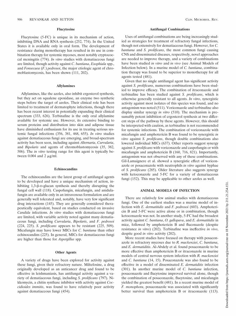
Flucytosine
Flucytosine (5-FC) is unique in its mechanism of action,inhibiting DNA and RNA synthesis (252, 774). In the UnitedStates it is available only in oral form. The development ofresistance during monotherapy has resulted in its use in com-bination therapy for systemic mycoses, most notably cryptococ-cal meningitis (774). In vitro studies with dematiaceous fungiare limited, though activity against C. bantiana, Exophiala spp.,and Fonsecaea (F.) pedrosoi, the major etiologic agent of chro-moblastomycosis, has been shown (111, 202).
Allylamines
Allylamines, like the azoles, also inhibit ergosterol synthesis,but they act on squalene epoxidase, an enzyme two syntheticsteps before the target of azoles. Their clinical role has beenlimited to treatment of dermatophyte infections, though therehas been recent interest in potentially expanding their clinicalspectrum (333, 626). Terbinafine is the only oral allylamineavailable for systemic use. However, its extensive binding toserum proteins and distribution into skin and adipose tissuehave diminished enthusiasm for its use in treating serious sys-temic fungal infections (356, 381, 468, 655). In vitro studiesagainst dematiaceous fungi are emerging, and broad-spectrumactivity has been seen, including against Alternaria, Curvularia,and Bipolaris and agents of chromoblastomycosis (35, 382,506). The in vitro testing range for this agent is typically be-tween 0.004 and 2 �g/ml.
Echinocandins
The echinocandins are the latest group of antifungal agentsto be developed and have a unique mechanism of action, in-hibiting 1,3-�-D-glucan synthesis and thereby disrupting thefungal cell wall (118). Caspofungin, micafungin, and anidula-fungin are available only in an intravenous formulation and aregenerally well tolerated and, notably, have very few significantdrug interactions (145). They are generally considered thera-peutically equivalent, based on studies conducted on invasiveCandida infections. In vitro studies with dematiaceous fungiare limited, with variable activity noted against many dematia-ceous fungi, including Curvularia, Bipolaris, and F. pedrosoi(224, 225). S. prolificans appears to be resistant (225, 509).Micafungin may have lower MICs for C. bantiana than otherechinocandins (225). In general, MICs for dematiaceous fungiare higher than those for Aspergillus spp.
Other Agents
A variety of drugs have been explored for activity againstthese fungi, given their refractory nature. Miltefosine, a drugoriginally developed as an anticancer drug and found to beeffective in leishmaniasis, has antifungal activity against a va-riety of dematiaceous fungi, including S. prolificans (797). Ni-kkomycin, a chitin synthase inhibitor with activity against Coc-cidioides immitis, was found to have relatively poor activityagainst dematiaceous fungi (454).
Antifungal Combinations
Uses of antifungal combinations are being increasingly stud-ied as strategies for treatment of refractory fungal infections,though not extensively for dematiaceous fungi. However, for C.bantiana and S. prolificans, the most common fungi causingCNS and disseminated diseases, respectively, novel approachesare needed to improve therapy, and a variety of combinationshave been studied in vitro and in vivo (see Animal Models ofInfection below). In a murine model of C. bantiana, combina-tion therapy was found to be superior to monotherapy for allagents tested (481).
Given that no single antifungal agent has significant activityagainst S. prolificans, numerous combinations have been stud-ied to improve efficacy. The combination of itraconazole andterbinafine has been studied against S. prolificans, which isotherwise generally resistant to all agents. In vitro, synergisticactivity against most isolates of this species was found, and noantagonism was noted (511). Voriconazole and terbinafine alsodisplay similar synergy in vitro (510). The mechanism is pre-sumably potent inhibition of ergosterol synthesis at two differ-ent steps of the pathway by these agents. However, this shouldbe interpreted with caution, as terbinafine is not recommendedfor systemic infections. The combination of voriconazole withmicafungin and amphotericin B was found to be synergistic invitro against S. prolificans, though double combinations alsolowered individual MICs (637). Other reports suggest synergyagainst S. prolificans with voriconazole and caspofungin or withmicafungin and amphotericin B (160, 716, 821). Importantly,antagonism was not observed with any of these combinations.Gil-Lamaignere et al. showed a synergistic effect of voricon-azole or posaconazole with neutrophils in vitro against hyphaeof S. prolificans (285). Older literature also suggests synergywith ketoconazole and 5-FC for a variety of dematiaceousfungi (152). This may be applicable to other azoles as well.
ANIMAL MODELS OF INFECTION
There are relatively few animal studies with dematiaceousfungi. One of the earliest studies was a murine model of in-fection with E. dermatitidis and F. pedrosoi (603). Amphoteri-cin B and 5-FC were active alone or in combination, thoughketoconazole was not. In another study, 5-FC had the broadestactivity against C. bantiana, O. gallopava, and E. dermatitidis inmice, followed by amphotericin B and fluconazole (despiteresistance in vitro) (202). Terbinafine was ineffective in vivo,despite good in vitro activity (202).
More recent studies have focused on therapy with posacon-azole in refractory mycoses due to R. mackenziei, C. bantiana,and E. dermatiditis. Al-Abdely et al. found posaconazole to bemore effective than amphotericin B or itraconazole in murinemodels of central nervous system infection with R. mackenzieiand C. bantiana (14, 15). Posaconazole was also found to beeffective in a model of disseminated E. dermatiditis infection(301). In another murine model of C. bantiana infection,posaconazole and flucytosine improved survival alone, thoughthe combination of posaconazole, flucytosine, and micafunginyielded the greatest benefit (481). In a recent murine model ofF. monophora, posaconazole was associated with significantlybetter survival than amphotericin B or itraconazole (113).
906 REVANKAR AND SUTTON CLIN. MICROBIOL. REV.
Dow
nloa
ded
from
http
s://j
ourn
als.
asm
.org
/jour
nal/c
mr
on 1
8 N
ovem
ber
2021
by
211.
21.2
54.1
51.
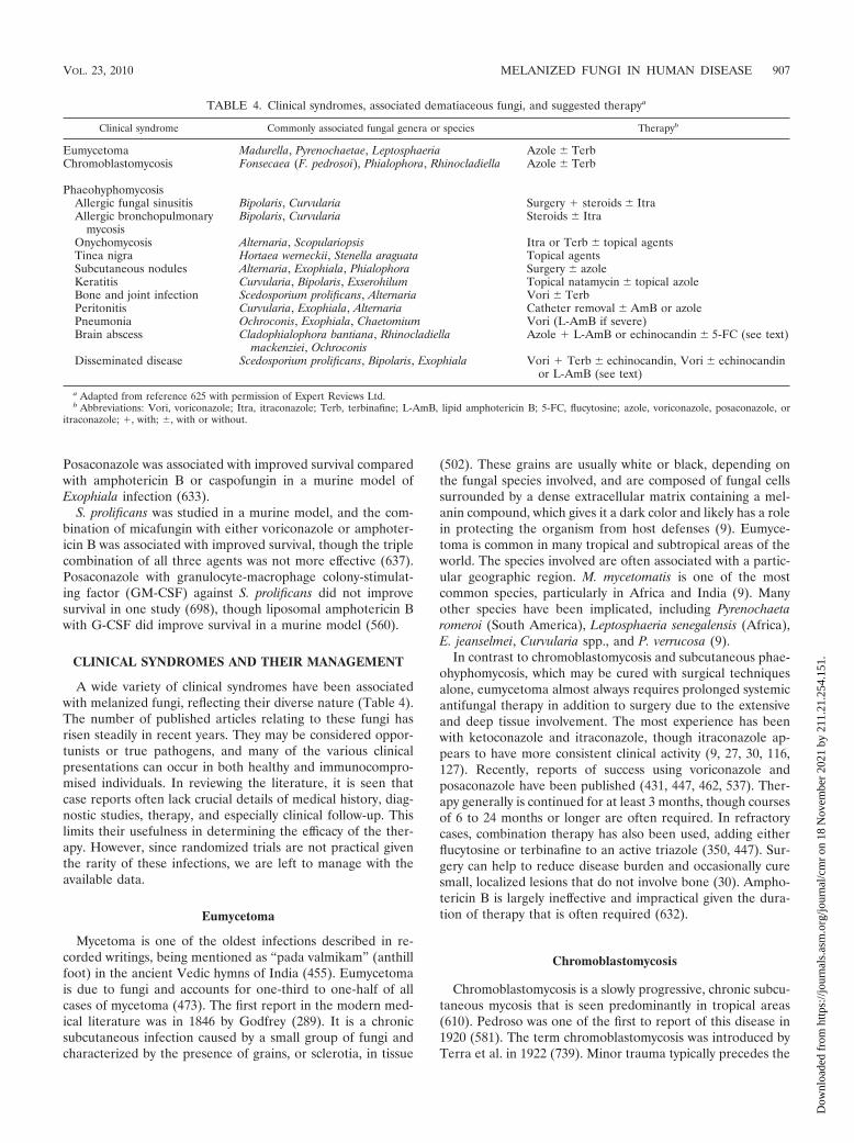
Posaconazole was associated with improved survival comparedwith amphotericin B or caspofungin in a murine model ofExophiala infection (633).
S. prolificans was studied in a murine model, and the com-bination of micafungin with either voriconazole or amphoter-icin B was associated with improved survival, though the triplecombination of all three agents was not more effective (637).Posaconazole with granulocyte-macrophage colony-stimulat-ing factor (GM-CSF) against S. prolificans did not improvesurvival in one study (698), though liposomal amphotericin Bwith G-CSF did improve survival in a murine model (560).
CLINICAL SYNDROMES AND THEIR MANAGEMENT
A wide variety of clinical syndromes have been associatedwith melanized fungi, reflecting their diverse nature (Table 4).The number of published articles relating to these fungi hasrisen steadily in recent years. They may be considered oppor-tunists or true pathogens, and many of the various clinicalpresentations can occur in both healthy and immunocompro-mised individuals. In reviewing the literature, it is seen thatcase reports often lack crucial details of medical history, diag-nostic studies, therapy, and especially clinical follow-up. Thislimits their usefulness in determining the efficacy of the ther-apy. However, since randomized trials are not practical giventhe rarity of these infections, we are left to manage with theavailable data.
Eumycetoma
Mycetoma is one of the oldest infections described in re-corded writings, being mentioned as “pada valmikam” (anthillfoot) in the ancient Vedic hymns of India (455). Eumycetomais due to fungi and accounts for one-third to one-half of allcases of mycetoma (473). The first report in the modern med-ical literature was in 1846 by Godfrey (289). It is a chronicsubcutaneous infection caused by a small group of fungi andcharacterized by the presence of grains, or sclerotia, in tissue
(502). These grains are usually white or black, depending onthe fungal species involved, and are composed of fungal cellssurrounded by a dense extracellular matrix containing a mel-anin compound, which gives it a dark color and likely has a rolein protecting the organism from host defenses (9). Eumyce-toma is common in many tropical and subtropical areas of theworld. The species involved are often associated with a partic-ular geographic region. M. mycetomatis is one of the mostcommon species, particularly in Africa and India (9). Manyother species have been implicated, including Pyrenochaetaromeroi (South America), Leptosphaeria senegalensis (Africa),E. jeanselmei, Curvularia spp., and P. verrucosa (9).
In contrast to chromoblastomycosis and subcutaneous phae-ohyphomycosis, which may be cured with surgical techniquesalone, eumycetoma almost always requires prolonged systemicantifungal therapy in addition to surgery due to the extensiveand deep tissue involvement. The most experience has beenwith ketoconazole and itraconazole, though itraconazole ap-pears to have more consistent clinical activity (9, 27, 30, 116,127). Recently, reports of success using voriconazole andposaconazole have been published (431, 447, 462, 537). Ther-apy generally is continued for at least 3 months, though coursesof 6 to 24 months or longer are often required. In refractorycases, combination therapy has also been used, adding eitherflucytosine or terbinafine to an active triazole (350, 447). Sur-gery can help to reduce disease burden and occasionally curesmall, localized lesions that do not involve bone (30). Ampho-tericin B is largely ineffective and impractical given the dura-tion of therapy that is often required (632).
Chromoblastomycosis
Chromoblastomycosis is a slowly progressive, chronic subcu-taneous mycosis that is seen predominantly in tropical areas(610). Pedroso was one of the first to report of this disease in1920 (581). The term chromoblastomycosis was introduced byTerra et al. in 1922 (739). Minor trauma typically precedes the
TABLE 4. Clinical syndromes, associated dematiaceous fungi, and suggested therapya
Clinical syndrome Commonly associated fungal genera or species Therapyb
Eumycetoma Madurella, Pyrenochaetae, Leptosphaeria Azole TerbChromoblastomycosis Fonsecaea (F. pedrosoi), Phialophora, Rhinocladiella Azole Terb
PhaeohyphomycosisAllergic fungal sinusitis Bipolaris, Curvularia Surgery � steroids ItraAllergic bronchopulmonary
mycosisBipolaris, Curvularia Steroids Itra
Onychomycosis Alternaria, Scopulariopsis Itra or Terb topical agentsTinea nigra Hortaea werneckii, Stenella araguata Topical agentsSubcutaneous nodules Alternaria, Exophiala, Phialophora Surgery azoleKeratitis Curvularia, Bipolaris, Exserohilum Topical natamycin topical azoleBone and joint infection Scedosporium prolificans, Alternaria Vori TerbPeritonitis Curvularia, Exophiala, Alternaria Catheter removal AmB or azolePneumonia Ochroconis, Exophiala, Chaetomium Vori (L-AmB if severe)Brain abscess Cladophialophora bantiana, Rhinocladiella
mackenziei, OchroconisAzole � L-AmB or echinocandin 5-FC (see text)
Disseminated disease Scedosporium prolificans, Bipolaris, Exophiala Vori � Terb echinocandin, Vori echinocandinor L-AmB (see text)
a Adapted from reference 625 with permission of Expert Reviews Ltd.b Abbreviations: Vori, voriconazole; Itra, itraconazole; Terb, terbinafine; L-AmB, lipid amphotericin B; 5-FC, flucytosine; azole, voriconazole, posaconazole, or
itraconazole; �, with; , with or without.
VOL. 23, 2010 MELANIZED FUNGI IN HUMAN DISEASE 907
Dow
nloa
ded
from
http
s://j
ourn
als.
asm
.org
/jour
nal/c
mr
on 1
8 N
ovem
ber
2021
by
211.
21.2
54.1
51.

lesions, though many patients do not recall this. Nodular le-sions can progress over years to form large, verrucous plaques.Histopathology is characterized by the presence of muriformsclerotic bodies (Medlar bodies or “copper pennies”) in tissue,which defines this condition (232, 610). By far the most com-mon species is F. pedrosoi, followed by Phialophora verrucosaand, less commonly, Cladophialophora carrioni and Rhinocla-diella aquaspersa (610). They can also cause other clinical syn-dromes, often leading to confusion in the literature as someauthors refer to any disease caused by these fungi as “_______chromoblastomycosis” (66). Other fungi have been implicated,though some reports do not clearly describe the pathogno-monic features and so are questionable (594). It is not under-stood how and under what conditions sclerotic bodies areformed in tissue. Melanin is thought to play an important role,though other compounds such as peptidases, glycosphingolip-ids, and sialidase may be involved in pathogenesis as well (666).
Therapy is difficult and various modalities have been used,usually over a period of several months and even years. Besidesantifungal therapy, surgery, cryotherapy, thermotherapy, andeven laser therapy have been tried (90). In a large series,cryotherapy, itraconazole, or the combination resulted in thelargest number of cures (88). In developing countries, wheresystemic antifungals are not easily available or are too expen-sive, the use of cryotherapy alone in a systematic manner overseveral months has led to good cure rates as well (126). Suchphysical therapies are most effective on small, localized lesions.The exact mechanism of this effect is unclear.
Antifungal therapy is essential for moderate to severe orwidespread disease. As a single agent, itraconazole appears tobe the most effective, and it is the agent with which there is themost clinical experience (88, 426, 610, 611, 620). A variety ofother treatments have also been successful, including ketocon-azole, flucytosine, local heat therapy, and amphotericin B (44,377, 515). However, the overall cure rate was only 57% in onelarge series of 100 cases from Brazil, despite use of multiplemodalities (515). Recently, terbinafine has been found to havein vitro and clinical activity (91, 684). In refractory cases, thecombination of itraconazole and terbinafine has been found tobe useful, and some experts recommend this as first-line ther-apy for moderate to severe disease (90, 317, 610).
Phaeohyphomycosis
The remainder of clinical syndromes can be grouped underthe term phaeohyphomycosis. For the purposes of this review,they will be arbitrarily divided into allergic disease, superficialinfection, deep local infection, pulmonary infection, centralnervous system infection, and disseminated infection.
Allergic disease. Allergic responses to dematiaceous fungimay actually represent the most common clinical manifestationof these fungi. Though asthma has many associated environ-mental factors, several studies have linked it with exposure tomolds and to dematiaceous fungi, Alternaria spp. and Clados-porium spp. in particular (108, 278, 456, 555). The effect hasalso correlated with seasonal fluctuations in outdoor moldcounts (555). In addition, Alternaria has been associated withsevere asthma exacerbation in some individuals (541). A fre-quent finding is the presence of elevated Alternaria-specific IgE(456). However, most melanized fungi do not elicit such a
response, and it remains unclear why only a few genera areassociated with allergic disease.
(i) Allergic fungal sinusitis. Allergic fungal sinusitis is arelatively common condition, with estimates of 6 to 9% of allcases of chronic sinusitis requiring surgery (673). Patientswith this condition usually present with chronic sinus symp-toms that are not responsive to antibiotics. Previously, As-pergillus was thought to be the most common fungus respon-sible for allergic sinusitis, but it is now appreciated thatdisease due to dematiaceous fungi actually comprises themajority of cases (239, 674). Geographic variation has alsobeen reported, with an increased incidence in the southernUnited States (240).
The most common species isolated are Alternaria, Bipolaris,and Curvularia, though other rare fungi (Epicoccum and Nodu-lisporium) have also been reported (155, 544, 673). However,fungi are frequently isolated from normal individuals as well(668). Criteria have been suggested for this disease, and theseinclude (i) nasal polyps; (ii) the presence of allergic mucin,containing Charcot-Leyden crystals and eosinophils; (iii) hy-phal elements in the mucosa without evidence of tissue inva-sion, (iv) positive skin test to fungal allergens; and (v) oncomputed tomography (CT) scans, characteristic areas of cen-tral hyperattenuation within the sinus cavity. Not all are con-sidered by experts to be necessary for diagnosis (357, 675).Diagnosis generally depends on demonstration of allergic mu-cin, with or without actual culture of the organism. Therapyconsists of surgery to remove the mucin, which is often tena-cious, and systemic steroids, though patients have been curedby surgical therapy alone (498, 631, 732). Antifungal therapy,usually in the form of itraconazole, may play a role in reducingthe requirement for steroids, but this is not routinely recom-mended and small, randomized studies showed no benefitwhen it is used as primary therapy in addition to surgery (425,654). However, in refractory cases, itraconazole may improveoutcomes (135, 680). Other azoles (voriconazole) have onlyrarely been used for this disease (223).
In rare cases, patients may present with often chronic symp-toms of mass effect due to the inflammation extending fromsinuses into adjacent structures, including the orbits (102, 125,150, 379). These are almost always immunocompetent pa-tients. In addition to surgery and steroids, systemic antifungaltherapy is often given.
(ii) ABPM. Allergic bronchopulmonary mycosis (ABPM) issimilar in presentation to allergic bronchopulmonary aspergil-losis (ABPA), which is typically seen in patients with asthma orcystic fibrosis (5, 658). There is a suggestion that allergic fungalsinusitis and allergic bronchopulmonary mycosis may actuallybe a continuum of disease and should be referred to as sino-bronchial allergic mycosis (SAM) (771). Criteria for the diag-nosis of ABPA in patients with asthma include (i) asthma, (ii)positive skin test for fungal allergens, (iii) elevated IgE levels,(iv) Aspergillus-specific IgE, and (v) proximal bronchiectasis(5). Similar criteria for ABPM are not established but mayinclude elevated IgE levels, positive skin tests, and response tosystemic steroids.
In reviewing cases of ABPM due to dematiaceous fungi,essentially all cases are found to be due to Bipolaris orCurvularia (323, 432, 526, 622, 631, 658, 753). Asthma wascommon in these cases, but bronchiectasis was often not
908 REVANKAR AND SUTTON CLIN. MICROBIOL. REV.
Dow
nloa
ded
from
http
s://j
ourn
als.
asm
.org
/jour
nal/c
mr
on 1
8 N
ovem
ber
2021
by
211.
21.2
54.1
51.

present, perhaps reflecting somewhat different pathogenicmechanisms. All cases had either eosinophilia or elevatedIgE levels. Therapy was primarily systemic steroids, with aslow taper over 2 to 3 months or longer, if necessary. Itra-conazole has been used as a steroid-sparing agent in ABPA,but its efficacy is not clear and routine use of itraconazole isnot generally recommended (5).
Superficial infections. These cases of superficial infectionsinvolve only keratinized tissues, such as the fingernails andtoenails and the stratum corneum. Consequences of these in-fections are generally cosmetic. Relatively few fungi are re-sponsible for the majority of infections.
(i) Onychomycosis. Dematiaceous fungi are rare causes ofonychomycosis. Clinical features may include a history oftrauma, involvement of only one or two toenails, and lackof response to standard systemic therapy (316). Alternaria,Scopulariopsis, and Neoscytalidium have been reported, withthe last genus being highly resistant to therapy (68, 316, 643,752). In one study, Neoscytalidium infection was associatedwith plucking of green tea leaves (68). Itraconazole andterbinafine are the most commonly used systemic agents andmay be combined with topical therapy for refractory cases(316, 752). No published data are available for the newerazole agents.
(ii) Tinea nigra. Tinea nigra is an uncommon infection con-fined to the stratum corneum. The characteristic appearance isthat of a pigmented macule, usually on the palms or soles, andmay be bilateral (440, 682). It is usually asymptomatic. Themost common reported cause is Hortaea werneckii (previouslyPhaeoannellomyces werneckii), with some cases due to Stenellaaraguata (87, 588, 682). Most cases are associated with expo-sure to sandy beaches in tropical regions, where H. werneckii isfound in areas of high salinity (588). However, individual casesdue to Scopulariopsis brevicaulis, Phoma eupyrena, and Chaeto-mium globosum with findings consistent with tinea nigra havealso been reported (57, 154, 287). Diagnosis is made by skinscraping, and biopsy is not needed. Although systemic antifun-gals have been given with success (318), topical therapy withazoles or keratolytics is very effective (87, 588).
Deep local infections. Deep local infections are a heteroge-nous group of infectious syndromes that are typically caused bylocal trauma. Virtually any of the melanized fungi discussed inthis review may cause these infections. While they are rarelylife-threatening, even in immunocompromised patients, con-siderable morbidity can result due to difficulties in treatmentand complications.
(i) Subcutaneous lesions. Subcutaneous lesions are the mostcommon case reports of infection due to melanized fungi in theliterature. Alternaria spp. are by far the most common etiologicagent, with a recent review cataloguing over 156 cases up to2007 (195, 191, 275, 373, 542, 577, 710, 751). Exophiala spp.and Phialophora spp. are the next most common fungi, fol-lowed by Cladosporium spp., Exserohilum spp., Veronaea bot-ryosa, and many others with scattered case reports (7, 12, 19,23, 32, 36, 56, 61, 65, 81, 140, 143, 194, 209, 230, 241, 251, 258,306, 313, 342, 362, 365, 369, 387, 413, 418, 421, 457, 469, 482,486, 487, 495, 539, 559, 556, 568, 571, 576, 590, 609, 613, 614,646, 659, 694, 699, 720, 728, 749, 764, 776, 807, 818). Minortrauma is the usual inciting factor, though it is frequently
unrecognized by the patient. Occasionally wood splinters orother vegetable matter is found upon skin biopsy or excision ofthe lesion (499, 524, 559, 660).
Many patients are immunocompetent, and they often arefrom a rural background, i.e., farmers with frequent, minortrauma from plant material or gardeners (133, 192, 206, 233,277, 359, 576, 751, 815). Organ transplantation is also a com-mon risk factor (28, 38, 112, 142, 235, 236, 262, 271, 286, 296,465, 484, 514, 556, 587, 645, 679, 704, 725). Apparently noso-comial cases have also been reported, with skin irritations fromdressings or i.v. sites as possible risk factors (237). Lesionstypically occur on exposed areas of the body and often appearas isolated cystic or papular lesions. Presentation is usuallyindolent, with weeks to months of gradual enlarging mass,though pain is often absent. Severely immunocompromisedpatients are at increased risk of subsequent dissemination,though this may rarely occur in apparently immunocompetentpatients as well. Occasionally, infection may extend to involvejoints or bone, requiring more extensive surgery or prolongedantifungal therapy (198).
Multiple therapeutic options are available, usually depend-ing on the immune status of the patient and the extent oflesions. Oral systemic therapy with an azole antifungal agent inconjunction with surgery is frequently employed and has beenused successfully, particularly in immunocompromised pa-tients. This is to prevent possible disseminated infection,though this is actually very rare in all but the most immuno-suppressed patients. Terbinafine has also been used success-fully, particularly in patients failing azole therapy (7). Surgicalexcision alone has been successful in a number of cases, evenin organ transplant patients (12, 194, 235, 387, 418, 558, 587,646, 686, 720, 764). The Mohs surgical technique, which wasdeveloped for removing melanoma, may be a useful surgicalapproach, as it spares tissue and completely removes thepathological lesion in staged surgeries (86). Patients for whomprolonged antifungal therapy is problematic may also benefitfrom this technique, such as transplant patients, who often areon immunosuppressive medications that interact with oral tri-azole antifungals. For multiple lesions where resection may bedifficult, antifungal therapy alone has also been successful,even in immunocompromised patients (251, 514). A variety ofalternative therapies have been successfully employed as well,including cryotherapy, thermotherapy (local heat application),and supersaturated potassium iodide (SSKI) (286, 313, 751).These are particularly relevant in developing countries wheresystemic antifungal therapy is difficult to obtain or too expen-sive for patients. Recurrences may occur several months toover a year after therapy is complete, so careful clinical fol-low-up is important (38, 142).
(ii) Keratitis. Fungal keratitis is an important ophthalmo-logic problem, particularly in tropical areas of the world (298).In one large series, 40% of all infectious keratitis was caused byfungi, almost exclusively molds (298). The most common fungiare Fusarium and Aspergillus, followed by dematiaceous fungi(up to 8 to 17% of cases) (298, 715). Many species can causedisease, with Bipolaris and Curvularia most common, thoughLasiodiplodia theobromae may cause more severe disease (55,75, 213, 298, 743, 744). Approximately half the cases are asso-ciated with trauma; prior eye surgery, diabetes, and contactlens use have also been noted as important risk factors (298,
VOL. 23, 2010 MELANIZED FUNGI IN HUMAN DISEASE 909
Dow
nloa
ded
from
http
s://j
ourn
als.
asm
.org
/jour
nal/c
mr
on 1
8 N
ovem
ber
2021
by
211.
21.2
54.1
51.

743). Diagnosis rests on potassium hydroxide (KOH) smearand culture, with many dematiaceous fungi associated withpigmented plaques (270).
Many cases of keratitis due to dematiaceous fungi havecome from India (77, 136, 210, 269, 582). In a large experienceof keratitis due to dematiaceous fungi, 88 cases were examined(269). The most common dematiaceous genus causing keratitiswas Curvularia, followed by Bipolaris, Exserohilum, and Lasio-diplodia. Almost half the cases were associated with trauma.Most patients received topical agents only (5% natamycin withor without an azole), though more severe cases also receivedoral ketoconazole. Overall response was 72% in those avail-able for follow-up. Surgery was needed in 13 patients, with anadditional 6 requiring enucleation due to poor response. Itra-conazole topically has also been used with success (582).
In a study from the United States of 43 cases of Curvulariakeratitis, almost all were associated with trauma (800). Plantswere the most common source, though several cases of metalinjury were seen as well. Topical natamycin was used almostexclusively, with only a few severe cases requiring adjunctivetherapy, usually with an azole. Of the oral agents, itraconazolehad the best in vitro activity, though the majority of isolateswere resistant to flucytosine. Surgery, including penetratingkeratoplasty, was required in 19% of patients. At the end oftherapy, only 78% had a visual acuity of 20/40 or better. Othercase series from the United States have noted a rise in contactlens use as a risk factor in recent years (374, 388). In thesouthern United States, cases are more frequently seen duringwarm, humid months (799).
Topical polyenes, such as amphotericin B and natamycin,are commonly used, but oral and topical itraconazole havebeen found to be useful as well (298, 744). Use of voriconazolehas become more common, with topical preparations well de-scribed (211), but published cases involving dematiaceousfungi are infrequent (564). A series of Alternaria keratitis casesthat were refractory to natamycin responded to topical azoles(758). A recent analysis of clinical trials involving fungal ker-atitis suggested that none of the available agents was highlyeffective (247). Many patients are left with residual visual def-icits at the end of therapy. Clearly, further advances in therapyare needed for this debilitating disease.
(iii) Bone and joint infections. There are relatively few casereports of isolated osteoarticular infections due to dematia-ceous fungi, perhaps reflecting the significant trauma oftenrequired for implantation into these deeper tissues, thoughsome cases did not have noticeable trauma. S. prolificans is themost common cause, with all except two cases occurring inyoung children (166, 299, 325, 406, 453, 474, 716, 718). Inaddition to surgery, various antifungal therapies have beenemployed, with three cases using the combination of voricon-azole and terbinafine with success (166, 299, 406). Unusualtherapies have also been tried, including irrigation with poly-hexamethylbiguanide and oral therapy with miltefosine (406,716). Whether these actually improved the likelihood of aclinical response is unknown.
Alternaria alternata has been reported to cause palatal ulcerswith associated osteomyelitis in patients with chronic sinusitis(198, 265, 297). Although immune tolerance has been sug-gested in these cases, no convincing evidence is available. Re-
currences were common despite prolonged antifungal therapy.Other organisms associated with single case reports include
P. obovatum, E. oligosperma, C. arxii, M. mycetomatis, P. rich-ardsiae, F. pedrosoi, and P. parasiticum (94, 390, 398, 472, 512,689, 812). Itraconazole was the most common single agentused with success (398, 512, 689). Antifungal therapy for allthese cases is usually prolonged, i.e., 6 months and up to 2years.
(iv) Peritonitis. Peritonitis occurs essentially only in patientsreceiving peritoneal dialysis (6, 8, 69, 97, 106, 115, 117, 144,197, 261, 302, 305, 367, 405, 416, 461, 466, 563, 598, 605, 618,619, 656, 690, 762, 765, 783, 784). The presentation is usuallysubacute, with many patients being without significant symp-toms. The genera isolated included Curvularia (eight cases);Exophiala (five); Alternaria (four); Bipolaris (four); Aureobasi-dium (three); and Lecythophora, Hormonema, and Phiale-monium (one each). Eosinophils in peritoneal fluid were notuncommon and were associated with a variety of species. Cath-eter removal was considered critical, though one case withamphotericin B lock therapy in the catheter with systemicfluconazole resulted in cure with catheter retention (106). Out-comes were generally good, with only three deaths, two asso-ciated with persistent infection and the other with a retainedcatheter (405, 618, 690).
(v) Miscellaneous infections. Various anecdotal cases of un-usual infectious syndromes have been reported. A case ofepididymitis due to E. jeanselmei was reported in a 54-year-oldmale who had received multiple needle aspirations for a symp-tomatic hydrocele (248). Surgical excision alone resulted incure. In another case, a 5-year-old asymptomatic girl was notedto have “black grains” in her urine 3 weeks after treatment fora urinary tract infection. Hyphae and conidia identified as aCurvularia sp. were observed in a wet mount but did not growin culture (635). She received no therapy, and the conditionspontaneously resolved. E. dermatitidis was isolated from acase of otitis externa in a 19-year-old immunocompetent fe-male, along with Pseudomonas (404). She responded to anti-biotics and topical antimycotics. E. jeanselmei was isolatedfrom esophageal brushings of a patient with Barrett’s esopha-gus, and biopsy revealed hyphal elements as well (669). Thepatient was treated with ketoconazole, though little clinicalimprovement was noted after 5 months. A case of acute, inva-sive sinusitis due to E. rostratum was reported in an 18-year-oldfemale with aplastic anemia and persistent neutropenia(442). She underwent surgery and prolonged antifungaltherapy with liposomal amphotericin B and voriconazole-itraconazole, although she died of her underlying diseasewith persistent evidence of infection at autopsy. In contrastto the case for allergic sinusitis, such presentations are rarein immunocompromised patients.
Pulmonary infection. Pulmonary infection is usually seen inimmunocompromised patients or those with underlying lungdisease, and it may be due to a wide variety of species, includ-ing S. prolificans, C. bantiana, Chaetomium spp., Ochroconisgallopava, Exophiala spp., Alternaria, Cladophialophora boppii,F. pedrosoi, L. theobromae, Aureobasidium pullulans, Curvu-laria spp., Sarcinosporin inkin, and P. verrucosa (1, 17, 63, 92,101, 107, 199, 219, 267, 303, 348, 351, 352, 402, 427, 429, 443,464, 477, 520, 528, 554, 583, 701, 734, 736, 805, 814). Clinicalmanifestations include pneumonia, asymptomatic solitary pul-
910 REVANKAR AND SUTTON CLIN. MICROBIOL. REV.
Dow
nloa
ded
from
http
s://j
ourn
als.
asm
.org
/jour
nal/c
mr
on 1
8 N
ovem
ber
2021
by
211.
21.2
54.1
51.
monary nodules, and endobronchial lesions which may causehemoptysis. Therapy has consisted of systemic antifungalagents, usually amphotericin B or itraconazole initially, fol-lowed by itraconazole for a more prolonged period. Mortalityrates are high in immunocompromised patients (40%). Ex-perience with voriconazole is accumulating and appears prom-ising (17, 199, 219, 348). Posaconazole was effective in a case ofAlternaria pneumonia in a patient with leukemia that was re-fractory to amphotericin B and voriconazole (528). Occasionalcases of solitary pulmonary nodules in immunocompetent pa-tients may be cured with surgical resection alone (92, 303).
Central nervous system infection. Central nervous systeminfection is a rare but frequently fatal manifestation of phae-ohyphomycosis, often in immunocompetent individuals. In areview of 101 cases of central nervous system infection due todematiaceous fungi (628), the most common presentation wasfound to be brain abscess (11, 24, 21, 34, 42, 53, 60, 79, 82, 100,110, 114, 137, 138, 156, 168, 169, 193, 212, 222, 254, 255, 290,314, 319, 329, 337, 343, 371, 394, 396, 408, 410, 412, 415, 422,458, 500, 505, 513, 523, 527, 529–531, 562, 570, 578, 602, 629,649, 661, 662, 665, 667, 678, 681, 692, 702, 711, 726, 738, 746,763, 786, 787, 790, 791, 803, 816). What is truly unique aboutthis disease is that over half the cases were in patients with norisk factor or immunodeficiency. In addition, no specific expo-sures were associated with onset of infection, though manycases seem to occur in rural areas. Typical symptoms includedheadache, neurologic deficits, and seizures, though rarely allthree. The most common species was C. bantiana, accountingfor half the cases. Other species included Rhinocladiella mack-enziei, Ochroconis gallopava, Bipolaris spicifera, Exophiala der-matitidis, and Chaetomium strumarium. Mortality was 70%.Since that review in 2004, over 50 cases have been reported,with C. bantiana remaining the most frequently seen isolate(131, 4, 17, 16, 22, 31, 40, 70, 78, 93, 121, 139, 170, 189, 243,244, 268, 272, 276, 328, 332, 341, 363, 380, 399, 420, 433, 448,450, 467, 476, 545, 575, 636, 657, 696, 700, 721, 731, 733, 754,759). A new species, Fonsecaea monophora, has been reportedsince that time and appears to have a predilection for causingCNS disease, in contrast to its related species, F. pedrosoi (420,721, 733). Encephalitis with diffuse brain involvement is a rarepresentation, with essentially 100% mortality (1, 80).
The pathogenesis may be hematogenous spread from aninitial, presumably subclinical pulmonary focus, though thisremains speculation. Animal models of C. bantiana reliablyreplicate CNS infection with intravenous or intranasal inocu-lation, though these are generally immunosuppressed mice(15, 200). However, it remains unclear why these fungi pref-erentially cause CNS disease in immunocompetent individuals.
Meningitis has also been described, usually in immunocom-promised patients (3, 17, 59, 74, 260, 366, 423, 444, 470, 729).However, cases with iatrogenic complications related to con-taminated steroid preparations injected epidurally have beenreported (131). These can be difficult to treat, and mortality ishigh (60%).
Many therapeutic strategies have been used in the treatmentof brain abscess, though it is unclear if any result in significantlyimproved outcomes. The retrospective analysis of 101 reportedcases mentioned above suggested that the combination of am-photericin B, flucytosine, and itraconazole may be associatedwith improved survival, though it was not frequently used.
Subsequent reports have documented various regimens, someusing voriconazole or posaconazole with clinical success,though failures have also been reported. Voriconazole wasunsuccessful in treating three out of four cases of C. bantianabrain abscess, though two of these patients were immunocom-promised, and one received concomitant phenytoin, which mayhave reduced levels of voriconazole (243, 450, 467, 754). De-spite these reports, voriconazole may have a role in therapy ofphaeohyphomycotic brain abscess, as it has been successfullyused in cases of Aspergillus and S. apiospermum brain abscess(188, 540). Posaconazole has been reported to be effective in acase of R. mackenziei brain abscess, which represents the firstreported survival of infection due to this species (16). For casesdue to C. bantiana, addition of flucytosine to azole therapy maybe useful given its in vitro activity against this species specifi-cally and in vivo and clinical data (481, 628). Based on theexperience described above and animal studies, a combinationof agents is likely to be more effective than monotherapy,though the optimal combination remains unclear and shouldbe based on the individual case.
What does appear to be consistent is that complete excisionof brain abscess whenever feasible is associated with betteroutcomes than aspiration or partial excision. In a series of 10cases due to C. bantiana at one institution, all surviving pa-tients were able to have complete resection of the brain lesion(268). In another case, repeated surgical excisions alone re-sulted in cure (189). However, outcomes remain poor, with anoverall mortality of 70%.
Disseminated infection. Disseminated infection is the mostuncommon manifestation of infection caused by melanizedfungi. In a review of 72 cases (627), most patients were immu-nocompromised, though occasional patients without knownimmunodeficiency or risk factors developed disseminated dis-ease as well (3, 24, 73, 76, 95, 105, 119, 171, 208, 238, 242, 246,288, 291, 308, 321, 345, 386, 389, 397, 400, 402, 403, 409, 423,424, 435, 471, 485, 516, 538, 543, 550, 557, 578, 593, 595, 596,612, 621, 627, 640, 647, 672, 688, 712, 713, 740, 755, 769, 772,794, 804, 806, 822). In contrast to most invasive mold infec-tions, blood cultures were positive in over half the cases. Themost common isolate was S. prolificans, accounting for over athird of cases. Since that review, S. prolificans remains the mostfrequent cause of disseminated disease, almost exclusively inimmunocompromised patients (33, 62, 96, 151, 304, 358, 364,368, 434, 697, 709, 747, 750, 796). E. dermatitidis, in contrast, iscommonly seen in immunocompetent patients, particularlyfrom Asia (13, 349, 565). A variety of other molds were re-ported in disseminated disease, including E. oligosperma,Chaetomium perlucidum, O. gallopava, Lecythophora mutabilis,P. parasiticum, B. spicifera, Exserohilum sp., E. spinifera, andCurvularia lunata (25, 64, 85, 207, 256, 335, 417, 451, 536, 741,788). Interestingly, peripheral eosinophilia has been observedin 9% of cases, and these were generally due to Bipolaris andCurvularia. These same species are often associated with aller-gic disease.
The mortality rate was 70%, despite aggressive antifungaltherapy. There were no antifungal regimens associated withimproved survival for disseminated infection. Scedosporiumprolificans is generally resistant to all available antifungalagents, and infection with S. prolificans was associated withnearly 100% mortality in the absence of recovery from neu-
VOL. 23, 2010 MELANIZED FUNGI IN HUMAN DISEASE 911
Dow
nloa
ded
from
http
s://j
ourn
als.
asm
.org
/jour
nal/c
mr
on 1
8 N
ovem
ber
2021
by
211.
21.2
54.1
51.
tropenia, indicating the importance of the host response in thisinfection. However, recent case reports have suggested that thecombination of itraconazole or voriconazole with terbinafinemay be synergistic against this species and improve outcomes,though clinical experience is limited (358, 750, 796). Some casereports utilized colony-stimulating factors and/or leukocyte in-fusions to augment antifungal therapy (96, 368, 796).
Other combinations or therapies have not been shown to beconsistently effective, though clinical experience is limited, andwill likely be confined to anecdotal reports, given the rarity ofthis infection. Recent successful case reports have used itra-conazole, voriconazole, and posaconazole for a variety of dif-ferent species (207, 368, 417, 536, 565). Amphotericin B aloneis not generally effective (25, 64, 368).
CONCLUSIONS
Melanized fungi remain uncommon causes of infection inhumans but have become increasingly recognized in a widevariety of clinical syndromes. Many species across a broadrange of genera are associated with disease, which leads todaunting challenges in diagnostic testing. However, relativelyfew are responsible for the majority of clinical cases. Alternariais a frequent cause of subcutaneous lesions, Bipolaris and Cur-vularia are often associated with allergic disease, and C. ban-tiana and S. prolificans are the most common causes of brainabscess and disseminated disease, respectively. Taxonomy isconstantly evolving as molecular methods shed new light onrelationships between species. Melanin appears to be an im-portant virulence factor for these fungi, though much addi-tional work is needed to better understand the pathogenicmechanisms underlying these infections, particularly in immu-nocompetent patients. Life-threatening infections are rare butmay be seen even in individuals with no apparent risk factors,especially in cases of brain abscess. As these are typically soilorganisms and common laboratory contaminants, sometimesthey are disregarded as nonpathogenic. However, the clinicalsetting in which they are isolated should always be consideredwhen evaluating their potential as etiologic agents and beforemaking decisions regarding therapy. Diagnosis depends on ahigh degree of clinical suspicion and careful mycological andpathological examination of clinical specimens. Molecular di-agnostic techniques are progressing but are not standardizedor reliable for the diverse species encountered.
Therapy for many infectious syndromes has evolved with theadvent of several new antifungal agents in recent years. Theoral triazoles voriconazole, posaconazole, and itraconazoledemonstrate the most consistent in vitro activity against thisgroup of fungi and are widely used, though voriconazole isusually the drug of choice in most clinical settings. High dosesof amphotericin B lipid formulations may have a role in thetreatment of refractory cases or for severe infections in unsta-ble patients, though it is usually not effective as a single agent.Once the patient is stable, “consolidation” therapy with abroad-spectrum oral azole is often employed until a completeresponse is achieved. Terbinafine has broad activity againstmelanized fungi, and interest in its use beyond dermatophyteinfections is increasing. It appears to provide synergistic activ-ity with azole antifungals, and this may be a useful strategyagainst refractory subcutaneous infections such as chromoblas-
tomycosis and mycetoma that often do not respond to conven-tional monotherapy. In addition, the use of terbinafine withvoriconazole for disseminated S. prolificans infection has beensuccessful with what is otherwise an almost universally fatalinfection. It should be pointed out that in these disseminatedcases, recovery of immune function, especially phagocytic cells,is critically important as well. Flucytosine has limited activityagainst dematiaceous fungi, though it may have a role in ther-apy of chromoblastomycosis and of brain abscess due to C.bantiana, in particular. Echinocandins do not appear to beuseful as single agents but may be considered in combinationtherapy of difficult cases. Combination therapy is a potentiallyuseful therapeutic strategy for refractory infections, particu-larly brain abscess and disseminated disease. However, it is notclear which antifungal drug combinations are most effective.Therapy is evolving for many of the clinical syndromes de-scribed, and randomized clinical trials to address this issue areimpractical given the sporadic nature of cases. Detailed casereporting of both successful and unsuccessful clinical experi-ences will be important in attempting to define optimal therapyfor infections caused by dematiaceous fungi.
REFERENCES
1. Abbott, S. P., L. Sigler, R. McAleer, D. A. McGough, M. G. Rinaldi, and G.Mizell. 1995. Fatal cerebral mycoses caused by the ascomycete Chaetomiumstrumarium. J. Clin. Microbiol. 33:2692–2698.
2. Abliz, P., K. Fukushima, K. Takizawa, and K. Nishimura. 2004. Identifi-cation of pathogenic dematiaceous fungi and related taxa based on largesubunit ribosomal DNA D1/D2 domain sequence analysis. FEMS Immu-nol. Med. Microbiol. 40:41–49.
3. Adam, R. D., M. L. Paquin, E. A. Petersen, M. A. Saubolle, M. G. Rinaldi,J. G. Corcoran, J. N. Galgiani, and R. E. Sobonya. 1986. Phaeohyphomy-cosis caused by the fungal genera Bipolaris and Exserohilum. A report of 9cases and review of the literature. Medicine 65:203–217.
4. Adeyemi, O. A., O. Lie, R. Bernstein, N. Gottardi-Littell, K. Muro, D. Patil,and G. A. Noskin. 2007. Woman with multiple brain abscesses. Clin. Infect.Dis. 45:1351–1352:1397-1399.
5. Agarwal, R. 2009. Allergic bronchopulmonary aspergillosis. Chest 135:805–826.
6. Agarwal, S., N. L. Goodman, and H. H. Malluche. 1993. Peritonitis due toExophiala jeanselmei in a patient undergoing continuous ambulatory peri-toneal dialysis. Am. J. Kidney Dis. 21:673–675.
7. Agger, W. A., D. Andes, and J. W. Burgess. 2004. Exophiala jeanselmeiinfection in a heart transplant recipient successfully treated with oral ter-binafine. Clin. Infect. Dis. 38:e112–115.
8. Ahmad, S., R. J. Johnson, S. Hillier, W. R. Shelton, and M. G. Rinaldi.1985. Fungal peritonitis caused by Lecythophora mutabilis. J. Clin. Micro-biol. 22:182–186.
9. Ahmed, A. O., W. van Leeuwen, A. Fahal, W. van de Sande, H. Verbrugh,and A. van Belkum. 2004. Mycetoma caused by Madurella mycetomatis: aneglected infectious burden. Lancet Infect. Dis. 4:566–574.
10. Ahmed, A. O., W. van Vianen, M. T. ten Kate, W. W. van de Sande, A. vanBelkum, A. H. Fahal, H. A. Verbrugh, and I. A. Bakker-Woudenberg. 2003.A murine model of Madurella mycetomatis eumycetoma. FEMS Immunol.Med. Microbiol. 37:29–36.
11. Ajanee, N., M. Alam, K. Holmberg, and J. Khan. 1996. Brain abscess causedby Wangiella dermatitidis: case report. Clin. Infect. Dis. 23:197–198.
12. Ajello, L., L. K. Georg, R. T. Steigbigel, and C. J. Wang. 1974. A case ofphaeohyphomycosis caused by a new species of Phialophora. Mycologia66:490–498.
13. Alabaz, D., F. Kibar, S. Arikan, B. Sancak, U. Celik, N. Aksaray, and M.Turgut. 2009. Systemic phaeohyphomycosis due to Exophiala (Wangiella) inan immunocompetent child. Med. Mycol. 47:653–657.
14. Al-Abdely, H. M., L. Najvar, R. Bocanegra, A. Fothergill, D. Loebenberg,M. G. Rinaldi, and J. R. Graybill. 2000. SCH 56592, amphotericin B, oritraconazole therapy of experimental murine cerebral phaeohyphomycosisdue to Ramichloridium obovoideum (“Ramichloridium mackenziei”). Anti-microb. Agents Chemother. 44:1159–1162.
15. Al-Abdely, H. M., L. K. Najvar, R. Bocanegra, and J. R. Graybill. 2005.Antifungal therapy of experimental cerebral phaeohyphomycosis due toCladophialophora bantiana. Antimicrob. Agents Chemother. 49:1701–1707.
16. Al Abdely, H. M., A. M. Alkhunaizi, J. A. Al Tawfiq, M. Hassounah, M. G.Rinaldi, and D. A. Sutton. 2005. Successful therapy of cerebral phaeohy-
912 REVANKAR AND SUTTON CLIN. MICROBIOL. REV.
Dow
nloa
ded
from
http
s://j
ourn
als.
asm
.org
/jour
nal/c
mr
on 1
8 N
ovem
ber
2021
by
211.
21.2
54.1
51.
phomycosis due to Ramichloridium mackenziei with the new triazoleposaconazole. Med. Mycol. 43:91–95.
17. Al-Aidaroos, A., I. Bin-Hussain, H. El Solh, A. Kofide, S. Thawadi, A.Belgaumi, and A. Al Ahmari. 2007. Invasive Chaetomium infection in twoimmunocompromised pediatric patients. Pediatr. Infect. Dis. J. 26:456–458.
18. Al-Astruey-Izquierdo, A., M. Cuenca-Estrella, A. Monzon, and J. L. Ro-driguez-Tudela. 2007. Prevalence and susceptibility testing of new speciesof Pseudallescheria and Scedosporium in a collection of clinical mold iso-lates. Antimicrob. Agents Chemother. 51:748–751.
19. Al-Attar, A., C. G. Williams, and R. J. Redett. 2006. Rare lower extremityinvasive fungal infection in an immunosuppressed patient: Exserohilumlongirostratum. Plast. Reconstr. Surg. 117:e44–47.
20. Alcorn, J. L. 1983. Generic concepts in Drechslera, Bipolaris and Exserohi-lum. Mycotaxon 17:1–86.
21. Aldape, K. D., H. S. Fox, J. P. Roberts, N. L. Ascher, J. R. Lake, and H. A.Rowley. 1991. Cladosporium trichoides cerebral phaeohyphomycosis in aliver transplant recipient. Report of a case. Am. J. Clin. Pathol. 95:499–502.
22. Alhabib, K. F., and E. A. Bryce. 2003. Xylohypha bantiana multiple brainabscesses in a patient with systemic lupus erythematosus. Can. J. Infect. Dis.14:119–120.
23. Allred, B. J. 1990. Subcutaneous phaeohyphomycosis due to Exophialajeanselmei in an immunosuppressed patient: case report. New ZealandMed. J. 103:321–322.
24. Al-Mohsen, I. Z., D. A. Sutton, L. Sigler, E. Almodovar, N. Mahgoub, H.Frayha, S. Al-Hajjar, M. G. Rinaldi, and T. J. Walsh. 2000. Acrophialo-phora fusispora brain abscess in a child with acute lymphoblastic leukemia:review of cases and taxonomy. J. Clin. Microbiol. 38:4569–4576.
25. Al-Obaid, I., S. Ahmad, Z. U. Khan, B. Dinesh, and H. M. Hejab. 2006.Catheter-associated fungemia due to Exophiala oligosperma in a leukemicchild and review of fungemia cases caused by Exophiala species. Eur.J. Clin. Microbiol. Infect. Dis. 25:729–732.
26. Al-Rajhi, A. A., A. H. Awad, S. S. al-Hedaithy, R. K. Forster, and K. C.Caldwell. 1993. Scytalidium dimidiatum fungal endophthalmitis. Br. J. Oph-thalmol. 77:388–390.
27. Al-Tawfiq, J. A., and S. S. Amr. 2009. Madura leg due to Exophialajeanselmei successfully treated with surgery and itraconazole therapy. Med.Mycol. 47:648–652.
28. Altomare, G. F., G. L. Capella, V. Boneschi, and M. A. Viviani. 2000.Effectiveness of terbinafine in cutaneous alternariosis. Br. J. Dermatol.142:840–841.
29. Alvarez, M., P. B. Lopez, C. Rayon, G. J. Garcia, M. C. Roson Porto, M.Gonzalez, J. V. Martinez-Suarez, and J. L. Rodriguez-Tudela. 1995. Noso-comial outbreak caused by Scedosporium prolificans (inflatum): four fatalcases in leukemic patients. J. Clin. Microbiol. 33:3290–3295.
30. Ameen, M., and R. Arenas. 2009. Developments in the management ofmycetomas. Clin. Exp. Dermatol. 34:1–7.
31. Amr, S. S., and J. A. Al-Tawfiq. 2007. Aspiration cytology of brain abscessfrom a fatal case of cerebral phaeohyphomycosis due to Ramichloridiummackenziei. Diagn. Cytopathol. 35:695–699.
32. Anandan, V., V. Nayak, S. Sundaram, and P. Srikanth. 2008. An associa-tion of Alternaria alternata and Scopulariopsis brevicaulis in cutaneous phaeo-hyphomycosis. Indian J. Dermatol. Venereol. Leprol. 74:244–247.
33. Ananda-Rajah, M. R., A. Grigg, and M. A. Slavin. 2008. Breakthroughdisseminated Scedosporium prolificans infection in a patient with relapsedleukaemia on prolonged voriconazole followed by posaconazole prophy-laxis. Mycopathologia 166:83–86.
34. Anandi, V., T. J. John, A. Walter, J. C. Shastry, M. K. Lalitha, A. A. Padhye,L. Ajello, and F. W. Chandler. 1989. Cerebral phaeohyphomycosis causedby Chaetomium globosum in a renal transplant recipient. J. Clin. Microbiol.27:2226–2229.
35. Andrade, T. S., L. G. Castro, R. S. Nunes, V. M. Gimenes, and A. E. Cury.2004. Susceptibility of sequential Fonsecaea pedrosoi isolates from chromo-blastomycosis patients to antifungal agents. Mycoses 47:216–221.
36. Aoyama, Y., M. Nomura, S. Yamanaka, Y. Ogawa, and Y. Kitajima. 2009.Subcutaneous phaeohyphomycosis caused by Exophiala xenobiotica in anon-Hodgkin lymphoma patient. Med. Mycol. 47:95–99.
37. Aquino, V. M., J. M. Norvell, K. Krisher, and M. M. Mustafa. 1995. Fataldisseminated infection due to Exserohilum rostratum in a patient with aplas-tic anemia: case report and review. Clin. Infect. Dis. 20:176–178.
38. Ara, M., C. Aspiroz, P. Zaballos, V. Alcalde, R. Alvarez, A. Rezusta, andJ. A. Gimenez. 2006. Relapse of cutaneous Alternaria infectoria in a renaltransplant recipient after 2 years. Acta Derm. Venereol. 86:154–155.
39. Arango, M., C. Jaramillo, A. Cortes, and A. Restrepo. 1998. Auricularchromoblastomycosis caused by Rhinocladiella aquaspersa. Med. Mycol.36:43–45.
40. Aribandi, M., C. Bazan III, and M. G. Rinaldi. 2005. Magnetic resonanceimaging findings in fatal primary cerebral infection due to Chaetomiumstrumarium. Australas. Radiol. 49:166–169.
41. Arthur, S., L. L. Steed, D. J. Apple, Q. Peng, G. Howard, and M. Escobar-Gomez. 2001. Scedosporium prolificans keratouveitis in association with acontact lens retained intraocularly over a long term. J. Clin. Microbiol.39:4579–4582.
42. Arunkumar, M. J., V. Rajshekhar, M. J. Chandy, P. P. Thomas, and C. K.Jacob. 2000. Management and outcome of brain abscess in renal transplantrecipients. Postgrad. Med. J. 76:207–211.
43. Arzanlou, M., J. Z. Groenewald, W. Gams, U. Braun, H. D. Shin, and P. W.Crous. 2007. Phylogenetic and morphotaxonomic revision of Ramichlo-ridium and allied genera. Stud. Mycol. 58:57–93.
44. Attapattu, M. C. 1997. Chromoblastomycosis—a clinical and mycologicalstudy of 71 cases from Sri Lanka. Mycopathologia 137:145–151.
45. Attili, D. S., G. S. De Hoog, and A. A. Pizzirani-Kleiner. 1998. rDNA-RFLPand ITS1 sequencing of species of the genus Fonsecaea, agents of chromo-blastomycosis. Med. Mycol. 36:219–225.
46. Aviv, J. E., W. Lawson, E. J. Bottone, V. P. Sachdev, P. M. Som, and H. F.Biller. 1990. Multiple intracranial mucoceles associated with phaeohypho-mycosis of the paranasal sinuses. Arch. Otolaryngol. Head Neck Surg.116:1210–1213.
47. Badali, H., V. O. Carvalho, V. Vicente, D. Attili-Angelis, I. B. Kwiatkowski,A. H. Gerrits Van Den Ende, and G. S. De Hoog. 2009. Cladophialophorasaturnica sp. nov., a new opportunistic species of Chaetothyriales revealedusing molecular data. Med. Mycol. 47:51–62.
48. Badali, H., J. Chander, S. Bansal, A. Aher, S. S. Borkar, J. F. Meis, andG. S. De Hoog. 2010. First autochthonous case of Rhinocladiella mackenzieicerebral abscess outside the Middle East. J. Clin. Microbiol. 48:646–649.
49. Badali, H., G. S. De Hoog, I. Curfs-Breuker, B. Andersen, and J. F. Meis.2009. In vitro activities of eight antifungal drugs against 70 clinical andenvironmental isolates of Alternaria species. J. Antimicrob. Chemother.63:1295–1297.
50. Badali, H., G. S. de Hoog, I. Curfs-Breuker, and J. F. Meis. 2010. In vitroactivities of antifungal drugs against Rhinocladiella mackenziei, an agent offatal brain infection. J. Antimicrob. Chemother. 65:175–177.
51. Badali, H., C. Gueidan, M. J. Najafzadeh, A. Bonifaz, A. H. van den Ende,and G. S. de Hoog. 2008. Biodiversity of the genus Cladophialophora. Stud.Mycol. 61:175–191.
52. Badali, H., M. J. Najafzadeh, M. Van Esbroeck, E. van den Enden, B.Tarazooie, J. F. Meis, and G. S. de Hoog. 2010. The clinical spectrum ofExophiala jeanselmei, with a case report and in vitro antifungal susceptibilityof the species. Med. Mycol. 48:318–327.
53. Baddley, J. W., S. A. Moser, D. A. Sutton, and P. G. Pappas. 2000. Microas-cus cinereus (anamorph Scopulariopsis) brain abscess in a bone marrowtransplant recipient. J. Clin. Microbiol. 38:395–397.
54. Baddley, J. W., L. Mostert, R. C. Summerbell, and S. A. Moser. 2006.Phaeoacremonium parasiticum infections confirmed by beta-tubulin se-quence analysis of case isolates. J. Clin. Microbiol. 44:2207–2211.
55. Badenoch, P. R., C. L. Halliday, D. H. Ellis, K. J. Billing, and R. A. Mills.2006. Ulocladium atrum keratitis. J. Clin. Microbiol. 44:1190–1193.
56. Baker, J. G., I. F. Salkin, P. Forgacs, J. H. Haines, and M. E. Kemna. 1987.First report of subcutaneous phaeohyphomycosis of the foot caused byPhoma minutella. J. Clin., Microbiol. 25:2395–2397.
57. Bakerspigel, A. 1970. The isolation of Phoma hibernica from a lesion on aleg. Sabouraudia 7:261–264.
58. Balajee, S. A., A. M. Borman, M. E. Brandt, J. Cano, M. Cuenca-Estrella,E. Dannaoui, J. Guarro, G. Haase, C. C. Kibbler, W. Meyer, K. O’Donnell,C. A. Petti, J. L. Rodriguez-Tudela, D. Sutton, A. Velegraki, and B. L.Wickes. 2009. Sequence-based identification of Aspergillus, Fusarium, andMucorales species in the clinical mycology laboratory: where are we andwhere should we go from here? J. Clin. Microbiol. 47:877–884.
59. Banerjee, T. K., A. K. Patwari, R. Dutta, V. K. Anand, and A. Chabra. 2002.Cladosporium bantianum meningitis in a neonate. Indian J. Pediatr. 69:721–723.
60. Banerjee, U., A. K. Mohapatra, C. Sarkar, and R. Chaudhery. 1989. Cla-dosporiosis (cerebral phaeohyphomycosis) of brain—a case report. Myco-pathologia 105:163–166.
61. Baradkar, V. P., M. Mathur, and S. Kumar. 2009. Phaeohyphomycosis ofsubcutaneous tissue caused by Phaeoacremonium parasiticum. IndianJ. Med. Microbiol. 27:66–69.
62. Barbaric, D., and P. J. Shaw. 2001. Scedosporium infection in immunocom-promised patients: successful use of liposomal amphotericin B and itracon-azole. Med. Pediatr. Oncol. 37:122–125.
63. Barenfanger, J., F. Ramirez, R. P. Tewari, and L. Eagleton. 1989. Pulmo-nary phaeohyphomycosis in a patient with hemoptysis. Chest 95:1158–1160.
64. Barron, M. A., D. A. Sutton, R. Veve, J. Guarro, M. Rinaldi, E. Thompson,P. J. Cagnoni, K. Moultney, and N. E. Madinger. 2003. Invasive mycoticinfections caused by Chaetomium perlucidum, a new agent of cerebralphaeohyphomycosis. J. Clin. Microbiol. 41:5302–5307.
65. Bartolome, B., R. Valks, J. Fraga, V. Buendia, J. Fernandez-Herrera, andA. Garcia-Diez. 1999. Cutaneous alternariosis due to Alternaria chlamydos-pora after bone marrow transplantation. Acta Dermatol. Venereol. 79:244.
66. Barton, K., D. Miller, and S. C. Pflugfelder. 1997. Corneal chromoblasto-mycosis. Cornea 16:235–239.
67. Bartynski, J. M., T. V. McCaffrey, and E. Frigas. 1990. Allergic fungalsinusitis secondary to dermatiaceous fungi—Curvularia lunata and Alterna-ria. Otolaryngol. Head Neck Surg. 103:32–39.
68. Barua, P., S. Barua, B. Borkakoty, and J. Mahanta. 2007. Onychomycosis
VOL. 23, 2010 MELANIZED FUNGI IN HUMAN DISEASE 913
Dow
nloa
ded
from
http
s://j
ourn
als.
asm
.org
/jour
nal/c
mr
on 1
8 N
ovem
ber
2021
by
211.
21.2
54.1
51.
by Scytalidium dimidiatum in green tea leaf pluckers: report of two cases.Mycopathologia 164:193–195.
69. Bava, A. J., A. Fayad, C. Cespedes, and M. Sandoval. 2003. Fungal perito-nitis caused by Bipolaris spicifera. Med. Mycol. 41:529–531.
70. Beeram, V., S. Challa, and P. Vannemreddy. 2008. Cerebral mycetoma withcranial osteomyelitis. J. Neurosurg. Pediatr. 1:493–495.
71. Ben-Ami, R., P. R. Lasala, R. E. Lewis, and D. P. Kontoyiannis. 2010. Lackof galactomannan reactivity in dematiaceous molds recovered from cancerpatients with phaeohyphomycosis. Diagn. Microbiol. Infect. Dis. 66:200–203.
72. Ben-Ami, R., R. E. Lewis, I. I. Raad, and D. P. Kontoyiannis. 2009. Phaeo-hyphomycosis in a tertiary care cancer center. Clin. Infect. Dis. 48:1033–1041.
73. Benne, C. A., C. Neeleman, M. Bruin, G. S. De Hoog, and A. Fleer. 1993.Disseminating infection with Scytalidium dimidiatum in a granulocytopenicchild. Eur. J. Clin. Microbiol. Infect. Dis. 12:118–121.
74. Bennett, J. E., H. Bonner, A. E. Jennings, and R. I. Lopez. 1973. Chronicmeningitis caused by Cladosporium trichoides. Am. J. Clin. Pathol. 59:398–407.
75. Ben-Simon, G. J., I. S. Barequet, and A. Grinbaum. 2002. More than tearsin your eyes (Exophiala jeanselmei keratitis). Cornea 21:230–231.
76. Berenguer, J., J. L. Rodriguez-Tudela, C. Richard, M. Alvarez, M. A. Sanz,L. Gaztelurrutia, J. Ayats, and J. V. Martinez-Suarez. 1997. Deep infec-tions caused by Scedosporium prolificans. A report on 16 cases in Spain anda review of the literature. Scedosporium prolificans Spanish Study Group.Medicine 76:256–265.
77. Bharathi, M. J., R. Ramakrishnan, S. Vasu, R. Meenakshi, and R.Palaniappan. 2003. Epidemiological characteristics and laboratory diagno-sis of fungal keratitis. A three-year study. Indian J. Ophthalmol. 51:315–321.
78. Bhat, S. V., D. L. Paterson, M. G. Rinaldi, and P. J. Veldkamp. 2007.Scedosporium prolificans brain abscess in a patient with chronic granulo-matous disease: successful combination therapy with voriconazole and ter-binafine. Scand. J. Infect. Dis. 39:87–90.
79. Bhatia, R., P. Tandon, and N. K. Misra. 1986. Inflammatory lesions of thebasal ganglia and thalamus: review of twenty-one cases. Neurosurgery 19:983–988.
80. Biggs, P. J., R. L. Allen, J. M. Powers, and H. P. Holley, Jr. 1986. Phaeo-hyphomycosis complicating compound skull fracture. Surg. Neurol. 25:393–396.
81. Bilu, D., S. Movahedi-Lankarani, R. A. Kazin, C. Shields, and M. Moresi.2004. Cutaneous Bipolaris infection in a neutropenic patient with acutelymphoblastic leukemia. J. Cutan. Med. Surg. 8:446–449.
82. Binford, C. H., R. K. Thompson, M. E. Gorham, and C. W. Emmons. 1952.Mycotic brain abscess due to Cladosporium trichoides, a new species. Am. J.Clin. Pathol. 22:535–542.
83. Bloomfield, B. J., and M. Alexander. 1967. Melanins and resistance of fungito lysis. J. Bacteriol. 93:1276–1280.
84. Boerema, G. H., J. de Gruyter, M. E. Noordeloos, and M. E. C. Hamers.2004. Phoma identification manual. Differentiation of specific and infra-specific taxa in culture. CABI Publishing, Cambridge, MA.
85. Boggild, A. K., S. M. Poutanen, S. Mohan, and M. A. Ostrowski. 2006.Disseminated phaeohyphomycosis due to Ochroconis gallopavum in thesetting of advanced HIV infection. Med. Mycol. 44:777–782.
86. Bogle, M. A., M. S. Rabkin, and A. K. Joseph. 2004. Mohs micrographicsurgery for the eradication of phaeohyphomycosis of the hand. Dermatol.Surg. 30:231–233.
87. Bonifaz, A., H. Badali, G. S. de Hoog, M. Cruz, J. Araiza, M. A. Cruz, L.Fierro, and R. M. Ponce. 2008. Tinea nigra by Hortaea werneckii, a reportof 22 cases from Mexico. Stud. Mycol. 61:77–82.
88. Bonifaz, A., E. Carrasco-Gerard, and A. Saul. 2001. Chromoblastomycosis:clinical and mycologic experience of 51 cases. Mycoses 44:1–7.
89. Bonifaz, A., S. De Hoog, M. R. McGinnis, A. Saul, O. Rodriguez-Cortes, J.Araiza, M. Cruz, and P. Mercadillo. 2009. Eumycetoma caused byCladophialophora bantiana successfully treated with itraconazole. Med. My-col. 47:111–114.
90. Bonifaz, A., V. Paredes-Solis, and A. Saul. 2004. Treating chromoblasto-mycosis with systemic antifungals. Expert. Opin. Pharmacother. 5:247–254.
91. Bonifaz, A., A. Saul, V. Paredes-Solis, J. Araiza, and L. Fierro-Arias. 2005.Treatment of chromoblastomycosis with terbinafine: experience with fourcases. J. Dermatolog. Treat. 16:47–51.
92. Borges, M. C., Jr., S. Warren, W. White, and E. V. Pellettiere. 1991.Pulmonary phaeohyphomycosis due to Xylohypha bantiana. Arch. Pathol.Lab. Med. 115:627–629.
93. Borkar, S. A., M. S. Sharma, G. Rajpal, M. Jain, I. Xess, and B. S. Sharma.2008. Brain abscess caused by Cladophialophora bantiana in an immuno-competent host: need for a novel cost-effective antifungal agent. IndianJ. Med. Microbiol. 26:271–274.
94. Bossler, A. D., S. S. Richter, A. J. Chavez, S. A. Vogelgesang, D. A. Sutton,A. M. Grooters, M. G. Rinaldi, G. S. de Hoog, and M. A. Pfaller. 2003.Exophiala oligosperma causing olecranon bursitis. J. Clin. Microbiol. 41:4779–4782.
95. Bourbeau, P., D. A. McGough, H. Fraser, N. Shah, and M. G. Rinaldi. 1992.Fatal disseminated infection caused by Myceliophthora thermophila, a newagent of mycosis: case history and laboratory characteristics. J. Clin. Mi-crobiol. 30:3019–3023.
96. Bouza, E., P. Munoz, L. Vega, M. Rodriguez-Creixems, J. Berenguer, andA. Escudero. 1996. Clinical resolution of Scedosporium prolificans fungemiaassociated with reversal of neutropenia following administration of granu-locyte colony-stimulating factor. Clin. Infect. Dis. 23:192–193.
97. Brackett, R. W., A. N. Shenouda, S. S. Hawkins, and W. B. Brock. 1988.Curvularia infection complicating peritoneal dialysis. South. Med. J. 81:943–944.
98. Brandt, M. E., and D. W. Warnock. 2003. Epidemiology, clinical manifes-tations, and therapy of infections caused by dematiaceous fungi. J. Che-mother. 15(Suppl. 2):36–47.
99. Brasch, J., J. O. Busch, and G. S. de Hoog. 2008. Cutaneous phaeohypho-mycosis caused by Alternaria infectoria. Acta Dermatol. Venereol. 88:160–161.
100. Brown, J. W., III, J. Nadell, C. V. Sanders, and L. Sardenga. 1976. Brainabscess caused by Cladosporium trichoides (bantianum): a case with para-nasal sinus involvement. South. Med. J. 69:1519–1521.
101. Brubaker, L. H., J. C. Steele, Jr., and J. P. Rissing. 1988. Cure of Curvulariapneumonia by amphotericin B in a patient with megakaryocytic leukemia.Arch. Pathol. Lab. Med. 112:1178–1179.
102. Brummund, W., V. P. Kurup, G. J. Harris, J. A. Duncavage, and J. A.Arkins. 1986. Allergic sino-orbital mycosis. A clinical and immunologicstudy. JAMA 256:3249–3253.
103. Brush, L., and N. P. Money. 1999. Invasive hyphal growth in Wangielladermatitidis is induced by stab inoculation and shows dependence uponmelanin biosynthesis. Fungal Genet. Biol. 28:190–200.
104. Bryan, C. S., C. W. Smith, D. E. Berg, and R. B. Karp. 1993. Curvularialunata endocarditis treated with terbinafine: case report. Clin. Infect. Dis.16:30–32.
105. Bryan, M. G., D. M. Elston, C. Hivnor, and B. A. Honl. 2000. Phaeohypho-mycosis in a premature infant. Cutis 65:137–140.
106. Buchanan, W. E., M. J. Quinn, and J. A. Hasbargen. 1994. Peritonealcatheter colonization with Alternaria: successful treatment with catheterpreservation. Perit. Dial. Int. 14:91–92.
107. Burns, K. E., N. P. Ohori, and A. T. Iacono. 2000. Dactylaria gallopavainfection presenting as a pulmonary nodule in a single-lung transplantrecipient. J. Heart Lung Transplant. 19:900–902.
108. Bush, R. K., and J. J. Prochnau. 2004. Alternaria-induced asthma. J. AllergyClin. Immunol. 113:227–234.
109. Butler, M. J., and A. W. Day. 1998. Fungal melanins: a review. Can. J.Microbiol. 44:1115–1136.
110. Buxi, T. B., K. Prakash, R. Vohra, and D. Bhatia. 1996. Imaging in phae-ohyphomycosis of the brain: case report. Neuroradiology 38:139–141.
111. Caligiorne, R. B., M. A. Resende, P. H. Melillo, C. P. Peluso, F. H. Carmo,and V. Azevedo. 1999. In vitro susceptibility of chromoblastomycosis andphaeohyphomycosis agents to antifungal drugs. Med. Mycol. 37:405–409.
112. Calista, D., M. Leardini, and F. Arcangeli. 2003. Subcutaneous Exophialajeanselmei infection in a heart transplant patient. Eur. J. Dermatol. 13:489.
113. Calvo, E., F. J. Pastor, M. M. Rodriguez, E. Mayayo, V. Salas, and J.Guarro. 2010. Murine model of a disseminated infection by the novelfungus Fonsecaea monophora and successful treatment with posaconazole.Antimicrob. Agents Chemother. 54:919–923.
114. Campbell, C. K., and S. S. A. Al-Hedaithy. 1993. Phaeohyphomycosis of thebrain caused by Ramichloridium mackenziei sp. nov. in Middle Easterncountries. J. Med. Vet. Mycol. 31:325–332.
115. Canon, H. L., S. C. Buckingham, R. J. Wyatt, and D. P. Jones. 2001. Fungalperitonitis caused by Curvularia species in a child undergoing peritonealdialysis. Pediatr. Nephrol. 16:35–37.
116. Capoor, M. R., G. Khanna, D. Nair, A. Hasan, Rajni, M. Deb, and P.Aggarwal. 2007. Eumycetoma pedis due to Exophiala jeanselmei. IndianJ. Med. Microbiol. 25:155–157.
117. Caporale, N. E., L. Calegari, D. Perez, and E. Gezuele. 1996. Peritonealcatheter colonization and peritonitis with Aureobasidium pullulans. Perit.Dial. Int. 16:97–98.
118. Cappelletty, D., and K. Eiselstein-McKitrick. 2007. The echinocandins.Pharmacotherapy 27:369–388.
119. Carreter de Granda, M. E., C. Richard, E. Conde, A. Iriondo, F. Marco deLucas, R. Salesa, and A. Zubizarreta. 2001. Endocarditis caused by Sce-dosporium prolificans after autologous peripheral blood stem cell transplan-tation. Eur. J. Clin. Microbiol. Infect. Dis. 20:215–217.
120. Carrillo, A. J., and J. Guarro. 2001. In vitro activities of four novel triazolesagainst Scedosporium spp. Antimicrob. Agents Chemother. 45:2151–2153.
121. Carter, E., and C. Boudreaux. 2004. Fatal cerebral phaeohyphomycosis dueto Curvularia lunata in an immunocompetent patient. J. Clin. Microbiol.42:5419–5423.
122. Carzaniga, R., D. Fiocco, P. Bowyer, and R. J. O’Connell. 2002. Localiza-tion of melanin in conidia of Alternaria alternata using phage display anti-bodies. Mol. Plant Microbe Interact. 15:216–224.
914 REVANKAR AND SUTTON CLIN. MICROBIOL. REV.
Dow
nloa
ded
from
http
s://j
ourn
als.
asm
.org
/jour
nal/c
mr
on 1
8 N
ovem
ber
2021
by
211.
21.2
54.1
51.
123. Casadevall, A., A. L. Rosas, and J. D. Nosanchuk. 2000. Melanin andvirulence in Cryptococcus neoformans. Curr. Opin. Microbiol. 3:354–358.
124. Castanet, J., J. P. Lacour, M. Toussaint-Gary, C. Perrin, S. Rodot, and J. P.Ortonne. 1995. Alternaria tenuissima plurifocal cutaneous infection. Ann.Dermatol. Venereol. 122:115–118.
125. Castelnuovo, P., F. De Bernardi, C. Cavanna, F. Pagella, P. Bossolesi, P.Marone, and C. Farina. 2004. Invasive fungal sinusitis due to Bipolarishawaiiensis. Mycoses 47:76–81.
126. Castro, L. G., E. R. Pimentel, and C. S. Lacaz. 2003. Treatment of chro-momycosis by cryosurgery with liquid nitrogen: 15 years’ experience. Int. J.Dermatol. 42:408–412.
127. Castro, L. G., and J. Piquero-Casals. 2008. Clinical and mycologic findingsand therapeutic outcome of 27 mycetoma patients from Sao Paulo, Brazil.Int. J. Dermatol. 47:160–163.
128. Cateau, E., T. Mergey, C. Kauffmann-Lacroix, and M. H. Rodier. 2009.Relationships between free living amoebae and Exophiala dermatitidis: apreliminary study. Med. Mycol. 47:115–118.
129. Celard, M., E. Dannaoui, M. A. Piens, E. Gueho, G. Kirkorian, T. Green-land, F. Vandenesch, and S. Picot. 1999. Early Microascus cinereus endo-carditis of a prosthetic valve implanted after Staphylococcus aureus endo-carditis of the native valve. Clin. Infect. Dis. 29:691–692.
130. Centers for Disease Control and Prevention. 2009. Biosafety in microbio-logical and biomedical laboratories, 5th ed., p. 170–177. Centers for DiseaseControl and Prevention, Atlanta, GA.
131. Centers for Disease Control and Prevention. 2002. Exophiala infectionfrom contaminated injectable steroids prepared by a compounding phar-macy—United States, July-November 2002. MMWR Morb. Mortal. Wkly.Rep. 51:1109–1112.
132. Cermeno-Vivas, J. R., J. M. Torres-Rodriguez, J. C. Fung-Tomc, E. Huczko,B. Minassian, and D. P. Bonner. 2001. In vitro susceptibility of dematia-ceous fungi to ten antifungal drugs using an agar diffusion test. Rev. Ibe-roam. Micol. 18:113–117.
133. Chabasse, D., C. de Bievre, E. Legrand, J. P. Saint-Andre, L. de Gentile, B.Cimon, and J. P. Bouchara. 1995. Subcutaneous abscess caused by Pleu-rophomopsis lignicola Petr: first case. J. Med. Vet. Mycol. 33:415–417.
134. Chai, L. Y., M. G. Netea, J. Sugui, A. G. Vonk, W. W. van de Sande, A.Warris, K. J. Kwon-Chung, and B. J. Kullberg. 2009. Aspergillus fumigatusconidial melanin modulates host cytokine response. Immunobiology [Epubahead of print.] doi:10.1016/j.imbio.2009.10.002.
135. Chan, K. O., K. A. Genoway, and A. R. Javer. 2008. Effectiveness ofitraconazole in the management of refractory allergic fungal rhinosinusitis.J. Otolaryngol. Head Neck Surg. 37:870–874.
136. Chander, J., N. Singla, N. Agnihotri, S. K. Arya, and A. Deep. 2008.Keratomycosis in and around Chandigarh: a five-year study from a northIndian tertiary care hospital. Indian J. Pathol. Microbiol. 51:304–306.
137. Chandramukhi, A., M. G. Ramadevi, and S. K. Shankar. 1983. Cerebralcladosporiosis—a neuropathological and microbiological study. Clin. Neu-rol. Neurosurg. 85:245–253.
138. Chang, C. L., D. S. Kim, D. J. Park, H. J. Kim, C. H. Lee, and J. H. Shin.2000. Acute cerebral phaeohyphomycosis due to Wangiella dermatitidis ac-companied by cerebrospinal fluid eosinophilia. J. Clin. Microbiol. 38:1965–1966.
139. Chang, X., R. Li, J. Yu, X. Bao, and J. Qin. 2009. Phaeohyphomycosis of thecentral nervous system caused by Exophiala dermatitidis in a 3-year-oldimmunocompetent host. J. Child Neurol. 24:342–345.
140. Chen, Y. T., H. C. Lin, C. C. Huang, and Y. H. Lo. 2006. Cutaneousphaeohyphomycosis caused by an itraconazole and amphotericin B resistantstrain of Veronaea botryosa. Int. J. Dermatol. 45:429–432.
141. Cherian, R. S., M. Betty, M. T. Manipadam, V. M. Cherian, P. M. Poon-noose, A. T. Oommen, and R. A. Cherian. 2009. The “dot-in-circle” sign—acharacteristic MRI finding in mycetoma foot: a report of three cases. Br. J.Radiol. 82:662–665.
142. Chua, J. D., S. M. Gordon, J. Banbury, G. S. Hall, and G. W. Procop. 2001.Relapsing Exophiala jeanselmei phaeohyphomycosis in a lung-transplantpatient. Transplant. Infect. Dis. 3:235–238.
143. Chuan, M. T., and M. C. Wu. 1995. Subcutaneous phaeohyphomycosiscaused by Exophiala jeanselmei: successful treatment with itraconazole. Int.J. Dermatol. 34:563–566.
144. Clark, E. C., S. M. Silver, G. E. Hollick, and M. G. Rinaldi. 1995. Contin-uous ambulatory peritoneal dialysis complicated by Aureobasidium pullu-lans peritonitis. Am. J. Nephrol. 15:353–355.
145. Cleary, J. D. 2009. Echinocandins: pharmacokinetic and therapeutic issues.Curr. Med. Res. Opin. 25:1741–1750.
146. Clinical Laboratory Standards Institute. 2008. Interpretive criteria foridentification of bacteria and fungi by sequencing; approved guideline.CLSI MM18-A. Clinical Laboratory Standards Institute, Wayne, PA.
147. Clinical Laboratory Standards Institute. 2008. Reference method for brothdilution antifungal susceptibility testing of conidium-forming filamentousfungi. Revised M38-A2. Clinical Laboratory Standards Institute,Wayne, PA.
148. Clinical Laboratory Standards Institute. 2008. Reference method for broth
dilution antifungal susceptibility testing of yeasts. Approved standard M27-A3, 3rd ed. Clinical Laboratory Standards Institute, Wayne, PA.
149. Coldiron, B. M., E. L. Wiley, and M. G. Rinaldi. 1990. Cutaneous phaeo-hyphomycosis caused by a rare fungal pathogen, Hormonema dematioides:successful treatment with ketoconazole. J. Am. Acad. Dermatol. 23:363–367.
150. Colton, R., A. Zeharia, B. Karmazyn, N. Buller, Y. Levy, R. Inbal, S. Zmira,and E. Yaniv. 2002. Exserohilum sinusitis presenting as proptosis in ahealthy adolescent male. J. Adolesc. Health 30:73–75.
151. Cooley, L., D. Spelman, K. Thursky, and M. Slavin. 2007. Infection withScedosporium apiospermum and S. prolificans, Australia. Emerg. Infect. Dis.13:1170–1177.
152. Corrado, M. L., M. Kramer, M. Cummings, and R. H. Eng. 1982. Suscep-tibility of dematiaceous fungi to amphotericin B, miconazole, ketoconazole,flucytosine and rifampin alone and in combination. Sabouraudia 20:109–113.
153. Cortez, K. J., E. Roilides, F. Quiroz-Telles, J. Meletiadis, C. Antachopou-los, T. Knudsen, W. Buchanan, J. Milanovich, D. A. Sutton, A. Fothergill,M. G. Rinaldi, Y. R. Shea, T. Zaoutis, S. Kottilil, and T. J. Walsh. 2008.Infections caused by Scedosporium spp. Clin. Microbiol. Rev. 21:157–197.
154. Costa, A. R., E. Porto, C. d. Lacaz, N. T. de Melo, M. d. Calux, and N. Y.Valente. 1988. Cutaneous and ungual phaeohyphomycosis caused by speciesof Chaetomium Kunze (1817) ex Fresenius, 1829. J. Med. Vet. Mycol.26:261–268.
155. Cox, G. M., W. A. Schell, R. L. Scher, and J. R. Perfect. 1994. First reportof involvement of Nodulisporium species in human disease. J. Clin. Micro-biol. 32:2301–2304.
156. Crichlow, D. K., F. T. Enrile, and M. Y. Memon. 1973. Cerebellar abscessdue to Cladosporium trichoides (bantianum): case report. Am. J. Clin.Pathol. 60:416–421.
157. Crous, P. W., U. Braun, K. Schubert, and J. Z. Groenewald. 2007. Delim-iting Cladosporium from morphologically similar genera. Stud. Mycol. 58:33–56.
158. Crous, P. W., B. Slippers, M. J. Wingfield, J. Rheeder, W. F. Marasas, A. J.Philips, A. Alves, T. Burgess, P. Barber, and J. Z. Groenewald. 2006.Phylogenetic lineages in the Botryosphaeriaceae. Stud. Mycol. 55:235–253.
159. Crous, P. W., W. Gams, M. J. Wingfield, and P. S. van Wyk. 1996. Phae-oacremonium gen. nov. associated with wilt and decline disease of woodyhosts and human infections. Mycologia 88:786–796.
160. Cuenca-Estrella, M., A. Alastruey-Izquierdo, L. Alcazar-Fuoli, L. Bernal-Martinez, A. Gomez-Lopez, M. J. Buitrago, E. Mellado, and J. L. Ro-driguez-Tudela. 2008. In vitro activities of 35 double combinations of an-tifungal agents against Scedosporium apiospermum and Scedosporiumprolificans. Antimicrob. Agents Chemother. 52:1136–1139.
161. Cuenca-Estrella, M., A. Gomez-Lopez, E. Mellado, M. J. Buitrago, A. Mon-zon, and J. L. Rodriguez-Tudela. 2006. Head-to-head comparison of theactivities of currently available antifungal agents against 3,378 Spanishclinical isolates of yeasts and filamentous fungi. Antimicrob. Agents Che-mother. 50:917–921.
162. Cuenca-Estrella, M., A. Gomez-Lopez, E. Mellado, G. Garcia-Effron, A.Monzon, and J. L. Rodriguez-Tudela. 2005. In vitro activity of ravuconazoleagainst 923 clinical isolates of nondermatophyte filamentous fungi. Anti-microb. Agents Chemother. 49:5136–5138.
163. Cuenca-Estrella, M., B. Ruiz-Diez, J. V. Martinez-Suarez, A. Monzon, andJ. L. Rodriguez-Tudela. 1999. Comparative in-vitro activity of voriconazole(UK-109,496) and six other antifungal agents against clinical isolates ofScedosporium prolificans and Scedosporium apiospermum. J. Antimicrob.Chemother. 43:149–151.
164. Cuetara, M. S., A. Alhambra, M. D. Moragues, E. Gonzalez-Elorza, J.Ponton, and A. del Palacio. 2009. Detection of (1,3)-beta-D-glucan as anadjunct to diagnosis in a mixed population with uncommon proven invasivefungal diseases or with an unusual clinical presentation. Clin. VaccineImmunol. 16:423–426.
165. Dadachova, E., and A. Casadevall. 2008. Ionizing radiation: how fungi cope,adapt, and exploit with the help of melanin. Curr. Opin. Microbiol. 11:525–531.
166. Dalton, P. A., W. J. Munckhof, and D. W. Walters. 2006. Scedosporiumprolificans: an uncommon cause of septic arthritis. Aust. N. Z. J. Surg.76:661–663.
167. Dan, M., O. Yossepowitch, D. Hendel, O. Shwartz, and D. A. Sutton. 2006.Phialemonium curvatum arthritis of the knee following intra-articular injec-tion of a corticosteroid. Med. Mycol. 44:571–574.
168. Dar, L., S. Singh, U. Banerjee, A. Verma, and R. Bhatia. 1993. Brain abscessdue to Xylohypha bantiana. Indian J. Med. Microbiol. 11:148–150.
169. Dastur, H. M., A. P. Chaukar, and M. D. Rebello. 1966. Cerebral chromo-blastomycosis due to Cladosporium trichoides (bantianum). I. A review andcase report. Neurol. India 14:1–5.
170. Deb, S., A. K. Khan, B. Debasish, and B. Subroto. 2005. Intracranialnecrotizing granuloma caused by Cladophialophora bantiana. Neurol. India53:335–336.
171. De Battle, J., M. Motje, R. Balanza, R. Guardia, and R. Ortiz. 2000.
VOL. 23, 2010 MELANIZED FUNGI IN HUMAN DISEASE 915
Dow
nloa
ded
from
http
s://j
ourn
als.
asm
.org
/jour
nal/c
mr
on 1
8 N
ovem
ber
2021
by
211.
21.2
54.1
51.
Disseminated infection caused by Scedosporium prolificans in a patient withmultilineal leukemia. J. Clin. Microbiol. 38:1694–1695.
172. De Hoog, G. S., D. Adelmann, A. O. Ahmed, and A. van Belkum. 2004.Phylogeny and typification of Madurella mycetomatis, with a comparison ofother agents of eumycetoma. Mycoses 47:121–130.
173. De Hoog, G. S., and N. A. Yurlova. 1994. Conidiogenesis, nutritional phys-iology and taxonomy of Aureobasidium and Hormonema. Antonie VanLeeuwenhoek 65:41–54.
174. De Hoog, G. S., D. Attili-Angelis, V. A. Vicente, A. H. Van Den Ende, andF. Queiroz-Telles. 2004. Molecular ecology and pathogenic potential ofFonsecaea species. Med. Mycol. 42:405–416.
175. De Hoog, G. S., J. Guarro, J. Gene, and M. J. Figueras. 2000. Atlas ofclinical fungi. Centraalbureau voor Schimmelcultures, Utrecht, Nether-lands.
176. De Hoog, G. S., and R. Horre. 2002. Molecular taxonomy of the Alternariaand Ulocladium species from humans and their identification in the routinelaboratory. Mycoses 45:259–276.
177. De Hoog, G. S., J. Guarro, J. Gene, and M. J. Fígueras. 2009. Atlas ofclinical fungi, pilot version of 3rd ed., CD-ROM. Centraalbureau voorSchimmelcultures, Baarn, Netherlands.
178. De Hoog, G. S., T. Matos, M. Sudhadham, K. F. Luijsterburg, and G.Haase. 2005. Intestinal prevalence of the neurotropic black yeast Exophiala(Wangiella) dermatitidis in healthy and impaired individuals. Mycoses 48:142–145.
179. De Hoog, G. S., P. Mayser, G. Haase, R. Horre, and A. M. Horrevorts. 2000.A new species, Phialophora europaea, causing superficial infections inhumans. Mycoses 43:409–416.
180. De Hoog, G. S., N. Poonwan, and A. H. G. Gerrits van den Ende. 1999.Taxonomy of Exophiala spinifera and its relationship to E. jeanselmei. Stud.Mycol. 43:133–142.
181. De Hoog, G. S., A. S. Nishikaku, G. Fernandez-Zeppenfeldt, C. Padin-Gonzalez, E. Burger, H. Badali, N. Richard-Yegres, and A. H. van denEnde. 2007. Molecular analysis and pathogenicity of the Cladophialophoracarrionii complex, with the description of a novel species. Stud. Mycol.58:219–234.
182. De Hoog, G. S., K. Takeo, E. Gottlich, K. Nishimura, and M. Miyaji. 1995.A human isolate of Exophiala (Wangiella) dermatitidis forming a catenatesynanamorph that links the genera Exophiala and Cladophialophora.J. Med. Vet. Mycol. 33:355–358.
183. De Hoog, G. S., K. Takeo, S. Yoshida, E. Gottlich, K. Nishimura, and M.Miyaji. 1994. Pleoanamorphic life cycle of Exophiala (Wangiella) dermati-tidis. Antonie Van Leeuwenhoek 65:143–153.
184. De Hoog, G. S., V. Vicente, R. B. Caligiorne, S. Kantarcioglu, K. Tintelnot,A. H. Gerrits van den Ende, and G. Haase. 2003. Species diversity andpolymorphism in the Exophiala spinifera clade containing opportunisticblack yeast-like fungi. J. Clin. Microbiol. 41:4767–4778.
185. De Hoog, G. S., X. O. Weenink, and A. H. G. Gerrits van den Ende. 1999.Taxonomy of the Phialophora verrucosa complex with the description offour new species. Stud. Mycol. 43:107–142.
186. De Hoog, G. S., and A. H. G. Gerrits van den Ende. 1992. Nutritionalpattern and ecophysiology of Hortae werneckii, agent of human tinea nigra.Antonie Van Leeuwenhoek 62:321–329.
187. De Hoog, G. S., and R. G. Vitale. 2007. Bipolaris, Exophiala, Scedosporium,Sporothrix, and other dematiaceous fungi, p. 1898–1917. In P. R. Murray,E. J. Baron, J. H. Jorgensen, M. L. Landry, and M. A. Pfaller (ed.), Manualof clinical microbiology, 9th ed. ASM Press, Washington, DC.
188. De Lastours, V., A. Lefort, M. Zappa, V. Dufour, N. Belmatoug, and B.Fantin. 2003. Two cases of cerebral aspergillosis successfully treated withvoriconazole. Eur. J. Clin. Microbiol. Infect. Dis. 22:297–299.
189. Delfino, D., S. De Hoog, L. Polonelli, M. Benecchi, F. Fanti, S. Galatioto, G.Manti, and V. Cusumano. 2006. Survival of a neglected case of brainabscess caused by Cladophialophora bantiana. Med. Mycol. 44:651–654.
190. Delhaes, L., A. Harun, S. C. Chen, Q. Nguyen, M. Slavin, C. H. Heath, K.Maszewska, C. Halliday, V. Robert, T. C. Sorrell, T. A. Auscedo StudyGroup, and W. Meyer. 2008. Molecular typing of Australian Scedosporiumisolates showing genetic variability and numerous S. aurantiacum. Emerg.Infect. Dis. 14:282–290.
191. Del Palacio, A., C. Gomez-Hernando, F. Revenga, E. Carabias, A. Gonzalez,M. S. Cuetara, and E. M. Johnson. 1996. Cutaneous Alternaria alternatainfection successfully treated with itraconazole. Clin. Exp. Dermatol. 21:241–243.
192. Del Palacio, H. A., J. M. Conde-Zurita, P. S. Reyes, and N. A. Rodriguez.1983. A case of Alternaria alternata (Fr.) Keissler infection of the knee. Clin.Exp. Dermatol. 8:641–646.
193. Del Palacio-Hernanz, A., M. K. Moore, C. K. Campbell, A. del Palacio-Perez-Medel, and R. del Castillo-Cantero. 1989. Infection of the centralnervous system by Rhinocladiella atrovirens in a patient with acquired im-munodeficiency syndrome. J. Med. Vet. Mycol. 27:127–130.
194. De Monbrison, F., M. A. Piens, B. Ample, S. Euvrard, P. Cochat, and S.Picot. 2004. Two cases of subcutaneous phaeohyphomycosis due to Ex-ophiala jeanselmei, in cardiac transplant and renal transplant patients. Br. J.Dermatol. 150:597–598.
195. De Moragas, J. M., G. Prats, and G. Verger. 1981. Cutaneous alternariosistreated with miconazole. Arch. Dermatol. 117:292–294.
196. Destino, L., D. A. Sutton, A. L. Helon, P. L. Havens, J. G. Thometz, R. E.Willoughby, Jr., and M. J. Chusid. 2006. Severe osteomyelitis caused byMyceliophthora thermophila after a pitchfork injury. Ann. Clin. Microbiol.Antimicrob. 5:21.
197. DeVault, G. A., Jr., S. T. Brown III, J. W. King, M. Fowler, and A. Oberle.1985. Tenckhoff catheter obstruction resulting from invasion by Curvularialunata in the absence of peritonitis. Am. J. Kidney Dis. 6:124–127.
198. Diaz, M., R. Puente, and M. A. Trevino. 1990. Response of long-runningAlternaria alternata infection to fluconazole. Lancet 336:513.
199. Diemert, D., D. Kunimoto, C. Sand, and R. Rennie. 2001. Sputum isolationof Wangiella dermatitidis in patients with cystic fibrosis. Scand. J. Infect. Dis.33:777–779.
200. Dixon, D. M., W. G. Merz, H. L. Elliott, and S. Macleay. 1987. Experimentalcentral nervous system phaeohyphomycosis following intranasal inoculationof Xylohypha bantiana in cortisone-treated mice. Mycopathologia 100:145–153.
201. Dixon, D. M., J. Migliozzi, C. R. Cooper, Jr., O. Solis, B. Breslin, and P. J.Szaniszlo. 1992. Melanized and non-melanized multicellular form mutantsof Wangiella dermatitidis in mice: mortality and histopathology studies.Mycoses 35:17–21.
202. Dixon, D. M., and A. Polak. 1987. In vitro and in vivo drug studies withthree agents of central nervous system phaeohyphomycosis. Chemotherapy33:129–140.
203. Dixon, D. M., A. Polak, and P. J. Szaniszlo. 1987. Pathogenicity and viru-lence of wild-type and melanin-deficient Wangiella dermatitidis. J. Med. Vet.Mycol. 25:97–106.
204. Dixon, D. M., T. J. Walsh, W. G. Merz, and M. R. McGinnis. 1989. Infec-tions due to Xylohypha bantiana (Cladosporium trichoides). Rev. Infect. Dis.11:515–525.
205. Doering, T. L., J. D. Nosanchuk, W. K. Roberts, and A. Casadevall. 1999.Melanin as a potential cryptococcal defence against microbicidal proteins.Med. Mycol. 37:175–181.
206. Dooley, D. P., M. L. Beckius, B. S. Jeffery, C. K. McAllister, W. H. Radentz,A. R. Feldman, M. G. Rinaldi, S. R. Bailey, and J. H. Keeling. 1989.Phaeohyphomycotic cutaneous disease caused by Pleurophoma in a cardiactransplant patient. J. Infect. Dis. 159:503–507.
207. Drees, M., B. L. Wickes, M. Gupta, and S. Hadley. 2007. Lecythophoramutabilis prosthetic valve endocarditis in a diabetic patient. Med. Mycol.45:463–467.
208. Drouhet, E., and B. Dupont. 1983. Laboratory and clinical assessment ofketoconazole in deep-seated mycoses. Am. J. Med. 74:30–47.
209. Duggan, J. M., M. D. Wolf, and C. A. Kauffman. 1995. Phialophora verru-cosa infection in an AIDS patient. Mycoses 38:215–218.
210. Dunlop, A. A., E. D. Wright, S. A. Howlader, I. Nazrul, R. Husain, K.McClellan, and F. A. Billson. 1994. Suppurative corneal ulceration in Ban-gladesh. A study of 142 cases examining the microbiological diagnosis,clinical and epidemiological features of bacterial and fungal keratitis. Aust.N. Z. J. Ophthalmol. 22:105–110.
211. Dupuis, A., N. Tournier, G. Le Moal, and N. Venisse. 2009. Preparation andstability of voriconazole eye drop solution. Antimicrob. Agents Chemother.53:798–799.
212. Duque, O. 1961. Meningo-encephalitis and brain abscess caused by Clados-porium and Fonsecaea. Am. J. Clin. Pathol. 36:505–517.
213. Durkin, S. R., T. Henderson, R. Raju, and D. Ellis. 2008. Successful treat-ment of phaeohyphomycotic keratitis caused by Bipolaris australiensis. Clin.Exp. Ophthalmol. 36:697–699.
214. Dutriaux, C., I. Saint-Cyr, N. Desbois, D. Cales-Quist, A. Diedhou, andA. M. Boisseau-Garsaud. 2005. Subcutaneous phaeohyphomycosis due toExophiala spinifera in a renal transplant recipient. Ann. Dermatol. Vene-reol. 132:259–262.
215. Ebright, J. R., P. H. Chandrasekar, S. Marks, M. R. Fairfax, A. Aneziokoro,and M. R. McGinnis. 1999. Invasive sinusitis and cerebritis due to Curvu-laria clavata in an immunocompetent adult. Clin. Infect. Dis. 28:687–689.
216. El-Ani, A. S. 1966. A new species of Leptosphaeria, an etiologic agent ofmycetoma. Mycologia 58:406–411.
217. El-Ani, A. S., and M. A. Gordon. 1965. The ascospore sheath and taxonomyof Leptosphaeria senegalensis. Mycologia 57:275–278.
218. Elewski, B. E. 1996. Onychomycosis caused by Scytalidium dimidiatum.J. Am. Acad. Dermatol. 35:336–338.
219. Elinav, H., U. Izhar, S. Benenson, D. Admon, C. Hidalgo-Grass, I. Pola-check, and M. Korem. 2009. Invasive Scytalidium dimidiatum infection in animmunocompetent adult. J. Clin. Microbiol. 47:1259–1263.
220. Ellis, M. B. 1971. Dematiaceous hyphomycetes. Commonwealth Mycolog-ical Institute, Kew, United Kingdom.
221. Ellis, M. B. 1976. More dematiaceous hyphomycetes. Commonwealth My-cological Institute, Kew, United Kingdom.
222. Emmens, R. K., D. Richardson, W. Thomas, S. Hunter, R. A. Hennigar,J. R. Wingard, and F. S. Nolte. 1996. Necrotizing cerebritis in an allogeneicbone marrow transplant recipient due to Cladophialophora bantiana.J. Clin. Microbiol. 34:1330–1332.
916 REVANKAR AND SUTTON CLIN. MICROBIOL. REV.
Dow
nloa
ded
from
http
s://j
ourn
als.
asm
.org
/jour
nal/c
mr
on 1
8 N
ovem
ber
2021
by
211.
21.2
54.1
51.

223. Erwin, G. E., and J. E. Fitzgerald. 2007. Case report: allergic bronchopul-monary aspergillosis and allergic fungal sinusitis successfully treated withvoriconazole. J. Asthma 44:891–895.
224. Espinel-Ingroff, A. 1998. Comparison of In vitro activities of the new tria-zole SCH56592 and the echinocandins MK-0991 (L-743,872) andLY303366 against opportunistic filamentous and dimorphic fungi andyeasts. J. Clin. Microbiol. 36:2950–2956.
225. Espinel-Ingroff, A. 2003. In vitro antifungal activities of anidulafungin andmicafungin, licensed agents and the investigational triazole posaconazole asdetermined by NCCLS methods for 12,052 fungal isolates: review of theliterature. Rev. Iberoam. Micol. 20:121–136.
226. Espinel-Ingroff, A. 2001. In vitro fungicidal activities of voriconazole, itra-conazole, and amphotericin B against opportunistic moniliaceous and de-matiaceous fungi. J. Clin. Microbiol. 39:954–958.
227. Espinel-Ingroff, A., K. Boyle, and D. J. Sheehan. 2001. In vitro antifungalactivities of voriconazole and reference agents as determined by NCCLSmethods: review of the literature. Mycopathologia 150:101–115.
228. Espinel-Ingroff, A., E. Johnson, H. Hockey, and P. Troke. 2008. Activities ofvoriconazole, itraconazole and amphotericin B in vitro against 590 mouldsfrom 323 patients in the voriconazole phase III clinical studies. J. Antimi-crob. Chemother. 61:616–620.
229. Esterre, P., A. Andriantsimahavandy, E. R. Ramarcel, and J. L. Pecarrere.1996. Forty years of chromoblastomycosis in Madagascar: a review. Am. J.Trop. Med. Hyg. 55:45–47.
230. Estes, S. A., W. G. Merz, and L. G. Maxwell. 1977. Primary cutaneousphaeohyphomycosis caused by Drechslera spicifera. Arch. Dermatol. 113:813–815.
231. European Economic Community. 2000. Directive 2000/54/EC, p. 19. InOfficial journal of the European Communities. Office for Official Publica-tions of the European Communities, Luxembourg.
232. Fader, R. C., and M. R. McGinnis. 1988. Infections caused by dematiaceousfungi: chromoblastomycosis and phaeohyphomycosis. Infect. Dis. Clin. N.Am. 2:925–938.
233. Fan, Y. M., W. M. Huang, S. F. Li, G. F. Wu, W. Li, and R. Y. Chen. 2009.Cutaneous phaeohyphomycosis of foot caused by Curvularia clavata. My-coses 52:544–546.
234. Farina, C., A. Gamba, R. Tambini, H. Beguin, and J. L. Trouillet. 1998.Fatal aortic Myceliophthora thermophila infection in a patient affected bycystic medial necrosis. Med. Mycol. 36:113–118.
235. Farina, C., E. Gotti, D. Mouniee, P. Boiron, and A. Goglio. 2007. Phae-oacremonium parasiticum subcutaneous infection in a kidney-transplantedpatient successfully treated by surgery. Transpl. Infect. Dis. 9:253–255.
236. Farina, C., E. Gotti, A. Parma, L. Naldi, and A. Goglio. 2007. Pheohypho-mycotic soft tissue disease caused by Alternaria alternata in a kidney trans-plant patient: a case report and literature review. Transplant. Proc. 39:1655–1659.
237. Feldman, D. L., E. Fitzpatrick, O. Schaening, and L. I. Lutwick. 1995.Nosocomial phaeomycotic cyst of the hand. Ann. Plastic Surg. 35:113–115.
238. Feltkamp, M. C., M. J. Kersten, J. van der Lelie, J. D. Burggraaf, G. S. deHoog, and E. J. Kuijper. 1997. Fatal Scedosporium prolificans infection in aleukemic patient. Eur. J. Clin. Microbiol. Infect. Dis. 16:460–464.
239. Ferguson, B. J. 2000. Definitions of fungal rhinosinusitis. Otolaryngol. Clin.N. Am. 33:227–235.
240. Ferguson, B. J., L. Barnes, J. M. Bernstein, D. Brown, C. E. Clark III, P. R.Cook, W. S. DeWitt, S. M. Graham, B. Gordon, A. R. Javer, J. H. Krouse,F. A. Kuhn, H. L. Levine, S. C. Manning, B. F. Marple, A. H. Morgan, J. D.Osguthorpe, D. Skedros, B. M. Rains III, H. H. Ramadan, J. E. Terrell, andA. J. Yonkers. 2000. Geographic variation in allergic fungal rhinosinusitis.Otolaryngol. Clin. N. Am. 33:441–449.
241. Fernandez, M., D. E. Noyola, S. N. Rossmann, and M. S. Edwards. 1999.Cutaneous phaeohyphomycosis caused by Curvularia lunata and a review ofCurvularia infections in pediatrics. Pediatr. Infect. Dis. J. 18:727–731.
242. Ferraro, F. A., and M. A. Morgan. 1989. A case of disseminated Phialophoraparasitica infection. Arch. Pathol. Lab. Med. 113:1379–1381.
243. Fica, A., M. C. Diaz, M. Luppi, R. Olivares, L. Saez, M. Baboor, and P.Vasquez. 2003. Unsuccessful treatment with voriconazole of a brain abscessdue to Cladophialophora bantiana. Scand. J. Infect. Dis. 35:892–893.
244. Filizzola, M. J., F. Martinez, and S. J. Rauf. 2003. Phaeohyphomycosis ofthe central nervous system in immunocompetent hosts: report of a case andreview of the literature. Int. J. Infect. Dis. 7:282–286.
245. Fincher, R. M., J. F. Fisher, A. A. Padhye, L. Ajello, and J. C. Steele, Jr.1988. Subcutaneous phaeohyphomycotic abscess caused by Phialophoraparasitica in a renal allograft recipient. J. Med. Vet. Mycol. 26:311–314.
246. Flanagan, K. L., and A. D. Bryceson. 1997. Disseminated infection due toBipolaris australiensis in a young immunocompetent man: case report andreview. Clin. Infect. Dis. 25:311–313.
247. Florcruz, N. V., and I. Peczon, Jr. 2008. Medical interventions for fungalkeratitis. Cochrane Database Syst. Rev. 23:CD004241.
248. Flynn, B. J., P. P. Bourbeau, P. J. Cera, L. M. Scicchitano, R. L. Jordan,and W. T. Yap. 1999. Phaeohyphomycosis of the epididymis caused byExophiala jeanselmei. J. Urol. 162:492–493.
249. Fogarty, R. V., and J. M. Tobin. 1996. Fungal melanins and their interac-tions with metals. Enzyme Microb. Technol. 19:311–317.
250. Fothergill, A. W., M. G. Rinaldi, and D. A. Sutton. 2009. Antifungalsusceptibility testing of Exophiala spp.: a head-to-head comparison ofamphotericin B, itraconazole, posaconazole and voriconazole. Med. My-col. 47:41–43.
251. Foulet, F., C. Duvoux, C. de Bievre, C. Hezode, and S. Bretagne. 1999.Cutaneous phaeohyphomycosis caused by Veronaea bothryosa in a livertransplant recipient successfully treated with itraconazole. Clin. Infect. Dis.29:689–690.
252. Francis, P., and T. J. Walsh. 1992. Evolving role of flucytosine in immu-nocompromised patients: new insights into safety, pharmacokinetics, andantifungal therapy. Clin. Infect. Dis. 15:1003–1018.
253. Franzen, A. J., M. M. Cunha, K. Miranda, J. Hentschel, H. Plattner, M. B.da Silva, C. G. Salgado, W. de Souza, and S. Rozental. 2008. Ultrastructuralcharacterization of melanosomes of the human pathogenic fungus Fonse-caea pedrosoi. J. Struct. Biol. 162:75–84.
254. Freitas, A., D. B. Pedral-Sampaio, N. L. Espinheira, E. Daltro, J. Sampaio,P. Athanasio, C. Lacaz, and R. Badaro. 1997. Cladophialophora bantiana(previously Cladosporium trichoides): first report of a case in Brazil. Braz.J. Infect. Dis. 1:313–316.
255. Friedman, A. D., J. M. Campos, L. B. Rorke, D. A. Bruce, and A. M.Arbeter. 1981. Fatal recurrent Curvularia brain abscess. J. Pediatr. 99:413–415.
256. Fukushima, N., K. Mannen, S. Okamoto, T. Shinogi, K. Nishimoto, and E.Sueoka. 2005. Disseminated Ochroconis gallopavum infection in a chroniclymphocytic leukemia: a case report and review of the literature on hema-tological malignancies. Intern. Med. 44:879–882.
257. Fukushiro, R. 1983. Chromomycosis in Japan. Int. J. Dermatol. 22:221–229.258. Fukushiro, R., S. Udagawa, Y. Kawashima, and Y. Kawamura. 1986. Sub-
cutaneous abscesses caused by Ochroconis gallopavum. J. Med. Vet. Mycol.24:175–182.
259. Fung-Tomc, J. C., E. Huczko, B. Minassian, and D. P. Bonner. 1998. Invitro activity of a new oral triazole, BMS-207147 (ER-30346). Antimicrob.Agents Chemother. 42:313–318.
260. Fuste, F. J., L. Ajello, R. Threlkeld, and J. E. Henry, Jr. 1973. Drechslerahawaiiensis: causative agent of a fatal fungal meningo-encephalitis. Sab-ouraudia 11:59–63.
261. Gadallah, M. F., R. White, M. A. el Shahawy, F. Abreo, A. Oberle, and J.Work. 1995. Peritoneal dialysis complicated by Bipolaris hawaiiensis peri-tonitis: successful therapy with catheter removal and oral itraconazol with-out the use of amphotericin-B. Am. J. Nephrol. 15:348–352.
262. Gallelli, B., M. Viviani, M. Nebuloni, A. V. Marzano, C. Pozzi, P. Messa,and G. B. Fogazzi. 2006. Skin infection due to Alternaria species in kidneyallograft recipients: report of a new case and review of the literature. J.Nephrol. 19:668–672.
263. Gams, W. 2000. Phialophora and some similar morphologically little-differ-entiated anamorphs of divergent ascomycetes. Stud. Mycol. 45:187–199.
264. Gams, W., and M. R. McGinnis. 1983. Phialemonium, a new anamorphgenus intermediate between Phialophora and Acremonium. Mycologia 75:977–987.
265. Garau, J., R. D. Diamond, L. B. Lagrotteria, and S. A. Kabins. 1977.Alternaria osteomyelitis. Ann. Intern. Med. 86:747–748.
266. Garcia-Effron, G., A. Gomez-Lopez, E. Mellado, A. Monzon, J. L. Ro-driguez-Tudela, and M. Cuenca-Estrella. 2004. In vitro activity of terbin-afine against medically important non-dermatophyte species of filamentousfungi. J. Antimicrob. Chemother. 53:1086–1089.
267. Garcia-Ruiz, J. C., E. Amutio, I. Hernandez, C. Alvarez, F. Floristan, I.Zuazua, A. Alvarez-Blanco, and J. Ponton. 1998. Clinical resolution ofScedosporium prolificans pneumonia associated with treatment with liposo-mal amphotericin B in a patient with acute leukemia. Rev. Iberoam. Micol.15:158–159.
268. Garg, N., I. B. Devi, G. V. Vajramani, S. Nagarathna, S. Sampath, B. A.Chandramouli, A. Chandramuki, and S. K. Shankar. 2007. Central nervoussystem cladosporiosis: an account of ten culture-proven cases. Neurol. India55:282–288.
269. Garg, P., U. Gopinathan, K. Choudhary, and G. N. Rao. 2000. Keratomy-cosis: clinical and microbiologic experience with dematiaceous fungi. Oph-thalmology 107:574–580.
270. Garg, P., G. K. Vemuganti, S. Chatarjee, U. Gopinathan, and G. N. Rao.2004. Pigmented plaque presentation of dematiaceous fungal keratitis: aclinicopathologic correlation. Cornea 23:571–576.
271. Garrison, A. P., G. W. Procop, V. Vincek, J. Moon, M. I. Morris, S.Doblecki-Lewis, T. J. Cleary, D. Brust, and I. Rosa-Cunha. 2008. A case ofsubcutaneous Mycoleptodiscus indicus infection in a liver transplant recip-ient successfully treated with antifungal therapy. Transpl. Infect. Dis. 10:218–220.
272. Garzoni, C., L. Markham, P. Bijlenga, and J. Garbino. 2008. Cladophialo-phora bantiana: a rare cause of fungal brain abscess. Clinical aspects andnew therapeutic options. Med. Mycol. 46:481–486.
273. Gavin, P. J., D. A. Sutton, and B. Z. Katz. 2002. Fatal endocarditis in a
VOL. 23, 2010 MELANIZED FUNGI IN HUMAN DISEASE 917
Dow
nloa
ded
from
http
s://j
ourn
als.
asm
.org
/jour
nal/c
mr
on 1
8 N
ovem
ber
2021
by
211.
21.2
54.1
51.
neonate caused by the dematiaceous fungus Phialemonium obovatum: casereport and review of the literature. J. Clin. Microbiol. 40:2207–2212.
274. Geis, P. A., M. H. Wheeler, and P. J. Szaniszlo. 1984. Pentaketide metab-olites of melanin synthesis in the dematiaceous fungus Wangiella dermati-tidis. Arch. Microbiol. 137:324–328.
275. Gene, J., A. Azon-Masoliver, J. Guarro, F. Ballester, I. Pujol, M. Llovera,and C. Ferrer. 1995. Cutaneous phaeohyphomycosis caused by Alternarialongipes in an immunosuppressed patient. J. Clin. Microbiol. 33:2774–2776.
276. George, I. A., M. S. Mathews, R. Karthik, L. John, A. Sundar, O. C.Abraham, and V. Joseph. 2008. Fatal cerebral abscess caused by Cladophia-lophora bantiana. J. Assoc. Physicians India 56:470–472.
277. Gerdsen, R., M. Uerlich, G. S. De Hoog, T. Bieber, and R. Horre. 2001.Sporotrichoid phaeohyphomycosis due to Alternaria infectoria. Br. J. Der-matol. 145:484–486.
278. Gergen, P. J., P. C. Turkeltaub, and M. G. Kovar. 1987. The prevalence ofallergic skin test reactivity to eight common aeroallergens in the U.S.population: results from the second National Health and Nutrition Exam-ination Survey. J. Allergy Clin. Immunol. 80:669–679.
279. Gerrits van den Ende, A. H. G., and G. S. de Hoog. 1999. Variability andmolecular diagnostics of the neurotropic species Cladophialophora banti-ana. Stud. Mycol. 43:151–162.
280. Gilaberte, M., R. Bartralot, J. M. Torres, F. S. Reus, V. Rodriguez, A.Alomar, and R. M. Pujol. 2005. Cutaneous alternariosis in transplant re-cipients: clinicopathologic review of 9 cases. J. Am. Acad. Dermatol. 52:653–659.
281. Gilgado, F., J. Cano, J. Gene, and J. Guarro. 2005. Molecular phylogeny ofthe Pseudallescheria boydii species complex: proposal of two new species.J. Clin. Microbiol. 43:4930–4942.
282. Gilgado, F., J. Cano, J. Gene, D. A. Sutton, and J. Guarro. 2008. Molecularand phenotypic data supporting distinct species statuses for Scedosporiumapiospermum and Pseudallescheria boydii and the proposed new speciesScedosporium dehoogii. J. Clin. Microbiol. 46:766–771.
283. Gilgado, F., J. Gene, J. Cano, and J. Guarro. 2010. Heterothallism inScedosporium apiospermum and description of its teleomorph Pseudall-escheria apiosperma sp. nov. Med. Mycol. 48:122–128.
284. Gil-Lamaignere, C., A. Maloukou, J. L. Rodriguez-Tudela, and E. Roilides.2001. Human phagocytic cell responses to Scedosporium prolificans. Med.Mycol. 39:169–175.
285. Gil-Lamaignere, C., E. Roilides, J. Mosquera, A. Maloukou, and T. J.Walsh. 2002. Antifungal triazoles and polymorphonuclear leukocytessynergize to cause increased hyphal damage to Scedosporium prolificansand Scedosporium apiospermum. Antimicrob. Agents Chemother. 46:2234–2237.
286. Gilmour, T. K., E. Rytina, P. B. O’Connell, and J. C. Sterling. 2001.Cutaneous alternariosis in a cardiac transplant recipient. Australas. J. Der-matol. 42:46–49.
287. Ginarte, M., M. Pereiro, Jr., V. Fernandez-Redondo, and J. Toribio. 1996.Plantar infection by Scopulariopsis brevicaulis. Dermatology 193:149–151.
288. Girardi, L. S., R. Malowitz, G. T. Tortora, and E. D. Spitzer. 1993. Aureo-basidium pullulans septicemia. Clin. Infect. Dis. 16:338–339.
289. Godfrey, J. 1846. Diseases of the foot not hitherto described. Lancet i:593–594.
290. Goel, A., A. Satoskar, A. P. Desai, and S. K. Pandya. 1992. Brain abscesscaused by Cladosporium trichoides. Br. J. Neurosurg. 6:591–593.
291. Gold, W. L., H. Vellend, I. E. Salit, I. Campbell, R. Summerbell, M. Rinaldi,and A. E. Simor. 1994. Successful treatment of systemic and local infectionsdue to Exophiala species. Clin. Infect. Dis. 19:339–341.
292. Gomez, B. L., and J. D. Nosanchuk. 2003. Melanin and fungi. Curr. Opin.Infect. Dis. 16:91–96.
293. Gomez, B. L., J. D. Nosanchuk, S. Diez, S. Youngchim, P. Aisen, L. E. Cano,A. Restrepo, A. Casadevall, and A. J. Hamilton. 2001. Detection of mela-nin-like pigments in the dimorphic fungal pathogen Paracoccidioides bra-siliensis in vitro and during infection. Infect. Immun. 69:5760–5767.
294. Gonzalez, G. M. 2009. In vitro activities of isavuconazole against opportu-nistic filamentous and dimorphic fungi. Med. Mycol. 47:71–76.
295. Gonzalez, M. S., B. Alfonso, D. Seckinger, A. A. Padhye, and L. Ajello. 1984.Subcutaneous phaeohyphomycosis caused by Cladosporium devriesii, sp.nov. Sabouraudia 22:427–432.
296. Gonzalez-Lopez, M. A., R. Salesa, M. C. Gonzalez-Vela, H. Fernandez-Llaca, J. F. Val-Bernal, and J. Cano. 2007. Subcutaneous phaeohyphomy-cosis caused by Exophiala oligosperma in a renal transplant recipient. Br. J.Dermatol. 156:762–764.
297. Goodpasture, H. C., T. Carlson, B. Ellis, and G. Randall. 1983. Alternariaosteomyelitis. Evidence of specific immunologic tolerance. Arch. Pathol.Lab. Med. 107:528–530.
298. Gopinathan, U., P. Garg, M. Fernandes, S. Sharma, S. Athmanathan, andG. N. Rao. 2002. The epidemiological features and laboratory results offungal keratitis: a 10-year review at a referral eye care center in South India.Cornea 21:555–559.
299. Gosbell, I. B., V. Toumasatos, J. Yong, R. S. Kuo, D. H. Ellis, and R. C.Perrie. 2003. Cure of orthopaedic infection with Scedosporium prolificans,
using voriconazole plus terbinafine, without the need for radical surgery.Mycoses 46:233–236.
300. Gottlich, E., W. van der Lubbe, B. Lange, S. Fiedler, I. Melchert, M.Reifenrath, H. C. Flemming, and S. de Hoog. 2002. Fungal flora in ground-water-derived public drinking water. Int. J. Hyg. Environ. Health 205:269–279.
301. Graybill, J. R., L. K. Najvar, E. Johnson, R. Bocanegra, and D. Loebenberg.2004. Posaconazole therapy of disseminated phaeohyphomycosis in a mu-rine model. Antimicrob. Agents Chemother. 48:2288–2291.
302. Greig, J., M. Harkness, P. Taylor, C. Hashmi, S. Liang, and J. Kwan. 2003.Peritonitis due to the dermatiaceous mold Exophiala dermatitidis compli-cating continuous ambulatory peritoneal dialysis. Clin. Microbiol. Infect.9:713–715.
303. Greig, J. R., M. A. Khan, N. S. Hopkinson, B. G. Marshall, P. O. Wilson,and S. U. Rahman. 2001. Pulmonary infection with Scedosporium prolificansin an immunocompetent individual. J. Infect. 43:15–17.
304. Grenouillet, F., F. Botterel, J. Crouzet, F. Larosa, Y. Hicheri, J. M. Forel,P. Helias, S. Ranque, and L. Delhaes. 2009. Scedosporium prolificans: anemerging pathogen in France? Med. Mycol. 47:343–350.
305. Guarner, J., C. Del Rio, P. Williams, and J. E. McGowan, Jr. 1989. Fungalperitonitis caused by Curvularia lunata in a patient undergoing peritonealdialysis. Am. J. Med. Sci. 298:320–323.
306. Guarro, J., H. C. Gugnani, N. Sood, R. Batra, E. Mayayo, J. Gene, and S.Kakkar. 2008. Subcutaneous phaeohyphomycosis caused by Wallemia sebiin an immunocompetent host. J. Clin. Microbiol. 46:1129–1131.
307. Guarro, J., E. Mayayo, J. Tapiol, C. Aguilar, and J. Cano. 1999. Microspha-eropsis olivacea as an etiological agent of human skin infection. Med. Mycol.37:133–137.
308. Guarro, J., M. Nucci, T. Akiti, J. Gene, J. Cano, M. D. Barreiro, and C.Aguilar. 1999. Phialemonium fungemia: two documented nosocomial cases.J. Clin. Microbiol. 37:2493–2497.
309. Guarro, J., A. M. Silvestre, Jr., G. Verkley, J. Cano, O. F. Gompertz,J. Gene, M. M. Ogawa, J. Tomimori-Yamashita, S. P. Teixeira, and F. A. deAlmeida. 2006. Limitations of DNA sequencing for diagnosis of a mixedinfection by two fungi, Phaeoacremonium venezuelense and a Plectophomellasp., in a transplant recipient. J. Clin. Microbiol. 44:4279–4282.
310. Gubbins, P. O., and S. Heldenbrand. 2010. Clinically relevant drug inter-actions of current antifungal agents. Mycoses 53:95–113.
311. Gueho, E., A. Bonnefoy, J. Luboinski, J. C. Petit, and G. S. De Hoog. 1989.Subcutaneous granuloma caused by Phialophora richardsiae: case reportand review of the literature. Mycoses 32:219–223.
312. Guerrero, A., P. Torres, M. T. Duran, B. Ruiz-Diez, M. Rosales, and J. L.Rodriguez-Tudela. 2001. Airborne outbreak of nosocomial Scedosporiumprolificans infection. Lancet 357:1267–1268.
313. Gugnani, H. C., V. Ramesh, N. Sood, J. Guarro, H. Moin Ul, A. Paliwal-Joshi, and B. Singh. 2006. Cutaneous phaeohyphomycosis caused by Cla-dosporium oxysporum and its treatment with potassium iodide. Med. Mycol.44:285–288.
314. Guppy, K. H., C. Thomas, K. Thomas, and D. Anderson. 1998. Cerebralfungal infections in the immunocompromised host: a literature review anda new pathogen—Chaetomium atrobrunneum: case report. Neurosurgery43:1463–1469.
315. Gupta, A. K., and Y. Kohli. 2003. In vitro susceptibility testing of ciclopirox,terbinafine, ketoconazole and itraconazole against dermatophytes and non-dermatophytes, and in vitro evaluation of combination antifungal activity.Br. J. Dermatol. 149:296–305.
316. Gupta, A. K., J. E. Ryder, R. Baran, and R. C. Summerbell. 2003. Non-dermatophyte onychomycosis. Dermatol. Clin. 21:257–268.
317. Gupta, A. K., P. R. Taborda, and A. D. Sanzovo. 2002. Alternate week andcombination itraconazole and terbinafine therapy for chromoblastomycosiscaused by Fonsecaea pedrosoi in Brazil. Med. Mycol. 40:529–534.
318. Gupta, G., A. D. Burden, G. S. Shankland, M. E. Fallowfield, and M. D.Richardson. 1997. Tinea nigra secondary to Exophiala werneckii respondingto itraconazole. Br. J. Dermatol. 137:483–484.
319. Gupta, S. K., K. S. Manjunath-Prasad, B. S. Sharma, V. K. Khosla, V. K.Kak, M. Minz, and V. K. Sakhuja. 1997. Brain abscess in renal transplantrecipients: report of three cases. Surg. Neurol. 48:284–287.
320. Haase, G., H. Skopnik, T. Groten, G. Kusenbach, and H. G. Posselt. 1991.Long-term fungal cultures from sputum of patients with cystic fibrosis.Mycoses 34:373–376.
321. Halaby, T., H. Boots, A. Vermeulen, A. Van Der Ven, H. Beguin, H. VanHooff, and J. Jacobs. 2001. Phaeohyphomycosis caused by Alternaria infec-toria in a renal transplant patient. J. Clin. Microbiol. 39:1952–1955.
322. Haldane, D. J., and E. Robart. 1990. A comparison of calcofluor white,potassium hydroxide, and culture for the laboratory diagnosis of superficialfungal infection. Diagn. Microbiol. Infect. Dis. 13:337–339.
323. Halwig, J. M., D. A. Brueske, P. A. Greenberger, R. B. Dreisin, and H. M.Sommers. 1985. Allergic bronchopulmonary curvulariosis. Am. Rev. Resp.Dis. 132:186–188.
324. Hamilton, A. J., and B. L. Gomez. 2002. Melanins in fungal pathogens.J. Med. Microbiol. 51:189–191.
325. Harkness, B., D. Andresen, A. Kesson, and D. Isaacs. 2009. Infections
918 REVANKAR AND SUTTON CLIN. MICROBIOL. REV.
Dow
nloa
ded
from
http
s://j
ourn
als.
asm
.org
/jour
nal/c
mr
on 1
8 N
ovem
ber
2021
by
211.
21.2
54.1
51.
following lawnmower and farm machinery-related injuries in children. J.Paediatr. Child Health 45:525–528.
326. Harrington, B. J., and G. J. Hageage. 2003. Calcofluor white: a review of itsuses and applications in clinical mycology and parasitology. Lab. Med.34:361–367.
327. Harris, J. E., D. A. Sutton, A. Rubin, B. Wickes, G. S. De Hoog, and C.Kovarik. 2009. Exophiala spinifera as a cause of cutaneous phaeohyphomy-cosis: case study and review of the literature. Med. Mycol. 47:87–93.
328. Harrison, D. K., S. Moser, and C. A. Palmer. 2008. Central nervous systeminfections in transplant recipients by Cladophialophora bantiana. South.Med. J. 101:292–296.
329. Hart, A. P., D. A. Sutton, P. J. McFeeley, and M. Kornfeld. 2001. Cerebralphaeohyphomycosis caused by a dematiaceous Scopulariopsis species. Clin.Neuropathol. 20:224–228.
330. Harun, A., H. Perdomo, F. Gilgado, S. C. Chen, J. Cano, J. Guarro, and W.Meyer. 2009. Genotyping of Scedosporium species: a review of molecularapproaches. Med. Mycol. 47:406–414.
331. Haselwandter, K., and M. R. Ebner. 1994. Microorganisms surviving for5300 years. FEMS Microbiol. Lett. 116:189–193.
332. Hauck, E. F., M. McGinnis, and H. J. Nauta. 2008. Cerebral phaeohypho-mycosis mimics high-grade astrocytoma. J. Clin. Neurosci. 15:1061–1066.
333. Hay, R. J. 1999. Therapeutic potential of terbinafine in subcutaneous andsystemic mycoses. Br. J. Dermatol. 141:36–40.
334. Hayakawa, M., E. E. Ghosn, M. da Gloria Teixeria de Sousa, K. S. Ferreira,and S. R. Almeida. 2006. Phagocytosis, production of nitric oxide andpro-inflammatory cytokines by macrophages in the presence of dematia-ceous fungi that cause chromoblastomycosis. Scand. J. Immunol. 64:382–387.
335. Heath, C. H., J. L. Lendrum, B. L. Wetherall, S. L. Wesselingh, and D. L.Gordon. 1997. Phaeoacremonium parasiticum infective endocarditis follow-ing liver transplantation. Clin. Infect. Dis. 25:1251–1252.
336. Heath, C. H., M. A. Slavin, T. C. Sorrell, R. Handke, A. Harun, M. Phillips,Q. Nguyen, L. Delhaes, D. Ellis, W. Meyer, and S. C. Chen. 2009. Popula-tion-based surveillance for scedosporiosis in Australia: epidemiology, dis-ease manifestations and emergence of Scedosporium aurantiacum infection.Clin. Microbiol. Infect. 15:689–693.
337. Heney, C., E. Song, A. Kellen, F. Raal, S. D. Miller, and V. Davis. 1989.Cerebral phaeohyphomycosis caused by Xylohypha bantiana. Eur. J. Clin.Microbiol. Infect. Dis. 8:984–988.
338. Herbrecht, R. 2004. Posaconazole: a potent, extended-spectrum triazoleanti-fungal for the treatment of serious fungal infections. Int. J. Clin. Pract.58:612–624.
339. Herbrecht, R., S. Natarajan-Ame, Y. Nivoix, and V. Letscher-Bru. 2003.The lipid formulations of amphotericin B. Expert Opin. Pharmacother.4:1277–1287.
340. Hibbett, D. S., M. Binder, J. F. Bischoff, M. Blackwell, P. F. Cannon, O. E.Eriksson, S. Huhndorf, T. James, P. M. Kirk, R. Lucking, H. ThorstenLumbsch, F. Lutzoni, P. B. Matheny, D. J. McLaughlin, M. J. Powell, S.Redhead, C. L. Schoch, J. W. Spatafora, J. A. Stalpers, R. Vilgalys, M. C.Aime, A. Aptroot, R. Bauer, D. Begerow, G. L. Benny, L. A. Castlebury,P. W. Crous, Y. C. Dai, W. Gams, D. M. Geiser, G. W. Griffith, C. Gueidan,D. L. Hawksworth, G. Hestmark, K. Hosaka, R. A. Humber, K. D. Hyde,J. E. Ironside, U. Koljalg, C. P. Kurtzman, K. H. Larsson, R. Lichtwardt,J. Longcore, J. Miadlikowska, A. Miller, J. M. Moncalvo, S. Mozley-Stan-dridge, F. Oberwinkler, E. Parmasto, V. Reeb, J. D. Rogers, C. Roux, L.Ryvarden, J. P. Sampaio, A. Schussler, J. Sugiyama, R. G. Thorn, L. Tibell,W. A. Untereiner, C. Walker, Z. Wang, A. Weir, M. Weiss, M. M. White, K.Winka, Y. J. Yao, and N. Zhang. 2007. A higher-level phylogenetic classi-fication of the fungi. Mycol. Res. 111:509–547.
341. Hipolito, E., E. Faria, A. F. Alves, G. S. de Hoog, J. Anjos, T. Goncalves,P. V. Morais, and H. Estevao. 2009. Alternaria infectoria brain abscess in achild with chronic granulomatous disease. Eur. J. Clin. Microbiol. Infect.Dis. 28:377–380.
342. Hironaga, M., K. Nakano, I. Yokoyama, and J. Kitajima. 1989. Phialophorarepens, an emerging agent of subcutaneous phaeohyphomycosis in humans.J. Clin. Microbiol. 27:394–399.
343. Hironaga, M., and S. Watanabe. 1980. Cerebral phaeohyphomycosis causedby Cladosporium bantianum: a case in a female who had cutaneous alter-nariosis in her childhood. Sabouraudia 18:229–235.
344. Hirsch, B. E., B. F. Farber, J. F. Shapiro, and S. Kennelly. 1996. Successfultreatment of Aureobasidium pullulans prosthetic hip infection. Infect. Dis.Clin. Pract. 5:205–207.
345. Hiruma, M., A. Kawada, H. Ohata, Y. Ohnishi, H. Takahashi, M.Yamazaki, A. Ishibashi, K. Hatsuse, M. Kakihara, and M. Yoshida. 1993.Systemic phaeohyphomycosis caused by Exophiala dermatitidis. Mycoses36:1–7.
346. Hohl, P. E., H. P. Holley, Jr., E. Prevost, L. Ajello, and A. A. Padhye. 1983.Infections due to Wangiella dermatitidis in humans: report of the first doc-umented case from the United States and a review of the literature. Rev.Infect. Dis. 5:854–864.
347. Holker, U., J. Bend, R. Pracht, L. Tetsch, T. Muller, M. Hofer, and G. S. de
Hoog. 2004. Hortaea acidophila, a new acid-tolerant black yeast from lignite.Antonie Van Leeuwenhoek 86:287–294.
348. Hollingsworth, J. W., S. Shofer, and A. Zaas. 2007. Successful treatment ofOchroconis gallopavum infection in an immunocompetent host. Infection35:367–369.
349. Hong, K. H., J. W. Kim, S. J. Jang, E. Yu, and E. C. Kim. 2009. Livercirrhosis caused by Exophiala dermatitidis. J. Med. Microbiol. 58:674–677.
350. Hood, S. V., C. B. Moore, J. S. Cheesbrough, A. Mene, and D. W. Denning.1997. Atypical eumycetoma caused by Phialophora parasitica successfullytreated with itraconazole and flucytosine. Br. J. Dermatol. 136:953–956.
351. Hoppin, E. C., E. L. McCoy, and M. G. Rinaldi. 1983. Opportunistic my-cotic infection caused by Chaetomium in a patient with acute leukemia.Cancer 52:555–556.
352. Hopwood, V., E. G. Evans, J. Matthews, and D. W. Denning. 1995. Scedos-porium prolificans, a multi-resistant fungus, from a U.K. AIDS patient.J. Infect. 30:153–155.
353. Horre, R., and G. S. De Hoog. 1999. Primary cerebral infections by mela-nized fungi: a review. Stud. Mycol. 43:176–193.
354. Horre, R., G. S. de Hoog, C. Kluczny, G. Marklein, and K. P. Schaal. 1999.rDNA diversity of Ochroconis and Scolecobasidium species isolated fromhumans and animals. Stud. Mycol. 43:194–205.
355. Horre, R., K. P. Schaal, B. Siekmeier, B. Sterzik, G. S. de Hoog, and N.Schnitzler. 2004. Isolation of fungi, especially Exophiala dermatitidis, inpatients suffering from cystic fibrosis. Respiration 71:360–366.
356. Hosseini-Yeganeh, M., and A. J. McLachlan. 2002. Physiologically basedpharmacokinetic model for terbinafine in rats and humans. Antimicrob.Agents Chemother. 46:2219–2228.
357. Houser, S. M., and J. P. Corey. 2000. Allergic fungal rhinosinusitis: patho-physiology, epidemiology, and diagnosis. Otolaryngol. Clin. N. Am. 33:399–409.
358. Howden, B. P., M. A. Slavin, A. P. Schwarer, and A. M. Mijch. 2003.Successful control of disseminated Scedosporium prolificans infection with acombination of voriconazole and terbinafine. Eur. J. Clin. Microbiol. Infect.Dis. 22:111–113.
359. Hsu, M. M., and J. Y. Lee. 1993. Cutaneous and subcutaneous phaeohy-phomycosis caused by Exserohilum rostratum. J. Am. Acad. Dermatol. 28:340–344.
360. Huang, Y. T., S. J. Liaw, C. H. Liao, J. L. Yang, D. M. Lai, Y. C. Lee, andP. R. Hsueh. 2008. Catheter-related septicemia due to Aureobasidium pul-lulans. Int. J. Infect. Dis. 12:e137–139.
361. Huhndorf, S., A. Miller, and F. Fernadez. 2004. Molecular systematics ofthe Sordariales: the order and the family Lasiosphaeriaceaae redefined.Mycologia 2:368–387.
362. Hurley, M. A., and B. Saffran. 1997. Subcutaneous phaeohyphomycosis:overview and case report. J. Foot Ankle Surg. 36:230–235.
363. Husain, S., B. D. Alexander, P. Munoz, R. K. Avery, S. Houston, T. Pruett,R. Jacobs, E. A. Dominguez, J. G. Tollemar, K. Baumgarten, C. M. Yu,M. M. Wagener, P. Linden, S. Kusne, and N. Singh. 2003. Opportunisticmycelial fungal infections in organ transplant recipients: emerging impor-tance of non-Aspergillus mycelial fungi. Clin. Infect. Dis. 37:221–229.
364. Husain, S., P. Munoz, G. Forrest, B. D. Alexander, J. Somani, K. Brennan,M. M. Wagener, and N. Singh. 2005. Infections due to Scedosporium apio-spermum and Scedosporium prolificans in transplant recipients: clinicalcharacteristics and impact of antifungal agent therapy on outcome. Clin.Infect. Dis. 40:89–99.
365. Hussey, S. M., R. Gander, P. Southern, and M. P. Hoang. 2005. Subcuta-neous phaeohyphomycosis caused by Cladophialophora bantiana. Arch.Pathol. Lab. Med. 129:794–797.
366. Huttova, M., K. Kralinsky, J. Horn, I. Marinova, K. Iligova, J. Fric, S.Spanik, J. Filka, J. Uher, J. Kurak, and V. Krcmery, Jr. 1998. Prospectivestudy of nosocomial fungal meningitis in children—report of 10 cases.Scand. J. Infect. Dis. 30:485–487.
367. Ibanez, P. R., J. Chacon, A. Fidalgo, J. Martin, V. Paraiso, and J. L.Munoz-Bellido. 1997. Peritonitis by Aureobasidium pullulans in continuousambulatory peritoneal dialysis. Nephrol. Dial. Transplant. 12:1544–1545.
368. Idigoras, P., E. Perez-Trallero, L. Pineiro, J. Larruskain, M. C. Lopez-Lopategui, N. Rodriguez, and J. M. Gonzalez. 2001. Disseminated infectionand colonization by Scedosporium prolificans: a review of 18 cases, 1990–1999. Clin. Infect. Dis. 32:e158–e165.
369. Ikai, K., H. Tomono, and S. Watanabe. 1988. Phaeohyphomycosis caused byPhialophora richardsiae. J. Am. Acad. Dermatol. 19:478–481.
370. Ikeda, R., T. Sugita, E. S. Jacobson, and T. Shinoda. 2003. Effects ofmelanin upon susceptibility of Cryptococcus to antifungals. Microbiol. Im-munol. 47:271–277.
371. Imwidthaya, P. 1994. Systemic fungal infections in Thailand. J. Med. Vet.Mycol. 32:395–399.
372. Inoue, Y., Y. Matsuwaki, S. H. Shin, J. U. Ponikau, and H. Kita. 2005.Nonpathogenic, environmental fungi induce activation and degranulationof human eosinophils. J. Immunol. 175:5439–5447.
373. Iwatsu, T. 1988. Cutaneous alternariosis. Arch. Dermatol. 124:1822–1825.374. Iyer, S. A., S. S. Tuli, and R. C. Wagoner. 2006. Fungal keratitis: emerging
trends and treatment outcomes. Eye Contact Lens 32:267–271.
VOL. 23, 2010 MELANIZED FUNGI IN HUMAN DISEASE 919
Dow
nloa
ded
from
http
s://j
ourn
als.
asm
.org
/jour
nal/c
mr
on 1
8 N
ovem
ber
2021
by
211.
21.2
54.1
51.
375. Jacobson, E. S. 2000. Pathogenic roles for fungal melanins. Clin. Microbiol.Rev. 13:708–717.
376. Jacobson, E. S., and G. M. Compton. 1996. Discordant regulation of phe-noloxidase and capsular polysaccharide in Cryptococcus neoformans.J. Med. Vet. Mycol. 34:289–291.
377. Jacyk, W. K. 1979. Chromomycosis due to Cladosporium carrionii treatedwith 5-fluorocytosine. A case report from northern Nigeria. Cutis 23:649–650.
378. Janaki, C., G. Sentamilselvi, V. R. Janaki, S. Devesh, and K. Ajithados.1999. Case report. Eumycetoma due to Curvularia lunata. Mycoses 42:345–346.
379. Jay, W. M., R. W. Bradsher, B. LeMay, N. Snyderman, and E. J. Angtuaco.1988. Ocular involvement in mycotic sinusitis caused by Bipolaris. Am. J.Ophthalmol. 105:366–370.
380. Jayakeerthi, S. R., M. Dias, S. Nagarathna, B. Anandh, A. Mahadevan, andA. Chandramuki. 2004. Brain abscess due to Cladophialophora bantiana.Indian J. Med. Microbiol. 22:193–195.
381. Jensen, J. C. 1989. Clinical pharmacokinetics of terbinafine (Lamisil). Clin.Exp. Dermatol. 14:110–113.
382. Jessup, C. J., N. S. Ryder, and M. A. Ghannoum. 2000. An evaluation of thein vitro activity of terbinafine. Med. Mycol. 38:155–159.
383. Johnson, E. M., A. Szekely, and D. W. Warnock. 1998. In-vitro activity ofvoriconazole, itraconazole and amphotericin B against filamentous fungi. J.Antimicrob. Chemother. 42:741–745.
384. Johnson, E. M., A. Szekely, and D. W. Warnock. 1999. In vitro activity ofSyn-2869, a novel triazole agent, against emerging and less common moldpathogens. Antimicrob. Agents Chemother. 43:1260–1263.
385. Johnson, L. B., and C. A. Kauffman. 2003. Voriconazole: a new triazoleantifungal agent. Clin. Infect. Dis. 36:630–637.
386. Juma, A. 1993. Phialophora richardsiae endocarditis of aortic and mitralvalves in a diabetic man with a porcine mitral valve. J. Infect. 27:173–175.
387. Jumaa, P. A., C. Lightowler, L. R. Baker, and S. S. Das. 1995. Cutaneousinfection caused by Phialophora richardsiae treated successfully by surgicalexcision in an immunocompromised patient. J. Infect. 30:261–262.
388. Jurkunas, U., I. Behlau, and K. Colby. 2009. Fungal keratitis: changingpathogens and risk factors. Cornea 28:638–643.
389. Kaczmarski, E. B., J. A. Liu Yin, J. A. Tooth, E. M. Love, and I. W.Delamore. 1986. Systemic infection with Aureobasidium pullulans in a leu-kaemic patient. J. Infect. 13:289–291.
390. Kaell, A. T., and I. Weitzman. 1983. Acute monoarticular arthritis due toPhialophora parasitica. Am. J. Med. 74:519–522.
391. Kahn, J. N., M. J. Hsu, F. Racine, R. Giacobbe, and M. Motyl. 2006.Caspofungin susceptibility in Aspergillus and non-Aspergillus molds: inhibi-tion of glucan synthase and reduction of beta-D-1,3 glucan levels in culture.Antimicrob. Agents Chemother. 50:2214–2216.
392. Kainer, M. A., H. Keshavarz, B. J. Jensen, M. J. Arduino, M. E. Brandt,A. A. Padhye, W. R. Jarvis, and L. K. Archibald. 2005. Saline-filled breastimplant contamination with Curvularia species among women who under-went cosmetic breast augmentation. J. Infect. Dis. 192:170–177.
393. Kaltseis, J., J. Rainer, and G. S. De Hoog. 2009. Ecology of Pseudallescheriaand Scedosporium species in human-dominated and natural environmentsand their distribution in clinical samples. Med. Mycol. 47:398–405.
394. Kanj, S. S., S. S. Amr, and G. D. Roberts. 2001. Ramichloridium mackenzieibrain abscess: report of two cases and review of the literature. Med. Mycol.39:97–102.
395. Kantarcioglu, A. S., and G. S. de Hoog. 2004. Infections of the centralnervous system by melanized fungi: a review of cases presented between1999 and 2004. Mycoses 47:4–13.
396. Kantarcioglu, A. S., A. Yucel, and G. S. De Hoog. 2002. Case report.Isolation of Cladosporium cladosporioides from cerebrospinal fluid. Myco-ses 45:500–503.
397. Karim, M., H. Sheikh, M. Alam, and Y. Sheikh. 1993. Disseminated Bipo-laris infection in an asthmatic patient: case report. Clin. Infect. Dis. 17:248–253.
398. Karuppal, R., C. M. Kumaran, A. Marthya, C. V. Manoj Kumar, M. P.Narayanan, R. V. Raman, and S. Thomas. 2009. Tibial osteomyelitis due toFonsecaea pedrosoi in an immunocompetent patient: case report. J. FootAnkle Surg. 48:569–572.
399. Kashgari, T. Q., H. Al-Miniawi, and M. K. Moawad Hanna. 2000. Cerebralphaeohyphomycosis caused by Ramichloridium mackenziei in the EasternProvince of Saudi Arabia. Ann. Saudi Med. 20:457–460.
400. Kaufman, S. M. 1971. Curvularia endocarditis following cardiac surgery.Am. J. Clin. Pathol. 56:466–470.
401. Keating, G. M. 2005. Posaconazole. Drugs 65:1553–1567.402. Kenney, R. T., K. J. Kwon-Chung, A. T. Waytes, D. A. Melnick, H. I. Pass,
M. J. Merino, and J. I. Gallin. 1992. Successful treatment of systemicExophiala dermatitidis infection in a patient with chronic granulomatousdisease. Clin. Infect. Dis. 14:235–242.
403. Kent, D., T. Wong, R. Osgood, K. Kosinski, G. Coste, and D. Bor. 1998.Fungemia due to Hormonema dematioides following intense avian expo-sure. Clin. Infect. Dis. 26:759–760.
404. Kerkmann, M. L., K. Piontek, H. Mitze, and G. Haase. 1999. Isolation of
Exophiala (Wangiella) dermatitidis in a case of otitis externa. Clin. Infect.Dis. 29:939–940.
405. Kerr, C. M., J. R. Perfect, P. C. Craven, J. H. Jorgensen, D. J. Drutz, J. D.Shelburne, H. A. Gallis, and R. A. Gutman. 1983. Fungal peritonitis inpatients on continuous ambulatory peritoneal dialysis. Ann. Intern. Med.99:334–336.
406. Kesson, A. M., M. C. Bellemore, T. J. O’Mara, D. H. Ellis, and T. C. Sorrell.2009. Scedosporium prolificans osteomyelitis in an immunocompetent childtreated with a novel agent, hexadecylphospocholine (miltefosine), in com-bination with terbinafine and voriconazole: a case report. Clin. Infect. Dis.48:1257–1261.
407. Kethireddy, S., and D. Andes. 2007. CNS pharmacokinetics of antifungalagents. Expert Opin. Drug Metab. Toxicol. 3:573–581.
408. Keyser, A., F. X. Schmid, H. J. Linde, J. Merk, and D. E. Birnbaum. 2002.Disseminated Cladophialophora bantiana infection in a heart transplantrecipient. J. Heart Lung Transplant. 21:503–505.
409. Khan, J. A., S. T. Hussain, S. Hasan, P. McEvoy, and A. Sarwari. 2000.Disseminated Bipolaris infection in an immunocompetent host: an atypicalpresentation. J. Pak. Med. Assoc. 50:68–71.
410. Khan, Z. U., S. J. Lamdhade, M. Johny, J. Al Khalidi, A. Thussu, H. N.Yossef, I. Al Obaid, and A. A. Nasser. 2002. Additional case of Ramichlo-ridium mackenziei cerebral phaeohyphomycosis from the Middle East.Med. Mycol. 40:429–433.
411. Kiehn, T. E., B. Polsky, E. Punithalingam, F. F. Edwards, A. E. Brown, andD. Armstrong. 1987. Liver infection caused by Coniothyrium fuckelii in apatient with acute myelogenous leukemia. J. Clin. Microbiol. 25:2410–2412.
412. Kim, R. C., C. J. Hodge, Jr., H. V. Lamberson, Jr., and L. B. Weiner. 1981.Traumatic intracerebral implantation of Cladosporium trichoides. Neurol-ogy 31:1145–1148.
413. Kimura, M., A. Goto, T. Furuta, T. Satou, S. Hashimoto, and K. Nish-imura. 2003. Multifocal subcutaneous phaeohyphomycosis caused by Phi-alophora verrucosa. Arch. Pathol. Lab. Med. 127:91–93.
414. Kimura, M., and M. R. McGinnis. 1998. Fontana-Masson-stained tissuefrom culture-proven mycoses. Arch. Pathol. Lab. Med. 122:1107–1111.
415. King, A. B., and T. S. Collette. 1952. Brain abscess due to Cladosporiumtrichoides. Bull. Johns Hopkins Hosp. 91:298–305.
416. King, D., L. Pasarell, D. M. Dixon, M. R. McGinnis, and W. G. Merz. 1993.A phaeohyphomycotic cyst and peritonitis caused by Phialemonium speciesand a reevaluation of its taxonomy. J. Clin. Microbiol. 31:1804–1810.
417. Kobayashi, H., A. Sano, N. Aragane, M. Fukuoka, M. Tanaka, F. Kawaura,Y. Fukuno, E. Matsuishi, and S. Hayashi. 2008. Disseminated infection byBipolaris spicifera in an immunocompetent subject. Med. Mycol. 46:361–365.
418. Kondo, Y., M. Hiruma, A. Matsushita, S. Matsuba, K. Nishimura, and K.Takamori. 2007. Cutaneous phaeohyphomycosis caused by Veronaea bot-ryosa observed as sclerotic cells in tissue. Int. J. Dermatol. 46:625–627.
419. Koo, S., J. M. Bryar, J. H. Page, L. R. Baden, and F. M. Marty. 2009.Diagnostic performance of the (1,3)-beta-D-glucan assay for invasive fungaldisease. Clin. Infect. Dis. 49:1650–1659.
420. Koo, S., M. Klompas, F. M. Marty, J. M. Bryar, J. H. Page, and L. R.Baden. 2010. Fonsecaea monophora cerebral phaeohyphomycosis: case re-port of successful surgical excision and voriconazole treatment and review.Med. Mycol. 48:769–774.
421. Kotylo, P. K., K. S. Israel, J. S. Cohen, and M. S. Bartlett. 1989. Subcuta-neous phaeohyphomycosis of the finger caused by Exophiala spinifera.Am. J. Clin. Pathol. 91:624–627.
422. Kralovic, S. M., and J. C. Rhodes. 1995. Phaeohyphomycosis caused byDactylaria (human dactylariosis): report of a case with review of the liter-ature. J. Infect. 31:107–113.
423. Krcmery, V., Jr., S. Spanik, A. Danisovicova, Z. Jesenska, and M. Blahova.1994. Aureobasidium mansoni meningitis in a leukemia patient successfullytreated with amphotericin B. Chemotherapy 40:70–71.
424. Krisher, K. K., N. B. Holdridge, M. M. Mustafa, M. G. Rinaldi, and D. A.McGough. 1995. Disseminated Microascus cirrosus infection in pediatricbone marrow transplant recipient. J. Clin. Microbiol. 33:735–737.
425. Kuhn, F. A., and A. R. Javer. 2000. Allergic fungal rhinosinusitis: periop-erative management, prevention of recurrence, and role of steroids andantifungal agents. Otolaryngol. Clin. N. Am. 33:419–433.
426. Kumarasinghe, S. P., and M. P. Kumarasinghe. 2000. Itraconazole pulsetherapy in chromoblastomycosis. Eur. J. Dermatol. 10:220–222.
427. Kusenbach, G., H. Skopnik, G. Haase, F. Friedrichs, and H. Dohmen. 1992.Exophiala dermatitidis pneumonia in cystic fibrosis. Eur. J. Ped. 151:344–346.
428. Kwon-Chung, K. J., I. Polacheck, and T. J. Popkin. 1982. Melanin-lackingmutants of Cryptococcus neoformans and their virulence for mice. J. Bac-teriol. 150:1414–1421.
429. Kwon-Chung, K. J., I. S. Schwartz, and B. J. Rybak. 1975. A pulmonaryfungus ball produced by Cladosporium cladosporioides. Am. J. Clin. Pathol.64:564–568.
430. Labarca, J. A., E. A. Wagar, A. E. Grasmick, H. M. Kokkinos, and D. A.Bruckner. 1998. Critical evaluation of 4-week incubation for fungal cul-tures: is the fourth week useful? J. Clin. Microbiol. 36:3683–3685.
920 REVANKAR AND SUTTON CLIN. MICROBIOL. REV.
Dow
nloa
ded
from
http
s://j
ourn
als.
asm
.org
/jour
nal/c
mr
on 1
8 N
ovem
ber
2021
by
211.
21.2
54.1
51.
431. Lacroix, C., E. de Kerviler, P. Morel, F. Derouin, and M. Feuilhade deChavin. 2005. Madurella mycetomatis mycetoma treated successfully withoral voriconazole. Br. J. Dermatol. 152:1067–1068.
432. Lake, F. R., J. H. Froudist, R. McAleer, R. L. Gillon, A. E. Tribe, and P. J.Thompson. 1991. Allergic bronchopulmonary fungal disease caused by Bi-polaris and Curvularia. Aust. N. Z. J. Med. 21:871–874.
433. Lakshmi, V., C. Padmasri, P. Umabala, C. Sundaram, and M. Panigrahi.2008. Cerebral phaeohyphomycosis due to Cladophialophora bantiana. In-dian J. Med. Microbiol. 26:392–395.
434. Lamaris, G. A., G. Chamilos, R. E. Lewis, A. Safdar, I. I. Raad, and D. P.Kontoyiannis. 2006. Scedosporium infection in a tertiary care cancer center:a review of 25 cases from 1989–2006. Clin. Infect. Dis. 43:1580–1584.
435. Lampert, R. P., J. H. Hutto, W. H. Donnelly, and S. T. Shulman. 1977.Pulmonary and cerebral mycetoma caused by Curvularia pallescens. J. Pe-diatr. 91:603–605.
436. Langdon, J. S., and W. L. McDonald. 1987. Cranial Exophiala pisciphilainfection in Salmo salar in Australia. Bull. Eur. Assoc. Fish Pathol. 7:35–37.
437. Langfelder, K., M. Streibel, B. Jahn, G. Haase, and A. A. Brakhage. 2003.Biosynthesis of fungal melanins and their importance for human pathogenicfungi. Fungal Genet. Biol. 38:143–158.
438. Langvad, F., O. Pedersen, and K. Engjom. 1985. A fungal disease caused byExophiala sp. nova in farmed Atlantic salmon in Western Norway, p. 323–328. In A. E. Ellis (ed.), Fish and shellfish pathology. Academic Press,London, United Kingdom.
439. Lanisnik Rizner, T., and M. H. Wheeler. 2003. Melanin biosynthesis in thefungus Curvularia lunata (teleomorph: Cochliobolus lunatus). Can. J. Mi-crobiol. 49:110–119.
440. Larangeira de Almeida, H., Jr., R. N. Dallazem, L. S. Dossantos, and S. A.Hallal. 2007. Bilateral tinea nigra in a temperate climate. Dermatol. OnlineJ. 13:25.
441. LaRocco, M. T. 2007. Reagents, stains, and media: mycology, p. 1737–1744.In P. R. Murray, E. J. Baron, J. H. Jorgensen, M. L. Landry, and M. A.Pfaller (ed.), Manual of clinical microbiology, 9th ed. ASM Press, Wash-ington, DC.
442. Lasala, P. R., M. B. Smith, M. R. McGinnis, K. Sackey, J. A. Patel, and S.Qiu. 2005. Invasive Exserohilum sinusitis in a patient with aplastic anemia.Pediatr. Infect. Dis. J. 24:939–941.
443. Lastoria, C., A. Cascina, F. Bini, A. Di Matteo, C. Cavanna, C. Farina, E.Carretto, and F. Meloni. 2009. Pulmonary Cladophialophora boppii infec-tion in a lung transplant recipient: case report and literature review.J. Heart Lung Transplant. 28:635–637.
444. Latham, R. H. 2000. Bipolaris spicifera meningitis complicating a neurosur-gical procedure. Scand. J. Infect. Dis. 32:102–103.
445. Lau, A., S. Chen, T. Sorrell, D. Carter, R. Malik, P. Martin, and C.Halliday. 2007. Development and clinical application of a panfungal PCRassay to detect and identify fungal DNA in tissue specimens. J. Clin. Mi-crobiol. 45:380–385.
446. Lavelle, P. 1980. Chromoblastomycosis in Mexico. PAHO Sci. Publ. 396:235–247.
447. Lee, D. K., and A. K. Schwartz. 2007. Primary mycetoma osteomyelitis ofthe calcaneus with active subcutaneous nodules. J. Foot Ankle Surg. 46:302–306.
448. Lee, Y. M., P. A. Tambyah, K. H. Lee, K. C. Tan, and S. G. Lim. 2003.Successful treatment of Xylohypha bantiana brain abscess mimicking inva-sive cerebral aspergillosis in a liver transplant recipient. J. Infect. 47:348–351.
449. Lesire, V., E. Hazouard, P. F. Dequin, M. Delain, M. Therizol-Ferly, and A.Legras. 1999. Possible role of Chaetomium globosum in infection afterautologous bone marrow transplantation. Intensive Care Med. 25:124–125.
450. Levin, T. P., D. E. Baty, T. Fekete, A. L. Truant, and B. Suh. 2004.Cladophialophora bantiana brain abscess in a solid-organ transplant recip-ient: case report and review of the literature. J. Clin. Microbiol. 42:4374–4378.
451. Levy, I., J. Stein, S. Ashkenazi, Z. Samra, G. Livni, and I. Yaniv. 2003.Ecthyma gangrenosum caused by disseminated Exserohilum in a child withleukemia: a case report and review of the literature. Pediatr. Dermatol.20:495–497.
452. Li, D. M., R. Y. Li, G. S. De Hoog, Y. X. Wang, and D. L. Wang. 2009.Exophiala asiatica, a new species from a fatal case in China. Med. Mycol.47:101–109.
453. Li, J. Y., T. Y. Yong, D. I. Grove, and P. T. Coates. 2008. Successful controlof Scedosporium prolificans septic arthritis and probable osteomyelitis with-out radical surgery in a long-term renal transplant recipient. Transpl. Infect.Dis. 10:63–65.
454. Li, R. K., and M. G. Rinaldi. 1999. In vitro antifungal activity of nikkomycinZ in combination with fluconazole or itraconazole. Antimicrob. AgentsChemother. 43:1401–1405.
455. Lichon, V., and A. Khachemoune. 2006. Mycetoma: a review. Am. J. Clin.Dermatol. 7:315–321.
456. Lin, R. Y., and K. D. Williams. 2003. Hypersensitivity to molds in New YorkCity in adults who have asthma. Allergy Asthma Proc. 24:13–18.
457. Lin, S. C., P. L. Sun, Y. M. Ju, and Y. J. Chan. 2009. Cutaneous phaeohy-
phomycosis caused by Exserohilum rostratum in a patient with cutaneousT-cell lymphoma. Int. J. Dermatol. 48:295–298.
458. Lirng, J. F., R. D. Tien, A. K. Osumi, J. F. Madden, R. P. McLendon, andD. Sexton. 1995. Cerebral phaeohyphomycosis complicated with brain ab-scess: a case report. Chin. Med. J. 55:491–495.
459. Little, M. G., and M. L. Hammond. 1995. Scytalidium dimidiatum in Aus-tralia. Australas. J. Dermatol. 36:204–205.
460. Liu, G. Y., and V. Nizet. 2009. Color me bad: microbial pigments as viru-lence factors. Trends Microbiol. 17:406–413.
461. Lopes, J. O., S. H. Alves, J. P. Benevenga, F. B. Brauner, M. S. Castro, andE. Melchiors. 1994. Curvularia lunata peritonitis complicating peritonealdialysis. Mycopathologia 127:65–67.
462. Loulergue, P., A. Hot, E. Dannaoui, A. Dallot, S. Poiree, B. Dupont, and O.Lortholary. 2006. Successful treatment of black-grain mycetoma with vori-conazole. Am. J. Trop. Med. Hyg. 75:1106–1107.
463. Lumbsch, H. T., and S. M. Huhndorf. 2007. Outline of Ascomycota. My-conet 13:1–58.
464. Lundstrom, T. S., M. R. Fairfax, M. C. Dugan, J. A. Vazquez, P. H.Chandrasekar, E. Abella, and C. Kasten-Sportes. 1997. Phialophora verru-cosa infection in a BMT patient. Bone Marrow Transplant. 20:789–791.
465. Luque, P., F. A. Garcia-Gil, J. Larraga, B. Jimenez, E. Tome-Zelaya, M. T.Serrano, and M. E. Barrao. 2006. Treatment of cutaneous infection byAlternaria alternata with voriconazole in a liver transplant patient. Trans-plant. Proc. 38:2514–2515.
466. Lye, W. C. 1993. Peritonitis due to Wangiella dermatitidis in a patient onCAPD. Perit. Dial. Int. 13:319–320.
467. Lyons, M. K., J. E. Blair, and K. O. Leslie. 2005. Successful treatment withvoriconazole of fungal cerebral abscess due to Cladophialophora bantiana.Clin. Neurol. Neurosurg. 107:532–534.
468. Machard, B., P. Misslin, and M. Lemaire. 1989. Influence of plasma pro-tein binding on the brain uptake of an antifungal agent, terbinafine, in rats.J. Pharm. Pharmacol. 41:700–704.
469. Madan, V., D. Bisset, P. Harris, S. Howard, and M. H. Beck. 2006. Phae-ohyphomycosis caused by Exophiala salmonis. Br. J. Dermatol. 155:1082–1084.
470. Madrigal, V., J. Alonso, E. Bureo, F. J. Figols, and R. Salesa. 1995. Fatalmeningoencephalitis caused by Scedosporium inflatum (Scedosporium pro-lificans) in a child with lymphoblastic leukemia. Eur. J. Clin. Microbiol.Infect. Dis. 14:601–603.
471. Maertens, J., K. Lagrou, H. Deweerdt, I. Surmont, G. E. Verhoef, J. Ver-haegen, and M. A. Boogaerts. 2000. Disseminated infection by Scedospo-rium prolificans: an emerging fatality among haematology patients. Casereport and review. Ann. Hematol. 79:340–344.
472. Magnon, K. C., M. Jalbert, and A. A. Padhye. 1993. Osteolytic phaeohy-phomycosis caused by Phialemonium obovatum. Arch. Pathol. Lab. Med.117:841–843.
473. Maiti, P. K., A. Ray, and S. Bandyopadhyay. 2002. Epidemiological aspectsof mycetoma from a retrospective study of 264 cases in West Bengal. Trop.Med. Int. Health 7:788–792.
474. Malekzadeh, M., G. D. Overturf, S. B. Auerbach, L. Wong, and M. Hirsch.1990. Chronic, recurrent osteomyelitis caused by Scedosporium inflatum.Pediatr. Infect. Dis. J. 9:357–359.
475. Malloch, D., and I. F. Salkin. 1984. A new species of Scedosporium asso-ciated with osteomyelitis in humans. Mycotaxon 21:247–255.
476. Mani, R. S., Y. T. Chickabasaviah, S. Nagarathna, A. Chandramuki, M. R.Shivprakash, J. Vijayan, D. K. Prashantha, P. S. Vasudevan, A. Natarajan,and J. Kovoor. 2008. Cerebral phaeohyphomycosis caused by Scytalidiumdimidiatum: a case report from India. Med. Mycol. 46:705–711.
477. Manian, F. A., and M. J. Brischetto. 1993. Pulmonary infection due toExophiala jeanselmei: successful treatment with ketoconazole. Clin. Infect.Dis. 16:445–446.
478. Manzouri, B., G. C. Vafidis, and R. K. Wyse. 2001. Pharmacotherapy offungal eye infections. Expert Opin. Pharmacother. 2:1849–1857.
479. Marimon, R., J. Cano, J. Gene, D. A. Sutton, M. Kawasaki, and J. Guarro.2007. Sporothrix brasiliensis, S. globosa, and S. mexicana, three new Sporo-thrix species of clinical interest. J. Clin. Microbiol. 45:3198–3206.
480. Marimon, R., J. Gene, J. Cano, L. Trilles, M. Dos Santos Lazera, and J.Guarro. 2006. Molecular phylogeny of Sporothrix schenckii. J. Clin. Micro-biol. 44:3251–3256.
481. Marine, M., F. J. Pastor, and J. Guarro. 2009. Combined antifungal ther-apy in a murine model of disseminated infection by Cladophialophorabantiana. Med. Mycol. 47:45–49.
482. Markham, W. D., R. D. Key, A. A. Padhye, and L. Ajello. 1990. Phaeohy-phomycotic cyst caused by Tetraploa aristata. J. Med. Vet. Mycol. 28:147–150.
483. Marques, A. R., K. J. Kwon-Chung, S. M. Holland, M. L. Turner, and J. I.Gallin. 1995. Suppurative cutaneous granulomata caused by Microascuscinereus in a patient with chronic granulomatous disease. Clin. Infect. Dis.20:110–114.
484. Marques, S. A., R. M. Camargo, R. C. Summerbell, G. S. De Hoog, P.Ishioka, L. M. Chambo-Cordaro, and M. E. Marques. 2006. Subcutaneous
VOL. 23, 2010 MELANIZED FUNGI IN HUMAN DISEASE 921
Dow
nloa
ded
from
http
s://j
ourn
als.
asm
.org
/jour
nal/c
mr
on 1
8 N
ovem
ber
2021
by
211.
21.2
54.1
51.
phaeohyphomycosis caused by Phaeoacremonium parasiticum in a renaltransplant patient. Med. Mycol. 44:671–676.
485. Marriott, D. J., K. H. Wong, E. Aznar, J. L. Harkness, D. A. Cooper, andD. Muir. 1997. Scytalidium dimidiatum and Lecythophora hoffmannii: un-usual causes of fungal infections in a patient with AIDS. J. Clin. Microbiol.35:2949–2952.
486. Martinez-Gonzalez, M. C., M. M. Verea, D. Velasco, F. Sacristan, J. DelPozo, J. Garcia-Silva, and E. Fonseca. 2008. Three cases of cutaneousphaeohyphomycosis by Exophiala jeanselmei. Eur. J. Dermatol. 18:313–316.
487. Maslen, M. M., T. Collis, and R. Stuart. 1996. Lasiodiplodia theobromaeisolated from a subcutaneous abscess in a Cambodian immigrant to Aus-tralia. J. Med. Vet. Mycol. 34:279–283.
488. Mathews, M. S., and S. V. Maharajan. 1999. Exserohilum rostratum causingkeratitis in India. Med. Mycol. 37:131–132.
489. Matos, T., G. S. de Hoog, A. G. de Boer, I. de Crom, and G. Haase. 2002.High prevalence of the neurotrope Exophiala dermatitidis and related oli-gotrophic black yeasts in sauna facilities. Mycoses 45:373–377.
490. Matos, T., G. Haase, A. H. Gerrits van den Ende, and G. S. de Hoog. 2003.Molecular diversity of oligotrophic and neurotropic members of the blackyeast genus Exophiala, with accent on E. dermatitidis. Antonie Van Leeu-wenhoek 83:293–303.
491. Matsui, T., K. Nishimoto, S. Udagawa, H. Ishihara, and T. Ono. 1999.Subcutaneous phaeohyphomycosis caused by Phaeoacremonium rubrigenumin an immunosuppressed patient. Nippon Ishinkin. Gakkai Zasshi. 40:99–102.
492. Matsumoto, T., A. A. Padhye, L. Ajello, and P. G. Standard. 1984. Criticalreview of human isolates of Wangiella dermatitidis. Mycologia 76:232–249.
493. Matsumoto, T., L. Ajello, T. Matsuda, P. J. Szaniszlo, and T. J. Walsh.1994. Developments in hyalohyphomycosis and phaeohyphomycosis.J. Med. Vet. Mycol. 32(Suppl. 1):329–349.
494. Matsumoto, T., K. Nishimoto, K. Kimura, A. A. Padhye, L. Ajello, andM. R. McGinnis. 1984. Phaeohyphomycosis caused by Exophiala moniliae.Sabouraudia 22:17–26.
495. Matsushita, A., L. Jilong, M. Hiruma, M. Kobayashi, T. Matsumoto, H.Ogawa, and A. A. Padhye. 2003. Subcutaneous phaeohyphomycosis causedby Veronaea botryosa in the People’s Republic of China. J. Clin. Microbiol.41:2219–2222.
496. Matsuwaki, Y., K. Wada, T. A. White, L. M. Benson, M. C. Charlesworth,J. L. Checkel, Y. Inoue, K. Hotta, J. U. Ponikau, C. B. Lawrence, and H.Kita. 2009. Recognition of fungal protease activities induces cellular acti-vation and eosinophil-derived neurotoxin release in human eosinophils.J. Immunol. 183:6708–6716.
497. Mayser, P., M. Nilles, and G. S. De Hoog. 2002. Case report. Cutaneousphaeohyphomycosis due to Alternaria alternata. Mycoses 45:338–340.
498. McAleer, R., D. B. Kroenert, J. L. Elder, and J. H. Froudist. 1981. Allergicbronchopulmonary disease caused by Curvularia lunata and Drechslera ha-waiiensis. Thorax 36:338–344.
499. McCown, H. F., and E. E. Sahn. 1997. Subcutaneous phaeohyphomycosisand nocardiosis in a kidney transplant patient. J. Am. Acad. Dermatol.36:863–866.
500. McGill, H. C., and J. W. Brueck. 1956. Brain abscess due to Hormodendrumspecies. Arch. Pathol. 62:303–311.
501. McGinnis, M. R. 1983. Chromoblastomycosis and phaeohyphomycosis: newconcepts, diagnosis, and mycology. J. Am. Acad. Dermatol. 8:1–16.
502. McGinnis, M. R. 1996. Mycetoma. Dermatol. Clin. 14:97–104.503. McGinnis, M. R., A. A. Padhye, and L. Ajello. 1982. Pseudallescheria boydii
Negroni et Fischer, 1943, and its later synonym Petriellidium Mallock, 1970.Mycotaxon 14:94–102.
504. McGinnis, M. R., and A. A. Padhye. 2003. Fungi causing eumycotic myce-toma, p. 1848–1856. In P. R. Murray, E. J. Baron, J. H. Jorgensen, M. A.Pfaller, and R. H. Yolken (ed.), Manual of clinical microbiology, 8th ed.ASM Press, Washington, DC.
505. McGinnis, M. R., S. M. Lemon, D. H. Walker, G. S. De Hoog, and G. Haase.1999. Fatal cerebritis caused by a new species of Cladophialophora. Stud.Mycol. 43:166–171.
506. McGinnis, M. R., and L. Pasarell. 1998. In vitro evaluation of terbinafineand itraconazole against dematiaceous fungi. Med. Mycol. 36:243–246.
507. McGinnis, M. R., and L. Pasarell. 1998. In vitro testing of susceptibilities offilamentous ascomycetes to voriconazole, itraconazole, and amphotericin B,with consideration of phylogenetic implications. J. Clin. Microbiol. 36:2353–2355.
508. McGinnis, M. R., M. G. Rinaldi, and R. E. Winn. 1986. Emerging agents ofphaeohyphomycosis: pathogenic species of Bipolaris and Exserohilum.J. Clin. Microbiol. 24:250–259.
509. Meletiadis, J., J. F. Meis, J. W. Mouton, J. L. Rodriquez-Tudela, J. P.Donnelly, and P. E. Verweij. 2002. In vitro activities of new and conven-tional antifungal agents against clinical Scedosporium isolates. Antimicrob.Agents Chemother. 46:62–68.
510. Meletiadis, J., J. W. Mouton, J. F. Meis, and P. E. Verweij. 2003. In vitrodrug interaction modeling of combinations of azoles with terbinafineagainst clinical Scedosporium prolificans isolates. Antimicrob. Agents Che-mother. 47:106–117.
511. Meletiadis, J., J. W. Mouton, J. L. Rodriguez-Tudela, J. F. Meis, and P. E.Verweij. 2000. In vitro interaction of terbinafine with itraconazole againstclinical isolates of Scedosporium prolificans. Antimicrob. Agents Che-mother. 44:470–472.
512. Menon, S., and J. C. Edwards. 1994. Mycotic arthritis of the knee due toMadurella grisea. Br. J. Rheumatol. 33:292–295.
513. Middleton, F. G., P. F. Jurgenson, J. P. Utz, S. Shadomy, and H. J.Shadomy. 1976. Brain abscess caused by Cladosporium trichoides. Arch.Intern. Med. 136:444–448.
514. Miele, P. S., C. S. Levy, M. A. Smith, E. M. Dugan, R. H. Cooke, J. A. Light,and D. R. Lucey. 2002. Primary cutaneous fungal infections in solid organtransplantation: a case series. Am. J. Transplant. 2:678–683.
515. Minotto, R., C. D. Bernardi, L. F. Mallmann, M. I. Edelweiss, and M. L.Scroferneker. 2001. Chromoblastomycosis: a review of 100 cases in the stateof Rio Grande do Sul, Brazil. J. Am. Acad. Dermatol. 44:585–592.
516. Mitchell, D. M., M. Fitz-Henley, and J. Horner-Bryce. 1990. A case ofdisseminated phaeohyphomycosis caused by Cladosporium devriesii. WestIndian Med. J. 39:118–123.
517. Mohammedi, I., M. A. Piens, C. Audigier-Valette, J. C. Gantier, L. Argaud,O. Martin, and D. Robert. 2004. Fatal Microascus trigonosporus (anamorphScopulariopsis) pneumonia in a bone marrow transplant recipient. Eur.J. Clin. Microbiol. Infect. Dis. 23:215–217.
518. Moore, M. K. 1986. Hendersonula toruloidea and Scytalidium hyalinuminfections in London, England. J. Med. Vet. Mycol. 24:219–230.
519. Moore, M. K. 1992. The infection of human skin and nail by Scytalidiumspecies. Curr. Top. Med. Mycol. 4:1–42.
520. Morris, A., W. A. Schell, D. McDonagh, S. Chaffee, and J. R. Perfect. 1995.Pneumonia due to Fonsecaea pedrosoi and cerebral abscesses due to Emeri-cella nidulans in a bone marrow transplant recipient. Clin. Infect. Dis.21:1346–1348.
521. Morris-Jones, R., B. L. Gomez, S. Diez, M. Uran, S. D. Morris-Jones, A.Casadevall, J. D. Nosanchuk, and A. J. Hamilton. 2005. Synthesis of mel-anin pigment by Candida albicans in vitro and during infection. Infect.Immun. 73:6147–6150.
522. Morris-Jones, R., S. Youngchim, J. M. Hextall, B. L. Gomez, S. D. Morris-Jones, R. J. Hay, A. Casadevall, J. D. Nosanchuk, and A. J. Hamilton. 2004.Scytalidium dimidiatum causing recalcitrant subcutaneous lesions producesmelanin. J. Clin. Microbiol. 42:3789–3794.
523. Morton, S. J., K. Midthun, and W. G. Merz. 1986. Granulomatous enceph-alitis caused by Bipolaris hawaiiensis. Arch. Pathol. Lab. Med. 110:1183–1185.
524. Moskowitz, L. B., T. J. Cleary, M. R. McGinnis, and C. B. Thomson. 1983.Phialophora richardsiae in a lesion appearing as a giant cell tumor of thetendon sheath. Arch. Pathol. Lab. Med. 107:374–376.
525. Mostert, L., J. Z. Groenewald, R. C. Summerbell, V. Robert, D. A. Sutton,A. A. Padhye, and P. W. Crous. 2005. Species of Phaeoacremonium associ-ated with infections in humans and environmental reservoirs in infectedwoody plants. J. Clin. Microbiol. 43:1752–1767.
526. Mroueh, S., and A. Spock. 1992. Allergic bronchopulmonary disease causedby Curvularia in a child. Ped. Pulm. 12:123–126.
527. Mukherji, S. K., and M. Castillo. 1995. Cerebral phaeohyphomycosiscaused by Xylohypha bantiana: MR findings. Am. J. Roentgenol. 164:1304–1305.
528. Mullane, K., A. A. Toor, C. Kalnicky, T. Rodriguez, J. Klein, and P. Stiff.2007. Posaconazole salvage therapy allows successful allogeneic hemato-poietic stem cell transplantation in patients with refractory invasive moldinfections. Transpl. Infect. Dis. 9:89–96.
529. Musella, R. A., and G. H. Collins. 1971. Cerebral chromoblastomycosis.Case report. J. Neurosurg. 35:219–222.
530. Nadkarni, T. D., A. Goel, A. Shenoy, and A. P. Karapurkar. 1993. Clados-porium bantianum (trichoides) infection of the brain. J. Postgrad. Med.39:43–44.
531. Naim, U. R., E. S. Mahgoub, and A. H. Chagla. 1988. Fatal brain abscessescaused by Ramichloridium obovoideum: report of three cases. Acta Neuro-chirurg. 93:92–95.
532. Najafzadeh, M. J., H. Badali, M. T. Illnait-Zaragozi, G. S. De Hoog, andJ. F. Meis. 2010. In vitro activities of eight antifungal drugs against 55clinical isolates of Fonsecaea spp. Antimicrob. Agents Chemother. 54:1636–1638.
533. Najafzadeh, M. J., C. Gueidan, H. Badali, A. H. Van Den Ende, L. Xi, andG. S. De Hoog. 2009. Genetic diversity and species delimitation in theopportunistic genus Fonsecaea. Med. Mycol. 47:17–25.
534. National Committe for Clinical Laboratory Standards. 1997. Referencemethod for broth dilution antifungal susceptibility testing of yeasts. Ap-proved standard M27-A. National Committe for Clinical Laboratory Stan-dards, Wayne, PA.
535. Ndiaye, B., M. Develoux, M. T. Dieng, A. Kane, O. Ndir, G. Raphenon, andM. Huerre. 2000. Current report of mycetoma in Senegal: report of 109cases. J. Mycol. Med. 10:140–144.
536. Negroni, R., S. H. Helou, N. Petri, A. M. Robles, A. Arechavala, and M. H.Bianchi. 2004. Case study: posaconazole treatment of disseminated phaeo-hyphomycosis due to Exophiala spinifera. Clin. Infect. Dis. 38:e15–20.
922 REVANKAR AND SUTTON CLIN. MICROBIOL. REV.
Dow
nloa
ded
from
http
s://j
ourn
als.
asm
.org
/jour
nal/c
mr
on 1
8 N
ovem
ber
2021
by
211.
21.2
54.1
51.
537. Negroni, R., A. Tobon, B. Bustamante, M. A. Shikanai-Yasuda, H. Patino,and A. Restrepo. 2005. Posaconazole treatment of refractory eumycetomaand chromoblastomycosis. Rev. Inst. Med. Trop. Sao Paulo 47:339–346.
538. Nenoff, P., U. Gutz, K. Tintelnot, A. Bosse-Henck, M. Mierzwa, J. Hof-mann, L. C. Horn, and U. F. Haustein. 1996. Disseminated mycosis due toScedosporium prolificans in an AIDS patient with Burkitt lymphoma. My-coses 39:461–465.
539. Neoh, C. Y., S. H. Tan, and P. Perera. 2007. Cutaneous phaeohyphomycosisdue to Cladophialophora bantiana in an immunocompetent patient. Clin.Exp. Dermatol. 32:539–540.
540. Nesky, M. A., E. C. McDougal, and J. E. Peacock, Jr. 2000. Pseudallescheriaboydii brain abscess successfully treated with voriconazole and surgicaldrainage: case report and literature review of central nervous system pseu-dallescheriasis. Clin. Infect. Dis. 31:673–677.
541. Neukirch, C., C. Henry, B. Leynaert, R. Liard, J. Bousquet, and F. Neu-kirch. 1999. Is sensitization to Alternaria alternata a risk factor for severeasthma? A population-based study. J. Allergy Clin. Immunol. 103:709–711.
542. Neumeister, B., W. Hartmann, M. Oethinger, B. Heymer, and R. Marre.1994. A fatal infection with Alternaria alternata and Aspergillus terreus in achild with agranulocytosis of unknown origin. Mycoses 37:181–185.
543. Nielsen, K., H. Lang, A. C. Shum, K. Woodruff, and J. D. Cherry. 1993.Disseminated Scedosporium prolificans infection in an immunocompro-mised adolescent. Pediatr. Infect. Dis. J. 12:882–884.
544. Noble, J. A., S. A. Crow, D. G. Ahearn, and F. A. Kuhn. 1997. Allergic fungalsinusitis in the southeastern U. S. A.: involvement of a new agent Epicoc-cum nigrum Ehrenb. ex Schlecht. 1824. J. Med. Vet. Mycol. 35:405–409.
545. Nobrega, J. P., S. Rosemberg, A. M. Adami, E. M. Heins-Vaccari, S. LacazCda, and T. de Brito. 2003. Fonsecaea pedrosoi cerebral phaeohyphomyco-sis (“chromoblastomycosis”): first human culture-proven case reported inBrazil. Rev. Inst. Med. Trop. Sao Paulo. 45:217–220.
546. Nosanchuk, J. D., and A. Casadevall. 2003. The contribution of melanin tomicrobial pathogenesis. Cell. Microbiol. 5:203–223.
547. Nosanchuk, J. D., and A. Casadevall. 2006. Impact of melanin on microbialvirulence and clinical resistance to antimicrobial compounds. Antimicrob.Agents Chemother. 50:3519–3528.
548. Nosanchuk, J. D., B. L. Gomez, S. Youngchim, S. Diez, P. Aisen, R. M.Zancope-Oliveira, A. Restrepo, A. Casadevall, and A. J. Hamilton. 2002.Histoplasma capsulatum synthesizes melanin-like pigments in vitro and dur-ing mammalian infection. Infect. Immun. 70:5124–5131.
549. Nosanchuk, J. D., A. L. Rosas, S. C. Lee, and A. Casadevall. 2000. Mela-nisation of Cryptococcus neoformans in human brain tissue. Lancet 355:2049–2050.
550. Nucci, M., T. Akiti, G. Barreiros, F. Silveira, S. G. Revankar, D. A. Sutton,and T. F. Patterson. 2001. Nosocomial fungemia due to Exophialajeanselmei var. jeanselmei and a Rhinocladiella species: newly describedcauses of bloodstream infection. J. Clin. Microbiol. 39:514–518.
551. Nulens, E., E. De Laere, H. Vandevelde, L. B. Hilbrands, A. J. Rijs, W. J.Melchers, and P. E. Verweij. 2006. Alternaria infectoria phaeohyphomycosisin a renal transplant patient. Med. Mycol. 44:379–382.
552. Oberto-Perdigon, L., H. Romero, M. Perez-Blanco, and R. Apitz-Castro.2005. An ELISA test for the study of the therapeutic evolution of chromo-blastomycosis by Cladophialophora carrionii in the endemic area of FalconState, Venezuela. Rev. Iberoam. Micol. 22:39–43.
553. Odabasi, Z., V. L. Paetznick, J. R. Rodriguez, E. Chen, and L. Ostrosky-Zeichner. 2004. In vitro activity of anidulafungin against selected clinicallyimportant mold isolates. Antimicrob. Agents Chemother. 48:1912–1915.
554. Odell, J. A., S. Alvarez, D. G. Cvitkovich, D. A. Cortese, and B. L. McComb.2000. Multiple lung abscesses due to Ochroconis gallopavum, a dematia-ceous fungus, in a nonimmunocompromised wood pulp worker. Chest 118:1503–1505.
555. O’Driscoll, B. R., L. C. Hopkinson, and D. W. Denning. 2005. Mold sensi-tization is common amongst patients with severe asthma requiring multiplehospital admissions. BMC Pulm. Med. 5:4.
556. Ogawa, M. M., N. Z. Galante, P. Godoy, O. Fischman-Gompertz, F. Mar-telli, A. L. Colombo, J. Tomimori, and J. O. Medina-Pestana. 2009. Treat-ment of subcutaneous phaeohyphomycosis and prospective follow-up of 17kidney transplant recipients. J. Am. Acad. Dermatol. 61:977–985.
557. Ogden, P. E., D. L. Hurley, and P. T. Cain. 1992. Fatal fungal endarteritiscaused by Bipolaris spicifera following replacement of the aortic valve. Clin.Infect. Dis. 14:596–598.
558. Ohira, S., K. Isoda, H. Hamanaka, K. Takahashi, K. Nishimoto, and H.Mizutani. 2002. Case report. Phaeohyphomycosis caused by Phialophoraverrucosa developed in a patient with non-HIV acquired immunodeficiencysyndrome. Mycoses 45:50–54.
559. O’Quinn, R. P., J. L. Hoffmann, and A. S. Boyd. 2001. Colletotrichumspecies as emerging opportunistic fungal pathogens: a report of 3 cases ofphaeohyphomycosis and review. J. Am. Acad. Dermatol. 45:56–61.
560. Ortoneda, M., J. Capilla, I. Pujol, F. J. Pastor, E. Mayayo, J. Fernandez-Ballart, and J. Guarro. 2002. Liposomal amphotericin B and granulocytecolony-stimulating factor therapy in a murine model of invasive infection byScedosporium prolificans. J. Antimicrob. Chemother. 49:525–529.
561. Osherov, A., E. Schwammenthal, R. Kuperstein, J. Strahilevitz, and M. S.
Feinberg. 2006. Phialemonium curvatum prosthetic valve endocarditis withan unusual echocardiographic presentation. Echocardiography 23:503–505.
562. Osiyemi, O. O., L. M. Dowdy, S. M. Mallon, and T. Cleary. 2001. Cerebralphaeohyphomycosis due to a novel species: report of a case and review ofthe literature. Transplantation 71:1343–1346.
563. O’Sullivan, F. X., B. R. Stuewe, J. M. Lynch, J. W. Brandsberg, T. B.Wiegmann, R. V. Patak, W. G. Barnes, and G. R. Hodges. 1981. Peritonitisdue to Drechslera spicifera complicating continuous ambulatory peritonealdialysis. Ann. Intern. Med. 94:213–214.
564. Ozbek, Z., S. Kang, J. Sivalingam, C. J. Rapuano, E. J. Cohen, and K. M.Hammersmith. 2006. Voriconazole in the management of Alternaria kera-titis. Cornea 25:242–244.
565. Oztas, E., B. Odemis, M. Kekilli, M. Kurt, B. M. Dinc, E. Parlak, A.Kalkanci, and N. Sasmaz. 2009. Systemic phaeohyphomycosis resemblingprimary sclerosing cholangitis caused by Exophiala dermatitidis. J. Med.Microbiol. 58:1243–1246.
566. Padhye, A. A., L. Ajello, M. A. Wieden, and K. K. Steinbronn. 1986. Phae-ohyphomycosis of the nasal sinuses caused by a new species of Exserohilum.J. Clin. Microbiol. 24:245–249.
567. Padhye, A. A., M. S. Davis, D. Baer, A. Reddick, K. K. Sinha, and J. Ott.1998. Phaeohyphomycosis caused by Phaeoacremonium inflatipes. J. Clin.Microbiol. 36:2763–2765.
568. Padhye, A. A., W. B. Helwig, N. G. Warren, L. Ajello, F. W. Chandler, andM. R. McGinnis. 1988. Subcutaneous phaeohyphomycosis caused by Xylo-hypha emmonsii. J. Clin. Microbiol. 26:709–712.
569. Padhye, A. A., M. R. McGinnis, and L. Ajello. 1978. Thermotolerance ofWangiella dermatitidis. J. Clin. Microbiol. 8:424–426.
570. Palaoglu, S., A. Sav, T. Basak, Y. Yalcinlar, and B. W. Scheithauer. 1993.Cerebral phaeohyphomycosis. Neurosurgery 33:894–897.
571. Palencarova, E., Z. Jesenska, L. Plank, S. Straka, T. Baska, A. Hajtman,and J. Pec. 1995. Phaeohyphomycosis caused by Alternaria species andPhaeosclera dematioides Sigler, Tsuneda and Carmichael. Clin. Exp. Der-matol. 20:419–422.
572. Pang, K. R., J. J. Wu, D. B. Huang, and S. K. Tyring. 2004. Subcutaneousfungal infections. Dermatol. Ther. 17:523–531.
573. Paolo, W. F., Jr., E. Dadachova, P. Mandal, A. Casadevall, P. J. Szaniszlo,and J. D. Nosanchuk. 2006. Effects of disrupting the polyketide synthasegene WdPKS1 in Wangiella [Exophiala] dermatitidis on melanin productionand resistance to killing by antifungal compounds, enzymatic degradation,and extremes in temperature. BMC Microbiol. 6:55.
574. Pappagianis, D., and L. Ajello. 1994. Dematiaceous—a mycologic misno-mer? J. Med. Vet. Mycol. 32:319–321.
575. Pardo, F., E. Ferrer, P. A. Romero, and M. L. Perez del Molino. 2006.Cerebral phaeiohyphomycosis due to Cladophialophora bantiana. Enferm.Infecc. Microbiol. Clin. 24:593–594.
576. Parra, I. H., R. Galimberti, G. Galimberti, B. Guanella, and A. Kowalczuk.2008. Lymphocutaneous nocardiosis and cutaneous pheohyphomycosis in aliver transplant recipient. Int. J. Dermatol. 47:571–574.
577. Pastor, F. J., and J. Guarro. 2008. Alternaria infections: laboratory diagno-sis and relevant clinical features. Clin. Microbiol. Infect. 14:734–746.
578. Patel, R., C. A. Gustaferro, R. A. Krom, R. H. Wiesner, G. D. Roberts, andC. V. Paya. 1994. Phaeohyphomycosis due to Scopulariopsis brumptii in aliver transplant recipient. Clin. Infect. Dis. 19:198–200.
579. Pathengay, A., G. Y. Shah, T. Das, and S. Sharma. 2006. Curvularia lunataendophthalmitis presenting with a posterior capsular plaque. Indian J.Ophthalmol. 54:65–66.
580. Pauzner, R., A. Goldschmied-Reouven, I. Hay, Z. Vered, Z. Ziskind, N.Hassin, and Z. Farfel. 1997. Phaeohyphomycosis following cardiac surgery:case report and review of serious infection due to Bipolaris and Exserohilumspecies. Clin. Infect. Dis. 25:921–923.
581. Pedroso, A. G. 1920. 4 casos de dermatite verrucosa produzida pela Phia-lophora verrucosa. Ann. Paulistas Med. Cirurgia 11:53–61.
582. Peerapur, B. V., S. D. Rao, S. Patil, and B. G. Mantur. 2004. Keratomycosisdue to Exserohilum rostratum—a case report. Indian J. Med. Microbiol.22:126–127.
583. Pellon Daben, R., E. Marco de Lucas, L. Martin Cuesta, T. Piedra Velasco,J. Arnaiz Garcia, R. Landeras, M. Lopez Duarte, and A. Bermudez. 2008.Imaging findings of pulmonary infection caused by Scedosporium prolificansin a deep immunocompromised patient. Emerg. Radiol. 15:47–49.
584. Peltroche-Llacsahuanga, H., N. Schnitzler, S. Jentsch, A. Platz, S. De Hoog,K. G. Schweizer, and G. Haase. 2003. Analyses of phagocytosis, evokedoxidative burst, and killing of black yeasts by human neutrophils: a tool forestimating their pathogenicity? Med. Mycol. 41:7–14.
585. Pendle, S., K. Weeks, M. Priest, A. Gill, B. Hudson, G. Kotsiou, and R.Pritchard. 2004. Phaeohyphomycotic soft tissue infections caused by thecoelomycetous fungus Microsphaeropsis arundinis. J. Clin. Microbiol. 42:5315–5319.
586. Pereiro, M., Jr., J. Jo-Chu, and J. Toribio. 1998. Phaeohyphomycotic cystdue to Cladosporium cladosporioides. Dermatology 197:90–92.
587. Pereiro, M., Jr., M. M. Pereiro Ferreiros, G. S. De Hoog, and J. Toribio.2004. Cutaneous infection caused by Alternaria in patients receiving tacroli-mus. Med. Mycol. 42:277–282.
VOL. 23, 2010 MELANIZED FUNGI IN HUMAN DISEASE 923
Dow
nloa
ded
from
http
s://j
ourn
als.
asm
.org
/jour
nal/c
mr
on 1
8 N
ovem
ber
2021
by
211.
21.2
54.1
51.
588. Perez, C., M. T. Colella, C. Olaizola, C. Hartung de Capriles, S. Magaldi,and S. Mata-Essayag. 2005. Tinea nigra: report of twelve cases in Vene-zuela. Mycopathologia 160:235–238.
589. Perez-Blanco, M., G. Fernandez-Zeppenfeldt, R. Hernandez, F. Yegres, andD. Borelli. 1998. Chromomycosis by Rhinocladiella aquaspera: the first casein Venezuela. Rev. Iberoam. Micol. 15:51–54.
590. Petrini, B., F. Farnebo, M. A. Hedblad, and P. Appelgren. 2006. Concom-itant late soft tissue infections by Cladophialophora bantiana and Mycobac-terium abscessus following tsunami injuries. Med. Mycol. 44:189–192.
591. Pfaller, M. A., and M. R. McGinnis. 2009. The laboratory and clinicalmycology, p. 55–77. In E. J. Anaissie, M. R. McGinnis, M. A. Pfaller (ed.),Clinical mycology, 2nd ed. Churchill Livingstone, Philadelphia, PA.
592. Pfaller, M. A., S. A. Messer, R. J. Hollis, and R. N. Jones. 2002. Antifungalactivities of posaconazole, ravuconazole, and voriconazole compared tothose of itraconazole and amphotericin B against 239 clinical isolates ofAspergillus spp. and other filamentous fungi: report from SENTRY Anti-microbial Surveillance Program, 2000. Antimicrob. Agents Chemother. 46:1032–1037.
593. Pickles, R. W., D. E. Pacey, D. B. Muir, and W. H. Merrell. 1996. Experi-ence with infection by Scedosporium prolificans including apparent curewith fluconazole therapy. J. Infect. 33:193–197.
594. Piepenbring, M., O. A. Caceres Mendez, A. A. Espino Espinoza, R. Kirsch-ner, and H. Schofer. 2007. Chromoblastomycosis caused by Chaetomiumfunicola: a case report from Western Panama. Br. J. Dermatol. 157:1025–1029.
595. Pierach, C. A., G. Gulmen, G. J. Dhar, and J. C. Kiser. 1973. Phialophoramutabilis endocarditis. Ann. Intern. Med. 79:900–901. (Letter.)
596. Pierce, N. F., J. C. Millan, B. S. Bender, and J. L. Curtis. 1986. Dissemi-nated Curvularia infection. Arch. Pathol. Lab. Med. 110:871.
597. Pihet, M., J. Carrere, B. Cimon, D. Chabasse, L. Delhaes, F. Symoens, andJ. P. Bouchara. 2009. Occurrence and relevance of filamentous fungi inrespiratory secretions of patients with cystic fibrosis—a review. Med. Mycol.47:387–397.
598. Pimentel, J. D., K. Mahadevan, A. Woodgyer, L. Sigler, C. Gibas, O. C.Harris, M. Lupino, and E. Athan. 2005. Peritonitis due to Curvularia inae-qualis in an elderly patient undergoing peritoneal dialysis and a review of sixcases of peritonitis associated with other Curvularia spp. J. Clin. Microbiol.43:4288–4292.
599. Pirt, S. J., and B. I. Rowley. 1969. Melanin production in Aspergillus nidu-lans. Biochem. J. 114:9–10.
600. Pitisuttithum, P., R. Negroni, J. R. Graybill, B. Bustamante, P. Pappas, S.Chapman, R. S. Hare, and C. J. Hardalo. 2005. Activity of posaconazole inthe treatment of central nervous system fungal infections. J. Antimicrob.Chemother. 56:745–755.
601. Pitrak, D. L., E. W. Koneman, R. C. Estupinan, and J. Jackson. 1988.Phialophora richardsiae infection in humans. Rev. Infect. Dis. 10:1195–1203.
602. Podnos, Y. D., P. Anastasio, L. De La Maza, and R. B. Kim. 1999. Cerebralphaeohyphomycosis caused by Ramichloridium obovoideum (Ramichlo-ridium mackenziei): case report. Neurosurgery 45:372–375.
603. Polak, A. 1984. Antimycotic therapy of experimental infections caused bydematiaceous fungi. Sabouraudia 22:279–289.
604. Porteous, N. B., A. M. Grooters, S. W. Redding, E. H. Thompson, M. G.Rinaldi, G. S. De Hoog, and D. A. Sutton. 2003. Identification of Exophialamesophila isolated from treated dental unit waterlines. J. Clin. Microbiol.41:3885–3889.
605. Prasad, K. N., N. Prasad, A. Gupta, R. K. Sharma, A. K. Verma, and A.Ayyagari. 2004. Fungal peritonitis in patients on continuous ambulatoryperitoneal dialysis: a single centre Indian experience. J. Infect. 48:96–101.
606. Prenafeta-Boldu, F. X., R. Summerbell, and G. Sybren de Hoog. 2006.Fungi growing on aromatic hydrocarbons: biotechnology’s unexpected en-counter with biohazard? FEMS Microbiol. Rev. 30:109–130.
607. Pritchard, R. C., and D. B. Muir. 1987. Black fungi: a survey of dematia-ceous hyphomycetes from clinical specimens identified over a five-yearperiod in a reference laboratory. Pathology 19:281–284.
608. Proia, L. A., M. K. Hayden, P. L. Kammeyer, J. Ortiz, D. A. Sutton, T.Clark, H. J. Schroers, and R. C. Summerbell. 2004. Phialemonium: anemerging mold pathogen that caused 4 cases of hemodialysis-associatedendovascular infection. Clin. Infect. Dis. 39:373–379.
609. Qiu-Xia, C., L. Chang-Xing, H. Wen-Ming, S. Jiang-Qiang, L. Wen, and L.Shun-Fang. 2008. Subcutaneous phaeohyphomycosis caused by Cladospo-rium sphaerospermum. Mycoses 51:79–80.
610. Queiroz-Telles, F., P. Esterre, M. Perez-Blanco, R. G. Vitale, C. G. Salgado,and A. Bonifaz. 2009. Chromoblastomycosis: an overview of clinical mani-festations, diagnosis and treatment. Med. Mycol. 47:3–15.
611. Queiroz-Telles, F., K. S. Purim, J. N. Fillus, G. F. Bordignon, R. P.Lameira, J. Van Cutsem, and G. Cauwenbergh. 1992. Itraconazole in thetreatment of chromoblastomycosis due to Fonsecaea pedrosoi. Int. J. Der-matol. 31:805–812.
612. Rabodonirina, M., S. Paulus, F. Thevenet, R. Loire, E. Gueho, O. Bastien,J. F. Mornex, M. Celard, and M. A. Piens. 1994. Disseminated Scedospo-rium prolificans (S. inflatum) infection after single-lung transplantation.Clin. Infect. Dis. 19:138–142.
613. Rajendran, C., B. K. Khaitan, R. Mittal, M. Ramam, M. Bhardwaj, andK. K. Datta. 2003. Phaeohyphomycosis caused by Exophiala spinifera inIndia. Med. Mycol. 41:437–441.
614. Rallis, E., and E. Frangoulis. 2006. Successful treatment of subcutaneousphaeohyphomycosis owing to Exophiala jeanselmei with oral terbinafine.Int. J. Dermatol. 45:1369–1370.
615. Rebell, G., and R. K. Forster. 1976. Lasiodiplodia theobromae as a cause ofkeratomycoses. Sabouraudia 14:155–170.
616. Redondo-Bellon, P., M. Idoate, M. Rubio, and J. Ignacio Herrero. 1997.Chromoblastomycosis produced by Aureobasidium pullulans in an immu-nosuppressed patient. Arch. Dermatol. 133:663–664.
617. Rees, J. R., R. W. Pinner, R. A. Hajjeh, M. E. Brandt, and A. L. Reingold.1998. The epidemiological features of invasive mycotic infections in the SanFrancisco Bay area, 1992–1993: results of population-based laboratory ac-tive surveillance. Clin. Infect. Dis. 27:1138–1147.
618. Reiss-Levy, E., and P. Clingan. 1981. Peritonitis caused by Alternaria alter-nata. Med. J. Aust. 2:44.
619. Remon, C., I. J. de la Calle, F. Vallejo Carrion, S. Perez-Ramos, and E.Fernandez Ruiz. 1996. Exophiala jeanselmei peritonitis in a patient onCAPD. Perit. Dial. Int. 16:536–538.
620. Restrepo, A., A. Gonzalez, I. Gomez, M. Arango, and C. de Bedout. 1988.Treatment of chromoblastomycosis with itraconazole. Ann. N. Y. Acad. Sci.544:504–516.
621. Restrepo, A., M. R. McGinnis, D. Malloch, A. Porras, N. Giraldo, A.Villegas, and J. Herrera. 1984. Fungal endocarditis caused by Arniumleporinum following cardiac surgery. Sabouraudia 22:225–234.
622. Revankar, S. G. 2004. Dematiaceous fungi. Semin. Resp. Crit. Care Med.25:183–189.
623. Revankar, S. G. 2006. Phaeohyphomycosis. Infect. Dis. Clin. North Am.20:609–620.
624. Revankar, S. G. 2007. Phaeohyphomycosis, p. 243–252. In C. A. Kauffman(ed.), Atlas of fungal infections, 2nd ed. Current Medicine, Philadelphia,PA.
625. Revankar, S. G. 2005. Therapy of infections caused by dematiaceous fungi.Expert Rev. Anti Infect. Ther. 3:601–612.
626. Revankar, S. G., M. D. Nailor, and J. D. Sobel. 2008. Use of terbinafine inrare and refractory mycoses. Future Microbiol. 3:9–17.
627. Revankar, S. G., J. E. Patterson, D. A. Sutton, R. Pullen, and M. G. Rinaldi.2002. Disseminated phaeohyphomycosis: review of an emerging mycosis.Clin. Infect. Dis. 34:467–476.
628. Revankar, S. G., D. A. Sutton, and M. G. Rinaldi. 2004. Primary centralnervous system phaeohyphomycosis: a review of 101 cases. Clin. Infect. Dis.38:206–216.
629. Riley, O., and S. H. Mann. 1960. Brain abscess caused by Cladosporioumtrichoides. Am. J. Clin. Pathol. 33:525–531.
630. Rinaldi, M. 1996. G. Phaeohyphomycosis. Dermatol. Clin. 14:147–153.631. Rinaldi, M. G., P. Phillips, J. G. Schwartz, R. E. Winn, G. R. Holt, F. W.
Shagets, J. Elrod, G. Nishioka, and T. B. Aufdemorte. 1987. Human Cur-vularia infections. Report of five cases and review of the literature. Diagn.Microbiol. Infect. Dis. 6:27–39.
632. Rios-Fabra, A., A. R. Moreno, and R. E. Isturiz. 1994. Fungal infection inLatin American countries. Infect. Dis. Clin. North Am. 8:129–154.
633. Rivard, R. G., S. McCall, M. E. Griffith, J. S. Hawley, R. A. Ressner, H.Borra, J. E. Moon, M. L. Beckius, C. K. Murray, and D. R. Hospenthal.2007. Efficacy of caspofungin and posaconazole in a murine model ofdisseminated Exophiala infection. Med. Mycol. 45:685–689.
634. Rivero, M., A. Hidalgo, A. Alastruey-Izquierdo, M. Cia, L. Torroba, andJ. L. Rodriguez-Tudela. 2009. Infections due to Phialemonium species: casereport and review. Med. Mycol. 47:766–774.
635. Robson, A. M., and R. D. Craver. 1994. Curvularia urinary tract infection:a case report. Pediatr. Nephrol. 8:83–84.
636. Roche, M., R. M. Redmond, S. O’Neill, and E. Smyth. 2005. A case ofmultiple cerebral abscesses due to infection with Cladophialophora banti-ana. J. Infect. 51:e285–288.
637. Rodriguez, M. M., E. Calvo, C. Serena, M. Marine, F. J. Pastor, and J.Guarro. 2009. Effects of double and triple combinations of antifungal drugsin a murine model of disseminated infection by Scedosporium prolificans.Antimicrob. Agents Chemother. 53:2153–2155.
638. Rodriguez-Tudela, J. L., J. Berenguer, J. Guarro, A. S. Kantarcioglu, R.Horre, G. S. de Hoog, and M. Cuenca-Estrella. 2009. Epidemiology andoutcome of Scedosporium prolificans infection, a review of 162 cases. Med.Mycol. 47:359–370.
639. Roeijmans, H. J., G. S. De Hoog, C. S. Tan, and M. J. Figge. 1997. Molec-ular taxonomy and GC/MS of metabolites of Scytalidium hyalinum andNattrassia mangiferae (Hendersonula toruloidea). J. Med. Vet. Mycol. 35:181–188.
640. Rohwedder, J. J., J. L. Simmons, H. Colfer, and B. Gatmaitan. 1979.Disseminated Curvularia lunata infection in a football player. Arch. Intern.Med. 139:940–941.
641. Romano, C., R. Bilenchi, C. Alessandrini, and C. Miracco. 1999. Casereport. Cutaneous phaeohyphomycosis caused by Cladosporium oxysporum.Mycoses 42:111–115.
924 REVANKAR AND SUTTON CLIN. MICROBIOL. REV.
Dow
nloa
ded
from
http
s://j
ourn
als.
asm
.org
/jour
nal/c
mr
on 1
8 N
ovem
ber
2021
by
211.
21.2
54.1
51.
642. Romano, C., M. Fimiani, M. Pellegrino, L. Valenti, L. Casini, C. Miracco,and E. Faggi. 1996. Cutaneous phaeohyphomycosis due to Alternaria tenuis-sima. Mycoses 39:211–215.
643. Romano, C., E. Paccagnini, and E. M. Difonzo. 2001. Onychomycosiscaused by Alternaria spp. in Tuscany, Italy from 1985 to 1999. Mycoses44:73–76.
644. Romano, C., L. Valenti, C. Miracco, C. Alessandrini, E. Paccagnini, E.Faggi, and E. M. Difonzo. 1997. Two cases of cutaneous phaeohyphomy-cosis by Alternaria alternata and Alternaria tenuissima. Mycopathologia 137:65–74.
645. Romano, C., L. Vanzi, D. Massi, and E. M. Difonzo. 2005. Subcutaneousalternariosis. Mycoses 48:408–412.
646. Ronan, S. G., I. Uzoaru, V. Nadimpalli, J. Guitart, and J. R. Manaligod.1993. Primary cutaneous phaeohyphomycosis: report of seven cases. J.Cutan. Pathol. 20:223–228.
647. Roncoroni, A. J., and J. Smayevsky. 1988. Arthritis and endocarditis fromExophiala jeanselmei infection. Ann. Intern. Med. 108:773.
648. Roosje, P. J., G. S. de Hoog, J. P. Koeman, and T. Willemse. 1993. Phaeo-hyphomycosis in a cat caused by Alternaria infectoria E. G. Simmons. My-coses 36:451–454.
649. Rossmann, S. N., P. L. Cernoch, and J. R. Davis. 1996. Dematiaceous fungiare an increasing cause of human disease. Clin. Infect. Dis. 22:73–80.
650. Rotowa, N. A., H. J. Shadomy, and S. Shadomy. 1990. In vitro activities ofpolyene and imidazole antifungal agents against unusual opportunistic fun-gal pathogens. Mycoses 33:203–211.
651. Rowland, M. D., and W. E. Farrar. 1987. Thorn-induced Phialophora para-sitica arthritis treated successfully with synovectomy and ketaconazole.Am. J. Med. Sci. 30:393–395.
652. Ruchel, R., M. Schaffrinski, K. R. Seshan, and G. T. Cole. 2000. Vitalstaining of fungal elements in deep-seated mycotic lesions during experi-mental murine mycoses using the parenterally applied optical brightenerBlankophor. Med. Mycol. 38:231–237.
653. Ruiz-Diez, B., and J. V. Martinez-Suarez. 2003. Isolation, characterization,and antifungal susceptibility of melanin-deficient mutants of Scedosporiumprolificans. Curr. Microbiol. 46:228–232.
654. Rupa, V., M. Jacob, M. S. Mathews, A. Job, M. Kurien, and S. M. Chandi.2002. Clinicopathological and mycological spectrum of allergic fungal si-nusitis in South India. Mycoses 45:364–367.
655. Ryder, N. S., and I. Frank. 1992. Interaction of terbinafine with humanserum and serum proteins. J. Med. Vet. Mycol. 30:451–460.
656. Ryoo, N. H., J. S. Ha, D. S. Jeon, J. R. Kim, and E. A. Hwang. 2009.Alternaria peritonitis after contact with a cat. Perit. Dial. Int. 29:235–236.
657. Saberi, H., A. Kashfi, S. Hamidi, S. A. Tabatabai, and P. Mansouri. 2003.Cerebral phaeohyphomycosis masquerading as a parafalcian mass: casereport. Surg. Neurol. 60:354–359.
658. Saenz, R. E., W. D. Brown, and C. V. Sanders. 2001. Allergic bronchopul-monary disease caused by Bipolaris hawaiiensis presenting as a necrotizingpneumonia: case report and review of literature. Am. J. Med. Sci. 321:209–212.
659. Safdar, A. 2003. Curvularia—favorable response to oral itraconazole ther-apy in two patients with locally invasive phaeohyphomycosis. Clin. Micro-biol. Infect. 9:1219–1223.
660. Saint-Jean, M., G. St-Germain, C. Laferriere, and B. Tapiero. 2007. Hos-pital-acquired phaeohyphomycosis due to Exserohilum rostratum in a childwith leukemia. Can. J. Infect. Dis. Med. Microbiol. 18:200–202.
661. Salama, A. D., T. Rogers, G. M. Lord, R. I. Lechler, and P. D. Mason. 1997.Multiple Cladosporium brain abscesses in a renal transplant patient: ag-gressive management improves outcome. Transplantation 63:160–162.
662. Salem, F. A., D. W. Kannangara, and R. Nachum. 1983. Cerebral chromo-mycosis. Arch. Neurol. 40:173–174.
663. Salkin, I. F., J. A. Martinez, and M. E. Kemna. 1986. Opportunistic infec-tion of the spleen caused by Aureobasidium pullulans. J. Clin. Microbiol.23:828–831.
664. Salkin, I. F., M. R. McGinnis, M. J. Dykstra, and M. G. Rinaldi. 1988.Scedosporium inflatum, an emerging pathogen. J. Clin. Microbiol. 26:498–503.
665. Sandhyamani, S., R. Bhatia, L. N. Mohapatra, and S. Roy. 1981. Cerebralcladosporiosis. Surg. Neurol. 15:431–434.
666. Santos, A. L., V. F. Palmeira, S. Rozental, L. F. Kneipp, L. Nimrichter, D. S.Alviano, M. L. Rodrigues, and C. S. Alviano. 2007. Biology and pathogen-esis of Fonsecaea pedrosoi, the major etiologic agent of chromoblastomy-cosis. FEMS Microbiol. Rev. 31:570–591.
667. Santosh, V., N. Khanna, S. K. Shankar, L. Pal, S. Das, A. Chandramukhi,and V. R. Kolluri. 1995. Primary mycotic abscess of the brain caused byFonsecaea pedrosoi. Case report. J. Neurosurg. 82:128–130.
668. Sasama, J., D. A. Sherris, S. H. Shin, G. M. Kephart, E. B. Kern, and J. U.Ponikau. 2005. New paradigm for the roles of fungi and eosinophils inchronic rhinosinusitis. Curr. Opin. Otolaryngol. Head Neck Surg. 13:2–8.
669. Sautter, R. E., M. D. Bliss, D. Morrow, and R. E. Lee. 1984. Isolation ofExophiala jeanselmei associated with esophageal pathology—three cases,laboratory and clinical features. Mycopathologia 87:105–109.
670. Schnadig, V. J., and G. L. Woods. 2009. Histology of fungal infections, p.
79–108. In E. J. Anaissie, M. R. McGinnis, and M. A. Pfaller (ed.), Clinicalmycology, 2nd ed. Churchill Livingstone, Philadelphia, PA.
671. Schnitzler, N., H. Peltroche-Llacsahuanga, N. Bestier, J. Zundorf, R. Lut-ticken, and G. Haase. 1999. Effect of melanin and carotenoids of Exophiala(Wangiella) dermatitidis on phagocytosis, oxidative burst, and killing byhuman neutrophils. Infect. Immun. 67:94–101.
672. Schonheyder, H. C., H. E. Jensen, W. Gams, O. Nyvad, P. Van Nga, B.Aalbaek, and J. Stenderup. 1996. Late bioprosthetic valve endocarditiscaused by Phialemonium aff. curvatum and Streptococcus sanguis: a casereport. J. Med. Vet. Mycol. 34:209–214.
673. Schubert, M. S. 2004. Allergic fungal sinusitis. Otolaryngol. Clin. N. Am.37:301–326.
674. Schubert, M. S. 2004. Allergic fungal sinusitis: pathogenesis and manage-ment strategies. Drugs 64:363–374.
675. Schubert, M. S. 2009. Allergic fungal sinusitis: pathophysiology, diagnosisand management. Med. Mycol. 47(Suppl. 1):S324–S330.
676. Schubert, M. S., P. S. Hutcheson, R. J. Graff, L. Santiago, and R. G. Slavin.2004. HLA-DQB1 *03 in allergic fungal sinusitis and other chronic hyper-trophic rhinosinusitis disorders. J. Allergy Clin. Immunol. 114:1376–1383.
677. Scott, I. U., V. Cruz-Villegas, H. W. Flynn, Jr., and D. Miller. 2004. De-layed-onset, bleb-associated endophthalmitis caused by Lecythophora mu-tabilis. Am. J. Ophthalmol. 137:583–585.
678. Seaworth, B. J., K. J. Kwon-Chung, J. D. Hamilton, and J. R. Perfect. 1983.Brain abscess caused by a variety of Cladosporium trichoides. Am. J. Clin.Pathol. 79:747–752.
679. Segner, S., F. Jouret, J. F. Durant, L. Marot, and N. Kanaan. 2009. Cuta-neous infection by Alternaria infectoria in a renal transplant patient.Transpl. Infect. Dis. 11:330–332.
680. Seiberling, K., and P. J. Wormald. 2009. The role of itraconazole in recal-citrant fungal sinusitis. Am. J. Rhinol. Allergy 23:303–306.
681. Sekhon, A. S., J. Galbraith, B. W. Mielke, A. K. Garg, and G. Sheehan.1992. Cerebral phaeohyphomycosis caused by Xylohypha bantiana, with areview of the literature. Eur. J. Epidemiol. 8:387–390.
682. Severo, L. C., M. C. Bassanesi, and A. T. Londero. 1994. Tinea nigra: reportof four cases observed in Rio Grande do Sul (Brazil) and a review ofBrazilian literature. Mycopathologia 126:157–162.
683. Severo, L. C., F. M. Oliveira, G. Vettorato, and A. T. Londero. 1999.Mycetoma caused by Exophiala jeanselmei. Report of a case successfullytreated with itraconazole and review of the literature. Rev. Iberoam. Micol.16:57–59.
684. Sevigny, G. M., and F. A. Ramos-Caro. 2000. Treatment of chromoblas-tomycosis due to Fonsecaea pedrosoi with low-dose terbinafine. Cutis66:45–46.
685. Shah, C. V., D. B. Jones, and E. R. Holz. 2001. Microsphaeropsis olivaceakeratitis and consecutive endophthalmitis. Am. J. Ophthalmol. 131:142–143.
686. Sheikh, S. S., and S. S. Amr. 2007. Mycotic cysts: report of 21 casesincluding eight pheomycotic cysts from Saudi Arabia. Int. J. Dermatol.46:388–392.
687. Shelton, B. G., K. H. Kirkland, W. D. Flanders, and G. K. Morris. 2002.Profiles of airborne fungi in buildings and outdoor environments in theUnited States. Appl. Environ. Microbiol. 68:1743–1753.
688. Shigemori, M., K. Kawakami, T. Kitahara, O. Ijichi, M. Mizota, N. Ikari-moto, and K. Miyata. 1996. Hepatosplenic abscess caused by Curvulariaboedijn in a patient with acute monocytic leukemia. Pediatr. Infect. Dis. J.15:1128–1129.
689. Shigemura, T., K. Agematsu, T. Yamazaki, K. Eriko, G. Yasuda, K. Nish-imura, and K. Koike. 2009. Femoral osteomyelitis due to Cladophialophoraarxii in a patient with chronic granulomatous disease. Infection 37:469–473.
690. Shin, J. H., S. K. Lee, S. P. Suh, D. W. Ryang, N. H. Kim, M. G. Rinaldi,and D. A. Sutton. 1998. Fatal Hormonema dematioides peritonitis in apatient on continuous ambulatory peritoneal dialysis: criteria for organismidentification and review of other known fungal etiologic agents. J. Clin.Microbiol. 36:2157–2163.
691. Shukla, P. K., Z. A. Khan, B. Lal, P. K. Agrawal, and O. P. Srivastava. 1983.Clinical and experimental keratitis caused by the Colletotrichum state ofGlomerella cingulata and Acrophialophora fusispora. Sabouraudia 21:137–147.
692. Sides, E. H., III, J. D. Benson, and A. A. Padhye. 1991. Phaeohyphomycoticbrain abscess due to Ochroconis gallopavum in a patient with malignantlymphoma of a large cell type. J. Med. Vet. Mycol. 29:317–322.
693. Sidrim, J. J. C., R. H. O. Menezes, G. C. Paixao, M. F. G. Rocha, R. S. N.Brilhante, A. M. A. Oliveria, and M. J. N. Diogenes. 1999. Rhinocladiellaaquaspersa: limite imprecise entre chromoblastomycose et phaeohyphomy-cose? J. Mycol. Med. 9:114–118.
694. Sigler, L., R. C. Summerbell, L. Poole, M. Wieden, D. A. Sutton, M. G.Rinaldi, M. Aguirre, G. W. Estes, and J. N. Galgiani. 1997. Invasive Nat-trassia mangiferae infections: case report, literature review, and therapeuticand taxonomic appraisal. J. Clin. Microbiol. 35:433–440.
695. Silva, J. P., W. de Souza, and S. Rozental. 1998. Chromoblastomycosis: aretrospective study of 325 cases on Amazonic Region (Brazil). Mycopatho-logia 143:171–175.
VOL. 23, 2010 MELANIZED FUNGI IN HUMAN DISEASE 925
Dow
nloa
ded
from
http
s://j
ourn
als.
asm
.org
/jour
nal/c
mr
on 1
8 N
ovem
ber
2021
by
211.
21.2
54.1
51.
696. Silveira, E. R., M. A. Resende, V. S. Mariano, W. A. Coura, L. D. Alkmim,L. B. Vianna, C. E. Starling, G. G. Cruz, L. H. Benicio, A. M. Paula, J. A.Gomes, G. D. Santos, M. A. Macedo, R. E. Salum, M. Gontijo, A. L.Rabello, and R. B. Caligiorne. 2003. Brain abscess caused by Cladophialo-phora (Xylohypha) bantiana in a renal transplant patient. Transpl. Infect.Dis. 5:104–107.
697. Simarro, E., F. Marin, A. Morales, E. Sanz, J. Perez, and J. Ruiz. 2001.Fungemia due to Scedosporium prolificans: a description of two cases withfatal outcome. Clin. Microbiol. Infect. 7:645–647.
698. Simitsopoulou, M., C. Gil-Lamaignere, N. Avramidis, A. Maloukou, S.Lekkas, E. Havlova, L. Kourounaki, D. Loebenberg, and E. Roilides. 2004.Antifungal activities of posaconazole and granulocyte-macrophage colony-stimulating factor ex vivo and in mice with disseminated infection due toScedosporium prolificans. Antimicrob. Agents Chemother. 48:3801–3805.
699. Singal, A., D. Pandhi, S. N. Bhattacharya, S. Das, S. Aggarwal, and K.Mishra. 2008. Pheohyphomycosis caused by Exophiala spinifera: a rareoccurrence. Int. J. Dermatol. 47:44–47.
700. Singh, H., S. Irwin, S. Falowski, M. Rosen, L. Kenyon, D. Jungkind, and J.Evans. 2008. Curvularia fungi presenting as a large cranial base meningi-oma: case report. Neurosurgery 63:e177.
701. Singh, N., R. Agarwal, D. Gupta, M. R. Shivaprakash, and A. Chakrabarti.2006. An unusual case of mediastinal mass due to Fonsecaea pedrosoi. Eur.Respir. J. 28:662–664.
702. Singh, N., F. Y. Chang, T. Gayowski, and I. R. Marino. 1997. Infections dueto dematiaceous fungi in organ transplant recipients: case report and re-view. Clin. Infect. Dis. 24:369–374.
703. Singh, S. M., J. Naidu, and M. Pouranik. 1990. Ungual and cutaneousphaeohyphomycosis caused by Alternaria alternata and Alternaria chlamy-dospora. J. Med. Vet. Mycol. 28:275–278.
704. Siu, K., and A. K. Izumi. 2004. Phaeohyphomycosis caused by Coniothy-rium. Cutis 73:127–130.
705. Slomovic, A. R., R. K. Forster, and H. Gelender. 1985. Lasiodiplodia theo-bromae panophthalmitis. Can. J. Ophthalmol. 20:225–228.
706. Smith, J., and D. Andes. 2008. Therapeutic drug monitoring of antifungals:pharmacokinetic and pharmacodynamic considerations. Ther. Drug Monit.30:167–172.
707. Smith, W. J., R. H. Drew, and J. R. Perfect. 2009. Posaconazole’s impact onprophylaxis and treatment of invasive fungal infections: an update. ExpertRev. Anti Infect. Ther. 7:165–181.
708. Sole, M., J. Cano, J. L. Rodriguez-Tudela, J. Ponton, D. A. Sutton, R.Perrie, J. Gene, V. Rodriguez, and J. Guarro. 2003. Molecular typing ofclinical and environmental isolates of Scedosporium prolificans by inter-simple-sequence-repeat polymerase chain reaction. Med. Mycol. 41:293–300.
709. Song, M. J., J. H. Lee, and N. Y. Lee. 2009. Fatal Scedosporium prolificansinfection in a paediatric patient with acute lymphoblastic leukaemia. My-coses [Epub ahead of print.] doi:10.1111/j.1439–0507.2009.01765.x.
710. Sood, N., H. C. Gugnani, J. Guarro, A. Paliwal-Joshi, and V. K. Vijayan.2007. Subcutaneous phaeohyphomycosis caused by Alternaria alternata in animmunocompetent patient. Int. J. Dermatol. 46:412–413.
711. Sood, P., V. Dogra, A. Thakur, B. Mishra, A. Mandal, and S. Sinha. 2000.Brain abscess due to Xylohypha bantiana. Scand. J. Infect. Dis. 32:708–709.
712. Sparrow, S. A., L. A. Hallam, B. E. Wild, and D. L. Baker. 1992. Scedos-porium inflatum: first case report of disseminated infection and review ofthe literature. Pediatr. Hematol. Oncol. 9:293–295.
713. Spielberger, R. T., B. R. Tegtmeier, M. R. O’Donnell, and J. I. Ito. 1995.Fatal Scedosporium prolificans (S. inflatum) fungemia following allogeneicbone marrow transplantation: report of a case in the United States. Clin.Infect. Dis. 21:1067.
714. Srinivasan, A., B. L. Wickes, A. M. Romanelli, L. Debelenko, J. E. Rubnitz,D. A. Sutton, E. H. Thompson, A. W. Fothergill, M. G. Rinaldi, R. T.Hayden, and J. L. Shenep. 2009. Cutaneous infection caused by Macropho-mina phaseolina in a child with acute myeloid leukemia. J. Clin. Microbiol.47:1969–1972.
715. Srinivasan, M. 2004. Fungal keratitis. Curr. Opin. Ophthalmol. 15:321–327.716. Steinbach, W. J., W. A. Schell, J. L. Miller, and J. R. Perfect. 2003. Sce-
dosporium prolificans osteomyelitis in an immunocompetent child treatedwith voriconazole and caspofungin, as well as locally applied polyhexam-ethylene biguanide. J. Clin. Microbiol. 41:3981–3985.
717. Strahilevitz, J., G. Rahav, H. J. Schroers, R. C. Summerbell, Z. Amitai, A.Goldschmied-Reouven, E. Rubinstein, Y. Schwammenthal, M. S. Feinberg,Y. Siegman-Igra, E. Bash, I. Polacheck, A. Zelazny, S. J. Howard, P. Cib-otaro, O. Shovman, and N. Keller. 2005. An outbreak of Phialemoniuminfective endocarditis linked to intracavernous penile injections for thetreatment of impotence. Clin. Infect. Dis. 40:781–786.
718. Studahl, M., T. Backteman, F. Stalhammar, E. Chryssanthou, and B.Petrini. 2003. Bone and joint infection after traumatic implantation ofScedosporium prolificans treated with voriconazole and surgery. Acta Pae-diatr. 92:980–982.
719. Sudduth, E. J., A. J. Crumbley III, and W. E. Farrar. 1992. Phaeohypho-mycosis due to Exophiala species: clinical spectrum of disease in humans.Clin. Infect. Dis. 15:639–644.
720. Summerbell, R. C., S. Krajden, R. Levine, and M. Fuksa. 2004. Subcuta-neous phaeohyphomycosis caused by Lasiodiplodia theobromae and suc-cessfully treated surgically. Med. Mycol. 42:543–547.
721. Surash, S., A. Tyagi, G. S. De Hoog, J. S. Zeng, R. C. Barton, and R. P.Hobson. 2005. Cerebral phaeohyphomycosis caused by Fonsecaea mono-phora. Med. Mycol. 43:465–472.
722. Sutton, D. A. 2008. Basic mycology, p. 15–35. In D. R. Hospenthal andM. G. Rinaldi (ed.), Diagnosis and treatment of human mycoses. HumanaPress, Towata, NJ.
723. Sutton, D. A. 2007. Specimen collection, transport, and processing: mycol-ogy, p. 1728–1735. In P. R. Murray, E. J. Baron, J. H. Jorgensen, M. L.Landry, and M. A. Pfaller (ed.), Manual of clinical microbiology, 9th ed.ASM Press, Washington, DC.
724. Sutton, D. A., M. G. Rinaldi, S. E. Sanche. 2009. Dematiaceous fungi, p.329–354. In E. J. Anaissie, M. R. McGinnis, and M. A. Pfaller (ed.). Clinicalmycology, 2nd ed. Elsevier, Philadelphia, PA.
725. Sutton, D. A., M. G. Rinaldi, and M. Kielhofner. 2004. First U.S. report ofsubcutaneous phaeohyphomycosis caused by Veronaea botryosa in a hearttransplant recipient and review of the literature. J. Clin. Microbiol. 42:2843–2846.
726. Sutton, D. A., M. Slifkin, R. Yakulis, and M. G. Rinaldi. 1998. U.S. casereport of cerebral phaeohyphomycosis caused by Ramichloridium obovoi-deum (R. mackenziei): criteria for identification, therapy, and review ofother known dematiaceous neurotropic taxa. J. Clin. Microbiol. 36:708–715.
727. Sutton, D. A., W. D. Timm, G. Morgan-Jones, and M. G. Rinaldi. 1999.Human phaeohyphomycotic osteomyelitis caused by the coelomycete Pho-mopsis Saccardo 1905: criteria for identification, case history, and therapy.J. Clin. Microbiol. 37:807–811.
728. Suzuki, Y., S. Udagawa, H. Wakita, N. Yamada, H. Ichikawa, F. Furukawa,and M. Takigawa. 1998. Subcutaneous phaeohyphomycosis caused byGeniculosporium species; a new fungal pathogen. Br. J. Dermatol. 138:346–350.
729. Symmers, W. S. C. 1960. A case of cerebral chromoblastomycosis (Clados-poriosis) occurring in Britain as a complication of polyarteritis treated withcortisone. Brain 83:37–51.
730. Szaniszlo, P. J. 2002. Molecular genetic studies of the model dematiaceouspathogen Wangiella dermatitidis. Int. J. Med. Microbiol. 292:381–390.
731. Taj-Aldeen, S. J., M. Almaslamani, A. Alkhal, I. A. Bozom, A. M. Ro-manelli, B. L. Wickes, A. W. Fothergill, and D. A. Sutton. 2010. Cerebralphaeohyphomycosis due to Rhinocladiella mackenziei (formerly Ramichlo-ridium mackenziei): a taxonomic update and review of the literature. Med.Mycol. 48:546–556.
732. Taj-Aldeen, S. J., A. A. Hilal, and W. A. Schell. 2004. Allergic fungalrhinosinusitis: a report of 8 cases. Am. J. Otolaryngol. 25:213–218.
733. Takei, H., J. C. Goodman, and S. Z. Powell. 2007. Cerebral phaeohypho-mycosis caused by Cladophialophora bantiana and Fonsecaea monophora:report of three cases. Clin. Neuropathol. 26:21–27.
734. Tamm, M., M. Malouf, and A. Glanville. 2001. Pulmonary Scedosporiuminfection following lung transplantation. Transplant. Infect. Dis. 3:189–194.
735. Tan, D. H., L. Sigler, C. F. Gibas, and I. W. Fong. 2008. Disseminatedfungal infection in a renal transplant recipient involving Macrophominaphaseolina and Scytalidium dimidiatum: case report and review of taxo-nomic changes among medically important members of the Botryosphaeri-aceae. Med. Mycol. 46:285–292.
736. Tan, H. P., H. E. Wahlstrom, J. U. Zamora, and T. Hassanein. 1997.Aureobasidium pneumonia in a post liver transplant recipient: a case report.Hepatol. Gastroenterol. 44:1215–1218.
737. Taylor, J. W., D. J. Jacobson, S. Kroken, T. Kasuga, D. M. Geiser, D. S.Hibbett, and M. C. Fisher. 2000. Phylogenetic species recognition andspecies concepts in fungi. Fungal Genet. Biol. 31:21–32.
738. Tekkok, I. H., M. J. Higgins, and E. C. Ventureyra. 1996. Posttraumaticgas-containing brain abscess caused by Clostridium perfringens with uniquesimultaneous fungal suppuration by Myceliophthora thermophila: case re-port. Neurosurgery 39:1247–1251.
739. Terra, F., T. M. Fonseca, and O. E. Area Leao. 1922. Novo typo de der-matite verrucosa mycose por Achroteca com associacao de leishmaniose.Brazil Med. 36:363–368.
740. Terreni, A. A., A. F. DiSalvo, A. S. Baker, Jr., W. B. Crymes, P. R. Morris,and H. Dowda, Jr. 1990. Disseminated Dactylaria gallopava infection in adiabetic patient with chronic lymphocytic leukemia of the T-cell type.Am. J. Clin. Pathol. 94:104–107.
741. Tessari, G., A. Forni, R. Ferretto, M. Solbiati, G. Faggian, A. Mazzucco,and A. Barba. 2003. Lethal systemic dissemination from a cutaneous infec-tion due to Curvularia lunata in a heart transplant recipient. J. Eur. Acad.Dermatol. Venereol. 17:440–442.
742. Thomas, C., D. Mileusnic, R. B. Carey, M. Kampert, and D. Anderson.1999. Fatal Chaetomium cerebritis in a bone marrow transplant patient.Hum. Pathol. 30:874–879.
743. Thomas, P. A. 2003. Current perspectives on ophthalmic mycoses. Clin.Microbiol. Rev. 16:730–797.
744. Thomas, P. A. 2003. Fungal infections of the cornea. Eye 17:852–862.
926 REVANKAR AND SUTTON CLIN. MICROBIOL. REV.
Dow
nloa
ded
from
http
s://j
ourn
als.
asm
.org
/jour
nal/c
mr
on 1
8 N
ovem
ber
2021
by
211.
21.2
54.1
51.
745. Thompson, G. R., and J. S. Lewis. 2010. Pharmacology and clinical use ofvoriconazole. Expert Opin. Drug Metab. Toxicol. 6:83–94.
746. Tintelnot, K., G. S. De Hoog, E. Thomas, W. I. Steudel, K. Huebner, andH. P. Seeliger. 1991. Cerebral phaeohyphomycosis caused by an Exophialaspecies. Mycoses 34:239–244.
747. Tintelnot, K., G. Just-Nubling, R. Horre, B. Graf, I. Sobottka, M. Seibold,A. Haas, U. Kaben, and G. S. De Hoog. 2009. A review of German Scedos-porium prolificans cases from 1993 to 2007. Med. Mycol. 47:351–358.
748. Tintelnot, K., P. von Hunnius, G. S. de Hoog, A. Polak-Wyss, E. Gueho, andF. Masclaux. 1995. Systemic mycosis caused by a new Cladophialophoraspecies. J. Med. Vet. Mycol. 33:349–354.
749. Tokuhisa, Y., Y. Hagiya, M. Hiruma, and K. Nishimura. 2010. Phaeohy-phomycosis of the face caused by Exophiala oligosperma. Mycoses [Epubahead of pring.] doi:10.1111/j.1439–0507.2009.01845.x.
750. Tong, S. Y., A. Y. Peleg, J. Yoong, R. Handke, J. Szer, and M. Slavin. 2007.Breakthrough Scedosporium prolificans infection while receiving voricon-azole prophylaxis in an allogeneic stem cell transplant recipient. Transpl.Infect. Dis. 9:241–243.
751. Torres-Rodriguez, J. M., M. P. Gonzalez, J. M. Corominas, and R. M.Pujol. 2005. Successful thermotherapy for a subcutaneous infection due toAlternaria alternata in a renal transplant recipient. Arch. Dermatol. 141:1171–1173.
752. Tosti, A., B. M. Piraccini, S. Lorenzi, and M. Iorizzo. 2003. Treatment ofnon-dermatophyte mold and Candida onychomycosis. Dermatol. Clin. 21:491–497.
753. Travis, W. D., K. J. Kwon-Chung, D. E. Kleiner, A. Geber, W. Lawson, H. I.Pass, and D. Henderson. 1991. Unusual aspects of allergic bronchopulmo-nary fungal disease: report of two cases due to Curvularia organisms asso-ciated with allergic fungal sinusitis. Hum. Pathol. 22:1240–1248.
754. Trinh, J. V., W. J. Steinbach, W. A. Schell, J. Kurtzberg, S. S. Giles, andJ. R. Perfect. 2003. Cerebral phaeohyphomycosis in an immunodeficientchild treated medically with combination antifungal therapy. Med. Mycol.41:339–345.
755. Tsai, C. Y., Y. C. Lu, L. Wang, T. L. Hsu, and J. Sung. 1966. Systemicchromoblastomycosis due to Hormodendrum dermatitidis (Kano) Conant.Am. J. Clin. Pathol. 46:103–114.
756. Tsai, H. F., Y. C. Chang, R. G. Washburn, M. H. Wheeler, and K. J.Kwon-Chung. 1998. The developmentally regulated alb1 gene of Aspergillusfumigatus: its role in modulation of conidial morphology and virulence. J.Bacteriol. 180:3031–3038.
757. Tsai, H. F., M. H. Wheeler, Y. C. Chang, and K. J. Kwon-Chung. 1999. Adevelopmentally regulated gene cluster involved in conidial pigment bio-synthesis in Aspergillus fumigatus. J. Bacteriol. 181:6469–6477.
758. Tu, E. Y. 2009. Alternaria keratitis: clinical presentation and resolution withtopical fluconazole or intrastromal voriconazole and topical caspofungin.Cornea 28:116–119.
759. Tunuguntla, A., M. M. Saad, J. Abdalla, and J. W. Myers. 2005. Multiplebrain abscesses caused by Cladophialophora bantianum: a challenging case.Tenn. Med. 98:227-. 228:235.
760. Turiansky, G. W., P. M. Benson, L. C. Sperling, P. Sau, I. F. Salkin, M. R.McGinnis, and W. D. James. 1995. Phialophora verrucosa: a new cause ofmycetoma. J. Am. Acad. Dermatol. 32:311–315.
761. Uberti-Foppa, C., L. Fumagalli, N. Gianotti, A. M. Viviani, R. Vaiani, andE. Gieho. 1995. First case of osteomyelitis due to Phialophora richardsiae ina patient with HIV infection. AIDS 9:975–976.
762. Ujhelyi, M. R., R. H. Raasch, C. M. van der Horst, and W. D. Mattern.1990. Treatment of peritonitis due to Curvularia and Trichosporon withamphotericin B. Rev. Infect. Dis. 12:621–627.
763. Umabala, P., V. Lakshmi, A. R. Murthy, V. S. Prasad, C. Sundaram, and H.Beguin. 2001. Isolation of a Nodulisporium species from a case of cerebralphaeohyphomycosis. J. Clin. Microbiol. 39:4213–4218.
764. Umemoto, N., T. Demitsu, M. Kakurai, K. Sasaki, R. Azuma, E. Iida, K.Yoneda, M. Kawasaki, and T. Mochizuki. 2009. Two cases of cutaneousphaeohyphomycosis due to Exophiala jeanselmei: diagnostic significance ofdirect microscopical examination of the purulent discharge. Clin. Exp. Der-matol. 34:e351–353.
765. Vachharajani, T. J., F. Zaman, S. Latif, R. Penn, and K. D. Abreo. 2005.Curvularia geniculata fungal peritonitis: a case report with review of liter-ature. Int. Urol. Nephrol. 37:781–784.
766. van de Sande, W. W., J. de Kat, J. Coppens, A. O. Ahmed, A. Fahal, H.Verbrugh, and A. van Belkum. 2007. Melanin biosynthesis in Madurellamycetomatis and its effect on susceptibility to itraconazole and ketocon-azole. Microbes Infect. 9:1114–1123.
767. van de Sande, W. W., A. Luijendijk, A. O. Ahmed, I. A. Bakker-Wouden-berg, and A. van Belkum. 2005. Testing of the in vitro susceptibilities ofMadurella mycetomatis to six antifungal agents by using the Sensititre sys-tem in comparison with a viability-based 2,3-bis(2-methoxy-4-nitro-5-sulfo-phenyl)-5-[(phenylamino)carbonyl]-2H-tetrazolium hydroxide (XTT) assayand a modified NCCLS method. Antimicrob. Agents Chemother. 49:1364–1368.
768. van Duin, D., A. Casadevall, and J. D. Nosanchuk. 2002. Melanization ofCryptococcus neoformans and Histoplasma capsulatum reduces their suscep-
tibilities to amphotericin B and caspofungin. Antimicrob. Agents Che-mother. 46:3394–3400.
769. Vartian, C. V., D. M. Shlaes, A. A. Padhye, and L. Ajello. 1985. Wangielladermatitidis endocarditis in an intravenous drug user. Am. J. Med. 78:703–707.
770. Velazquez, L. F., A. Restrepo, and G. Calle. 1976. Cromomicosis: experi-encia de doce azos. Acta Med. Colomb. 1:165–171.
771. Venarske, D. L., and R. D. deShazo. 2002. Sinobronchial allergic mycosis:the SAM syndrome. Chest 121:1670–1676.
772. Ventin, M., C. Ramirez, and J. Garau. 1987. Exophiala dermatitidis de Hoogfrom a valvular aortal prothesis. Mycopathologia 99:45–46.
773. Verkley, G. J. M., M. da Silva, D. T. Wicklow, and P. W. Crous. 2004.Paraconiothyrium, a new genus to accommodate the mycoparasite Conio-thyrium minitans, anamorphs of Paraphaeosphaeria, and four new species.Stud. Mycol. 50:323–335.
774. Vermes, A., H. J. Guchelaar, and J. Dankert. 2000. Flucytosine: a review ofits pharmacology, clinical indications, pharmacokinetics, toxicity and druginteractions. J. Antimicrob. Chemother. 46:171–179.
775. Vidal, M. S., L. G. de Castro, S. C. Cavalecate, and S. Lacaz Cda. 2003.Immunoprecipitation techniques and Elisa in the detection of anti-Fonse-caea pedrosoi antibodies in chromoblastomycosis. Rev. Inst. Med. Trop.Sao Paulo 45:315–318.
776. Vieira, M. R., A. Milheiro, and F. A. Pacheco. 2001. Phaeohyphomycosisdue to Cladosporium cladosporioides. Med. Mycol. 39:135–137.
777. Vijaykrishna, D., L. Mostert, R. Jeewon, W. Gams, K. D. Hyde, and P. W.Crous. 2005. Pleurostomophora, an anamorph of Pleurostoma (Calospha-eriales), a new anamorph genus morphologically similar to Phialophora.Stud. Mycol. 50:387–395.
778. Villalba, E., and J. F. Yegres. 1988. Detection of circulating antibodies inpatients affected by chromoblastomycosis by Cladosporium carrionii usingdouble immunodiffusion. Mycopathologia 102:17–19.
779. Vincente, V. A., D. Attili-Angelis, M. R. Pie, F. Queiroz-Telles, L. M. Cruz,M. J. Najafzadeh, G. S. de Hoog, J. Zhao, and A. Pizzirani-Kleiner. 2008.Environmental isolation of black yeast-like fungi involved in human infec-tion. Stud. Mycol. 61:137–144.
780. Vitale, R. G., and G. S. De Hoog. 2002. Molecular diversity, new species andantifungal susceptibilities in the Exophiala spinifera clade. Med. Mycol.40:545–556.
781. Vitale, R. G., G. S. De Hoog, and P. E. Verweij. 2003. In vitro activity ofamphotericin B, itraconazole, terbinafine and 5-fluocytosine against Ex-ophiala spinifera and evaluation of post-antifungal effects. Med. Mycol.41:301–307.
782. Vitale, R. G., M. Perez-Blanco, and G. S. De Hoog. 2009. In vitro activity ofantifungal drugs against Cladophialophora species associated with humanchromoblastomycosis. Med. Mycol. 47:35–40.
783. Vlassopoulos, D., G. Kouppari, D. Arvanitis, K. Papaefstathiou, A. Dou-navis, A. Velegraki, and V. Hadjiconstantinou. 2001. Wangiella dermatitidisperitonitis in a CAPD patient. Perit. Dial. Int. 21:96–97.
784. Vogelgesang, S. A., J. W. Lockard, M. J. Quinn, and J. A. Hasbargen. 1990.Alternaria peritonitis in a patient undergoing continuous ambulatory peri-toneal dialysis. Perit. Dial. Int. 10:313.
785. Vollmer, T., M. Stormer, K. Kleesiek, and J. Dreier. 2008. Evaluation ofnovel broad-range real-time PCR assay for rapid detection of human patho-genic fungi in various clinical specimens. J. Clin. Microbiol. 46:1919–1926.
786. Vukmir, R. B., S. Kusne, P. Linden, W. Pasculle, A. W. Fothergill, J.Sheaffer, J. Nieto, R. Segal, H. Merhav, and A. J. Martinez. 1994. Successfultherapy for cerebral phaeohyphomycosis due to Dactylaria gallopava in aliver transplant recipient. Clin. Infect. Dis. 19:714–719.
787. Walz, R., M. Bianchin, M. L. Chaves, M. R. Cerski, L. C. Severo, and A. T.Londero. 1997. Cerebral phaeohyphomycosis caused by Cladophialophorabantiana in a Brazilian drug abuser. J. Med. Vet. Mycol. 35:427–431.
788. Wang, T. K., W. Chiu, S. Chim, T. M. Chan, S. S. Wong, and P. L. Ho. 2003.Disseminated Ochroconis gallopavum infection in a renal transplant recip-ient: the first reported case and a review of the literature. Clin. Nephrol.60:415–423.
789. Warnock, D. W. 2007. Taxonomy and classification of fungi, p. 1721-1727. In P. R. Murray, E. J. Baron, J. H. Jorgensen, M. L. Landry, andM. A. Pfaller (ed.), Manual of clinical microbiology, 9th ed. ASM Press,Washington, DC.
790. Watson, K. C. 1962. Cerebral chromoblastomycosis. J. Pathol. Bacteriol.84:233–237.
791. Watson, K. C., and G. M. Lines. 1957. Brain abscess due to the fungusHormodendrum. S. African Med. J. 31:1081–1082.
792. Weber, E., C. Gorke, and D. Begerow. 2002. The Lecythophora-Coniochaetacomplelx: II. Molecular studies based on sequences of the large subunit ofribosomal DNA. Nova Hedwigia 74:187–200.
793. Weinberger, M., I. Mahrshak, N. Keller, A. Goldscmied-Reuven, N. Am-ariglio, M. Kramer, A. Tobar, Z. Samra, S. D. Pitlik, M. G. Rinaldi, E.Thompson, and D. Sutton. 2006. Isolated endogenous endophthalmitis dueto a sporodochial-forming Phialemonium curvatum acquired through intra-cavernous autoinjections. Med. Mycol. 44:253–259.
794. Westerman, D. A., B. R. Speed, and H. M. Prince. 1999. Fatal disseminated
VOL. 23, 2010 MELANIZED FUNGI IN HUMAN DISEASE 927
Dow
nloa
ded
from
http
s://j
ourn
als.
asm
.org
/jour
nal/c
mr
on 1
8 N
ovem
ber
2021
by
211.
21.2
54.1
51.
infection by Scedosporium prolificans during induction therapy for acuteleukemia: a case report and literature review. Pathology 31:393–394.
795. Wheeler, M. H., and A. A. Bell. 1988. Melanins and their importance inpathogenic fungi. Curr. Top. Med. Mycol. 2:338–387.
796. Whyte, M., H. Irving, P. O’Regan, M. Nissen, D. Siebert, and R. Labrom.2005. Disseminated Scedosporium prolificans infection and survival of achild with acute lymphoblastic leukemia. Pediatr. Infect. Dis. J. 24:375–377.
797. Widmer, F., L. C. Wright, D. Obando, R. Handke, R. Ganendren, D. H.Ellis, and T. C. Sorrell. 2006. Hexadecylphosphocholine (miltefosine) hasbroad-spectrum fungicidal activity and is efficacious in a mouse model ofcryptococcosis. Antimicrob. Agents Chemother. 50:414–421.
798. Wiest, P. M., K. Wiese, M. R. Jacobs, A. B. Morrissey, T. I. Abelson, W.Witt, and M. M. Lederman. 1987. Alternaria infection in a patient withacquired immunodeficiency syndrome: case report and review of invasivealternaria infections. Rev. Infect. Dis. 9:799–803.
799. Wilhelmus, K. R. 2005. Climatology of dematiaceous fungal keratitis.Am. J. Ophthalmol. 140:1156–1157.
800. Wilhelmus, K. R., and D. B. Jones. 2001. Curvularia keratitis. Trans. Am.Ophthalmol. Soc. 99:111–130.
801. Willinger, B., G. Kopetzky, F. Harm, P. Apfalter, A. Makristathis, A. Berer,A. Bankier, and S. Winkler. 2004. Disseminated infection with Nattrassiamangiferae in an immunosuppressed patient. J. Clin. Microbiol. 42:478–480.
802. Wilson, C. M., E. J. O’Rourke, M. R. McGinnis, and I. F. Salkin. 1990.Scedosporium inflatum: clinical spectrum of a newly recognized pathogen.J. Infect. Dis. 161:102–107.
803. Wilson, E. 1982. Cerebral abscess caused by Cladosporium bantianum. Casereport. Pathology 14:91–96.
804. Wise, K. A., B. R. Speed, D. H. Ellis, and J. H. Andrew. 1993. Two fatalinfections in immunocompromised patients caused by Scedosporium infla-tum. Pathology 25:187–189.
805. Woo, P. C., S. K. Lau, A. H. Ngan, H. Tse, E. T. Tung, and K. Y. Yuen. 2008.Lasiodiplodia theobromae pneumonia in a liver transplant recipient. J. Clin.Microbiol. 46:380–384.
806. Wood, G. M., J. G. McCormack, D. B. Muir, D. H. Ellis, M. F. Ridley, R.Pritchard, and M. Harrison. 1992. Clinical features of human infection withScedosporium inflatum. Clin. Infect. Dis. 14:1027–1033.
807. Woollons, A., C. R. Darley, S. Pandian, P. Arnstein, J. Blackee, and J. Paul.1996. Phaeohyphomycosis caused by Exophiala dermatitidis following intra-articular steroid injection. Br. J. Dermatol. 135:475–477.
808. Xi, L., C. Lu, J. Sun, X. Li, H. Liu, J. Zhang, Z. Xie, and G. S. De Hoog.2009. Chromoblastomycosis caused by a meristematic mutant of Fonsecaeamonophora. Med. Mycol. 47:77–80.
809. Xi, L., J. Sun, C. Lu, H. Liu, Z. Xie, K. Fukushima, K. Takizawa, M. J.Najafzadeh, and G. S. De Hoog. 2009. Molecular diversity of Fonsecaea(Chaetothyriales) causing chromoblastomycosis in southern China. Med.Mycol. 47:27–33.
810. Xie, Z., J. Zhang, L. Xi, X. Li, L. Wang, C. Lu, and J. Sun. 2010. A chronic
chromoblastomycosis model by Fonsecaea monophora in Wistar rat. Med.Mycol. 48:201–206.
811. Yamagishi, Y., K. Kawasaki, and H. Ishizaki. 1997. Mitochondrial DNAanalysis of Phialophora verrucosa. Mycoses 40:329–334.
812. Yangco, B. G., D. TeStrake, and J. Okafor. 1984. Phialophora richardsiaeisolated from infected human bone: morphological, physiological and an-tifungal susceptibility studies. Mycopathologia 86:103–111.
813. Yau, Y. C., J. de Nanassy, R. C. Summerbell, A. G. Matlow, and S. E.Richardson. 1994. Fungal sternal wound infection due to Curvularia lunatain a neonate with congenital heart disease: case report and review. Clin.Infect. Dis. 19:735–740.
814. Yeghen, T., L. Fenelon, C. K. Campbell, D. W. Warnock, A. V. Hoffbrand,H. G. Prentice, and C. C. Kibbler. 1996. Chaetomium pneumonia in patientwith acute myeloid leukaemia. J. Clin. Pathol. 49:184–186.
815. Yehia, M., M. Thomas, H. Pilmore, W. Van Der Merwe, and I. Dittmer.2004. Subcutaneous black fungus (phaeohyphomycosis) infection in renaltransplant recipients: three cases. Transplantation 77:140–142.
816. Yoshimori, R. N., R. A. Moore, H. H. Itabashi, and D. G. Fujikawa. 1982.Phaeohyphomycosis of brain: granulomatous encephalitis caused by Drech-slera spicifera. Am. J. Clin. Pathol. 77:363–370.
817. Young, C. N., J. G. Swart, D. Ackermann, and K. Davidge-Pitts. 1978. Nasalobstruction and bone erosion caused by Drechslera hawaiiensis. J. Laryngol.Otol. 92:137–143.
818. Yu, J., S. Yang, Y. Zhao, and R. Li. 2006. A case of subcutaneous phae-ohyphomycosis caused by Chaetomium globosum and the sequences anal-ysis of C. globosum. Med. Mycol. 44:541–545.
819. Yurlova, N. A., and G. S. de Hoog. 2002. Exopolysaccharides and capsulesin human pathogenic Exophiala species. Mycoses 45:443–448.
820. Yurlova, N. A., G. S. de Hoog, and A. H. G. Gerrits van den Ende. 1999.Taxonomy of Aureobasidium and allied genera. Stud. Mycol. 43:63–69.
821. Yustes, C., and J. Guarro. 2005. In vitro synergistic interaction betweenamphotericin B and micafungin against Scedosporium spp. Antimicrob.Agents Chemother. 49:3498–3500.
822. Zaharopoulos, P., V. J. Schnadig, K. D. Davie, R. E. Boudreau, and V. W.Weedn. 1988. Multiseptate bodies in systemic phaeohyphomycosis diag-nosed by fine needle aspiration cytology. Acta Cytol. 32:885–891.
823. Zalar, P., G. S. de Hoog, and N. Gunde-Cimerman. 1999. Ecology ofhalotolerant dothideaceous black yeasts. Stud. Mycol. 43:38–48.
824. Zalar, P., C. Gostincar, G. S. de Hoog, V. Ursic, M. Sudhadham, and N.Gunde-Cimerman. 2008. Redefinition of Aureobasidium pullulans and itsvarieties. Stud. Mycol. 61:21–38.
825. Zeng, J. S., D. A. Sutton, A. W. Fothergill, M. G. Rinaldi, M. J. Harrak, andG. S. de Hoog. 2007. Spectrum of clinically relevant Exophiala species in theUnited States. J. Clin. Microbiol. 45:3713–3720.
826. Zeppenfeldt, G., N. Richard-Yegres, F. Yegres, and R. Hernandez. 1994.Cladosporium carrionii: hongo dimorfo en cactaceas de la zona endemicapara la cromomicosis en Venezuela. Rev. Iberoam. Micol. 11:61–63.
Sanjay Revankar, M.D., is an AssociateProfessor at Wayne State University, De-partment of Medicine, Division of Infec-tious Diseases, Detroit, MI. He completedhis residency in internal medicine at theUniversity of Michigan and fellowships ininfectious diseases and mycology at the Uni-versity of Texas Health Science Center, SanAntonio, and the Fungus Testing Labora-tory, respectively. His research interests in-clude basic and clinical mycology, especiallyunusual mold infections.
Deanna A. Sutton, Ph.D., MT,SM(ASCP),RM,SM(NRCM), is an Associate Professorin the Department of Pathology at the Uni-versity of Texas Health Science Center inSan Antonio, TX, and the AdministrativeDirector of the Fungus Testing Laboratory(FTL). As a member of the FTL, her re-search interests encompass fungal taxon-omy, antifungal susceptibility trends, andidentification of rare molds encountered inhuman and veterinary medicine. She hastaught numerous medical mycology courses and has been an invitedworkshop presenter both nationally and internationally. She currentlyserves as an Associate Editor of Medical Mycology; is on the editorialboard of several peer-reviewed journals, including the Journal of Clin-ical Microbiology and Antimicrobial Agents and Chemotherapy; and haswritten extensively on invasive fungal infections caused by uncommonand emerging molds in humans and animals.
928 REVANKAR AND SUTTON CLIN. MICROBIOL. REV.
Dow
nloa
ded
from
http
s://j
ourn
als.
asm
.org
/jour
nal/c
mr
on 1
8 N
ovem
ber
2021
by
211.
21.2
54.1
51.
![Department of Agricultural Microbiology COURSE …] with New... · Fungi, Algae and Protozoa-Distinguished characteristics of fungi, general account on morphology, reproduction, physiology](https://img.pdfslide.us/doc/110x75/5ab90ee77f8b9ab62f8d58a4/department-of-agricultural-microbiology-course-with-newfungi-algae-and.jpg)