Embed Size (px)
Citation preview
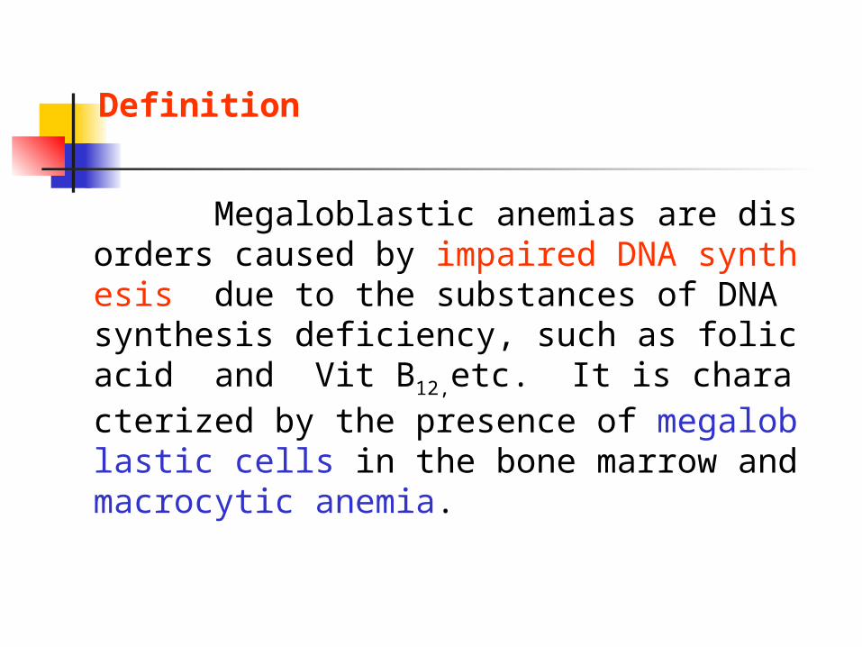
Megaloblastic anemias are disorders caused by impaired DNA synthesis due to the substances of DNA synthesis deficiency, such as folic acid and Vit B12,etc. It is characterized by the presence of megaloblastic cells in the bone marrow and macrocytic anemia.
Definition

Classification of MA
1.Vitamin B12 deficiency
2.Folate deficiency
3.Penicious anemia

The important role of folate and Vitamin B12 pl
aying in the pathways of deoxynucleotide and DNA synthesis:
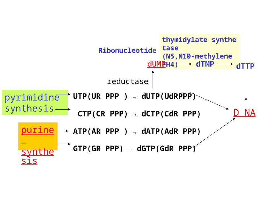
reductase
purine synthesis
pyrimidine synthesis
thymidylate synthetase(N5,N10-methylene FH4)
UTP(UR PPP ) → dUTP(UdRPPP)
CTP(CR PPP) → dCTP(CdR PPP)
ATP(AR PPP ) → dATP(AdR PPP)
GTP(GR PPP) → dGTP(GdR PPP)
Ribonucleotide
dUMP
D NA
dTMP dTTP
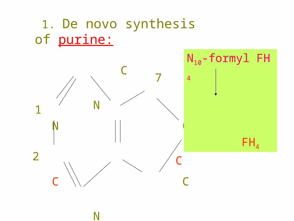
1. De novo synthesis of purine:
C
N
N C
C
C C
N
N
1
2
7
8
N10-formyl FH4
FH4
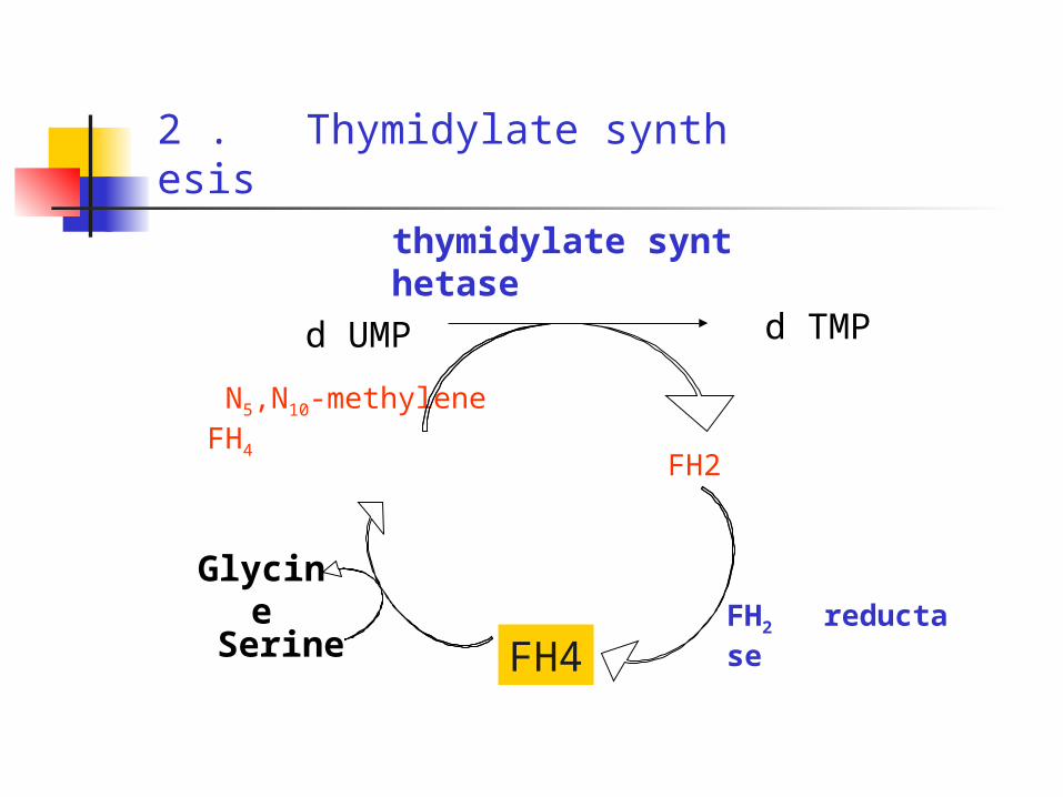
2 . Thymidylate synthesis
FH2 reductase
Glycine
N5,N10-methylene FH4
thymidylate synthetase
FH2
FH4
d UMP d TMP
Serine
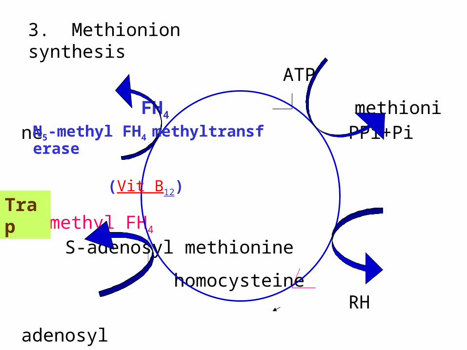
ATP
FH4 methionine PPi+Pi
N5-methyl FH4 S-adenosyl methionine
homocysteine RH
adenosyl
R-CH3
H2O S-adenosyl homocysteine
N5-methyl FH4 methyltransferase (Vit B12)
3. Methionion synthesis
Trap
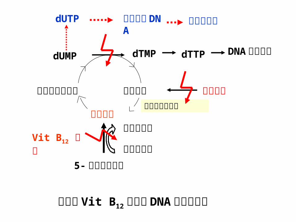
dUMP dTMP dTTP DNA 正常合成
亚甲基四氢叶酸 二氢叶酸
四氢叶酸二氢叶酸还原酶
叶酸缺乏
5- 甲基四氢叶酸高半胱氨酸
甲硫蛋氨酸Vit B12 缺乏
叶酸和 Vit B12 缺乏对 DNA 合成的影响
dUTP 合成异常DNA
巨幼变细胞
Diet
Methyltetrahydrofolate
Tetrahydrofolate
Dihydrofolate
N5,N10-methylene FH4
VitaminB12
methylB12 CH3
Methionine
Homocysteine
Thymidylate DNA
Deoxyuridylate
purine and pyrimidine synthesis
Serine
B6
glycine

Megaloblastic Anemias
Morphologically and functionally change
Megaloblastic cells
Defective DNA synthesis
Different disorders
Deficiency of folate and
Vit B12,etc. Non-deficiency
Ineffective erythropoiesis
Pathogenesis of MgA Etiology of MgA
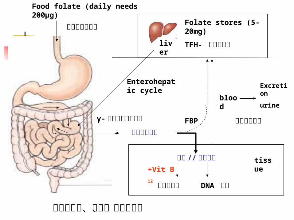
单谷氨酸叶酸
Enterohepatic cycle
叶酸的吸收、利用、贮存和排泄
γ- 谷氨酰胺羧基肽酶
甲基 // 四氢叶酸
DNA 合成蛋氨酸循环
+Vit B12
tissue
FBP 叶酸结合蛋白
blood
Excretion
urine
Food folate (daily needs 200μg)
多聚谷氨酸叶酸 Folate stores (5-20mg)
TFH- 多谷氨酸盐liver

绿叶蔬菜、柠檬、香蕉、瓜类、香菇、酵母及动物内脏(尤其是肝脏)等都含大量叶酸。
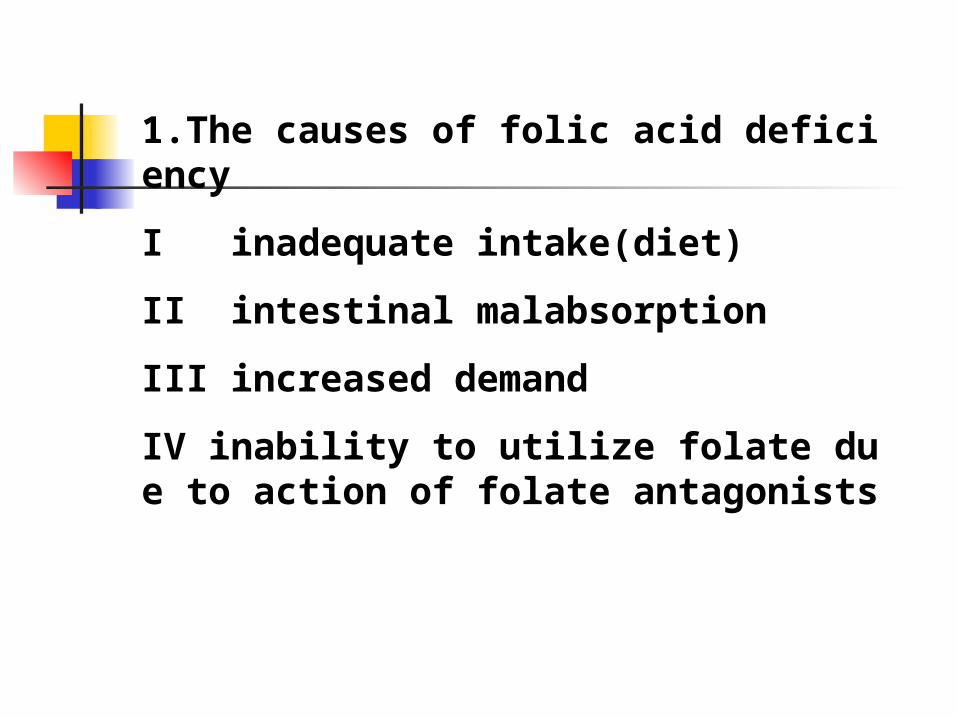
1.The causes of folic acid deficiency
I inadequate intake(diet)
II intestinal malabsorption
III increased demand
IV inability to utilize folate due to action of folate antagonists
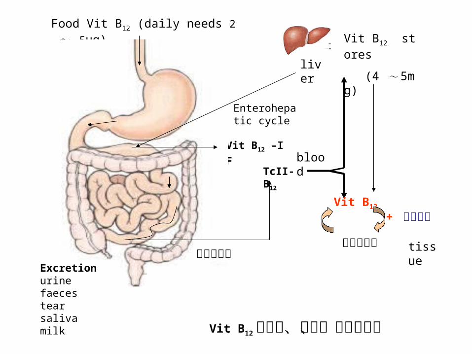
Food Vit B12 (daily needs 2 ~ 5μg)Vit B12 stores
(4 ~ 5mg) liver
Vit B12 的吸收、利用、贮存和排泄
TcII-B12
tissue
+ 甲基叶酸
蛋氨酸循环
Vit B12
Enterohepatic cycle
Vit B12 –IF
IF Vit B12
–R
蛋白
blood
释放内因子Excretionurinefaecestearsalivamilk

动物的肝、肾、肉类、禽蛋、乳类和海洋生物等 B12 含量较多。
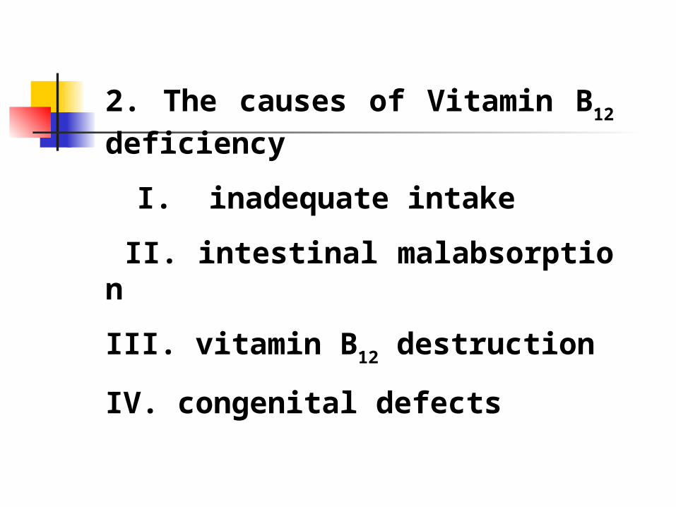
2. The causes of Vitamin B12 deficiency
I. inadequate intake
II. intestinal malabsorption
III. vitamin B12 destruction
IV. congenital defects

LaboratoryFeatures Clinical Features
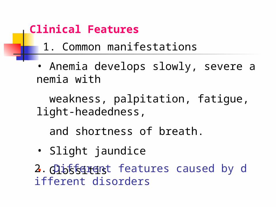
1. Common manifestations
• Anemia develops slowly, severe anemia with
weakness, palpitation, fatigue, light-headedness,
and shortness of breath.
• Slight jaundice
• Glossitis
Clinical Features
2. Different features caused by different disorders

Vit B12 deficiency:
•peripheral neuropathy and subacute combined degeneration of the spinal cord with nervous system symptoms such as numbness and tingling of the extremities, loss of position sense, muscle weakness and decreased tendon reflexes.
Folate deficiency :
not produce nervous system manifestations.
Pernicious anemia
• abdominal pain, diarrhea, nausea, and vomiting
• nervous system symptoms.
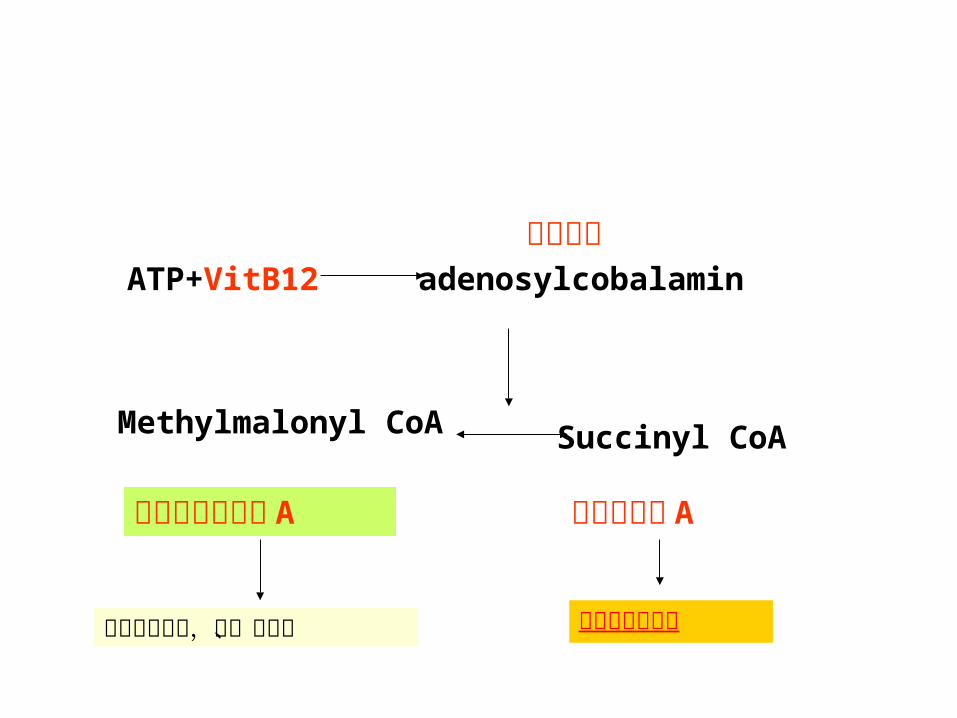
甲基丙二酰辅酶A
琥珀酰辅酶 A
腺苷钴胺ATP+VitB12 adenosylcobalamin
Methylmalonyl CoA Succinyl CoA
合成神经鞘磷脂代谢产物堆积,血、尿可测
Blood
•macrocytic anemia, with MCV↑(100~150fl or more).
•But coexisting IDA, Thalassemia trait, or inflammation may prevent macrocytosis.
•Erythrocytes show marked anisocytosis and poikilocytosis, with many oval macrocytes and in severe cases, basophilic stippling, Howell-Jolly bodies, and Cabot rings
•Megaloblastic normoblasts may be seen.
•Ret count is low.
Laboratory Features
•Leukopenia and thrombocytopenia are frequently present.
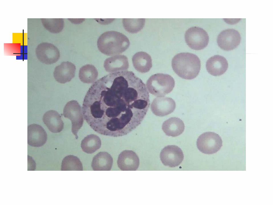
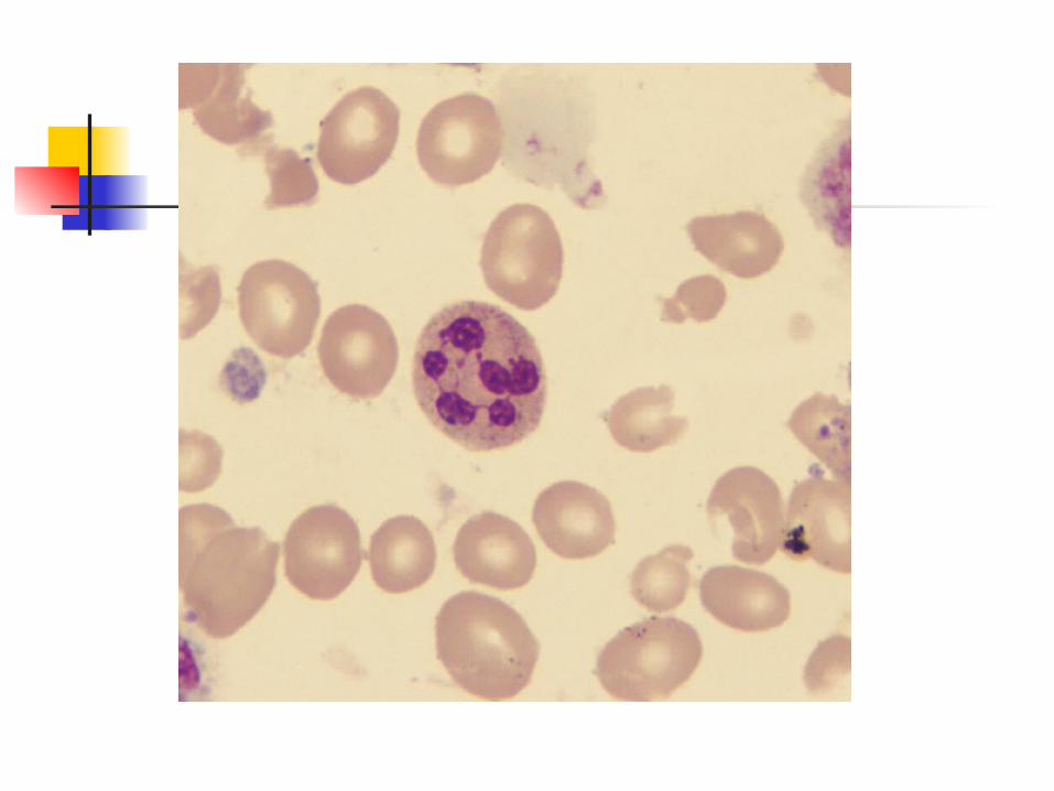
•“Hypersegmented neutrophils” (more than five lobes) are an early sign of megaloblastosis.
--Platelets:smaller and vary more widely in size.(PDW increase)

Bone Marrow:
--Moderate or marked hypercellularity.
-- M/E ratio ↓
--Erythroid hyperplasia with striking megaloblasts (more than 10%). Promegaloblasts and early megalonormoblasts increase with mitotic figures abundant in severe cases.
•basophilic stippling, Howell-Jolly bodies, and Cabot rings may be seen.
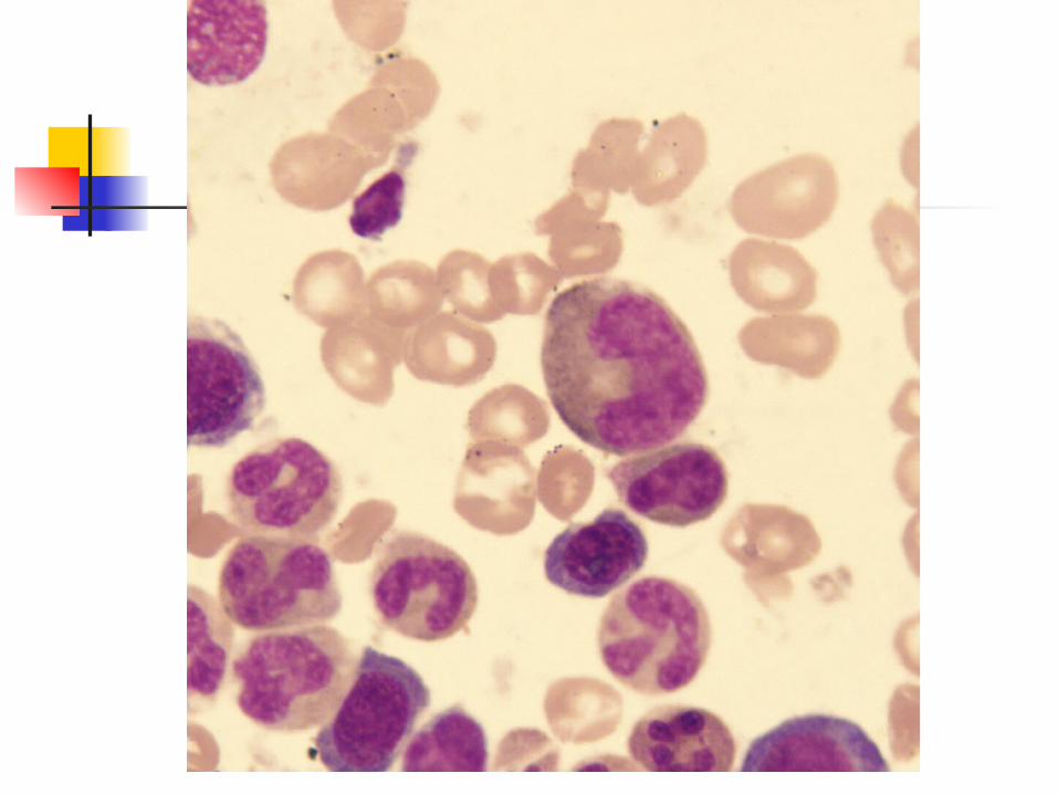
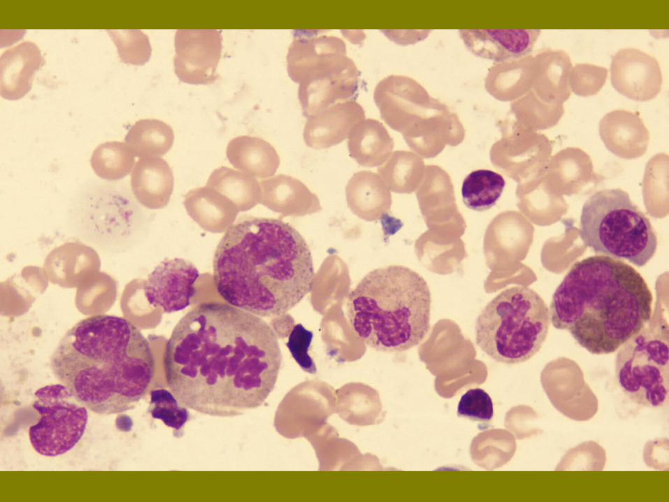
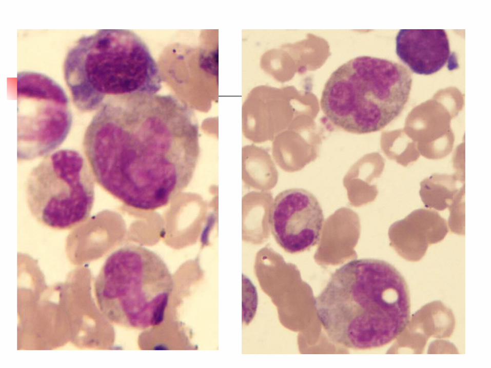
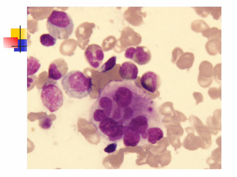
The characteristics of megaloblasts:
1. Cells and nuclei are larger in size and the chromatin is more looser.
2. Marked nuclear-cytoplasmic asynchrony.
The development of the nucleus is behind that of cytoplasm, called “young nucleus and old cytoplasm”
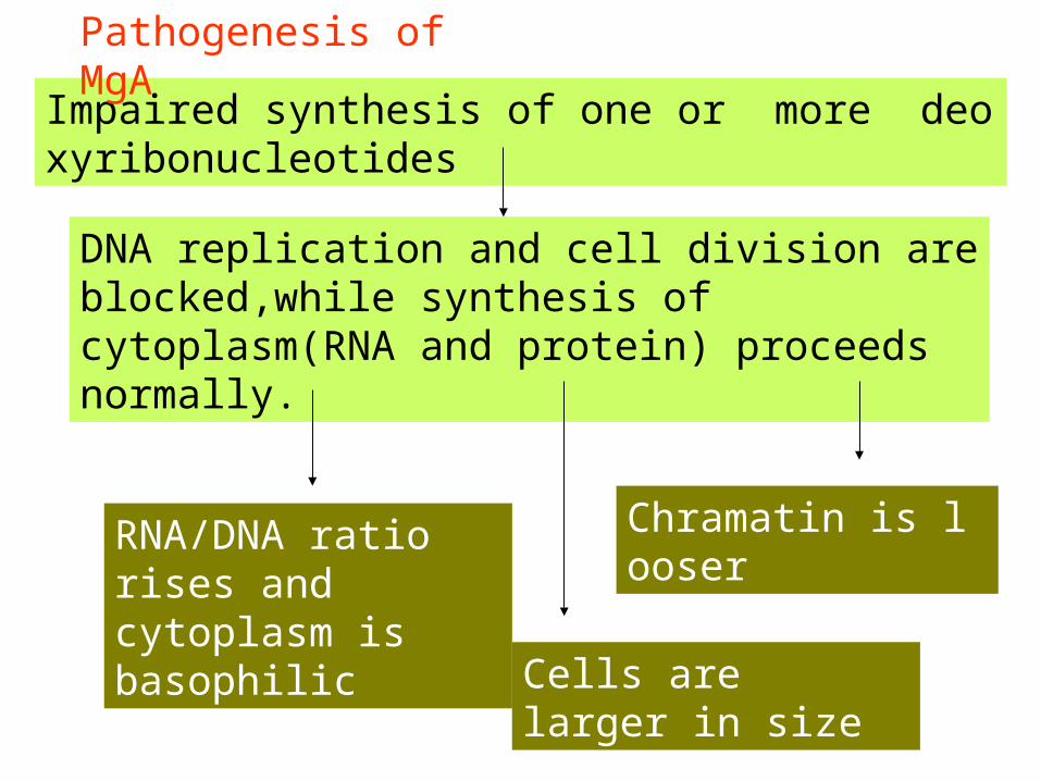
Impaired synthesis of one or more deoxyribonucleotides
DNA replication and cell division are blocked,while synthesis of cytoplasm(RNA and protein) proceeds normally.
RNA/DNA ratio rises and cytoplasm is basophilic
Chramatin is looser
Cells are larger in size
Pathogenesis of MgA
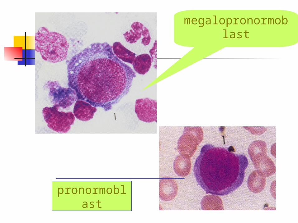
pronormoblast
megalopronormoblast
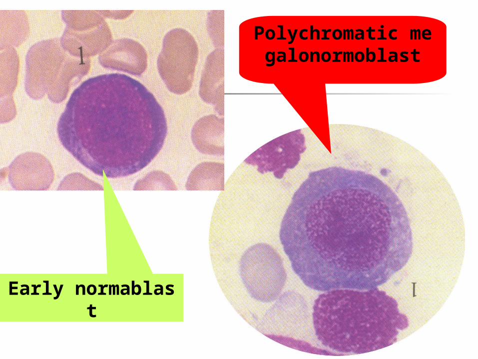
Early normablast
Polychromatic megalonormoblast
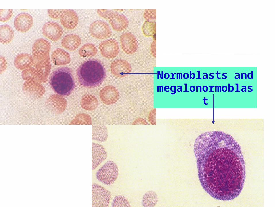
Normoblasts and megalonormoblast
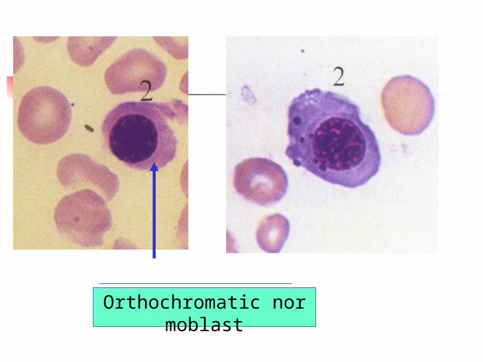
Orthochromatic normoblast

Megaloblastic changes 核畸形、多核、核碎裂的巨幼红细胞

Howell-Jolley bodies
Basophilic stippling normoblast
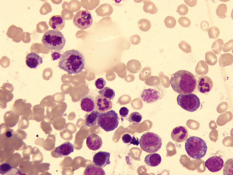
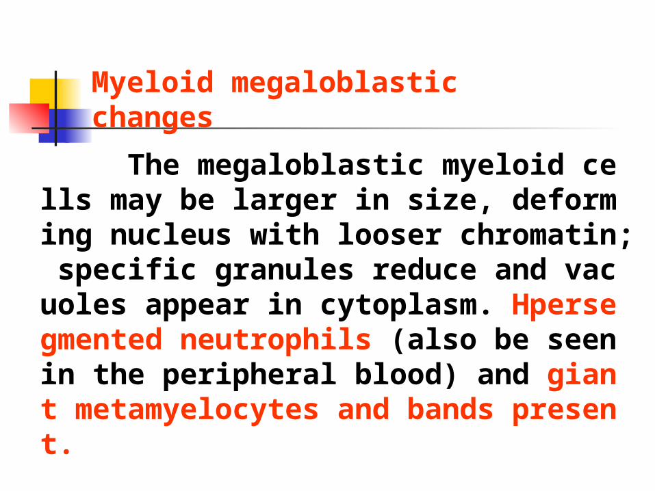
The megaloblastic myeloid cells may be larger in size, deforming nucleus with looser chromatin; specific granules reduce and vacuoles appear in cytoplasm. Hpersegmented neutrophils (also be seen in the peripheral blood) and giant metamyelocytes and bands present.
Myeloid megaloblastic changes
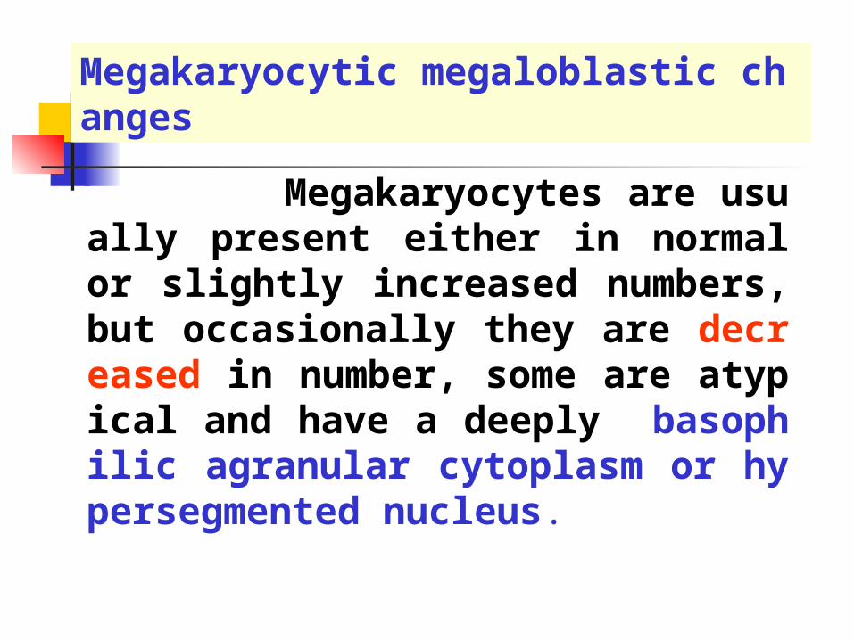
Megakaryocytes are usually present either in normal or slightly increased numbers, but occasionally they are decreased in number, some are atypical and have a deeply basophilic agranular cytoplasm or hypersegmented nucleus.
Megakaryocytic megaloblastic changes
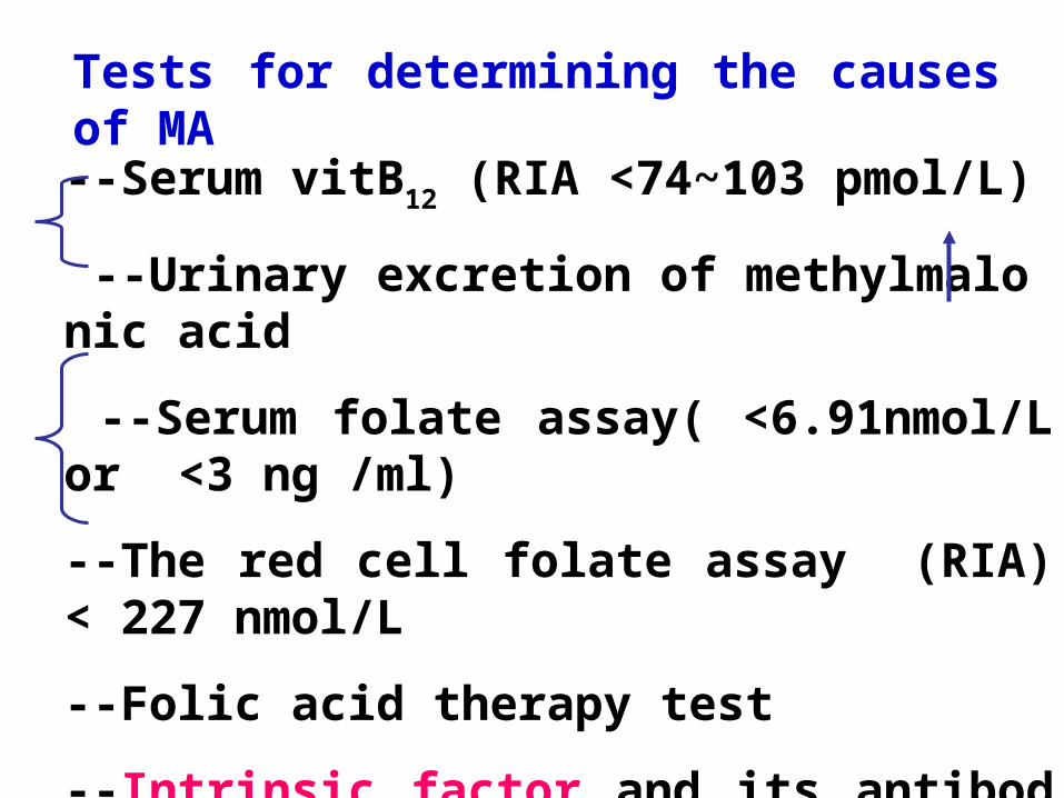
--Serum vitB12 (RIA <74~103 pmol/L)
--Urinary excretion of methylmalonic acid
--Serum folate assay( <6.91nmol/L or <3 ng /ml)
--The red cell folate assay (RIA)< 227 nmol/L
--Folic acid therapy test
--Intrinsic factor and its antibody
Tests for determining the causes of MA
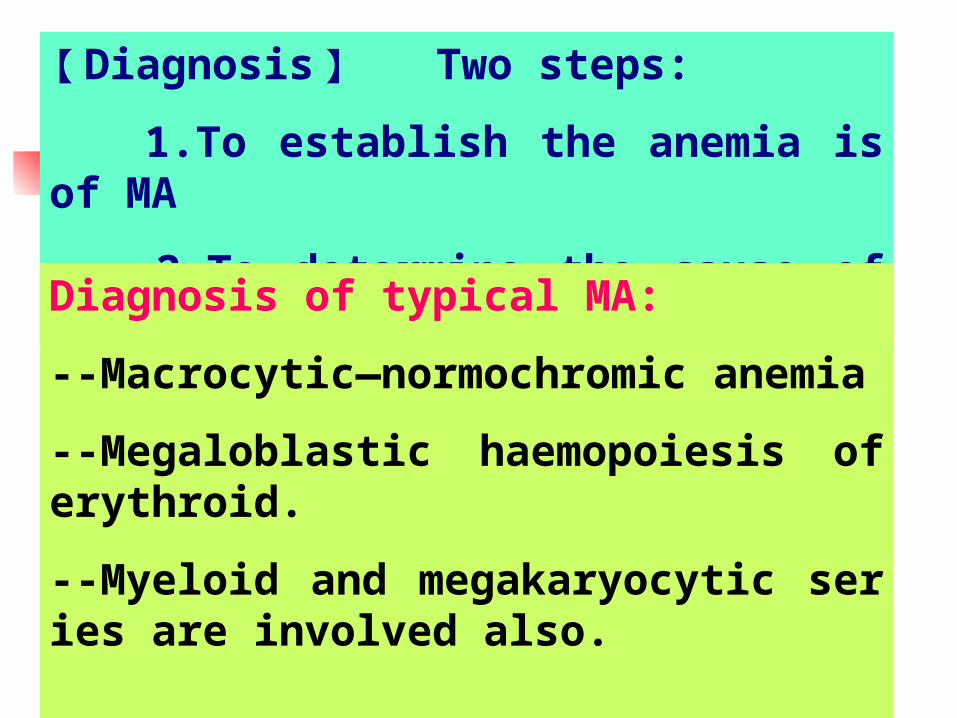
【 Diagnosis 】 Two steps:
1.To establish the anemia is of MA
2.To determine the cause of the anemia.
Diagnosis of typical MA:
--Macrocytic—normochromic anemia
--Megaloblastic haemopoiesis of erythroid.
--Myeloid and megakaryocytic series are involved also.

When MA is atypical, how to diagnose it?
Clinical features: history, signs, and specific tests
Myeloid megaloblastic changes
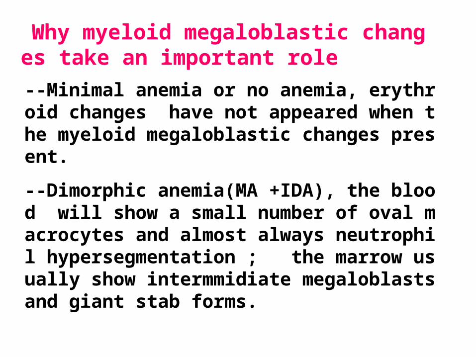
--Minimal anemia or no anemia, erythroid changes have not appeared when the myeloid megaloblastic changes present.
--Dimorphic anemia(MA +IDA), the blood will show a small number of oval macrocytes and almost always neutrophil hypersegmentation ; the marrow usually show intermmidiate megaloblasts and giant stab forms.
Why myeloid megaloblastic changes take an important role
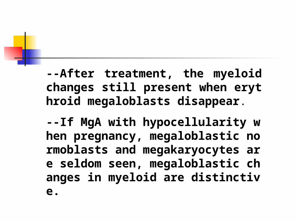
--After treatment, the myeloid changes still present when erythroid megaloblasts disappear.
--If MgA with hypocellularity when pregnancy, megaloblastic normoblasts and megakaryocytes are seldom seen, megaloblastic changes in myeloid are distinctive.
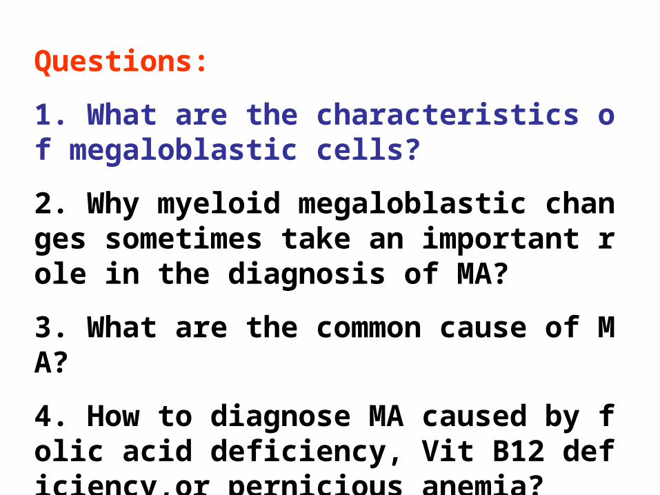
Questions:
1. What are the characteristics of megaloblastic cells?
2. Why myeloid megaloblastic changes sometimes take an important role in the diagnosis of MA?
3. What are the common cause of MA?
4. How to diagnose MA caused by folic acid deficiency, Vit B12 deficiency,or pernicious anemia?

ANEMIA
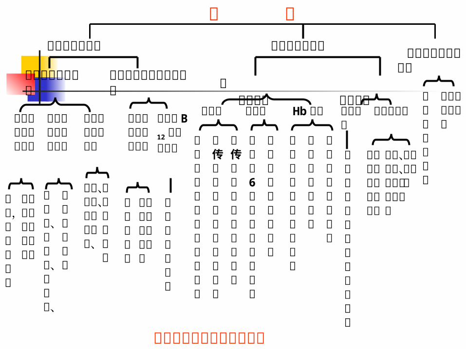
贫血的病因及发病机制分类
贫 血红细胞生成减少 红细胞破坏过多 红细胞丢
失增加 骨髓造血功能障碍
造血物质缺乏或利用障碍
铁缺乏和铁利用障碍
维生素 B12
或叶酸缺乏
红细胞 内在缺陷 外在异常
干细胞增殖分化障碍
骨髓被异常组织侵害
骨髓造血功能低下
免疫因素
非免疫因素
急性失血性贫血
慢性失血性贫血
微血管病性溶血性贫血
化学、物理、生物因素致溶血
脾功能亢进
各种原因致免疫性 溶血性贫血
巨幼细胞贫血等
缺铁性贫血
铁粒幼细胞性贫血等
再障,纯红再障等
骨髓增生异常综合征等
膜异常 酶异常 Hb 异常
珠蛋白生成障碍性贫血
异常血红蛋白病
不稳定血红蛋白病
葡萄糖6磷酸脱氢酶缺乏症
丙酮酸激酶缺乏症等
遗传性球形红细胞增多症
遗传性椭圆红细胞增多症等
阵发性睡眠性血红蛋白尿症
肾病、肝病、感染性疾病、
内分泌疾病等
白血病、骨髓瘤、癌转移、
骨髓纤维化等
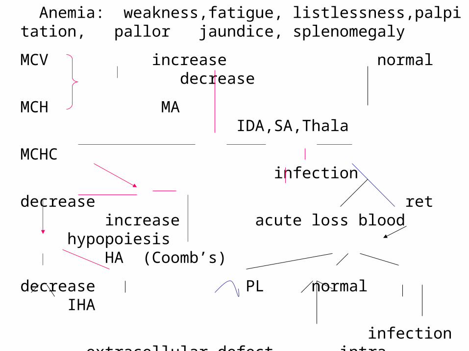
Anemia: weakness,fatigue, listlessness,palpitation, pallor jaundice, splenomegaly
MCV increase normal decrease
MCH MA IDA,SA,Thala
MCHC infection
decrease ret increase acute loss blood hypopoiesis HA (Coomb’s)
decrease PL normal IHA
infection extracellular defect intra-
WBC chronic renal disease osmotic fragility
decrease increase increase normal decrease
AA,MF AM HS,HE, G-6PD, PK Thala
PNH (ham’s) abnormalHb
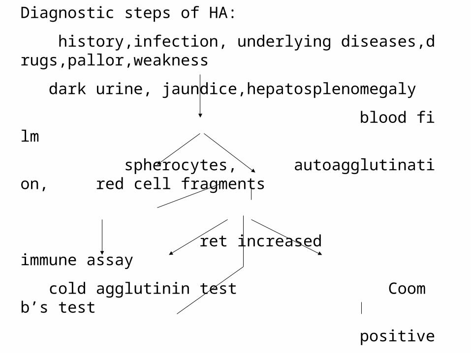
Diagnostic steps of HA:
history,infection, underlying diseases,drugs,pallor,weakness
dark urine, jaundice,hepatosplenomegaly
blood film
spherocytes, autoagglutination, red cell fragments
ret increased immune assay
cold agglutinin test Coomb’s test
positive
CAS AIHA negative cold warm
hemolysis test
transfusion reaction infection, congenital syphilis

Normocytic normochromic anemia
history, underlying diseases, pallor,weakness
blood film
decrease/ increase ret increase
morphologic features acute blood loss HA
normal abnormal
secondary hypoplasia abnormal proliferation
anemia infiltration in marrow
Infection AA MDS leukemia
renal disease MF
liver disease metastatic cancer
endocrinic disease
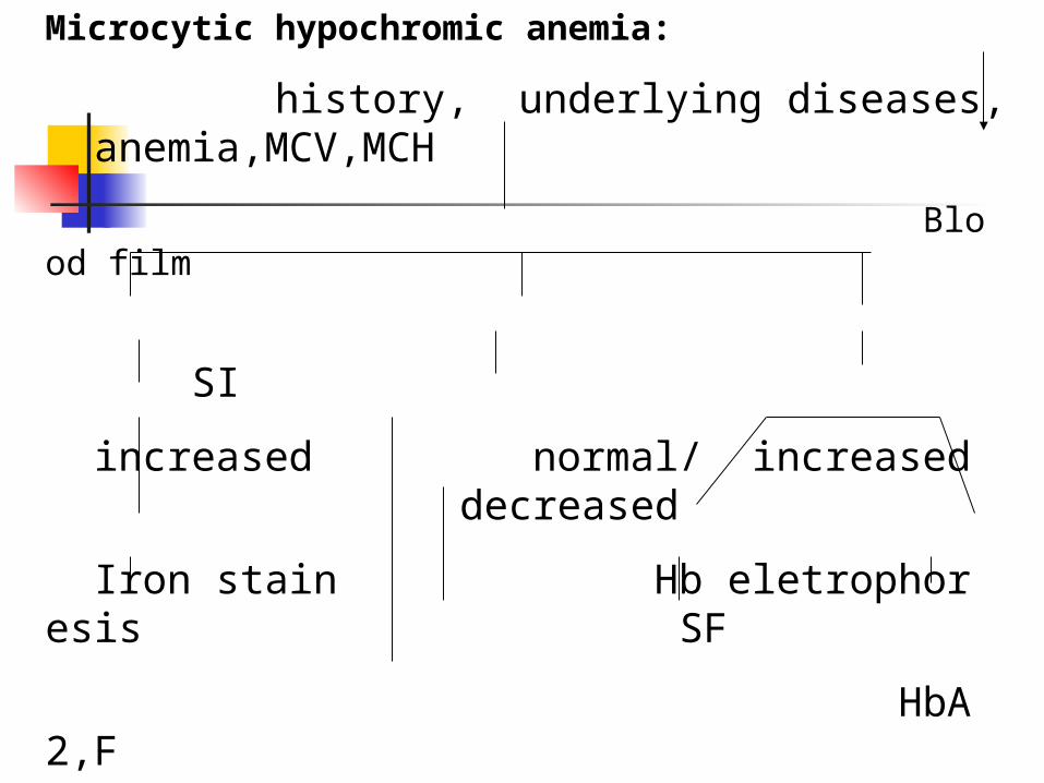
Microcytic hypochromic anemia:
history, underlying diseases, anemia,MCV,MCH
Blood film
SI
increased normal/ increased decreased
Iron stain Hb eletrophoresis SF
HbA2,F
increased normal/ increased decreased
SA Thalasemia anemia of chronic IDA
HbC,S, D,E disorders
etc.
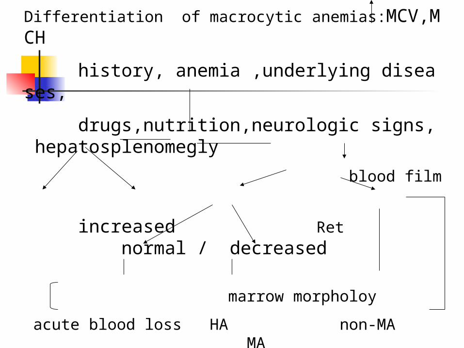
Differentiation of macrocytic anemias:MCV,MCH
history, anemia ,underlying diseases,
drugs,nutrition,neurologic signs, hepatosplenomegly
blood film
increased Ret normal / decreased
marrow morpholoy
acute blood loss HA non-MA MA
erythroblastic anemia abnormal proliferation
Alchohol poisoning MDS Folate deficiency
Liver disease VitB12 deficiency
Pernicious anemia