Embed Size (px)
Citation preview
Rev Med Hosp Gen Méx. 2015;78(3):135---143
www.elsevier.es/hgmx
´
´
REVIEW ARTICLE
Megaloblastic anaemia: Folic acid and vitamin B12metabolism
H.B. Castellanos-Sincoa,b, C.O. Ramos-Penafiela,∗, A. Santoyo-Sánchezc,J. Collazo-Jalomaa, C. Martínez-Murilloa, E. Montano-Figueroaa, A. Sinco-Ángelesd
a Servicio de Hematología, Hospital General de México ‘‘Dr. Eduardo Liceaga’’, Mexico City, Mexicob Servicio de Hematología, Hospital General de Zona con UMAA No. 48 ‘‘San Pedro Xalpa’’, Instituto Mexicano del Seguro Social,Mexico City, Mexicoc Facultad de Medicina, Universidad Nacional Autónoma de México, Mexico City, Mexicod Servicio de Hematología, Hospital General de Pachuca, Secretaría de Salud de Hidalgo, Pachuca, Mexico
Received 17 April 2015; accepted 6 July 2015Available online 28 September 2015
KEYWORDSMegaloblasticanaemia;Vitamin B12deficiency;Folic acid;Megaloblasts
Abstract Folic acid and cobalamin are B-group vitamins that play an essential role in manycellular processes. Deficiency in one or both of these vitamins causes megaloblastic anaemia, adisease characterized by the presence of megaloblasts. Megaloblasts occur when inhibition ofDNA synthesis causes asynchronous maturation between the nucleus and the cytoplasm. Clinicalmanifestations are similar to those of other types of anaemia, with the exception of cobalamindeficiency megaloblastic anaemia, which presents distinctive neurological symptoms. An under-standing of the metabolism of these vitamins will enable clinicians to make the best use andinterpretation of laboratory studies and monitor therapeutic strategies, which consist mainlyof administering supplements to restore body reserves.© 2015 Published by Masson Doyma México S.A. on behalf of Sociedad Médica del HospitalGeneral de México.
PALABRAS CLAVEAnemia
Anemias megaloblásticas: Metabolismodelácidofólico y vitamina B12
megaloblástica; Resumen Anemias megaloblásticas: metabolismo del ácido fólico y vitamina B12 El ácidoitaminas del complejo B, indispensables para un número importantedéficit de una o ambas vitaminas ocasiona anaemia megaloblás-do por la presencia de megaloblastos, resultado de la asincroníanúcleo y el citoplasma del eritrocito debido a la alteración en lastaciones clínicas son similares a otras anemias, salvo la anaemia
Deficiencia devitamina B 12;Ácido fólico;Megaloblastos
fólico y la cobalamina son vde procesos celulares. El
tica, síndrome caracterizade la maduración entre el
síntesis de ADN. Las manife
∗ Corresponding author at: Dr. Balmis 148, Col. Doctores, C.P. 06726 Mexico City, Mexico.E-mail address: [email protected] (C.O. Ramos-Penafiel).
http://dx.doi.org/10.1016/j.hgmx.2015.07.0010185-1063/© 2015 Published by Masson Doyma México S.A. on behalf of Sociedad Médica del Hospital General de México.
136 H.B. Castellanos-Sinco et al.
megaloblástica ocasionada por déficit de cobalamina que presenta alteraciones neurológicasde forma distintiva. Es importante conocer el metabolismo de las vitaminas en cuestión paraun correcto uso e interpretación de los estudios de laboratorio, así como para el monitoreo dela terapéutica, basada esencialmente en reponer el déficit y restaurar las reservas corporales.© 2015 Publicado por Masson Doyma México S.A. en nombre de Sociedad Médica del HospitalGeneral de México.
B
TwranywaMbCftspoo
D
Masb(tbi
F
Fta
maicPs
tsma
fa
4
oarfieitactiai
dtTmotfitrittaet
ssu
gss
g
ackground
he discovery of megaloblastic anaemia and its aetiologyas the result of the efforts of many different medical
esearchers. It was first characterized by Addison in 1849s anaemia, general languor and debility.1 Osler and Gard-er in 1877 noted the association with neuropathy, and 10ears later Lichtheim documented myelopathy. Megaloblastsere identified for the first time by Ehrlich in 1880. In 1920,bnormalities in white blood cells were described. In 1926,inot and Murphy showed that the disease could be reversedy the intake of large quantities of liver.2 Three years later,astle established that gastric acid contains an ‘‘intrinsicactor’’ that combines with an ‘‘extrinsic factor’’ to allowhis latter to be absorbed.3 Hodgkin later identified thetructure of vitamin B12, for which he received the Nobelrize.4 Years later, in 1948, Herbert discovered the structuref folic acid and described its association with the aetiologyf megaloblastic anaemia.5
efinition
egaloblastic anaemia is a general term used to describe group of anaemias caused by impaired DNA synthe-is. It is characterized by abnormal findings in peripherallood smear (macroovalocytes) and bone marrow samplesmegaloblastic hyperplasia). Megaloblasts, the hallmark ofhese anaemias, are caused by asynchronous maturationetween the nucleus and the cytoplasm due to DNA synthesismpairment.6---8
olic acid metabolism
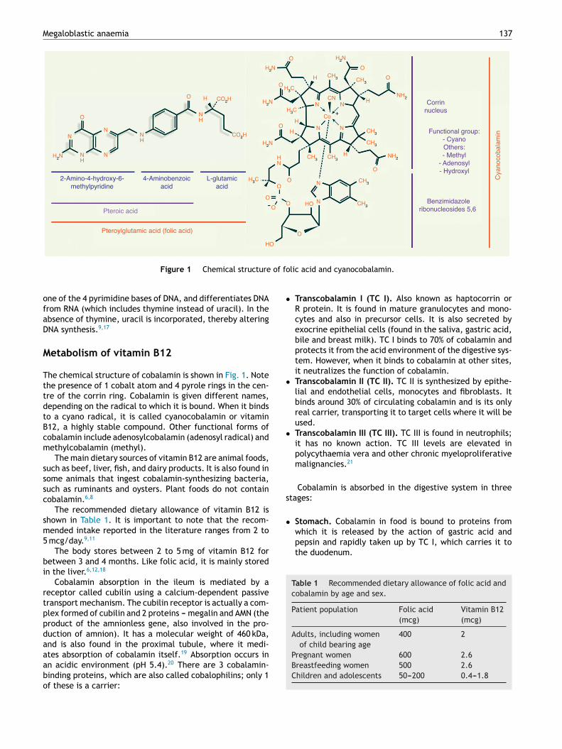
olic acid, also known as pteroyl-glutamate or pteroylglu-amic acid, is made up of: (1) pteroic acid; and (2) l-glutamiccid (one or more strands) (see Fig. 1).8,9
The functional form of folate is tetrahydrofolic acid. Theain dietary sources of folic acid are green vegetables, such
s asparagus, broccoli, spinach and lettuce. It is also foundn fruit, such as lemons, oranges, bananas and melons, and inereals, grains, nuts, beans, beef, fish, liver and kidneys.6
rolonged storage or over-cooking in abundant water canignificantly reduce the folate content of food.10
Daily adult requirements of folic acid range from 50o 100 mcg. The recommended dietary allowance (RDA)
hown in Table 1, however, is far higher than the mini-um requirement. This is because the bioavailability of foliccid depends on hydrolysation of the polyglutamate form of
mtd
olate to its monoglutamate form to facilitate absorptioncross the small intestine.11
The body stores around 5 mg of folate for between 3 and months. Folate is primarily stored in the liver.12
Folic acid is mainly absorbed in the jejunum by meansf passive transport following the concentration gradient,nd by means of active transport when folate binds toeduced-folate transporter 1 and 2 (RFT-1 and RFT-2) andolate binding protein (FBP). Folic acid is also absorbedn the ilium, solely by passive transport.12---14 Folate, inither its monoglutamate or reduced monoglutamate form,s absorbed in a neutral pH environment (pH 7.4) facili-ated by neutralization of the acid gastric environment bylkaline pancreatic juices.14 One of 2 enzymes, glutamatearboxypeptidase or polyglutamate hydrolase, are neededo hydrolyse folic acid polyglutamate (the form in which its found in food) to its monoglutamate form. These enzymesre found on the luminal surface of the jejunum andleum.13,14
Once taken up by the enterocyte, the enzyme dihy-rofolate reductase mediates the conversion of folic acido methyltetrahydrofolate through a two-step reaction.he folate then exits the enterocyte via the basolateralembrane and is either taken into the systemic circulation
r the enterohepatic cycle (liver, bile acids, intestine). Inhe systemic circulation, 66% binds to albumin, 33% remainsree, and a small amount (1%) binds to FBP. This is how its transported to the cells where it will be used. It entershe cells either by binding to RTF-1 or RTF-2 or to folateeceptor 1 (alpha) or 2 (beta). Intracellular folate transports mediated by clathrin-mediated endocytosis. Once insidehe cell (methyltetrahydrofolate), it must be demethylatedo become tetrahydrofolate (functional folate that canccept glutamic acid chains; these in turn prevent it fromxiting the cell, in other words, they ‘‘anchor’’ it insidehe cell).13,15
Excess intracellular folic acid can pass into the bloodtream and then be filtered through the glomerulus,ecreted into the proximal tubule, and eliminated in therine at a rate of 2---5 mcg/day.16
The biological functions of folic acid include: serine-lycine conversion, histidine catabolism, purine synthe-is, and more importantly, thymidylate and methionineynthesis.17
In thymidylate synthesis, folic acid carries one-carbonroups. Thymidylate is synthesized from deoxyuridine
onophosphate (dUMP) and methylenetetrahydrofolate byhymidylate synthase, which converts these elements intoihydrofolate and thymidylate. Thymidylate, or thymine, is
Megaloblastic anaemia 137
O
O
NN
NNH
2-Amino-4-hydroxy-6-methylpyridine
Pteroic acid
Pteroylglutamic acid (folic acid)
Cya
noco
bala
min
4-Aminobenzoicacid
L-glutamicacid
Corrinnucleus
Functional group:- CyanoOthers:- Methyl
- Adenosyl- Hydroxyl
Benzimidazoleribonucleosides 5,6
NH
H
NH
HN
H
H
H
HN
CN
N N
N
+
H
O
O
O
OO
O
OO
O
HO
O
HO
N
N
O
O
H2N
CO2H
CO2H
Co
H2N
H3C
H2N
H2N
H2N
H3C
H3C
CH3
NH2
NH2
CH3
CH3
CH3 CH3
CH3 CH3
CH3
of fo
•
•
•
s
•which it is released by the action of gastric acid andpepsin and rapidly taken up by TC I, which carries it tothe duodenum.
Table 1 Recommended dietary allowance of folic acid andcobalamin by age and sex.
Patient population Folic acid(mcg)
Vitamin B12(mcg)
Adults, including womenof child bearing age
400 2
Figure 1 Chemical structure
one of the 4 pyrimidine bases of DNA, and differentiates DNAfrom RNA (which includes thymine instead of uracil). In theabsence of thymine, uracil is incorporated, thereby alteringDNA synthesis.9,17
Metabolism of vitamin B12
The chemical structure of cobalamin is shown in Fig. 1. Notethe presence of 1 cobalt atom and 4 pyrole rings in the cen-tre of the corrin ring. Cobalamin is given different names,depending on the radical to which it is bound. When it bindsto a cyano radical, it is called cyanocobalamin or vitaminB12, a highly stable compound. Other functional forms ofcobalamin include adenosylcobalamin (adenosyl radical) andmethylcobalamin (methyl).
The main dietary sources of vitamin B12 are animal foods,such as beef, liver, fish, and dairy products. It is also found insome animals that ingest cobalamin-synthesizing bacteria,such as ruminants and oysters. Plant foods do not containcobalamin.6,8
The recommended dietary allowance of vitamin B12 isshown in Table 1. It is important to note that the recom-mended intake reported in the literature ranges from 2 to5 mcg/day.9,11
The body stores between 2 to 5 mg of vitamin B12 forbetween 3 and 4 months. Like folic acid, it is mainly storedin the liver.6,12,18
Cobalamin absorption in the ileum is mediated by areceptor called cubilin using a calcium-dependent passivetransport mechanism. The cubilin receptor is actually a com-plex formed of cubilin and 2 proteins --- megalin and AMN (theproduct of the amnionless gene, also involved in the pro-duction of amnion). It has a molecular weight of 460 kDa,and is also found in the proximal tubule, where it medi-
ates absorption of cobalamin itself.19 Absorption occurs inan acidic environment (pH 5.4).20 There are 3 cobalamin-binding proteins, which are also called cobalophilins; only 1of these is a carrier:lic acid and cyanocobalamin.
Transcobalamin I (TC I). Also known as haptocorrin orR protein. It is found in mature granulocytes and mono-cytes and also in precursor cells. It is also secreted byexocrine epithelial cells (found in the saliva, gastric acid,bile and breast milk). TC I binds to 70% of cobalamin andprotects it from the acid environment of the digestive sys-tem. However, when it binds to cobalamin at other sites,it neutralizes the function of cobalamin.
Transcobalamin II (TC II). TC II is synthesized by epithe-lial and endothelial cells, monocytes and fibroblasts. Itbinds around 30% of circulating cobalamin and is its onlyreal carrier, transporting it to target cells where it will beused.
Transcobalamin III (TC III). TC III is found in neutrophils;it has no known action. TC III levels are elevated inpolycythaemia vera and other chronic myeloproliferativemalignancies.21
Cobalamin is absorbed in the digestive system in threetages:
Stomach. Cobalamin in food is bound to proteins from
Pregnant women 600 2.6Breastfeeding women 500 2.6Children and adolescents 50---200 0.4---1.8
1
•
•
iacIeot3cb(cpc
sec
•
•
cf
•
•
afpltwmc
P
Tiai
mp(
psmistrt
tddm
E
FiapT
C
Ticbh
P
Ct
38
Duodenum and jejunum. The alkalizing action of thepancreatic juices together with the action of the pancre-atic enzymes (tripsin, chymotrypsin and elastase) degradethe TC I and release the cobalamin, which is now taken upby the intrinsic factor (IF). IF is produced by the parietalcells of the fundus and cardia of the stomach. It protectthe cobalamin and carries it to the cubilin in the ileum.
Ileum. The IF-cobalamin complex binds to cubilin andis taken up into the enterocyte by means of a calcium-dependent passive transport mechanism.
Once inside the enterocyte, the IF-cobalamin complexs engulfed by lysosomes, where enzymes degrade the IFnd release the cobalamin. The cobalamin then exits to theytoplasm, where it is taken up by TC II. In this way, the TCI/cobalamin complex exits the enterocyte via its basolat-ral membrane and is released into the systemic circulationr the enterohepatic cycle (liver, bile acids, intestine). Inhe systemic circulation, cobalamin can bind to any of the
aforementioned cobalophilins. TC II transports it to theells where it will be used. It is taken up into the cellsy binding to either TC II receptors (TC IIR) or megalina protein). Cobalamin is taken up by cells by means oflathrin-mediated endocytosis, the same intracellular trans-ort mechanisms used in folic acid uptake. Once inside theells, TC II is degraded, thus releasing the cobalamin.13,15,22
Excess intracellular cobalamin can pass into the bloodtream, from where it is filtered through the glomerulus andliminated in the urine.19In humans, the 2 active forms ofobalamin involved in biological functions are:
Methycobalamin. A co-enzyme of methionine synthase(also known as methyltetrahydrofolate-homocysteinemethyltransferase), an enzyme involved in methionineand tetrahydrofolate synthesis from methyltetrahydro-folate and homocysteine. This is where the folate andcobalamin metabolism pathways meet, and it is some-times called the ‘‘folate trap.’’
Adenosylcobalamin. A co-enzyme of methylmalonyl-CoA,an enzyme involved in the production of succinic acid frommethylmalonyl acid (accumulation of which causes neu-ropathy). Succinate plays a role in the Krebs cycle, anenergy-releasing pathway.
Two hypotheses have been developed to explain howobalamin-deficiency anaemia is in fact caused by functionalolate deficiency.23,24
Methyltetrahydrofolate trapping, or the ‘‘folate trap’’.Without cobalamin, methyltetrahydrofolate cannot bedemethylated by methionine synthesis. Remember,methyltetrahydrofolate cannot be polyglutamised, andtherefore cannot be ‘‘anchored’’ to the cell. This meansthat it can escape without being used.
Formate deficiency. When tetrahydrofolate is depleted(as described above), stored methyltetrahydrofolate can-
not be converted into polyglutamisable formate-mediatedformyltetrahydrofolate, which is another functional formof folate (in addition to the oft-mentioned tetrahydrofo-late) used in purine synthesis.cfdt
H.B. Castellanos-Sinco et al.
In addition to the foregoing hypotheses, cobalaminnd folic acid metabolization share another commoneature: they both require methylenetetrahydrofolate (aroduct of tetrahydrofolate) and dUMP to form thymidy-ate synthase-mediated thymidylate and dihydrofolate. Forhe reasons stated above, tetrahydrofolate cannot occurithout cobalamin, and without tetrahydrofolate, neitherethylenetetrahydrofolate nor its product, thymidylate,
an occur.23,24
athophysiology of megaloblastic anemia
he pathophysiology of this group of anaemias has its originsn ineffective erythropoiesis secondary to intramedullarypoptosis of hematopoietic precursor cells. This, in turn,s caused by DNA synthesis abnormalities.
Remember, both folate and cobalamin deficiency ulti-ately lead to thymidylate deficiency. DNA contains 2urine bases (adenine and guanine) and 2 pyrimidine basesthymine and cytosine).8,25
When there is insufficient thymidylate or thymine at theosition in the DNA strand where these nitrogenous baseshould occur, they are replaced by uracil. This happens pri-arily when uracil is incorporated at 2 similar positions
n opposite strands. When uracil is incorporated into whathould be a purely DNA structure, the repair enzymes detecthe error and try to correct it, albeit unsuccessfully. As aesult, first 1, then both DNA strands are destroyed, withhe resulting p53-mediated cellular apoptosis.8,25,26
This in turn leads to asynchronous maturation betweenhe nucleus and the cytoplasm. The latter, devoid of DNA,oes not fully mature, and the former, in which RNA pro-uction continues and haemoglobin synthesis is unaltered,ature at the normal rate.8,9,25,26
tiology of megaloblastic anemia
olic acid deficiency is usually due to low folate contentn the diet, or to an imbalance between folate demandnd intake. Cobalamin deficiency is usually caused byoor absorption of this vitamin in the digestive tract (seeable 2).25,27
linical picture
he clinical spectrum of megaloblastic anaemia is shownn Table 3, where the minor differences between thelinical manifestations of megaloblastic anaemia causedy folate deficiency and by cobalamin deficiency areighlighted.6,25,28
athophysiology of neurological changes
obalamin deficiency causes subacute combined degenera-ion of the posterior and lateral grey column of the spinal
ord due to methionine deficiency. Methionine is neededor the production of myelin. Myelin deficiency causesemyelination and gliosis of the grey column, which is fur-her aggravated by the neurotoxicity of methylmalonic acid.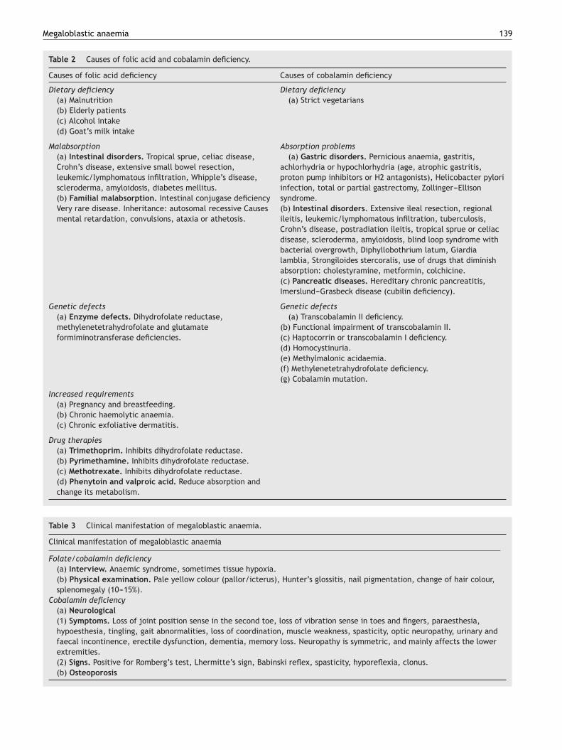
Megaloblastic anaemia 139
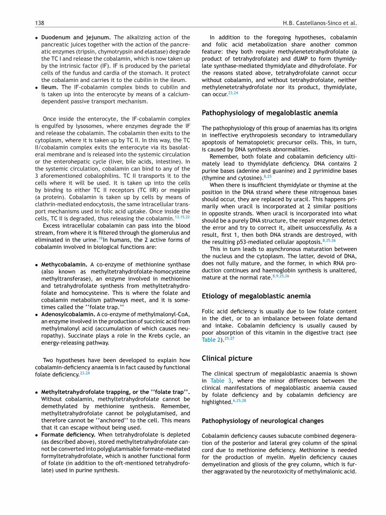
Table 2 Causes of folic acid and cobalamin deficiency.
Causes of folic acid deficiency Causes of cobalamin deficiency
Dietary deficiency Dietary deficiency(a) Malnutrition(b) Elderly patients(c) Alcohol intake(d) Goat’s milk intake
(a) Strict vegetarians
Malabsorption Absorption problems(a) Intestinal disorders. Tropical sprue, celiac disease,Crohn’s disease, extensive small bowel resection,leukemic/lymphomatous infiltration, Whipple’s disease,scleroderma, amyloidosis, diabetes mellitus.(b) Familial malabsorption. Intestinal conjugase deficiencyVery rare disease. Inheritance: autosomal recessive Causesmental retardation, convulsions, ataxia or athetosis.
(a) Gastric disorders. Pernicious anaemia, gastritis,achlorhydria or hypochlorhydria (age, atrophic gastritis,proton pump inhibitors or H2 antagonists), Helicobacter pyloriinfection, total or partial gastrectomy, Zollinger---Ellisonsyndrome.(b) Intestinal disorders. Extensive ileal resection, regionalileitis, leukemic/lymphomatous infiltration, tuberculosis,Crohn’s disease, postradiation ileitis, tropical sprue or celiacdisease, scleroderma, amyloidosis, blind loop syndrome withbacterial overgrowth, Diphyllobothrium latum, Giardialamblia, Strongiloides stercoralis, use of drugs that diminishabsorption: cholestyramine, metformin, colchicine.(c) Pancreatic diseases. Hereditary chronic pancreatitis,Imerslund---Grasbeck disease (cubilin deficiency).
Genetic defects Genetic defects(a) Enzyme defects. Dihydrofolate reductase,methylenetetrahydrofolate and glutamateformiminotransferase deficiencies.
(a) Transcobalamin II deficiency.(b) Functional impairment of transcobalamin II.(c) Haptocorrin or transcobalamin I deficiency.(d) Homocystinuria.(e) Methylmalonic acidaemia.(f) Methylenetetrahydrofolate deficiency.(g) Cobalamin mutation.
Increased requirements(a) Pregnancy and breastfeeding.(b) Chronic haemolytic anaemia.(c) Chronic exfoliative dermatitis.
Drug therapies(a) Trimethoprim. Inhibits dihydrofolate reductase.(b) Pyrimethamine. Inhibits dihydrofolate reductase.(c) Methotrexate. Inhibits dihydrofolate reductase.(d) Phenytoin and valproic acid. Reduce absorption andchange its metabolism.
Table 3 Clinical manifestation of megaloblastic anaemia.
Clinical manifestation of megaloblastic anaemia
Folate/cobalamin deficiency(a) Interview. Anaemic syndrome, sometimes tissue hypoxia.(b) Physical examination. Pale yellow colour (pallor/icterus), Hunter’s glossitis, nail pigmentation, change of hair colour,splenomegaly (10---15%).
Cobalamin deficiency(a) Neurological(1) Symptoms. Loss of joint position sense in the second toe, loss of vibration sense in toes and fingers, paraesthesia,hypoesthesia, tingling, gait abnormalities, loss of coordination, muscle weakness, spasticity, optic neuropathy, urinary andfaecal incontinence, erectile dysfunction, dementia, memory loss. Neuropathy is symmetric, and mainly affects the lowerextremities.(2) Signs. Positive for Romberg’s test, Lhermitte’s sign, Babinski reflex, spasticity, hyporeflexia, clonus.(b) Osteoporosis
1
Eg
B
Aat
•
•
•
•
•
40
levation of tumour necrosis factor alpha and epidermalrowth factor also contribute to neurological changes.28,29
lood picture
ll types of megaloblastic anaemia, whether caused by foliccid or cobalamin deficiency, present the following labora-ory findings.25
Flow cytometry. In addition to anaemia, macrocytosisis found in 75% of cases (note that in 25% of patientsmean corpuscular volume [MCV] is normal, above all incases with concurrent iron deficiency or thalassemia).Macrocytosis can be classified as mild (100---105 fL), mod-erate (106---115 fL) or severe (>116 fL). Another findingis increased blood cell distribution width. In some cases(associated with severe, chronic folic acid or cobalamindeficiency) varying degrees of leukoneutropaenia andthrombocytopenia can be present (usually mild to mod-erate, but occasionally severe).6,7,25
Peripheral blood samples. The most notable findingsare macrocytosis and hypersegmented neutrophils. Froma morphological point of view, these abnormalitiestogether, while not pathognomonic, are highly suggestive
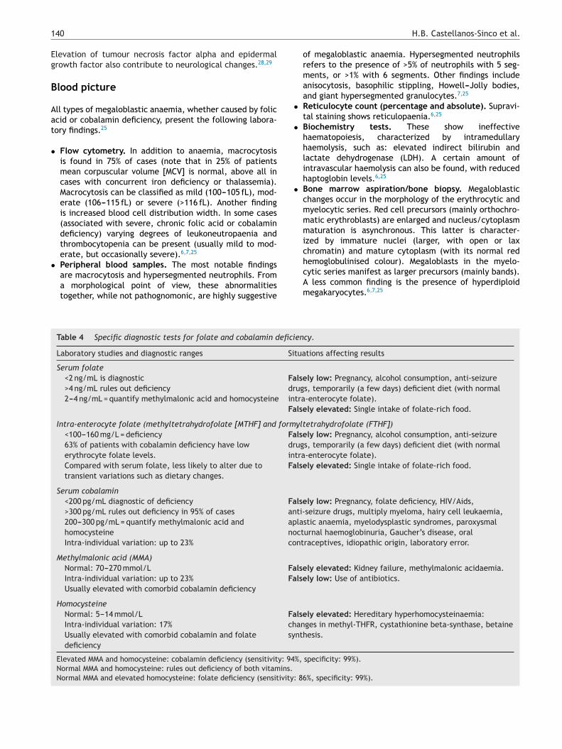
Table 4 Specific diagnostic tests for folate and cobalamin defici
Laboratory studies and diagnostic ranges Si
Serum folate<2 ng/mL is diagnostic>4 ng/mL rules out deficiency2---4 ng/mL = quantify methylmalonic acid and homocysteine
FadrinFa
Intra-enterocyte folate (methyltetrahydrofolate [MTHF] and form<100---160 mg/L = deficiency63% of patients with cobalamin deficiency have lowerythrocyte folate levels.Compared with serum folate, less likely to alter due totransient variations such as dietary changes.
FadrinFa
Serum cobalamin<200 pg/mL diagnostic of deficiency>300 pg/mL rules out deficiency in 95% of cases200---300 pg/mL = quantify methylmalonic acid andhomocysteineIntra-individual variation: up to 23%
Faanapnoco
Methylmalonic acid (MMA)Normal: 70---270 mmol/LIntra-individual variation: up to 23%Usually elevated with comorbid cobalamin deficiency
FaFa
HomocysteineNormal: 5---14 mmol/LIntra-individual variation: 17%Usually elevated with comorbid cobalamin and folatedeficiency
Fachsy
Elevated MMA and homocysteine: cobalamin deficiency (sensitivity: 94Normal MMA and homocysteine: rules out deficiency of both vitamins.Normal MMA and elevated homocysteine: folate deficiency (sensitivity:
H.B. Castellanos-Sinco et al.
of megaloblastic anaemia. Hypersegmented neutrophilsrefers to the presence of >5% of neutrophils with 5 seg-ments, or >1% with 6 segments. Other findings includeanisocytosis, basophilic stippling, Howell---Jolly bodies,and giant hypersegmented granulocytes.7,25
Reticulocyte count (percentage and absolute). Supravi-tal staining shows reticulopaenia.6,25
Biochemistry tests. These show ineffectivehaematopoiesis, characterized by intramedullaryhaemolysis, such as: elevated indirect bilirubin andlactate dehydrogenase (LDH). A certain amount ofintravascular haemolysis can also be found, with reducedhaptoglobin levels.6,25
Bone marrow aspiration/bone biopsy. Megaloblasticchanges occur in the morphology of the erythrocytic andmyelocytic series. Red cell precursors (mainly orthochro-matic erythroblasts) are enlarged and nucleus/cytoplasmmaturation is asynchronous. This latter is character-ized by immature nuclei (larger, with open or laxchromatin) and mature cytoplasm (with its normal red
hemoglobulinised colour). Megaloblasts in the myelo-cytic series manifest as larger precursors (mainly bands).A less common finding is the presence of hyperdiploidmegakaryocytes.6,7,25ency.
tuations affecting results
lsely low: Pregnancy, alcohol consumption, anti-seizureugs, temporarily (a few days) deficient diet (with normaltra-enterocyte folate).lsely elevated: Single intake of folate-rich food.
yltetrahydrofolate (FTHF])lsely low: Pregnancy, alcohol consumption, anti-seizureugs, temporarily (a few days) deficient diet (with normaltra-enterocyte folate).lsely elevated: Single intake of folate-rich food.
lsely low: Pregnancy, folate deficiency, HIV/Aids,ti-seizure drugs, multiply myeloma, hairy cell leukaemia,lastic anaemia, myelodysplastic syndromes, paroxysmalcturnal haemoglobinuria, Gaucher’s disease, oralntraceptives, idiopathic origin, laboratory error.
lsely elevated: Kidney failure, methylmalonic acidaemia.lsely low: Use of antibiotics.
lsely elevated: Hereditary hyperhomocysteinaemia:anges in methyl-THFR, cystathionine beta-synthase, betainenthesis.
%, specificity: 99%).
86%, specificity: 99%).
Megaloblastic anaemia 141
Cyanocobalamin
1000 µg/day, intramuscular, for 2 weeks.
Continue with 1000 μg/week
Continue therapy until…
Response time
Cyanocobalamin
1,000 to 2,000 μg/dayp.o. until blood
levels normalizeContinue with
1,000 µg/day p.o.
Folic acid
1-2 days 3-4 days
- Reduction of serum iron, indirect bilirubin and lactate dehydrogenase.- Normal haemopoiesis
- Reticulocytosis
- Haemoglobin starts to increase.- Reduction of mean corpuscular volume
-Disappearance of hypersegmented neutrophilsados
- Resolution of anaemia
- Resolution ofneuropathy
10 days 14 days 2 months 3-12 months
- Blood count normalizes- Underlying condition resolves (if no underlying condition, continue indefinitely).
1 to 5 mg/day p.o.
for 3 to 4 months
agem
tgdfpwF
C
Atmreawta
C
Ihh
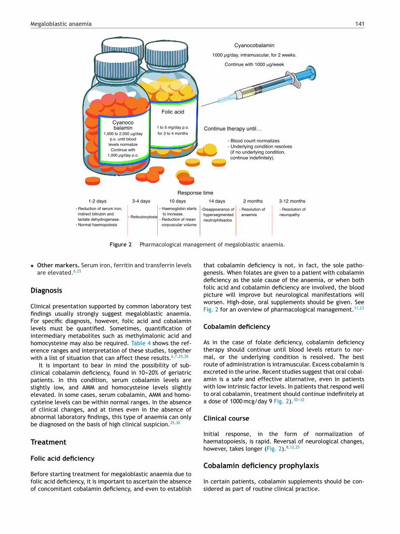
Figure 2 Pharmacological man
• Other markers. Serum iron, ferritin and transferrin levelsare elevated.6,25
Diagnosis
Clinical presentation supported by common laboratory testfindings usually strongly suggest megaloblastic anaemia.For specific diagnosis, however, folic acid and cobalaminlevels must be quantified. Sometimes, quantification ofintermediary metabolites such as methylmalonic acid andhomocysteine may also be required. Table 4 shows the ref-erence ranges and interpretation of these studies, togetherwith a list of situation that can affect these results.6,7,25,30
It is important to bear in mind the possibility of sub-clinical cobalamin deficiency, found in 10---20% of geriatricpatients. In this condition, serum cobalamin levels areslightly low, and AMM and homocysteine levels slightlyelevated. In some cases, serum cobalamin, AMM and homo-cysteine levels can be within normal ranges. In the absenceof clinical changes, and at times even in the absence ofabnormal laboratory findings, this type of anaemia can onlybe diagnosed on the basis of high clinical suspicion.25,30
Treatment
Folic acid deficiency
Before starting treatment for megaloblastic anaemia due tofolic acid deficiency, it is important to ascertain the absenceof concomitant cobalamin deficiency, and even to establish
C
Is
ent of megaloblastic anaemia.
hat cobalamin deficiency is not, in fact, the sole patho-enesis. When folates are given to a patient with cobalamineficiency as the sole cause of the anaemia, or when botholic acid and cobalamin deficiency are involved, the bloodicture will improve but neurological manifestations willorsen. High-dose, oral supplements should be given. Seeig. 2 for an overview of pharmacological management.11,23
obalamin deficiency
s in the case of folate deficiency, cobalamin deficiencyherapy should continue until blood levels return to nor-al, or the underlying condition is resolved. The best
oute of administration is intramuscular. Excess cobalamin isxcreted in the urine. Recent studies suggest that oral cobal-min is a safe and effective alternative, even in patientsith low intrinsic factor levels. In patients that respond well
o oral cobalamin, treatment should continue indefinitely at dose of 1000 mcg/day 9 Fig. 2).30---32
linical course
nitial response, in the form of normalization ofaematopoiesis, is rapid. Reversal of neurological changes,owever, takes longer (Fig. 2).8,12,25
obalamin deficiency prophylaxis
n certain patients, cobalamin supplements should be con-idered as part of routine clinical practice.
1
V
Soaod
P
Psoab
E
Itsl
E
NssnoobnIdd
C
Tmmdccd
wbccbptr
C
T
A
Tfpe
R
1
1
1
1
1
1
1
1
1
1
42
egetarians
trict vegetarians should receive between 2 and 6 mcg/dayf oral supplement. Pregnant vegetarians (strict or not) thatlso intend to breastfeed need an even higher dose, as theirffspring are at high risk of presenting severe cobalamineficiency.33,34
atients with prior gastric surgery
atients with prior partial gastrectomy or gastric bypassurgery are at high risk for subclinical cobalamin deficiencyr deficiency associated with impaired absorption of cobal-min. These patients should take 1000 mcg/day cobalaminefore meals.35,36
lderly patients
n the absence of well-designed studies investigating thisopic, the benefit of routine administration of cobalaminupplements is unclear. In special circumstances, cobalaminevels can be quantified.6,37
xposure to nitrous oxide
itrous oxide is known to inactivate cobalamin. For this rea-on, untreated or undiagnosed clinical cobalamin in patientscheduled for surgery using nitrous oxide may present rapideuropsychiatric deterioration. The pre-operative workupf these patients, therefore, should include cobalamin testsr flow cytometry, and deficiency should be fully resolvedefore surgery. It is also important to bear in mind thatitrous oxide is sometimes used as a recreational drug,f chronic, this abuse can cause serious neuropsychiatricisorders, even when not associated with vitamin B12eficiency.38,39
onclusions
he aim of this review has been to outline the essential infor-ation needed for the correct management of patients withegaloblastic anaemia. Cases not associated with a simpleietary deficiency, such as intestinal absorption disorders orellular abnormalities, merit particular attention, as physi-ians not familiar with this aetiology will often have troubleiagnosing and treating these patients.
Pharmacological management appears to be straightfor-ard. It is based supplementing deficits and building upody reserves. Follow-up of the latter is the key to a suc-essful outcome in these patients. It is also important toonsider the patient’s dietary habits. Dietary advice shoulde given in the absence of an underlying medical conditionreventing absorption of nutrients. In these cases, alterna-ive strategies should be implemented to cover nutritional
equirements.onflict of interest
he authors declare that they have no conflict of interests.
2
2
H.B. Castellanos-Sinco et al.
cknowledgements
he chemical structure figures were taken and modifiedrom the website Temas de Farmacognosia (http://www.lantas-medicinal-farmacognosia.com/gr%C3%A1fica/structuras-de-las-vitaminas/).
eferences
1. Addison T. Anaemia-disease of the supra-renal capsules. LondonMed Gaz. 1849;43:517---8.
2. Minot GR, Murphy WP. Treatment of pernicious anemia by aspecial diet. Blood. 1948;3:8---21.
3. Castle WB. The effect of the administration to patients withpernicious anemia of the contents of the normal human stomachrecovered after the ingestion of beef muscle. Am J Med Sci.1929;87:470---6.
4. Hodgkin DC, Kamper J, Mackey M, et al. Structure of VitaminB12. Nature. 1956;178:64---6.
5. Herbert V. Experimental nutritional folate deficiency in man.Trans Assoc Am Physicians. 1962;75:307---20.
6. Rodríguez-de Santiago E, Ferre-Aracil C, García García-de Pare-des A., Moreira-Vicente V.F. Pernicious anemia. From past topresent. Rev Clin Esp 2015 (Trabajo En Prensa, 10 Febrero 2015).
7. Pruthi RK, Tefferi A. Pernicious anemia revisited. Mayo Clin Proc.1994;69:144---50.
8. Allen RH, Stabler SP, Savage DG, et al. Metabolic abnormali-ties in cobalamin (vitamin B12) and folate deficiency. FASEB J.1993;7:1344---53.
9. Longo DL, Bunn HF. Vitamin B 12 and pernicious anemia ---- thedawn of molecular medicine. N Engl J Med. 2014;370:773---6.
0. McKillop DJ, Pentieva K, Daly D, et al. The effect of differentcooking methods on folate retention in various foods that areamongst the major contributors to folate intake in the UK diet.Br J Nutr. 2002;88:681---8.
1. Pitkin RM, Allen LB, Bailey LB, et al. Dietary reference intakesfor thiamin, riboflavin, niacin, vitamin B6, folate, vitamin B12,pantothenic acid, biotin and choline. Washington, DC: NationalAcademies Press (US); 1998.
2. Butler CC, Vidal-Alaball J, Cannings-John R, et al. Oral vitaminB12 versus intramuscular vitamin B12 for vitamin B12 defi-ciency: a systematic review of randomized controlled trials.Fam Pract. 2006;23:279---85.
3. Moestrup SK. New insights into carrier binding and epithelialuptake of the erythropoietic nutrients cobalamin and folate.Curr Opin Hematol. 2006;13:119---23.
4. Qiu A, Jansen M, Sakaris A, et al. Identification of an intestinalfolate transporter and the molecular basis for hereditary folatemalabsorption. Cell. 2006;127:917---28.
5. Rijnboutt S, Jansen G, Posthuma G, et al. Endocytosisof GPI-linked membrane folate receptor-alpha. J Cell Biol.1996;132:35---47.
6. O’Brien JS. Urinary excretion of folic and folinic acids in normaladults. Proc Soc Exp Biol Med. 1960;104:354---5.
7. Bailey LB, Gregory JF. Folate metabolism and requirements. JNutr. 1999;129:779---82.
8. Hsu JM, Kawin B, Minor P, et al. Vitamin B12 concentrations inhuman tissues. Nature. 1966;210:1264---5.
9. Birn H. The kidney in vitamin B12 and folate homeostasis: char-acterization of receptors for tubular uptake of vitamins andcarrier proteins. Am J Physiol Renal Physiol. 2006;291:F22---36.
0. Chanarin I, Deacon R, Lumb M, et al. Cobalamin and folate:
recent developments. J Clin Pathol. 1992;45:277---83.1. Carmel R. Cobalamin-binding proteins in man. In: Silber R, Gor-don AS, LoBue J, editors. Contemporary hematology-oncology,vol. 2. New York: Plenum Publishing; 1981.
3
3
3
3
3
3
3
3
Megaloblastic anaemia
22. Seetharam B. Receptor-mediated endocytosis of cobalamin(vitamin B12). Annu Rev Nutr. 1999;19:173---95.
23. Watanabe F, Nakano Y. Comparative biochemistry of vitaminB12 (cobalamin) metabolism: biochemical diversity in the sys-tems for intracellular cobalamin transfer and synthesis of thecoenzymes. Int J Biochem. 1991;23:1353---9.
24. Herbert V, Zalusky R. Interrelations of vitamin B12 and folicacid metabolism: folic acid clearance studies. J Clin Invest.1962;41:1263---76.
25. Carmel R, Green R, Rosenblatt DS, et al. Update on cobalamin,folate, and homocysteine. Hematology Am Soc Hematol EducProgram. 2003:62---81.
26. Shane B. Folate and vitamin B12 metabolism: overview andinteraction with riboflavin, vitamin B6, and polymorphisms.Food Nutr Bull. 2008;29:S5---16 (discussion S17---S19).
27. Truswell AS. Vitamin B12. Nutr Diet. 2007;64:S120---5.28. Hemmer B, Glocker FX, Schumacher M, et al. Subacute
combined degeneration: clinical, electrophysiological, andmagnetic resonance imaging findings. J Neurol Neurosurg Psy-chiatry. 1998;65:822---7.
29. Beck WS. Neuropsychiatric consequences of cobalamin defi-ciency. Adv Intern Med. 1991;36:33---56.
30. Green R, Kinsella LJ. Current concepts in the diagnosis of cobal-amin deficiency. Neurology. 1995;45:1435---40.
3
143
1. Carmel R. How I treat cobalamin (vitamin B12) deficiency.Blood. 2008;112:2214---21.
2. Andrès E, Dali-Youcef N, Vogel T, et al. Oral cobalamin (vita-min B(12)) treatment. An update. Int J Lab Hematol. 2009;31:1---8.
3. Antony AC. Vegetarianism and vitamin B-12 (cobalamin) defi-ciency. Am J Clin Nutr. 2003;78:3---6.
4. Centers for Disease Control and Prevention (CDC). Neurologicimpairment in children associated with maternal dietary defi-ciency of cobalamin----Georgia, 2001. JAMA. 2003;289:979.
5. Stein J, Stier C, Raab H, et al. Review article: the nutritionaland pharmacological consequences of obesity surgery. AlimentPharmacol Ther. 2014;40:582---609.
6. Sawaya RA, Jaffe J, Friedenberg L, et al. Vitamin, mineral, anddrug absorption following bariatric surgery. Curr Drug Metab.2012;13:1345---55.
7. Carmel R. Efficacy and safety of fortification and supplemen-tation with vitamin B12: biochemical and physiological effects.Food Nutr Bull. 2008;29:S177---87.
8. Schilling RF. Is nitrous oxide a dangerous anesthetic for vitamin
B12-deficient subjects? JAMA. 1986;255:1605---6.9. Ng J, O’Grady G, Pettit T, et al. Nitrous oxide use infirst-year students at Auckland University. Lancet. 2003;361:1349---50.