Embed Size (px)
Citation preview
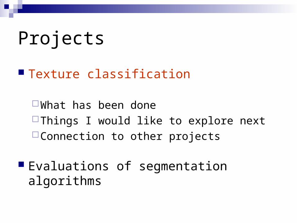
Projects
Texture classification
What has been doneThings I would like to explore nextConnection to other projects
Evaluations of segmentation algorithms
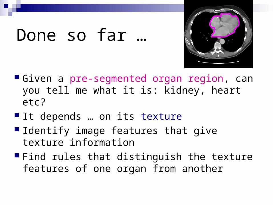
Done so far …
Given a pre-segmented organ region, can you tell me what it is: kidney, heart etc?
It depends … on its texture Identify image features that give texture
information Find rules that distinguish the texture
features of one organ from another
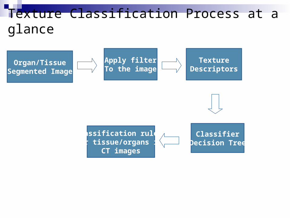
Texture Classification Process at a glance
Apply filterTo the image
Organ/TissueSegmented Image
TextureDescriptors
Classifier(Decision Tree)
Classification rules for tissue/organs in
CT images
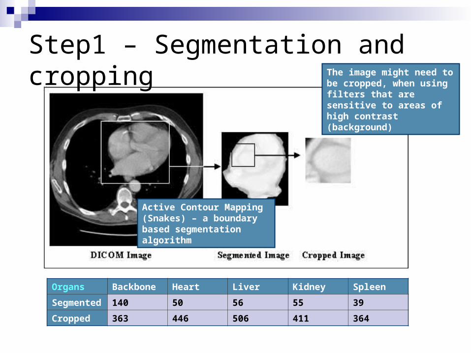
Step1 – Segmentation and cropping
Organs Backbone Heart Liver Kidney Spleen
Segmented 140 50 56 55 39
Cropped 363 446 506 411 364
The image might need to be cropped, when using filters that are sensitive to areas of high contrast (background)
Active Contour Mapping (Snakes) – a boundary based segmentation algorithm
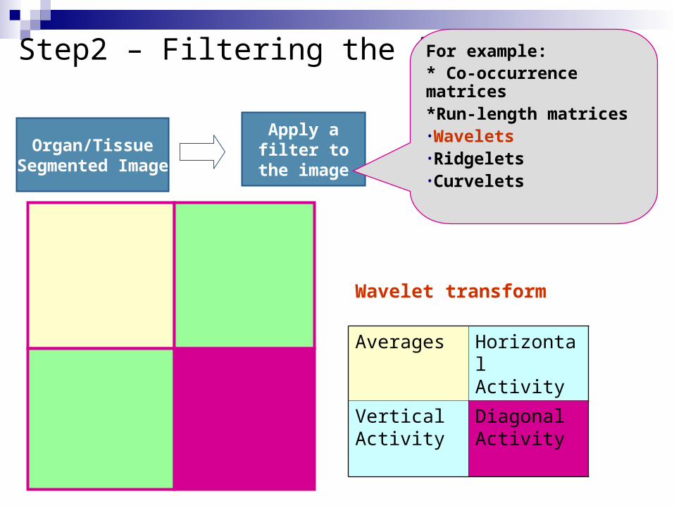
Step2 – Filtering the image
Apply a filter to the image
Organ/TissueSegmented Image
For example:* Co-occurrence matrices *Run-length matrices•Wavelets•Ridgelets•Curvelets
Averages Horizontal Activity
Vertical Activity
Diagonal Activity
Wavelet transform
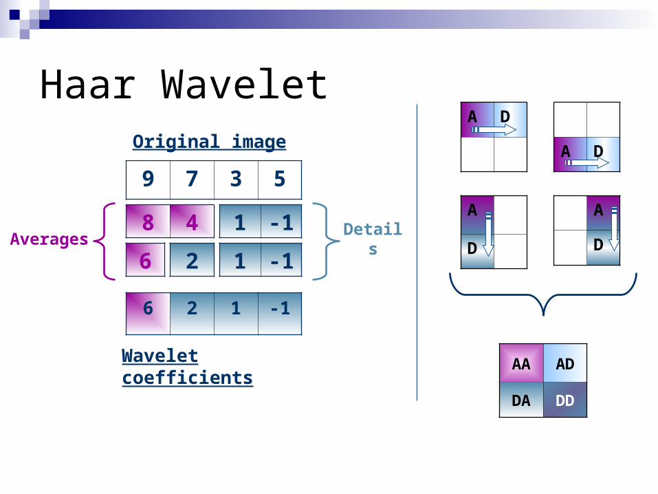
Haar Wavelet
8 4
9 7 3 5
6
1 -1
2 1 -1
Original image
Wavelet coefficients
6 2 1 -1
Averages Details
A D
A D
A
D
A
D
AA AD
DA DD
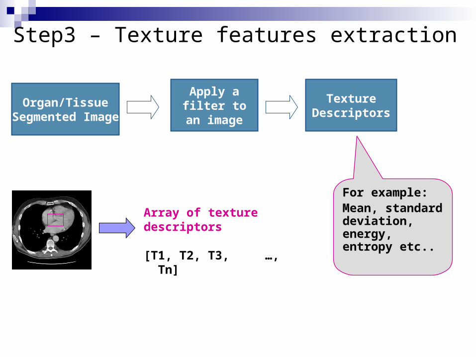
Step3 – Texture features extraction
Apply a filter to an image
Organ/TissueSegmented Image
TextureDescriptors
For example:Mean, standard deviation, energy, entropy etc..
Array of texture descriptors
[T1, T2, T3, …, Tn]
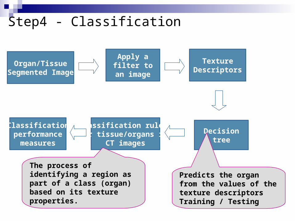
Step4 - Classification
Apply a filter to an image
Organ/TissueSegmented Image
TextureDescriptors
Classification rules for tissue/organs in
CT images
The process of identifying a region as part of a class (organ) based on its texture properties.
Decisiontree
Predicts the organ from the values of the texture descriptorsTraining / Testing
Classificationperformance
measures
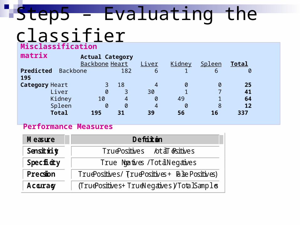
Step5 – Evaluating the classifier
Measure Definition
Sensitivity True Positives / Total Positives
Specificity True Negatives / Total Negatives
Precision True Positives / (True Positives + False Positives)
Accuracy (True Positives + True Negatives) / Total Samples
Actual CategoryBackbone Heart Liver Kidney Spleen Total
Predicted Backbone 182 6 1 6 0 195Category Heart 3 18 4 0 0 25
Liver 0 3 30 1 7 41Kidney 10 4 0 49 1 64Spleen 0 0 4 0 8 12Total 195 31 39 56 16 337
Misclassification matrix
Performance Measures
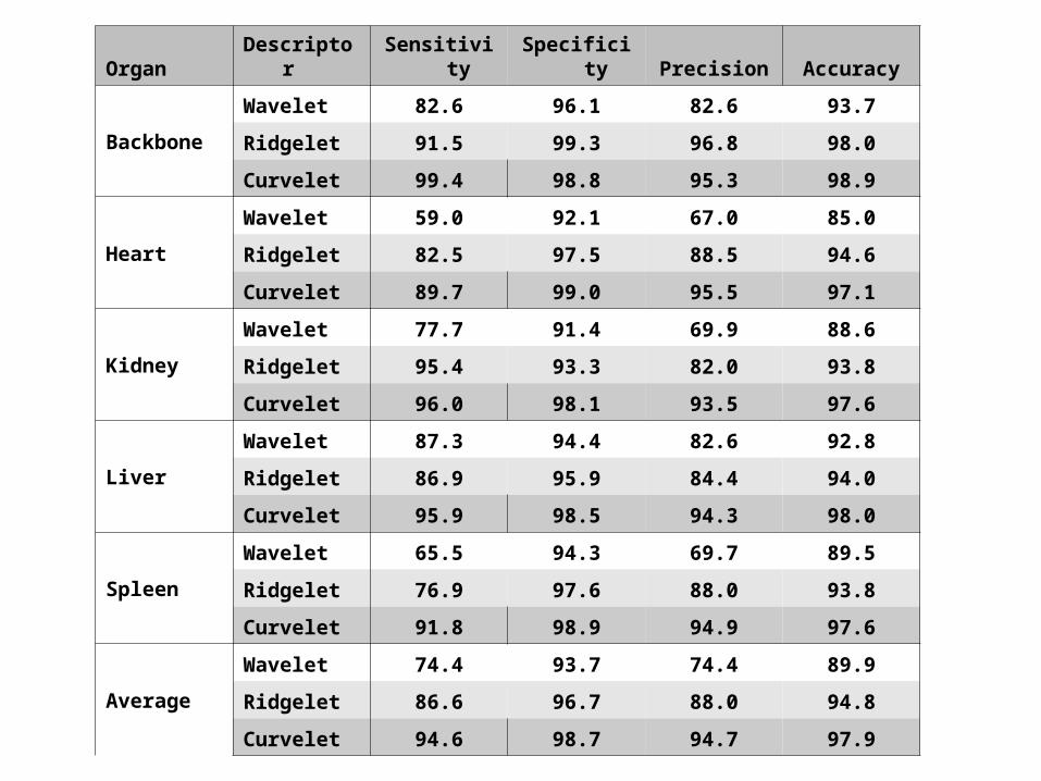
Organ Descriptor Sensitivity Specificity Precision Accuracy
Backbone
Wavelet 82.6 96.1 82.6 93.7
Ridgelet 91.5 99.3 96.8 98.0
Curvelet 99.4 98.8 95.3 98.9
Heart
Wavelet 59.0 92.1 67.0 85.0
Ridgelet 82.5 97.5 88.5 94.6
Curvelet 89.7 99.0 95.5 97.1
Kidney
Wavelet 77.7 91.4 69.9 88.6
Ridgelet 95.4 93.3 82.0 93.8
Curvelet 96.0 98.1 93.5 97.6
Liver
Wavelet 87.3 94.4 82.6 92.8
Ridgelet 86.9 95.9 84.4 94.0
Curvelet 95.9 98.5 94.3 98.0
Spleen
Wavelet 65.5 94.3 69.7 89.5
Ridgelet 76.9 97.6 88.0 93.8
Curvelet 91.8 98.9 94.9 97.6
Average
Wavelet 74.4 93.7 74.4 89.9
Ridgelet 86.6 96.7 88.0 94.8
Curvelet 94.6 98.7 94.7 97.9
Apply a filter to an image
Organ/TissueSegmented Image
TextureDescriptors
Classification rules for tissue/organs in
CT images
Decisiontree
Things I would like to explore
Gabor filtersFractal Dimensions
Performancemeasures
Different patientsDifferent organs Abnormal texture
Different modalities
Connections to other project
Can we use wavelet, ridgelet, curvelet-based texture descriptors for content based image retrieval?
Can we use these descriptors in the volumetric segmentation?
Instead of many 2D images, can we use the same process for 3D stack of slices?
Projects
Texture classification
Evaluations of segmentation algorithms
What has been doneThings I would like to explore nextConnection to other projects
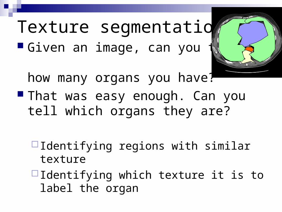
Texture segmentation Given an image, can you tell me
how many organs you have? That was easy enough. Can you
tell which organs they are?
Identifying regions with similar texture Identifying which texture it is to label the
organ
A couple of key questions
Can you do it better by varying a parameter? How do you choose the values of your segmentation parameters?
If it looks better is it really better?
A couple of key questions
Parameter optimization
Performance evaluation
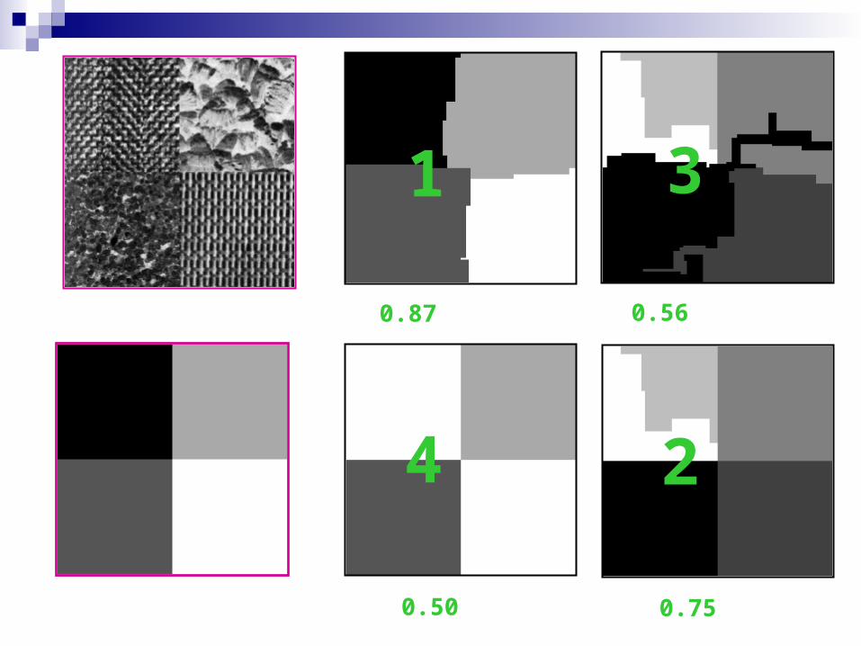
1 3
4 2
0.87 0.56
0.50 0.75
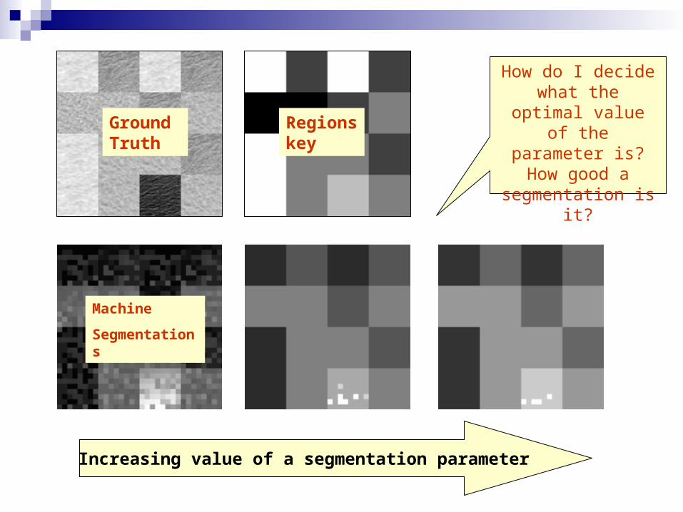
Increasing value of a segmentation parameter
GroundTruth
Regionskey
Machine
Segmentations
How do I decide what the optimal value of the parameter is?
How good a segmentation is it?
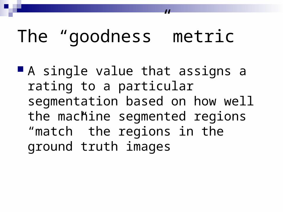
The “goodness” metric
A single value that assigns a rating to a particular segmentation based on how well the machine segmented regions “match” the regions in the ground truth images

Region Categories
Ground Truth vs. Machine Segmented Correctly Detected Over Segmented Under Segmented Missed Noise
GT
MS
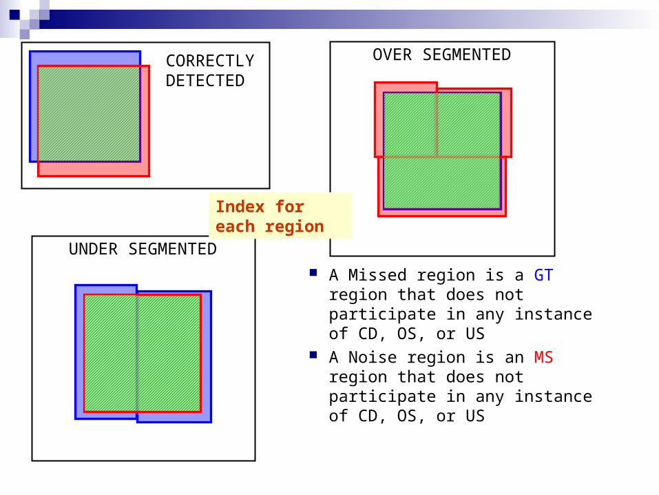
CORRECTLY DETECTED
OVER SEGMENTED
UNDER SEGMENTED
A Missed region is a GT region that does not participate in any instance of CD, OS, or US
A Noise region is an MS region that does not participate in any instance of CD, OS, or US
Index for each region
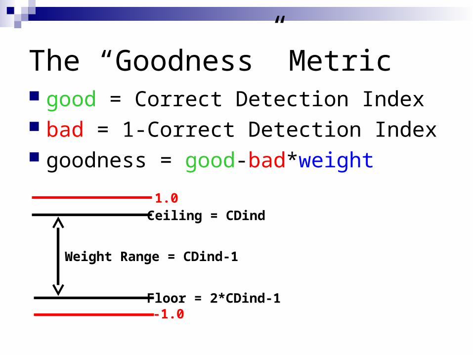
The “Goodness” Metric good = Correct Detection Index bad = 1-Correct Detection Index goodness = good-bad*weight
1.0
-1.0
Ceiling = CDind
Floor = 2*CDind-1
Weight Range = CDind-1
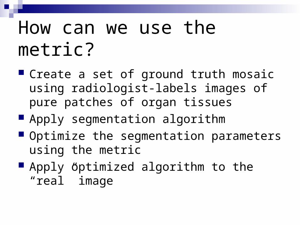
How can we use the metric?
Create a set of ground truth mosaic using radiologist-labels images of pure patches of organ tissues
Apply segmentation algorithm Optimize the segmentation parameters using the
metric Apply optimized algorithm to the “real” image
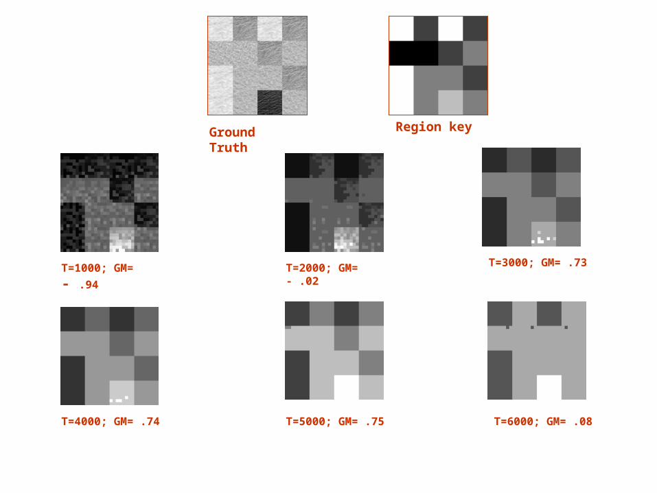
Ground Truth Region key
T=1000; GM= - .94
T=4000; GM= .74
T=3000; GM= .73T=2000; GM= - .02
T=5000; GM= .75 T=6000; GM= .08
Done so far
Used the metric on a block-wise walevet-based segmentation algorithm on some sample mosaic
To be done
Fully test the metric on a wide range of segmentation algorithms
Decouple the various components of the metric and test the individual performance measures instead of the overall score
Extend the metric to measure one region vs background segmentation
To be done
Improve the wavelet-based algorithms we have implement to include other texture features
Explore and compare other texture-based segmentation algorithm
Use regions and metric to calculate changes in time of an abnormal region
Connections to other projects
Use one of these algorithms to create a rough segmentation that will generate the starting point for a more sophisticated segmentation algorithm.
Some references ”Wavelet-based Texture Classification of Tissues in Computed
Tomography”, L. Semler, L, Dettori, and Jacob Furst.18th IEEE International Symposium on Computer-based Medical Systems, Dublin, Ireland, June 2005.
“Ridgelet-based Texture Classification in Computed Tomography”, L. Semler, L. Dettori. and W.Kerr. 8th IASTED International Conference on Signal and Image Processing, Honolulu, HW, August 2006.
“Curvelet-based Texture Classification of Tissues in Computed Tomography”, L. Semler, & L. Dettori. International Conference on Image Processing, Atlanta, GA, October 2006.
“A Comparison of Wavelet-based and Ridgelet-based texture classification of Tissues in Computed Tomography”, with Lindsay Semler, International Conference on Computer Vision Theory and Applications, Setubal, Portugal, February 2006
“A Methodology and Metric for Quantitative Analysis and Parameter Optimization of Unsupervised, Multi-Region Image Segmentation”, William Kerr, Lucia Dettori, and Lindsay Semler, 8th IASTED International Conference on Signal and Image Processing, Honolulu, HW, August 2006.


![Jesi [AN] JCube Open Day SPRING 2014€¦ · Claudio Bedino - Italian Crowdfunding Andrea Colombo - U-Start Gianluca Dettori - dPixel Enrico Martini - Min. Sviluppo Economico Francesca](https://img.pdfslide.us/doc/110x75/5e8cca71e3be365b3c328d8d/jesi-an-jcube-open-day-spring-claudio-bedino-italian-crowdfunding-andrea-colombo.jpg)