Embed Size (px)
DESCRIPTION
....
Citation preview
Mar. Drugs 2012, 10, 987-997; doi:10.3390/md10050987
Marine Drugs ISSN 1660-3397
www.mdpi.com/journal/marinedrugs
Article
Cytotoxic Sesterterpenoids from a Sponge Hippospongia sp.
Yu-Chia Chang 1,2, Shang-Wei Tseng 1,3, Li-Lian Liu 4,5, Yalan Chou 4, Yuan-Shing Ho 6,
Mei-Chin Lu 1,3 and Jui-Hsin Su 1,3,5,*
1 National Museum of Marine Biology & Aquarium, Pingtung 944, Taiwan;
E-Mails: [email protected] (Y.-C.C.); [email protected] (S.-W.T.);
[email protected] (M.-C.L.) 2 Doctoral Degree Program in Marine Biotechnology, National Sun Yat-sen University and
Academia Sinica, Kaohsiung 804, Taiwan 3 Graduate Institute of Marine Biotechnology, National Dong Hwa University, Pingtung 944, Taiwan 4 Institute of Marine Biology, National Sun Yat-sen University, Kaohsiung 804, Taiwan;
E-Mails: [email protected] (L.-L.L.); [email protected] (Y.C.) 5 Asia-Pacific Ocean Research Center, National Sun Yat-sen University, Kaohsiung 804, Taiwan 6 Eastern Marine Biology Research Center, Fisheries Research Institute, Taitung 961, Taiwan;
E-Mail: [email protected]
* Author to whom correspondence should be addressed; E-Mail: [email protected];
Tel.: +886-8-8825001 (ext. 3126); Fax: +886-8-8825087.
Received: 28 March 2012; in revised form: 18 April 2012 / Accepted: 24 April 2012 /
Published: 27 April 2012
Abstract: One new pentacyclic sesterterpene, hippospongide A (1), and one new
scalarane sesterterpenoid, hippospongide B (2), along with six previously reported known
scalarane–type sesterterpenes (3–8), were isolated from a sponge Hippospongia sp. The
structures of these compounds were elucidated on the basis of their spectroscopic data and
comparison of the NMR data with those of known analogues. These metabolites are the
first pentacyclic sesterterpene and scalarane-type sesterterpenes to be reported from this
genus. Compounds 3–5 exhibited significant cytotoxicity against DLD-1, HCT-116, T-47D
and K562 cancer cell lines.
Keywords: sesterterpenoid; scalarane; sponge; Hippospongia
OPEN ACCESS
Mar. Drugs 2012, 10 988
1. Introduction
In previous reports, scalarane sesterterpenoids have been identified from sponges and nudibranchs [1].
Research into the pharmacological properties of this class of natural products is of particular interest.
In fact, many scalarane metabolites show a variety of biological activities, such as antimicrobial,
cytotoxic, antifeedant, ichthyotoxic, anti-inflammatory, antitubercular, platelet aggregation inhibition,
RCE-protease inhibition and nerve growth factor synthesis-stimulating [1]. Our investigation of the
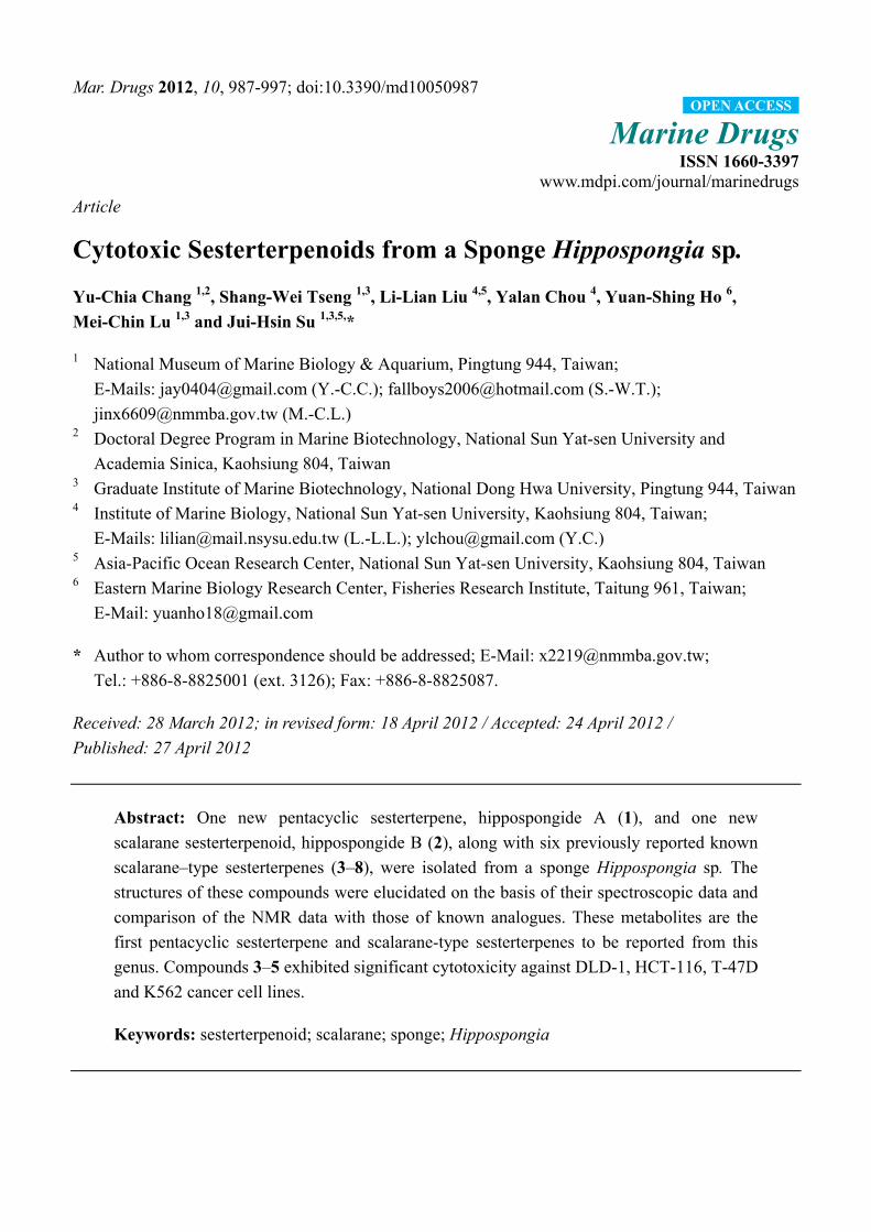
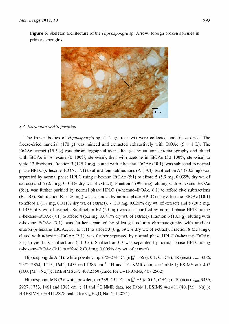
chemical constituents of a sponge Hippospongia sp. (Figure 1) yielded one new pentacyclic
sesterterpene, hippospongide A (1), and one new scalarane sesterterpenoid, hiposppongide B (2), along
with six known sesterterpenoids, heteronemin (3) [2], heteronemin acetate (4) [3], hyrtiosin E (5) [4],
12-deacetoxyscalarin 19-acetate (6) [5], hyrtiosal (7) [6] and scalarafuran (8) [7]. The cytotoxicity of
metabolites 1–8 against human colon adenocarcinoma (DLD-1 and HCT-116), hormone-dependent
breast cancer (T-47D) and human chronic myelogenous leukemia (K562) cell lines was evaluated.
Figure 1. Sponge Hippospongia sp.
2. Results and Discussion
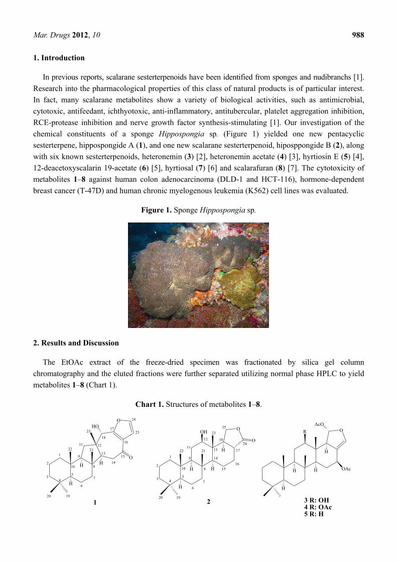
The EtOAc extract of the freeze-dried specimen was fractionated by silica gel column
chromatography and the eluted fractions were further separated utilizing normal phase HPLC to yield
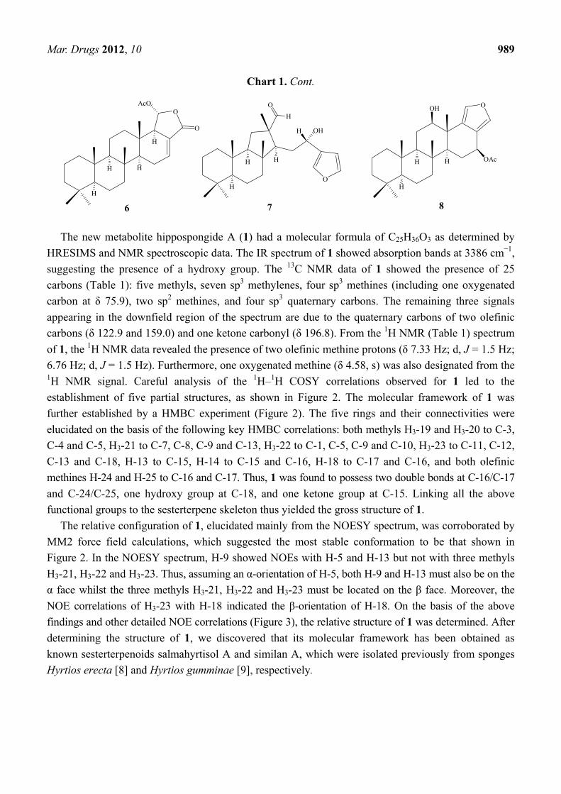
metabolites 1–8 (Chart 1).
Chart 1. Structures of metabolites 1–8.
O
O
HO
H
H H
1
2
34
5
6
7
8
9
10
11 12
13
14
15
16
17
18
1920
2122
23
24
25
H
H
O
H
H
O
OH
1
2
3 45
6
7
8
9
10
11
12
13
14
1516
17
18
1920
2122
23
24
25
H
H
OAcO
H
H
R
OAc
3 R: OH4 R: OAc5 R: H
1 2
Mar. Drugs 2012, 10 989
Chart 1. Cont.
O
OH
O
H
H H
H
H
7
H
H
O
H
OH
OAc
8H
H
OAcO
H
H
6
O
The new metabolite hippospongide A (1) had a molecular formula of C25H36O3 as determined by
HRESIMS and NMR spectroscopic data. The IR spectrum of 1 showed absorption bands at 3386 cm−1,
suggesting the presence of a hydroxy group. The 13C NMR data of 1 showed the presence of 25
carbons (Table 1): five methyls, seven sp3 methylenes, four sp3 methines (including one oxygenated
carbon at δ 75.9), two sp2 methines, and four sp3 quaternary carbons. The remaining three signals
appearing in the downfield region of the spectrum are due to the quaternary carbons of two olefinic
carbons (δ 122.9 and 159.0) and one ketone carbonyl (δ 196.8). From the 1H NMR (Table 1) spectrum
of 1, the 1H NMR data revealed the presence of two olefinic methine protons (δ 7.33 Hz; d, J = 1.5 Hz;
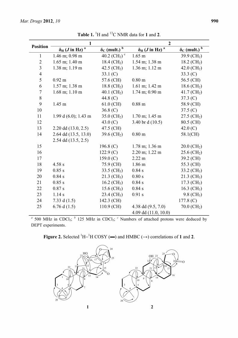
6.76 Hz; d, J = 1.5 Hz). Furthermore, one oxygenated methine (δ 4.58, s) was also designated from the 1H NMR signal. Careful analysis of the 1H–1H COSY correlations observed for 1 led to the
establishment of five partial structures, as shown in Figure 2. The molecular framework of 1 was
further established by a HMBC experiment (Figure 2). The five rings and their connectivities were
elucidated on the basis of the following key HMBC correlations: both methyls H3-19 and H3-20 to C-3,
C-4 and C-5, H3-21 to C-7, C-8, C-9 and C-13, H3-22 to C-1, C-5, C-9 and C-10, H3-23 to C-11, C-12,
C-13 and C-18, H-13 to C-15, H-14 to C-15 and C-16, H-18 to C-17 and C-16, and both olefinic
methines H-24 and H-25 to C-16 and C-17. Thus, 1 was found to possess two double bonds at C-16/C-17
and C-24/C-25, one hydroxy group at C-18, and one ketone group at C-15. Linking all the above
functional groups to the sesterterpene skeleton thus yielded the gross structure of 1.
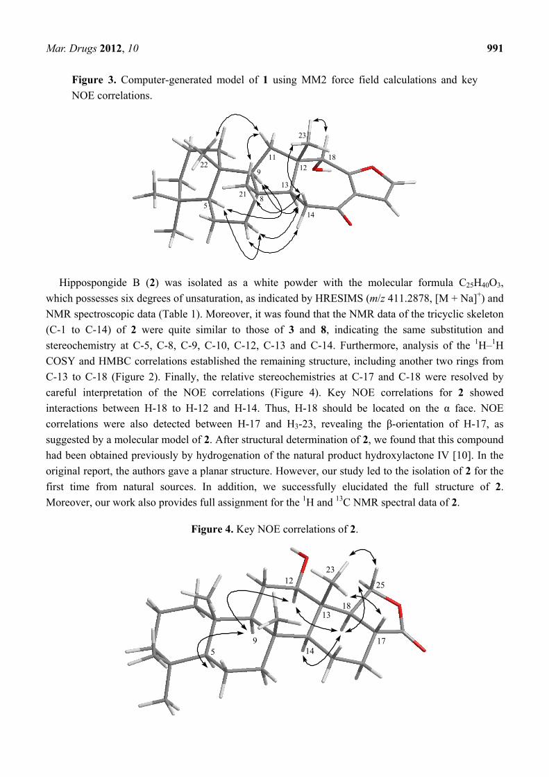
The relative configuration of 1, elucidated mainly from the NOESY spectrum, was corroborated by
MM2 force field calculations, which suggested the most stable conformation to be that shown in
Figure 2. In the NOESY spectrum, H-9 showed NOEs with H-5 and H-13 but not with three methyls
H3-21, H3-22 and H3-23. Thus, assuming an α-orientation of H-5, both H-9 and H-13 must also be on the
α face whilst the three methyls H3-21, H3-22 and H3-23 must be located on the β face. Moreover, the
NOE correlations of H3-23 with H-18 indicated the β-orientation of H-18. On the basis of the above
findings and other detailed NOE correlations (Figure 3), the relative structure of 1 was determined. After
determining the structure of 1, we discovered that its molecular framework has been obtained as
known sesterterpenoids salmahyrtisol A and similan A, which were isolated previously from sponges
Hyrtios erecta [8] and Hyrtios gumminae [9], respectively.
Mar. Drugs 2012, 10 990
Table 1. 1H and 13C NMR data for 1 and 2.
Position 1 2
δH (J in Hz) a δC (mult.) b δH (J in Hz) a δC (mult.) b 1 1.46 m; 0.98 m 40.2 (CH2)
c 1.65 m 39.9 (CH2) 2 1.65 m; 1.40 m 18.4 (CH2) 1.54 m; 1.38 m 18.2 (CH2) 3 1.38 m; 1.19 m 42.5 (CH2) 1.36 m; 1.12 m 42.0 (CH2) 4 33.1 (C) 33.3 (C) 5 0.92 m 57.6 (CH) 0.80 m 56.5 (CH) 6 1.57 m; 1.38 m 18.8 (CH2) 1.61 m; 1.42 m 18.6 (CH2) 7 1.68 m; 1.10 m 40.1 (CH2) 1.74 m; 0.90 m 41.7 (CH2) 8 44.8 (C) 37.3 (C) 9 1.45 m 61.0 (CH) 0.88 m 58.9 (CH)
10 36.8 (C) 37.5 (C) 11 1.99 d (6.0); 1.43 m 35.0 (CH2) 1.70 m; 1.45 m 27.5 (CH2) 12 43.0 (C) 3.40 br d (10.5) 80.5 (CH) 13 2.20 dd (13.0, 2.5) 47.5 (CH) 42.0 (C) 14 2.64 dd (13.5, 13.0) 39.6 (CH2) 0.80 m 58.1(CH)
2.54 dd (13.5, 2.5) 15 196.8 (C) 1.78 m; 1.36 m 20.0 (CH2) 16 122.9 (C) 2.20 m; 1.22 m 25.6 (CH2) 17 159.0 (C) 2.22 m 39.2 (CH) 18 4.58 s 75.9 (CH) 1.86 m 55.3 (CH) 19 0.85 s 33.5 (CH3) 0.84 s 33.2 (CH3) 20 0.84 s 21.3 (CH3) 0.80 s 21.3 (CH3) 21 0.85 s 16.2 (CH3) 0.84 s 17.3 (CH3) 22 0.87 s 15.6 (CH3) 0.84 s 16.3 (CH3) 23 1.14 s 23.4 (CH3) 0.91 s 9.8 (CH3) 24 7.33 d (1.5) 142.3 (CH) 177.8 (C) 25 6.76 d (1.5) 110.9 (CH) 4.38 dd (9.5, 7.0)
4.09 dd (11.0, 10.0) 70.0 (CH2)
a 500 MHz in CDCl3; b 125 MHz in CDCl3;
c Numbers of attached protons were deduced by DEPT experiments.
Figure 2. Selected 1H−1H COSY (▬) and HMBC (→) correlations of 1 and 2.
O
O
HO
45
7
810
11 12
1314
15
1718
20
2122
23
24
25
1
3
O
O
OH
12
13
14
17
23
24
25
16
18
9
5
10
22 21
8
4
3
20
1 2
Mar. Drugs 2012, 10 991
Figure 3. Computer-generated model of 1 using MM2 force field calculations and key
NOE correlations.
22
2113
23
1211 18
145
8
9
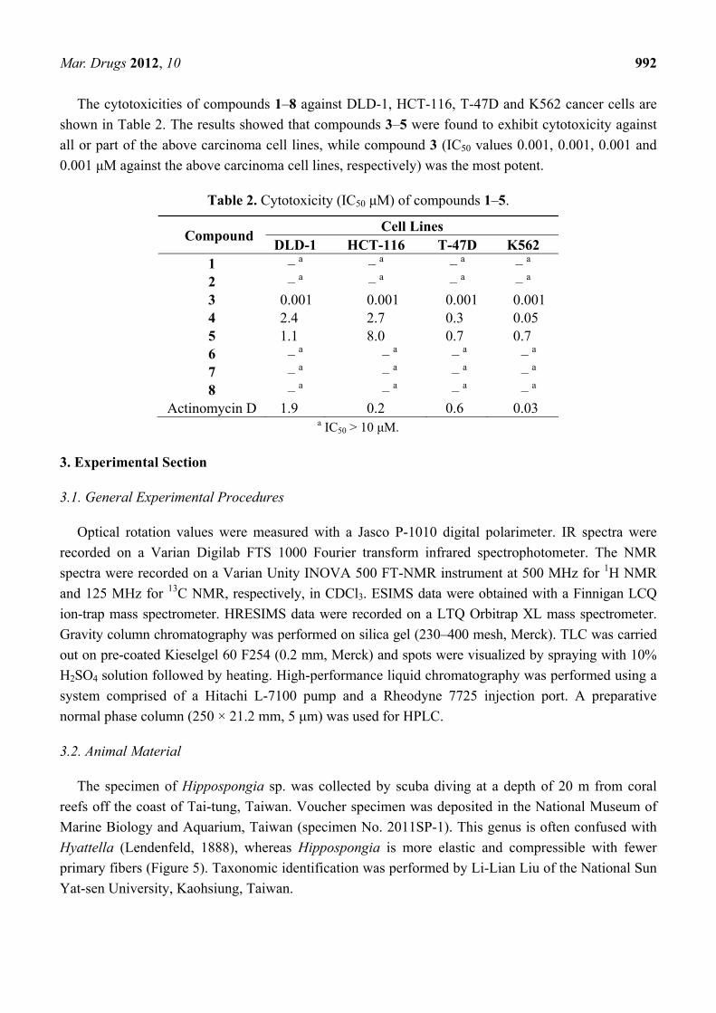
Hippospongide B (2) was isolated as a white powder with the molecular formula C25H40O3,
which possesses six degrees of unsaturation, as indicated by HRESIMS (m/z 411.2878, [M + Na]+) and
NMR spectroscopic data (Table 1). Moreover, it was found that the NMR data of the tricyclic skeleton
(C-1 to C-14) of 2 were quite similar to those of 3 and 8, indicating the same substitution and
stereochemistry at C-5, C-8, C-9, C-10, C-12, C-13 and C-14. Furthermore, analysis of the 1H–1H
COSY and HMBC correlations established the remaining structure, including another two rings from
C-13 to C-18 (Figure 2). Finally, the relative stereochemistries at C-17 and C-18 were resolved by
careful interpretation of the NOE correlations (Figure 4). Key NOE correlations for 2 showed
interactions between H-18 to H-12 and H-14. Thus, H-18 should be located on the α face. NOE
correlations were also detected between H-17 and H3-23, revealing the β-orientation of H-17, as
suggested by a molecular model of 2. After structural determination of 2, we found that this compound
had been obtained previously by hydrogenation of the natural product hydroxylactone IV [10]. In the
original report, the authors gave a planar structure. However, our study led to the isolation of 2 for the
first time from natural sources. In addition, we successfully elucidated the full structure of 2.
Moreover, our work also provides full assignment for the 1H and 13C NMR spectral data of 2.
Figure 4. Key NOE correlations of 2.
14
18
17
2512
13
23
95
Mar. Drugs 2012, 10 992
The cytotoxicities of compounds 1–8 against DLD-1, HCT-116, T-47D and K562 cancer cells are
shown in Table 2. The results showed that compounds 3–5 were found to exhibit cytotoxicity against
all or part of the above carcinoma cell lines, while compound 3 (IC50 values 0.001, 0.001, 0.001 and
0.001 μM against the above carcinoma cell lines, respectively) was the most potent.
Table 2. Cytotoxicity (IC50 μM) of compounds 1–5.
Compound Cell Lines
DLD-1 HCT-116 T-47D K562 1 – a – a – a – a 2 – a – a – a – a 3 0.001 0.001 0.001 0.001 4 2.4 2.7 0.3 0.05 5 1.1 8.0 0.7 0.7 6 – a – a – a – a 7 – a – a – a – a 8 – a – a – a – a
Actinomycin D 1.9 0.2 0.6 0.03 a IC50 > 10 μM.
3. Experimental Section
3.1. General Experimental Procedures
Optical rotation values were measured with a Jasco P-1010 digital polarimeter. IR spectra were
recorded on a Varian Digilab FTS 1000 Fourier transform infrared spectrophotometer. The NMR
spectra were recorded on a Varian Unity INOVA 500 FT-NMR instrument at 500 MHz for 1H NMR
and 125 MHz for 13C NMR, respectively, in CDCl3. ESIMS data were obtained with a Finnigan LCQ
ion-trap mass spectrometer. HRESIMS data were recorded on a LTQ Orbitrap XL mass spectrometer.
Gravity column chromatography was performed on silica gel (230–400 mesh, Merck). TLC was carried
out on pre-coated Kieselgel 60 F254 (0.2 mm, Merck) and spots were visualized by spraying with 10%
H2SO4 solution followed by heating. High-performance liquid chromatography was performed using a
system comprised of a Hitachi L-7100 pump and a Rheodyne 7725 injection port. A preparative
normal phase column (250 × 21.2 mm, 5 μm) was used for HPLC.
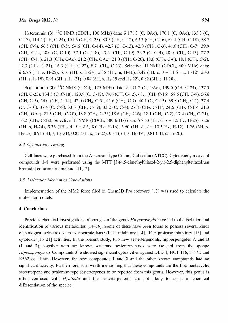
3.2. Animal Material
The specimen of Hippospongia sp. was collected by scuba diving at a depth of 20 m from coral
reefs off the coast of Tai-tung, Taiwan. Voucher specimen was deposited in the National Museum of
Marine Biology and Aquarium, Taiwan (specimen No. 2011SP-1). This genus is often confused with
Hyattella (Lendenfeld, 1888), whereas Hippospongia is more elastic and compressible with fewer
primary fibers (Figure 5). Taxonomic identification was performed by Li-Lian Liu of the National Sun
Yat-sen University, Kaohsiung, Taiwan.
Mar. Drugs 2012, 10 993
Figure 5. Skeleton architecture of the Hippospongia sp. Arrow: foreign broken spicules in
primary spongins.
3.3. Extraction and Separation
The frozen bodies of Hippospongia sp. (1.2 kg fresh wt) were collected and freeze-dried. The
freeze-dried material (170 g) was minced and extracted exhaustively with EtOAc (5 × 1 L). The
EtOAc extract (15.3 g) was chromatographed over silica gel by column chromatography and eluted
with EtOAc in n-hexane (0–100%, stepwise), then with acetone in EtOAc (50–100%, stepwise) to
yield 13 fractions. Fraction 3 (125.7 mg), eluted with n-hexane–EtOAc (10:1), was subjected to normal
phase HPLC (n-hexane–EtOAc, 7:1) to afford four subfractions (A1–A4). Subfraction A4 (30.5 mg) was
separated by normal phase HPLC using n-hexane–EtOAc (5:1) to afford 5 (5.9 mg, 0.039% dry wt. of
extract) and 6 (2.1 mg, 0.014% dry wt. of extract). Fraction 4 (996 mg), eluting with n-hexane–EtOAc
(8:1), was further purified by normal phase HPLC (n-hexane–EtOAc, 6:1) to afford five subfractions
(B1–B5). Subfraction B1 (120 mg) was separated by normal phase HPLC using n-hexane–EtOAc (10:1)
to afford 1 (1.7 mg, 0.011% dry wt. of extract), 7 (3.0 mg, 0.020% dry wt. of extract) and 8 (20.5 mg,
0.133% dry wt. of extract). Subfraction B2 (20 mg) was also purified by normal phase HPLC using
n-hexane–EtOAc (7:1) to afford 4 (6.2 mg, 0.041% dry wt. of extract). Fraction 6 (10.5 g), eluting with
n-hexane–EtOAc (3:1), was further separated by silica gel column chromatography with gradient
elution (n-hexane–EtOAc, 3:1 to 1:1) to afford 3 (6 g, 39.2% dry wt. of extract). Fraction 8 (524 mg),
eluted with n-hexane–EtOAc (2:1), was further separated by normal phase HPLC (n-hexane–EtOAc,
2:1) to yield six subfractions (C1–C6). Subfraction C3 was separated by normal phase HPLC using
n-hexane–EtOAc (3:1) to afford 2 (0.8 mg, 0.005% dry wt. of extract).
Hippospongide A (1): white powder; mp 272–274 °C; 25D[ ]α −66 (c 0.1, CHCl3); IR (neat) νmax 3386,
2922, 2854, 1715, 1642, 1455 and 1385 cm−1; 1H and 13C NMR data, see Table 1; ESIMS m/z 407
(100, [M + Na]+); HRESIMS m/z 407.2560 (calcd for C25H36O3Na, 407.2562).
Hippospongide B (2): white powder; mp 289–291 °C; 25D[ ]α −3 (c 0.05, CHCl3); IR (neat) νmax 3436,
2927, 1753, 1461 and 1383 cm−1; 1H and 13C NMR data, see Table 1; ESIMS m/z 411 (80, [M + Na]+);
HRESIMS m/z 411.2878 (calcd for C25H40O3Na, 411.2875).
Mar. Drugs 2012, 10 994
Heteronmin (3): 13C NMR (CDCl3, 100 MHz) data: δ 171.3 (C, OAc), 170.1 (C, OAc), 135.3 (C,
C-17), 114.4 (CH, C-24), 101.6 (CH, C-25), 80.5 (CH, C-12), 69.3 (CH, C-16), 64.1 (CH, C-18), 58.7
(CH, C-9), 56.5 (CH, C-5), 54.6 (CH, C-14), 42.7 (C, C-13), 42.0 (CH2, C-3), 41.8 (CH2, C-7), 39.9
(CH2, C-1), 38.0 (C, C-10), 37.4 (C, C-8), 33.2 (CH3, C-19), 33.2 (C, C-4), 28.0 (CH2, C-15), 27.2
(CH2, C-11), 21.3 (CH3, OAc), 21.2 (CH3, OAc), 21.0 (CH3, C-20), 18.6 (CH2, C-6), 18.1 (CH2, C-2),
17.3 (CH3, C-21), 16.3 (CH3, C-22), 8.7 (CH3, C-23). Selective 1H NMR (CDCl3, 400 MHz) data:
δ 6.76 (1H, s, H-25), 6.16 (1H, s, H-24), 5.35 (1H, m, H-16), 3.42 (1H, d, J = 11.6 Hz, H-12), 2.43
(1H, s, H-18), 0.91 (3H, s, H3-21), 0.84 (6H, s, H3-19 and H3-22), 0.82 (3H, s, H-20).
Scalarafuran (8): 13C NMR (CDCl3, 125 MHz) data: δ 171.2 (C, OAc), 139.0 (CH, C-24), 137.3
(CH, C-25), 134.5 (C, C-18), 120.9 (C, C-17), 79.6 (CH, C-12), 68.1 (CH, C-16), 58.6 (CH, C-9), 56.6
(CH, C-5), 54.0 (CH, C-14), 42.0 (CH2, C-3), 41.6 (CH2, C-7), 40.1 (C, C-13), 39.8 (CH2, C-1), 37.4
(C, C-10), 37.4 (C, C-8), 33.3 (CH3, C-19), 33.2 (C, C-4), 27.8 (CH2, C-11), 24.6 (CH2, C-15), 21.3
(CH3, OAc), 21.3 (CH3, C-20), 18.8 (CH3, C-23),18.6 (CH2, C-6), 18.1 (CH2, C-2), 17.4 (CH3, C-21),
16.2 (CH3, C-22), Selective 1H NMR (CDCl3, 500 MHz) data: δ 7.53 (1H, d, J = 1.5 Hz, H-25), 7.26
(1H, s, H-24), 5.76 (1H, dd, J = 8.5, 8.0 Hz, H-16), 3.60 (1H, d, J = 10.5 Hz, H-12), 1.26 (3H, s,
H3-23), 0.91 (3H, s, H3-21), 0.85 (3H, s, H3-22), 0.84 (3H, s, H3-19), 0.81 (3H, s, H3-20).
3.4. Cytotoxicity Testing
Cell lines were purchased from the American Type Culture Collection (ATCC). Cytotoxicity assays of
compounds 1–8 were performed using the MTT [3-(4,5-dimethylthiazol-2-yl)-2,5-diphenyltetrazolium
bromide] colorimetric method [11,12].
3.5. Molecular Mechanics Calculations
Implementation of the MM2 force filed in Chem3D Pro software [13] was used to calculate the
molecular models.
4. Conclusions
Previous chemical investigations of sponges of the genus Hippospongia have led to the isolation and
identification of various metabolites [14–36]. Some of these have been found to possess several kinds
of biological activities, such as isocitrate lyase (ICL) inhibitory [14], RCE protease inhibitory [15] and
cytotoxic [16–21] activities. In the present study, two new sesterterpenoids, hippospongides A and B
(1 and 2), together with six known scalarane sesterterpenoids were isolated from the sponge
Hippospongia sp. Compounds 3–5 showed significant cytoxicities against DLD-1, HCT-116, T-47D and
K562 cell lines. However, the new compounds 1 and 2 and the other known compounds had no
significant activity. Furthermore, it is worth mentioning that these compounds are the first pentacyclic
sesterterpene and scalarane-type sesterterpenes to be reported from this genus. However, this genus is
often confused with Hyattella and the sesterterpenoids are not likely to assist in chemical
differentiation of the species.
Mar. Drugs 2012, 10 995
Acknowledgements
This work was supported by grants from the Ministry of Education (00C030205) and National
Museum of Marine Biology & Aquarium and the National Science Council (NSC 100-2320-B-291-001),
Taiwan, awarded to J.-H. Su.
References
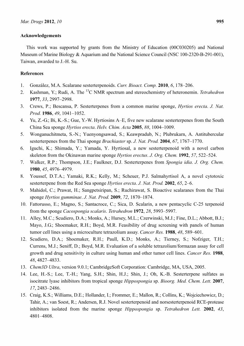
1. González, M.A. Scalarane sesterterpenoids. Curr. Bioact. Comp. 2010, 6, 178–206.
2. Kashman, Y.; Rudi, A. The 13C NMR spectrum and stereochemistry of heteronemin. Tetrahedron
1977, 33, 2997–2998.
3. Crews, P.; Bescansa, P. Sesterterpenes from a common marine sponge, Hyrtios erecta. J. Nat.
Prod. 1986, 49, 1041–1052.
4. Yu, Z.-G.; Bi, K.-S.; Gue, Y.-W. Hyrtiosins A–E, five new scalarane sesterterpenes from the South
China Sea sponge Hyrtios erecta. HeIv. Chim. Acta 2005, 88, 1004–1009.
5. Wonganuchitmeta, S.-N.; Yuenyongsawad, S.; Keawpradub, N.; Plubrukarn, A. Antitubercular
sesterterpenes from the Thai sponge Brachiaster sp. J. Nat. Prod. 2004, 67, 1767–1770.
6. Iguchi, K.; Shimada, Y.; Yamada, Y. Hyrtiosal, a new sesterterpenoid with a novel carbon
skeleton from the Okinawan marine sponge Hyrtios erectus. J. Org. Chem. 1992, 57, 522–524.
7. Walker, R.P.; Thompson, J.E.; Faulkner, D.J. Sesterterpenes from Spongia idia. J. Org. Chem.
1980, 45, 4976–4979.
8. Youssef, D.T.A.; Yamaki, R.K.; Kelly, M.; Scheuer, P.J. Salmahyrtisol A, a novel cytotoxic
sesterterpene from the Red Sea sponge Hyrtios erecta. J. Nat. Prod. 2002, 65, 2–6.
9. Mahidol, C.; Prawat, H.; Sangpetsiripan, S.; Ruchirawat, S. Bioactive scalaranes from the Thai
sponge Hyrtios gumminae. J. Nat. Prod. 2009, 72, 1870–1874.
10. Fattorusso, E.; Magno, S.; Santacroce, C.; Sica, D. Scalarin, a new pentacyclic C-25 terpenoid
from the sponge Cacospongia scalaris. Tetrahedron 1972, 28, 5993–5997.
11. Alley, M.C.; Scudiero, D.A.; Monks, A.; Hursey, M.L.; Czerwinski, M.J.; Fine, D.L.; Abbott, B.J.;
Mayo, J.G.; Shoemaker, R.H.; Boyd, M.R. Feasibility of drug screening with panels of human
tumor cell lines using a microculture tetrazolium assay. Cancer Res. 1988, 48, 589–601.
12. Scudiero, D.A.; Shoemaker, R.H.; Paull, K.D.; Monks, A.; Tierney, S.; Nofziger, T.H.;
Currens, M.J.; Seniff, D.; Boyd, M.R. Evaluation of a soluble tetrazolium/formazan assay for cell
growth and drug sensitivity in culture using human and other tumor cell lines. Cancer Res. 1988,
48, 4827–4833.
13. Chem3D Ultra, version 9.0.1; CambridgeSoft Corporation: Cambridge, MA, USA, 2005.
14. Lee, H.-S.; Lee, T.-H.; Yang, S.H.; Shin, H.J.; Shin, J.; Oh, K.-B. Sesterterpene sulfates as
isocitrate lyase inhibitors from tropical sponge Hippospongia sp. Bioorg. Med. Chem. Lett. 2007,
17, 2483–2486.
15. Craig, K.S.; Williams, D.E.; Hollander, I.; Frommer, E.; Mallon, R.; Collins, K.; Wojciechowicz, D.;
Tahir, A.; van Soest, R.; Andersen, R.J. Novel sesterterpenoid and norsesterterpenoid RCE-protease
inhibitors isolated from the marine sponge Hippospongia sp. Tetrahedron Lett. 2002, 43,
4801–4808.
Mar. Drugs 2012, 10 996
16. Liu, H.; Wang, G.; Namikoshi, M.; Kobayashi, H.; Yao, X.; Cai, G. Sesquiterpene quinones from a
marine sponge Hippospongia sp. that inhibit maturation of starfish oocytes and induce cell cycle
arrest with HepG2 cells. Pharm. Biol. 2006, 44, 522–527.
17. Shen, Y.-C.; Chen, C.-Y.; Kuo, Y.-H. New sesquiterpene hydroquinones from a Taiwanese marine
sponge, Hippospongia metachromia. J. Nat. Prod. 2001, 64, 801–803.
18. Ishibashi, M.; Ohizumi, Y.; Cheng, J.-f.; Nakamura, H.; Hirata, Y.; Sasaki, T.; Kobayashi, J.
Metachromins A and B, novel antineoplastic sesquiterpenoids from the Okinawan sponge
Hippospongia cf. metachromia. J. Org. Chem. 1988, 53, 2855–2858.
19. Musman, M.; Ohtani, I.I.; Nagaoka, D.; Tanaka, J.; Higa, T. Hipposulfates A and B, new
sesterterpene sulfates from an Okinawan sponge, Hippospongia cf. metachromia. J. Nat. Prod.
2001, 64, 350–352.
20. Piao, S.-J.; Zhang, H.-J.; Lu, H.-Y.; Yang, F.; Jiao, W.-H.; Yi, Y.-H.; Chen, W.-S.; Lin, H.-W.
Hippolides A–H, acyclic manoalide derivatives from the marine sponge Hippospongia lachne.
J. Nat. Prod. 2011, 74, 1248–1254.
21. Oda, T.; Wang, W.; Fujita, A.; Mochizuki, M.; Ukai, K.; Namikoshi, M. Promotion of IL-8
production in PMA-stimulated HL-60 cells by sesquiterpene quinones from a marine sponge,
Hippospongia sp. J. Nat. Med. 2007, 61, 434–437.
22. Madaio, A.; Piccialli, V.; Sica, D.; Corriero, G. New polyhydroxysterols from the dictyoceratid
sponges Hippospongia communis, Spongia officinalis, Ircinia variabilis, and Spongionella
gracilis. J. Nat. Prod. 1989, 52, 952–961.
23. Madaio, A.; Notaro, G.; Piccialli, V.; Sica, D. Minor 5,6-secosterols from the marine sponge
Hisppospongia communis. Isolation and synthesis of (7Z,22E,24R)-24-methyl-5,6-secocholesta-7,
22-diene-3β,5β,6-triol. J. Nat. Prod. 1990, 53, 565–572.
24. Cimino, G.; de Stefano, S.; Minale, L. Furospongin-1, a new C-21 furanoterpene from the
sponges Spongia officinalis and Hippospongia communzs. Tetrahedron 1971, 27, 4673–4679.
25. Cimino, G.; de Stefano, S.; Minale, L. Minor C-21 furanoterpenes from the sponges Spongia
officinalis and Hippospongia communis. Tetrahedron 1972, 28, 267–273.
26. Madaio, A.; Piccialli, V.; Sica, D. Hipposterol, a unique trihydroxylated 5,6-secosterol from the
marine sponge Hippospongia communis. Tetrahedron Lett. 1988, 29, 5999–6000.
27. Kobayashi, J.; Murayama, T.; Ohizumi, Y. Metachromin C, a new cytotoxic sesquiterpenoid from
the Okinawan marine sponge Hippospongia metachromia. J. Nat. Prod. 1989, 52, 1173–1176.
28. Kobayashi, J.; Naitoh, K.; Saaski, T.; Shigemori, H. Metachromins D–H, new cytotoxic
sesquiterpenoids from the Okinawan marine sponge Hippospongia metachromia. J. Org. Chen.
1992, 57, 5773–5776.
29. Kobayashi, J.; Shinonaga, H.; Shigemori, H.; Sasaki, T. Untenospongin C, a new C21 furanoterpene
from the Okinawan marine sponge Hippospongia sp. Chem. Pharm. Bull. 1993, 41, 381–382.
30. Rochfort, S.J.; Atkin, D.; Hobbs, L.; Capon, R.J. Hippospongins A–F: New furanoterpenes from a
Southern Australian marine sponge Hippospongia sp. J. Nat. Prod. 1996, 59, 1024–1028.
31. Kobayashi, J.; Ohizumi, Y.; Nakamura, H.; Hirata, Y. Hippospongin, a novel furanosesterterpene
possessing antispasmodic activity from the Okinawan marine sponge Hippospongia sp.
Tetrahedron Lett. 1986, 27, 2113–2116.
Mar. Drugs 2012, 10 997
32. Ohta, S.; Uno, M.; Tokumasu, M.; Hiraga, Y.; Ikegami, S. Hippospongic acid A: An unusual
triterpenoic acid from a marine sponge, Hippospongia sp., which inhibits gastrulation of starfish
embryos. Tetrahedron Lett. 1996, 37, 7765–7766.
33. Guo, Y.W.; Trivellone, E. Ent-untenospongin A, a new C21 furanoterpene from the Indian marine
sponge Hippospongia sp. Chin. Chem. Lett. 2000, 11, 327–330.
34. Nakamura, H.; Deng, S.; Kobayashi, J.; Ohizumi, Y. Dictyoceratin-A and -B, novel antimicrobial
terpenoids from the Okinawan marine sponge Hippospongia sp. Tetrahedron 1986, 42, 4197–4201.
35. Ishiyama, H.; Ishibashi, M.; Ogawa, A.; Yoshida, S.; Kobayashi, J. Taurospongin A, a novel
acetylenic fatty acid derivative inhibiting DNA polymerase and HIV reverse transcriptase from
sponge Hippospongia sp. J. Org. Chem. 1997, 62, 3831–3836.
36. Guo, Y.-W.; Trivellone, E. New hurgamides from a Red Sea sponge of the genus Hippospongia.
J. Asian Nat. Prod. Res. 2006, 2, 251–256.
Samples Availability: Not available.
© 2012 by the authors; licensee MDPI, Basel, Switzerland. This article is an open access article
distributed under the terms and conditions of the Creative Commons Attribution license
(http://creativecommons.org/licenses/by/3.0/).