Embed Size (px)
Citation preview
review article
T h e n e w e ng l a nd j o u r na l o f m e dic i n e
n engl j med 354;1 www.nejm.org january 5, 200654
medical progress
Autoimmune HepatitisEdward L. Krawitt, M.D.
From the Department of Medicine, Uni-versity of Vermont, Burlington; and the Department of Medicine, Dartmouth Col-lege, Hanover, N.H. Address reprint re-quests to Dr. Krawitt at the University of Vermont, Given C-246, Burlington, VT 05405-0068, or at [email protected].
N Engl J Med 2006;354:54-66.Copyright © 2006 Massachusetts Medical Society.
A utoimmune hepatitis is a generally progressive, chronic hep-
atitis of unknown cause that occurs in children and adults of all ages. Oc-casionally, it has a fluctuating course, with periods of increased or de-
creased activity. The diagnosis is based on histologic abnormalities, characteristic clinical and biochemical findings, and abnormal levels of serum globulins, includ-ing autoantibodies. Since the first descriptions of this disorder more than 50 years ago,1 many labels have been applied, but “autoimmune hepatitis” has been accepted as the most appropriate and least redundant term.2,3 Variant, overlapping, or mixed forms of autoimmune hepatitis that share features with other putative autoimmune liver diseases, primary biliary cirrhosis, and primary sclerosing cholangitis occur as well. The distinctions among these disorders at present are necessarily descriptive.
It remains important to distinguish autoimmune hepatitis from other forms of chronic hepatitis, because a high percentage of cases respond to antiinflammatory or immunosuppressive therapy, or both. Although appropriate management can pro-long survival, improve the quality of life, and avoid the need for liver transplanta-tion, considerable therapeutic challenges remain in the treatment of this disorder.4
pathogenesis
A conceptual framework for the pathogenesis of autoimmune hepatitis postulates an environmental agent that triggers a cascade of T-cell–mediated events directed at liver antigens in a host genetically predisposed to this disease, leading to a pro-gressive necroinflammatory and fibrotic process in the liver.
potential triggers
The environmental agents assumed to induce autoimmune hepatitis have not been delineated but include viruses. The finding of molecular mimicry by cross-reactivity between epitopes of viruses and certain liver antigens adds credence to a hypothesis of virally triggered disease. Because the trigger or triggers of autoimmune hepatitis may be part of a so-called hit-and-run phenomenon, in which induction occurs many years before overt autoimmune disease, identifying an infectious agent may prove impossible. There has been evidence implicating measles virus, hepatitis viruses, cytomegalovirus, and Epstein–Barr virus as initiators of the disease; the most con-vincing evidence is related to hepatitis viruses.5-7
Certain drugs, including oxyphenisatin, methyldopa, nitrofurantoin, diclofenac, interferon, pemoline, minocycline, and atorvastatin, can induce hepatocellular injury that mimics autoimmune hepatitis.8-12 It has also been suggested that herbal agents such as black cohosh and dai-saiko-to might trigger autoimmune hepatitis. Wheth-er drugs and herbs unmask or induce autoimmune hepatitis or simply cause a drug-induced hepatitis with accompanying autoimmune features is unclear. Minocycline10,11
Copyright © 2006 Massachusetts Medical Society. All rights reserved. Downloaded from www.nejm.org by LUIGI GRECO on January 13, 2006 .
medical progress
n engl j med 354;1 www.nejm.org january 5, 2006 55
and atorvastatin, which induce other autoimmune syndromes, have been implicated most recently as potential triggering agents of this disease.
genetic susceptibility
Most knowledge concerning the genetics of au-toimmune hepatitis comes from studies of the HLA genes that reside in the major histocompati-bility complex (MHC), located on the short arm of chromosome 6. The MHC is a genetic system with extensive polymorphism. Although multiple genes are probably involved, HLA genes appear to play the dominant role in a predisposition to autoimmune hepatitis.13,14
Type 1 autoimmune hepatitis, characterized by circulating antinuclear antibodies (ANA), smooth-muscle antibodies, antiactin antibodies, atypical perinuclear antineutrophilic cytoplasmic antibod-ies (pANCA), and autoantibodies against soluble liver antigen and liver–pancreas antigen (SLA/LP), is associated with the HLA-DR3 serotype (found in linkage disequilibrium with HLA-B8 and HLA-A1), particularly among white patients. There is an as-sociation with HLA-DR4 among patients who are HLA-DR3–negative. HLA-DR3–associated disease is more common in the early-onset, severe form of autoimmune hepatitis, which often occurs in girls and young women. In comparison, the asso-ciation with HLA-DR4 is more common in adults and may be associated with an increased incidence of extrahepatic manifestations, milder disease, and a better response to corticosteroid therapy. In Japan, where HLA-DR3 is rare, the most com-mon associated HLA locus is HLA-DR4.
The results of serotyping studies have been confirmed with the use of genotyping for HLA-DRB, DQA, and DQB with polymerase-chain-reac-tion techniques. A high frequency of the HLA-DRB1*0301DRB3*0101DQAl*0501DQB1*0201 haplotype (the first two elements correspond to the serologic determinants DR3 and DR52) and the HLA-DRB1*0401 allele have been observed in association with autoimmune hepatitis. In South American populations, an increased frequency of the HLA-DRB1*1301 allele was reported,15,16 where-as in Japan, autoimmune hepatitis has been asso-ciated with the DRB1*0405DQB1*0401 haplo-type.17 In children, type 1 autoimmune hepatitis is commonly associated with the HLA-DRB1*03 and HLA-DRB1*13 alleles.
Type 2 autoimmune hepatitis, a rare disorder characterized by antibodies against liver–kidney
microsome 1 (LKM-1) and liver cytosol 1 (ALC-1), has been associated with the HLA-DRB1 and HLA-DQB1 alleles.18 HLA-DR2 appears to be pro-tective in white northern Europeans, and a study of white Argentineans suggested that the HLA-DRB1*1302 allele is protective.14,15
Susceptibility to autoimmune hepatitis has been reported to be associated with tumor ne-crosis factor (TNF) genes, the loci of which are in the class III region of the MHC, although this finding has been disputed.19,20 A polymorphism at position 308 of the TNF-α gene has been as-sociated with susceptibility to type 1 autoimmune hepatitis in both European and North American patients, but it may simply represent linkage dis-equilibrium with HLA-DRB1*0301. There were no significant differences in the response to thera-py between those with and those without the 308 polymorphism.19 Furthermore, this associa-tion was not present in Japanese or Brazilian pa-tients with autoimmune hepatitis.17,20 Similar as-sociations of susceptibility with polymorphisms of cytotoxic T-lymphocyte antigen 4 observed in northern European patients were not seen in Brazilian patients.21,22 Potential associations with loci in other chromosomes are under investiga-tion.23,24
mechanisms of aberrant autoreactivity
Knowledge concerning autoantigens responsible for initiating the cascades of events in autoimmune hepatitis is still rudimentary. A leading candidate for many years has been the asialoglycoprotein receptor, a liver-specific membrane protein with high levels of expression in periportal hepatocytes. Information based on the identification of SLA/LP autoantibodies and the cloning and character-ization of the SLA/LP antigen, which shares some amino acid sequences with the asialoglycopro-tein receptor, suggests that this 50-kD cytosolic protein may represent a relevant antigen in at least some patients with type 1 autoimmune hep-atitis.25,26
Evidence of an autoimmune process in the type 2 form of the disease is more compelling. The presence of immunodominant B-cell epitopes of cytochrome P-450 2D6 (CYP2D6) and evidence of cross-reactivity with homologues of different vi-ruses suggest that relevant antigens exist within CYP2D6.27
The identification of CD4+ regulatory T cells has reinvoked the concept that failure of or escape
Copyright © 2006 Massachusetts Medical Society. All rights reserved. Downloaded from www.nejm.org by LUIGI GRECO on January 13, 2006 .
T h e n e w e ng l a nd j o u r na l o f m e dic i n e
n engl j med 354;1 www.nejm.org january 5, 200656
from normal suppression of reactivity against the self has an essential role in the development of autoimmune disease. The hypothesis that this escape phenomenon occurs in autoimmune hep-atitis has remained attractive and is based on early studies of immune regulation.28-31 Recent experi-mental evidence suggests that immunoregulatory dysfunction characterized by decreased numbers of CD4+CD25+ regulatory T cells and decreased levels of scurfin, the protein product of the FOXP3 gene that is a member of the forkhead family of transcription factors, may occur in autoimmune hepatitis.32 Such observations suggest that a de-crease in the number of regulatory T cells and their ability to expand may lead to autoimmune liver disease.
clinical characteristics
Autoimmune hepatitis is more common among women than men, but it occurs globally in chil-dren and adults of both sexes in diverse ethnic groups.16-18,33-41 Since chronic viral hepatitis ap-pears to be very common, the prevalence of auto-immune hepatitis may be higher than reported because of concomitant chronic hepatitis C or B or both.38
presentation
The presentation of autoimmune hepatitis is het-erogeneous, and the clinical course may be char-acterized by periods of decreased or increased activity; thus, clinical manifestations are variable. The spectrum of presentation ranges from no symptoms to debilitating symptoms and even ful-minant hepatic failure.
Patients may present with nonspecific symp-toms of varying severity, such as fatigue, lethargy, malaise, anorexia, nausea, abdominal pain, and itching. Arthralgia involving small joints is com-mon. Physical examination may reveal no abnor-malities, but it may also reveal hepatomegaly, splenomegaly, jaundice, and signs and symptoms of chronic liver disease.
Patients with severe or fulminant symptoms accompanied by profound jaundice and a pro-longed prothrombin time may have aminotrans-ferase levels in the thousands.42 Many patients with an acute presentation have histologic evi-dence of chronic disease on liver biopsy, indicat-ing that they probably have had subclinical dis-
ease for a long time. Long periods of subclinical disease may also occur after presentation.
Autoimmune hepatitis may first become evi-dent during pregnancy or in the early postpartum period. Furthermore, postpartum exacerbations may occur in patients whose condition improved during pregnancy.43-45
One clue to diagnosing autoimmune hepati-tis is the presence of other diseases with autoim-mune features, commonly thyroiditis, ulcerative colitis, type 1 diabetes, rheumatoid arthritis, and celiac disease.46,47 Occasionally, circulating anti-endomysial antibodies, antigluten antibodies, and anti–tissue transglutaminase antibodies may be found in patients with autoimmune hepatitis; this finding generally reflects the coexistence of ce-liac sprue and autoimmune hepatitis.
laboratory abnormalities
In general, aminotransferase elevations are more striking than abnormalities in bilirubin and alkaline phosphatase levels in patients with autoimmune hepatitis. Some cases, however, are characterized by cholestasis, with high levels of conjugated bilirubin and alkaline phosphatase. In such circumstances, extrahepatic obstruction and cholestatic forms of viral hepatitis, drug-induced disease, primary biliary cirrhosis, primary sclerosing cholangitis, and variant syndromes must be considered.
One characteristic laboratory feature of auto-immune hepatitis, although not invariant, is a generalized elevation of serum globulins, in par-ticular, gamma globulin and IgG, which are gen-erally 1.2 to 3.0 times normal. The characteristic circulating autoantibodies seen in autoimmune hepatitis include ANA, smooth-muscle antibody, antiactin antibody, SLA/LP autoantibodies, pANCA, anti–LKM-1, and anti–LC-1. Antimitochondrial antibodies are sometimes present in patients with autoimmune hepatitis. It should be noted, how-ever, that autoantibodies are found in various liver diseases, and their presence, by itself, is not diag-nostic of autoimmune hepatitis. There is little evi-dence that autoantibodies play a part in its patho-genesis.
classification and autoantibodies
Classification of autoimmune hepatitis on the ba-sis of autoantibody patterns has been helpful to clinicians (Table 1). Although the distinction was
Copyright © 2006 Massachusetts Medical Society. All rights reserved. Downloaded from www.nejm.org by LUIGI GRECO on January 13, 2006 .
medical progress
n engl j med 354;1 www.nejm.org january 5, 2006 57
initially based on circulating antibodies alone, other differences have become apparent. The main serologic markers of type 1 autoimmune hepati-tis are ANA and smooth-muscle antibody. Titers of at least 1:80 are generally accepted as positive,3 but results vary, depending on the assays used; lower titers may signify a positive response in children. Antiactin antibodies are more specific for type 1 autoimmune hepatitis.48 Anti–LKM-1 and anti–LC-1 characterize type 2 disease.18,33,34
The identification of other circulating auto-antibodies, in particular SLA/LP autoantibod-ies25,26,49 and atypical pANCA,50-52 are sometimes helpful in diagnosing type 1 disease. SLA/LP auto-antibodies are the most specific autoantibody identified in type 1 autoimmune hepatitis but is found in only 10 to 30 percent of cases. Atypical pANCA is frequently present, and on rare occa-sions, it occurs as an isolated autoantibody.52
Anti–LKM-1 and anti–LC-1 can occur alone or together in type 2 autoimmune hepatitis.18,33,34,53 Anti–LKM-1, which is directed at CYP2D6, can occur in chronic hepatitis C, though the anti-body response to immunodominant epitopes dif-fers.27 Anti–LC-1 generally occurs in conjunc-
tion with anti–LKM-1, but it may be the sole autoantibody.34 It recognizes formiminotrans-ferase cyclodeaminase, a liver-specific 58-kD met-abolic enzyme.54
complications
The complications of autoimmune hepatitis are the same as in any progressive liver disease. Pri-mary hepatocellular carcinoma is a known con-sequence of autoimmune hepatitis; in some pa-tients, chronic hepatitis progresses to cirrhosis and, ultimately, to carcinoma. However, carcinoma oc-curs in association with autoimmune hepatitis less frequently than does chronic viral hepatitis.
histologic appearance
The histologic appearance of autoimmune hepa-titis is the same as that of chronic hepatitis, and although certain changes are characteristic, no findings are specific for autoimmune hepatitis.55 The histologic differential diagnosis of chronic hepatitis is provided in Table 2. Advances in viro-logic studies and refinements in cholangiograph-ic methods have made it easier to rule out other clinical entities.
Table 1. Classification of Autoimmune Hepatitis.
Variable Type 1 Autoimmune Hepatitis Type 2 Autoimmune Hepatitis
Characteristic autoantibodies Antinuclear antibody*Smooth-muscle antibody*Antiactin antibody†Autoantibodies against soluble
liver antigen and liver–pancreas antigen‡
Atypical perinuclear antineutrophil cytoplasmic antibody
Antibody against liver–kidney micro-some 1*
Antibody against liver cytosol 1*
Geographic variation Worldwide Worldwide; rare in North America
Age at presentation Any age Predominantly childhood and young adulthood
Sex of patients Female in approximately 75% of cases
Female in approximately 95% of cases
Association with other autoimmune diseases
Common Common§
Clinical severity Broad range Generally severe
Histopathologic features at presen-tation
Broad range Generally advanced
Treatment failure Infrequent Frequent
Relapse after drug withdrawal Variable Common
Need for long-term maintenance Variable Approximately 100%
* The conventional method of detection is immunofluorescence.† Tests for this antibody are rarely available in commercial laboratories.‡ This antibody is detected by enzyme-linked immunosorbent assay.§ Autoimmune polyendocrinopathy–candidiasis–ectodermal dystrophy is seen only in patients with type 2 disease.47
Copyright © 2006 Massachusetts Medical Society. All rights reserved. Downloaded from www.nejm.org by LUIGI GRECO on January 13, 2006 .
T h e n e w e ng l a nd j o u r na l o f m e dic i n e
n engl j med 354;1 www.nejm.org january 5, 200658
Autoimmune hepatitis is generally character-ized by a mononuclear-cell infiltrate invading the limiting plate — that is, the sharply demar-cated hepatocyte boundary that surrounds the portal triad and permeates the surrounding pa-renchyma (periportal infiltrate, also called piece-meal necrosis or interface hepatitis that progress-es to lobular hepatitis). There may be an abundance of plasma cells, a finding that in the past led to the use of the term “plasma-cell hepatitis.” Eo-sinophils are frequently present. The portal le-sion generally spares the biliary tree. Fibrosis is present in all but the mildest forms of autoim-mune hepatitis. In advanced disease, the fibrosis is extensive, and with the distortion of the he-patic lobule and the appearance of regenerative nodules, it results in cirrhosis.55 Occasionally, cen-trizonal lesions occur.42,57
The findings in patients with acute-onset auto-immune hepatitis differ somewhat from those with an insidious presentation. Patients present-ing with fulminant hepatic failure tend to have interface and lobular hepatitis, lobular disarray, and hepatocyte, central, and submassive necro-
sis. However, they have less fibrosis than patients who present with a more chronic course.42 Ste-atosis occurs in a minority of patients,55 although nonalcoholic fatty liver disease may occur in con-junction with autoimmune hepatitis. The various histologic appearances are depicted in Figure 1.
In patients who have a spontaneous or phar-macologically induced remission, the histologic findings may revert to normal or inflammation may be confined to portal areas. In this setting, cirrhosis may become inactive and fibrosis may diminish or disappear.55,59-61
diagnosis
In the presence of a compatible histologic pic-ture, the diagnosis of autoimmune hepatitis is based on characteristic clinical and biochemical findings, circulating autoantibodies, and abnor-mal levels of serum globulins. Circulating anti-bodies are absent in about 10 percent of patients. A scoring system proposed and subsequently re-vised by the International Autoimmune Hepatitis Group3 to standardize the diagnosis for clinical trials and population studies has had limited
Table 2. Histologic Differential Diagnosis of Chronic Hepatitis.
Disease Distinguishing Features*
Autoimmune hepatitis Conspicuous plasma-cell infiltrates
Primary biliary cirrhosis Lymphocytic and granulomatous infiltrates of bile ducts; ductopenia
Primary sclerosing cholangitis Fibrous obliterative cholangitis; ductopenia
Autoimmune cholangitis† Lymphocytic and granulomatous infiltrates of bile ducts; ductopenia
Chronic viral hepatitis Ground-glass hepatocytes; immunoperoxidase staining for hepatitis B surface and core antigens in patients with chronic hepatitis B; nodu-lar-appearing infiltrates characteristic in patients with chronic hepati-tis C; steatosis possible in patients infected with hepatitis C virus genotype 3
Chronic drug-induced hepatitis No helpful distinguishing histologic features
Alpha1-antitrypsin deficiency Intracytoplasmic globules
Wilson’s disease Heavy copper deposition
Granulomatous hepatitis Conspicuous and frequent granulomas
Graft-versus-host disease Lymphocytic and granulomatous infiltrates of bile ducts; ductopenia
Alcoholic steatohepatitis Steatosis; central inflammation and fibrosis; Mallory bodies
Nonalcoholic steatohepatitis Glycogenated nuclei; steatosis; central inflammation and fibrosis; Mallory bodies
* These histologic features may be helpful in distinguishing among the causes of chronic hepatitis. Differences in histo-pathological findings among the diseases may be more apparent depending on the grade and stage of disease.55
† There is still debate as to whether this entity is antimitochondrial-antibody–negative primary biliary cirrhosis.56
Copyright © 2006 Massachusetts Medical Society. All rights reserved. Downloaded from www.nejm.org by LUIGI GRECO on January 13, 2006 .
medical progress
n engl j med 354;1 www.nejm.org january 5, 2006 59
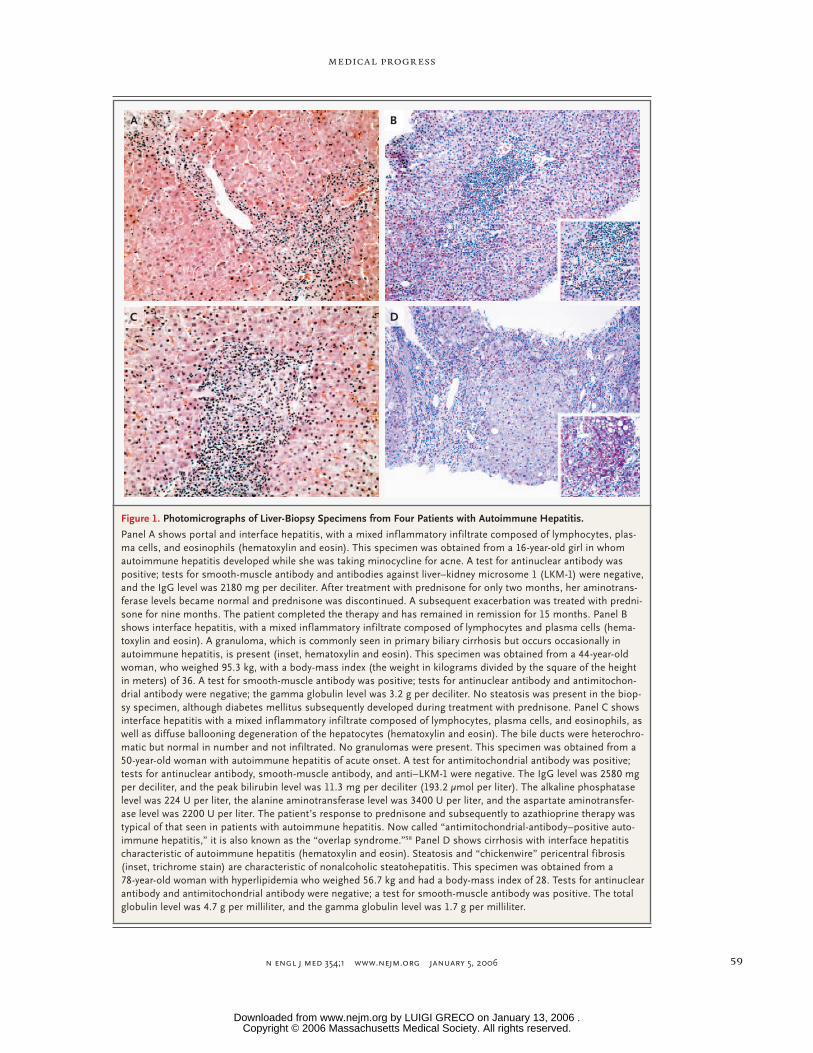
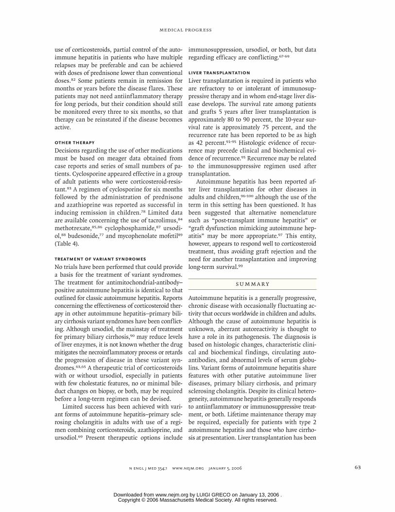
A B
C D
Figure 1. Photomicrographs of Liver-Biopsy Specimens from Four Patients with Autoimmune Hepatitis.
Panel A shows portal and interface hepatitis, with a mixed inflammatory infiltrate composed of lymphocytes, plas-ma cells, and eosinophils (hematoxylin and eosin). This specimen was obtained from a 16-year-old girl in whom autoimmune hepatitis developed while she was taking minocycline for acne. A test for antinuclear antibody was positive; tests for smooth-muscle antibody and antibodies against liver–kidney microsome 1 (LKM-1) were negative, and the IgG level was 2180 mg per deciliter. After treatment with prednisone for only two months, her aminotrans-ferase levels became normal and prednisone was discontinued. A subsequent exacerbation was treated with predni-sone for nine months. The patient completed the therapy and has remained in remission for 15 months. Panel B shows interface hepatitis, with a mixed inflammatory infiltrate composed of lymphocytes and plasma cells (hema-toxylin and eosin). A granuloma, which is commonly seen in primary biliary cirrhosis but occurs occasionally in autoimmune hepatitis, is present (inset, hematoxylin and eosin). This specimen was obtained from a 44-year-old woman, who weighed 95.3 kg, with a body-mass index (the weight in kilograms divided by the square of the height in meters) of 36. A test for smooth-muscle antibody was positive; tests for antinuclear antibody and antimitochon-drial antibody were negative; the gamma globulin level was 3.2 g per deciliter. No steatosis was present in the biop-sy specimen, although diabetes mellitus subsequently developed during treatment with prednisone. Panel C shows interface hepatitis with a mixed inflammatory infiltrate composed of lymphocytes, plasma cells, and eosinophils, as well as diffuse ballooning degeneration of the hepatocytes (hematoxylin and eosin). The bile ducts were heterochro-matic but normal in number and not infiltrated. No granulomas were present. This specimen was obtained from a 50-year-old woman with autoimmune hepatitis of acute onset. A test for antimitochondrial antibody was positive; tests for antinuclear antibody, smooth-muscle antibody, and anti–LKM-1 were negative. The IgG level was 2580 mg per deciliter, and the peak bilirubin level was 11.3 mg per deciliter (193.2 μmol per liter). The alkaline phosphatase level was 224 U per liter, the alanine aminotransferase level was 3400 U per liter, and the aspartate aminotransfer-ase level was 2200 U per liter. The patient’s response to prednisone and subsequently to azathioprine therapy was typical of that seen in patients with autoimmune hepatitis. Now called “antimitochondrial-antibody–positive auto-immune hepatitis,” it is also known as the “overlap syndrome.”58 Panel D shows cirrhosis with interface hepatitis characteristic of autoimmune hepatitis (hematoxylin and eosin). Steatosis and “chickenwire” pericentral fibrosis (inset, trichrome stain) are characteristic of nonalcoholic steatohepatitis. This specimen was obtained from a 78-year-old woman with hyperlipidemia who weighed 56.7 kg and had a body-mass index of 28. Tests for antinuclear antibody and antimitochondrial antibody were negative; a test for smooth-muscle antibody was positive. The total globulin level was 4.7 g per milliliter, and the gamma globulin level was 1.7 g per milliliter.
Copyright © 2006 Massachusetts Medical Society. All rights reserved. Downloaded from www.nejm.org by LUIGI GRECO on January 13, 2006 .
T h e n e w e ng l a nd j o u r na l o f m e dic i n e
n engl j med 354;1 www.nejm.org january 5, 200660
value and may be inaccurate when applied to in-dividual patients, especially children. Attempts are under way to devise a less complicated and more accurate system.62
variant syndromes
Although we have long known that the clinical, his-tologic, and serologic profiles of so-called overlap, mixed, or variant syndromes differ from the clas-sic features of autoimmune hepatitis, primary bili-ary cirrhosis, and primary sclerosing cholangitis, no consensus regarding categorization has been reached. Terms such as “overlap syndrome,” “anti-mitochondrial-antibody–negative primary biliary cirrhosis,” “the hepatic form of primary biliary cirrhosis,” “autoimmune cholangitis,” “autoim-mune cholangiopathy,” “chronic autoimmune cho-lestasis,” “immunocholangitis,” “immune cholan-giopathy,” and “combined hepatitic/cholestatic syndrome” have all been used to describe patients with features of both autoimmune hepatitis and primary biliary cirrhosis. The presentation of pu-tative coincidental diseases, consecutive diseases, and evolution from one disease to another have highlighted the complexity of this issue.56,58,63-66
One approach is to consider the variant syn-dromes of autoimmune hepatitis and primary bili-ary cirrhosis as part of a continuum that extends from classic autoimmune hepatitis to classic pri-mary biliary cirrhosis. Examination of a biopsy specimen with histologic features of autoimmune hepatitis but serologic findings characteristic of primary biliary cirrhosis, such as an isolated anti-mitochondrial antibody directed toward enzymes in the 2-oxo acid dehydrogenase family, would
be indicative of the overlap syndrome,58 or anti-mitochondrial-antibody–positive autoimmune hep-atitis (Table 3). The clinical course and response to therapy in this syndrome appear to be identical to those in classic autoimmune hepatitis.
There is disagreement as to whether the vari-ant most commonly called autoimmune chol-angitis56,58 merely represents antimitochondrial-antibody–negative primary biliary cirrhosis (Table 3). Immunoblotting and enzyme-linked immuno-sorbent assays for antimitochondrial antibodies and primary biliary cirrhosis–specific antinucle-ar antibodies (anti-Sp100 and anti-gp210) have yielded different autoantibody profiles for the two conditions, underscoring the heterogeneity of these syndromes.66
Identifying and classifying autoimmune hep-atitis–primary sclerosing cholangitis overlap syn-dromes is also difficult, particularly in chil-dren.53,67-72 “Autoimmune sclerosing cholangitis” is the term applied to this disease in affected chil-dren and could arguably be applied to that in adults as well. Although primary sclerosing chol-angitis can evolve to autoimmune hepatitis, au-toimmune hepatitis more commonly evolves to autoimmune sclerosing cholangitis.72 Autoimmune sclerosing cholangitis cannot be diagnosed in the absence of cholangiographic abnormalities. Pa-tients suspected of having autoimmune hepatitis who also have histologic bile-duct abnormalities, cholestatic laboratory changes (e.g., elevations of alkaline phosphatase, γ-glutamyltransferase, or both), pruritus, inflammatory bowel disease, or loss of response to antiinflammatory or immuno-suppressive therapy may have autoimmune scle-rosing cholangitis.
Table 3. Characteristics of Autoimmune Hepatitis–Primary Biliary Cirrhosis Variant Syndromes.
Characteristic Overlap Syndrome* Autoimmune Cholangitis†
Antinuclear antibody Absent Generally present
Smooth-muscle antibody Absent Generally present
Antimitochondrial antibody Present Absent
Biochemical cholestasis† Absent Present
Histologic evidence of bile-duct abnormalities Absent Present
Cholangiographic abnormalities Absent Absent
Responsiveness to immunosuppression Present Variable
* This syndrome is also called antimitochondrial-antibody–positive autoimmune hepatitis.58 There is debate as to wheth-er autoimmune cholangitis and antimitochondrial-antibody–negative primary biliary cirrhosis represent different enti-ties.56,63-66
† This condition is characterized by elevated levels of serum alkaline phosphatase, γ-glutamyltransferase, or both.
Copyright © 2006 Massachusetts Medical Society. All rights reserved. Downloaded from www.nejm.org by LUIGI GRECO on January 13, 2006 .
medical progress
n engl j med 354;1 www.nejm.org january 5, 2006 61
treatment
In the 1970s, evidence that mercaptopurine and azathioprine were effective in treating autoim-mune diseases, together with controlled studies of corticosteroids, led to the opinion that auto-immune hepatitis is a treatable disease. Antiin-flammatory or immunosuppressive therapy has been a mainstay in the treatment of both type 1 and type 2 disease. Depending on the definition of a response, therapy is reported to be successful in 65 to 80 percent of cases, which indicates that a substantial percentage of patients require ther-apy beyond standard treatment. Current response rates appear better than those in early trials, pre-sumably because earlier trials involved more pa-tients with severe disease and antedated the pres-ent ability to test for chronic viral hepatitis B and C. Ten-year survival rates (with the end point be-ing death or transplantation) among treated pa-tients are now considered to exceed 90 percent; but the 20-year survival rate may be less than 80 per-cent among patients without cirrhosis and less than 40 percent among those with cirrhosis at presen-tation.73 Once the disease is in remission, main-tenance therapy with azathioprine alone is suc-cessful in approximately 80 percent of patients.74
Response to treatment is helpful in estab-lishing the diagnosis of autoimmune hepatitis, but the response rate to standard therapy is not 100 percent. Thus, a lack of response cannot rule out this diagnosis. Moreover, not all patients re-ceive treatment, and the prescribed doses of pred-nisone and azathioprine or mercaptopurine vary. In addition, other diseases, including some vari-ant syndromes, may respond to corticosteroids.
Progress in the medical management of auto-immune hepatitis has been slow. Considerable challenges still exist in the areas of initial and maintenance regimens, management of relapse, management of a lack of response to therapy, drug toxicity and intolerance, noncompliance, and treat-ment during pregnancy. Although guidelines for the treatment of autoimmune hepatitis have been published by the American Association for the Study of Liver Diseases, these are meant to be flex-ible.75 The heterogeneity of autoimmune hepatitis underscores the need for individualized therapy in adults and children.4,75,76
standard treatment
Initial treatment with prednisone (or predniso-lone) alone or in combination with azathioprine
should be instituted in nearly all patients in whom the histologic findings include interface hepatitis, with or without fibrosis or cirrhosis. The magnitude of aminotransferase and gamma glob-ulin elevations does not necessarily correlate with the histologic extent of injury and provides little help with respect to the initiation of treatment. In patients with only portal inflammation, the decision to treat is often determined on the basis of the levels of aminotransferase, gamma globu-lin, or both; the symptoms; or the combination of levels and symptoms. Asymptomatic patients and those with portal inflammation without fi-brosis may be followed without treatment, but their clinical status, including the findings on liver biopsy, should be monitored carefully for evidence of progression of disease, since the activ-ity of autoimmune hepatitis sometimes fluctuates. Initial treatment consists of combination therapy in order to avoid or mitigate the side effects of corticosteroid treatment. An alternative approach is to wait until remission is achieved before cor-ticosteroid-sparing treatment with azathioprine or mercaptopurine is initiated (Table 4).
Adverse effects or intolerance of azathioprine, mercaptopurine, or both is an issue of particu-lar concern.79,80 Azathioprine is a prodrug of mercaptopurine. The methylation of mercapto-purine and 6-thioguanosine 5'-monophosphate is catalyzed by thiopurine methyltransferase, which is encoded by highly polymorphic genes. Patients who are homozygous for a mutation of thiopurine methyltransferase associated with low enzyme activity are at high risk for severe com-plications, including death. Patients who are het-erozygous for a mutation of thiopurine methyl-transferase probably are at intermediate risk. Given these findings, some investigators have suggest-ed performing thiopurine methyltransferase geno-typing before prescribing azathioprine or mer-captopurine. However, some patients who cannot tolerate azathioprine appear to be able to toler-ate mercaptopurine without side effects, indicating that azathioprine-induced toxicity is not simply due to a deficiency of thiopurine methyltrans-ferase.81 Despite the availability of reliable meth-ods for genoptying thiopurine methyltransfer-ase and determining levels of mer cap topurine metabolites, their use in the clinical manage-ment of autoimmune hepatitis is not estab-lished.79,80
In general, a patient’s progress is followed by monitoring levels of serum aminotransferases
Copyright © 2006 Massachusetts Medical Society. All rights reserved. Downloaded from www.nejm.org by LUIGI GRECO on January 13, 2006 .
T h e n e w e ng l a nd j o u r na l o f m e dic i n e
n engl j med 354;1 www.nejm.org january 5, 200662
and circulating globulins (total or gamma glob-ulin, or both, with or without IgG). The histologic response typically lags behind the biochemical response, and a clinical remission does not nec-essarily mean that there is histologic evidence of resolution. Reasonable intervals for repeated liver biopsy appear to be one year after levels of as-partate aminotransferase and alanine aminotrans-ferase have become normal or approximately two years after presentation.
Although some patients remain in remission after drug treatment is withdrawn, most require long-term maintenance therapy. In general, pa-
tients with milder disease have a better response. Adults and children with cirrhosis at the time of the initial biopsy, particularly children with type 2 disease, rarely stay in remission when treat-ment is withdrawn. Thus, lifelong maintenance therapy is generally indicated in such cases. The wisdom of the administration of azathioprine alone or as a corticosteroid-sparing agent should be approached by weighing the side effects of long-term cortico steroid use against those of long-term azathioprine use; patients treated with aza-thioprine alone frequently have arthralgia.74
In the presence of severe side effects from the
Table 4. Drugs Used in the Treatment of Autoimmune Hepatitis in Adults and Children.
Drug Initial Therapy Maintenance Therapy Comments
Prednisone or prednisolone
Used as monotherapy in adults (20–60 mg/day) and children (1–2 mg per kilogram of body weight/day); also used in com-bination therapy in adults (15–30 mg/day) and children (1–2 mg/kg/day) with azathioprine or mercaptopurine
Used as monotherapy in adults (5–15 mg/day) and children (1 mg/kg/day); also used in combination therapy in adults (5–10 mg/day) and children (0.5–1.0 mg/kg/day) with aza-thioprine or mercaptopurine
Relatively contraindicated in pa-tients with osteoporosis, diabe-tes mellitus, glaucoma, cata-racts, arterial hypertension, major depression, and femoral avascular necrosis; reduced doses may work; use of budesonide under investiga-tion77
Azathioprine Used in combination with predni-sone or prednisolone in adults (50–100 mg/day) and children (1.5–2.0 mg/kg/day)
Used as monotherapy in adults (50–200 mg/day) and children (1.5–2.0 mg/kg/day); also used in combination therapy in adults (50–150 mg/day) and children (1.5–2.0 mg/kg/day)
Contraindicated in patients with homozygous thiopurine meth-yltransferase deficiency; rela-tively contraindicated in pa-tients with heterozygous thio-purine methyltransferase defi-ciency, cancer, or cytopenia, and pregnant patients
6-Mercaptopurine May be substituted for azathio-prine in combination therapy in adults (25–100 mg/day) and children (0.75–1.0 mg/kg/day)
Used as monotherapy in adults (25–100 mg/day) and children (0.75–1.0 mg/kg/day); also used in combination therapy in adults (25–100 mg/day) and children (0.5–1.0 mg/kg/day)
Contraindicated in patients with homozygous thiopurine meth-yltransferase deficiency; rela-tively contraindicated in pa-tients with heterozygous thio-purine methyltransferase defi-ciency, cancer, or cytopenia, and pregnant patients
Cyclosporine Sometimes used as monotherapy in children78; sometimes used as an alternative drug in adults with treatment-refractory dis-ease
Sometimes used as an alternative drug in adults with treatment-refractory disease
Once remission achieved in chil-dren, maintenance therapy ini-tiated with a combination of prednisone and azathioprine78; role of tacrolimus in place of cyclosporine not established
Mycophenolate mofetil Sometimes used in patients with treatment-refractory disease or in patients with adverse drug reactions to or intolerance of azathioprine, mercaptopurine, or both
Sometimes used in patients with treatment-refractory disease or in patients with adverse drug reactions to or intolerance of azathioprine, mercaptopurine, or both
Role of mycophenolate mofetil, methotrexate, and cyclophos-phamide not established
Ursodiol Sometimes used in combination with prednisone, azathioprine, or both
Sometimes used in combination with prednisone, azathioprine, or both
Role of ursodiol not established
Copyright © 2006 Massachusetts Medical Society. All rights reserved. Downloaded from www.nejm.org by LUIGI GRECO on January 13, 2006 .
medical progress
n engl j med 354;1 www.nejm.org january 5, 2006 63
use of corticosteroids, partial control of the auto-immune hepatitis in patients who have multiple relapses may be preferable and can be achieved with doses of prednisone lower than conventional doses.82 Some patients remain in remission for months or years before the disease flares. These patients may not need antiinflammatory therapy for long periods, but their condition should still be monitored every three to six months, so that therapy can be reinstated if the disease becomes active.
other therapy
Decisions regarding the use of other medications must be based on meager data obtained from case reports and series of small numbers of pa-tients. Cyclosporine appeared effective in a group of adult patients who were corticosteroid-resis-tant.83 A regimen of cyclosporine for six months followed by the administration of prednisone and azathioprine was reported as successful in inducing remission in children.78 Limited data are available concerning the use of tacrolimus,84 methotrexate,85,86 cyclophosphamide,87 ursodi-ol,88 budesonide,77 and mycophenolate mofetil89 (Table 4).
treatment of variant syndromes
No trials have been performed that could provide a basis for the treatment of variant syndromes. The treatment for antimitochondrial-antibody–positive autoimmune hepatitis is identical to that outlined for classic autoimmune hepatitis. Reports concerning the effectiveness of corticosteroid ther-apy in other autoimmune hepatitis–primary bili-ary cirrhosis variant syndromes have been conflict-ing. Although ursodiol, the mainstay of treatment for primary biliary cirrhosis,90 may reduce levels of liver enzymes, it is not known whether the drug mitigates the necroinflammatory process or retards the progression of disease in these variant syn-dromes.63,65 A therapeutic trial of corticosteroids with or without ursodiol, especially in patients with few cholestatic features, no or minimal bile-duct changes on biopsy, or both, may be required before a long-term regimen can be devised.
Limited success has been achieved with vari-ant forms of autoimmune hepatitis–primary scle-rosing cholangitis in adults with use of a regi-men combining corticosteroids, azathioprine, and ursodiol.69 Present therapeutic options include
immunosuppression, ursodiol, or both, but data regarding efficacy are conflicting.67-69
liver transplantation
Liver transplantation is required in patients who are refractory to or intolerant of immunosup-pressive therapy and in whom end-stage liver dis-ease develops. The survival rate among patients and grafts 5 years after liver transplantation is approximately 80 to 90 percent, the 10-year sur-vival rate is approximately 75 percent, and the recurrence rate has been reported to be as high as 42 percent.91-95 Histologic evidence of recur-rence may precede clinical and biochemical evi-dence of recurrence.95 Recurrence may be related to the immunosuppressive regimen used after transplantation.
Autoimmune hepatitis has been reported af-ter liver transplantation for other diseases in adults and children,96-100 although the use of the term in this setting has been questioned. It has been suggested that alternative nomenclature such as “post-transplant immune hepatitis” or “graft dysfunction mimicking autoimmune hep-atitis” may be more appropriate.97 This entity, however, appears to respond well to corticosteroid treatment, thus avoiding graft rejection and the need for another transplantation and improving long-term survival.99
summary
Autoimmune hepatitis is a generally progressive, chronic disease with occasionally fluctuating ac-tivity that occurs worldwide in children and adults. Although the cause of autoimmune hepatitis is unknown, aberrant autoreactivity is thought to have a role in its pathogenesis. The diagnosis is based on histologic changes, characteristic clini-cal and biochemical findings, circulating auto-antibodies, and abnormal levels of serum globu-lins. Variant forms of autoimmune hepatitis share features with other putative autoimmune liver diseases, primary biliary cirrhosis, and primary sclerosing cholangitis. Despite its clinical hetero-geneity, autoimmune hepatitis generally responds to antiinflammatory or immunosuppressive treat-ment, or both. Lifetime maintenance therapy may be required, especially for patients with type 2 autoimmune hepatitis and those who have cirrho-sis at presentation. Liver transplantation has been
Copyright © 2006 Massachusetts Medical Society. All rights reserved. Downloaded from www.nejm.org by LUIGI GRECO on January 13, 2006 .
T h e n e w e ng l a nd j o u r na l o f m e dic i n e
n engl j med 354;1 www.nejm.org january 5, 200664
successful in patients who have no response to medical management.
Dr. Krawitt reports having received lecture fees from Axcan Scandipharm. No other potential conflict of interest relevant to this article was reported.
I am indebted to Dr. Ian Mackay for the education and inspira-tion provided by his writings and discussions, to my colleagues Drs. Paul Mayer and Alex John, to Dr. Abdel Elhosseiny for his advice and expertise preparing the histologic images, and to Ms. Margo Mertz for editorial assistance.
references
Reuben A. A sheep in wolf’s clothing. Hepatology 2003;38:1596-601.
Mackay IR, Weiden S, Hasker J. Auto-immune hepatitis. Ann N Y Acad Sci 1965;124:767-80.
Alvarez F, Berg PA, Bianchi FB, et al. International Autoimmune Hepatitis Group report: review of criteria for diagnosis of autoimmune hepatitis. J Hepatol 1999;31:929-38.
Czaja AJ, Bianchi FB, Carpenter HA, et al. Treatment challenges and investiga-tional opportunities in autoimmune hep-atitis. Hepatology 2005;41:207-15.
Huppertz H-I, Treichel U, Gassel AM, Jeschke R, Meyer zum Buschenfelde KH. Autoimmune hepatitis following hepatitis A virus infection. J Hepatol 1995;23:204-8.
Skoog SM, Rivard RE, Batts KP, Smith CI. Autoimmune hepatitis preceded by acute hepatitis A infection. Am J Gastro-enterol 2002;97:1568-9.
Vento S, Cainelli F, Renzini C, Concia E. Autoimmune hepatitis type 2 induced by HCV and persisting after viral clearance. Lancet 1997;350:1298-9.
Lewis JL, Zimmerman HJ. Drug-induced autoimmune liver disease. In: Krawitt EL, Wiesner RH, Nishioka M, eds. Autoimmune liver diseases. 2nd ed. Amsterdam: Else-vier, 1998:627-49.
Sterling MJ, Kane M, Grace ND. Pem-oline-induced autoimmune hepatitis. Am J Gastroenterol 1996;91:2233-4.
Gough A, Chapman S, Wagstaff K, Emery P, Elias E. Minocycline induced autoimmune hepatitis and systemic lupus erythematosus-like syndrome. BMJ 1996;312:169-72.
Nietsch HH, Libman BS, Pansze TW, Eicher JN, Reeves JR, Krawitt EL. Minocy-cline-induced hepatitis. Am J Gastroen-terol 2000;95:2993-5.
Graziadei IW, Obermoser GE, Sepp NT, Erhart KH, Vogel W. Drug-induced lupus-like syndrome associated with se-vere autoimmune hepatitis. Lupus 2003;12:409-12.
Donaldson PT, Albertini RJ, Krawitt EL. Immunogenetic studies of autoimmune hepatitis and primary sclerosing cholan-gitis. In: Krawitt EL, Wiesner RH, Nishioka M, eds. Autoimmune liver diseases. 2nd ed. Amsterdam: Elsevier, 1998:141-65.
Donaldson PT. Genetics in autoimmune hepatitis. Semin Liver Dis 2002;22:353-64.
Pando M, Larriba J, Fernandez GC, et al. Pediatric and adult forms of type I au-toimmune hepatitis in Argentina: evidence
1.
2.
3.
4.
5.
6.
7.
8.
9.
10.
11.
12.
13.
14.
15.
for differential genetic predisposition. Hep-atology 1999;30:1374-80.
Czaja AJ, Souto EO, Bittencourt PL, et al. Clinical distinctions and pathogenic implications of type 1 autoimmune hepa-titis in Brazil and the United States. J Hep-atol 2002;37:302-8.
Yoshizawa K, Ota M, Katsuyama Y, et al. Genetic analysis of HLA region of Jap-anese patients with type 1 autoimmune hepatitis. J Hepatol 2005;42:578-84.
Djilali-Saiah I, Renous R, Caillat-Zuc-man S, Debray D, Alvarez F. Linkage dis-equilibrium between HLA class II region and autoimmune hepatitis in pediatric patients. J Hepatol 2004;40:904-9.
Czaja AJ, Cookson S, Constantini PK, Clare M, Underhill JA, Donaldson PT. Cy-tokine polymorphisms associated with clinical features and treatment outcome in type 1 autoimmune hepatitis. Gastro-enterology 1999;117:645-52.
Bittencourt PL, Palacios SA, Cancado EL, et al. Autoimmune hepatitis in Brazil-ian patients is not linked to tumor necro-sis factor alpha polymorphisms at posi-tion −308. J Hepatol 2001;35:24-8.
Agarwal K, Czaja AJ, Jones DE, Don-aldson PT. Cytotoxic T lymphocyte anti-gen-4 (CTLA-4) gene polymorphisms and susceptibility to type 1 autoimmune hepa-titis. Hepatology 2000;31:49-53.
Bittencourt PL, Palacios SA, Cancado ELR, et al. Cytotoxic T lymphocyte anti-gen-4 gene polymorphisms do not confer susceptibility to autoimmune hepatitis types 1 and 2 in Brazil. Am J Gastroenterol 2003;98:1616-20.
Fukagawa NK, Liang P, Li M, Ashika-ga T, Reddy KR, Krawitt EL. Glutathione-S-transferase M1 null genotype in auto-immune hepatitis. Dig Dis Sci 2001;46:2080-3.
Vogel A, Strassburg CP, Manns MP. Genetic association of vitamin D receptor polymorphisms with primary biliary cir-rhosis and autoimmune hepatitis. Hepa-tology 2002;35:126-31.
Wies I, Brunner S, Henninger J, et al. Identification of target antigen for SLA/LP autoantibodies in autoimmune hepatitis. Lancet 2000;355:1510-5.
Ma Y, Okamoto M, Thomas MG, et al. Antibodies to conformational epitopes of soluble liver antigen define a severe form of autoimmune liver disease. Hepatology 2002;35:658-64.
Kerkar N, Choudhuri K, Ma Y, et al. Cytochrome P4502D6(193-212): a new im-munodominant epitope and target of vi-
16.
17.
18.
19.
20.
21.
22.
23.
24.
25.
26.
27.
rus/self cross-reactivity in liver kidney mi-crosomal autoantibody type 1-positive liver disease. J Immunol 2003;170:1481-9.
Hodgson HJ, Wands JR, Isselbacher KJ. Alteration in suppressor cell activity in chronic active hepatitis. Proc Natl Acad Sci U S A 1978;75:1549-53.
Nouri-Aria KT, Hegarty JE, Alexander GJM, Eddleston ALWF, Williams R. Effect of corticosteroids on suppressor-cell ac-tivity in “autoimmune” and viral chronic active hepatitis. N Engl J Med 1982;307:1301-4.
Krawitt EL, Kilby AE, Albertini RJ, et al. An immunogenetic study of suppressor cell activity in autoimmune chronic active hepatitis. Clin Immunol Immunopathol 1988;46:249-57.
Lohse AW, Kögel M, Meyer zum Büschenfelde KH. Evidence for spontane-ous immunosuppression in autoimmune hepatitis. Hepatology 1995;22:381-8.
Longhi MS, Ma Y, Bogdanos DP, Cheese man P, Mieli-Vergani G, Vergani D. Impairment of CD4(+)CD25(+) regulatory T-cells in autoimmune liver disease. J Hep-atol 2004;41:31-7.
Duchini A, McHutchison JG, Pockros PJ. LKM-positive autoimmune hepatitis in the western United States: a case series. Am J Gastroenterol 2000;95:3238-41.
Bridoux-Henno L, Maggiore G, Johanet C, et al. Features and outcome of autoim-mune hepatitis type 2 presenting with iso-lated positivity for anti-liver cytosol anti-body. Clin Gastroenterol Hepatol 2004;2:825-30.
Schramm C, Kanzler S, zum Buschen-felde KH, Galle PR, Lohse AW. Autoimmune hepatitis in the elderly. Am J Gastroen-terol 2001;96:1587-91.
Lim KN, Casanova RL, Boyer TD, Bru-no CJ. Autoimmune hepatitis in African Americans: presenting features and re-sponse to therapy. Am J Gastroenterol 2001;96:3390-4.
Hurlburt KJ, McMahon BJ, Deubner H, Hsu-Trawinski B, Williams JL, Kowdley KV. Prevalence of autoimmune liver dis-ease in Alaska natives. Am J Gastroenterol 2002;97:2402-7.
Toda G, Zeniya M, Watanabe F, et al. Present status of autoimmune hepatitis in Japan — correlating the characteristics with international criteria in an area with a high rate of HCV infection. J Hepatol 1997;26:1207-12.
Boberg KM, Aadland E, Jahnsen J, Raknerud N, Stiris M, Bell H. Incidence and prevalence of primary biliary cirrho-
28.
29.
30.
31.
32.
33.
34.
35.
36.
37.
38.
39.
Copyright © 2006 Massachusetts Medical Society. All rights reserved. Downloaded from www.nejm.org by LUIGI GRECO on January 13, 2006 .
medical progress
n engl j med 354;1 www.nejm.org january 5, 2006 65
sis, primary sclerosing cholangitis, and autoimmune hepatitis in a Norwegian pop-ulation. Scand J Gastroenterol 1998;33:99-103.
Nishioka M, Morshed SA, McFarlane IG, et al. Geographical variation in the fre-quency and characteristics of autoimmune liver disease. In: Krawitt EL, Wiesner RH, Nishioka M, eds. Autoimmune liver diseas-es. 2nd ed. Amsterdam: Elsevier, 1998:413-24.
Kosar Y, Kacar S, Sasmaz N, et al. Type 1 autoimmune hepatitis in Turkish patients: absence of association with HLA B8. J Clin Gastroenterol 2002;35:185-90.
Kessler WR, Cummings OW, Eckert G, Chalasani N, Lumeng L, Kwo PY. Fulminant hepatic failure as the initial presentation of acute autoimmune hepatitis. Clin Gas-troenterol Hepatol 2004;2:625-31.
Heneghan MA, Norris SM, O’Grady JG, Harrison PM, McFarlane IG. Manage-ment and outcome of pregnancy in auto-immune hepatitis. Gut 2001;48:97-102.
Buchel E, Van Steenbergen W, Nevens F, Fevery J. Improvement of autoimmune hepatitis during pregnancy followed by flare-up after delivery. Am J Gastroenterol 2002;97:3160-5.
Samuel D, Riordan S, Strasser S, Kur-tovic J, Singh-Grewel I, Koorey D. Severe autoimmune hepatitis first presenting in early post partum period. Clin Gastroen-terol Hepatol 2004;2:622-4.
Abdo A, Meddings J, Swain M. Liver abnormalities in celiac disease. Clin Gas-troenterol Hepatol 2004;2:107-12.
Obermayer-Straub P, Perheentupa J, Braun S, et al. Hepatic autoantigens in pa-tients with autoimmune polyendocrinop-athy-candidiasis-ectodermal dystrophy. Gastroenterology 2001;121:668-77.
Czaja AJ, Cassani F, Cataleta M, Val-entini P, Bianchi FB. Frequency and sig-nificance of antibodies to actin in type 1 autoimmune hepatitis. Hepatology 1996;24:1068-73.
Herkel J, Heidrich B, Nieraad N, Wies I, Rother M, Lohse AW. Fine specificity of autoantibodies to soluble liver antigen and liver/pancreas. Hepatology 2002;35:403-8.
Roozendaal C, de Jong MA, van den Berg AP, van Wijk RT, Limburg PC, Kallen-berg CGM. Clinical significance of anti-neutrophil cytoplasmic antibodies (ANCA) in autoimmune liver diseases. J Hepatol 2000;32:734-41.
Terjung B, Spengler U, Sauerbruch T, Worman HJ. “Atypical p-ANCA” in IBD and hepatobiliary disorders react with a 50-kilodalton nuclear envelope protein of neu-trophils and myeloid cell lines. Gastroen-terology 2000;119:310-22.
Krawitt EL. Sudden jaundice with iso-lated atypical perinuclear antineutrophil cytoplasmic antibodies. Ann Intern Med 1999;131:796.
40.
41.
42.
43.
44.
45.
46.
47.
48.
49.
50.
51.
52.
Gregorio GV, Portmann B, Reid F, et al. Autoimmune hepatitis in childhood: a 20-year experience. Hepatology 1997;25:541-7.
Lapierre P, Hajoui O, Homberg JC, Alvarez F. Formiminotransferase cyclode-aminase is an organ-specific autoantigen recognized by sera of patients with auto-immune hepatitis. Gastroenterology 1999;116:643-9.
Batts KP, Ludwig J. Histopathology of autoimmune hepatitis, primary biliary cir-rhosis, and primary sclerosing cholangitis. In: Krawitt EL, Wiesner RH, Nishioka M, eds. Autoimmune liver diseases. 2nd ed. Amsterdam: Elsevier, 1998:115-40.
Goodman ZD, McNally PR, Davis DR, Ishak KG. Autoimmune cholangitis: a variant of primary biliary cirrhosis: clini-copathologic and serologic correlations in 200 cases. Dig Dis Sci 1995;40:1232-42.
Pratt DS, Fawaz KA, Rabson A, Del-lelis R, Kaplan MM. A novel histological lesion in glucocorticoid-responsive chron-ic hepatitis. Gastroenterology 1997;113:664-8.
Davis PA, Leung P, Manns MP, et al. M4 and M9 antibodies in the overlap syn-drome of primary biliary cirrhosis and chronic active hepatitis: epitopes or epiphe-nomena. Hepatology 1992;16:1128-36.
Dufour J-F, DeLellis R, Kaplan MM. Reversibility of hepatic fibrosis in auto-immune hepatitis. Ann Intern Med 1997;127:981-5.
Cotler SJ, Jakate S, Jensen DM. Reso-lution of cirrhosis in autoimmune hepati-tis with corticosteroid therapy. J Clin Gas-troenterol 2001;32:428-30.
Czaja AJ, Carpenter HA. Decreased fibrosis during corticosteroid therapy of autoimmune hepatitis. J Hepatol 2004;40:646-52.
Hennes EM, Zeniya M, Czaja AJ, et al. Simplified diagnostic criteria for autoim-mune hepatitis. Hepatology 2005;42:295A. abstract.
Chazouilleres O, Wendum D, Serfaty L, Montembault S, Rosmorduc O, Poupon R. Primary biliary cirrhosis-autoimmune hepatitis overlap syndrome: clinical fea-tures and response to therapy. Hepatology 1998;28:296-301.
Lohse AW, zum Buschenfelde KH, Franz B, Kanzler S, Gerken G, Dienes HP. Characterization of the overlap syndrome of primary biliary cirrhosis (PBC) and autoimmune hepatitis: evidence for it being a hepatitic form of PBC in geneti-cally susceptible individuals. Hepatology 1999;29:1078-84.
Joshi S, Cauch-Dudek K, Wanless IR, et al. Primary biliary cirrhosis with addi-tional features of autoimmune hepatitis: response to therapy with ursodeoxycholic acid. Hepatology 2002;35:409-13.
Romero-Gomez M, Wichmann I, Cre-
53.
54.
55.
56.
57.
58.
59.
60.
61.
62.
63.
64.
65.
66.
spo J, et al. Serum immunological profile in patients with chronic autoimmune cholestasis. Am J Gastroenterol 2004;99:2150-7.
Gregorio GV, Portmann B, Karani J, et al. Autoimmune hepatitis/sclerosing chol-angitis overlap syndrome in childhood: a 16-year prospective study. Hepatology 2001;33:544-53.
Feldstein AE, Perrault J, El-Youssif M, Lindor KD, Freese DK, Angulo P. Primary sclerosing cholangitis in children: a long-term follow-up study. Hepatology 2003;38:210-7.
Gohlke F, Lohse AW, Dienes HP, et al. Evidence for an overlap syndrome of auto-immune hepatitis and primary sclerosing cholangitis. J Hepatol 1996;24:699-705.
McNair AN, Moloney M, Portmann BC, Williams R, McFarlane IG. Autoimmune hepatitis overlapping with primary scle-rosing cholangitis in five cases. Am J Gas-troenterol 1998;93:777-84.
van Buuren HR, van Hoogstraten JF, Terkivatan T, Schalm SW, Vleggaar FP. High prevalence of autoimmune hepatitis among patients with primary sclerosing cholangitis. J Hepatol 2000;33:543-8.
Abdo AA, Bain VG, Kichian K, Lee SS. Evolution of autoimmune hepatitis to pri-mary sclerosing cholangitis: a sequential syndrome. Hepatology 2002;36:1393-9.
Roberts SK, Therneau TM, Czaja AJ. Prognosis of histological cirrhosis in type 1 autoimmune hepatitis. Gastroenterolo-gy 1996;110:848-57.
Johnson P, McFarlane IG, Williams R. Azathioprine for long-term maintenance of remission in autoimmune hepatitis. N Engl J Med 1995;333:958-63.
Czaja AJ, Freese DK. Diagnosis and treatment of autoimmune hepatitis. Hep-atology 2002;36:479-97.
Krawitt EL, Bonis PAL. Treatment of autoimmune hepatitis. In: Rose BD, ed. UpToDate, version 13.1. Wellesley, Mass.: UpToDate, 2005.
Czaja AJ, Lindor KD. Failure of budesonide in a pilot study of treatment-dependent autoimmune hepatitis. Gastro-enterology 2000;119:1312-6.
Alvarez F, Ciocca M, Canero-Velasco C, et al. Short-term cyclosporine induces a remission of autoimmune hepatitis in children. J Hepatol 1999;30:222-7.
Rumbo C, Emerick KM, Emre S, Shnei-der BL. Azathioprine metabolite measure-ments in the treatment of autoimmune hepatitis in pediatric patients: a prelimi-nary report. J Pediatr Gastroenterol Nutr 2002;35:391-8.
Langley PG, Underhill J, Tredger JM, Norris S, McFarlane IG. Thiopurine meth-yltransferase phenotype and genotype in relation to azathioprine therapy in auto-immune hepatitis. J Hepatol 2002;37:441-7.
Pratt DS, Flavin DP, Kaplan MM. The
67.
68.
69.
70.
71.
72.
73.
74.
75.
76.
77.
78.
79.
80.
81.
Copyright © 2006 Massachusetts Medical Society. All rights reserved. Downloaded from www.nejm.org by LUIGI GRECO on January 13, 2006 .
n engl j med 354;1 www.nejm.org january 5, 200666
medical progress
successful treatment of autoimmune hep-atitis with 6-mercaptopurine after failure with azathioprine. Gastroenterology 1996;110:271-4.
Czaja AJ. Low-dose corticosteroid ther-apy after multiple relapses of severe HBsAg-negative chronic active hepatitis. Hepatology 1990;11:1044-9.
Fernandes NF, Redeker AG, Vierling JM, Villamil FG, Fong TL. Cyclosporine therapy in patients with steroid resistant autoimmune hepatitis. Am J Gastroenter-ol 1999;94:241-8.
Van Thiel DH, Wright H, Carroll P, et al. Tacrolimus: a potential new treatment for autoimmune chronic active hepatitis: results of an open-label preliminary trial. Am J Gastroenterol 1995;90:771-6.
Burak KW, Urbanski SJ, Swain MG. Successful treatment of refractory type 1 autoimmune hepatitis with methotrexate. J Hepatol 1998;29:990-3.
Venkataramani A, Jones MB, Sorrell MF. Methotrexate therapy for refractory chronic active autoimmune hepatitis. Am J Gastroenterol 2001;96:3432-4.
Kanzler S, Gerken G, Dienes HP, Mey-er zum Buschenfelde KH, Lohse AW. Cy-clophosphamide as alternative immuno-suppressive therapy for autoimmune hepatitis — report of three cases. Z Gas-troenterol 1997;35:571-8.
82.
83.
84.
85.
86.
87.
Czaja AJ, Carpenter HA, Lindor KD. Ursodeoxycholic acid as adjunctive thera-py for problematic type 1 autoimmune hepa-titis: a randomized placebo-controlled treat-ment trial. Hepatology 1999;30:1381-6.
Richardson PD, James PD, Ryder SD. Mycophenolate mofetil for maintenance of remission in autoimmune hepatitis in patients resistant to or intolerant of aza-thioprine. J Hepatol 2000;33:371-5.
Kaplan MM, Gershwin ME. Primary biliary cirrhosis. N Engl J Med 2005;353:1261-73.
Sanchez-Urdazpal LS, Czaja AJ, van Hoek B, Krom RAF, Wiesner RH. Prog-nostic features and role of liver transplan-tation in severe corticosteroid-treated autoimmune chronic active hepatitis. Hep-atology 1992;15:215-21.
Reich DJ, Fiel I, Guarrera JV, et al. Liver transplantation for autoimmune hepati-tis. Hepatology 2000;32:693-700.
Ratziu V, Samuel D, Sebagh M, et al. Long-term follow-up after liver transplan-tation for autoimmune hepatitis: evidence of recurrence of primary disease. J Hepa-tol 1999;30:131-41.
Gonzalez-Koch A, Czaja AJ, Carpenter HA, et al. Recurrent autoimmune hepati-tis after orthotopic liver transplantation. Liver Transpl 2001;7:302-10.
Duclos-Vallee JC, Sebagh M, Rifai K,
88.
89.
90.
91.
92.
93.
94.
95.
et al. A 10 year follow up study of patients transplanted for autoimmune hepatitis: histological recurrence precedes clinical and biochemical recurrence. Gut 2003;52:893-7.
Kerkar N, Hadzic N, Davies ET, et al. De-novo autoimmune hepatitis after liver transplantation. Lancet 1998;351:409-13.
Heneghan MA, Portmann BC, Norris SM, et al. Graft dysfunction mimicking autoimune hepatitis following liver trans-plantation in adults. Hepatology 2001;34:464-70.
Salcedo M, Vaquero J, Banares R, et al. Response to steroids in de novo autoim-mune hepatitis after liver transplantation. Hepatology 2002;35:349-56.
Mieli-Vergani G, Vergani D. De novo autoimmune hepatitis after liver transplan-tation. J Hepatol 2004;40:3-7.
Inui A, Sogo T, Komatsu H, Miyaka-wa H, Fujisawa T. Antibodies against cy-tokeratin 8/18 in a patient with de novo autoimmune hepatitis after living-donor liver transplantation. Liver Transpl 2005;11:504-7.Copyright © 2006 Massachusetts Medical Society.
96.
97.
98.
99.
100.
VIEW CURRENT JOB POSTINGS AT THE NEJM CAREERCENTER
Visit our online CareerCenter for physicians at www.nejmjobs.org to see the expanded features and services available. Physicians can conduct a quick search of the public data base by specialty and view hundreds
of current openings that are updated daily online at the CareerCenter.
Copyright © 2006 Massachusetts Medical Society. All rights reserved. Downloaded from www.nejm.org by LUIGI GRECO on January 13, 2006 .


![TAPAtion fSLer clinical practice guidanc:t of˜patients … · 2021. 5. 24. · 2005 Muratorietal.[28] ItalianandCaucasian 1and2 B8-DR3-DQ2 – 2006 Teufeletal.[29] German 1 B8-DR3-DQ2](https://img.pdfslide.us/doc/110x75/614633ac8f9ff81254201cd7/tapation-fsler-clinical-practice-guidanct-ofoepatients-2021-5-24-2005-muratorietal28.jpg)