Embed Size (px)
Citation preview

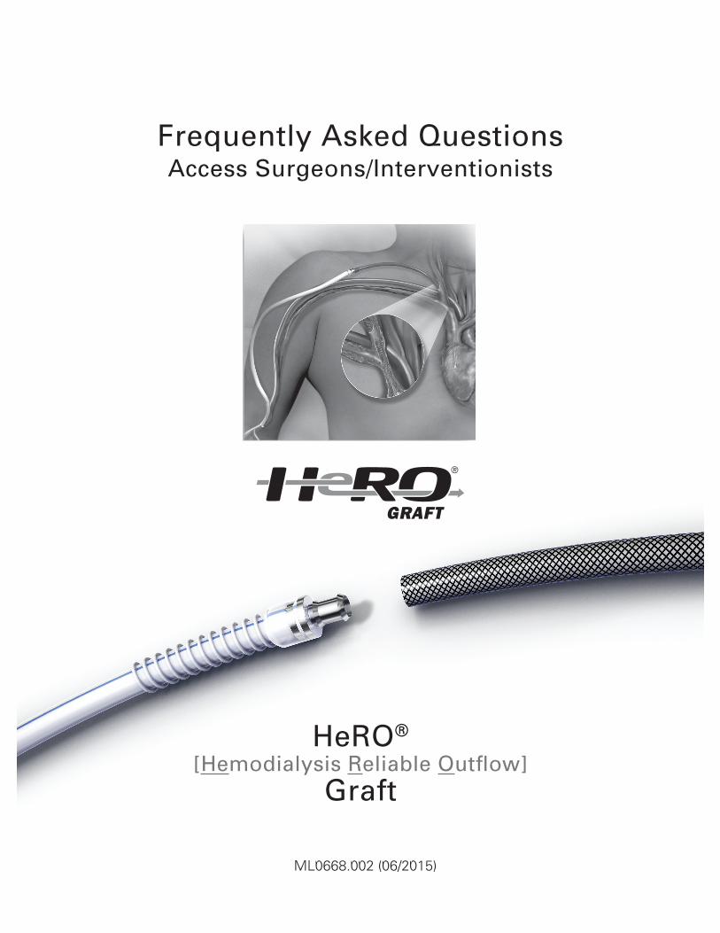
Frequently Asked Questions
HeRO®
[Hemodialysis Reliable Outflow]Graft
Medical Professionals
ML0668.002 (06/2015)
2HeRO Graft FAQ - Medical Professionals 13-0061 Rev. B 2012-10
Only qualified healthcare providers should place, manipulate, de-clot, revise or explant the HeRO Graft. For greater detail, please consult the HeRO Graft Instructions for Use manual: www.herograft.com
Q What is the HeRO Graft?A The HeRO Graft is a fully subcutaneous vascular access system that bypasses the
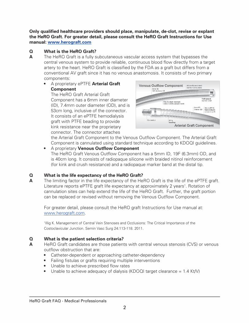
central venous system to provide reliable, continuous blood flow directly from a target artery to the heart. HeRO Graft is classified by the FDA as a graft but differs from a conventional AV graft since it has no venous anastomosis. It consists of two primary components:• A proprietary ePTFE Arterial Graft
Component The HeRO Graft Arterial Graft Component has a 6mm inner diameter (ID), 7.4mm outer diameter (OD), and is 53cm long, inclusive of the connector. It consists of an ePTFE hemodialysis graft with PTFE beading to provide kink resistance near the proprietary connector. The connector attaches the Arterial Graft Component to the Venous Outflow Component. The Arterial Graft Component is cannulated using standard technique according to KDOQI guidelines.
• A proprietary Venous Outflow Component The HeRO Graft Venous Outflow Component has a 5mm ID, 19F (6.3mm) OD, and is 40cm long. It consists of radiopaque silicone with braided nitinol reinforcement (for kink and crush resistance) and a radiopaque marker band at the distal tip.
Q What is the life expectancy of the HeRO Graft?A The limiting factor in the life expectancy of the HeRO Graft is the life of the ePTFE graft.
Literature reports ePTFE graft life expectancy at approximately 2 years1. Rotation of cannulation sites can help extend the life of the HeRO Graft. Further, the graft portion can be replaced or revised without removing the Venous Outflow Component. For greater detail, please consult the HeRO graft Instructions for Use manual at: www.herograft.com. 1Illig K. Management of Central Vein Stenoses and Occlusions: The Critical Importance of the
Costoclavicular Junction. Semin Vasc Surg 24:113-118. 2011.
Q What is the patient selection criteria?A HeRO Graft candidates are those patients with central venous stenosis (CVS) or venous
outflow obstruction that are:• Catheter-dependent or approaching catheter-dependency• Failing fistulas or grafts requiring multiple interventions• Unable to achieve prescribed flow rates• Unable to achieve adequacy of dialysis (KDOQI target clearance = 1.4 Kt/V)
3HeRO Graft FAQ - Medical Professionals 13-0061 Rev. B 2012-10
Q How is patient surgical suitability determined?A The following surgical assessments should be completed prior to initiating the implant
procedure:• Central venography to confirm central venous stenosis• Vessel mapping to confirm artery inner diameter ≥3mm for arterial anastomosis• Verify the ejection fraction is >20%• Verify systolic blood pressure is ≥100mmHg• Obtain screening blood cultures to rule out existing systemic infection. In the event
blood cultures are positive, postpone implant of the HeRO Graft pending antibiotic treatment and confirmation of infection resolution.
• Medically managed for hypercoagulation• Swab the patient’s nose prior to HeRO Graft implant for potential methicillin
resistant staphylococcus aureus; treat accordingly.
Patient surgical suitability information can also be found in the Instructions For Use manual and available for download at www.herograft.com.
Q How extensive is the implant surgery?A The placement is similar to both tunneled cuff catheter placement and graft placement.
It may require angioplasty of the central veins prior to placement of the Venous Outflow Component. The HeRO Graft implant can be done under general anesthesia or conscious sedation. The average procedure time is 60-90 minutes. Complete implant instructions are provided in the Instructions for Use manual available at www.herograft.com. See also the HeRO Graft Implant Video available at www.herograft.com.
Q Where is the device intended to be implanted?A Use of the HeRO Graft was clinically studied in the upper extremity utilizing the internal
jugular vein. Central venous access through any other veins has NOT been studied and may increase the risk of adverse events not encountered in the clinical trial. Surgeons must use discretion in selecting appropriately sized target vessels to accommodate the 6.3mm outer diameter of the Venous Outflow Component based on each patient’s anatomy.
Q Are there any contraindications for placement of a HeRO Graft?A Implantation of the HeRO Graft is contraindicated if:
• The brachial or target artery inner diameter (ID) is less than 3mm• The internal jugular vein (IJV) or target vasculature cannot be dilated to
accommodate the 19F Venous Outflow Component• There is significant arterial occlusive disease that would preclude safe placement of
an upper extremity hemodialysis access• There is known or suspected allergy to device materials (i.e., ePTFE, silicone,
titanium, nitinol)• The patient has a topical or subcutaneous infection associated with the implantation
site• The patient has known or suspected systemic infection, bacteremia, or septicemia
4HeRO Graft FAQ - Medical Professionals 13-0061 Rev. B 2012-10
Q How long is the incorporation period after surgical implantation?A Follow the KDOQI graft guidelines of 2-4 weeks for incorporation of the ePTFE graft
material before cannulating the HeRO Graft.
Q Should an antibiotic regimen be continued after a HeRO Graft implant?A When Vancomycin and Gentamycin are prescribed during the implant, the antibiotics
will stay in the patient’s system for 7 days without dialyzing out during their dialysis treatments. If a patient has a history of chronic infection, the nephrologist might consider keeping the patient on an antibiotic course until the bridging catheter is removed. Additional considerations to reduce the risk of infection, especially during the bridging period, include:• Apply antibiotic ointment to the bridging catheter exit site.• Remove the bridging catheter as soon as possible once the HeRO Graft is being
cannulated to decrease the risk of an infection related to the bridging catheter. During clinical trials, the only infections seen were during the bridging period1.
• All bridging catheters should be cultured upon explant. In the event that the catheter tip cultures are positive, treat the patient with appropriate antibiotics to decrease the risk of the HeRO Graft becoming infected.
1Katzman H. Initial experience and outcome of a new hemodialysis access device for catheter-dependant patients. J. Vasc. Surg. 2009;50:600-7.
Q Will the HeRO Graft clot?A The HeRO Graft patency was clinically proven to be equivalent to a standard ePTFE
graft. The expected intervention rate is approximately 2 per year. Additional clinical trial information and a comparison of HeRO Graft to other AV accesses can be viewed at: www.herograft.com. As with conventional AV grafts, HeRO Graft may occlude in patients with:• A small brachial artery (e.g., inner diameter less than 3mm)• Insufficient arterial inflow or inflow stenosis• A history of clotted accesses for unknown reasons• A coagulability disorder or medical condition that is associated with clotting (i.e.,
cancer)• Insufficient anticoagulation or non-compliance with anticoagulation medication • Systemic low blood pressure or severe hypotension following fluid removal post
dialysis • A kinked graft• Incomplete thrombus removal in previous interventions • Intra-graft stenosis at site of multiple punctures• An event such as mechanical compression (i.e., spring loaded hemostasis clamps)
Q Does fibrin sheath build on the HeRO Graft?A No fibrin sheath formation is expected around the tip of the Venous Outflow
Component due to continuous blood flow from a target artery to the central venous system and into the heart.
5HeRO Graft FAQ - Medical Professionals 13-0061 Rev. B 2012-10
Q Is anticoagulant or antiplatelet therapy (such as Plavix®) required after an implant? If so, for how long?
A Patients on therapeutic doses of anticoagulation should have PTT and drug levels checked per standard treatment course prior to a HeRO Graft implant, and anticoagulants such as COUMADIN® or antiplatelets such as Plavix® prescribed prior to the HeRO Graft implant should continue after surgery. Special consideration may be needed to prevent occlusion in HeRO Graft patients, after surgery, since the HeRO Graft is a continuous flow system. Many physicians prescribe a course of proactive prophylaxis for the first 30 days and then re-evaluate.
• Place the HeRO Graft patient on therapeutic dose of a drug such as: ° Clopidogrel (Plavix®) starting at time of implant ° Low dose aspirin daily• COUMADIN® dose to maintain INR levels of 2.7 to 3.0 as tolerated by patient ° Maintain blood pressure at 100 systolic with fluids or pressors ° Use drugs such as Midrodrine or Florinef
Q What happens if the Venous Outflow Component of the HeRO Graft is mistakenly punctured?
A The HeRO Graft Venous Outflow Component may need to be replaced depending on the location and extent of damage if punctured. Puncture of the Venous Outflow Component is highly unlikely due to its location. Accidentally puncturing the Venous Outflow Component can be avoided by locating the Arterial Graft Component connector and cannulating 3 inches or more from the connector to avoid the Venous Outflow Component and graft beading near the connector region. Additional information is available in the Care and Cannulation guide at www.herograft.com.
Q In what situations will the HeRO Graft be removed?A If the HeRO Graft is abandoned for any reason, we recommend removal of the Venous
Outflow Component. The Arterial Graft Component would typically not be removed due to maturation/incorporation of surrounding tissue into the ePTFE material. It can be ligated and left in place similar to conventional AV grafts. For greater detail, please consult the HeRO graft Instructions for Use manual at www.herograft.com.
6HeRO Graft FAQ - Medical Professionals 13-0061 Rev. B 2012-10
Q What complications are seen with the HeRO Graft?A Complications may occur at any time during or after the endovascular portion of the
procedure. Possible complications include, but are not limited to:• Bleeding• Embolism• Hematoma • Infection• Trauma to major vasculature• Cardiac arrhythmia ° To avoid vessel or heart damage or cardiac arrhythmia, special care must be
taken with:• Guidewire ° Assure correct location of the guidewire tip in the inferior vena cava (IVC)
throughout the entire procedure using fluoroscopy ° Stabilize the guidewire in the IVC while advancing the dilator sheath ° Use of stiff guidewires requires extra care to prevent perforation during
advancement and manipulation• Dilators ° Consider balloon angioplasty for serial dilation• Delivery Stylet ° The stylet is not visible under fluoroscopy
DO NOT advance the tip of the delivery stylet into the right atrium DO NOT advance the delivery stylet independently of the Venous
Outflow Component
To reduce the risk of complications, perform vessel mapping in advance to ensure appropriate vessel size and location when determining an optimum strategy for treatment. Be aware that cannulation of the left internal jugular vein (IJV) may increase the risk of complications when compared to the right IJV.
Q What is the best treatment for steal syndrome after the HeRO Graft implant?A Steal syndrome is possible just as with any graft. Considerations to decrease the risk of
steal syndrome include:• Confirm the artery has a minimum inner diameter (ID) of 3mm with vessel mapping
(angiography or duplex ultrasonography) prior to implant.• Confirm with angiography or duplex ultrasonography that there is no significant
arterial occlusive disease on the implant side that would preclude safe placement of an upper extremity access.
Evaluate for steal syndrome during the implant procedure by checking distal pulses with Doppler of the radial and ulnar arteries. If steal syndrome symptoms occur, consider the following:• DRIL (distal revascularization-interval ligation) procedure1 • Banding, though this may reduce the flow in the HeRO Graft• Proximalization of the inflow• Explant the HeRO Graft if other corrective measures are not successful
1Knox RC, Berman SS, Hughes JD, Gentile AT, Mills JL. Distal revascularization-interval ligation: a durable and effective treatment for ischemic steal syndrome after hemodialysis access. J. Vasc. Surg.2002;36(2):250-5.
7HeRO Graft FAQ - Medical Professionals 13-0061 Rev. B 2012-10
Q What is the best way to prevent infection with the HeRO Graft?A The following considerations are suggested to reduce the risk of bacteremia with the
HeRO Graft: Pre-Implant• Obtain screening blood cultures to rule out asymptomatic bacteremia prior to HeRO
Graft implant for any patient dialyzing on a catheter; treat patient with antibiotics per culture outcome and ensure infection is resolved prior to HeRO Graft implant procedure.
• Swab the patient’s nose prior to HeRO Graft implant for potential methicillin resistant staphylococcus aureus; treat accordingly.
Implant and Bridging-Period• Prophylactically treat the patient in the peri-operative period with antibiotics based
upon the patient’s bacteremia history: ° Ancef® or combination Vancomycin and Gentamycin for native stick Venous
Outflow Component placement ° Vancomycin and Gentamycin for over-the-wire exchange of a tunneled cuffed
dialysis catheter ° Vancomycin and Gentamycin for femoral catheter placement and atypical HeRO
Graft placement• If an existing catheter is removed, suture the tract closed from the existing catheter
prior to placing the Venous Outflow Component. • Consider covering any catheter extensions with an antimicrobial incise drape to
protect the sterile area.• Plan for increased bacteremia risk after an ipsilateral HeRO Graft placement or
with femoral bridging catheters and treat prophylactically with antibiotics knowing patients are at higher infection risk.
• Culture any catheters removed at the time of HeRO Graft implant.• Apply antibiotic ointment to the bridging catheter exit site.• Remove the bridging catheter as soon as possible once the HeRO Graft is being
cannulated to decrease the risk of an infection related to the bridging catheter.• All bridging catheters should be cultured upon explant. In the event catheter tip
cultures are positive, treat the patient with appropriate antibiotics to decrease the risk of the HeRO Graft becoming infected.
Q Why does the patient’s blood pressure need to be over 100 systolic?A Occlusion is potentially a problem in HeRO Graft patients with low blood pressure and
ejection fractions due to HeRO Graft being a continuous flow system that provides reliable, continuous blood flow directly from a target artery to the central venous system and into the heart.
Q When was the HeRO Graft first available commercially?A After FDA Clearance, the first HeRO Graft kit sale occurred in April 2008, and the first
implant was in May 2008.
8HeRO Graft FAQ - Medical Professionals 13-0061 Rev. B 2012-10
Please see the Instructions for Use document provided in the HeRO Graft packaging and online at www.herograft.com for the full list of anticipated adverse events, contraindications, and complications.
The FDA regulation name for the HeRO Graft is vascular graft prosthesis.
INDICATIONS FOR USE: The HeRO Graft is indicated for end stage renal disease patients on hemodialysis who have ex-hausted all other access options. See instructions for use for full indication, contraindication and caution statements. Rx only.
CryoLife, the snowflake design and Life Restoring Technologies are trademarks owned by CryoLife, Inc. HeRO and Hemosphere are trademarks owned by Hemosphere, Inc. All other trade-marks are owned by their respective owners. © 2012 CryoLife, Inc. All rights reserved.
CryoLife, Inc.1655 Roberts Boulevard, NW Kennesaw, Georgia 30144United States
Customer Service888-427-9654 phone770-590-3753 faxwww.herograft.com
Q When will the HeRO Graft be sold internationally?A The HeRO Graft is not approved for sale outside of the United States. However, a
“Special Access Program” is available in Canada in certain cases. Please contact CryoLife customer service at 888-427-9654 for more information on this process.
Q Is it safe for a patient with the HeRO Graft to have an MRI?A The HeRO Graft was determined to be MR-conditional. A patient may be scanned safely
immediately after HeRO Graft placement under the following conditions:• Static magnetic field of 3-Tesla or less• Spatial gradient magnetic field of 720-Gauss/cm or less
See the full Instructions for Use manual at www.herograft.com for additional information regarding MRI safety, heating and artifact details.
Q How can I reach Customer Service to place an order for the HeRO Graft or ask a question?
A Customer service can be reached at the following contact information: Phone: 888-427-9654 Fax: 770-590-3753 Email: [email protected] Web: http://www.herograft.com/contact-us/
Please see the Instructions for Use document provided in the HeRO Graft packaging and online at www.herograft.com forthe full list of anticipated adverse events, contraindications, andcomplications.
The FDA regulation name for the HeRO Graft is vascular graft prosthesis.
HeRO, CryoLife, the snowflake design, and Life Restoring Tech-nologies are trademarks owned by CryoLife, Inc. All other trade-marks are owned by their respective owners. © 2015 CryoLife, Inc. All rights reserved.
Frequently Asked Questions
HeRO®
[Hemodialysis Reliable Outflow]Graft
Access Surgeons/Interventionists
ML0668.002 (06/2015)
2HeRO Graft FAQ - Access Surgeons/Interventionists 13-0061 Rev. B 2012-10
Only qualified healthcare providers should place, manipulate, de-clot, revise or explant the HeRO Graft. For greater detail, please consult the HeRO Graft Instructions for Use manual: www.herograft.com
Q Is there Medicare reimbursement for the HeRO Graft? How does the reimbursement rate compare to grafts?
A The Arterial Graft Component is reported using CPT® 36830. The Venous Outflow Component is reported with CPT® 36558 (procedure-to-device edit C1750, Catheter, Hemodialysis, Long-term). Please visit www.herograft.com for further details.
Q Is special training required to implant the HeRO Graft?A The physician must be trained in both surgical and endovascular skills and have
privileges to include interventional wires, balloon angioplasty and endovascular stent placements. The Venous Outflow Component is placed endovascularly under fluoroscopy. Consider a consult or assistance from an interventional radiologist or interventional nephrologist regarding central venous access in patients with severe stenosis, occlusion or tortuous vessels. Each access surgeon is typically proctored by a trained CryoLife representative with a minimum of 3 cases prior to commencing independent implantation of the device. See also the HeRO Graft Implant Procedure section available at www.herograft.com.
Q Is the HeRO Graft sutured for stabilization?A The Arterial Graft Component is anastamosed to the artery and will incorporate like a
standard ePTFE graft. The Venous Ouflow Component is connected to the ePTFE graft via a connector. No other forms of stabilization are required.
Q Where is the temporary bridging catheter typically placed?A Approximately 60% of the clinical trial patients had a femoral bridging catheter due
to presence of pacemaker leads, defibrillator leads or stenotic vessels in the chest. It is preferable to place the HeRO Graft in one side of the chest and the tunneled cuff catheter on the opposite side or contralaterally. Femoral catheters may carry a higher risk of infection, but can be used if necessary during the graft maturation period.
Q Are there known differences in the occlusion rates in left versus right-sided HeRO Graft implants?
A There are no known differences in occlusion rates in left vs. right-sided implants. Be aware that cannulation of the LEFT internal jugular vein (IJV) may increase the risk of complications when compared to the right IJV1. There also may be risk of device tip movement with left-sided implants. 1Sulek CA, Blas ML, Lobato EB. A randomized study of left versus right internal jugular vein cannulation in adults. J Clin Anesth. 2000 Mar; 12(2):142-5.
3HeRO Graft FAQ - Access Surgeons/Interventionists 13-0061 Rev. B 2012-10
Q Are there any specific methods or training that must be used when performing a thrombectomy on HeRO Graft?
A Percutaneous or surgical technique may be used to declot the HeRO Graft. A surgical technique is recommended during the graft incorporation period to avoid risk of seroma or other complications. Percutaneous technique is recommended after the graft is completely incorporated using a rheolytic thrombectomy system, balloon maceration, or balloon-assisted aspiration. A 90cm thrombectomy device is required to accommodate the entire length of the HeRO Graft. Mechanical/rotational devices (e.g., Arrow-Trerotola PTD®) are contraindicated as internal damage to the Venous Outflow Component and connector may occur. After the thrombectomy procedure, administration of drugs such as tPA or urokinase to lyse any residual thrombus is recommended. A Thrombectomy Guidelines brochure can be found in the Document Library or watch the Thrombectomy Video located in the www.herograft.com.
Q What surgical equipment is needed for the HeRO Graft implant?A The HeRO Graft is implanted in a standard operating room equipped for a vascular
access procedure with fluoroscopy and ultrasound guidance. In addition to the Accessory Component Kit, some vascular access surgical instruments may be required, such as: • 5F micro-puncture set• various 0.035” guidewires at least 150cm in length• heavy duty scissors• heparinized saline• 4 x 4 sterile gauze pads• various subcutaneous tissue and skin sutures• radiographic contrast fluid• tunneler set with 6mm and 7mm bullet tips• various atraumatic vascular clamps• standard vessels loops• syringe and syringe adapter• sterile surgical lubricant
Q Can the HeRO Graft be implanted in the femoral area?A Use of the HeRO Graft was clinically studied in the upper extremity using the internal
jugular vein for venous access. Implantation of the device in other vasculature, for example, the femoral vein, has NOT been studied and may increase the risk of adverse events not encountered in the clinical trial. Surgeons need to use their discretion in selecting appropriately-sized target vessels to accommodate the 6.3mm outer diameter of the Venous Outflow Component based on each individual patient’s anatomy. Also, consider using Vancomycin and Gentamycin for femoral placement.
4HeRO Graft FAQ - Access Surgeons/Interventionists 13-0061 Rev. B 2012-10
Q Can the HeRO Graft be implanted in the subclavian vein?A Use of the HeRO Graft was clinically studied utilizing the internal jugular vein.
Implantation of the device in other vasculature, for example, the subclavian vein, has NOT been studied and may increase the risk of adverse events not encountered in the clinical trial. When using the subclavian vein for venous access, a more lateral percutaneous approach might mitigate the risk of clavicle crush or occlusion of the HeRO Graft Venous Outflow Component1. Consideration should be made to follow these patients with clavicle imaging to monitor the potential of interaction of the clavicle and first rib with the Venous Outflow Component. 1Illig KA. Management of Central Vein Stenosis and Occlusions: The Critical Importance of the Costoclavicular Junction. Semin Vasc Surg 24:113-118, 2011.
Q Can the HeRO Graft be attached to an existing fistula or graft?A The HeRO Graft can be attached to any existing fistula or graft in an end-to-end fashion
making sure to keep the Arterial Graft Component’s PTFE beading and connector intact for connecting to the Venous Outflow Component. See commercial experience online at www.herograft.com, specifically Successful Use of HeRO Device to Salvage a Functional Arteriovenous Fistula In EJVES by Dr. Chen.
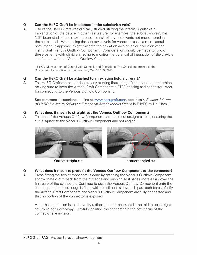
Q What does it mean to straight cut the Venous Outflow Component?A The end of the Venous Outflow Component should be cut straight across, ensuring the
cut is square to the Venous Outflow Component and not angled.
Correct straight cut Incorrect angled cut
Q What does it mean to press fit the Venous Outflow Component to the connector?A Press fitting the two components is done by grasping the Venous Outflow Component
approximately 2cm back from the cut edge and pushing so it slides more easily over the first barb of the connector. Continue to push the Venous Outflow Component onto the connector until the cut edge is flush with the silicone sleeve hub past both barbs. Verify the Arterial Graft Component and Venous Outflow Component are fully connected and that no portion of the connector is exposed. After the connection is made, verify radiopaque tip placement in the mid to upper right atrium using fluoroscopy. Carefully position the connector in the soft tissue at the connector site incision.
5HeRO Graft FAQ - Access Surgeons/Interventionists 13-0061 Rev. B 2012-10
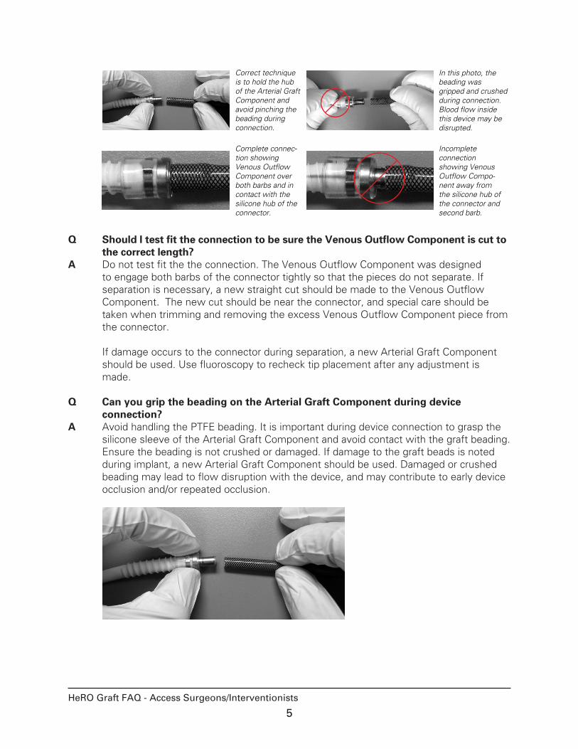
Correct technique is to hold the hub of the Arterial Graft Component and avoid pinching the beading during connection.
Complete connec-tion showing Venous Outflow Component over both barbs and in contact with the silicone hub of the connector.
In this photo, the beading was gripped and crushed during connection. Blood flow inside this device may be disrupted.
Incomplete connection showing Venous Outflow Compo-nent away from the silicone hub of the connector and second barb.
Q Should I test fit the connection to be sure the Venous Outflow Component is cut to the correct length?
A Do not test fit the the connection. The Venous Outflow Component was designed to engage both barbs of the connector tightly so that the pieces do not separate. If separation is necessary, a new straight cut should be made to the Venous Outflow Component. The new cut should be near the connector, and special care should be taken when trimming and removing the excess Venous Outflow Component piece from the connector. If damage occurs to the connector during separation, a new Arterial Graft Component should be used. Use fluoroscopy to recheck tip placement after any adjustment is made.
Q Can you grip the beading on the Arterial Graft Component during device connection?
A Avoid handling the PTFE beading. It is important during device connection to grasp the silicone sleeve of the Arterial Graft Component and avoid contact with the graft beading. Ensure the beading is not crushed or damaged. If damage to the graft beads is noted during implant, a new Arterial Graft Component should be used. Damaged or crushed beading may lead to flow disruption with the device, and may contribute to early device occlusion and/or repeated occlusion.
6HeRO Graft FAQ - Access Surgeons/Interventionists 13-0061 Rev. B 2012-10
Q How is the HeRO Graft explant procedure performed?A The Venous Outflow Component and connector do not incorporate into the surrounding
tissue and may be removed using manual traction similar to a conventional hemodialysis catheter. If thrombus is present, it may be dislodged during the explant procedure and therefore should be treated using a thrombolytic agent, or other appropriate therapy, prior to performing the explant procedure. To Explant the HeRO Graft Venous Outflow Component and Connector:1. Prep patient using aseptic surgical technique. 2. Open the incision at the deltopectoral groove (DPG) and dissect to expose at least
5cm of the graft, including the connector and PTFE beading.3. Carefully dissect the exposed graft and connector to free the incorporated material
for ease of revision.4. Ligate the graft approximately 1cm away from the PTFE beading.5. Cut the graft component between the ligation and the PTFE beading to separate the
Venous Outflow Component.6. Gently twist to loosen the Venous Outflow Component with attached connector.
Using appropriate technique, (i.e., syringe) apply negative pressure to remove potential intraluminal thrombus.
7. Pull gently using counter pressure applied at the original venous incision site until the Venous Outflow Component and connector is fully removed.
Caution: Upon removing the Venous Outflow Component and connector, continue applying pressure at the original venous incision site to decrease risk of bleeding.
8. After removal of the components, close the DPG incision site.
General Cautions:• During removal of the Venous Outflow Component, special care should
be used if there is a stent in the vessel. Use imaging (fluoroscopy) for visualization of the Venous Outflow Component and stent interaction to decrease the potential of Venous Outflow Component, stent, or vessel damage.
• Only qualified healthcare providers should explant the device.• Adhere to universal precautions when explanting the device.• The HeRO Graft has been in contact with body fluids and is a potential
biohazard. Handle the device using acceptable medical practice and applicable local, state and federal laws and regulations. Return the explanted portion of the device to CryoLife using the Explant Return Kit by contacting Customer Service.
Complete implant instructions are provided in the Instructions for Use manual available in the www.herograft.com.
Q Can the HeRO Graft be implanted in the same vein as a pacemaker lead, defibrillator, or bridging catheter?
A The Instructions for Use lists as a caution NOT to implant the HeRO Graft in the same vessel as a bridging catheter, defibrillator or pacemaker lead. Interaction of the Venous Outflow Component with another device in the same vessel is unknown.
7HeRO Graft FAQ - Access Surgeons/Interventionists 13-0061 Rev. B 2012-10
Q Can the HeRO Graft be made into an early cannulation access?A The HeRO Graft can be attached to any other graft material (such as Acuseal®or Vectra®)
in an end-to-end fashion making sure to keep the Arterial Graft Component’s PTFE beading and connector intact for connection to the Venous Outflow Component.
Q Is there a tapered graft option for the HeRO Graft?A There is no tapered graft, but the Arterial Graft Component can be cut at an angle to
create a more gradual transition at the arterial anastomosis. An alternative option is to perform an end-to-end anastomosis of the Arterial Graft Component to a tapered graft.
Q Can the HeRO Graft be implanted through a stented vessel?A The HeRO Graft was not implanted through any stented vessels during the clinical trial,
so the interaction of the HeRO Graft with stents is unknown. Special care should be used if the Venous Outflow Component is explanted and may come into contact with a stented vessel during the explant procedure. Use imaging (fluoroscopy) for visualization of the Venous Outflow Component and stent interaction to decrease the potential of Venous Outflow Component, stent, or vessel damage.
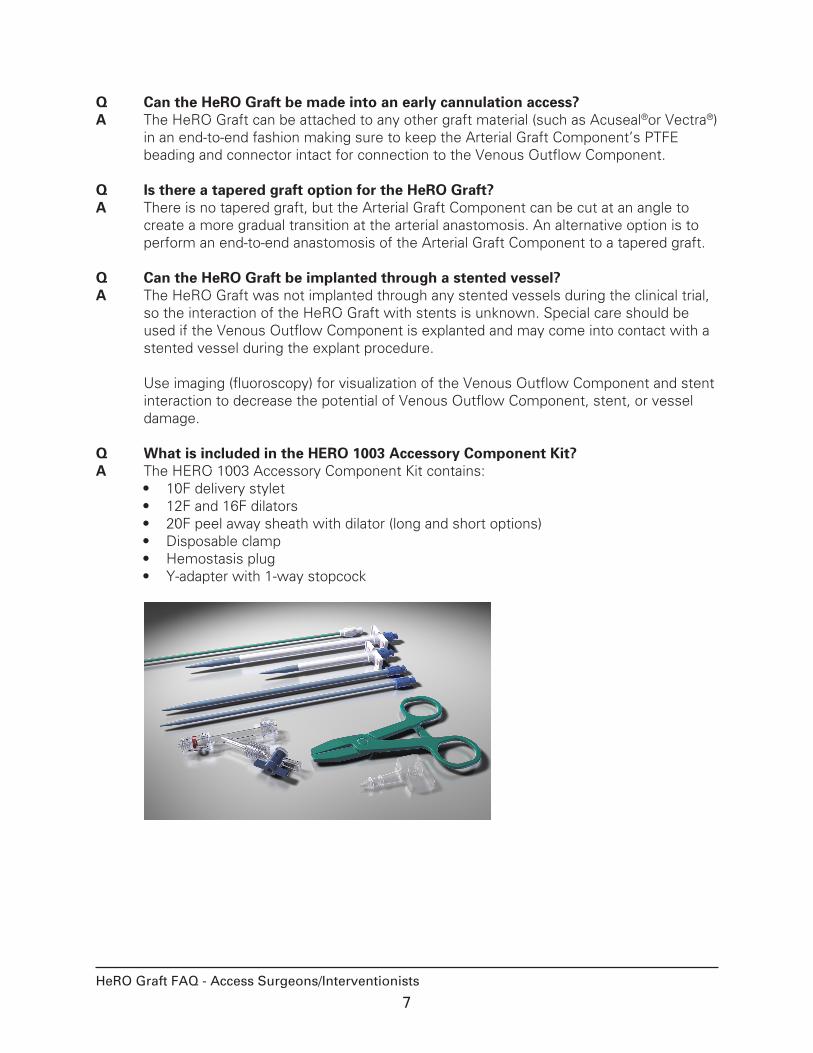
Q What is included in the HERO 1003 Accessory Component Kit?A The HERO 1003 Accessory Component Kit contains:
• 10F delivery stylet• 12F and 16F dilators• 20F peel away sheath with dilator (long and short options)• Disposable clamp • Hemostasis plug• Y-adapter with 1-way stopcock
8HeRO Graft FAQ - Access Surgeons/Interventionists 13-0061 Rev. B 2012-10
Q Do I need to put a suture around the section where the outflow component connects to the connector?
A Suturing the connection is not needed. The HeRO Graft is designed not to separate if properly connected. This is due to its unique design features, which include two barbs on the connector of the Arterial Graft Component and the nitinol reinforcement braid of the Venous Outflow Component. After cutting the Venous Outflow Component to length and discarding the silicone Luer End, the cut end of the nitinol braid engages both barbs of the connector tightly and should not separate once fully connected, that is the Venous Outflow Component is in contact with the center hub of the connector. Also, the compatibility of the Venous Outflow Component has not been tested with suture materials.
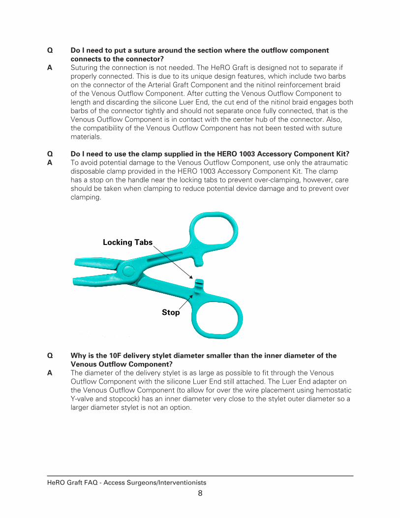
Q Do I need to use the clamp supplied in the HERO 1003 Accessory Component Kit?A To avoid potential damage to the Venous Outflow Component, use only the atraumatic
disposable clamp provided in the HERO 1003 Accessory Component Kit. The clamp has a stop on the handle near the locking tabs to prevent over-clamping, however, care should be taken when clamping to reduce potential device damage and to prevent over clamping.
Locking Tabs
Stop
Q Why is the 10F delivery stylet diameter smaller than the inner diameter of the Venous Outflow Component?
A The diameter of the delivery stylet is as large as possible to fit through the Venous Outflow Component with the silicone Luer End still attached. The Luer End adapter on the Venous Outflow Component (to allow for over the wire placement using hemostatic Y-valve and stopcock) has an inner diameter very close to the stylet outer diameter so a larger diameter stylet is not an option.
9HeRO Graft FAQ - Access Surgeons/Interventionists 13-0061 Rev. B 2012-10
Q Will the Venous Outflow Component move?A Since there is no venous anastomosis, the HeRO Graft Venous Outflow Component has
freedom of movement. This movement could result in the tip not remaining in the right atrium of the heart which may increase the risk of occlusion. This may be more of a concern in:• Left-sided implants• Subclavian vein access (use extra caution due to the increased risk of patient
complications)• Morbidly obese patients• Graft connector located lateral to the deltopectoral groove
Special consideration may be needed if an implant or revision is scheduled in a morbidly obese patient and/or a subclavian or left-sided HeRO Graft implant. In an effort to reduce the amount of movement of the Venous Outflow Component in these patients, consider:• Placing the tip of the Venous Outflow Component in the mid or lower right atrium• Placing the HeRO Graft connector medial (mid-clavicular) to the deltopectoral groove• Suturing the HeRO Graft connector to patient’s fascia to hold it in place during the
incorporation period• Performing fluoroscopy of the HeRO Graft tip while moving the patient’s arm as
much as possible (rotate at shoulder, adduct arm to side) after connecting the Arterial Graft Component and Venous Outflow Component
• Obtaining an upright chest X-ray to verify location of the marker band at the tip of the HeRO Graft in the patient’s heart
Q How is HeRO Graft revision performed?A The HeRO Graft Arterial Graft Component can be revised if necessary via a jump graft
procedure. If graft revision is necessary due to infection, resection and removal of the infected portion of the graft is required prior to completing the jump graft procedure. Return the excised portion of the graft to CryoLife. Follow the instructions for the jump graft procedure as detailed below. If damage occurs to the PTFE beading on the existing graft, replace the entire Arterial Graft Component including the connector. Replacement of the Arterial Graft Component will also require revision to the Venous Outflow Component and should be attended by a CryoLife representative. Contact Customer Service for your local representative.
To Revise the HeRO Graft Arterial Graft Component:1. Create incisions at the sites selected for the graft-to-graft anastomosis and dissect
to expose the existing graft. Caution: DO NOT peel or otherwise damage the graft beading as this may
adversely impact the integrity of the existing graft.2. Create a subcutaneous tunnel from new inflow incision site to the new outflow
incision site circumventing the existing graft. Graft routing may vary depending on patient-specific anatomy and the placement of the existing graft.
10HeRO Graft FAQ - Access Surgeons/Interventionists 13-0061 Rev. B 2012-10
3. Using standard graft tunneling techniques, gently pull the jump graft through the new tunnel. Utilize markings on the graft to verify it has not twisted.
4. Use a standard vascular clamp to occlude the existing graft near the new inflow anastomosis site.
5. Perform a standard graft-to-graft anastomosis.6. Remove the clamp, bleed the jump graft segment to remove air, and then reclamp
the jump graft segment next to the new outflow anastomosis site.7. Cut the graft to length, avoiding excessive tension or redundant graft material,
and perform the outflow anastomosis of the jump graft to the existing graft using standard technique.
8. Remove the clamp and check the device patency, utilizing standard Doppler technique.
9. Close both incisions.
If explanting a segment of the device, return using the Explant Return Kit by contacting CryoLife Customer Service.
Caution: Use only the disposable clamp included in the Accessory Component Kit. Use of other clamps may result in damage to the device. Be careful not to overclamp the disposable clamp.
Complete implant instructions are provided in the Instructions for Use manual available at www.herograft.com.
Q How is the Venous Outflow Component exchange procedure performed?A The Venous Outflow Component does not incorporate into venous anatomy and can
be removed or exchanged. Fluoroscopy is required during insertion of a new Venous Outflow Component to avoid vessel damage and ensure proper placement. Due to the complexity and permutations of this procedure, exchanges should be attended by a CryoLife representative. Contact Customer Service for your local representative. Tools Required:• HERO 1001 Venous Outflow Component• HERO 1003 Accessory Component Kit• 0.035” guidewire at least 150cm in length
Recommended Accessories:• Stiffened 5F Micropuncture® Introducer Set (such as Cook Inc. MPIS-501-10.0-SC-
NT-U)• Heavy duty scissors, such as Vantage® Iris Scissors 4-1/8 Ref#V95-304• Explant Return Kit. Request from CryoLife Customer Service
11HeRO Graft FAQ - Access Surgeons/Interventionists 13-0061 Rev. B 2012-10
Venous Outflow Component Exchange Procedure:1. Prep the patient according to standard surgical guidelines. Place the patient into
Trendelenberg position to reduce the potential for air embolus during exchanges. For patients undergoing general anesthesia, a positive breath can be forced during removal of the dilator from the sheath to prevent air induction.
2. Prepare the 5F microintroducer by removing the 0.014” wire-compatible dilator and securely attaching the sheath to the Y-adapter (from the Accessory Component Kit). Flush the sheath with heparanized saline via the Luer port.
3. Palpate to locate the device connector. Open the deltopectoral groove (DPG) incision to expose the PTFE graft rings and at least 5cm of the Venous Outflow Component.
4. Clamp the graft with an atraumatic vascular clamp near the PTFE graft beading. Inject the graft with heparinized saline to maintain patency.
Caution: Do not clamp the PTFE beading as damage to the beading may result. If damage occurs, replacement of the Arterial Graft Component is recommended.
5. Palpate the venous access site to confirm location of the Venous Outflow Component. Open the previous incision and expose the Venous Outflow Component nearest the point it enters/exits the vein.
6. Create a purse string suture at the venous access site and clamp the Venous Outflow Component nearest the point it enters/exits the vein.
7. Place 4x4 gauze under the connector to prevent debris from contaminating the incision site.
8. Ensure both clamps are secure and cut the Venous Outflow Component with a pair of heavy duty scissors approximately 3cm from the connector.
9. Using the heavy duty scissors, cut the remainder of the Venous Outflow Component from the connector starting at the connector shoulder and working toward the cut end.
Caution: Cutting through the nitinol braiding may be difficult. Do not damage the barbs on the connector. If damage occurs, replacement of the connector with a new Arterial Graft Component is recommended.
10. Once completed, remove the 4x4 gauze and inspect the wound for any potential debris left behind. Replace the gauze and continue the procedure. Note: Alternately, it may be possible to twist and pull the Venous Outflow Component until it can be removed from the connector without cutting. This may be a slow and time-consuming process.
Caution: Do not crush or otherwise damage the beading on the Arterial Graft Component. If damage occurs, replacement of the Arterial Graft Component is recommended.
11. At the venous access site, gently pull the Venous Outflow Component through the tunneled tract. Do not move or displace the tip of the Venous Outflow Component in the right atrium.
12. Insert the assembled 5F sheath into the exposed end of the Venous Outflow Component. Ensure that the hub is securely seated in the Venous Outflow Component, and remove the clamp.
13. Aspirate blood from the device. Use fluoroscopy while advancing the guidewire to the desired position in the inferior vena cava.
14. Maintaining wire position, gently remove the existing Venous Outflow Component over the wire. The purse string suture can help control bleeding at the venous
12HeRO Graft FAQ - Access Surgeons/Interventionists 13-0061 Rev. B 2012-10
access site.15. Load the 20F peel away sheath onto the guidewire and use fluoroscopy to advance.16. Withdraw the dilator and use the silicone hemostasis plug to occlude the sheath
opening, leaving the guidewire in place. Ensure both plug seal rings are fully seated within the sheath. Avoid pinching or clamping the sheath.
17. Remove the Y-adapter from the 5F micropuncture assembly and attach to the Luer End of the new Venous Outflow Component.
18. Advance the Venous Outflow Component over the guidewire. Remove the hemostasis plug and advance the Venous Outflow Component into the 20F sheath. Use fluoroscopy to advance the Venous Outflow Component to the superior vena cava. A twisting or rotational motion may be used to ease insertion. Surgical lubricant may be used, if necessary.
19. Place the radiopaque tip of the Venous Outflow Component in the mid to upper right atrium and use fluoroscopy to confirm proper tip placement.
20. Peel away the 20F sheath. Clamp the Venous Outflow Component with the disposable clamp.
Caution: Use only the disposable clamp included in the Accessory Component Kit. Use of other clamps may result in damage to the device.
21. Holding the Venous Outflow Component away from the incision sites, use heavy duty scissors to cut off the silicone Luer and Y-adapter assembly. Discard unused portion. Tunnel through the existing tract to the connection site.
22. Remove the clamp and flush with heparanized saline. Reclamp the Venous Outflow Component at the venous incision site.
23. Unclamp the graft, confirm patency and reclamp.24. Grasp the silicone sleeve on the connector in one hand. In the other hand, grasp the
Venous Outflow Component 2cm back from the cut edge and push so it slides more easily over the first barb of the connector. Continue to push the Venous Outflow Component onto the connector until the cut edge is flush with the silicone sleeve hub past both barbs. Verify the Arterial Graft Component and Venous Outflow Component are fully connected and that no portion of the connector is exposed.
Caution: Do not peel or otherwise damage the graft beads as this may adversely impact the integrity of the graft. If damage occurs, replacement of the Arterial Graft Component is recommended.
25. Verify radiopaque tip placement in the mid to upper right atrium using fluoroscopy.26. Gently tuck the connected device into the connector site incision and return the
patient to standard supine position.27. Remove all clamps and confirm device patency before closing incisions.
Return the explanted device to CryoLife using the Explant Return Kit by contacting CryoLife Customer Service. Complete implant instructions are provided in the Instructions for Use manual available at www.herograft.com.
13HeRO Graft FAQ - Access Surgeons/Interventionists 13-0061 Rev. B 2012-10
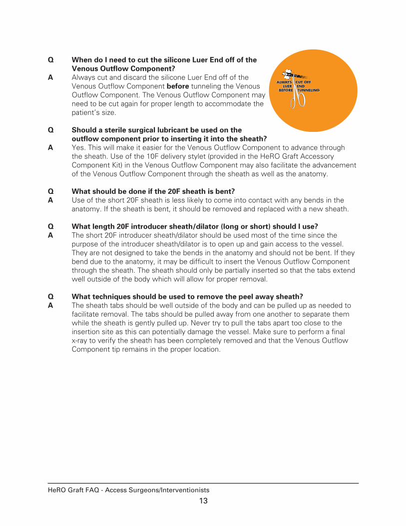
Q When do I need to cut the silicone Luer End off of the Venous Outflow Component?
A Always cut and discard the silicone Luer End off of the Venous Outflow Component before tunneling the Venous Outflow Component. The Venous Outflow Component may need to be cut again for proper length to accommodate the patient’s size.
Q Should a sterile surgical lubricant be used on the outflow component prior to inserting it into the sheath?
A Yes. This will make it easier for the Venous Outflow Component to advance through the sheath. Use of the 10F delivery stylet (provided in the HeRO Graft Accessory Component Kit) in the Venous Outflow Component may also facilitate the advancement of the Venous Outflow Component through the sheath as well as the anatomy.
Q What should be done if the 20F sheath is bent?A Use of the short 20F sheath is less likely to come into contact with any bends in the
anatomy. If the sheath is bent, it should be removed and replaced with a new sheath.
Q What length 20F introducer sheath/dilator (long or short) should I use?A The short 20F introducer sheath/dilator should be used most of the time since the
purpose of the introducer sheath/dilator is to open up and gain access to the vessel. They are not designed to take the bends in the anatomy and should not be bent. If they bend due to the anatomy, it may be difficult to insert the Venous Outflow Component through the sheath. The sheath should only be partially inserted so that the tabs extend well outside of the body which will allow for proper removal.
Q What techniques should be used to remove the peel away sheath?A The sheath tabs should be well outside of the body and can be pulled up as needed to
facilitate removal. The tabs should be pulled away from one another to separate them while the sheath is gently pulled up. Never try to pull the tabs apart too close to the insertion site as this can potentially damage the vessel. Make sure to perform a final x-ray to verify the sheath has been completely removed and that the Venous Outflow Component tip remains in the proper location.

14
Please see the Instructions for Use document provided in the HeRO Graft packaging and online at www.herograft.com for the full list of anticipated adverse events, contraindications, and complications.
The FDA regulation name for the HeRO Graft is vascular graft prosthesis.
INDICATIONS FOR USE: The HeRO Graft is indicated for end stage renal disease patients on hemodialysis who have ex-hausted all other access options. See instructions for use for full indication, contraindication and caution statements. Rx only.
CryoLife, the snowflake design and Life Restoring Technologies are trademarks owned by CryoLife, Inc. HeRO and Hemosphere are trademarks owned by Hemosphere, Inc. All other trade-marks are owned by their respective owners. © 2012 CryoLife, Inc. All rights reserved.
CryoLife, Inc.1655 Roberts Boulevard, NW Kennesaw, Georgia 30144United States
Customer Service888-427-9654 phone770-590-3753 faxwww.herograft.com
HeRO Graft FAQ - Access Surgeons/Interventionists
Please see the Instructions for Use document provided in the HeRO Graft packaging and online at www.herograft.com forthe full list of anticipated adverse events, contraindications, andcomplications.
The FDA regulation name for the HeRO Graft is vascular graft prosthesis.
HeRO, CryoLife, the snowflake design, and Life Restoring Tech-nologies are trademarks owned by CryoLife, Inc. All other trade-marks are owned by their respective owners.© 2015 CryoLife, Inc. All rights reserved.

Frequently Asked Questions
HeRO®
[Hemodialysis Reliable Outflow]Graft
Nephrologists
ML0668.002 (06/2015)
2HeRO Graft FAQ - Nephrologists 13-0061 Rev. B 2012-10
Only qualified healthcare providers should place, manipulate, de-clot, revise or explant the HeRO Graft. For greater detail, please consult the HeRO Graft Instructions for Use manual: www.herograft.com.
Q What do we do if we receive a transient patient with the HeRO Graft and it is not patent?
A Interventions should be performed by the specialist performing graft interventions. A detailed Thrombectomy Guidelines can be found at www.herograft.com.
Q Is there a maximum blood flow recommendation for the HeRO Graft?A Follow the provider/facility policy for setting blood flow rates. There is no special
requirement for the HeRO Graft.
Q When cannulating the HeRO Graft for the first time, can blood pump speeds of 350 to 400ml/min be used immediately?
A The HeRO Graft should be treated in the same manner as conventional ePTFE grafts. Flow rates of 450-500ml/min are usually achieved.
Q If infiltration occurs, is it more serious than infiltration with a regular graft?A As the HeRO Graft Arterial Graft Component is a standard ePTFE graft, infiltration is
expected to be as serious as with other standard grafts or fistulas.
Q Will HeRO Graft recipients be able to participate in access flow testing?A HeRO Graft is classified as a graft and should be treated the same way you treat any
graft when evaluating access flow. A baseline number should be determined for the HeRO Graft and then monitor changes accordingly per your protocols.
Q I know an access-challenged patient. How do I refer him/her to a physician who implants the HeRO Graft?
A Contact a HeRO Graft implanting physician at www.herograft.com. It is important to ask the surgeon if he/she wants vessel mapping done prior to seeing the patient, or if he/she will complete this at the time of the visit. NOTE: Not all HeRO Graft implanting physicians are located on our website. Please contact CryoLife Customer Service to find additional physicians in your area.
Q Does the use of Heparin during cannulation need to be changed when dialyzing using the HeRO Graft?
A The HeRO Graft is cannulated the same as any ePTFE graft, therefore Heparin use would be the same. However, since the HeRO Graft is a continuous flow system, no Heparin lock at the end of the dialysis session is needed.
3HeRO Graft FAQ - Nephrologists 13-0061 Rev. B 2012-03
Please see the Instructions for Use document provided in the HeRO Graft packaging and online at www.herograft.com for the full list of anticipated adverse events, contraindications, and complications.
The FDA regulation name for the HeRO Graft is vascular graft prosthesis.
INDICATIONS FOR USE: The HeRO Graft is indicated for end stage renal disease patients on hemodialysis who have ex-hausted all other access options. See instructions for use for full indication, contraindication and caution statements. Rx only.
CryoLife, the snowflake design and Life Restoring Technologies are trademarks owned by CryoLife, Inc. HeRO and Hemosphere are trademarks owned by Hemosphere, Inc. All other trade-marks are owned by their respective owners. © 2012 CryoLife, Inc. All rights reserved.
CryoLife, Inc.1655 Roberts Boulevard, NW Kennesaw, Georgia 30144United States
Customer Service888-427-9654 phone770-590-3753 faxwww.herograft.com
Please see the Instructions for Use document provided in the HeRO Graft packaging and online at www.herograft.com forthe full list of anticipated adverse events, contraindications, andcomplications.
The FDA regulation name for the HeRO Graft is vascular graft prosthesis.
HeRO, CryoLife, the snowflake design, and Life Restoring Tech-nologies are trademarks owned by CryoLife, Inc. All other trade-marks are owned by their respective owners.© 2015 CryoLife, Inc. All rights reserved.

Frequently Asked Questions
HeRO®
[Hemodialysis Reliable Outflow]Graft
Dialysis Providers
ML0668.002 (06/2015)
2HeRO Graft FAQ - Dialysis Providers 13-0061 Rev. B 2012-10
Only qualified healthcare providers should place, manipulate, de-clot, revise or explant the HeRO Graft. For greater detail, please consult the HeRO Graft Instructions for Use manual: www.herograft.com
Q What do we do if we receive a transient patient with the HeRO Graft and it is not patent?
A Interventions should be performed by the specialist performing graft interventions. A detailed Thrombectomy Guidelines can be found at www.herograft.com.
Q Is there a maximum blood flow recommendation for the HeRO Graft?A Follow the provider/facility policy for setting blood flow rates. There is no special
requirement for the HeRO Graft.
Q When cannulating the HeRO Graft for the first time, can blood pump speeds of 350 to 400ml/min be used immediately?
A The HeRO Graft should be treated in the same manner as conventional ePTFE grafts. Flow rates of 450-500ml/min are usually achieved.
Q If infiltration occurs, is it more serious than infiltration with a regular graft?A As the HeRO Graft Arterial Graft Component is a standard ePTFE graft, infiltration is
expected to be as serious as with other standard grafts or fistulas.
Q Will HeRO Graft recipients be able to participate in access flow testing?A HeRO Graft is classified as a graft and should be treated the same way you treat any
graft when evaluating access flow. A baseline number should be determined for the HeRO Graft and then monitor changes accordingly per your protocols.
Q Are small gauge needles required for the initial HeRO Graft cannulations?A It is not necessary to start with small needles. The provider/facility protocol should be
followed as with other ePTFE grafts.
Q Can fistula clamps be used on the HeRO Graft?A To achieve hemostasis after puncture, use moderate finger pressure rather than
mechanical fistula clamps. Since the HeRO Graft is a continuous flow system, a mechanical clamp may constrict flow and increase the risk of clotting.
Additional information on recommended technique can be found in the Care and Cannulation guide located at www.herograft.com.
Q When prepping cannulation sites, which anti-bacterial is preferred?A Follow the KDOQI guidelines for access assessment and preparation.
Q Why is using a light tourniquet recommended on the HeRO Graft?A A light tourniquet will assist in palpating the graft making the HeRO Graft feel more like
a conventional AV graft. The rationale for using a light tourniquet is related to the lack of a venous anastomosis with the HeRO Graft. It is similar to using a tourniquet for a native fistula as there is also no venous anastomosis with a fistula.
3HeRO Graft FAQ - Dialysis Providers 13-0061 Rev. B 2012-10
Q What is the risk to the graft if a tourniquet is applied too tightly or left on too long?
A As with any peripheral access, a tourniquet left on too long, or one that is applied too tightly, may constrict flow and increase clotting. The tourniquet should only be used for initiating cannulation and removed immediately when treatment begins.
Q Can all hemodialysis staff cannulate the HeRO Graft?A Follow your provider/facility policies with regard to graft cannulation.
Additional information on recommended technique can be found in the Care and Cannulation guide located at www.herograft.com.
Q How does a dialysis clinic know when a patient has received the HeRO Graft?A Dialysis facilities may receive an implant notification fax form from the implanting
surgeon’s office, or the nephrologist may have communicated the access surgery is scheduled to occur. If either of these have occurred and you have not been offered educational support, please contact CryoLife Customer Service. Each patient should receive a Patient Identification wallet card they can carry with them to identify them as a HeRO Graft patient. Additionally, HeRO Graft patients typically have 3 incision sites which identify the venous, connector, and arterial anastomosis sites.
Q Will a representative visit each dialysis facility with a HeRO Graft?A Educational opportunities are made available to each clinic via online learning,
brochures, and telephone education. A small group of clinical educators support the entire United States, which may limit inservice opportunities.
Tutorial videos on how the HeRO Graft works and an overview of Care and Cannulation are located at www.herograft.com.
Q Are there any activity limitations for a new HeRO Graft recipient?A The limitations are similar to other new AV accesses. HeRO Graft patients should check
with their doctor or nurse if they are not sure about beginning a new activity. Once their incisions heal, they may usually resume normal activities; however, they must remember to protect the HeRO Graft just as they would any hemodialysis access. The Patient Information brochure, located at www.herograft.com, addresses how the patient should take care of the HeRO Graft after implant.
Q How do I obtain a copy of the patient education materials or the Care and Cannulation guide?
A PDF versions of our educational materials are available for download at www.herograft.com. You may also contact Customer Service.
Visit www.herograft.com for HeRO Graft video tutorials. You also are welcome to register to attend a 20 minute webinar.
4HeRO Graft FAQ - Dialysis Providers 13-0061 Rev. B 2012-10
Please see the Instructions for Use document provided in the HeRO Graft packaging and online at www.herograft.com for the full list of anticipated adverse events, contraindications, and complications.
The FDA regulation name for the HeRO Graft is vascular graft prosthesis.
INDICATIONS FOR USE: The HeRO Graft is indicated for end stage renal disease patients on hemodialysis who have ex-hausted all other access options. See instructions for use for full indication, contraindication and caution statements. Rx only.
CryoLife, the snowflake design and Life Restoring Technologies are trademarks owned by CryoLife, Inc. HeRO and Hemosphere are trademarks owned by Hemosphere, Inc. All other trade-marks are owned by their respective owners. © 2012 CryoLife, Inc. All rights reserved.
CryoLife, Inc.1655 Roberts Boulevard, NW Kennesaw, Georgia 30144United States
Customer Service888-427-9654 phone770-590-3753 faxwww.herograft.com
Q Do I need to use wet sticks for cannulating the HeRO Graft?A HeRO Graft is cannulated the same as any ePTFE graft. Wet sticks are not required.
Q Does the use of Heparin during cannulation need to be changed when dialyzing using the HeRO Graft?
A The HeRO Graft is cannulated the same as any ePTFE graft, therefore Heparin use would be the same. However, since the HeRO Graft is a continuous flow system, no Heparin lock at the end of the dialysis session is needed.
Q Is needle placement the same as conventional grafts?A The provider/facility protocol should be followed as with other ePTFE grafts.
Please see the instructions for Use document provided in the HeRO Graft packaging and online at www.herograft.com forthe full list of anticipated adverse events, contraindications, andcomplications.
The FDA regulation name for the HeRO Graft is vascular graft prosthesis.
HeRO, CryoLife, the snowflake design, and Life Restoring Tech-nologies are trademarks owned by CryoLife, Inc. All other trade-marks are owned by their respective owners.© 2015 CryoLife, Inc. All rights reserved.

Frequently Asked Questions
HeRO®
[Hemodialysis Reliable Outflow]Graft
Hospital Administrators
ML0668.002 (06/2015)
2HeRO Graft FAQ - Hospital Administrators 13-0061 Rev. B 2012-10
Only qualified healthcare providers should place, manipulate, de-clot, revise or explant the HeRO Graft. For greater detail, please consult the HeRO Graft Instructions for Use manual: www.herograft.com
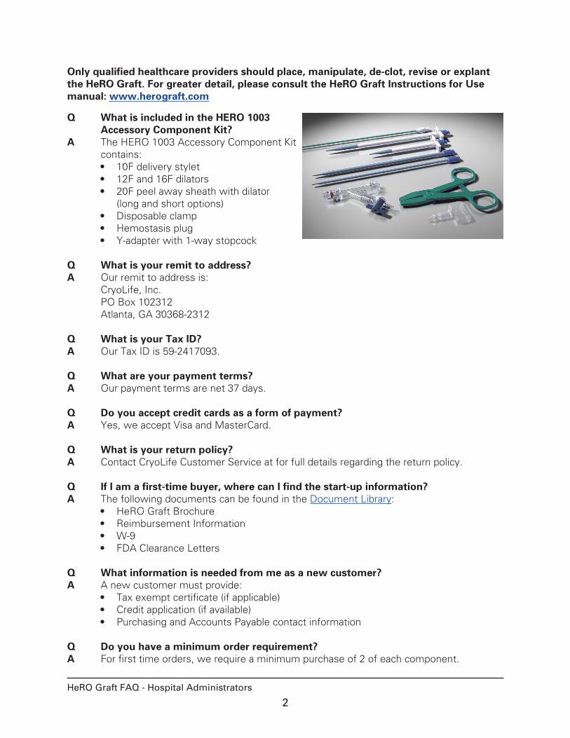
Q What is included in the HERO 1003 Accessory Component Kit?
A The HERO 1003 Accessory Component Kit contains:• 10F delivery stylet• 12F and 16F dilators• 20F peel away sheath with dilator
(long and short options)• Disposable clamp • Hemostasis plug• Y-adapter with 1-way stopcock
Q What is your remit to address?A Our remit to address is:
CryoLife, Inc. PO Box 102312 Atlanta, GA 30368-2312
Q What is your Tax ID?A Our Tax ID is 59-2417093.
Q What are your payment terms?A Our payment terms are net 37 days.
Q Do you accept credit cards as a form of payment?A Yes, we accept Visa and MasterCard.
Q What is your return policy?A Contact CryoLife Customer Service at for full details regarding the return policy.
Q If I am a first-time buyer, where can I find the start-up information?A The following documents can be found in the Document Library:
• HeRO Graft Brochure• Reimbursement Information• W-9 • FDA Clearance Letters
Q What information is needed from me as a new customer?A A new customer must provide:
• Tax exempt certificate (if applicable)• Credit application (if available)• Purchasing and Accounts Payable contact information
Q Do you have a minimum order requirement?A For first time orders, we require a minimum purchase of 2 of each component.
3HeRO Graft FAQ - Hospital Administrators 13-0061 Rev. B 2012-10
Q Is a Purchase Order required to place an order?A Yes, a Purchase Order is required to place an order.
Q What information is required on the Purchase Order?A The following information is required on the Purchase Order:
• Purchase Order Number• Billing Information• Shipping Information• Part Numbers, Desired Quantity and Pricing• Shipping Method and/or Delivery Date
Q What shipping methods are available?A Items are shipped UPS ground unless otherwise specified.
F.O.B. Ship point
Q Can I use my own shipping account number?A When possible, we will use your shipping account number when it is noted on the
order.
Q How should the HeRO Graft be stored?A The HeRO Graft should be stored in a cool, dry place. Avoid exposure to moisture and
excessive heat.
Q Is the HeRO Graft sterilized?A The HeRO Graft is ethylene oxide sterilized and non-pyrogenic. DO NOT re-sterilize.
Q What is the shelf life of the HeRO Graft?A The shelf life of the HeRO Graft is 2 years from the date of sterilization.
Q Does the HeRO Graft contain latex?A No. The HeRO Graft is latex free.
4HeRO Graft FAQ - Hospital Administrators 13-0061 Rev. B 2012-03
Please see the Instructions for Use document provided in the HeRO Graft packaging and online at www.herograft.com for the full list of anticipated adverse events, contraindications, and complications.
The FDA regulation name for the HeRO Graft is vascular graft prosthesis.
INDICATIONS FOR USE: The HeRO Graft is indicated for end stage renal disease patients on hemodialysis who have ex-hausted all other access options. See instructions for use for full indication, contraindication and caution statements. Rx only.
CryoLife, the snowflake design and Life Restoring Technologies are trademarks owned by CryoLife, Inc. HeRO and Hemosphere are trademarks owned by Hemosphere, Inc. All other trade-marks are owned by their respective owners. © 2012 CryoLife, Inc. All rights reserved.
CryoLife, Inc.1655 Roberts Boulevard, NW Kennesaw, Georgia 30144United States
Customer Service888-427-9654 phone770-590-3753 faxwww.herograft.com
Please see the instructions for Use document provided in the HeRO Graft packaging and online at www.herograft.com forthe full list of anticipated adverse events, contraindications, andcomplications.
The FDA regulation name for the HeRO Graft is vascular graft prosthesis.
HeRO, CryoLife, the snowflake design, and Life Restoring Tech-nologies are trademarks owned by CryoLife, Inc. All other trade-marks are owned by their respective owners.© 2015 CryoLife, Inc. All rights reserved.