Embed Size (px)
Citation preview
Medical Problems in Pregnancy
Tom Heaps
Consultant Acute Physician
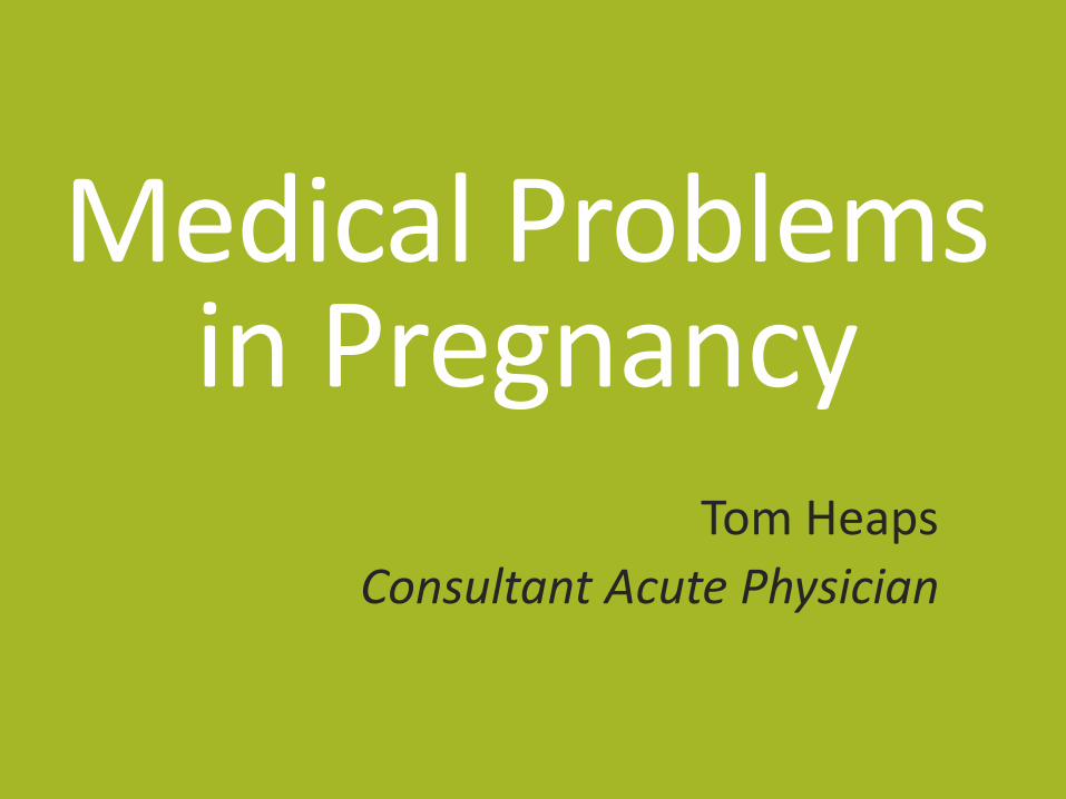
CMACE 2011: Saving Mother’s Lives
Overall mortality rate 11.39/100,000 pregnancies (2006-2008 data)
Failure to take symptoms seriously (‘healthy’ females)
Reluctance to investigate (perceived risks of radiation)
Fear of prescribing appropriate drugs
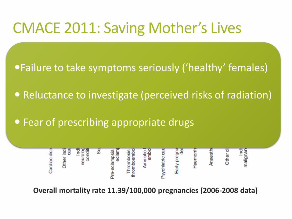
Considerations in pregnancy Two patients rather than one
Physiological changes in normal pregnancy
Differences in blood test parameters
Radiation exposure
Drug considerations
Differential diagnoses may be different
Effects of pregnancy on pre-existing conditions
Conditions unique to pregnancy
Early involvement of multispecialty team and ITU mandatory
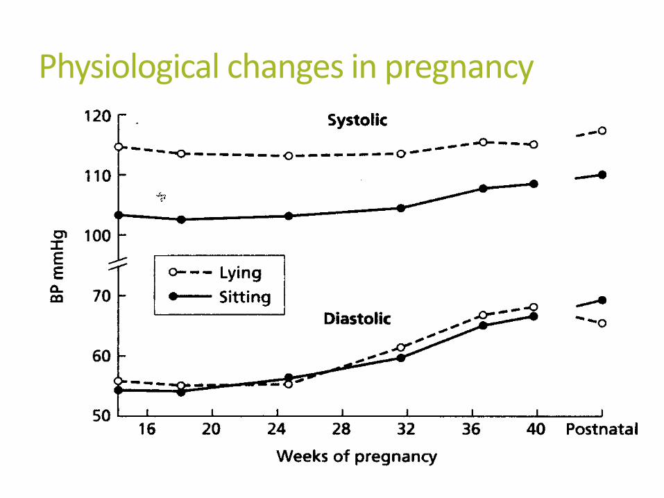
Physiological changes in pregnancy Peripheral vascular resistance falls by 50%
MAP falls by 10mmHg by 22-24w then slowly rises to term
Cardiac output and circulating volume increase by 50% (haemodilution)
Resting HR increases by 10-20/minute
Renal blood flow and GFR increase by 70-80%
Glycosuria, proteinuria (<300mg/d), bicarbonaturia, aminoaciduria, calciuria
Physiological hydronephrosis/hydroureter; kidney length increases by 1cm and renal pelvis dilates (AP diameter ≤2cm normal)
Hyperventilation/respiratory alkalosis due to progesterone-mediated stimulation of respiratory centre
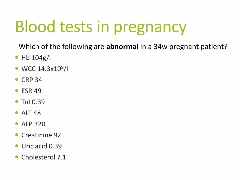
Blood tests in pregnancy
Hb 104g/l
WCC 14.3x10⁹/l
CRP 34
ESR 49
TnI 0.39
ALT 48
ALP 320
Creatinine 92
Uric acid 0.39
Cholesterol 7.1
Which of the following are abnormal in a 34w pregnant patient?
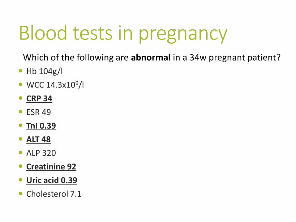
Blood tests in pregnancy
Hb 104g/l
WCC 14.3x10⁹/l
CRP 34
ESR 49
TnI 0.39
ALT 48
ALP 320
Creatinine 92
Uric acid 0.39
Cholesterol 7.1
Which of the following are abnormal in a 34w pregnant patient?
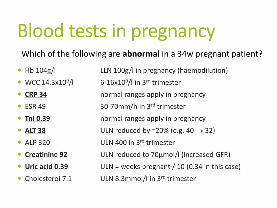
Blood tests in pregnancy
Hb 104g/l LLN 100g/l in pregnancy (haemodilution)
WCC 14.3x10⁹/l 6-16x109/l in 3rd trimester
CRP 34 normal ranges apply in pregnancy
ESR 49 30-70mm/h in 3rd trimester
TnI 0.39 normal ranges apply in pregnancy
ALT 38 ULN reduced by ~20% (e.g. 40 32)
ALP 320 ULN 400 in 3rd trimester
Creatinine 92 ULN reduced to 70µmol/l (increased GFR)
Uric acid 0.39 ULN = weeks pregnant / 10 (0.34 in this case)
Cholesterol 7.1 ULN 8.3mmol/l in 3rd trimester
Which of the following are abnormal in a 34w pregnant patient?
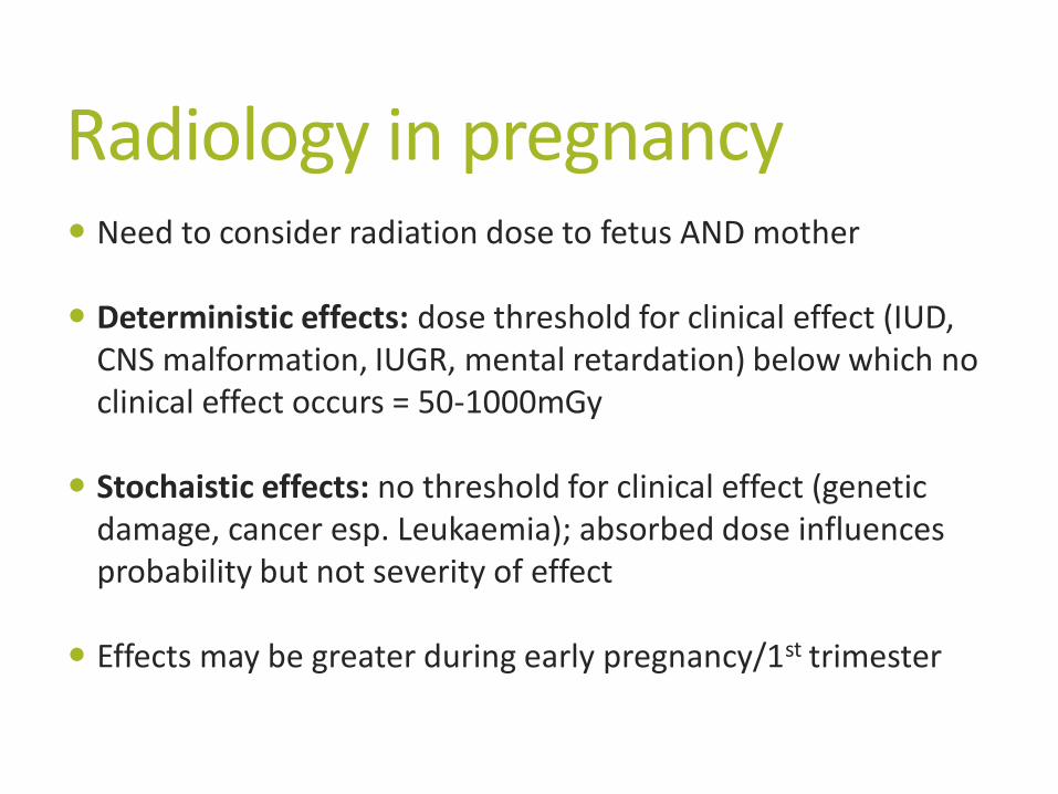
Radiology in pregnancy Need to consider radiation dose to fetus AND mother
Deterministic effects: dose threshold for clinical effect (IUD,
CNS malformation, IUGR, mental retardation) below which no clinical effect occurs = 50-1000mGy
Stochaistic effects: no threshold for clinical effect (genetic damage, cancer esp. Leukaemia); absorbed dose influences probability but not severity of effect
Effects may be greater during early pregnancy/1st trimester
Radiology in pregnancy
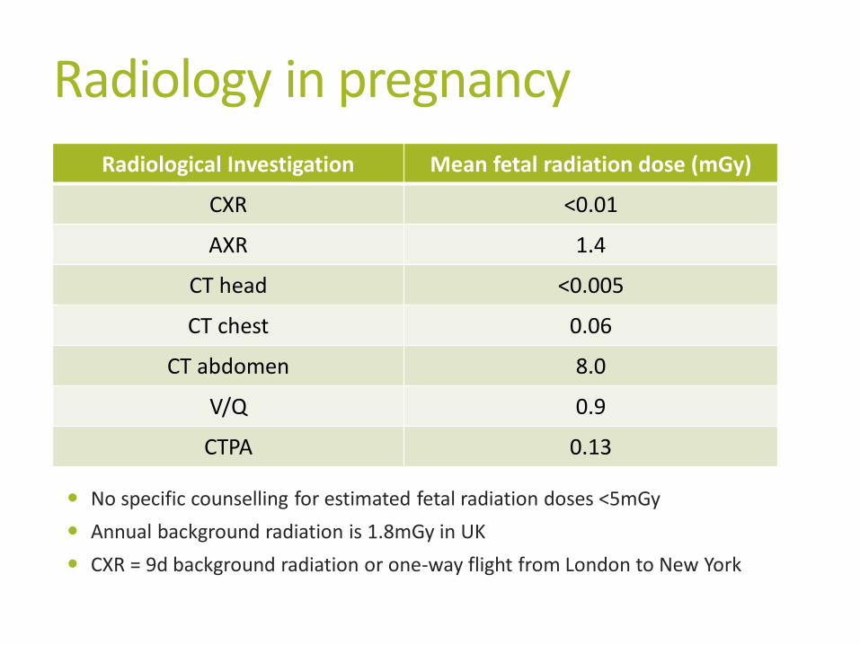
No specific counselling for estimated fetal radiation doses <5mGy
Annual background radiation is 1.8mGy in UK
CXR = 9d background radiation or one-way flight from London to New York
Radiological Investigation Mean fetal radiation dose (mGy)
CXR <0.01
AXR 1.4
CT head <0.005
CT chest 0.06
CT abdomen 8.0
V/Q 0.9
CTPA 0.13
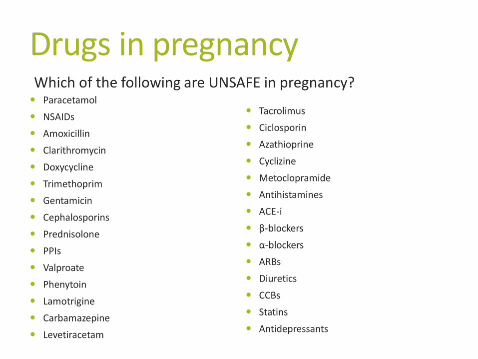
Drugs in pregnancy
Paracetamol
NSAIDs
Amoxicillin
Clarithromycin
Doxycycline
Trimethoprim
Gentamicin
Cephalosporins
Prednisolone
PPIs
Valproate
Phenytoin
Lamotrigine
Carbamazepine
Levetiracetam
Which of the following are UNSAFE in pregnancy?
Tacrolimus
Ciclosporin
Azathioprine
Cyclizine
Metoclopramide
Antihistamines
ACE-i
β-blockers
α-blockers
ARBs
Diuretics
CCBs
Statins
Antidepressants
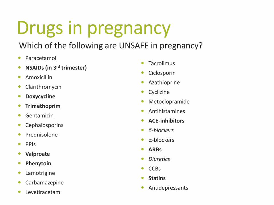
Drugs in pregnancy
Paracetamol
NSAIDs (in 3rd trimester)
Amoxicillin
Clarithromycin
Doxycycline
Trimethoprim
Gentamicin
Cephalosporins
Prednisolone
PPIs
Valproate
Phenytoin
Lamotrigine
Carbamazepine
Levetiracetam
Which of the following are UNSAFE in pregnancy?
Tacrolimus
Ciclosporin
Azathioprine
Cyclizine
Metoclopramide
Antihistamines
ACE-inhibitors
β-blockers
α-blockers
ARBs
Diuretics
CCBs
Statins
Antidepressants
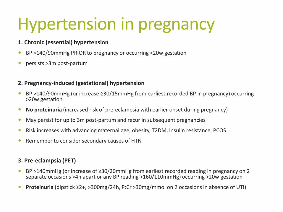
Hypertension in pregnancy 1. Chronic (essential) hypertension
BP >140/90mmHg PRIOR to pregnancy or occurring <20w gestation
persists >3m post-partum
2. Pregnancy-induced (gestational) hypertension
BP >140/90mmHg (or increase ≥30/15mmHg from earliest recorded BP in pregnancy) occurring >20w gestation
No proteinuria (increased risk of pre-eclampsia with earlier onset during pregnancy)
May persist for up to 3m post-partum and recur in subsequent pregnancies
Risk increases with advancing maternal age, obesity, T2DM, insulin resistance, PCOS
Remember to consider secondary causes of HTN
3. Pre-eclampsia (PET)
BP >140mmHg (or increase of ≥30/20mmHg from earliest recorded reading in pregnancy on 2 separate occasions >4h apart or any BP reading >160/110mmHg) occurring >20w gestation
Proteinuria (dipstick ≥2+, >300mg/24h, P:Cr >30mg/mmol on 2 occasions in absence of UTI)
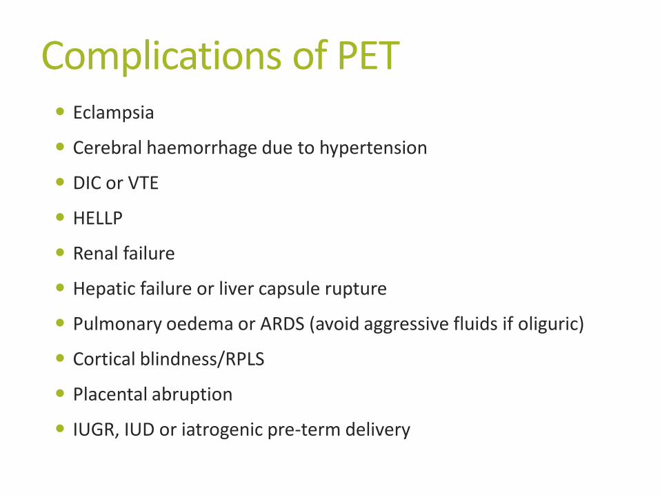
Complications of PET Eclampsia
Cerebral haemorrhage due to hypertension
DIC or VTE
HELLP
Renal failure
Hepatic failure or liver capsule rupture
Pulmonary oedema or ARDS (avoid aggressive fluids if oliguric)
Cortical blindness/RPLS
Placental abruption
IUGR, IUD or iatrogenic pre-term delivery
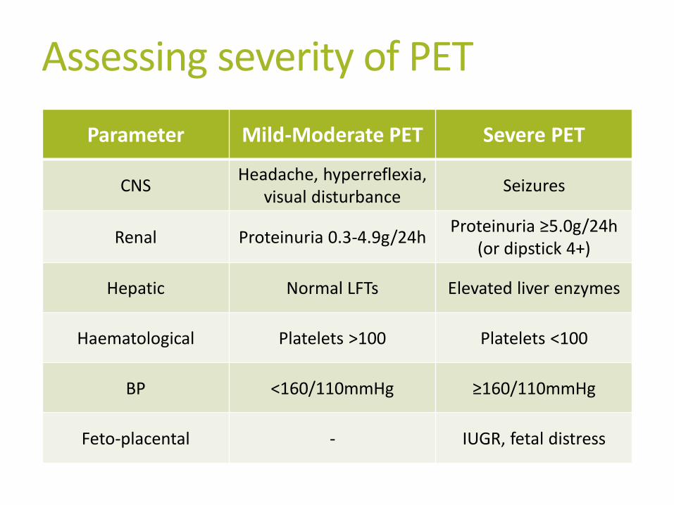
Assessing severity of PET
Parameter Mild-Moderate PET Severe PET
CNS Headache, hyperreflexia,
visual disturbance Seizures
Renal Proteinuria 0.3-4.9g/24h Proteinuria ≥5.0g/24h
(or dipstick 4+)
Hepatic Normal LFTs Elevated liver enzymes
Haematological Platelets >100 Platelets <100
BP <160/110mmHg ≥160/110mmHg
Feto-placental - IUGR, fetal distress
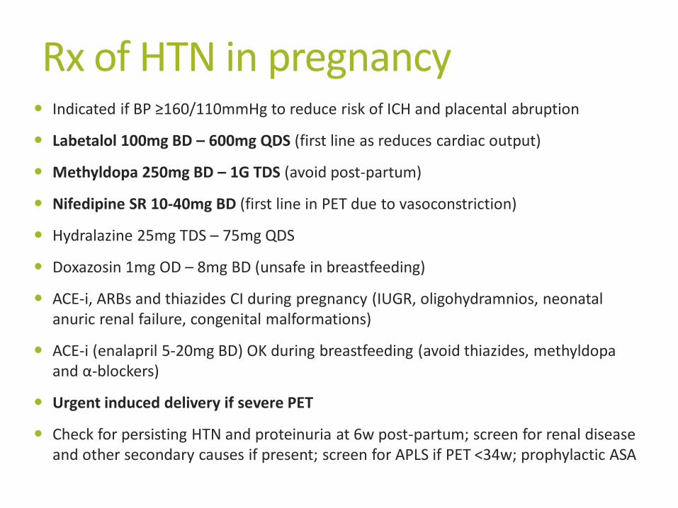
Rx of HTN in pregnancy Indicated if BP ≥160/110mmHg to reduce risk of ICH and placental abruption
Labetalol 100mg BD – 600mg QDS (first line as reduces cardiac output)
Methyldopa 250mg BD – 1G TDS (avoid post-partum)
Nifedipine SR 10-40mg BD (first line in PET due to vasoconstriction)
Hydralazine 25mg TDS – 75mg QDS
Doxazosin 1mg OD – 8mg BD (unsafe in breastfeeding)
ACE-i, ARBs and thiazides CI during pregnancy (IUGR, oligohydramnios, neonatal anuric renal failure, congenital malformations)
ACE-i (enalapril 5-20mg BD) OK during breastfeeding (avoid thiazides, methyldopa and α-blockers)
Urgent induced delivery if severe PET
Check for persisting HTN and proteinuria at 6w post-partum; screen for renal disease and other secondary causes if present; screen for APLS if PET <34w; prophylactic ASA
Clinical Case 35-year-old female presents to ED following generalized seizure
3-weeks post-partum (uncomplicated pregnancy and NVD)
Second seizure whilst being assessed in ED
BP 190/126mmHg
WHAT IS THE DIAGNOSIS?
HOW ARE YOU GOING TO TREAT THIS PATIENT?
Eclampsia 1/3000 pregnancies (incidence 0.03%) in UK
Case fatality 2%
Up to 44% of cases occur post-partum (usually within 4w)
60% have history of PET, 15% isolated HTN, 15% isolated proteinuria and 10% have no history of PET/HTN/proteinuria
IV MgSO4 4G (16mmol) over 10min then 1G (4mmol)/h for 24h (avoid loading dose if already taking CCB due to risk of hypotension)
Stop or reduce infusion if absent reflexes or RR <12/min
IV labetalol 50mg bolus then 20-160mg/h for severe HTN (insert arterial line)
Exclude other causes of seizure (ICH, CVT) with neuroimaging
Cautious fluid replacement (risk of pulmonary oedema/ARDS)
LMWH once BP controlled (providing no coagulopathy)
Headache/Seizures in pregnancy Migraine usually improves during pregnancy, may rebound after delivery
PET/eclampsia
CVST often post-partum, risk increased by thrombophilia, PET, dehydration e.g. HG
Stroke often SAH/ICH (also dissection), risk increased by HTN/PET/thrombophilia
Pituitary apoplexy headache, vomiting, hypotension and visual field defects
RCVS thunderclap headaches early post-partum
TTP thrombocytopenia, MAHA, normal clotting, renal failure
IIH early pregnancy, obesity, may resolve after delivery
PDPH
Gestational epilepsy
Others e.g. meningoencephalitis, alcohol/drugs, hypoglycaemia, hyponatraemia
Clinical Case
24-year-old female
28 weeks pregnant (first pregnancy)
pleuritic chest pain and dyspnoea
ECG sinus tachycardia, S1Q3T3
respiratory alkalosis on ABG
HOW WOULD YOU INVESTIGATE FOR PE?
HOW WOULD YOU TREAT IF PE WAS CONFIRMED?
VTE in pregnancy: the facts VTE is a leading cause of preventable maternal death
(2/100,000 pregnancies)
failure to take symptoms seriously and under-investigation are common
VTE is 6x more common in pregnancy
procoagulant state (increased factor VIII, IX, X, fibrinogen levels and activity of fibrinolytic inhibitors, decreased protein S, aPC resistance) plus venous stasis
risk maximal in late pregnancy/early puerperium (1st trimester - 3m postpartum)
‘The patient was discharged from ED after presenting with pleuritic chest pain and breathlessness. The
medical registrar believed that CXR and anticoagulation were contraindicated in pregnancy. The patient was later found dead at home. Coroner’s post-mortem revealed she had died from a massive pulmonary
embolus.’ CEMACH 2007
VTE in pregnancy: the facts up to 50% with VTE in pregnancy have heritable
thrombophilia (esp. FVL)
other RF include previous VTE, obesity, dehydration, smoking, age >35, twins, multiparity, pre-eclampsia, OHS
85% of DVTs occur on left (55% outside pregnancy)
72% of DVTs are ileofemoral/proximal (9% outside pregnancy)
leg oedema and calf pain are common in normal pregnancy
long-term complications in young healthy women; severe PTS (10%) and chronic pulmonary HTN (4%)
Investigating VTE in pregnancy d-dimer positive in 80% by 2nd trimester and 100% by 3rd trimester
negative d-dimer should NOT be used to exclude VTE in pregnancy (high risk)
sinus tachycardia, rightward axis and S1Q3T3 on ECG and respiratory alkalosis on ABG are NORMAL findings in pregnancy
CXR is safe and mandatory in ALL pregnant patients with ?PE; alternative pathologies, suitability for V/Q
Bilateral leg USS;
no radiation risk
very low yield in the absence of clinical DVT
3% risk of false +ves in pregnancy
may delay definitive Ix/expose patients to unnecessary antioagulation for longer
CTPA vs V/Q in pregnancy V/Q scan
usually diagnostic in pregnant population (20% indeterminate vs. 50-70% of general population)
more radiation than CTPA to fetus (0.32-0.74mGy in early pregnancy)
radiation may be limited half dose Q scan (perfusion), only proceeding to V scan if Q abnormal
1mGy radiation to maternal breast
CTPA
Less radiation than V/Q to fetus (0.03mGy in 1st trimester – 0.66mGy in 3rd trimester)
Significantly more radiation to ‘lactating’ maternal breast (20mGy)
Lifetime risk of breast cancer 1/8
Excess risk from CTPA 1/1200 at age 20 decreasing to 1/3500 by age 40
Overall increases relative risk of breast cancer by 1.004
Sequential combination of CXR, V/Q, CTPA and conventional pulmonary angiography together give <9/12
background radiation dose to fetus

Treating VTE in pregnancy warfarin is teratogenic in (early) pregnancy (safe during breastfeeding)
LMWH is safe during pregnancy
different dosing e.g. enoxaparin 1mg/kg BD
check platelet count after 2 weeks of Rx (HIT)
anti-factor Xa monitoring only if ↓↓ or ↑↑ BMI or GFR <30ml/min
discontinue LMWH temporarily 24h prior to delivery; restart after 6-12h
continue anticoagulation for ≥6w after delivery and for ≥6m in total
switch to warfarin 1w after delivery
prophylactic LMWH antenatally in subsequent pregnancies
case reports of successful thrombolysis for massive PE in pregnancy
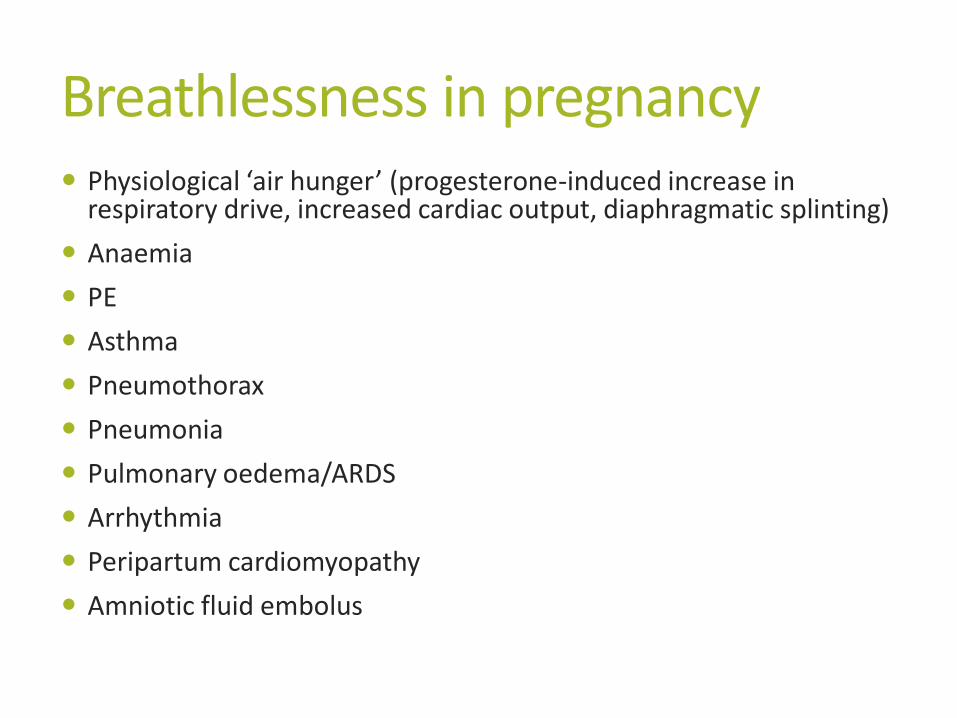
Breathlessness in pregnancy Physiological ‘air hunger’ (progesterone-induced increase in
respiratory drive, increased cardiac output, diaphragmatic splinting)
Anaemia
PE
Asthma
Pneumothorax
Pneumonia
Pulmonary oedema/ARDS
Arrhythmia
Peripartum cardiomyopathy
Amniotic fluid embolus
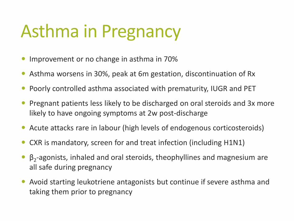
Asthma in Pregnancy Improvement or no change in asthma in 70%
Asthma worsens in 30%, peak at 6m gestation, discontinuation of Rx
Poorly controlled asthma associated with prematurity, IUGR and PET
Pregnant patients less likely to be discharged on oral steroids and 3x more likely to have ongoing symptoms at 2w post-discharge
Acute attacks rare in labour (high levels of endogenous corticosteroids)
CXR is mandatory, screen for and treat infection (including H1N1)
β2-agonists, inhaled and oral steroids, theophyllines and magnesium are all safe during pregnancy
Avoid starting leukotriene antagonists but continue if severe asthma and taking them prior to pregnancy
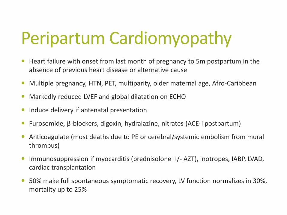
Peripartum Cardiomyopathy Heart failure with onset from last month of pregnancy to 5m postpartum in the
absence of previous heart disease or alternative cause
Multiple pregnancy, HTN, PET, multiparity, older maternal age, Afro-Caribbean
Markedly reduced LVEF and global dilatation on ECHO
Induce delivery if antenatal presentation
Furosemide, β-blockers, digoxin, hydralazine, nitrates (ACE-i postpartum)
Anticoagulate (most deaths due to PE or cerebral/systemic embolism from mural thrombus)
Immunosuppression if myocarditis (prednisolone +/- AZT), inotropes, IABP, LVAD, cardiac transplantation
50% make full spontaneous symptomatic recovery, LV function normalizes in 30%, mortality up to 25%
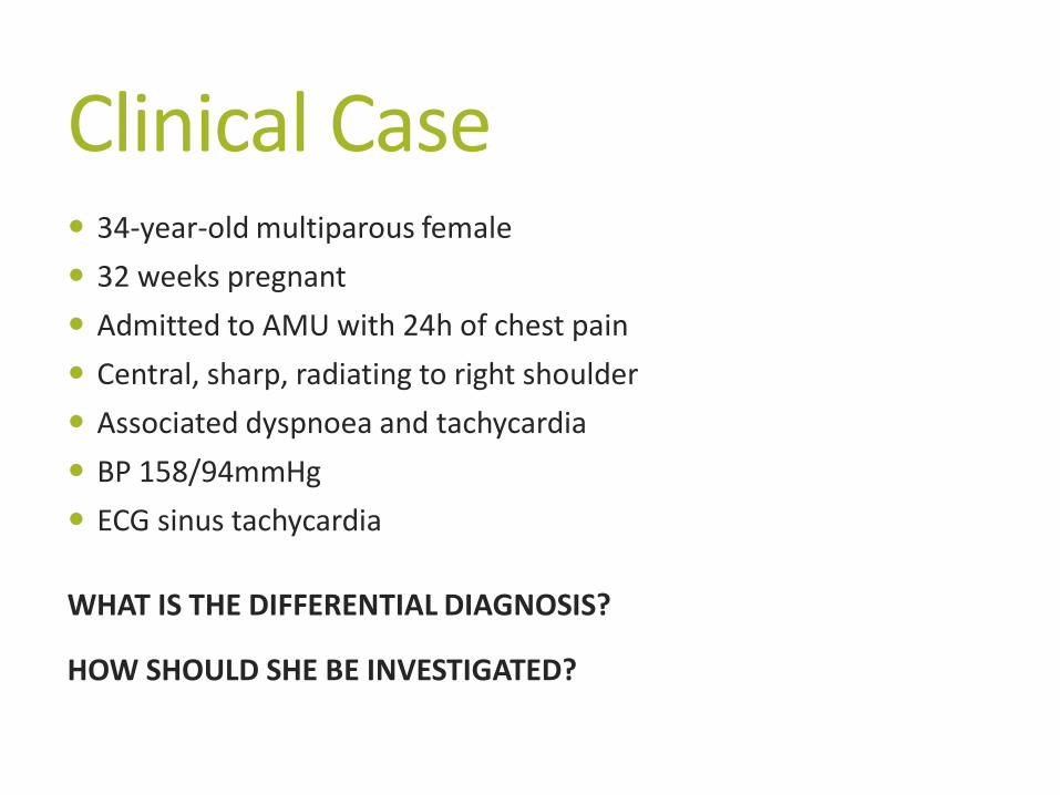
Clinical Case 34-year-old multiparous female
32 weeks pregnant
Admitted to AMU with 24h of chest pain
Central, sharp, radiating to right shoulder
Associated dyspnoea and tachycardia
BP 158/94mmHg
ECG sinus tachycardia
WHAT IS THE DIFFERENTIAL DIAGNOSIS? HOW SHOULD SHE BE INVESTIGATED?
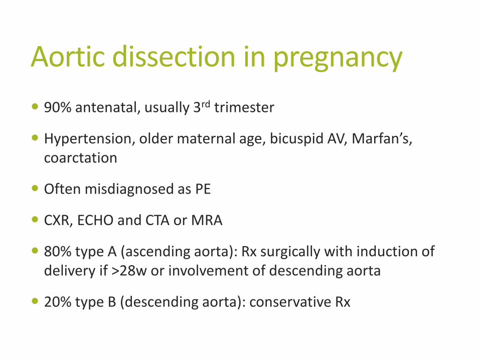
Aortic dissection in pregnancy
90% antenatal, usually 3rd trimester
Hypertension, older maternal age, bicuspid AV, Marfan’s, coarctation
Often misdiagnosed as PE
CXR, ECHO and CTA or MRA
80% type A (ascending aorta): Rx surgically with induction of delivery if >28w or involvement of descending aorta
20% type B (descending aorta): conservative Rx
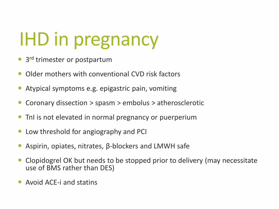
IHD in pregnancy 3rd trimester or postpartum
Older mothers with conventional CVD risk factors
Atypical symptoms e.g. epigastric pain, vomiting
Coronary dissection > spasm > embolus > atherosclerotic
TnI is not elevated in normal pregnancy or puerperium
Low threshold for angiography and PCI
Aspirin, opiates, nitrates, β-blockers and LMWH safe
Clopidogrel OK but needs to be stopped prior to delivery (may necessitate use of BMS rather than DES)
Avoid ACE-i and statins
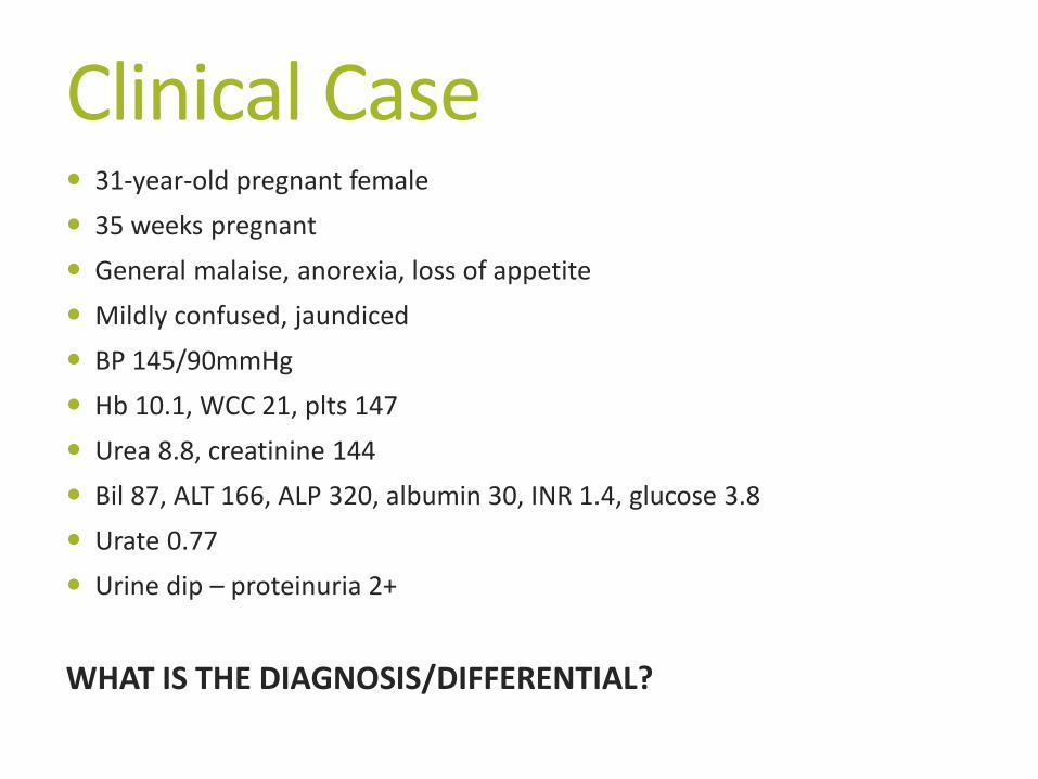
Clinical Case 31-year-old pregnant female
35 weeks pregnant
General malaise, anorexia, loss of appetite
Mildly confused, jaundiced
BP 145/90mmHg
Hb 10.1, WCC 21, plts 147
Urea 8.8, creatinine 144
Bil 87, ALT 166, ALP 320, albumin 30, INR 1.4, glucose 3.8
Urate 0.77
Urine dip – proteinuria 2+
WHAT IS THE DIAGNOSIS/DIFFERENTIAL?
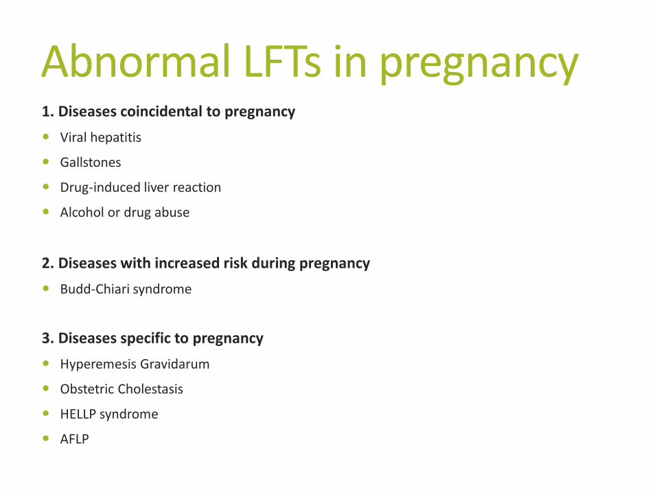
Abnormal LFTs in pregnancy 1. Diseases coincidental to pregnancy
Viral hepatitis
Gallstones
Drug-induced liver reaction
Alcohol or drug abuse
2. Diseases with increased risk during pregnancy
Budd-Chiari syndrome
3. Diseases specific to pregnancy
Hyperemesis Gravidarum
Obstetric Cholestasis
HELLP syndrome
AFLP
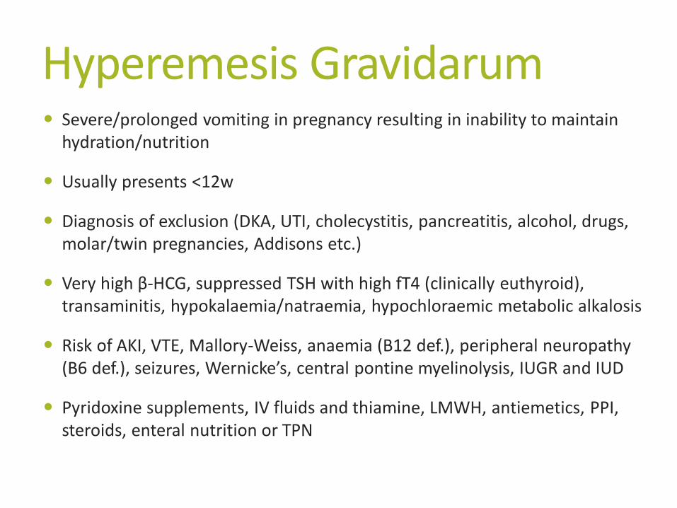
Hyperemesis Gravidarum Severe/prolonged vomiting in pregnancy resulting in inability to maintain
hydration/nutrition
Usually presents <12w
Diagnosis of exclusion (DKA, UTI, cholecystitis, pancreatitis, alcohol, drugs, molar/twin pregnancies, Addisons etc.)
Very high β-HCG, suppressed TSH with high fT4 (clinically euthyroid), transaminitis, hypokalaemia/natraemia, hypochloraemic metabolic alkalosis
Risk of AKI, VTE, Mallory-Weiss, anaemia (B12 def.), peripheral neuropathy (B6 def.), seizures, Wernicke’s, central pontine myelinolysis, IUGR and IUD
Pyridoxine supplements, IV fluids and thiamine, LMWH, antiemetics, PPI, steroids, enteral nutrition or TPN
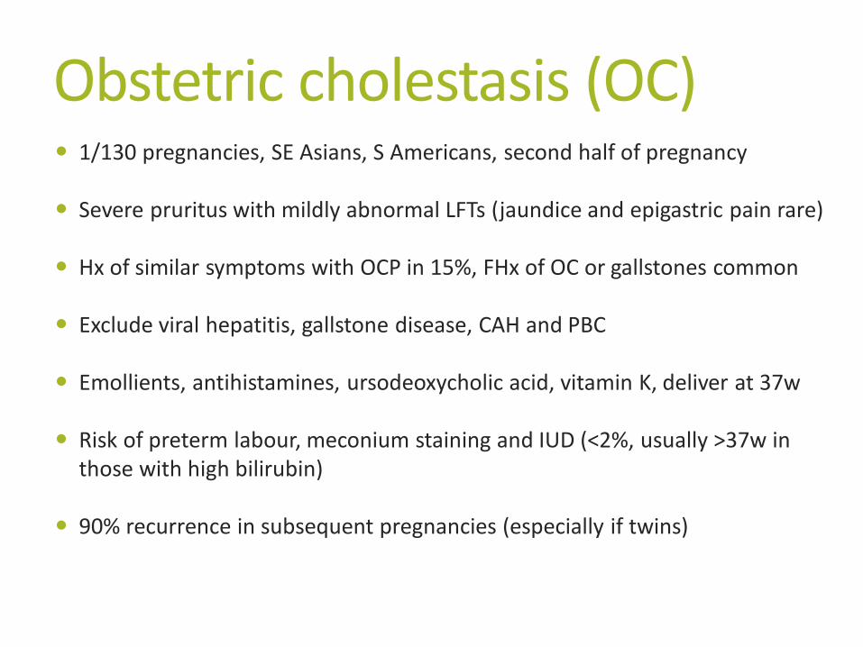
Obstetric cholestasis (OC) 1/130 pregnancies, SE Asians, S Americans, second half of pregnancy
Severe pruritus with mildly abnormal LFTs (jaundice and epigastric pain rare)
Hx of similar symptoms with OCP in 15%, FHx of OC or gallstones common
Exclude viral hepatitis, gallstone disease, CAH and PBC
Emollients, antihistamines, ursodeoxycholic acid, vitamin K, deliver at 37w
Risk of preterm labour, meconium staining and IUD (<2%, usually >37w in those with high bilirubin)
90% recurrence in subsequent pregnancies (especially if twins)

HELLP syndrome 30% postnatal, 70% antenatal, mortality <1%
complicates 20% of severe PET but HTN & proteinuria may be absent
N&V, epigastric pain (liver capsule oedema, may rupture with subcapsular haematoma)
Intravascular haemolysis, raised bilirubin and modestly elevated LFTs, platelets <100, renal failure may occur
Distinguish from AFLP and TTP/HUS
Antihypertensives, FFP/platelets as required, delivery, high-dose dexamethasone postnatally
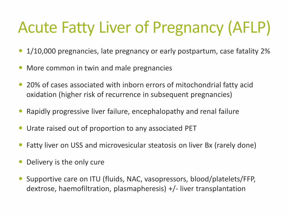
Acute Fatty Liver of Pregnancy (AFLP) 1/10,000 pregnancies, late pregnancy or early postpartum, case fatality 2%
More common in twin and male pregnancies
20% of cases associated with inborn errors of mitochondrial fatty acid oxidation (higher risk of recurrence in subsequent pregnancies)
Rapidly progressive liver failure, encephalopathy and renal failure
Urate raised out of proportion to any associated PET
Fatty liver on USS and microvesicular steatosis on liver Bx (rarely done)
Delivery is the only cure
Supportive care on ITU (fluids, NAC, vasopressors, blood/platelets/FFP, dextrose, haemofiltration, plasmapheresis) +/- liver transplantation
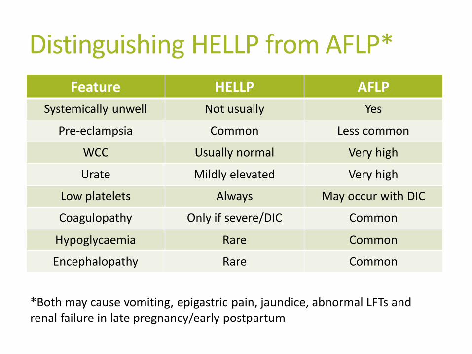
Distinguishing HELLP from AFLP*
Feature HELLP AFLP
Systemically unwell Not usually Yes
Pre-eclampsia Common Less common
WCC Usually normal Very high
Urate Mildly elevated Very high
Low platelets Always May occur with DIC
Coagulopathy Only if severe/DIC Common
Hypoglycaemia Rare Common
Encephalopathy Rare Common
*Both may cause vomiting, epigastric pain, jaundice, abnormal LFTs and renal failure in late pregnancy/early postpartum
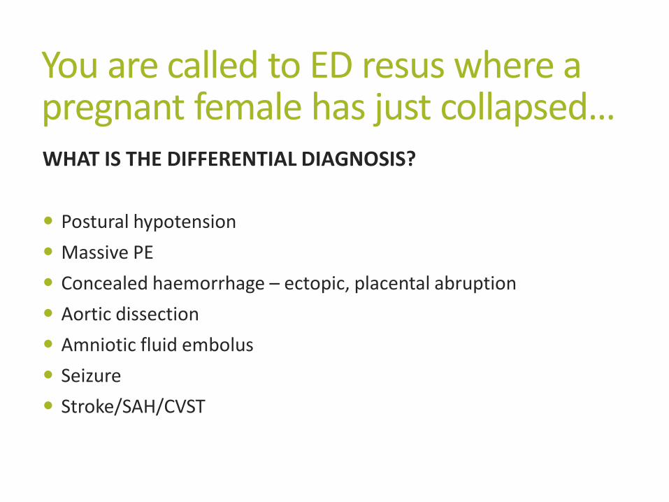
You are called to ED resus where a pregnant female has just collapsed… WHAT IS THE DIFFERENTIAL DIAGNOSIS?
Postural hypotension
Massive PE
Concealed haemorrhage – ectopic, placental abruption
Aortic dissection
Amniotic fluid embolus
Seizure
Stroke/SAH/CVST
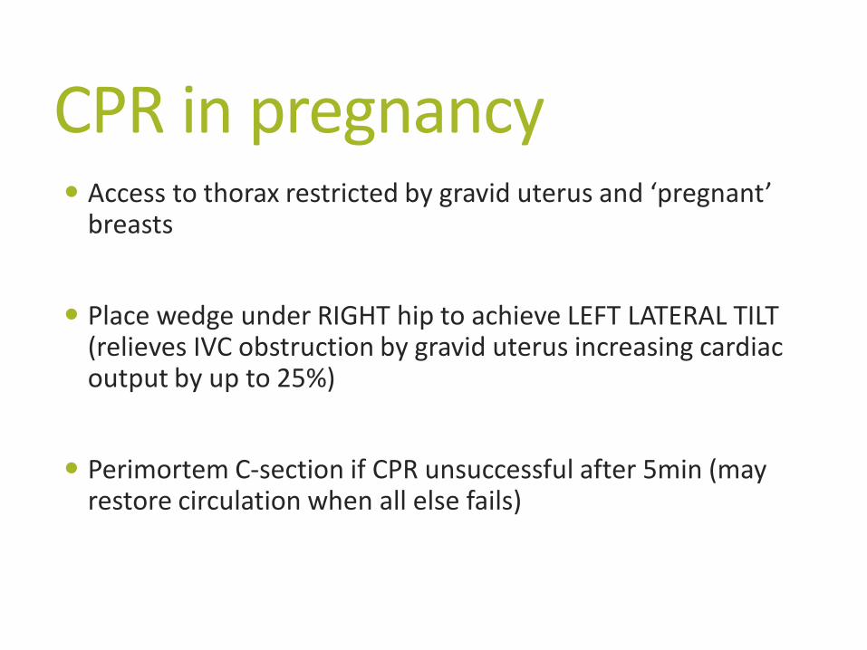
CPR in pregnancy Access to thorax restricted by gravid uterus and ‘pregnant’
breasts
Place wedge under RIGHT hip to achieve LEFT LATERAL TILT (relieves IVC obstruction by gravid uterus increasing cardiac output by up to 25%)
Perimortem C-section if CPR unsuccessful after 5min (may restore circulation when all else fails)
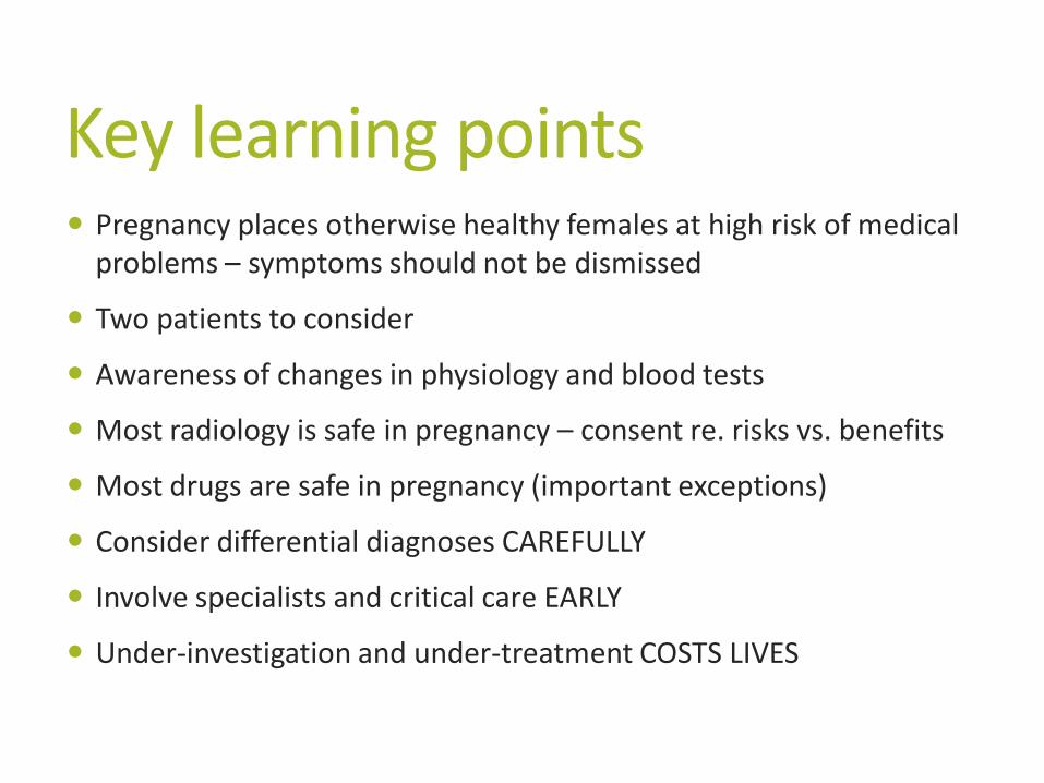
Key learning points Pregnancy places otherwise healthy females at high risk of medical
problems – symptoms should not be dismissed
Two patients to consider
Awareness of changes in physiology and blood tests
Most radiology is safe in pregnancy – consent re. risks vs. benefits
Most drugs are safe in pregnancy (important exceptions)
Consider differential diagnoses CAREFULLY
Involve specialists and critical care EARLY
Under-investigation and under-treatment COSTS LIVES


![Respiratory Distress.ppt [Read-Only]ocw.usu.ac.id/course/download/1110000107-growth-and-development... · hyaline membrane disease, and cerebral hyperventilation. Learning Objective](https://img.pdfslide.us/doc/110x75/5b6434a47f8b9a2e308d27a6/respiratory-read-onlyocwusuacidcoursedownload1110000107-growth-and-development.jpg)