Embed Size (px)
Citation preview

BRITISH MEDICAL JOURNALLONDON SATURDAY JULY 1 1961
OSTEOARTHROSIS IN PATIENTS AND POPULATIONS*BY
J. H. KELLGREN, M.Sc., F.R.C.P., F.R.C.S.Director, Rheumatism Research Centre, University of Manchester
and Manchester Royal Infirmary
The history of osteoarthrosis is long, for degenerativejoint changes can be seen in fossil skeletons of prehistoricanimals and in the joints of ancient Egyptian mummies.Despite this long history our knowledge of the diseaseis incomplete, perhaps because it is one of those dullcommonplace disorders that are hard to study withenthusiasm, but new knowledge of osteoarthrosis mustbe gained if the later years of our lengthening lives arenot to be plagued by increasing pain and disability.
Osteoarthrosis can be defined as an expression of ajoint's inadequacy to meet the mechanical stress placedupon it, and in certain patients and in certain joints,notably the hip, abnormal mechanical factors doobviously play a dominant part in both causation andtreatment. But are such joints representative of the totalproblem of degenerative joint disease ? May there notbe important constitutional factors which predispose topremature degeneration of multiple joints submitted tono more than average mechanical stress ? Detailedlaboratory studies of individual patients have beenunrewarding in this respect; except in such rareconditions as alkaptonuria, where congenital absence ofthe singre enzyme homogentisic acid oxidase (La Du,Zannoni, Laster, and Seegmiller, 1958) is associated withpremature degeneration of all the intervertebral disksand articular cartilages. This lack of positive labora-tory findings, however, may only reflect the inadequacyof our methods, and while we are waiting for thebiochemists to devise new methods it may beinteresting to review some of the information obtainedin recent years from the study of osteoarthrosis inpopulation groups by field-survey techniques.
OSTEOARTHROSIS IN POPULATIONSThe epidemiological approach requires both definition
and quantitation of the abnormality under study.Osteoarthrosis is best defined in terms of anatomical
abnormalities in the joints. The earliest changes, suchas softening, fibrillation, or disintegration of the articularcartilage, can be recognized with certainty only bydirect inspection of the joint surface-a procedureapplicable only to post-mortem surveys (Heine, 1926;Bennett, Waine, and Bauer 1942). The later changesof subchondral bone sclerosis and marginal osteophyticoutgrowths are, however, easily demonstrated radio-logically, and radiographs give a good index of theselater and more severe changes. Furthermore, radio-graphs provide evidence that can be studied in such a*Based on a Honyman Gillespie Lecture delivered in Edin-
burgh on November 3, 1960. The terms of the lecture precludethe mentioning of experiments on animals.
manner that inter- and intra-observer error can bereduced to a minimum and standard gradings for thedegree of radiological change can be elaborated(Kellgren and Lawrence, 1957), so providing forquantitation. Although radiographs provide the bestobjective evidence of the advanced degrees of osteo-arthrosis they give no information about pain anddisability, so that a clinical examination of the articularsystem and questionaries about rheumatic complaints arealso needed.The early surveys which were carried out in Lancashire
(Lawrence and Aitken-Swan, 1952; Kellgren, Lawrence,and Aitken-Swan, 1953) were planned jointly with theManchester University Department of OccupationalHealth and were done for the National Coal Boardbecause it has been suggested that miners suffered fromsome special form of rheumatism.
In the first of these studies information aboutrheumatic complaints of all kinds was collected fromvarious population samples totalling nearly 7,000.composed of miners and men from other occupationalgroups, and women from both mining and non-miningfamilies, living in the town of Leigh, in Lancashire.The complaints were classified by site, duration, and
degree of resulting disability, and only secondarily bydiagnostic categories, since diagnostic definition in therheumatic diseases is at best difficult and rarely preciseenough for use in field work. Nevertheless, it becameclear that osteoarthrosis in women and disk disorders in
MALES FEMALES251 OSTEOARTHROSIS201
w 101 I
Z 10 °iRHEUMATOID ARTHRITIS
(L is DISK DISORDERS
°o 1..... ..........
101 20 UNDETERMINED
020 470 60 > 60 20 40 60 > 60
AGE IN YEARSFIG. I.-Rheumatic complaints by age and sex occurring at sometime during a five-year period in a random sample of 1,619 men
and 1,896 women from the town of Leigh, Lancs.5243

OSTEOARTHROSIS
men were the most frequent and important causes ofpain and disability and that rheumatoid arthritis, thoughless frequent, often caused prolonged disability,especially in middle-aged women. In the younger age-groups there were many undiagnosable episodes of painand stiffness, some of which may well represent earlystages of these other more serious conditions (Fig. 1).
Survey in MinersWhen miners were compared with the surrounding
population it was found that the total complaint ratefor pains in the limbs and spine in miners differed littlefrom that in other men, but the miners lost moreworking-time and their pains were situated mainly inthe knees and back-sciatic distribution. Such roughdiagnostic classification as could be made suggested thatminers suffered more from disk disorders and osteo-arthrosis than non-miners, but rheumatoid arthritis wasequally distributed and possibly even less frequent amongminers in the older age-groups.
Since the problem in miners appeared to be one ofosteoarthrosis and disk degeneration, a sample of miners,manual workers, and office workers in the 40-49 age-group were studied radiologically as well as clinicallyto see whether the excess of back and knee pain inminers was associated with anatomical changes in thejoints (Kellgren and Lawrence, 1952). Severe radio-logical signs of disk degeneration in the lumbar spinewere found in 43% of miners but in only 7% of officeworkers." Conversely, only 8 % of miners had radio-logically normal spines compared with 67% of officeworkers (Fig. 2). The findings in the manual workers,who were engineers, were intermediate but nearer to theoffice workers than the miners. A similar excess ofradiological osteoarthrosis was noted in the knees of theminers, but radiological evidence of disk degenerationin the cervical spine was found equally in the threeoccupational groups. A fairly close association wasfound between these x-ray changes in the knees and thelumbar spine and pain in the knees and back-sciaticdistribution respectively, but not all men with severex-ray changes had symptoms.
1OO1
to
a
0
0
0.
90q
80
70
60-
50
40
30
20
I0
0.
aTYPE OF WORKER: M.
D
LUMBARt SPINE -KNEES
MINERSMANUAL (WALKDEN YARD)SEDENTARY (N.C. B. OFFICES)
HANDS CERVICAL SPINE
Oto o o S v. oL
DEGREE OF RADIOLOGICAL CHANGE
FIG. 2.-Incidence of osteoarthrosis and disk degeneration in 84miners, 45 manual workers, and 42 office workers aged 40-49
years.
A similar study of groups of men working underdifferent conditions (Lawrence, 1955) showed thatradiological signs of disk degeneration in the lumbarspine were especially related to heavy lifting and traumato the back, whereas working in a stooping position,stature, stem height, and exposure to wet aggravatedsymptoms and disability but had no influence on theradiological changes. Similarly, radiological osteo-arthrosis in the knees was related to injury and bodyweight but not to kneeling, whereas deformity of theknees aggravated symptoms and disability but had no
influence on the radiological changes.These results in miners were entirely in keeping with
the predominant role of mechanical factors in theproduction of osteoarthrosis in men, but there were alsosome unexpected findings.Although the miners had much more frequent and
severe anatomical changes in the joints, their complaintrate differed little from men in other occupations, whilethe women from mining families had fewer complaintsthan other women in Lancashire, and the male non-
mining members of the mining families had only halfas many rheumatic complaints as men from non-miningfamilies. These differences were noted at all sites ofpain and in all diagnostic categories except rheumatoidarthritis, and it seemed difficult to explain this by anyother means than a group difference in complaintthreshold. This question of complaint threshold mayin fact be one of the most important parameters to assess
in patients with rheumatic diseases, and the existenceof such group differences of complaint rate precludesthe use of complaints alone as an index of articulardisease.The second unexpected finding was the high prevalence
in the women of Leigh of what appeared to be osteo-arthrosis of multiple joints, affecting particularly thehands. Though this involvement of the hands might tosome extent be explained in terms of the mechanicalstresses peculiar to household chores, the widespreadinvolvement of many joints suggested a constitutionalfactor.At the same time it was noted that many of the female
patients attending the rheumatism clinic at theManchester Royal Infirmary appeared to be sufferingfrom multiple osteoarthrosis affecting the diarthrodialjoints in a symmetrical and characteristic manner. Thispolyarticular osteoarthrosis was usually associated withHeberden's nodes, and it was suggested that osteo-arthrosis in women might often be a generalizedcondition which, like Heberden's nodes, could resultfrom some inherited constitutional disorder (Kellgrenand Moore, 1952).To investigate this possibility further surveys were
made in which routine radiographs were taken of manyjoints and in which a blood sample was also obtainedfrom each respondent. The concentration of therheumatoid serum factor has been determined in thesesurveys by Dr. J. Ball, using his modification of thesensitized sheep-cell agglutination test (Ball, 1950;Kellgren and Ball, 1959), and the serum uric acid andserum cholesterol have been estimated by Miss V.Hewitt, whose work has been supported by the NuffieldFoundation.
Multiple Osteoarthrosis in an Urban PopulationIn the first of these studies, which was carried out
in co-operation with the Medical Research Council's
BRITISHMEDICAL JOUJRNAL
2 JULY 1. 1961
JULY 1, 1961BRITisH 3
MEDICAL JOURNAL
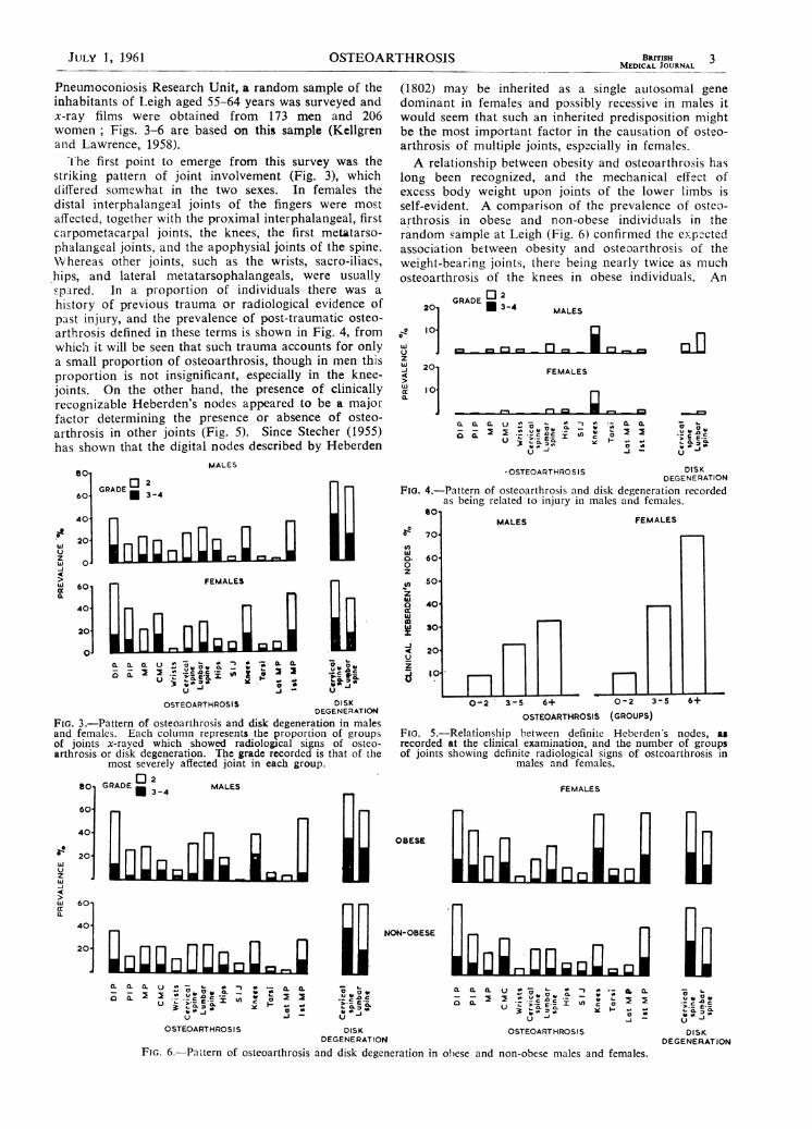
Pneumoconiosis Research Unit, a random sample of theinhabitants of Leigh aged 55-64 years was surveyed andx-ray films were obtained from 173 men and 206women; Figs. 3-6 are based on this sample (Kellgrenand Lawrence, 1958).
'The first point to emerge from this survey was thestriking pattern of joint involvement (Fig. 3), whichdiffered somewhat in the two sexes. In females thedistal interphalangeal joints of the fingers were mostaffected, together with the proximal interphalangeal, firstcarpometacarpal joints, the knees, the first metatarso-phalangeal joints, and the apophysial joints of the spine.Whereas other joints, such as the wrists, sacro-iliacs,hips, and lateral metatarsophalangeals, were usuallyspAred. In a proportion of individuals there was ahistory of previous trauma or radiological evidence ofpast injury, and the prevalence of post-traumatic osteo-arthrosis defined in these terms is shown in Fig. 4, fromwhich it will be seen that such trauma accounts for onlya small proportion of osteoarthrosis, though in men thisproportion is not insignificant, especially in the knee-joints. On the other hand, the presence of clinicallyrecognizable Heberden's nodes appeared to be a majorfactor determining the presence or absence of osteo-arthrosis in other joints (Fig. 5). Since Stecher (1955)has shown that the digital nodes described by Heberden
MALES
at
Uzw
4j
80.
GRADE 0 2 nt-60' * 3-4Hi
ki 0 FEMALES
0.
0000 *00.'U00. .'2
w'
(L > > 0 UC - w ,#l,§,!zX .................................U to
u ~ ~OSTEOARTHROSIS DISK
DEGENERATIONFiG. 3.-Pattern of osteoarthrosis and disk degeneration in malesand females. Each column represents the proportion of groupsof joints x-rayed which showed radiological signs of osteo-arthrosis or disk degeneration. The grade recorded is that of the
most severely affected joint in each group.
go, GRADE a 2 MALES
60'40'0 34 m
zw .I 1iI-
(1802) may be inherited as a single autosomal genedominant in females and possibly recessive in males itwould seem that such an inherited predisposition mightbe the most important factor in the causation of osteo-arthrosis of multiple joints, especially in females.A relationship between obesity and osteoarthrosis has
long been recognized, and the mechanical effect ofexcess body weight upon joints of the lower limbs isself-evident. A comparison of the prevalence of osteo-arthrosis in obese and non-obese individuals in therandom sample at Leigh (Fig. 6) confirmed the expzctedassociation between obesity and osteoarthrosis of theweight-bearing joints, there being nearly twice as muchosteoarthrosis of the knees in obese individuals. An
201
..o 101
wJzU2w
4w
a.UJ
201
GRADE °2U 3-4 MALES
- r-I - -D -
FEMALES
t-% r-% __0. 0. CL U - A 2
0 0. > E 0 ,in
-J.
a. a.
_-
- I-
I.) -
*OSTEOARTHROSIS DISKDEGENERATION
FiG. 4.-Pattern of osteoarthrosis and disk degeneration recordedas being related to injury in males and females.
SoI0
MALES FEMALES0 I
ti)w0.0z
nz
d
0
-j
2U
60j
50-
40
3O
20
O0
0-2 3-S 6+ 0-2 3-5 6+
OSTEOARTHROSIS (GROUPS)
L
FIo. 5.-Relationship between definite Heberden's nodes, asrecorded at the clinical examination, and the number of groupsof joints showing definite radiological signs of osteoarthrosis in
males and females.
FEMALES
OBESE ,L11;Iiw
Ui 60,0. Ill
NON-OBESE
20 Il[l oo 1
0. 0. 0.0U a -0. 0. '; L.00.. ' -
-~~~~~~~~~~~~-. -.0 U00. ~ ~ -0.50. *"%o- WX 0 C O Cl_
OSTEOARTHROSIS DISK OSTEOARTHROSISDEGENERATION
FIG. 6.-Pattern of osteoarthrosis and disk degeneration in obese and non-obese males and females.
0-j0
u
DISKDEGENERATION
JULY 1, 1961 OSTEOARTHROSIS

4 JULY 1, 1961 OSTEOARTHROSIS
unexpected finding, however, was an excess ofgeneralized osteoarthrosis in obese males; indeed, obesemales had nearly twice as much osteoarthrosis of thedistal interphalangeal joints, and the pattern of osteo-arthrosis in obese males resembled that of females.
Later such comprehensive surveys were extended byDr. J. S. Lawrence, as Director of the EmpireRheumatism Council's Field Unit, to include all agesover 15 years at Leigh. A mobile examination centreprovided by the Wellcome Trust was used in these latersurveys. Evidence about the possible relation of lipidmetabolism to osteoarthrosis was obtained by comparingserum cholesterol levels with the degree of osteoarthrosisfound in the hands in 438 males and 466 females over
35 years of age from the random sample at Leigh. Asignificant association was found between osteoarthrosisof the hands and above-average serum cholesterol levels(Fig. 7), especially in women, but this was less apparentin the few individuals with serum cholesterol of over 360mg./ l()0 ml.
100
90
In7, 8 00
_ 70a:
° 60-cn0 50
o
X 40
w
z 30uJ0
..0 20
I C
CHOLESTEROL
--- 301 - 360241-300
- - - - - -181-240
/
/
//
'I,I-,a0
/
/ " '
P
0 MALE
* FEMALE
'Ie
35+ 45+ 55+ 65+ 75+
AGE (years)
FiG. 7.-Radiological osteoarthrosis of the hands at threecholestcrol levels in males and females.
Inflammatory Joint Diseases as a Causative FactorInflammatory joint diseases such as rheumatoid
arthritis are often mentioned as a causative factor inosteoarthrosis, and in the random sample at Leigh(Kellgren and Lawrence, 1958) a clinical diagnosis ofpresent or past inflammatory polyarthritis was associatedwith an excess of osteoarthrosis, especially in certainjoints such as the metacarpophalangeals, the wrists, thelumbar spine, and the knees. However, there was .no
correlation between the rheumatoid serum factor andmultiple osteoarthrosis, nor was there any correlationbetween radiological signs of erosive joint disease andosteoarthrosis. Indeed, in another survey in which onlyindividuals with a history of pain and swelling of thehand joints were examined, a slight negative correlationwas observed between the radiological signs ofrheumatoid arthritis and osteoarthrosis in hand films(Miall, Ball, and Kellgren, 1958). This is perhaps notsurprising, since rheumatoid arthritis, especially whendefined in terms of the rheumatoid serum factor., isessentially a destructive atrophic process, which mightbe expected to inhibit the proliferative osteophytic out-growths of osteoarthrosis, and it seems likely thatosteoarthrosis only follows the milder forms ofinflammatory polyarthritis in which prolonged remissionsare freqtuent.
Gout is another disease which in its milder forms maypredispose to osteoarthrosis, but we have not yetanalysed our survey data to see whether there is anycorrelation between hyperuricaemia and osteoarthrosis.
Involvement of the Hip-jointThe hip-joint is not commonly affected by osteo-
arthrosis. Fig. 8, based on the same sample as Fig. 7,shows that some 80''Y of individuals retain radiologically
100'
z0
-J
D0.
0Q.
004o
80
60
40
20
MALE
----- FEMALE
HANDS '
20 30 40 50 60AGE (years)
FIG. 8.-Osteoarthrosis in hands and hips by sex.
70
normal hips throughout life, but when this joint isaffected the resulting disability is often severe, so thatosteoarthrosis of the hip is a relatively common reason
for consultation, especially in orthopaedic clinics. Thehip is also peculiar in that it is rarely affected by thegeneralized form of osteoarthrosis associated withHeberden's nodes. On the contrary, osteoarthrosis ofthe hip is usually secondary to local mechanical defectssuch as dysplasia of the acetabulum and femoral head,previous Perthes's disease, slipped epiphysis, or previousinflammatory arthritis. Thus the hip presents a very
special case, and it is probably unwise to extrapolatefrom studies of the hip-joint to the problem of osteo-arthrosis in general.There is, however, one form of osteoarthrosis of the
hip which is associated with multiple disk degenerationand sometimes with osteoarthrosis in all the joints, andthat is the group of the rare familial diseases classifiedunder the general terms of chondrodystrophy andepiphysial dysplasia. These conditions will almostcertainly be found to have a metabolic basis whenadequate methods of investigation are available, andminor degrees of these conditions may turn out to beanother significant cause of multiple osteoarthrosis inthe population when we know how to recognize them.
Conclusions from SurveysThese various considerations suggest that osteo-
arthrosis may often be the articular expression of a
generalized constitutional condition resulting frominherited metabolic abnormalities and/or dietary andother environmental factors. The future prospects forprevention and medical treatment may therefore not beas barren as they at present appear to be, but the eradi-cation of osteoarthrosis is still an ideal goal for theremote future. In the meantime there are millions ofpatients requiring diagnosis and treatment.
OSTEOARTHROSIS IN PATIENTS
Although the anatomical changes of osteoarthrosisbecome almost universal in the later decades of life, mostof these pathological changes are not associated withsignificant pain or disability, but in some joints in some
patients the symptoms may be severe and disabling.
BRITISHMEDICAL JOURNAL
_- I
41
t
tIf
ol.f
J.J,
/
D.

OSTEOARTHROSIS BR1TisH 5MEDICAL JOURNAL
The question of osteoarthrosis in patients is thereforebest considered in terms of the type of osteoarthrosisand the particular joint or joints affected.
The Worn JointIn osteoarthrosis secondary to mechanical factors the
process is a simple wearing-out of the joint, a processwhich is not necessarily painful; but the worn joint ismechanically defective and therefore more easilysprained than the normal joint. In the upper limbwhere the stresses are slight this is of little importance,and secondary osteoarthrosis of this type in joints suchas the wrist and elbow produces nothing more seriousthan some painless restriction of the range of motion,but in large weight-bearing joints, such as the hip andknee, recurrent ligamentous sprains or traumaticeffusions are common and persist for longer thansimilar sprains of a normal joint (Kellgren, 1940).The individual episode of pain and stiffness, which is
worse after rest and in the morning, can be rapidly andeffectively relieved by infiltrating the affected ligamentwith hydrocortisone suspension, but this procedure istechnically more difficult than it would appear, becausethe site at which pain is felt is a very poor guide to itssource (Kellgren, 1939, 1949), and in treatment by localinjection precise anatomical localization of the sourceof pain is essential to success. It is therefore useful tocombine some local anaesthetic with the hydrocortisonesuspension, as otherwise it is impossible to know whetherthe injection has been correctly placed. The risk ofintroducing bacterial infection during such injectiontherapy is great and the most scrupulous aseptictechnique must be used. In the knee, quadricepsexercises are also essential, since the quadriceps is theguardian of the knee-joint, but in the hip and other jointsit is doubtful whether exercise or other methods ofphysical treatment are of the same value. A worn-outinefficient joint can, of course, be protected from sprainsif the patient is prepared to live within the exercisetolerance of the affected joint with the help of simpleanalgesics of the aspirin type and any orthopaedicappliances that may be indicated, and this is often thebest method of dealing with the situation.As the wearing process progresses some joints will
become so disorganized that every attempt at weight-bearing produces a sprain. The bone ends may alsobecome crumbled and riddled with cysts causing incon-gruity of the joint surface and total disorganization ofthe joint which is often accompanied by severe pain anddisability. At this stage relief can be obtained only bysurgical reconstruction, which in suitable cases shouldnot be too long delayed.
Primary Generalized OsteoarthrosisIn this form of osteoarthrosis the problem is quite
different because in this condition there is a peculiarpain mechanism which is seen most clearly in the acuteHeberden's node. In such acute nodes the affected jointis swollen and hot and tender ; but the centre of theswelling is not an inflammatory exudate but a collectionof highly polymerized hyaluronic acid unconjugatedwith protein (Jackson and Kellgren, 1957). The painis continuous, often worse at night. If the collectionof hyaluronic acid is evacuated the painful statedisappears in a few days, but hyaluronic acid producesno pain when injected. The pain is aggravated by useof the joint and relieved by rest and immobilization,
it is not associated with pronounced stiffness after restor sleep, and it is little influenced by drugs such assalicylates, phenylbutazone, or corticosteroids. Thus inmany ways this pain differs from the pain of traumaor inflammatory conditions such as rheumatoid arthritis,and is presumably produced by some chemicalmechanisms which we do not yet understand.
Fortunately this painful phase subsides once theligaments and cartilages of the joint are largely replacedby new bone, so that the final result is a bony enlargedjoint with a limited range of motion but which causesno pain provided it is not subjected to mechanicalstresses that are beyond its capacity.
In the hands this process is easily seen and recognizedbecause of the characteristic symptoms and pattern ofjoint involvement, and the only condition with whichit is likely to be confused is psoriatic arthropathy.When the spine is affected the diagnosis is more
difficult; but backache without stiffness in a middle-agedwoman whose back shows exaggerated curves and onlyslight limitation of motion suggests this possibility, andthe presence of Heberden's nodes on the fingers servesto remind us that similar nodes may be forming on theapophysial joints of the spine. In the knee the acutephase presents as an aching warm knee containing aslight excess of highly viscous fluid but without synovialthickening and with little limitation of motion. Evidenceof generalized osteoarthrosis in other joints will help toestablish the diagnosis. This is important, since the painis rapidly relieved by rest and immobilization, but it isunaffected by most drugs, including local injections ofhydrocortisone, and aggravated by all forms of physio-therapy.When the hip is affected there is the same pain after
use with little stiffness. Although the range of motionmay become grossly reduced by osteophytic outgrowths,the femoral head and acetabulum retain their normalshape, and adduction and flexion deformities do notdevelop. Excess pain can be relieved by a period offweight-bearing, and surgical treatment is rarely needed.Only in the more florid and rapidly developing forms
of osteoarthrosis do the joints pass through this acutelypainful phase. In most individuals bony enlargementof the joints develops slowly and painlessly and with onlysuch minor aching after use as can be disregarded underfavourable conditions. If, however, such an individual'scomplaint threshold is lower than average or becomeslowered temporarily by some intercurrent disease,psycho-social difficulties, or the onset of a depressivestate, previously tolerable discomfort becomes intoler-able pain and a cause for medical consultation. A trueassessment of this situation is vital because in such casesit is the lowered complaint threshold and not the jointthat requires treatment.
Inflammatory ConditionsThe presence of generalized osteoarthrosis provides
no protection against inflammatory conditions such asrheumatoid arthritis, and the onset of such another morepainful disease is a common reason for medicalconsultation. This situation should be suspected whensymptoms become prominent some years after the onsetof bony enlargement of the joints ; especially when thereis generalized morning stiffness of more than a fewminutes' duration and the joint symptoms areaccompanied by malaise and loss of weight. Certainjoints, such as the wrist, ankle, and tarsus, are spared
JULY 1, 1961

6 JULY 1, 1961 OSTEOARTHROSIS BRITISH___MEDICAL JOURNAL
in generalized osteoarthrosis, and if these are abnormalsome other form of arthritis is present. A great excessof low-viscosity joint fluid, synovial thickening, andlesions in the bursae, tendon sheaths, and tendons arefurther indications of inflammatory polyarthritis.Although some subjects with a very high erythrocyte
sedimentation rate were included in our original studyof patients with generalized osteoarthrosis (Kellgren andMoore, 1952), subsequent experience has suggested thatin uncomplicated generalized osteoarthrosis the E.S.R. isnot greatly elevated, and a value of over 30 mm./hour(Westergren) is now regarded as evidence of theexistence of some other disease, as is the presence ofanaemia. A positive sheep-cell agglutination test is alsostrong evidence for the presence of concomitantrheumatoid arthritis. Rheumatoid arthritis in elderlymales may present as increasing pain and stiffness in asingle large joint, such as the hip, which may lead toan erroneous diagnosis of osteoarthrosis; but this canbe avoided by routine examination of all joints, becausesuch patients usually have definite signs of rheumatoidarthritis in other more accessible sites, especially in thefeet and hands, and it should be remembered that grosserosive changes in these joints in old men are oftenpainless. It is obviously essential to aim at a correctdiagnosis in all patients with osteoarthrosis in whomsome other diseases, such as rheumatoid arthritis, arepresent, since it is the latter conditions that determineprognosis and treatment.
Finally, pain and stiffness in the limbs and back in apatient with obvious osteoarthrosis may not be due tothe joint disease but to some concomitant non-articulardisorder, especially disease of the nervous and cardio-vascular systems. For instance, patients with a stiff hipmay get leg pain from a prolapsed disk, and st.ffness ofthe legs in patients with obvious osteoarthrosis of thehips may be due to a concurrent spastic paraplegia orParkinsonism, and it is surprising to what an extent aradiograph showing gross osteoarthrosis of the hipconcentrates our attention on the hip-joint.
SUMMARY AND CONCLUSIONSOsteoarthrosis has been studied in population groups
as well as in patients, and such studies have led to theconclusion that osteoarthrosis in women is predomi-nantly a polyarticular condition resulting from inheritedconstitutional factors which may also be influenced bydiet and other environmental conditions. In men thesame cauisative factors may operate; but trauma,occupational stress, and mechanical factors in the jointsprobably play a predominant part. Osteoarthrosis as asequel to inflammatory joint disease also contributes tothe sum of osteoarthrosis in both sexes. The futureprospe:ts for prevention and medical treatment ofosteoarthrosis may therefore be good, and furtherresearch along both epidemiological and experimentallines is urgently needed.
In the meantime a proper differential diagnosis of thetype of osteoarthrosis present and the true reasons forseeking medical consultation can lead to more successfulpalliative treatment.
Figs. 1, 3. 4. 5. and 6 are reproduced by permission fromthe Annals of Rheumatic Diseases (Kellgren et al., 1953;Kellgren and Lawrence. 1958). Fig. 2 is reproduced bypermission from the British Journal of Industrial Medicine(Kellgren and Lawrence, 1952).
REFERENCESBall, J. (195iO). Lanicet, 2, 520.Bennett, Qi. A., Waine, H., and Bauer, W. (1942). Chatnges in the
Kniee Joinit at Various Ages. Commonwealth Fund, London,Oxford.
Heberden, W. (1802). Conimentaries onz the History atnd Cure ofDiseases. Payne, London.
Heine, J. (1926). Virchows Arch. path. Anat., 260, 521.Jackson, D. S., and Kellgren, J. H. (1957). Ann. rheum. Dis.,
16, 238.Kellgren, J. H. (1939). Cliti. Sci., 4, 35.
(1940). Ibid., 4, 303.(1949). Lancet, 1, 943.and Ball, J. (1959). Brit. med. J., 1, 523.and Lawrence, J. S. (1952). Brit. J. intdustr. Med., 9, 197.
- - (1957). Ann. rheum. Dis., 16, 494.(1958). Ibid., 17, 388.
- and Aitkeri-Swan, J. (1953). Ibid., 12, 5.and Moore, R. (1952). Brit. med. J.. 1, 181.
La Du, B. N., Zannoni, V. G., Laster, L., and Seegmiller, J. E.(1958). J. biol. Chem., 230, 251.
Lawrence, J. S. (1955). Brit. J. -induistr. Med., 12, 249.- and Aitken-Swan, J. (1952). Ibid., 9, 1.Miall, W. E., Ball, J., and Kellgren, J. H. (1958). Ann. rheuin.
Dis., 17, 263.Stecher, R. M. (1955). Ibid., 14, 1.
METHIILLINBY
A. H. DOUTHWAITE, M.D., F.R.C.P.Consulting Physician
J. A. P. TRAFFORD, M.B., M.R.C.P.Medical Registrar
D. A. F. McGILL, M.D., M.R.C.P.Medical Registrar
AND
I. E. EVANS, M.B., M.R.C.P.Medical Registrar
Gluy's Hospital, Lonzdon
Methicillin (" celbenin ") is of proved value in the treat-ment of staphylococcal infections that are resistant tobenzylpenicillin (penicillin G) (Douthwaite andTrafford, 1960; Stewart, Nixon, and Coles, 1960).Investigation into the problems arising from its use havecontinued to be made. In particular, the question ofthe occurrence of resistance in vivo of staphylococcihas been studied. A method of achieving satisfactoryblood levels of methicillin in combination withprobenecid in a low dose regime is here described.
Clinical Material and MethodsThe series consists of 46 patients, all of whom had
benzylpenicillin-resistant staphylococcal infections ofvarying types. The types of cases treated were:
Cases CasesPneumcnia .. . 9 Bacterial endocarditis 4Empyema . .. 1 Lung abscess .. 2Septicaemia .. . 4 Cervical abscess ..Osteomyelitis 2 Neonatal pemplligus 2Meningitis .. . 1 Multiple boils and abscesses 4Cerebral abscess 1 Urinary tract infection 4Bronchiectasis with acute Wound infections 6
exacerbations 5
Levels of methicillin in the blood and cerebrospinalfluid were measured whenever practicable and routinehaematological, radiological, and bacteriological investi-gations were carried out.The dosage of methicillin was altered according to
the blood levels (see below). The maximum dose was