Embed Size (px)
Citation preview
Ch. 1 Introduction
Ch. 2 Signals & Systems
Ch. 3 Image Quality Metrics
HYOUNGSUK YOO
2011. 9. 1
Medical Imaging Engineering
Based on Prince and Links, Medical Imaging Signals and Systems and Lecture Notes by Prince. Figures are from the book.
Lecture Outline
• Overview of different imaging systems • Review of basic signals and systems • Image quality assessment
Medical Imaging Engineering Hyoungsuk Yoo, University of Ulsan 2
What is Medical Imaging?
• Using an instrument to see the inside of a human body • The properties imaged vary depending on the imaging modality
- Non-invasive
- Some with exposure to small amount of radiation
(X-ray, CT and nuclear medicine)
- Some w/o (MRI and ultrasound)
- X-ray (projection or CT): attenuation coefficient to X-ray
- Ultrasound: sound reflectivity
- MRI: hydrogen proton density, spin relaxation.
3 Medical Imaging Engineering Hyoungsuk Yoo, University of Ulsan
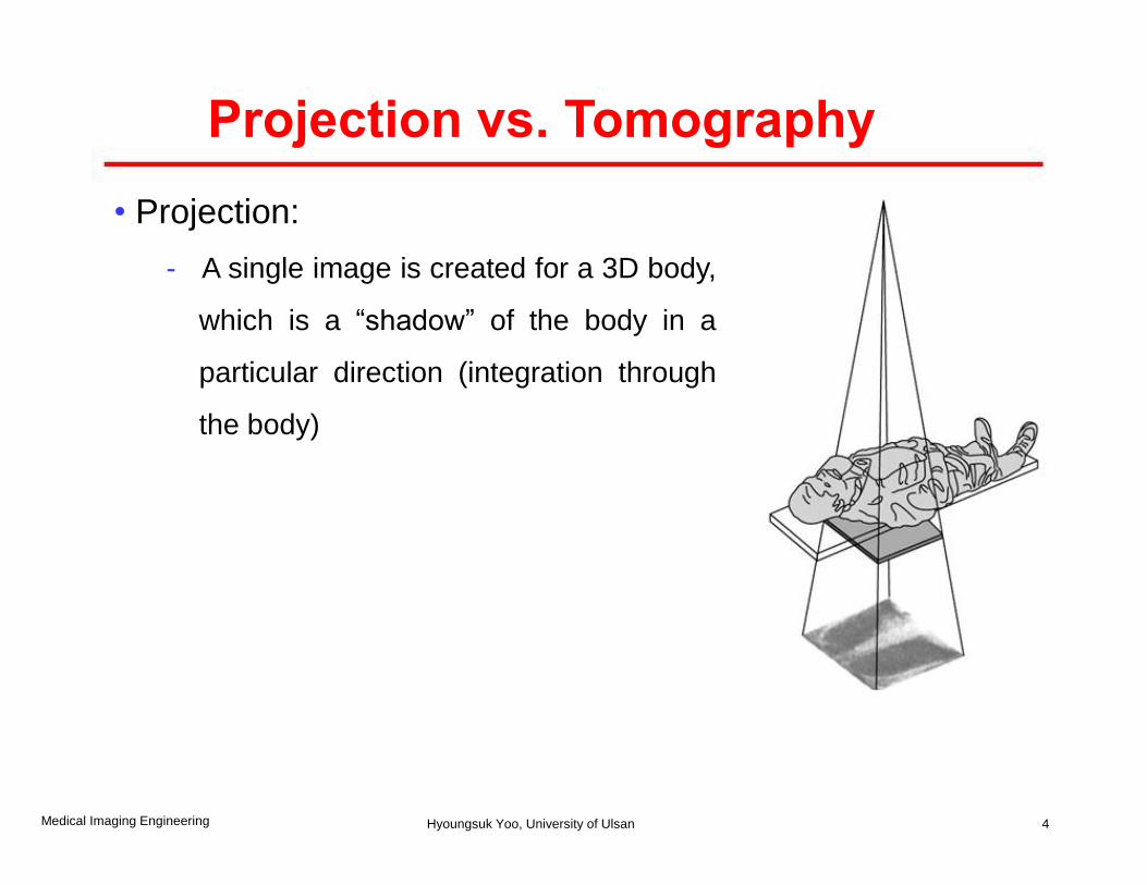
Projection vs. Tomography
• Projection:
- A single image is created for a 3D body,
which is a “shadow” of the body in a
particular direction (integration through
the body)
4 Medical Imaging Engineering Hyoungsuk Yoo, University of Ulsan
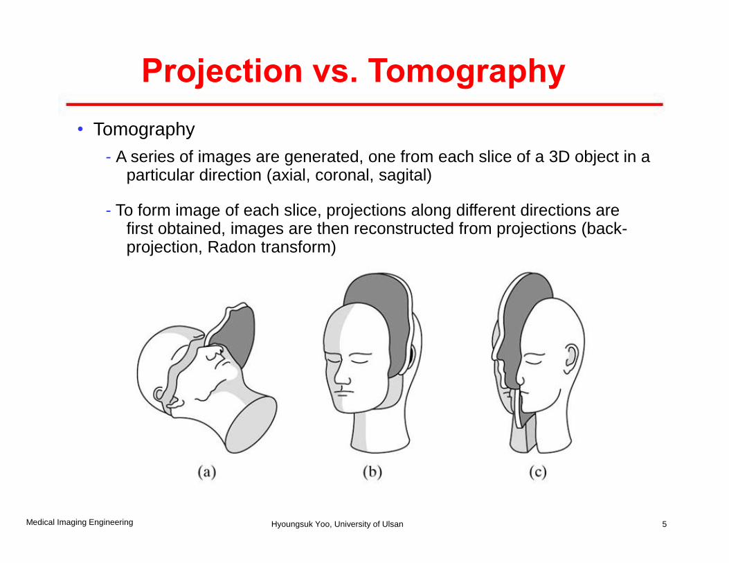
Projection vs. Tomography
• Tomography
- A series of images are generated, one from each slice of a 3D object in a particular direction (axial, coronal, sagital)
- To form image of each slice, projections along different directions are first obtained, images are then reconstructed from projections (back- projection, Radon transform)
5 Medical Imaging Engineering Hyoungsuk Yoo, University of Ulsan
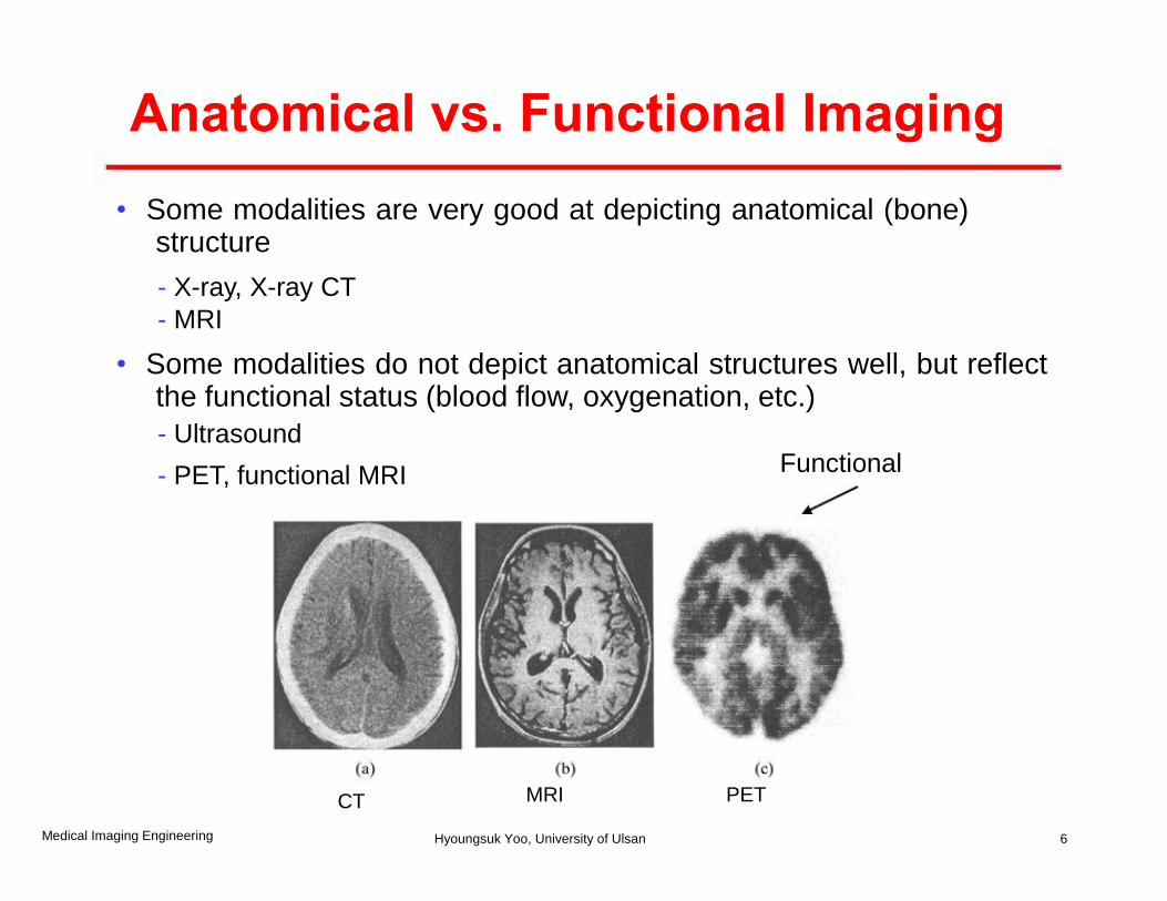
Anatomical vs. Functional Imaging
• Some modalities are very good at depicting anatomical (bone) structure
- X-ray, X-ray CT
- MRI
• Some modalities do not depict anatomical structures well, but reflect the functional status (blood flow, oxygenation, etc.)
- Ultrasound Functional - PET, functional MRI
MRI PET CT
6 Medical Imaging Engineering Hyoungsuk Yoo, University of Ulsan
Common Imaging Modalities
• Projection radiography (X-ray)
• Computed Tomography (CT scan or CAT Scan)
• Nuclear Medicine (SPECT, PET)
• Ultrasound imaging
• MRI
7 Medical Imaging Engineering Hyoungsuk Yoo, University of Ulsan
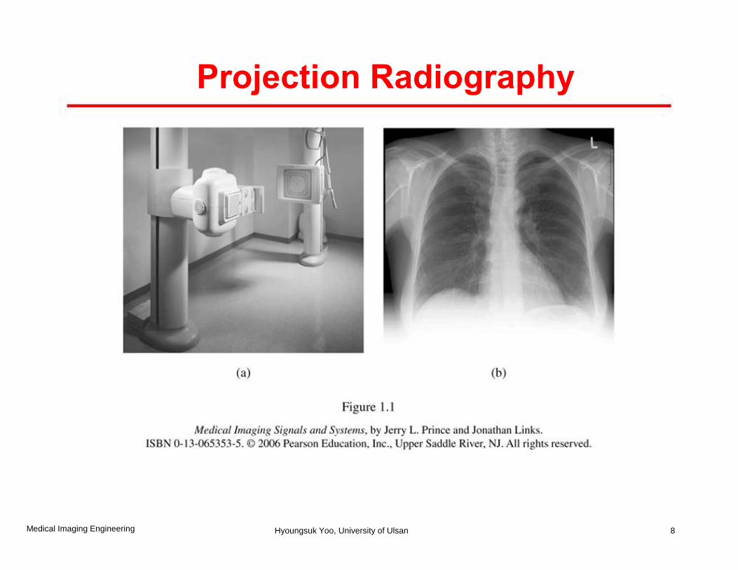
Projection Radiography
8 Medical Imaging Engineering Hyoungsuk Yoo, University of Ulsan
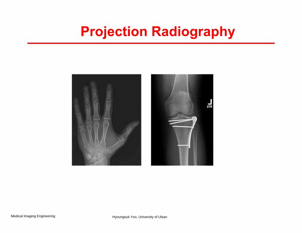
Projection Radiography
Medical Imaging Engineering Hyoungsuk Yoo, University of Ulsan
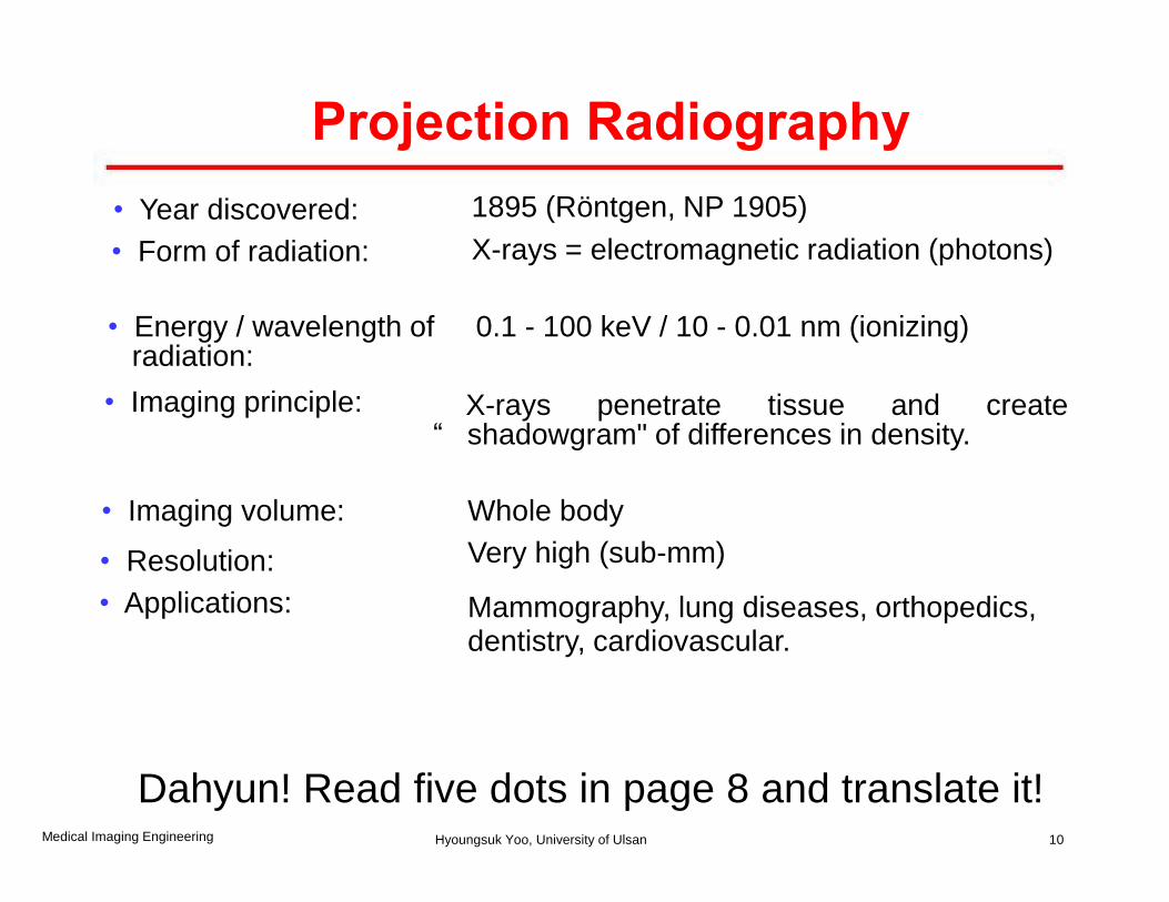
• Year discovered: 1895 (Röntgen, NP 1905)
• Form of radiation: X-rays = electromagnetic radiation (photons)
• Energy / wavelength of radiation:
0.1 - 100 keV / 10 - 0.01 nm (ionizing)
• Imaging principle: X-rays penetrate tissue and create “ shadowgram" of differences in density.
• Imaging volume: Whole body
• Resolution: Very high (sub-mm)
• Applications: Mammography, lung diseases, orthopedics, dentistry, cardiovascular.
10
Projection Radiography
Medical Imaging Engineering Hyoungsuk Yoo, University of Ulsan
Dahyun! Read five dots in page 8 and translate it!
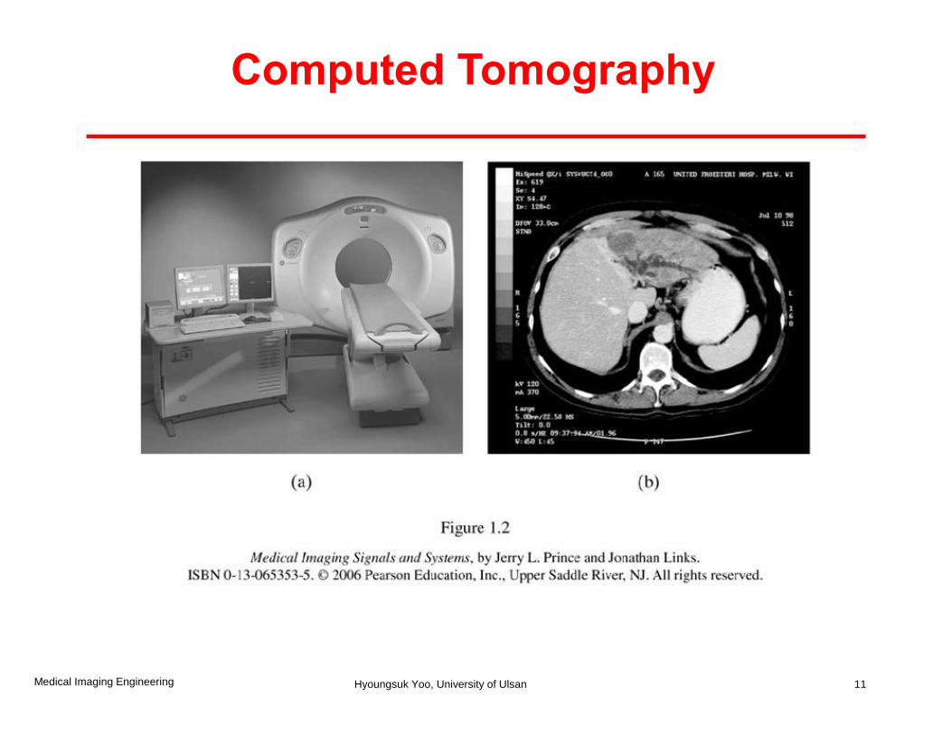
Computed Tomography
11 Medical Imaging Engineering Hyoungsuk Yoo, University of Ulsan
Computed Tomography
Medical Imaging Engineering Hyoungsuk Yoo, University of Ulsan
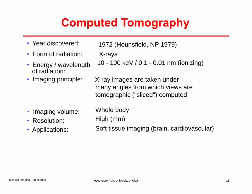
• Year discovered: 1972 (Hounsfield, NP 1979)
• Form of radiation: X-rays
• Energy / wavelength of radiation:
10 - 100 keV / 0.1 - 0.01 nm (ionizing)
• Imaging principle: X-ray images are taken under
many angles from which views are tomographic ("sliced") computed
• Imaging volume: Whole body
• Resolution: High (mm)
• Applications: Soft tissue imaging (brain, cardiovascular)
13
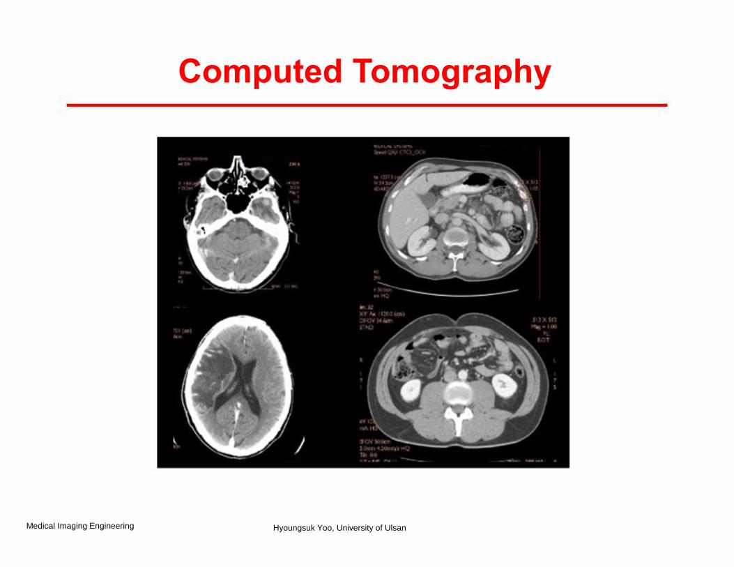
Computed Tomography
Medical Imaging Engineering Hyoungsuk Yoo, University of Ulsan
Nuclear Medicine
• Images can only be made when appropriate radioactive substances (called radiotracer) are introduced into the body that emit gamma rays.
• A nuclear medicine image reflects the local
concentration of a radiotracer within the body
• Three types
-Conventional radionuclide imaging or scintigraphy
-Single photon emission computed tomography (SPECT)
- Positron emission tomography (PET)
14 Medical Imaging Engineering Hyoungsuk Yoo, University of Ulsan
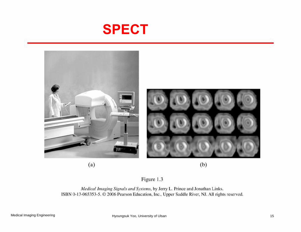
SPECT
15 Medical Imaging Engineering Hyoungsuk Yoo, University of Ulsan
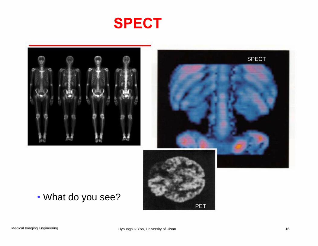
SPECT
• What do you see? PET
16
SPECT
Medical Imaging Engineering Hyoungsuk Yoo, University of Ulsan
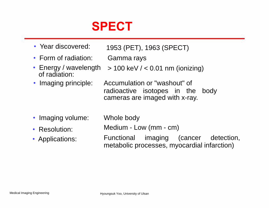
• Year discovered: 1953 (PET), 1963 (SPECT)
• Form of radiation: Gamma rays
• Energy / wavelength of radiation:
> 100 keV / < 0.01 nm (ionizing)
• Imaging principle: Accumulation or "washout" of radioactive isotopes in the body cameras are imaged with x-ray.
• Imaging volume: Whole body
• Resolution: Medium - Low (mm - cm)
• Applications: Functional imaging (cancer detection, metabolic processes, myocardial infarction)
SPECT
Medical Imaging Engineering Hyoungsuk Yoo, University of Ulsan
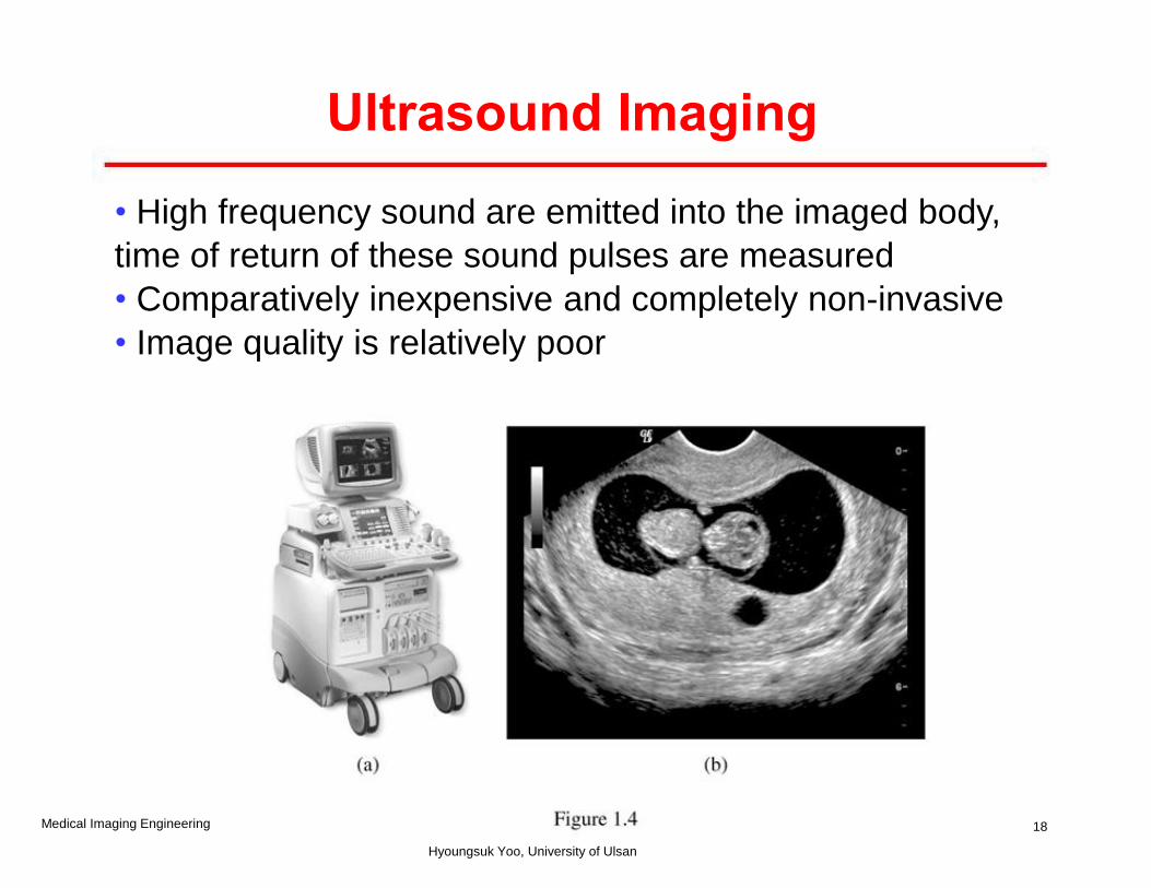
• High frequency sound are emitted into the imaged body,
time of return of these sound pulses are measured
• Comparatively inexpensive and completely non-invasive
• Image quality is relatively poor
18
Ultrasound Imaging
Medical Imaging Engineering
Hyoungsuk Yoo, University of Ulsan
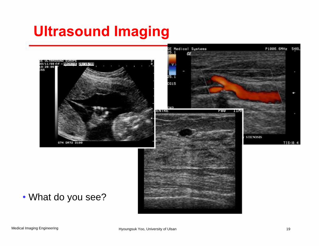
• What do you see?
19
Ultrasound Imaging
Medical Imaging Engineering Hyoungsuk Yoo, University of Ulsan
• Year discovered: 1952 (clinical: 1962)
• Form of radiation: Sound waves (non-ionizing)
NOT EM radiation! • Frequency / wavelength of radiation:
1 - 10 MHz / 1 - 0.1 mm
• Imaging principle: Echoes from discontinuities in
tissue density/speed of sound are registered.
• Imaging volume: < 20 cm
• Resolution: High (mm)
• Applications: Soft tissue, blood flow
20
Ultrasound Imaging
Medical Imaging Engineering Hyoungsuk Yoo, University of Ulsan
Jiyoung! Read four dots in page 11-12 and translate it!
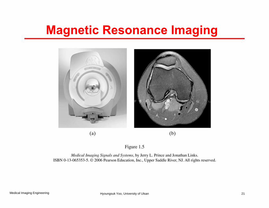
Magnetic Resonance Imaging
21 Medical Imaging Engineering Hyoungsuk Yoo, University of Ulsan
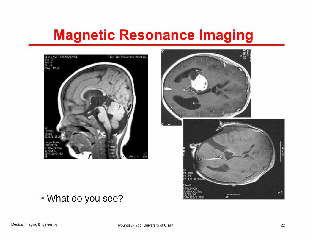
• What do you see?
22
Magnetic Resonance Imaging
Medical Imaging Engineering Hyoungsuk Yoo, University of Ulsan
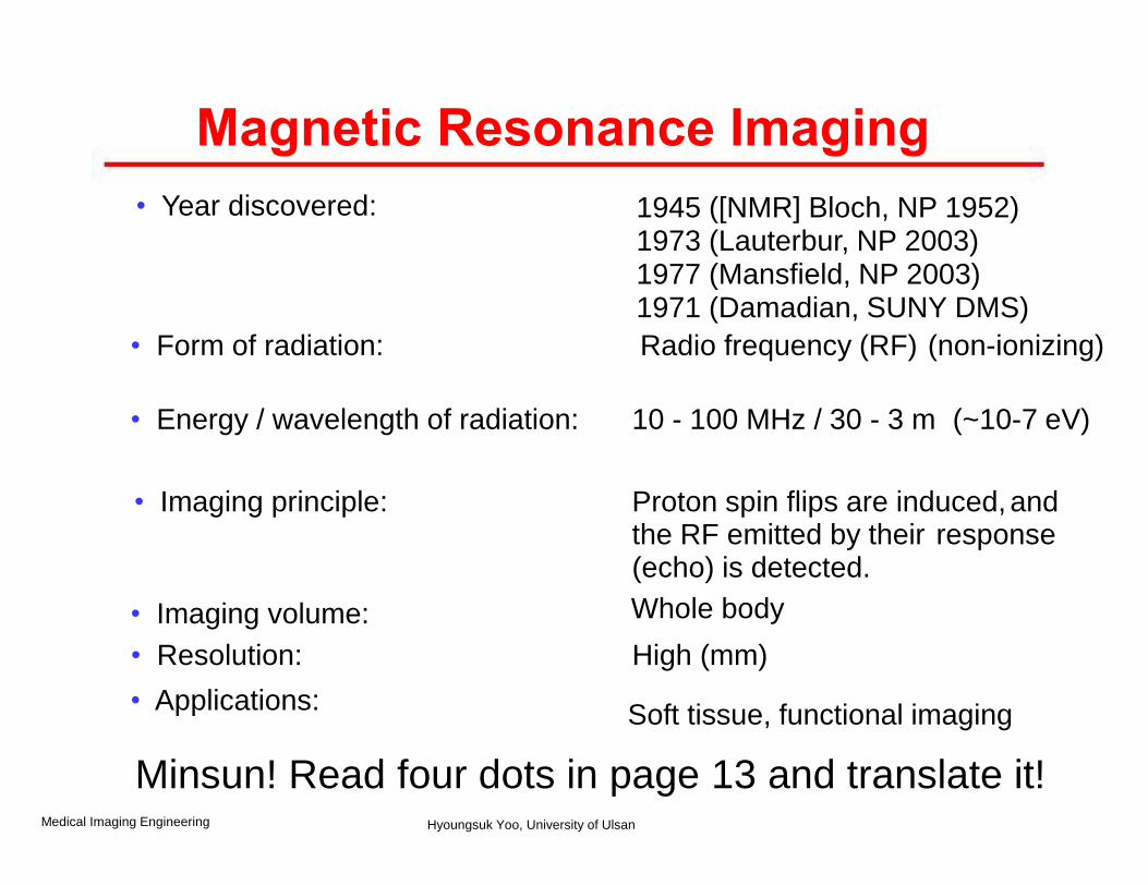
• Year discovered: 1945 ([NMR] Bloch, NP 1952) 1973 (Lauterbur, NP 2003) 1977 (Mansfield, NP 2003) 1971 (Damadian, SUNY DMS)
• Form of radiation: Radio frequency (RF) (non-ionizing)
• Energy / wavelength of radiation: 10 - 100 MHz / 30 - 3 m (~10-7 eV)
• Imaging principle: Proton spin flips are induced, and the RF emitted by their response (echo) is detected.
• Imaging volume: Whole body
• Resolution: High (mm)
• Applications: Soft tissue, functional imaging
Magnetic Resonance Imaging
Medical Imaging Engineering Hyoungsuk Yoo, University of Ulsan
Minsun! Read four dots in page 13 and translate it!
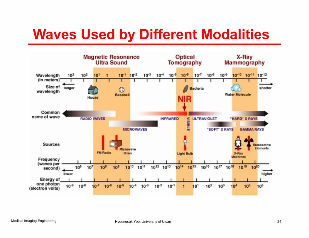
Waves Used by Different Modalities
24 Medical Imaging Engineering Hyoungsuk Yoo, University of Ulsan
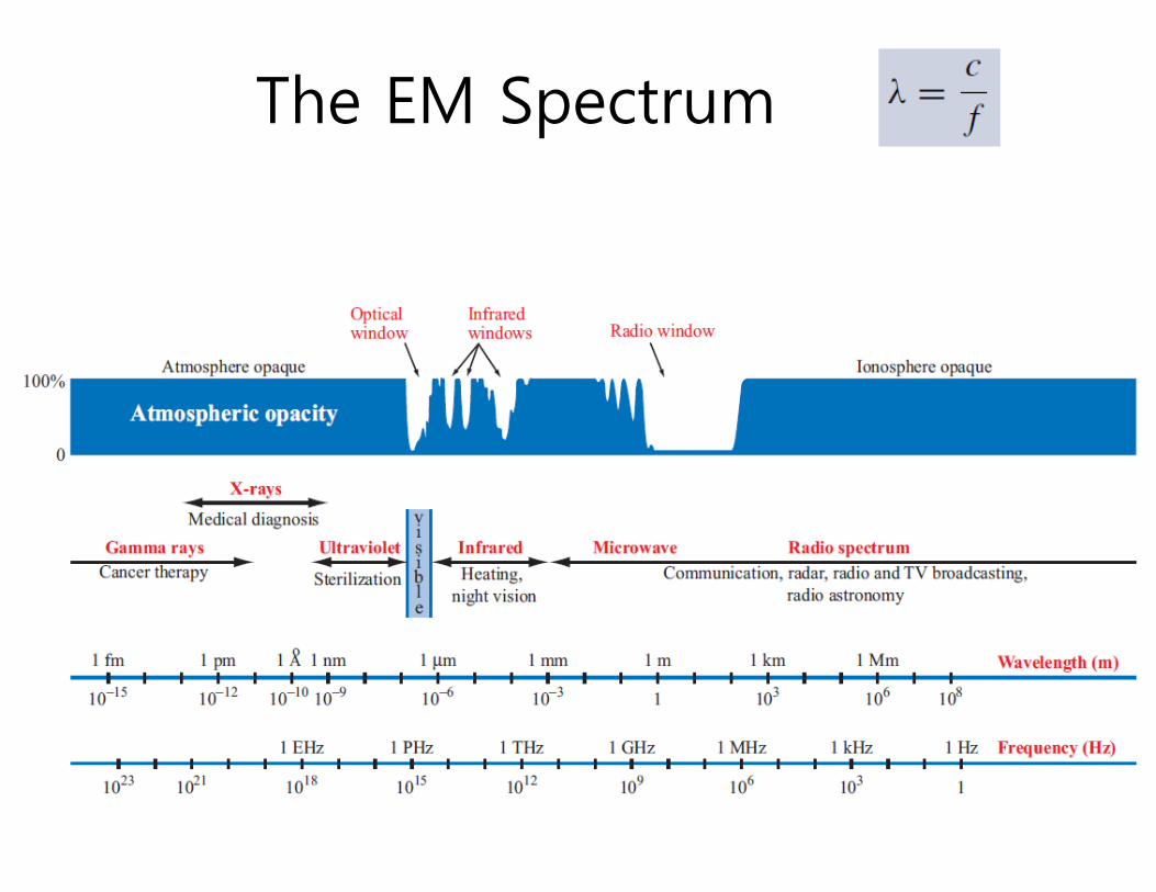
The EM Spectrum
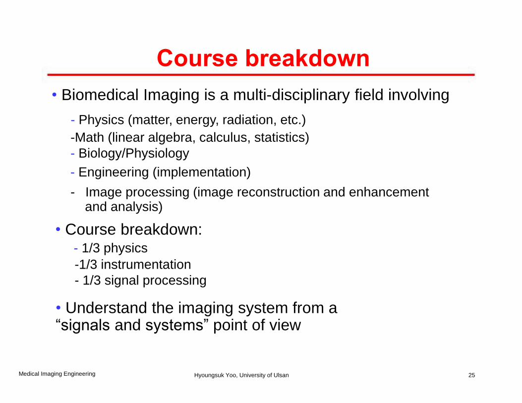
Course breakdown
• Biomedical Imaging is a multi-disciplinary field involving
- Physics (matter, energy, radiation, etc.)
-Math (linear algebra, calculus, statistics)
- Biology/Physiology
- Engineering (implementation)
- Image processing (image reconstruction and enhancement and analysis)
• Course breakdown: - 1/3 physics
-1/3 instrumentation
- 1/3 signal processing
• Understand the imaging system from a “signals and systems” point of view
25 Medical Imaging Engineering Hyoungsuk Yoo, University of Ulsan
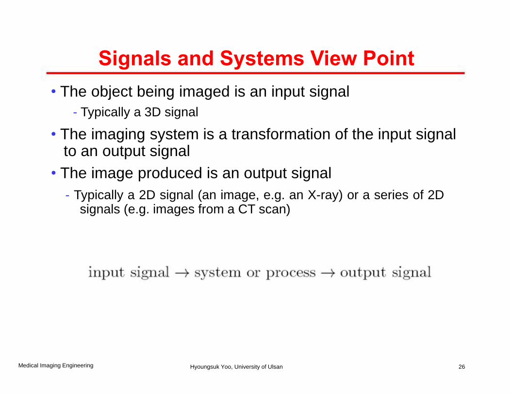
Signals and Systems View Point
• The object being imaged is an input signal
- Typically a 3D signal
• The imaging system is a transformation of the input signal to an output signal
• The image produced is an output signal
- Typically a 2D signal (an image, e.g. an X-ray) or a series of 2D signals (e.g. images from a CT scan)
26 Medical Imaging Engineering Hyoungsuk Yoo, University of Ulsan
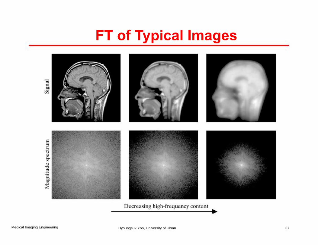
FT of Typical Images
37 Medical Imaging Engineering Hyoungsuk Yoo, University of Ulsan

Extra Readings
• See Chap 2 of textbook for more extensive reviews of signals and systems.
• I would like to recommend that you try to find the problem in the textbook as a reminder. Homework 1. Pick one problem from 2. 26 to 2. 33 (page 60~62) and solve. Due date : Sep. 20th, 2011
39 Medical Imaging Engineering Hyoungsuk Yoo, University of Ulsan
Image Quality
• Introduction
• Contrast
• Resolution
• Noise
• Artifacts
• Distortions
40 Medical Imaging Engineering Hyoungsuk Yoo, University of Ulsan
Measures of Quality
• Physics-oriented issues:
- contrast, resolution
-noise, artifacts, distortion
- Quantitative accuracy
• Task-oriented issues:
-sensitivity, specificity
-diagnostic accuracy
41 Medical Imaging Engineering Hyoungsuk Yoo, University of Ulsan
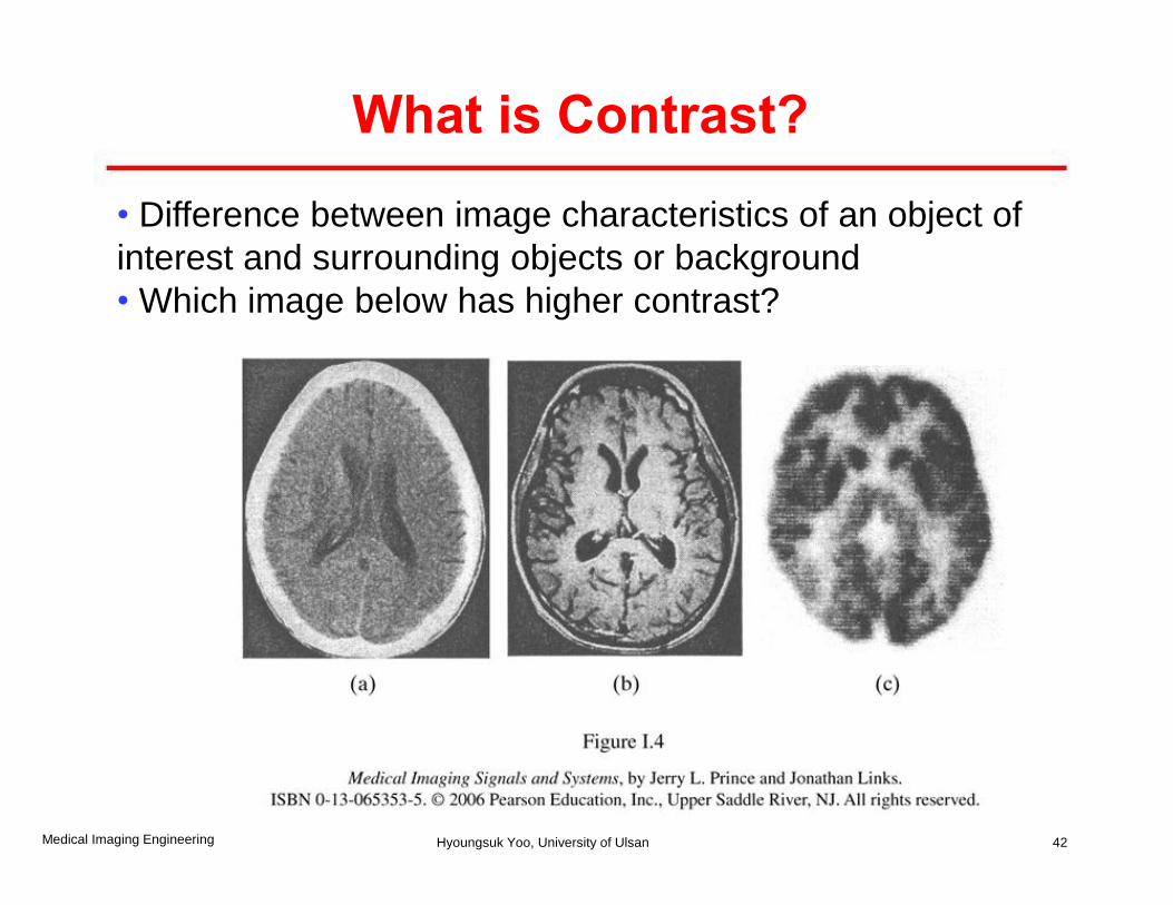
What is Contrast?
• Difference between image characteristics of an object of
interest and surrounding objects or background
• Which image below has higher contrast?
42 Medical Imaging Engineering Hyoungsuk Yoo, University of Ulsan

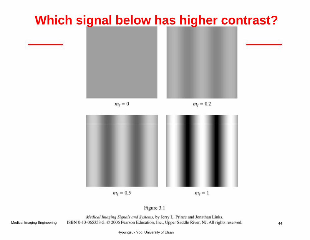
Contrast
• Contrast: Difference between image characteristics of an
object of interest and surrounding objects or background
• General definition
- fmax, fmin: maximum and minimum values of the signal in an image
• For a sinusoidal signal
43 Medical Imaging Engineering Hyoungsuk Yoo, University of Ulsan
44 Medical Imaging Engineering
Hyoungsuk Yoo, University of Ulsan
Which signal below has higher contrast?
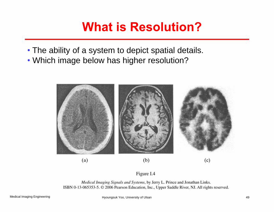
What is Resolution?
• The ability of a system to depict spatial details.
• Which image below has higher resolution?
49 Medical Imaging Engineering Hyoungsuk Yoo, University of Ulsan
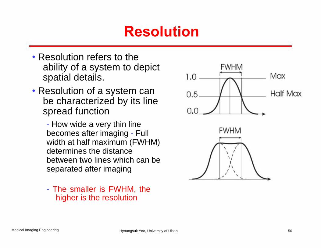
Resolution
• Resolution refers to the ability of a system to depict spatial details.
• Resolution of a system can be characterized by its line spread function
- How wide a very thin line becomes after imaging - Full width at half maximum (FWHM) determines the distance between two lines which can be separated after imaging
- The smaller is FWHM, the higher is the resolution
50 Medical Imaging Engineering Hyoungsuk Yoo, University of Ulsan
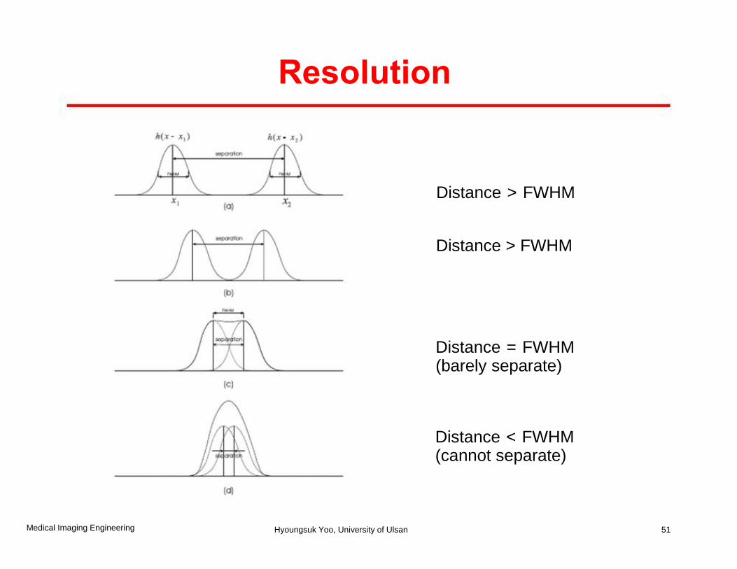
Distance > FWHM
Distance > FWHM
Distance = FWHM (barely separate)
Distance < FWHM (cannot separate)
51
Resolution
Medical Imaging Engineering Hyoungsuk Yoo, University of Ulsan
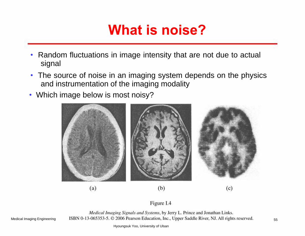
What is noise?
• Random fluctuations in image intensity that are not due to actual signal
• The source of noise in an imaging system depends on the physics and instrumentation of the imaging modality
• Which image below is most noisy?
55 Medical Imaging Engineering
Hyoungsuk Yoo, University of Ulsan
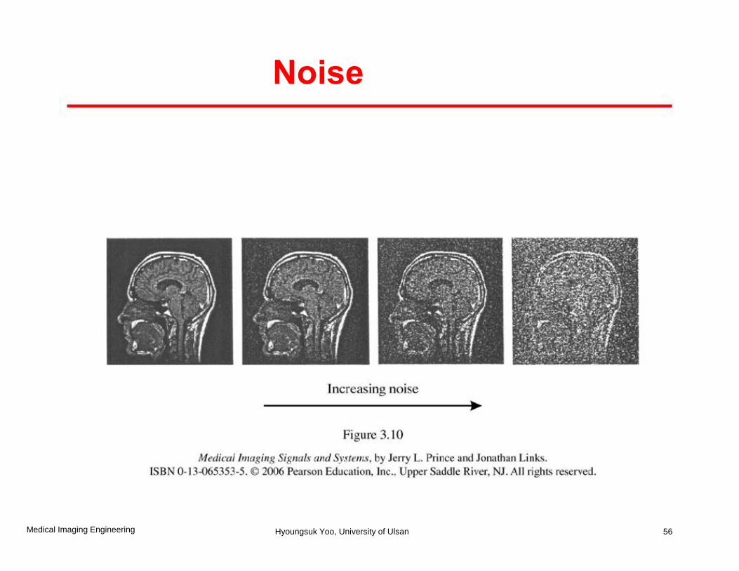
Noise
56 Medical Imaging Engineering Hyoungsuk Yoo, University of Ulsan
Artifacts, distortion & accuracy
• Artifacts:
- Some imaging systems can create image features that do not represent a valid object in the imaged patient, or false shapes/textures.
• Distortion
- Some imaging system may distort the actual shape/position and other geometrics of imaged object.
• Accuracy
- Conformity to truth and clinical utility
62 Medical Imaging Engineering Hyoungsuk Yoo, University of Ulsan
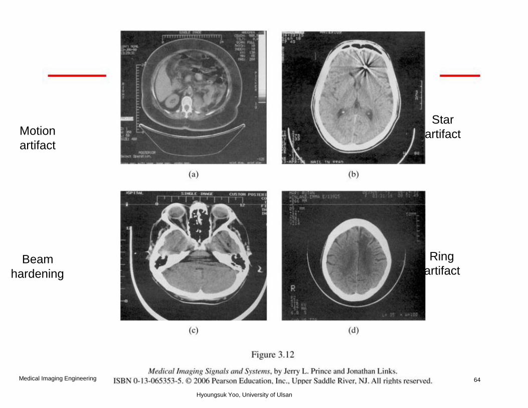
Star Motion artifact artifact
Ring Beam artifact hardening
64 Medical Imaging Engineering
Hyoungsuk Yoo, University of Ulsan
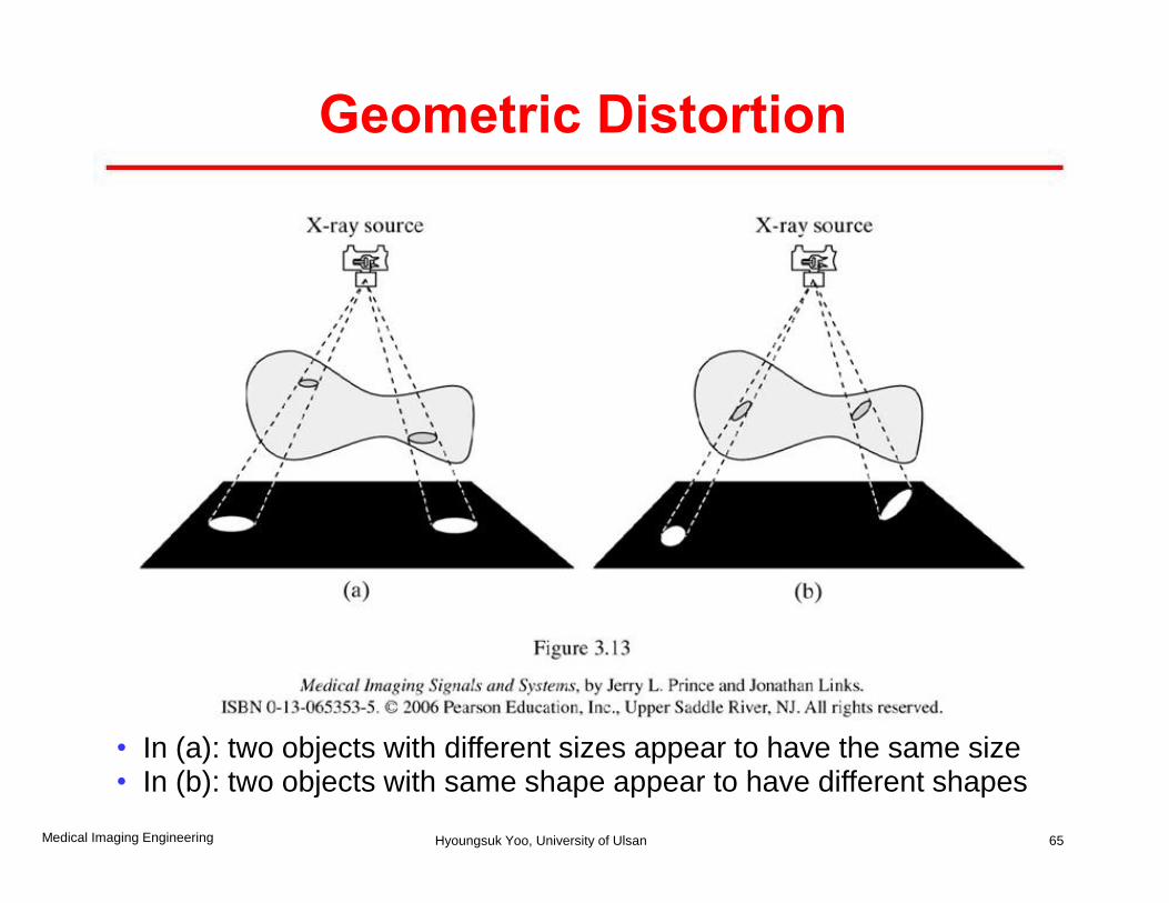
Geometric Distortion
• In (a): two objects with different sizes appear to have the same size • In (b): two objects with same shape appear to have different shapes
65 Medical Imaging Engineering Hyoungsuk Yoo, University of Ulsan
Reference
• Prince and Links, Medical Imaging Signals and Systems, Chap 1-3.
69 Medical Imaging Engineering Hyoungsuk Yoo, University of Ulsan