Embed Size (px)
Citation preview
Reagents for HospitalsMedical and Research Laboratories
Panreac Applichem
Reagentsfor Hospitals
2
About us
Medical Laboratories
Microscopy / p. 7
Indroduction / p. 7
Sample Processing / p. 8Getting the sample Types of processing Pathological anatomy laboratory process From sampling to processing
Techniques and Stages / p. 10Fixing / p. 10
Types of Chemical Fixative Agents Formaldehyde Fixation ProcedureHistofix pre-dosed and Substitutes of FormaldehydeReagents for FixingDecalcifiers
Drying and Clearing / p. 16Reagents for DryingReagents for Clearing
Inclusion / p. 19Embedding media Reagents for Embedding (paraffins)
Cutting / p. 20
Rehydration / p. 21Deparaffinization-HydrationReagents for Deparaffinization-Hydration
Staining / p. 22 Dyes for microscopyHematoxylin-Eosin StainHematoxylinsEosinsReagents for Staining
Powdered dyesDyes in solution
Mounting / p. 29Mounting media and Immersion oils
Reagents for Histology / p. 30Giemsa StainPAS StainingMasson’s Trichrome StainingReticulin fiber staining
Reagents for Cytology / p. 38Papanicolaou Stain
Reagents for Clinical Microbiology / p. 41Gram StainingZiehl-Neelsen StainOther staining solutions
Reagents for Hematology / p. 48Kit for Fast Staining in Haematology (Fast Panoptic)May Grünwald-Giemsa or Pappenheim StainWright’s StainOther Products
Auxiliary Products / p. 52General ReagentspH Indicator stripsDerquim detergents
Summary
4
6
3
Research Laboratories
Reagents for Genomics / p. 58PCR / p. 58DNA Decontamination / p. 59Gel electrophoresis / p. 59Nucleic Acid Isolation / p. 59Cloning Assays / p. 60Buffers and Solvents / p. 60
Reagents for Proteomics / p. 61Products for electrophoresis and blotting / p. 61
Reagents for Cell Culture / p. 64Banish cell culture contamination / p. 64
Trends on new techniques for Clinical Diagnosis
Liquid Biopsies / p. 65
Package pictograms
58
65
67
Panreac Applichem
Reagentsfor Hospitals
4
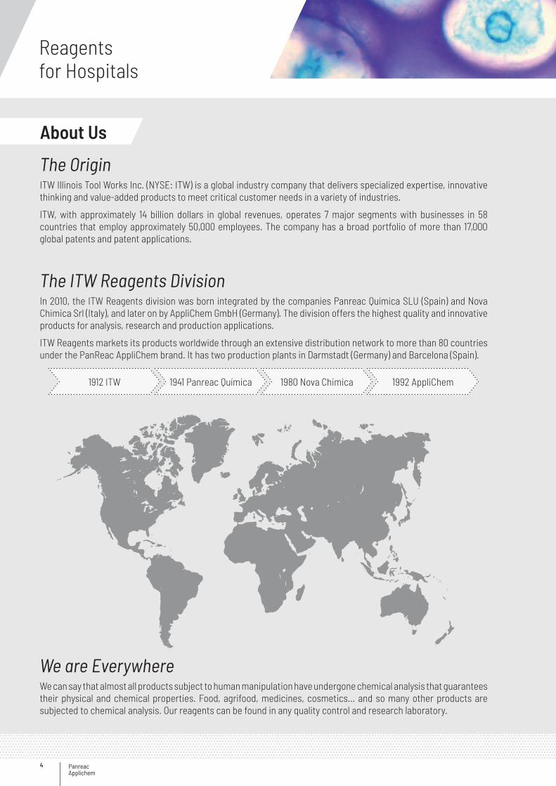
We are EverywhereWe can say that almost all products subject to human manipulation have undergone chemical analysis that guarantees their physical and chemical properties. Food, agrifood, medicines, cosmetics... and so many other products are subjected to chemical analysis. Our reagents can be found in any quality control and research laboratory.
The OriginITW Illinois Tool Works Inc. (NYSE: ITW) is a global industry company that delivers specialized expertise, innovative thinking and value-added products to meet critical customer needs in a variety of industries.
ITW, with approximately 14 billion dollars in global revenues, operates 7 major segments with businesses in 58 countries that employ approximately 50,000 employees. The company has a broad portfolio of more than 17,000 global patents and patent applications.
The ITW Reagents DivisionIn 2010, the ITW Reagents division was born integrated by the companies Panreac Química SLU (Spain) and Nova Chimica Srl (Italy), and later on by AppliChem GmbH (Germany). The division offers the highest quality and innovative products for analysis, research and production applications.
ITW Reagents markets its products worldwide through an extensive distribution network to more than 80 countries under the PanReac AppliChem brand. It has two production plants in Darmstadt (Germany) and Barcelona (Spain).
1912 ITW 1941 Panreac Química 1980 Nova Chimica 1992 AppliChem
About Us

5
Our range of Laboratory Chemicals include:
Analytical reagentsReagents for volumetric analysisReagents and solvents for general applicationsReagents and solvents for HPLCReagents and solvents for GCReagents for metallic traces analysisAnalytical standardsReagents and solvents for specific applicationsProducts for clinical diagnosisProducts for microbiology
ISO 9001:2015 ISO 14001:2015 OHSAS 18001:2007
Our range of Laboratory Biochemicals cover:
Cell Biology / Cell CultureProtein Biochemistry and ElectrophoresisNucleic Acid BiochemistryGeneral Biochemicals and Biological BuffersSpecial Biochemicals
ExcellenceOur products are strictly controlled in our laboratories and meet the highest quality requirements. A multi-site Integrated Management System for Quality, Environment and Safety is implemented in all activities and processes.
Service & BenefitsExceptional know-how and a wide range of chemicals and biochemicals for a great diversity of applications.
European production committed to corporate social responsibility (CSR).
Efficient global distribution network to export our products worldwide to more than 80 countries.
Qualified management team fully committed to our business project.

Panreac Applichem
Reagentsfor Hospitals
6
Clinical Pathology: Hematology, Histopathology, Cytology, Routine Pathology.
Clinical Microbiology: Bacteriology, Mycobacteriology, Virology, Mycology, Parasitology, Immunology, Serology.
Clinical Biochemistry: Biochemical analysis, Hormonal assays, etc.
Molecular diagnostic laboratory or cytogenetics and molecular biology lab.
Hospital laboratoriesAttached to a hospital to perform tests on
patients. We can find 4 different types.
Outside clinical laboratoriesFor extremely specialized tests, sample
may go to an external research laboratory.
Medical and Research LaboratoriesMedical Laboratories are focused on applied science mainly on a production-like basis, as opposed to Research Laboratories that focus on basic science on an academic basis.
A Medical Laboratory or clinical laboratory is where tests are usually done on clinical specimens in order to obtain information about the health of a patient as pertaining to the diagnosis, treatment, and prevention of disease.
Research Laboratories use the conventional techniques for Genomics, Proteomics and Cell Culture procedures.
PanReac AppliChem Products for Hospital Laboratories: Medical Laboratories: Products for Microscopy. Research Laboratories: Products for Genomics, Proteomics and Cell Culture.
In the first part of the brochure we will focus on the Clinical Pathology and Microbiology laboratories according to the type of investigation and the main fields that use microscopy for the analysis: Citology, Haematology, Microbiology and Histology. At the end you will find reagents for Research Laboratories.
Medical LaboratoriesIn many countries there are mainly two types of Medical Laboratories as per the types of investigations carried out.
7
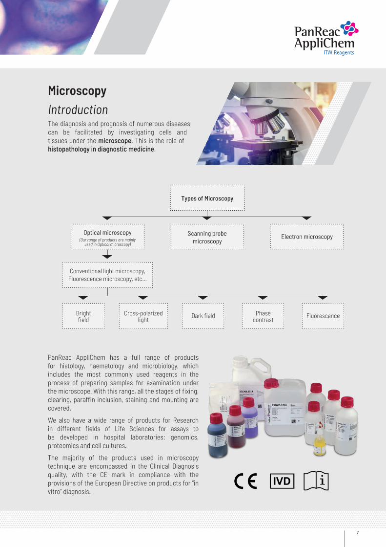
MicroscopyIntroductionThe diagnosis and prognosis of numerous diseases can be facilitated by investigating cells and tissues under the microscope. This is the role of histopathology in diagnostic medicine.
Types of Microscopy
Scanning probe microscopy
Optical microscopyElectron microscopy
Conventional light microscopy, Fluorescence microscopy, etc…
Bright field
Cross-polarized light
Phase contrast
FluorescenceDark field
(Our range of products are mainly used in Optical microscopy)
PanReac AppliChem has a full range of products for histology, haematology and microbiology, which includes the most commonly used reagents in the process of preparing samples for examination under the microscope. With this range, all the stages of fixing, clearing, paraffin inclusion, staining and mounting are covered.
We also have a wide range of products for Research in different fields of Life Sciences for assays to be developed in hospital laboratories: genomics, proteomics and cell cultures.
The majority of the products used in microscopy technique are encompassed in the Clinical Diagnosis quality, with the CE mark in compliance with the provisions of the European Directive on products for “in vitro” diagnosis.
Panreac Applichem
Reagentsfor Hospitals
8
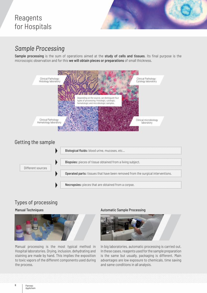
Getting the sample
Types of processing
Clinical Pathology: Histology laboratory
Clinical Pathology: Hematology laboratory
Clinical Pathology: Cytology laboratory
Clinical microbiology laboratory
Depending on the source, we distinguish four types of processing: histologic, cytologic, hematologic and microbiologic samples.
Different sources
Biological fluids: blood urine, mucoses, etc…
Biopsies: pieces of tissue obtained from a living subject.
Operated parts: tissues that have been removed from the surgical interventions.
Necropsies: pieces that are obtained from a corpse.
Manual Techniques
Manual processing is the most typical method in Hospital laboratories. Drying, inclusion, dehydrating and staining are made by hand. This implies the exposition to toxic vapors of the different components used during the process.
Automatic Sample Processing
In big laboratories, automatic processing is carried out. In these cases, reagents used for the sample preparation is the same but usually, packaging is different. Main advantages are low exposure to chemicals, time saving and same conditions in all analysis.
Sample ProcessingSample processing is the sum of operations aimed at the study of cells and tissues. Its final purpose is the microscopic observation and for this we will obtain pieces or preparations of small thickness.
9
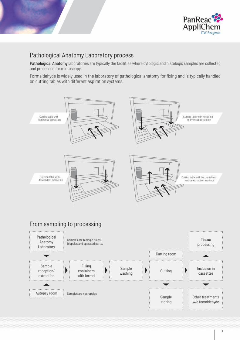
Pathological Anatomy Laboratory processPathological Anatomy laboratories are typically the facilities where cytologic and histologic samples are collected and processed for microscopy.
Formaldehyde is widely used in the laboratory of pathological anatomy for fixing and is typically handled on cutting tables with different aspiration systems.
From sampling to processing
Pathological Anatomy
Laboratory
Autopsy room Samples are necropsies
Samples are biologic fluids, biopsies and operated parts.
Cutting room
Sample reception/extraction
Inclusion in cassettes
Tissue processing
Filling containers with formol
Sample washing
Cutting
Sample storing
Other treatments w/o fomaldehyde
Cutting table with horizontal extraction
Cutting table with descendent extraction
Cutting table with horizontal and vertical extraction
Cutting table with horizontal and vertical extraction in a hood
Panreac Applichem
Reagentsfor Hospitals
10
Techniques and stagesAlthough most of the stages are common, some of the steps are exclusive only for one type of sample processing. For example, inclusion is only done on tissues and heat fixation only on blood samples.
Type of Sample FixingDrying and
ClearingInclusion Cutting Rehydration Staining Mounting Microscopy
Histologic
MicrobiologicHematologicCytologic
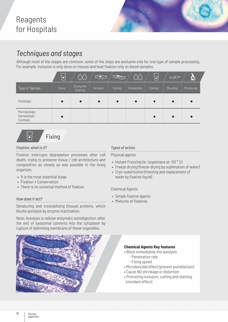
Fixation, what is it?
Fixation interrupts degradation processes after cell death, trying to preserve tissue / cell architecture and composition as closely as was possible in the living organism.
• It is the most essential stage• Fixation ≠ Conservation• There is no universal method of fixation
How does it act?
Denaturing and insolubilizing (tissue) proteins, which blocks autolysis by enzyme inactivation.
Note: Autolysis is cellular enzymatic autodigestion, after the exit of lysosomal contents into the cytoplasm by rupture of delimiting membrane of these organelles.
Types of action
Physical agents
• Instant Freezing (ie. isopentane at -50 ° C)• Freeze drying (freeze-drying by sublimation of water)• Cryo-substitution (freezing and replacement of
water by fixative liquid)
Chemical Agents
• Simple fixative agents• Mixtures of fixatives
Chemical Agents Key features• Block immediately the autolysis - Penetration rate - Fixing speed• Microbiocidal effect (prevent putrefaction)• Cause NO shrinkage or distortion• Promoting inclusion, cutting and staining
(mordant effect)
Fixing
11
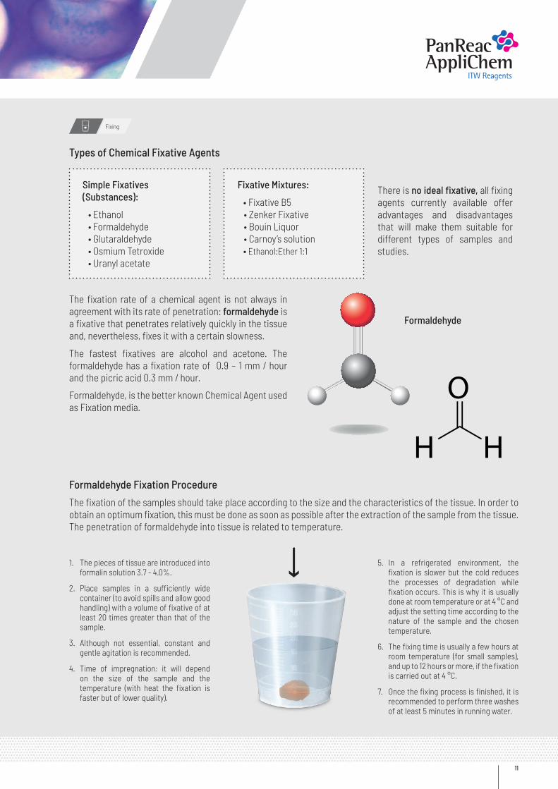
Formaldehyde Fixation Procedure
The fixation of the samples should take place according to the size and the characteristics of the tissue. In order to obtain an optimum fixation, this must be done as soon as possible after the extraction of the sample from the tissue. The penetration of formaldehyde into tissue is related to temperature.
1. The pieces of tissue are introduced into formalin solution 3.7 - 4.0%.
2. Place samples in a sufficiently wide container (to avoid spills and allow good handling) with a volume of fixative of at least 20 times greater than that of the sample.
3. Although not essential, constant and gentle agitation is recommended.
4. Time of impregnation: it will depend on the size of the sample and the temperature (with heat the fixation is faster but of lower quality).
5. In a refrigerated environment, the fixation is slower but the cold reduces the processes of degradation while fixation occurs. This is why it is usually done at room temperature or at 4 °C and adjust the setting time according to the nature of the sample and the chosen temperature.
6. The fixing time is usually a few hours at room temperature (for small samples), and up to 12 hours or more, if the fixation is carried out at 4 °C.
7. Once the fixing process is finished, it is recommended to perform three washes of at least 5 minutes in running water.
Simple Fixatives (Substances):
• Ethanol • Formaldehyde • Glutaraldehyde • Osmium Tetroxide • Uranyl acetate
Fixative Mixtures:
• Fixative B5 • Zenker Fixative • Bouin Liquor • Carnoy’s solution • Ethanol:Ether 1:1
There is no ideal fixative, all fixing agents currently available offer advantages and disadvantages that will make them suitable for different types of samples and studies.
The fixation rate of a chemical agent is not always in agreement with its rate of penetration: formaldehyde is a fixative that penetrates relatively quickly in the tissue and, nevertheless, fixes it with a certain slowness.
The fastest fixatives are alcohol and acetone. The formaldehyde has a fixation rate of 0.9 – 1 mm / hour and the picric acid 0.3 mm / hour.
Formaldehyde, is the better known Chemical Agent used as Fixation media.
Types of Chemical Fixative Agents
Fixing
Formaldehyde
Panreac Applichem
Reagentsfor Hospitals
12
Two different alternatives for manipulation
Use an alternative substance
Decrease the exposure times
Description Code Package
Histofix ® Substitute of FormaldehydeComposition:Glyoxal …………….......…… 15-25 %Ethanol absolute …......…… 5-8 %Acetic Acid glacial ….....….. < 5 %Methanol ……………......…. < 0.5 %
255805.2711 ~ 1000 ml
255805.2714 5 L
Histofix ® Substitute of Formaldehyde ready to use257157.1211 b 1000 ml
257157.1214 i 5 L
Description Code Package
Histofix ® Preservative ready to useAssay (Iodom.): 3.7-4.0 % Formaldehyde pH: 6.8-7.2
256462.0905 b 45x10 ml
256462.0967 b 24x75 ml
256462.0944 b 12x200 ml
256462.09118 G 1.5 L
Other sizes available
Histofix® is a trademark of Panreac Quimica SLU
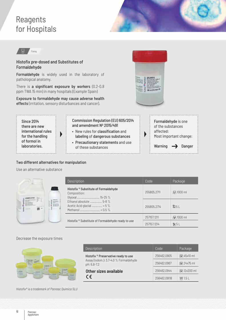
Histofix pre-dosed and Substitutes of Formaldehyde
Formaldehyde is widely used in the laboratory of pathological anatomy.
There is a significant exposure by workers (0.2-0.8 ppm TWA 15 min) in many hospitals (Example Spain)
Exposure to formaldehyde may cause adverse health effects (irritation, sensory disturbances and cancer).
Fixing
Since 2014 there are new international rules for the handling of formol in laboratories.
Commission Regulation (EU) 605/2014 and amendment Nº 2015/491• New rules for classification and
labeling of dangerous substances• Precautionary statements and use
of these substances
Formaldehyde is one of the substances affected:Most important change:
Warning Danger
13
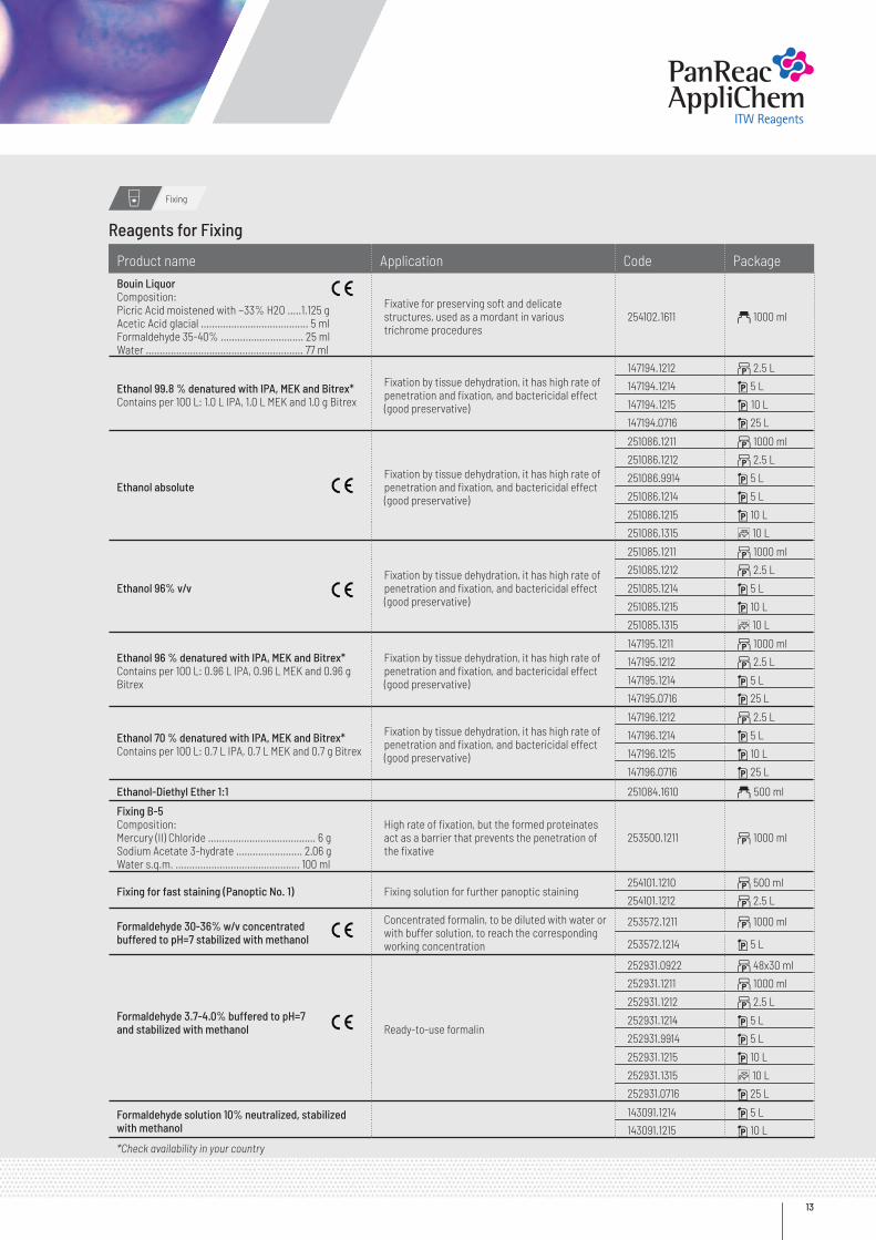
Fixing
Product name Application Code PackageBouin LiquorComposition:Picric Acid moistened with ~33% H2O .....1.125 gAcetic Acid glacial ....................................... 5 mlFormaldehyde 35-40% .............................. 25 mlWater ......................................................... 77 ml
Fixative for preserving soft and delicate structures, used as a mordant in various trichrome procedures
254102.1611 a 1000 ml
Ethanol 99.8 % denatured with IPA, MEK and Bitrex*Contains per 100 L: 1.0 L IPA, 1.0 L MEK and 1.0 g Bitrex
Fixation by tissue dehydration, it has high rate of penetration and fixation, and bactericidal effect (good preservative)
147194.1212 b 2.5 L
147194.1214 i 5 L
147194.1215 i 10 L
147194.0716 i 25 L
Ethanol absoluteFixation by tissue dehydration, it has high rate of penetration and fixation, and bactericidal effect (good preservative)
251086.1211 b 1000 ml
251086.1212 b 2.5 L
251086.9914 i 5 L
251086.1214 i 5 L
251086.1215 i 10 L
251086.1315 C 10 L
Ethanol 96% v/vFixation by tissue dehydration, it has high rate of penetration and fixation, and bactericidal effect (good preservative)
251085.1211 b 1000 ml
251085.1212 b 2.5 L
251085.1214 i 5 L
251085.1215 i 10 L
251085.1315 C 10 L
Ethanol 96 % denatured with IPA, MEK and Bitrex*Contains per 100 L: 0.96 L IPA, 0.96 L MEK and 0.96 g Bitrex
Fixation by tissue dehydration, it has high rate of penetration and fixation, and bactericidal effect (good preservative)
147195.1211 b 1000 ml
147195.1212 b 2.5 L
147195.1214 i 5 L
147195.0716 i 25 L
Ethanol 70 % denatured with IPA, MEK and Bitrex*Contains per 100 L: 0.7 L IPA, 0.7 L MEK and 0.7 g Bitrex
Fixation by tissue dehydration, it has high rate of penetration and fixation, and bactericidal effect (good preservative)
147196.1212 b 2.5 L
147196.1214 i 5 L
147196.1215 i 10 L
147196.0716 i 25 L
Ethanol-Diethyl Ether 1:1 251084.1610 a 500 ml
Fixing B-5Composition:Mercury (II) Chloride ....................................... 6 gSodium Acetate 3-hydrate ........................ 2.06 gWater s.q.m. ............................................. 100 ml
High rate of fixation, but the formed proteinates act as a barrier that prevents the penetration of the fixative
253500.1211 b 1000 ml
Fixing for fast staining (Panoptic No. 1) Fixing solution for further panoptic staining254101.1210 b 500 ml
254101.1212 b 2.5 L
Formaldehyde 30-36% w/v concentrated buffered to pH=7 stabilized with methanol
Concentrated formalin, to be diluted with water or with buffer solution, to reach the corresponding working concentration
253572.1211 b 1000 ml
253572.1214 i 5 L
Formaldehyde 3.7-4.0% buffered to pH=7and stabilized with methanol Ready-to-use formalin
252931.0922 b 48x30 ml
252931.1211 b 1000 ml
252931.1212 b 2.5 L
252931.1214 i 5 L
252931.9914 i 5 L
252931.1215 i 10 L
252931.1315 C 10 L
252931.0716 i 25 L
Formaldehyde solution 10% neutralized, stabilized with methanol
143091.1214 i 5 L
143091.1215 i 10 L
*Check availability in your country
Reagents for Fixing
Panreac Applichem
Reagentsfor Hospitals
14
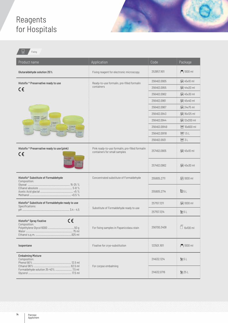
Product name Application Code Package
Glutaraldehyde solution 25% Fixing reagent for electronic microscopy 253857.1611 a 1000 ml
Histofix ® Preservative ready to use Ready-to-use formalin, pre-filled formalin containers
256462.0905 b 45x10 ml
256462.0955 b 44x20 ml
256462.0962 b 45x30 ml
256462.0961 b 45x40 ml
256462.0967 b 24x75 ml
256462.0943 b 16x125 ml
256462.0944 b 12x200 ml
256462.09149 G 10x600 ml
256462.09118 G 1.5 L
256462.0931 G 3 L
Histofix ® Preservative ready to use (pink) Pink ready-to-use formalin, pre-filled formalin containers for small samples 257462.0905 b 45x10 ml
257462.0962 b 45x30 ml
Histofix® Substitute of FormaldehydeComposition:Glyoxal …………………………....................……… 15-25 %Ethanol absolute ……….......................…………… 5-8 %Acetic Acid glacial ……............................………… <5 %Methanol …………………........................…………. <0.5 %
Concentrated substitute of Formaldehyde 255805.2711 ~ 1000 ml
255805.2714 5 L
Histofix® Substitute of Formaldehyde ready to useSpecifications:pH ………………………………….....................…… 3.4 – 4.5 Substitute of Formaldehyde ready to use
257157.1211 b 1000 ml
257157.1214 i 5 L
Histofix® Spray fixativeComposition:Polyethylene Glycol 6000 .....................................50 gWater ................................................................. 75 mlEthanol s.q.m. ..................................................925 ml
For fixing samples in Papanicolaou stain 256700.3408 6x100 ml
Isopentane Fixative for cryo-substitution 123501.1611 a 1000 ml
Embalming MixtureComposition:Phenol 90% ..................................................... 12.5 mlEthanol 96% ................................................... 62.5 mlFormaldehyde solution 35-40% ....................... 7.5 mlGlycerol ............................................................ 17.5 ml
For corpse embalming
214632.1214 i 5 L
214632.0716 i 25 L
Fixing
15
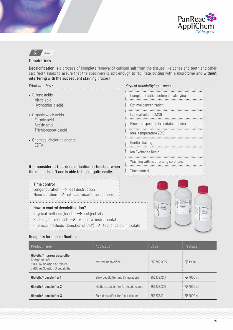
Decalcifiers
Decalcification is a process of complete removal of calcium salt from the tissues like bones and teeth and other calcified tissues to assure that the specimen is soft enough to facilitate cutting with a microtome and without interfering with the subsequent staining process.
It is considered that decalcification is finished when the object is soft and is able to be cut quite easily.
What are they?
• Strong acids - Nitric acid - Hydrochloric acid
Keys of decalcifiying process
• Organic weak acids - Formic acid - Acetic acid - Trichloroacetic acid
• Chemical chelating agents - EDTA
Optimal concentration
Blocks suspended in container center
Ideal temperature 25ºC
Gentle shaking
Ion Exchange Resin
Washing with neutralizing solutions
Time control
Complete fixation before decalcifying
Optimal volume (1:20)
Fixing
Time controlLonger duration cell destruction Minor duration difficult microtome sections
How to control decalcification?Physical methods (touch) subjectivity Radiological methods expensive instrumental Chemical methods (detection of Ca2+) test of calcium oxalate
Reagents for decalcification
Product name Application Code Package
Histofix ® marrow decalcifierComprised of:3x100 ml Solution A fixative3x100 ml Solution B decalcifier
Marrow decalcifier 256284.0922 b Pack
Histofix ® decalcifier 1 Slow decalcifier and fixing agent 256239.1211 b 1000 ml
Histofix® decalcifier 2 Medium decalcifier for fixed tissues 256238.1211 b 1000 ml
Histofix® decalcifier 3 Fast decalcifier for fixed tissues 256237.1211 b 1000 ml
Panreac Applichem
Reagentsfor Hospitals
16
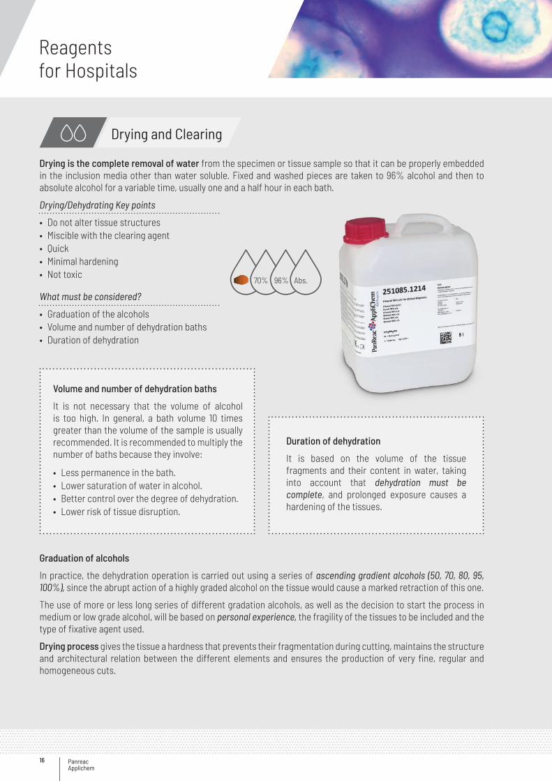
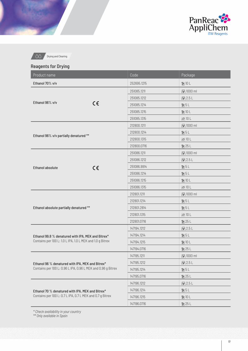
Drying is the complete removal of water from the specimen or tissue sample so that it can be properly embedded in the inclusion media other than water soluble. Fixed and washed pieces are taken to 96% alcohol and then to absolute alcohol for a variable time, usually one and a half hour in each bath.
Graduation of alcohols
In practice, the dehydration operation is carried out using a series of ascending gradient alcohols (50, 70, 80, 95, 100%), since the abrupt action of a highly graded alcohol on the tissue would cause a marked retraction of this one.
The use of more or less long series of different gradation alcohols, as well as the decision to start the process in medium or low grade alcohol, will be based on personal experience, the fragility of the tissues to be included and the type of fixative agent used.
Drying process gives the tissue a hardness that prevents their fragmentation during cutting, maintains the structure and architectural relation between the different elements and ensures the production of very fine, regular and homogeneous cuts.
Drying/Dehydrating Key points
What must be considered?
• Do not alter tissue structures• Miscible with the clearing agent• Quick• Minimal hardening• Not toxic
• Graduation of the alcohols• Volume and number of dehydration baths• Duration of dehydration
70% 96% Abs.
Drying and Clearing
Volume and number of dehydration baths
It is not necessary that the volume of alcohol is too high. In general, a bath volume 10 times greater than the volume of the sample is usually recommended. It is recommended to multiply the number of baths because they involve:
• Less permanence in the bath.• Lower saturation of water in alcohol.• Better control over the degree of dehydration.• Lower risk of tissue disruption.
Duration of dehydration
It is based on the volume of the tissue fragments and their content in water, taking into account that dehydration must be complete, and prolonged exposure causes a hardening of the tissues.
17
Product name Code Package
Ethanol 70% v/v 252695.1215 i 10 L
Ethanol 96% v/v
251085.1211 b 1000 ml
251085.1212 b 2.5 L
251085.1214 i 5 L
251085.1215 i 10 L
251085.1315 C 10 L
Ethanol 96% v/v partially denatured **
212800.1211 b 1000 ml
212800.1214 i 5 L
212800.1315 C 10 L
212800.0716 i 25 L
Ethanol absolute
251086.1211 b 1000 ml
251086.1212 b 2.5 L
251086.9914 i 5 L
251086.1214 i 5 L
251086.1215 i 10 L
251086.1315 C 10 L
Ethanol absolute partially denatured **
212801.1211 b 1000 ml
212801.1214 i 5 L
212801.2814 i 5 L
212801.1315 C 10 L
212801.0716 i 25 L
Ethanol 99.8 % denatured with IPA, MEK and Bitrex*Contains per 100 L: 1.0 L IPA, 1.0 L MEK and 1.0 g Bitrex
147194.1212 b 2.5 L
147194.1214 i 5 L
147194.1215 i 10 L
147194.0716 i 25 L
Ethanol 96 % denatured with IPA, MEK and Bitrex*Contains per 100 L: 0.96 L IPA, 0.96 L MEK and 0.96 g Bitrex
147195.1211 b 1000 ml
147195.1212 b 2.5 L
147195.1214 i 5 L
147195.0716 i 25 L
Ethanol 70 % denatured with IPA, MEK and Bitrex*Contains per 100 L: 0.7 L IPA, 0.7 L MEK and 0.7 g Bitrex
147196.1212 b 2.5 L
147196.1214 i 5 L
147196.1215 i 10 L
147196.0716 i 25 L
* Check availability in your country ** Only available in Spain
Drying and Clearing
Reagents for Drying
Panreac Applichem
Reagentsfor Hospitals
18
Abs. 96% 70%
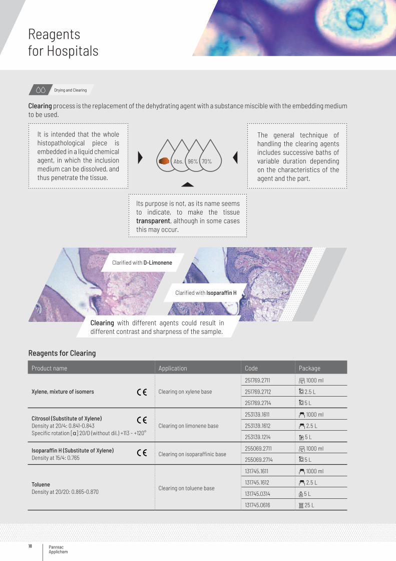
It is intended that the whole histopathological piece is embedded in a liquid chemical agent, in which the inclusion medium can be dissolved, and thus penetrate the tissue.
The general technique of handling the clearing agents includes successive baths of variable duration depending on the characteristics of the agent and the part.
Its purpose is not, as its name seems to indicate, to make the tissue transparent, although in some cases this may occur.
Reagents for Clearing
Product name Application Code Package
Xylene, mixture of isomers Clearing on xylene base
251769.2711 ~ 1000 ml
251769.2712 2.5 L
251769.2714 5 L
Citrosol (Substitute of Xylene)Density at 20/4: 0.841-0.843Specific rotation [α] 20/D (without dil.) +113 - +120°
Clearing on limonene base
253139.1611 a 1000 ml
253139.1612 a 2.5 L
253139.1214 i 5 L
Isoparaffin H (Substitute of Xylene)Density at 15/4: 0.765
Clearing on isoparaffinic base255069.2711 ~ 1000 ml
255069.2714 5 L
TolueneDensity at 20/20: 0.865-0.870
Clearing on toluene base
131745.1611 a 1000 ml
131745.1612 a 2.5 L
131745.0314 z 5 L
131745.0616 s 25 L
Drying and Clearing
Clearing process is the replacement of the dehydrating agent with a substance miscible with the embedding medium to be used.
Clearing with different agents could result in different contrast and sharpness of the sample.
Clarified with D-Limonene
Clarified with Isoparaffin H
19
Reagents for Embedding
Embedding media
Product name Application Code Package
Paraffin M.P. 51-53°Cpellets
For both infiltration and/or embedding 253209.1211 b 1000 g
Paraffin M.P. 55-58°Cplasticized +DMSO pellets
DMSO increases the rate of penetration of paraffin and provides additional preservation, the addition of polymers prevents sprinkling, air-filled slits between the paraffin crystals that can adversely affect the sectioning procedure
256993.0933 h 6 x 1 kg
256993.0415 G 10 kg
Paraffin M.P. 56-58°Cpellets
For both infiltration and/or embedding253211.1211 b 1000 g
253211.0914 1 5 kg
Paraffin M.P. ~ 42-44°CPieces, low melting point
Near to corporal temperature213206.0911 1 1000 g
213206.0914 G 5 kg
Paraffin CleanerComposition:Isoparaffin H .............. 425 ml1-Propanol .................. 75 ml
Microtomes cleaner used in the processing of human tissue
256876.3408 6 x 100 ml
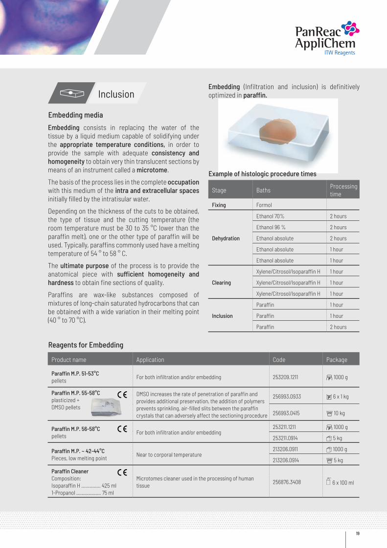
Stage BathsProcessing time
Fixing Formol
Dehydration
Ethanol 70% 2 hours
Ethanol 96 % 2 hours
Ethanol absolute 2 hours
Ethanol absolute 1 hour
Ethanol absolute 1 hour
Clearing
Xylene/Citrosol/Isoparaffin H 1 hour
Xylene/Citrosol/Isoparaffin H 1 hour
Xylene/Citrosol/Isoparaffin H 1 hour
Inclusion
Paraffin 1 hour
Paraffin 1 hour
Paraffin 2 hours
Embedding consists in replacing the water of the tissue by a liquid medium capable of solidifying under the appropriate temperature conditions, in order to provide the sample with adequate consistency and homogeneity to obtain very thin translucent sections by means of an instrument called a microtome.
The basis of the process lies in the complete occupation with this medium of the intra and extracellular spaces initially filled by the intratisular water.
Depending on the thickness of the cuts to be obtained, the type of tissue and the cutting temperature (the room temperature must be 30 to 35 °C lower than the paraffin melt), one or the other type of paraffin will be used. Typically, paraffins commonly used have a melting temperature of 54 ° to 58 ° C.
The ultimate purpose of the process is to provide the anatomical piece with sufficient homogeneity and hardness to obtain fine sections of quality.
Paraffins are wax-like substances composed of mixtures of long-chain saturated hydrocarbons that can be obtained with a wide variation in their melting point (40 ° to 70 °C).
Embedding (Infiltration and inclusion) is definitively optimized in paraffin.
Example of histologic procedure times
Inclusion
Panreac Applichem
Reagentsfor Hospitals
20
Cutting
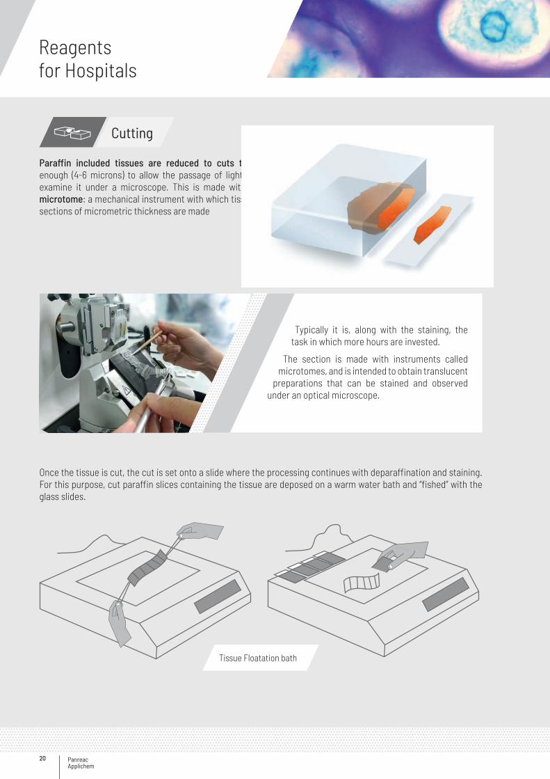
Paraffin included tissues are reduced to cuts thin enough (4-6 microns) to allow the passage of light to examine it under a microscope. This is made with a microtome: a mechanical instrument with which tissue sections of micrometric thickness are made
Once the tissue is cut, the cut is set onto a slide where the processing continues with deparaffination and staining. For this purpose, cut paraffin slices containing the tissue are deposed on a warm water bath and “fished” with the glass slides.
Typically it is, along with the staining, the task in which more hours are invested.
The section is made with instruments called microtomes, and is intended to obtain translucent
preparations that can be stained and observed under an optical microscope.
Tissue Floatation bath
21
Rehydration
Deparaffinization-Hydration is the process of removing the inclusion medium from paraffin-embedded tissue sections and rehydrating for proper penetration of the dyes.
Stage BathsProcessing time
Deparaffinization
Xylene/Citrosol/Isoparaffin H 10 min
Xylene/Citrosol/Isoparaffin H 10 min
Xylene/Citrosol/Isoparaffin H 10 min
HydrationEthanol absolute 1-2 min
Ethanol 96 % 1-2 min
Example of Deparaffinization-Hydration times
Reagents for Deparaffinization-Hydration
Deparaffinization-Hydration
Product name Code Package
Ethanol 70% v/v 252695.1215 i 10 L
Ethanol 96% v/v
251085.1211 b 1000 ml
251085.1212 b 2.5 L
251085.1214 i 5 L
251085.1215 i 10 L
251085.1315 C 10 L
Ethanol 96% v/v partially denatured **
212800.1211 b 1000 ml
212800.1214 i 5 L
212800.1315 C 10 L
212800.0716 i 25 L
Ethanol absolute
251086.1211 b 1000 ml
251086.1212 b 2.5 L
251086.9914 i 5 L
251086.1214 i 5 L
251086.1215 i 10 L
251086.1315 C 10 L
Ethanol absolute partially denatured **
212801.1211 b 1000 ml
212801.1214 i 5 L
212801.2814 i 5 L
212801.1315 C 10 L
212801.0716 i 25 L
Product name Code Package
Ethanol 99.8 % denatured with IPA, MEK and Bitrex*Contains per 100 L: 1.0 L IPA, 1.0 L MEK and 1.0 g Bitrex
147194.1212 b 2.5 L
147194.1214 i 5 L
147194.1215 i 10 L
147194.0716 i 25 L
Ethanol 96 % denatured with IPA, MEK and Bitrex*Contains per 100 L: 0.96 L IPA, 0.96 L MEK and 0.96 g Bitrex
147195.1211 b 1000 ml
147195.1212 b 2.5 L
147195.1214 i 5 L
147195.0716 i 25 L
Ethanol 70 % denatured with IPA, MEK and Bitrex*Contains per 100 L: 0.7 L IPA, 0.7 L MEK and 0.7 g Bitrex
147196.1212 b 2.5 L
147196.1214 i 5 L
147196.1215 i 10 L
147196.0716 i 25 L
Xylene, mixture of isomers
251769.2711 ~ 1000 ml
251769.2712 2.5 L
251769.2714 5 L
Citrosol(Substitute of Xylene)
253139.1611 a 1000 ml
253139.1612 a 2.5 L
253139.1214 { 5 L
Isoparaffin H (Substitute of Xylene)
131745.1611 a 1000 ml
131745.1612 a 2.5 L
131745.0314 z 5 L
131745.0616 s 25 L
>>
<<
* Check availability in your country ** Only available in Spain
Panreac Applichem
Reagentsfor Hospitals
22
Staining
Microscopy dyes are used mainly in histology, cytology and microbiology but also in other analytical techniques.
There are two types of microscopy dyes:
• Natural Dyes obtained in the form of extracts from certain plants or insects.- Nuclear: Hematoxylin and Carmine- Cytoplasmic: Safranin and Orcein
• Synthetic Dyes mostly derived from aniline.- Nuclear: Methyl Green, Basic Fuchsin, Cresyl Violet- Cytoplasmic: Eosin, Phloxine
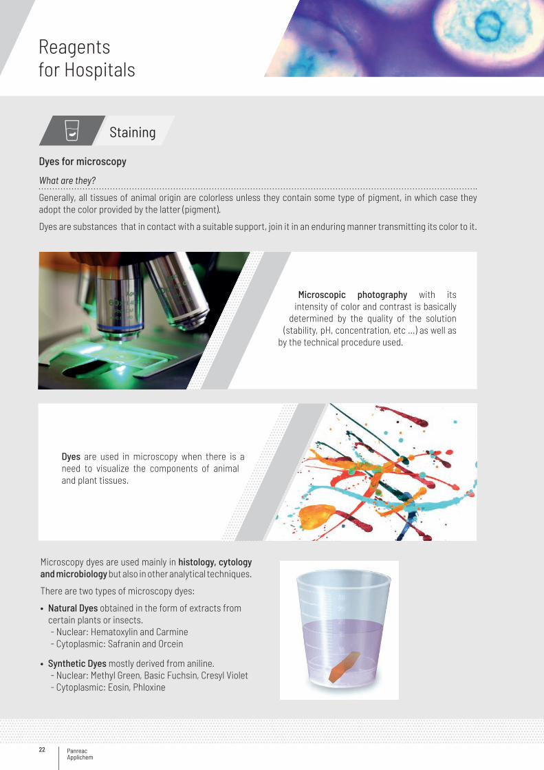
Dyes are used in microscopy when there is a need to visualize the components of animal and plant tissues.
What are they?
Generally, all tissues of animal origin are colorless unless they contain some type of pigment, in which case they adopt the color provided by the latter (pigment).
Dyes are substances that in contact with a suitable support, join it in an enduring manner transmitting its color to it.
Microscopic photography with its intensity of color and contrast is basically
determined by the quality of the solution (stability, pH, concentration, etc ...) as well as
by the technical procedure used.
Dyes for microscopy
23
Staining
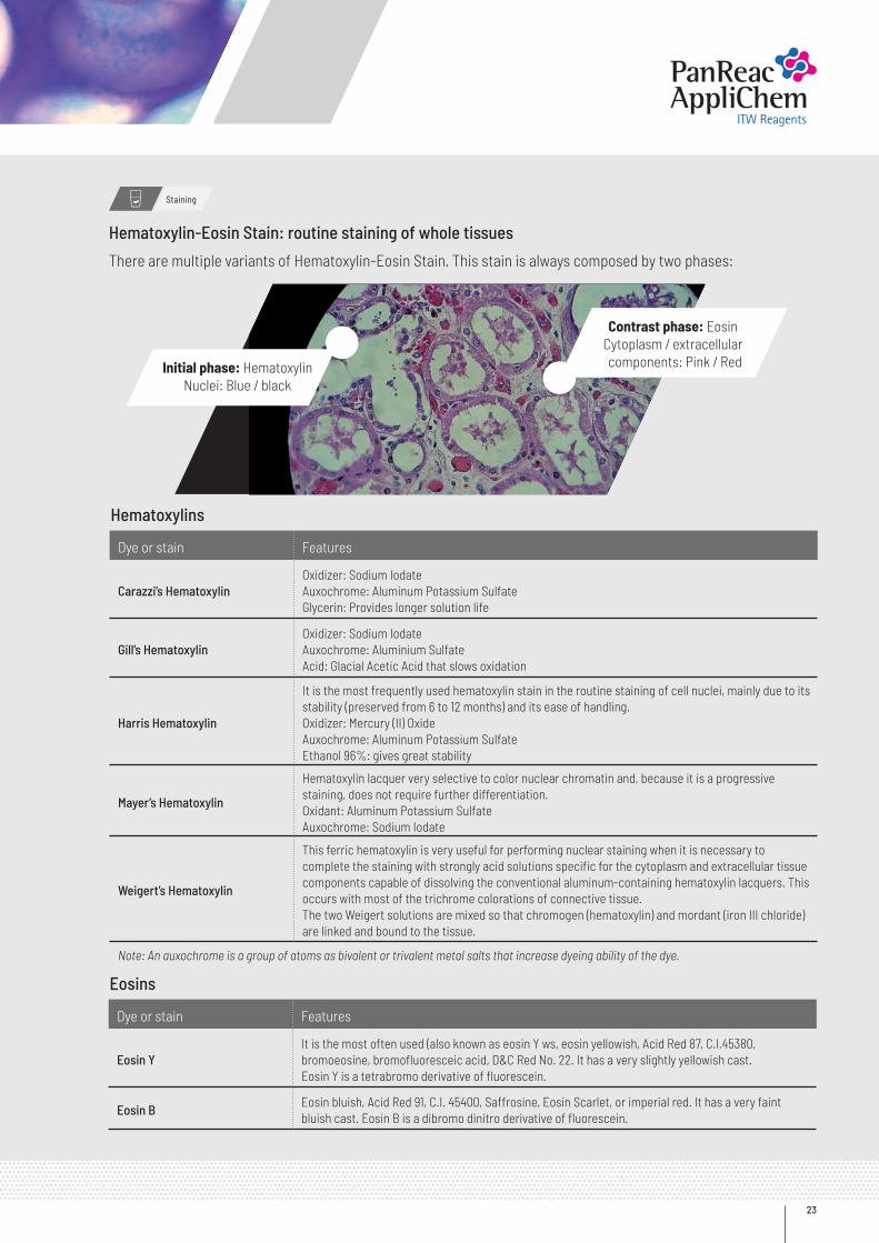
Hematoxylin-Eosin Stain: routine staining of whole tissues
There are multiple variants of Hematoxylin-Eosin Stain. This stain is always composed by two phases:
Initial phase: HematoxylinNuclei: Blue / black
Contrast phase: EosinCytoplasm / extracellular components: Pink / Red
Hematoxylins
Eosins
Dye or stain Features
Carazzi’s HematoxylinOxidizer: Sodium IodateAuxochrome: Aluminum Potassium SulfateGlycerin: Provides longer solution life
Gill’s HematoxylinOxidizer: Sodium IodateAuxochrome: Aluminium SulfateAcid: Glacial Acetic Acid that slows oxidation
Harris Hematoxylin
It is the most frequently used hematoxylin stain in the routine staining of cell nuclei, mainly due to its stability (preserved from 6 to 12 months) and its ease of handling.Oxidizer: Mercury (II) OxideAuxochrome: Aluminum Potassium SulfateEthanol 96%: gives great stability
Mayer’s Hematoxylin
Hematoxylin lacquer very selective to color nuclear chromatin and, because it is a progressive staining, does not require further differentiation.Oxidant: Aluminum Potassium SulfateAuxochrome: Sodium Iodate
Weigert’s Hematoxylin
This ferric hematoxylin is very useful for performing nuclear staining when it is necessary to complete the staining with strongly acid solutions specific for the cytoplasm and extracellular tissue components capable of dissolving the conventional aluminum-containing hematoxylin lacquers. This occurs with most of the trichrome colorations of connective tissue.The two Weigert solutions are mixed so that chromogen (hematoxylin) and mordant (iron III chloride) are linked and bound to the tissue.
Note: An auxochrome is a group of atoms as bivalent or trivalent metal salts that increase dyeing ability of the dye.
Dye or stain Features
Eosin Y It is the most often used (also known as eosin Y ws, eosin yellowish, Acid Red 87, C.I.45380, bromoeosine, bromofluoresceic acid, D&C Red No. 22. It has a very slightly yellowish cast. Eosin Y is a tetrabromo derivative of fluorescein.
Eosin BEosin bluish, Acid Red 91, C.I. 45400, Saffrosine, Eosin Scarlet, or imperial red. It has a very faint bluish cast. Eosin B is a dibromo dinitro derivative of fluorescein.
Panreac Applichem
Reagentsfor Hospitals
24
Staining
Powdered dyes
Reagents for Staining
Product name Application Code Package
Alcian Blue 8 GX (C.I. 74240) For histology, PAS-Alcian Blue staining,certified by the Biological Stain Comission
254584.1604 a 5 g
254584.1606 a 25 g
Aniline Blue WS (C.I. 42755) For collagen staining 253708.1606 a 25 g
Auramine O (C.I. 41000) Fluorescent staining 251162.1608 a 100 g
Azur II (C.I. 52010 + 52015) Blood smears staining 251178.1606 a 25 g
Brilliant Cresyl Blue (C.I. 51010) Platelets and thrombocytes staining 251169.1604 a 5 g
Brilliant Green (C.I. 42040) Vegetal tissue staining251758.1606 a 25 g
251758.1608 a 100 g
Bromophenol Blue Proteins staining131165.1604 a 5 g
131165.1606 a 25 g
Bromothymol Blue Vital staining131167.1604 a 5 g
131167.1606 a 25 gCarmine (Lacquer of carminic acid with calcium and aluminium) (C.I. 75470) Nucleus and glycogen staining 251824.1605 a 10 g
Coomassie Brilliant Blue G-250 (C.I. 42655) For electrophoresis A3480,0025 a 25 g
Coomassie Brilliant Blue R-250 (C.I. 42660) For electrophoresisA1092,0025 b 25 g
A1092,0100 b
Crystal Violet (C.I. 42555) Bacteria staining 251762.1606 a 25 g
DAPIChromosomes, ChlamydiaFluorescent dye
A1001,0010 a 10 mg
A1001,0025 a 25 mg
A1001,0100 a 100 mg
A1001,0500 a 500 mg
A1001,9001 a 1 g
A1001,9010 b 10 g
Eosin Yellowish (C.I. 45380) Vital staining and plasma staining251299.1606 a 25 g
251299.1608 a 100 g
Erythrosin B (C.I. 45430) Proteins, antigen-antibody reactions fluorescent dye 253982.1606 a 25 g
Fuchsin Acidic Disodium Salt (C.I. 42685) Blood smear staining 251331.1605 a 10 g
Fuchsin Basic (C.I. 42510) Nucleus and Koch's bacilli staining
251332.1606 a 25 g
251332.1608 a 100 g
251332.1610 a 500 g
Gentian Violet (C.I. 42535+42555) Bacteria staining according to Gram251765.1606 a 25 g
251765.1609 a 250 g
Giemsa stain Blood smears and protozoos staining 251337.1608 a 100 g
Hematoxylin 1-hydrate (C.I. 75290) Vagina smear staining251344.1604 a 5 g
251344.1606 a 25 g
Indigo Carmine (C.I. 73015) Nucleus and glycogen staining 251246.1605 a 10 g
Malachite Oxalate Green (C.I. 42000) Cytoplasm of vegetal cells staining251761.1606 a 25 g
251761.1608 a 100 g
>>
25
Staining
Dyes in solution
Product name Application Code Package
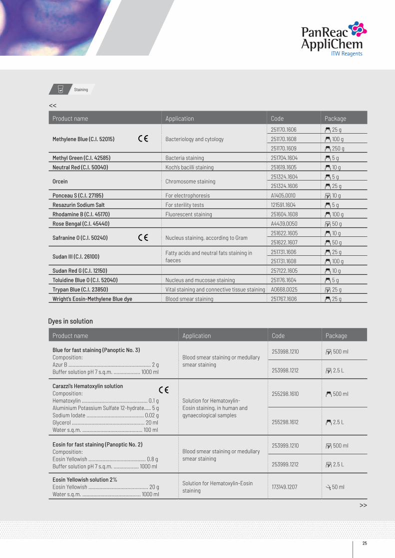
Methylene Blue (C.I. 52015) Bacteriology and cytology
251170.1606 a 25 g
251170.1608 a 100 g
251170.1609 a 250 g
Methyl Green (C.I. 42585) Bacteria staining 251704.1604 a 5 g
Neutral Red (C.I. 50040) Koch’s bacilli staining 251619.1605 a 10 g
Orcein Chromosome staining251324.1604 a 5 g
251324.1606 a 25 g
Ponceau S (C.I. 27195) For electrophoresis A1405,0010 b 10 g
Resazurin Sodium Salt For sterility tests 121591.1604 a 5 g
Rhodamine B (C.I. 45170) Fluorescent staining 251604.1608 a 100 g
Rose Bengal (C.I. 45440) A4439,0050 b 50 g
Safranine O (C.I. 50240) Nucleus staining, according to Gram251622.1605 a 10 g
251622.1607 a 50 g
Sudan III (C.I. 26100)Fatty acids and neutral fats staining in faeces
251731.1606 a 25 g
251731.1608 a 100 g
Sudan Red G (C.I. 12150) 257122.1605 a 10 g
Toluidine Blue O (C.I. 52040) Nucleus and mucosae staining 251176.1604 a 5 g
Trypan Blue (C.I. 23850) Vital staining and connective tissue staining A0668,0025 b 25 g
Wright’s Eosin-Methylene Blue dye Blood smear staining 251767.1606 a 25 g
Product name Application Code Package
Blue for fast staining (Panoptic No. 3)Composition:Azur B ........................................................... 2 gBuffer solution pH 7 s.q.m. ................... 1000 ml
Blood smear staining or medullary smear staining
253998.1210 b 500 ml
253998.1212 b 2.5 L
Carazzi’s Hematoxylin solutionComposition:Hematoxylin ............................................... 0.1 gAluminium Potassium Sulfate 12-hydrate..... 5 gSodium Iodate ......................................... 0.02 gGlycerol .................................................... 20 mlWater s.q.m. ........................................... 100 ml
Solution for Hematoxylin-Eosin staining, in human and gynaecological samples
255298.1610 a 500 ml
255298.1612 a 2.5 L
Eosin for fast staining (Panoptic No. 2)Composition:Eosin Yellowish ......................................... 0.8 gBuffer solution pH 7 s.q.m. .................. 1000 ml
Blood smear staining or medullary smear staining
253999.1210 b 500 ml
253999.1212 b 2.5 L
Eosin Yellowish solution 2%Eosin Yellowish ………………....................….. 20 gWater s.q.m. …………………..............……. 1000 ml
Solution for Hematoxylin-Eosin staining
173149.1207 l 50 ml
>>
<<
Panreac Applichem
Reagentsfor Hospitals
26
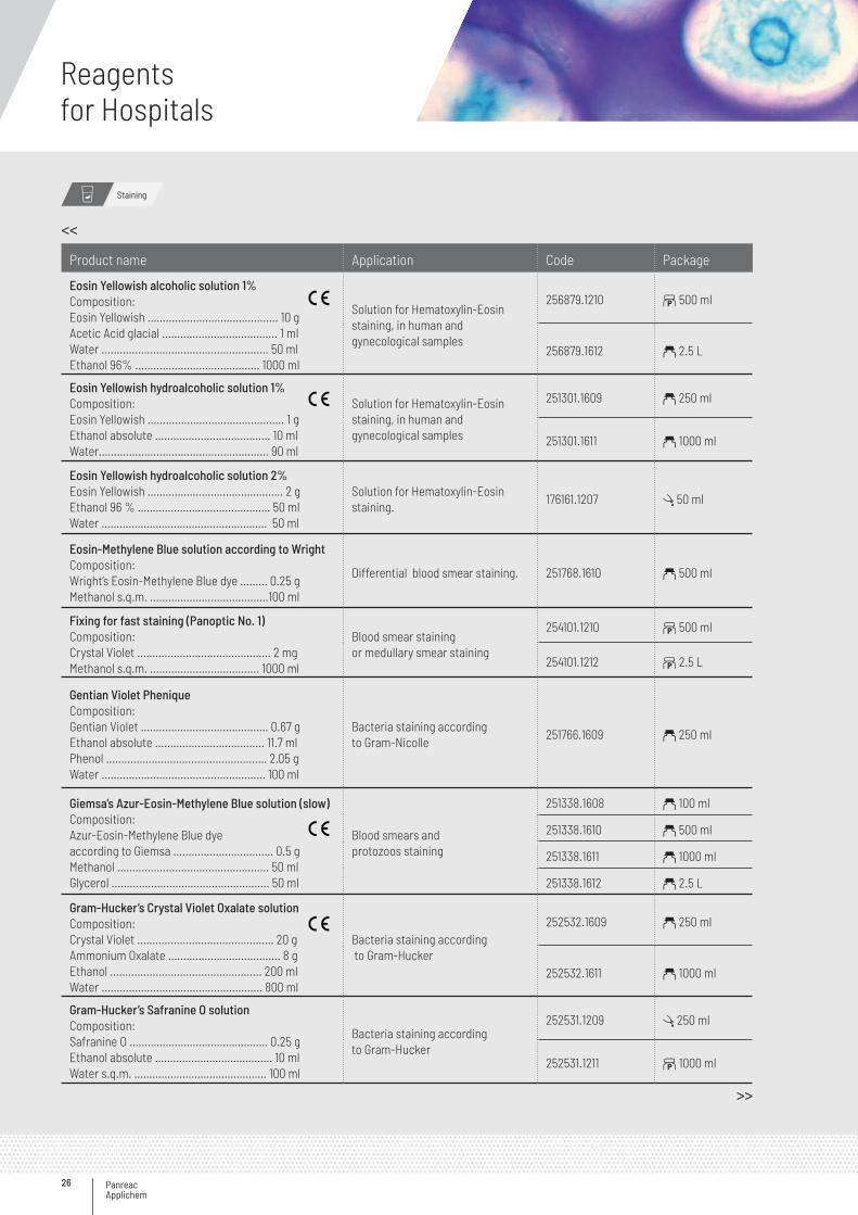
Product name Application Code Package
Eosin Yellowish alcoholic solution 1%Composition:Eosin Yellowish ........................................... 10 gAcetic Acid glacial ...................................... 1 mlWater ....................................................... 50 mlEthanol 96% ......................................... 1000 ml
Solution for Hematoxylin-Eosin staining, in human andgynecological samples
256879.1210 b 500 ml
256879.1612 a 2.5 L
Eosin Yellowish hydroalcoholic solution 1%Composition:Eosin Yellowish ............................................. 1 gEthanol absolute ...................................... 10 mlWater........................................................ 90 ml
Solution for Hematoxylin-Eosin staining, in human andgynecological samples
251301.1609 a 250 ml
251301.1611 a 1000 ml
Eosin Yellowish hydroalcoholic solution 2%Eosin Yellowish ……………………........…………. 2 gEthanol 96 % ……………………….......………. 50 mlWater ……………………………….......………… 50 ml
Solution for Hematoxylin-Eosin staining.
176161.1207 l 50 ml
Eosin-Methylene Blue solution according to Wright Composition:Wright’s Eosin-Methylene Blue dye ......... 0.25 gMethanol s.q.m. .......................................100 ml
Differential blood smear staining. 251768.1610 a 500 ml
Fixing for fast staining (Panoptic No. 1)Composition:Crystal Violet ............................................ 2 mgMethanol s.q.m. .................................... 1000 ml
Blood smear staining or medullary smear staining
254101.1210 b 500 ml
254101.1212 b 2.5 L
Gentian Violet PheniqueComposition:Gentian Violet .......................................... 0.67 gEthanol absolute .................................... 11.7 mlPhenol ..................................................... 2.05 gWater ...................................................... 100 ml
Bacteria staining according to Gram-Nicolle
251766.1609 a 250 ml
Giemsa’s Azur-Eosin-Methylene Blue solution (slow)Composition:Azur-Eosin-Methylene Blue dyeaccording to Giemsa ................................. 0.5 gMethanol .................................................. 50 mlGlycerol .................................................... 50 ml
Blood smears and protozoos staining
251338.1608 a 100 ml
251338.1610 a 500 ml
251338.1611 a 1000 ml
251338.1612 a 2.5 L
Gram-Hucker’s Crystal Violet Oxalate solutionComposition:Crystal Violet ............................................. 20 gAmmonium Oxalate ..................................... 8 gEthanol .................................................. 200 mlWater ..................................................... 800 ml
Bacteria staining according to Gram-Hucker
252532.1609 a 250 ml
252532.1611 a 1000 ml
Gram-Hucker’s Safranine O solutionComposition:Safranine O ………………………………….…… 0.25 gEthanol absolute ………………………………… 10 mlWater s.q.m. ………………………………….…. 100 ml
Bacteria staining accordingto Gram-Hucker
252531.1209 l 250 ml
252531.1211 b 1000 ml
<<
>>
Staining
27
<<
>>
Staining
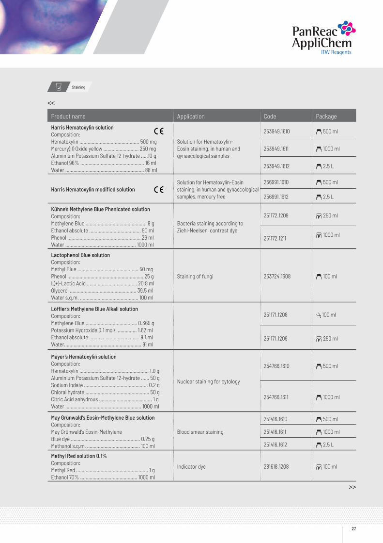
Product name Application Code Package
Harris Hematoxylin solution Composition:Hematoxylin ............................................ 500 mgMercury(II) Oxide yellow .......................... 250 mgAluminium Potassium Sulfate 12-hydrate .....10 gEthanol 96% ............................................... 16 mlWater .......................................................... 88 ml
Solution for Hematoxylin-Eosin staining, in human and gynaecological samples
253949.1610 a 500 ml
253949.1611 a 1000 ml
253949.1612 a 2.5 L
Harris Hematoxylin modified solutionSolution for Hematoxylin-Eosinstaining, in human and gynaecologicalsamples, mercury free
256991.1610 a 500 ml
256991.1612 a 2.5 L
Kühne’s Methylene Blue Phenicated solutionComposition:Methylene Blue ............................................. 9 gEthanol absolute ...................................... 90 mlPhenol ...................................................... 26 mlWater .................................................... 1000 ml
Bacteria staining according to Ziehl-Neelsen, contrast dye
251172.1209 b 250 ml
251172.1211b 1000 ml
Lactophenol Blue solutionComposition:Methyl Blue ............................................. 50 mgPhenol ........................................................ 25 gL(+)-Lactic Acid ..................................... 20.8 mlGlycerol ................................................. 39.5 mlWater s.q.m. ........................................... 100 ml
Staining of fungi 253724.1608 a 100 ml
Löffler’s Methylene Blue Alkali solutionComposition:Methylene Blue ...................................... 0.365 gPotassium Hydroxide 0.1 mol/l .............. 1.62 mlEthanol absolute ..................................... 9.1 mlWater......................................................... 91 ml
251171.1208 l 100 ml
251171.1209 b 250 ml
Mayer’s Hematoxylin solutionComposition:Hematoxylin ................................................... 1.0 gAluminium Potassium Sulfate 12-hydrate ...... 50 gSodium Iodate ............................................... 0.2 gChloral hydrate ............................................... 50 gCitric Acid anhydrous ....................................... 1 gWater ........................................................ 1000 ml
Nuclear staining for cytology
254766.1610 a 500 ml
254766.1611 a 1000 ml
May Grünwald’s Eosin-Methylene Blue solutionComposition:May Grünwald’s Eosin-MethyleneBlue dye ................................................... 0.25 gMethanol s.q.m. ....................................... 100 ml
Blood smear staining
251416.1610 a 500 ml
251416.1611 a 1000 ml
251416.1612 a 2.5 L
Methyl Red solution 0.1%Composition:Methyl Red ..................................................... 1 gEthanol 70% .......................................... 1000 ml
Indicator dye 281618.1208 b 100 ml
Panreac Applichem
Reagentsfor Hospitals
28
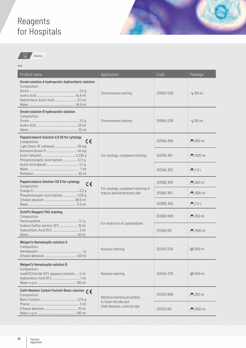
Product name Application Code Package
Orcein solution A hydroacetic-hydrochloric solutionComposition:Orcein ........................................................ 2.0 gAcetic Acid ............................................ 45.8 mlHydrochloric Acid 1 mol/l ................... ..... 8.3 mlWater ..................................................... 45.8 ml
Chromosome staining 251993.1208 l 100 ml
Orcein solution B hydroacetic solutionComposition:Orcein ........................................................ 2.0 gAcetic Acid ............................................... 55 mlWater ........................................................ 55 ml
Chromosome staining 251994.1208 l 100 ml
Papanicolaou’s Solution EA 50 for cytologyComposition:Light Green SF yellowish ......................... 58 mgBismarck Brown R ................................... 40 mgEosin Yellowish ...................................... 0.225 gPhosphotungstic Acid hydrate ................ 0.17 gAcetic Acid glacial ..................................... 0.1 gWater .......................................................... 7 mlMethanol ............................................. ..... 93 ml
For cytology, cytoplasm staining
253594.1610 a 500 ml
253594.1611 a 1000 ml
253594.1612 a 2.5 L
Papanicolaou’s Solution OG 6 for cytologyComposition:Orange G .................................................... 0.2 gPhosphotungstic Acid hydrate ................ 0.02 gEthanol absolute ................................... 88.5 mlWater ...................................................... 11.5 ml
For cytology, cytoplasm staining of mature and keratinized cells
253892.1610 a 500 ml
253892.1611 a 1000 ml
253892.1612 a 2.5 L
Schiff’s Reagent PAS stainingComposition:Pararosaniline ........................................... 0.1 gSodium Sulfite solution 10% .................... 10 mlHydrochloric Acid 35% .............................. 3 mlWater ....................................................... 50 ml
For detection of carbohydrate
251588.1609 a 250 ml
251588.1611 a 1000 ml
Weigert’s Hematoxylin solution A Composition:Hematoxylin .................................................. 1 gEthanol absolute .................................... 1 00 ml
Nucleus staining 253453.1210 b 500 ml
Weigert’s Hematoxylin solution BComposition:Iron(III) Chloride 30% aqueous solution .... 4 mlHydrochloric Acid 35% ............................... 1 mlWater s.q.m. ........................................... 100 ml
Nucleus staining 253454.1210 b 500 ml
Ziehl-Neelsen Carbol-Fuchsin Basic solution Composition:Basic Fuchsin .......................................... 0.74 gPhenol ........................................................ 5 mlEthanol absolute ...................................... 10 mlWater s.q.m. ........................................... 100 ml
Bacteria staining according to Gram-Nicolle and Ziehl-Neelsen, contrast dye
251333.1609 a 250 ml
251333.1611 a 1000 ml
<<
Staining
29
Mounting
Mounting and immersion media
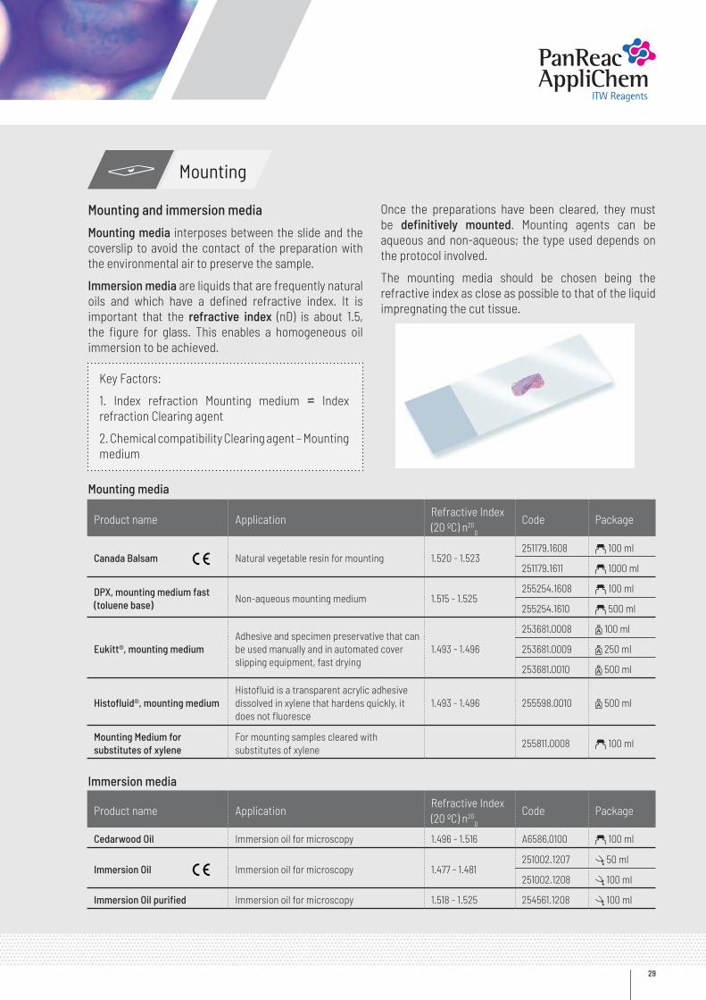
Mounting media interposes between the slide and the coverslip to avoid the contact of the preparation with the environmental air to preserve the sample.
Immersion media are liquids that are frequently natural oils and which have a defined refractive index. It is important that the refractive index (nD) is about 1.5, the figure for glass. This enables a homogeneous oil immersion to be achieved.
Once the preparations have been cleared, they must be definitively mounted. Mounting agents can be aqueous and non-aqueous; the type used depends on the protocol involved.
The mounting media should be chosen being the refractive index as close as possible to that of the liquid impregnating the cut tissue.
Key Factors:
1. Index refraction Mounting medium ⋍ Index refraction Clearing agent
2. Chemical compatibility Clearing agent – Mounting medium
Mounting media
Immersion media
Product name ApplicationRefractive Index(20 ºC) n20
D
Code Package
Canada Balsam Natural vegetable resin for mounting 1.520 - 1.523251179.1608 a 100 ml
251179.1611 a 1000 ml
DPX, mounting medium fast (toluene base)
Non-aqueous mounting medium 1.515 - 1.525255254.1608 a 100 ml
255254.1610 a 500 ml
Eukitt®, mounting mediumAdhesive and specimen preservative that can be used manually and in automated cover slipping equipment, fast drying
1.493 - 1.496
253681.0008 z 100 ml
253681.0009 z 250 ml
253681.0010 z 500 ml
Histofluid®, mounting mediumHistofluid is a transparent acrylic adhesive dissolved in xylene that hardens quickly, it does not fluoresce
1.493 - 1.496 255598.0010 z 500 ml
Mounting Medium for substitutes of xylene
For mounting samples cleared with substitutes of xylene
255811.0008 a 100 ml
Product name ApplicationRefractive Index(20 ºC) n20
D
Code Package
Cedarwood Oil Immersion oil for microscopy 1.496 - 1.516 A6586,0100 a 100 ml
Immersion Oil Immersion oil for microscopy 1.477 - 1.481251002.1207 l 50 ml
251002.1208 l 100 ml
Immersion Oil purified Immersion oil for microscopy 1.518 - 1.525 254561.1208 l 100 ml
Panreac Applichem
Reagentsfor Hospitals
30
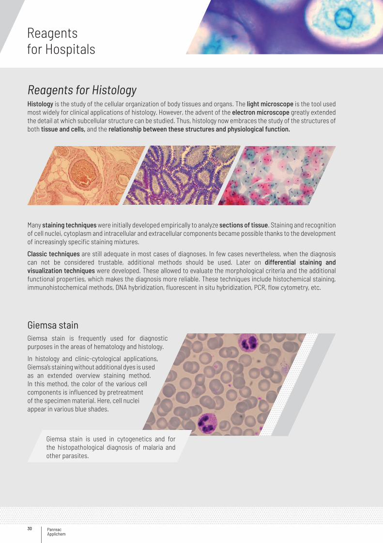
Reagents for HistologyHistology is the study of the cellular organization of body tissues and organs. The light microscope is the tool used most widely for clinical applications of histology. However, the advent of the electron microscope greatly extended the detail at which subcellular structure can be studied. Thus, histology now embraces the study of the structures of both tissue and cells, and the relationship between these structures and physiological function.
Many staining techniques were initially developed empirically to analyze sections of tissue. Staining and recognition of cell nuclei, cytoplasm and intracellular and extracellular components became possible thanks to the development of increasingly specific staining mixtures.
Classic techniques are still adequate in most cases of diagnoses. In few cases nevertheless, when the diagnosis can not be considered trustable, additional methods should be used. Later on differential staining and visualization techniques were developed. These allowed to evaluate the morphological criteria and the additional functional properties, which makes the diagnosis more reliable. These techniques include histochemical staining, immunohistochemical methods, DNA hybridization, fluorescent in situ hybridization, PCR, flow cytometry, etc.
Giemsa stainGiemsa stain is frequently used for diagnostic purposes in the areas of hematology and histology.
In histology and clinic-cytological applications, Giemsa’s staining without additional dyes is used as an extended overview staining method. In this method, the color of the various cell components is influenced by pretreatment of the specimen material. Here, cell nuclei appear in various blue shades.
Giemsa stain is used in cytogenetics and for the histopathological diagnosis of malaria and other parasites.
31
Product name Application Code Package
Giemsa’s Azur-Eosin-Methylene Blue solution (slow) Composition:Azur-Eosin-Methylene Blue dyeaccording to Giemsa ................................. 0.5 gMethanol .................................................. 50 mlGlycerol .................................................... 50 ml
Diagnosis of malaria and other parasites
251338.1608 a 100 ml
251338.1610 a 500 ml
251338.1611 a 1000 ml
251338.1612 a 2.5 L
Giemsa stain
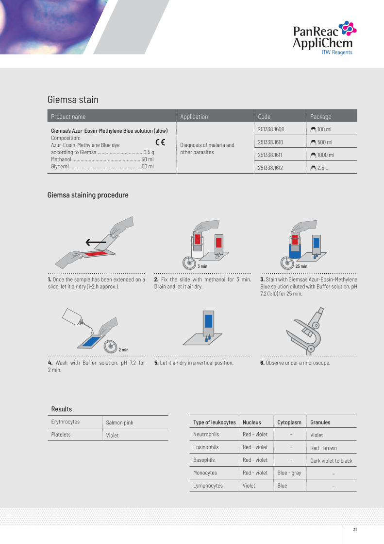
Giemsa staining procedure
2. Fix the slide with methanol for 3 min. Drain and let it air dry.
5. Let it air dry in a vertical position.
3. Stain with Giemsa’s Azur-Eosin-Methylene Blue solution diluted with Buffer solution, pH 7.2 (1:10) for 25 min.
6. Observe under a microscope.
1. Once the sample has been extended on a slide, let it air dry (1-2 h approx.).
4. Wash with Buffer solution, pH 7.2 for 2 min.
Results
Erythrocytes Salmon pink
Platelets Violet
Type of leukocytes Nucleus Cytoplasm Granules
Neutrophils Red - violet - Violet
Eosinophils Red - violet - Red - brown
Basophils Red - violet - Dark violet to black
Monocytes Red - violet Blue - gray -
Lymphocytes Violet Blue -
3 min
2 min
25 min
Panreac Applichem
Reagentsfor Hospitals
32
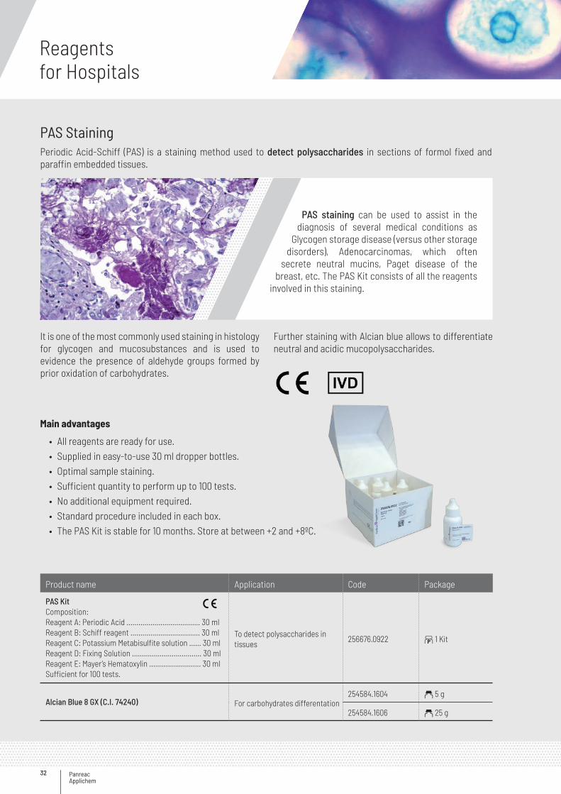
PAS StainingPeriodic Acid-Schiff (PAS) is a staining method used to detect polysaccharides in sections of formol fixed and paraffin embedded tissues.
Main advantages
• All reagents are ready for use.• Supplied in easy-to-use 30 ml dropper bottles.• Optimal sample staining.• Sufficient quantity to perform up to 100 tests.• No additional equipment required.• Standard procedure included in each box.• The PAS Kit is stable for 10 months. Store at between +2 and +8ºC.
Product name Application Code Package
PAS KitComposition:Reagent A: Periodic Acid ..................................... 30 mlReagent B: Schiff reagent ................................... 30 mlReagent C: Potassium Metabisulfite solution ...... 30 mlReagent D: Fixing Solution ................................... 30 mlReagent E: Mayer’s Hematoxylin .......................... 30 mlSufficient for 100 tests.
To detect polysaccharides in tissues
256676.0922 b 1 Kit
Alcian Blue 8 GX (C.I. 74240) For carbohydrates differentation254584.1604 a 5 g
254584.1606 a 25 g
PAS staining can be used to assist in the diagnosis of several medical conditions as
Glycogen storage disease (versus other storage disorders), Adenocarcinomas, which often
secrete neutral mucins, Paget disease of the breast, etc. The PAS Kit consists of all the reagents
involved in this staining.
It is one of the most commonly used staining in histology for glycogen and mucosubstances and is used to evidence the presence of aldehyde groups formed by prior oxidation of carbohydrates.
Further staining with Alcian blue allows to differentiate neutral and acidic mucopolysaccharides.
33
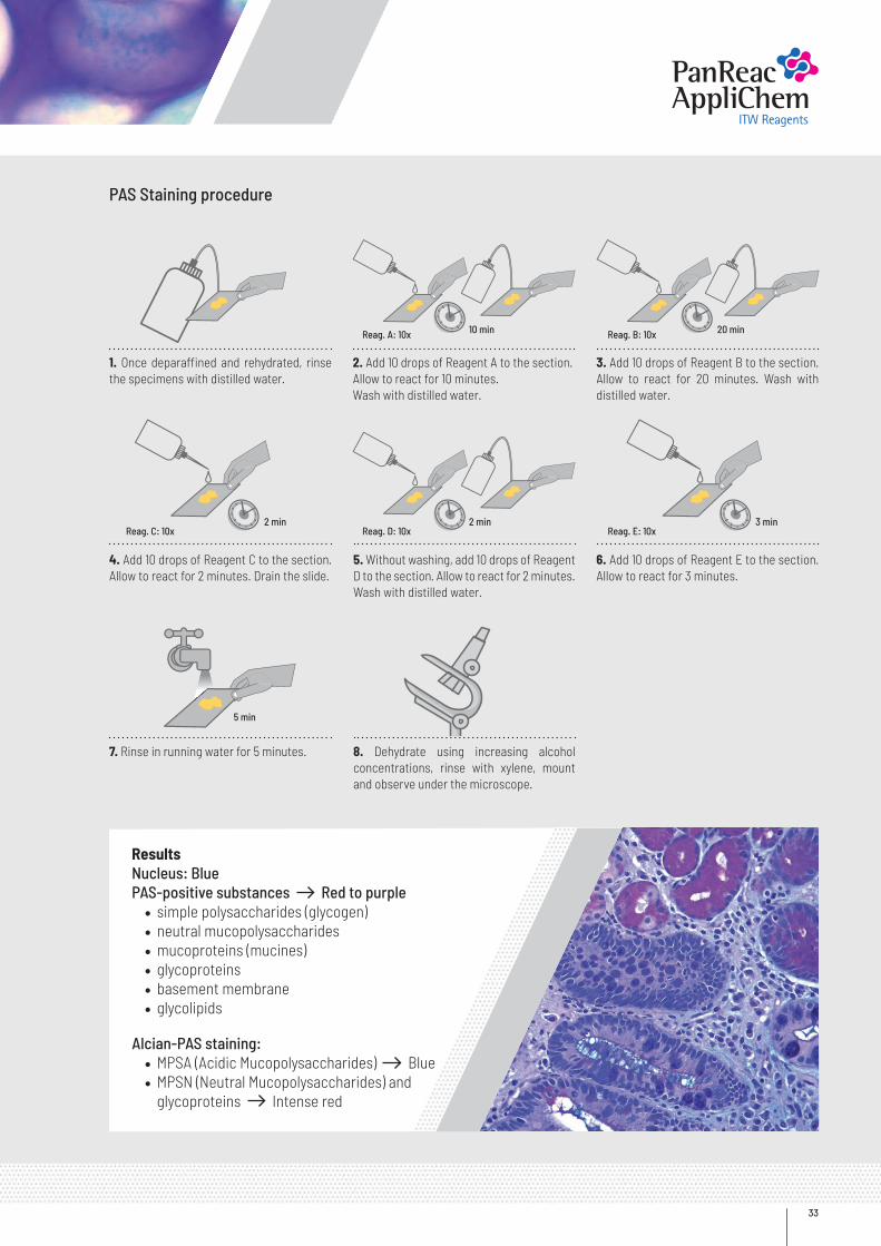
PAS Staining procedure
2. Add 10 drops of Reagent A to the section.Allow to react for 10 minutes.Wash with distilled water.
5. Without washing, add 10 drops of Reagent D to the section. Allow to react for 2 minutes. Wash with distilled water.
8. Dehydrate using increasing alcohol concentrations, rinse with xylene, mount and observe under the microscope.
3. Add 10 drops of Reagent B to the section. Allow to react for 20 minutes. Wash with distilled water.
6. Add 10 drops of Reagent E to the section. Allow to react for 3 minutes.
Reag. A: 10x
Reag. D: 10xReag. C: 10x Reag. E: 10x
1. Once deparaffined and rehydrated, rinse the specimens with distilled water.
4. Add 10 drops of Reagent C to the section. Allow to react for 2 minutes. Drain the slide.
7. Rinse in running water for 5 minutes.
Reag. B: 10x 20 min10 min
2 min
5 min
2 min 3 min
Results Nucleus: BluePAS-positive substances Red to purple
• simple polysaccharides (glycogen)• neutral mucopolysaccharides• mucoproteins (mucines)• glycoproteins• basement membrane• glycolipids
Alcian-PAS staining:
• MPSA (Acidic Mucopolysaccharides) Blue• MPSN (Neutral Mucopolysaccharides) and
glycoproteins Intense red
Panreac Applichem
Reagentsfor Hospitals
34
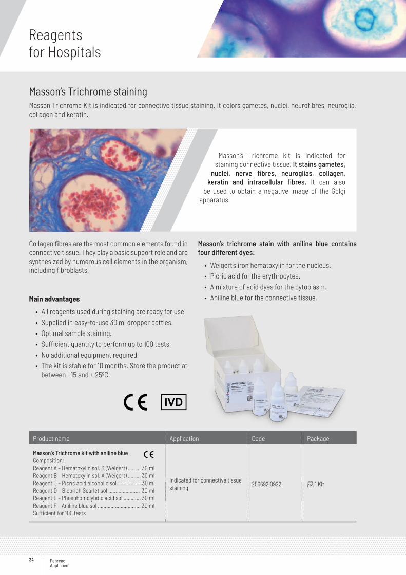
Masson’s Trichrome stainingMasson Trichrome Kit is indicated for connective tissue staining. It colors gametes, nuclei, neurofibres, neuroglia, collagen and keratin.
Masson’s Trichrome kit is indicated for staining connective tissue. It stains gametes,
nuclei, nerve fibres, neuroglias, collagen, keratin and intracellular fibres. It can also
be used to obtain a negative image of the Golgi apparatus.
Collagen fibres are the most common elements found in connective tissue. They play a basic support role and are synthesized by numerous cell elements in the organism, including fibroblasts.
Masson’s trichrome stain with aniline blue contains four different dyes:
• Weigert’s iron hematoxylin for the nucleus.• Picric acid for the erythrocytes.• A mixture of acid dyes for the cytoplasm.• Aniline blue for the connective tissue.Main advantages
• All reagents used during staining are ready for use• Supplied in easy-to-use 30 ml dropper bottles.• Optimal sample staining.• Sufficient quantity to perform up to 100 tests.• No additional equipment required.• The kit is stable for 10 months. Store the product at
between +15 and + 25ºC.
Product name Application Code Package
Masson’s Trichrome kit with aniline blueComposition:Reagent A – Hematoxylin sol. B (Weigert) ……… 30 mlReagent B – Hematoxylin sol. A (Weigert) ……… 30 mlReagent C – Picric acid alcoholic sol...………..... 30 mlReagent D – Biebrich Scarlet sol .…………….….. 30 mlReagent E – Phosphomolybdic acid sol ..………. 30 mlReagent F – Aniline blue sol ……….…………........ 30 mlSufficient for 100 tests
Indicated for connective tissue staining
256692.0922 b 1 Kit
35
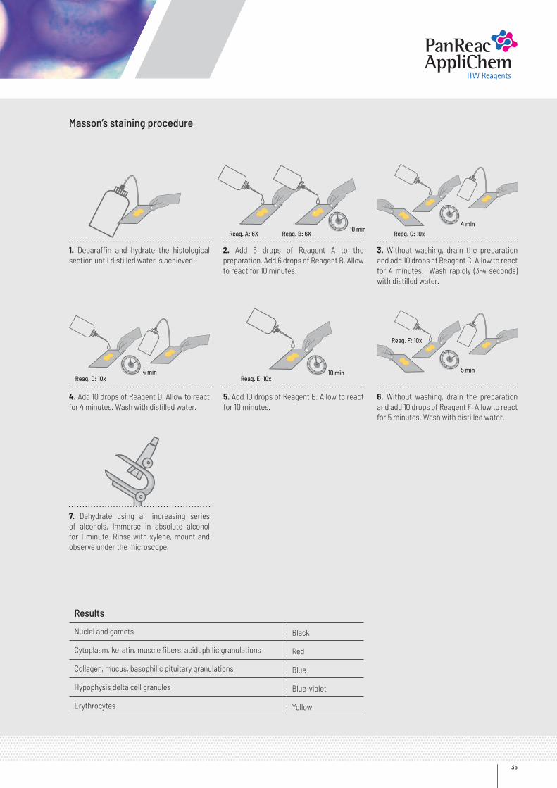
Masson’s staining procedure
Results
Nuclei and gamets Black
Cytoplasm, keratin, muscle fibers, acidophilic granulations Red
Collagen, mucus, basophilic pituitary granulations Blue
Hypophysis delta cell granules Blue-violet
Erythrocytes Yellow
2. Add 6 drops of Reagent A to the preparation. Add 6 drops of Reagent B. Allow to react for 10 minutes.
5. Add 10 drops of Reagent E. Allow to react for 10 minutes.
3. Without washing, drain the preparation and add 10 drops of Reagent C. Allow to react for 4 minutes. Wash rapidly (3-4 seconds) with distilled water.
6. Without washing, drain the preparation and add 10 drops of Reagent F. Allow to react for 5 minutes. Wash with distilled water.
1. Deparaffin and hydrate the histological section until distilled water is achieved.
4. Add 10 drops of Reagent D. Allow to react for 4 minutes. Wash with distilled water.
7. Dehydrate using an increasing series of alcohols. Immerse in absolute alcohol for 1 minute. Rinse with xylene, mount and observe under the microscope.
Reag. E: 10x10 min
Reag. D: 10x
Reag. C: 10x
Reag. F: 10x
4 min
Reag. A: 6X Reag. B: 6X10 min
4 min
5 min
Panreac Applichem
Reagentsfor Hospitals
36
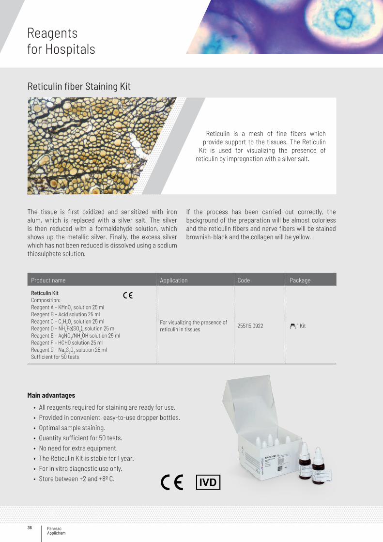
Reticulin fiber Staining Kit
Reticulin is a mesh of fine fibers which provide support to the tissues. The Reticulin
Kit is used for visualizing the presence of reticulin by impregnation with a silver salt.
The tissue is first oxidized and sensitized with iron alum, which is replaced with a silver salt. The silver is then reduced with a formaldehyde solution, which shows up the metallic silver. Finally, the excess silver which has not been reduced is dissolved using a sodium thiosulphate solution.
If the process has been carried out correctly, the background of the preparation will be almost colorless and the reticulin fibers and nerve fibers will be stained brownish-black and the collagen will be yellow.
Main advantages
• All reagents required for staining are ready for use.• Provided in convenient, easy-to-use dropper bottles.• Optimal sample staining.• Quantity sufficient for 50 tests.• No need for extra equipment.• The Reticulin Kit is stable for 1 year.• For in vitro diagnostic use only.• Store between +2 and +8º C.
Product name Application Code Package
Reticulin KitComposition:Reagent A – KMnO4 solution 25 mlReagent B – Acid solution 25 mlReagent C – C2H2O4 solution 25 mlReagent D – NH4Fe(SO4)2 solution 25 mlReagent E – AgNO3/NH4OH solution 25 mlReagent F – HCHO solution 25 mlReagent G – Na2S2O3 solution 25 mlSufficient for 50 tests
For visualizing the presence of reticulin in tissues
255115.0922 a 1 Kit
37
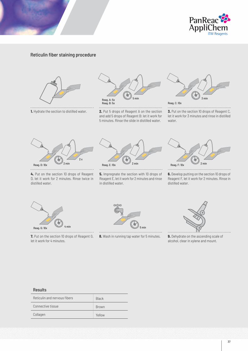
Reticulin fiber staining procedure
Results
Reticulin and nervous fibers Black
Connective tissue Brown
Collagen Yellow
2. Put 5 drops of Reagent A on the section and add 5 drops of Reagent B: let it work for 5 minutes. Rinse the slide in distilled water.
5. Impregnate the section with 10 drops of Reagent E, let it work for 2 minutes and rinse in distilled water.
8. Wash in running tap water for 5 minutes. 9. Dehydrate on the ascending scale ofalcohol, clear in xylene and mount.
3. Put on the section 10 drops of Reagent C, let it work for 3 minutes and rinse in distilled water.
6. Develop putting on the section 10 drops of Reagent F, let it work for 2 minutes. Rinse in distilled water.
1. Hydrate the section to distilled water.
4. Put on the section 10 drops of Reagent D, let it work for 2 minutes. Rinse twice in distilled water.
7. Put on the section 10 drops of Reagent G, let it work for 4 minutes.
Reag. A: 5xReag. B: 5x Reag. C: 10x
Reag. D: 10x
Reag. G: 10x
Reag. E: 10x Reag. F: 10x
5 min 3 min
2 min
4 min
2 min 2 min2 x
5 min
Panreac Applichem
Reagentsfor Hospitals
38
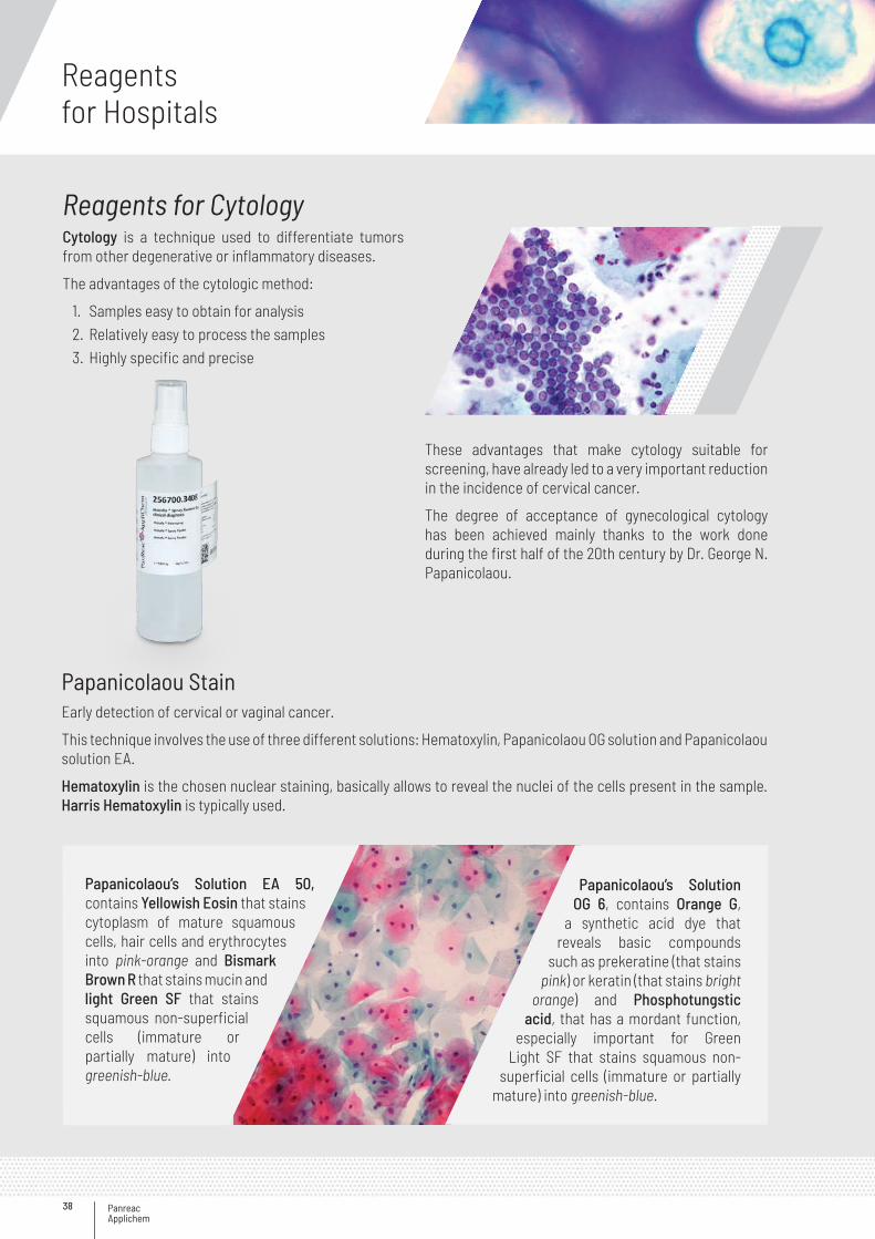
Reagents for CytologyCytology is a technique used to differentiate tumors from other degenerative or inflammatory diseases.
The advantages of the cytologic method:
1. Samples easy to obtain for analysis2. Relatively easy to process the samples3. Highly specific and precise
Papanicolaou StainEarly detection of cervical or vaginal cancer.
This technique involves the use of three different solutions: Hematoxylin, Papanicolaou OG solution and Papanicolaou solution EA.
Hematoxylin is the chosen nuclear staining, basically allows to reveal the nuclei of the cells present in the sample. Harris Hematoxylin is typically used.
These advantages that make cytology suitable for screening, have already led to a very important reduction in the incidence of cervical cancer.
The degree of acceptance of gynecological cytology has been achieved mainly thanks to the work done during the first half of the 20th century by Dr. George N. Papanicolaou.
Papanicolaou’s Solution EA 50, contains Yellowish Eosin that stains cytoplasm of mature squamous cells, hair cells and erythrocytes into pink-orange and Bismark Brown R that stains mucin and light Green SF that stains squamous non-superficial cells (immature or partially mature) into greenish-blue.
Papanicolaou’s Solution OG 6, contains Orange G,
a synthetic acid dye that reveals basic compounds
such as prekeratine (that stains pink) or keratin (that stains bright
orange) and Phosphotungstic acid, that has a mordant function,
especially important for Green Light SF that stains squamous non-
superficial cells (immature or partially mature) into greenish-blue.
39
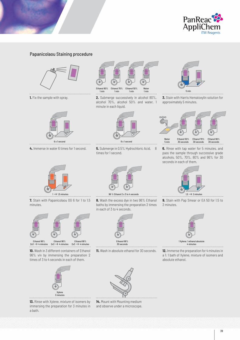
Papanicolaou Staining procedure
2. Submerge successively in alcohol 80%, alcohol 70%, alcohol 50% and water, 1 minute in each liquid.
5. Submerge in 0.5% Hydrochloric Acid, 8 times for 1 second.
8. Wash the excess dye in two 96% Ethanol baths by immersing the preparation 2 times in each of 3 to 4 seconds.
11. Wash in absolute ethanol for 30 seconds.
14. Mount with Mounting mediumand observe under a microscope.
9. Stain with Pap Smear or EA 50 for 1.5 to 2 minutes.
12. Immerse the preparation for 4 minutes in a 1: 1 bath of Xylene, mixture of isomers and absolute ethanol.
3. Stain with Harris Hematoxylin solution for approximately 5 minutes.
6. Rinse with tap water for 5 minutes, and pass the sample through successive grade alcohols, 50%, 70%, 80% and 96% for 30 seconds in each of them.
1. Fix the sample with spray.
4. Immerse in water 6 times for 1 second.
7. Stain with Papanicolaou OG 6 for 1 to 1.5 minutes.
10. Wash in 3 different containers of Ethanol 96% v/v by immersing the preparation 2 times of 3 to 4 seconds in each of them.
13. Rinse with Xylene, mixture of isomers by immersing the preparation for 3 minutes in a bath.
Ethanol 80%1 min
96 % Ethanol 2 x 3 to 4 seconds
Water5 min
Ethanol 70%1 min
Ethanol 50%30 seconds
Ethanol 96%2x3 4 minutes
Ethanol 50%1 min
Ethanol 70%30 seconds
Ethanol 96%2x3 4 minutes
Water1 min
Ethanol 96%30 seconds
Ethanol 96%2x3 4 minutes
Ethanol 96%30 seconds
Xylene3 minutes
1 Xylene: 1 ethanol absolute 4 minutes
5 min
1,5 2 minutes1 1,5 minutes
6 x 1 second 8 x 1 second
Panreac Applichem
Reagentsfor Hospitals
40
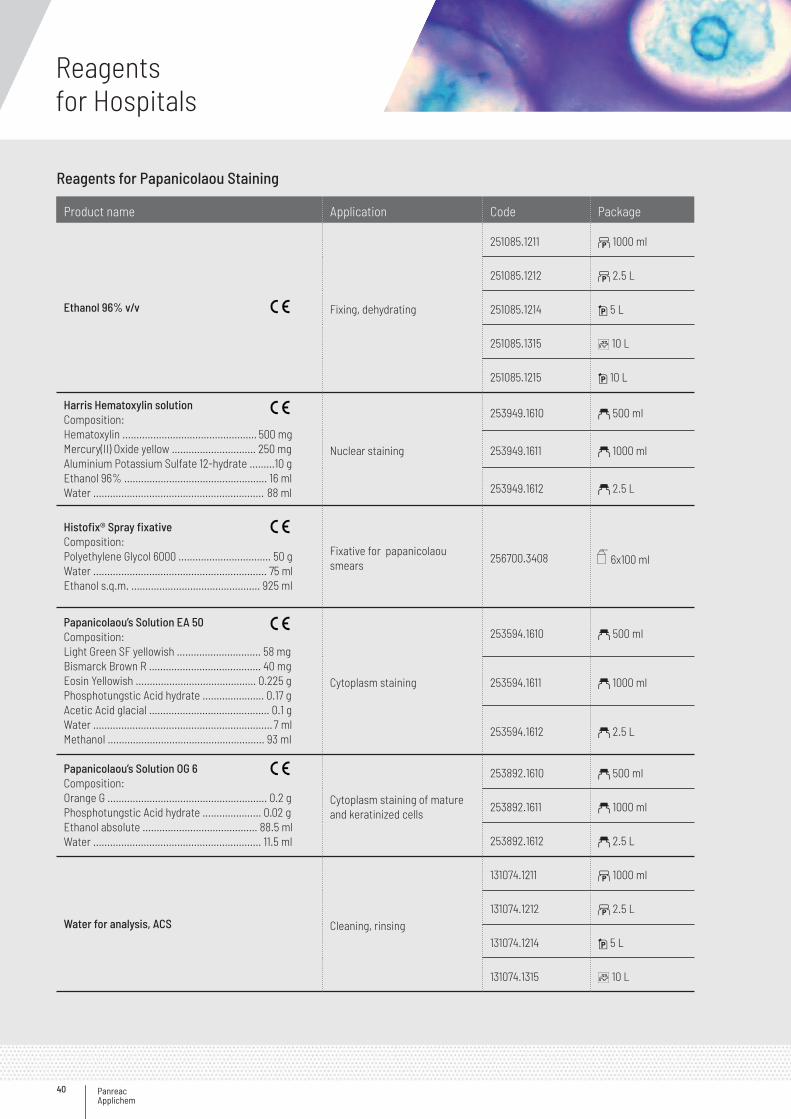
Reagents for Papanicolaou Staining
Product name Application Code Package
Ethanol 96% v/v Fixing, dehydrating
251085.1211 b 1000 ml
251085.1212 b 2.5 L
251085.1214 i 5 L
251085.1315 C 10 L
251085.1215 i 10 L
Harris Hematoxylin solution Composition:Hematoxylin ................................................ 500 mgMercury(II) Oxide yellow .............................. 250 mgAluminium Potassium Sulfate 12-hydrate .........10 gEthanol 96% ................................................... 16 mlWater ............................................................. 88 ml
Nuclear staining
253949.1610 a 500 ml
253949.1611 a 1000 ml
253949.1612 a 2.5 L
Histofix® Spray fixativeComposition:Polyethylene Glycol 6000 ................................. 50 gWater .............................................................. 75 mlEthanol s.q.m. .............................................. 925 ml
Fixative for papanicolaou smears
256700.3408 6x100 ml
Papanicolaou’s Solution EA 50Composition:Light Green SF yellowish .............................. 58 mgBismarck Brown R ........................................ 40 mgEosin Yellowish ........................................... 0.225 gPhosphotungstic Acid hydrate ...................... 0.17 gAcetic Acid glacial ........................................... 0.1 gWater ................................................................ 7 mlMethanol ........................................................ 93 ml
Cytoplasm staining
253594.1610 a 500 ml
253594.1611 a 1000 ml
253594.1612 a 2.5 L
Papanicolaou’s Solution OG 6Composition:Orange G ......................................................... 0.2 gPhosphotungstic Acid hydrate ..................... 0.02 gEthanol absolute ......................................... 88.5 mlWater ............................................................ 11.5 ml
Cytoplasm staining of mature and keratinized cells
253892.1610 a 500 ml
253892.1611 a 1000 ml
253892.1612 a 2.5 L
Water for analysis, ACS Cleaning, rinsing
131074.1211 b 1000 ml
131074.1212 b 2.5 L
131074.1214 i 5 L
131074.1315 C 10 L
41
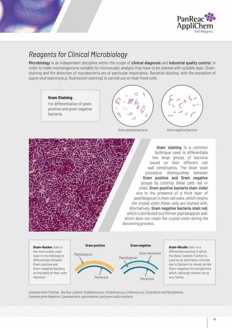
Reagents for Clinical MicrobiologyMicrobiology is an independent discipline within the scope of clinical diagnosis and industrial quality control. In order to make microorganisms suitable for microscopic analysis they have to be stained with suitable dyes. Gram-staining and the detection of mycobacteria are of particular importance. Bacterial staining, with the exception of supra-vital staining (e.g. fluorescent staining), is carried out on heat-fixed cells.
Gram staining is a common technique used to differentiate
two large groups of bacteria based on their different cell
wall constituents. The Gram stain procedure distinguishes between
Gram positive and Gram negative groups by coloring these cells red or
violet. Gram positive bacteria stain violet due to the presence of a thick layer of
peptidoglycan in their cell walls, which retains the crystal violet these cells are stained with.
Alternatively, Gram negative bacteria stain red, which is attributed to a thinner peptidoglycan wall,
which does not retain the crystal violet during the decoloring process.
Gram StainingFor differentiation of gram positive and gram negative bacteria.
Gram–Hucker stain is the most widely used stain in microbiology to differentiate between Gram-positive and Gram-negative bacteria on the basis of their color retention.
Gram–Nicolle stain is a differential staining in which the Basic Carbolic Fuchsin is used as an alternative contrast dye to Safranin to reveal certain Gram-negative microorganisms which, although colored, do so very faintly.
Example Gram Positive: Bacillus, Listeria, Staphylococcus, Streptococcus, Enterococcus, Clostridium and Mycoplasma.Example gram Negative: Cyanobacteria, spirochaetes, and green sulfur bacteria.
Gram positive bacteria
Gram positive
PeptidoglycanPeptidoglycan
Membrane Membrane
Outer membrane
Gram negative bacteria
Gram negative
Panreac Applichem
Reagentsfor Hospitals
42
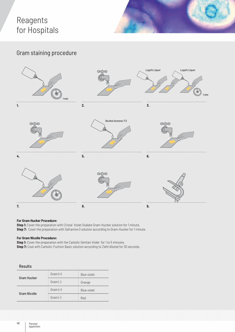
Results
Gram Hucker Gram (+) Blue violet
Gram (-) Orange
Gram Nicolle Gram (+) Blue violet
Gram (-) Red
2.
5.
8. 9.
3.
6.
1.
4.
7.
For Gram Hucker Procedure:Step 1: Cover the preparation with Cristal Violet Oxalate Gram-Hucker solution for 1 minute. Step 7: Cover the preparation with Safranine O solution according to Gram-Hucker for 1 minute.
For Gram Nicolle Procedure: Step 1: Cover the preparation with the Carbolic Gentian Violet for 1 to 5 minutes.Step 7: Coat with Carbolic-Fuchsin Basic solution according to Ziehl diluted for 30 seconds.
Lugol’s Liquor
Alcohol Acetone 7:3
Lugol’s Liquor
1 min1 min
Gram staining procedure
43
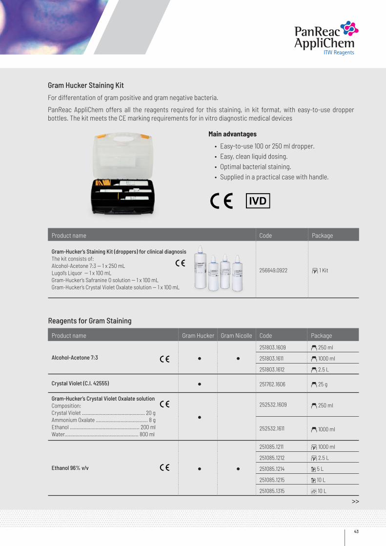
Gram Hucker Staining Kit
For differentation of gram positive and gram negative bacteria.
PanReac AppliChem offers all the reagents required for this staining, in kit format, with easy-to-use dropper bottles. The kit meets the CE marking requirements for in vitro diagnostic medical devices
Main advantages
• Easy-to-use 100 or 250 ml dropper.• Easy, clean liquid dosing.• Optimal bacterial staining.• Supplied in a practical case with handle.
Product name Code Package
Gram-Hucker’s Staining Kit (droppers) for clinical diagnosisThe kit consists of:Alcohol-Acetone 7:3 -- 1 x 250 mLLugol’s Liquor -- 1 x 100 mLGram-Hucker’s Safranine O solution -- 1 x 100 mLGram-Hucker’s Crystal Violet Oxalate solution -- 1 x 100 mL
256649.0922 b 1 Kit
Reagents for Gram Staining
Product name Gram Hucker Gram Nicolle Code Package
Alcohol-Acetone 7:3 · ·251803.1609 a 250 ml
251803.1611 a 1000 ml
251803.1612 a 2.5 L
Crystal Violet (C.I. 42555) · 251762.1606 a 25 g
Gram-Hucker’s Crystal Violet Oxalate solutionComposition:Crystal Violet .............................................. 20 gAmmonium Oxalate ...................................... 8 gEthanol ................................................... 200 mlWater...................................................... 800 ml
·252532.1609 a 250 ml
252532.1611 a 1000 ml
Ethanol 96% v/v · ·251085.1211 b 1000 ml
251085.1212 b 2.5 L
251085.1214 i 5 L
251085.1215 i 10 L
251085.1315 C 10 L
>>
Panreac Applichem
Reagentsfor Hospitals
44
<<
Product name Gram Hucker Gram Nicolle Code Package
Gentian Violet PheniqueComposition:Gentian Violet ……………………..….......….… 0.67 gPhenol ………………………………….…........... 2.05 gEthanol absolute …………………................ 11.7 mlWater ………………………………….......……… 100 ml
· 251766.1609 a 250 ml
Lugol’s Liquor with 0.33 % of Iodine (diluted)Composition:Iodine ……………………………...........……… 0.333 gPotassium Iodide ……………............…….. 0.666 gWater s.q.m. ……………..………...............… 100 ml
· · 256977.1609 a 250 ml
Lugol’s Liquor with 0.4 % of Iodine (diluted)Composition:Iodine …………………………….............……….. 0.4 gPotassium Iodide ……………..............…….. 0.66 gWater s.q.m. …..………………...............…… 100 ml
· ·251774.1608 a 100 ml
251774.1609 a 250 ml
251774.1611 a 1000 ml
Lugol’s Liquor with 5% of Iodine (concentrated)Composition:Iodine ........................................................... 5 gPotassium Iodide ........................................ 10 gWater s.q.m. ……..……………...............…… 100 ml
· ·257041.1608 a 100 ml
257041.1610 a 500 ml
257041.1611 a 1000 ml
Methanol (Reag. Ph. Eur.) for analysis, ACS, ISO · ·131091.1211 b 1000 ml
131091.1611 a 1000 ml
131091.1212 b 2.5 L
131091.1612 a 2.5 L
131091.1214 i 5 L
Safranine O (C.I. 50240) · · 251622.1605 a 10 g
251622.1607 a 50 g
Gram-Hucker’s Safranine O solutionComposition:Safranine O ……………………………………… 0.25 gEthanol absolute ……………………………..… 10 mlWater s.q.m. ……………………………………. 100 ml
· 252531.1209 l 250 ml
252531.1211 b 1000 ml
Water for analysis, ACS · ·131074.1211 b 1000 ml
131074.1212 b 2.5 L
131074.1214 i 5 L
131074.1315 C 10 L
Ziehl-Neelsen Carbol-Fuchsin Basic solutionComposition:Basic Fuchsin ……………………………..……. 0.74 gPhenol ………………………………………..……... 5 mlEthanol absolute ……………………………..…. 10 mlWater s.q.m. ………..……………………..……. 100 ml
·251333.1609 a 250 ml
251333.1611 a 1000 ml
45
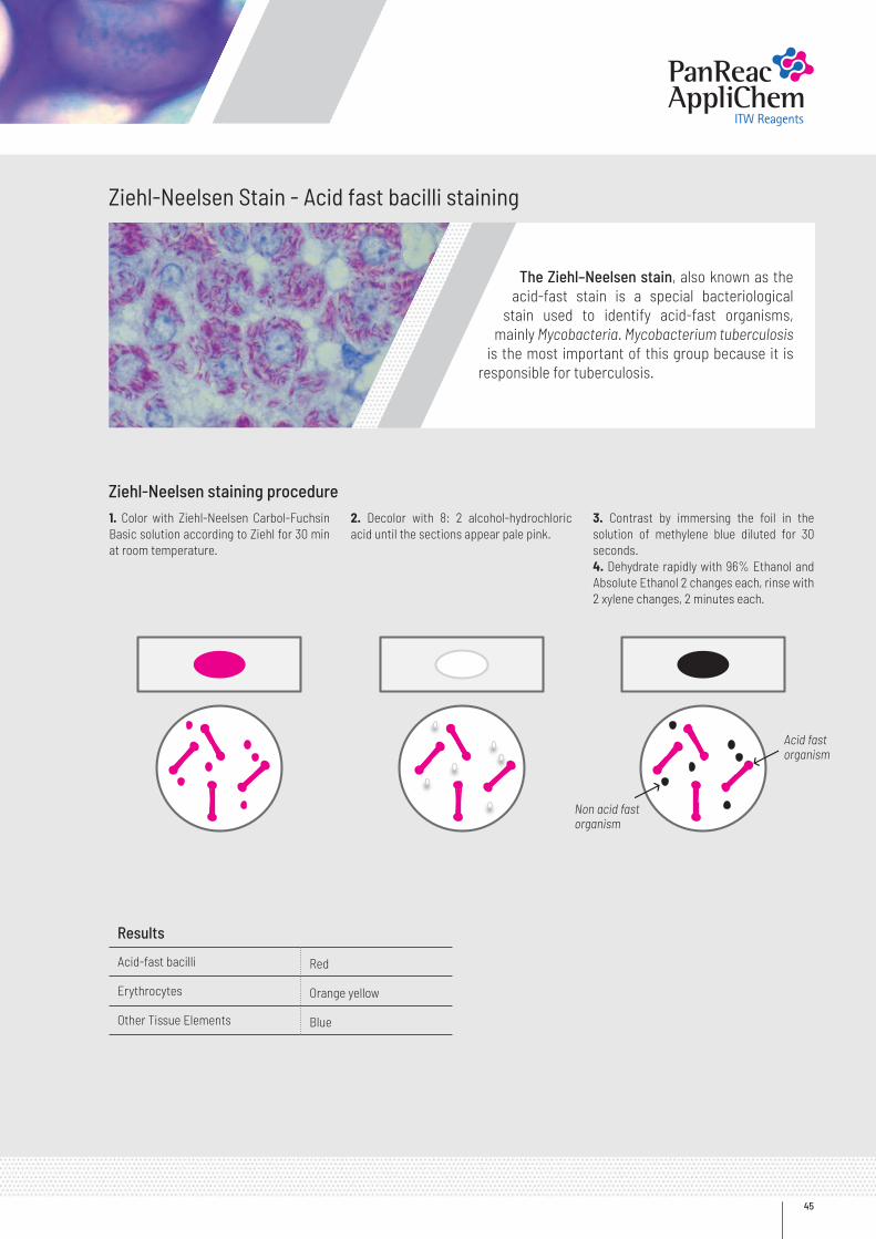
Ziehl-Neelsen Stain - Acid fast bacilli staining
Ziehl-Neelsen staining procedure
The Ziehl–Neelsen stain, also known as the acid-fast stain is a special bacteriological
stain used to identify acid-fast organisms, mainly Mycobacteria. Mycobacterium tuberculosis
is the most important of this group because it is responsible for tuberculosis.
Results
Acid-fast bacilli Red
Erythrocytes Orange yellow
Other Tissue Elements Blue
1. Color with Ziehl-Neelsen Carbol-Fuchsin Basic solution according to Ziehl for 30 min at room temperature.
2. Decolor with 8: 2 alcohol-hydrochloric acid until the sections appear pale pink.
3. Contrast by immersing the foil in the solution of methylene blue diluted for 30 seconds.4. Dehydrate rapidly with 96% Ethanol and Absolute Ethanol 2 changes each, rinse with 2 xylene changes, 2 minutes each.
Acid fast organism
Non acid fast organism
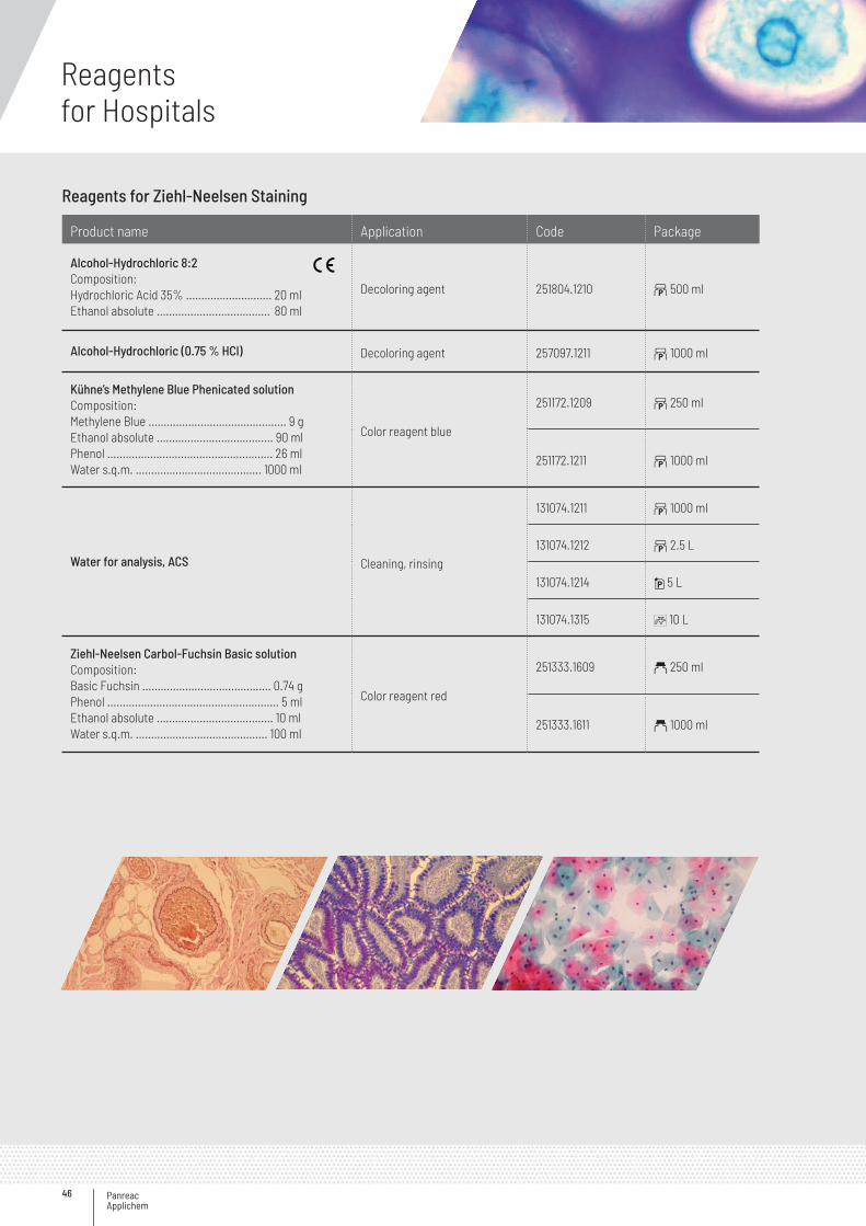
Panreac Applichem
Reagentsfor Hospitals
46
Reagents for Ziehl-Neelsen Staining
Product name Application Code Package
Alcohol-Hydrochloric 8:2Composition:Hydrochloric Acid 35% ............................ 20 mlEthanol absolute ..................................... 80 ml
Decoloring agent 251804.1210 b 500 ml
Alcohol-Hydrochloric (0.75 % HCl) Decoloring agent 257097.1211 b 1000 ml
Kühne’s Methylene Blue Phenicated solutionComposition:Methylene Blue ............................................. 9 gEthanol absolute ...................................... 90 mlPhenol ...................................................... 26 mlWater s.q.m. ......................................... 1000 ml
Color reagent blue
251172.1209 b 250 ml
251172.1211 b 1000 ml
Water for analysis, ACS Cleaning, rinsing
131074.1211 b 1000 ml
131074.1212 b 2.5 L
131074.1214 i 5 L
131074.1315 C 10 L
Ziehl-Neelsen Carbol-Fuchsin Basic solutionComposition:Basic Fuchsin .......................................... 0.74 gPhenol ........................................................ 5 mlEthanol absolute ...................................... 10 mlWater s.q.m. ........................................... 100 ml
Color reagent red
251333.1609 a 250 ml
251333.1611 a 1000 ml
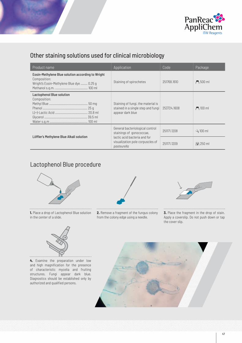
47
Other staining solutions used for clinical microbiologyOther staining solutions used for clinical microbiology
Lactophenol Blue procedure
Product name Application Code Package
Eosin-Methylene Blue solution according to WrightComposition:Wright’s Eosin-Methylene Blue dye ........ 0.25 gMethanol s.q.m. ...................................... 100 ml
Staining of spirochetes 251768.1610 a 500 ml
Lactophenol Blue solutionComposition:Methyl Blue ............................................. 50 mgPhenol .................................................... 25 gL(+)-Lactic Acid ...................................... 20.8 mlGlycerol .................................................. 39.5 mlWater s.q.m ............................................ 100 ml
Staining of fungi, the material isstained in a single step and fungiappear dark blue
253724.1608 a 100 ml
Löffler’s Methylene Blue Alkali solution
General bacteriological control stainings of gonococcae, lactic acid bacteria and for visualization pole corpuscles of pasteurella
251171.1208 l 100 ml
251171.1209 b 250 ml
2. Remove a fragment of the fungus colony from the colony edge using a needle.
3. Place the fragment in the drop of stain. Apply a coverslip. Do not push down or tap the cover slip.
1. Place a drop of Lactophenol Blue solution in the center of a slide.
4. Examine the preparation under low and high magnification for the presence of characteristic mycelia and fruiting structures. Fungi appear dark blue. Diagnostics should be established only by authorized and qualified persons.
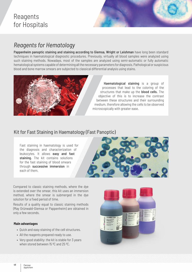
Panreac Applichem
Reagentsfor Hospitals
48
Reagents for HematologyPappenheim panoptic staining and staining according to Giemsa, Wright or Leishman have long been standard techniques in haematological diagnostic procedures. Previously, virtually all blood samples were analyzed using such staining methods. Nowadays, most of the samples are analyzed using semi-automatic or fully automatic hematological systems capable of determining all the necessary parameters for diagnosis. Pathological or suspicious blood and bone marrow smears are subjected to classical differential analysis using stains.
Compared to classic staining methods, where the dye is extended over the smear, this kit uses an immersion method, where the smear is submerged in the dye solution for a fixed period of time.Results of a quality equal to classic staining methods (May Grünwald-Giemsa or Pappenheim) are obtained in only a few seconds.
Kit for Fast Staining in Haematology (Fast Panoptic)
Haematological staining is a group of processes that lead to the coloring of the
structures that make up the blood cells. The objective of this is to increase the contrast
between these structures and their surrounding medium, therefore allowing the cells to be observed
microscopically with greater ease.
Main advantages
• Quick and easy staining of the cell structures. • All the reagents prepared ready to use. • Very good stability: the kit is stable for 3 years
when stored between 15 ºC and 25 ºC.
Fast staining in haematology is used for the diagnosis and characterization of leukocytes. It allows easy and fast staining. The kit contains solutions for the fast staining of blood smears through successive immersion in each of them.
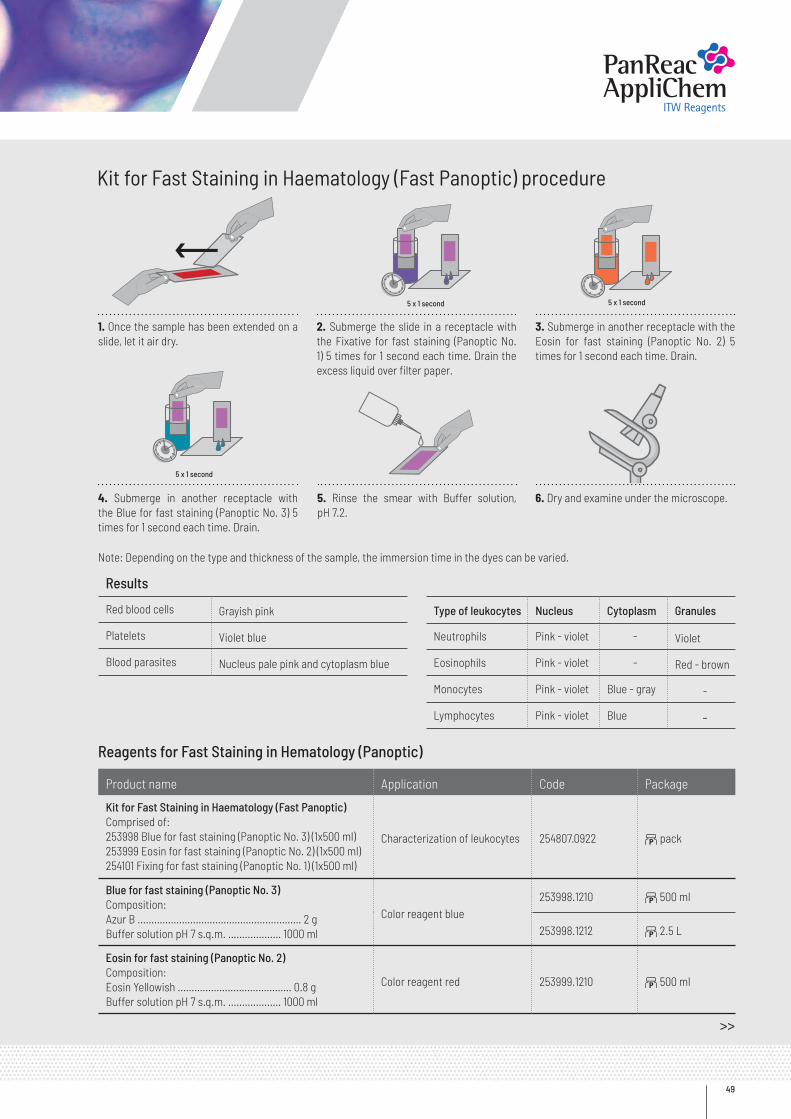
49
2. Submerge the slide in a receptacle with the Fixative for fast staining (Panoptic No. 1) 5 times for 1 second each time. Drain the excess liquid over filter paper.
5. Rinse the smear with Buffer solution, pH 7.2.
3. Submerge in another receptacle with the Eosin for fast staining (Panoptic No. 2) 5 times for 1 second each time. Drain.
6. Dry and examine under the microscope.
Note: Depending on the type and thickness of the sample, the immersion time in the dyes can be varied.
1. Once the sample has been extended on a slide, let it air dry.
4. Submerge in another receptacle with the Blue for fast staining (Panoptic No. 3) 5 times for 1 second each time. Drain.
Kit for Fast Staining in Haematology (Fast Panoptic) procedure
5 x 1 second
5 x 1 second5 x 1 second
Reagents for Fast Staining in Hematology (Panoptic)
Product name Application Code Package
Kit for Fast Staining in Haematology (Fast Panoptic) Comprised of:253998 Blue for fast staining (Panoptic No. 3) (1x500 ml)253999 Eosin for fast staining (Panoptic No. 2) (1x500 ml)254101 Fixing for fast staining (Panoptic No. 1) (1x500 ml)
Characterization of leukocytes 254807.0922 b pack
Blue for fast staining (Panoptic No. 3)Composition:Azur B ……………………………………........……… 2 gBuffer solution pH 7 s.q.m. ……..........… 1000 ml
Color reagent blue253998.1210 b 500 ml
253998.1212 b 2.5 L
Eosin for fast staining (Panoptic No. 2)Composition:Eosin Yellowish ……………........……………… 0.8 gBuffer solution pH 7 s.q.m. ……….......… 1000 ml
Color reagent red 253999.1210 b 500 ml
Results
Red blood cells Grayish pink
Platelets Violet blue
Blood parasites Nucleus pale pink and cytoplasm blue
Type of leukocytes Nucleus Cytoplasm Granules
Neutrophils Pink - violet - Violet
Eosinophils Pink - violet - Red - brown
Monocytes Pink - violet Blue - gray -
Lymphocytes Pink - violet Blue -
>>
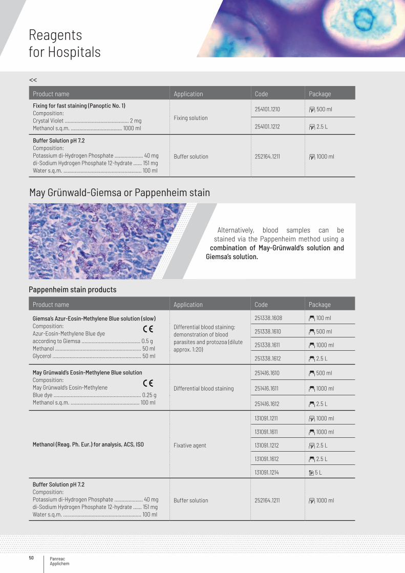
Panreac Applichem
Reagentsfor Hospitals
50
May Grünwald-Giemsa or Pappenheim stain
Pappenheim stain products
Product name Application Code Package
Giemsa’s Azur-Eosin-Methylene Blue solution (slow)Composition:Azur-Eosin-Methylene Blue dyeaccording to Giemsa ......................................... 0.5 gMethanol ............................................................ 50 mlGlycerol .............................................................. 50 ml
Differential blood staining; demonstration of blood parasites and protozoa (dilute approx. 1:20)
251338.1608 a 100 ml
251338.1610 a 500 ml
251338.1611 a 1000 ml
251338.1612 a 2.5 L
May Grünwald’s Eosin-Methylene Blue solutionComposition:May Grünwald’s Eosin-MethyleneBlue dye ............................................................. 0.25 gMethanol s.q.m. ................................................ 100 ml
Differential blood staining
251416.1610 a 500 ml
251416.1611 a 1000 ml
251416.1612 a 2.5 L
Methanol (Reag. Ph. Eur.) for analysis, ACS, ISO Fixative agent
131091.1211 b 1000 ml
131091.1611 a 1000 ml
131091.1212 b 2.5 L
131091.1612 a 2.5 L
131091.1214 i 5 L
Buffer Solution pH 7.2Composition:Potassium di-Hydrogen Phosphate ……………….. 40 mgdi-Sodium Hydrogen Phosphate 12-hydrate …… 151 mgWater s.q.m. …………………..………………………….. 100 ml
Buffer solution 252164.1211 b 1000 ml
Alternatively, blood samples can be stained via the Pappenheim method using a
combination of May-Grünwald’s solution and Giemsa’s solution.
Product name Application Code Package
Fixing for fast staining (Panoptic No. 1)Composition:Crystal Violet …………….........………………… 2 mgMethanol s.q.m. ……………….........……… 1000 ml
Fixing solution254101.1210 b 500 ml
254101.1212 b 2.5 L
Buffer Solution pH 7.2Composition:Potassium di-Hydrogen Phosphate ……………….. 40 mgdi-Sodium Hydrogen Phosphate 12-hydrate …… 151 mgWater s.q.m. …………………..………………………….. 100 ml
Buffer solution 252164.1211 b 1000 ml
>>
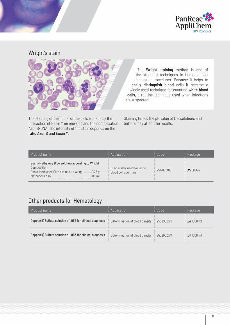
51
Wright’s stain
Other products for Hematology
Product name Application Code Package
Eosin-Methylene Blue solution according to Wright Composition:Eosin-Methylene Blue dye acc. to Wright ........ 0.25 gMethanol s.q.m. ............................................... 100 ml
Stain widely used for whiteblood cell counting
251768.1610 a 500 ml
Product name Application Code Package
Copper(II) Sulfate solution d.1.055 for clinical diagnosis Determination of blood density 253295.2711 ~ 1000 ml
Copper(II) Sulfate solution d.1.053 for clinical diagnosis Determination of blood density 253296.2711 ~ 1000 ml
The Wright staining method is one of the standard techniques in hematological
diagnostic procedures. Because it helps to easily distinguish blood cells it became a
widely used technique for counting white blood cells, a routine technique used when infections
are suspected.
The staining of the nuclei of the cells is made by the interaction of Eosin Y on one side and the complexation Azur B-DNA. The intensity of the stain depends on the ratio Azur B and Eosin Y.
Staining times, the pH value of the solutions and buffers may affect the results.
Panreac Applichem
Reagentsfor Hospitals
52
General ReagentsAuxiliary Products
Product name Code Package
Acetic Acid glacial (Reag. Ph. Eur.) for analysis, ACS, ISO
131008.1611 a 1000 ml
131008.1211 b 1000 ml
131008.1612 a 2.5 L
131008.1212 b 2.5 L
Acetic Acid glacial (USP, BP, Ph. Eur.) pure, pharma grade
141008.1611 a 1000 ml
141008.1211 b 1000 ml
141008.1612 a 2.5 L
141008.1212 b 2.5 L
Acetic Acid 96% for analysis122703.1611 a 1000 ml
122703.1612 a 2.5 L
Acetonitrile for UV, IR, HPLC, ACS361881.1611 a 1000 ml
361881.1612 a 2.5 L
Benedict’s Reagent qualitative for clinical diagnosis 251550.1211 b 1000 ml
Biuret’s Reagent for clinical diagnosis 251820.1208 b 100 ml
Boric Acid for analysis, ACS, ISO131015.1210 b 500 g
131015.1211 b 1000 g
Brij ® 35 aqueous solution 30% w/v for clinical diagnosis 252317.1611 a 1000 ml
Buffer solution pH 6.88 277091.1211 b 1000 ml
tert-Butyl Methyl Ether for UV, IR, HPLC363312.1611 a 1000 ml
363312.1612 a 2.5 L
Chloroform stabilized with ethanol (Reag. Ph. Eur.) for analysis, ACS, ISO131252.1611 a 1000 ml
131252.1612 a 2.5 L
Collodion solution 4% w/v (USP) pure, pharma grade141278.1609 a 250 ml
141278.1611 a 1000 ml
Dichloromethane stabilized with ~ 20 ppm of amylene for UV, IR, HPLC, GPC, ACS361254.1611 a 1000 ml
361254.1612 a 2.5 L
2,6-Dichlorophenol Indophenol Sodium Salt 2-hydrate (Reag. Ph. Eur.) for analysis, ACS 132056.1604 a 5 g
Diethyl Ether stabilized with ethanol for pesticide analysis 322551.1611 a 1000 ml
Diethyl Ether stabilized with ~ 6 ppm of BHT (Reag. Ph. Eur.) for analysis, ACS, ISO132770.0311 z 1000 ml
132770.1612 a 2.5 L
4-(Dimethylamino) Benzaldehyde (Reag. Ph. Eur.) for analysis, ACS 131293.1608 a 100 g
>>
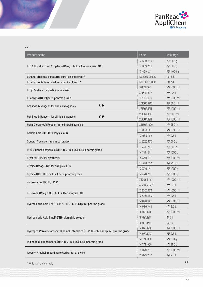
53
Product name Code Package
EDTA Disodium Salt 2-hydrate (Reag. Ph. Eur.) for analysis, ACS
131669.1209 b 250 g
131669.1210 b 500 g
131669.1211 b 1 000 g
Ethanol absolute denatured pure (pink colored) * NC808005000 i 5 L
Ethanol 94 % denatured pure (pink colored) * NC202005000 i 5 L
Ethyl Acetate for pesticide analysis321318.1611 a 1000 ml
321318.1612 a 2.5 L
Eucalyptol (USP) pure, pharma grade 142085.1611 a 1000 ml
Fehling's A Reagent for clinical diagnosis251563.1210 b 500 ml
251563.1211 b 1000 ml
Fehling’s B Reagent for clinical diagnosis251564.1210 b 500 ml
251564.1211 b 1000 ml
Folin-Ciocalteu’s Reagent for clinical diagnosis 251567.1609 a 250 ml
Formic Acid 98% for analysis, ACS131030.1611 a 1000 ml
131030.1612 a 2.5 L
General Absorbent technical grade 212520.1210 b 500 g
D(+)-Glucose anhydrous (USP, BP, Ph. Eur.) pure, pharma grade141341.1210 b 500 g
141341.1211 b 1000 g
Glycerol, 99% for synthesis 151339.1211 b 1000 ml
Glycine (Reag. USP) for analysis, ACS131340.1209 b 250 g
131340.1211 b 1000 g
Glycine (USP, BP, Ph. Eur.) pure, pharma grade 141340.1211 b 1000 g
n-Hexane for UV, IR, HPLC362063.1611 a 1000 ml
362063.1612 a 2.5 L
n-Hexane (Reag. USP, Ph. Eur.) for analysis, ACS132063.1611 a 1000 ml
132063.1612 a 2.5 L
Hydrochloric Acid 37% (USP-NF, BP, Ph. Eur.) pure, pharma grade141020.1611 a 1000 ml
141020.1612 a 2.5 L
Hydrochloric Acid 1 mol/l (1N) volumetric solution
181021.1211 b 1000 ml
181021.1214 i 5 l
181021.1315 C 10 L
Hydrogen Peroxide 33% w/v (110 vol.) stabilized (USP, BP, Ph. Eur.) pure, pharma grade141077.1211 b 1000 ml
141077.1212 b 2.5 L
Iodine resublimed pearls (USP, BP, Ph. Eur.) pure, pharma grade141771.1608 a 100 g
141771.1609 a 250 g
Isoamyl Alcohol according to Gerber for analysis121079.1211 b 1000 ml
121079.1212 b 2.5 L
>>
<<
* Only available in Italy
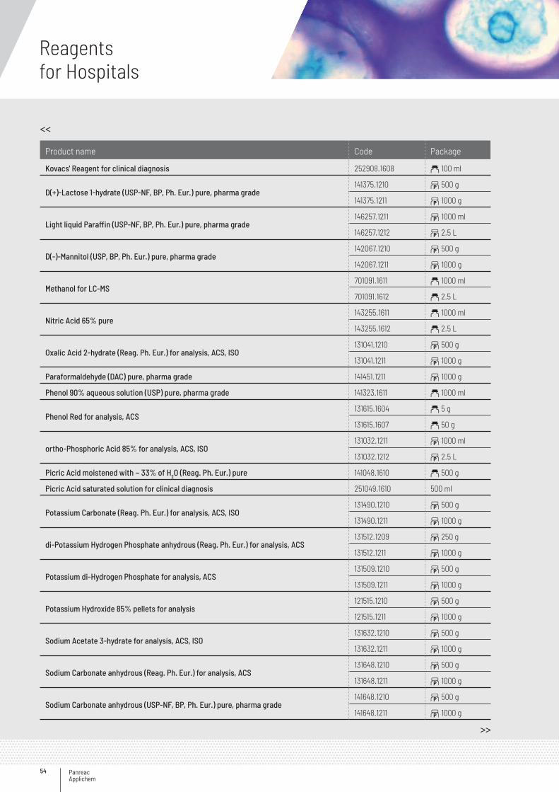
Panreac Applichem
Reagentsfor Hospitals
54
Product name Code Package
Kovacs' Reagent for clinical diagnosis 252908.1608 a 100 ml
D(+)-Lactose 1-hydrate (USP-NF, BP, Ph. Eur.) pure, pharma grade141375.1210 b 500 g
141375.1211 b 1000 g
Light liquid Paraffin (USP-NF, BP, Ph. Eur.) pure, pharma grade146257.1211 b 1000 ml
146257.1212 b 2.5 L
D(-)-Mannitol (USP, BP, Ph. Eur.) pure, pharma grade142067.1210 b 500 g
142067.1211 b 1000 g
Methanol for LC-MS701091.1611 a 1000 ml
701091.1612 a 2.5 L
Nitric Acid 65% pure143255.1611 a 1000 ml
143255.1612 a 2.5 L
Oxalic Acid 2-hydrate (Reag. Ph. Eur.) for analysis, ACS, ISO131041.1210 b 500 g
131041.1211 b 1000 g
Paraformaldehyde (DAC) pure, pharma grade 141451.1211 b 1000 g
Phenol 90% aqueous solution (USP) pure, pharma grade 141323.1611 a 1000 ml
Phenol Red for analysis, ACS131615.1604 a 5 g
131615.1607 a 50 g
ortho-Phosphoric Acid 85% for analysis, ACS, ISO131032.1211 b 1000 ml
131032.1212 b 2.5 L
Picric Acid moistened with ~ 33% of H2O (Reag. Ph. Eur.) pure 141048.1610 a 500 g
Picric Acid saturated solution for clinical diagnosis 251049.1610 500 ml
Potassium Carbonate (Reag. Ph. Eur.) for analysis, ACS, ISO131490.1210 b 500 g
131490.1211 b 1000 g
di-Potassium Hydrogen Phosphate anhydrous (Reag. Ph. Eur.) for analysis, ACS131512.1209 b 250 g
131512.1211 b 1000 g
Potassium di-Hydrogen Phosphate for analysis, ACS131509.1210 b 500 g
131509.1211 b 1000 g
Potassium Hydroxide 85% pellets for analysis121515.1210 b 500 g
121515.1211 b 1000 g
Sodium Acetate 3-hydrate for analysis, ACS, ISO131632.1210 b 500 g
131632.1211 b 1000 g
Sodium Carbonate anhydrous (Reag. Ph. Eur.) for analysis, ACS131648.1210 b 500 g
131648.1211 b 1000 g
Sodium Carbonate anhydrous (USP-NF, BP, Ph. Eur.) pure, pharma grade141648.1210 b 500 g
141648.1211 b 1000 g
<<
>>
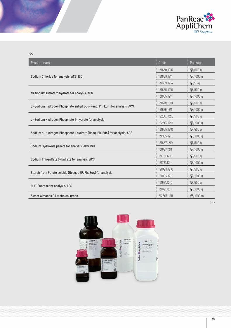
55
Product name Code Package
Sodium Chloride for analysis, ACS, ISO
131659.1210 b 500 g
131659.1211 b 1000 g
131659.1214 b 5 kg
tri-Sodium Citrate 2-hydrate for analysis, ACS131655.1210 b 500 g
131655.1211 b 1000 g
di-Sodium Hydrogen Phosphate anhydrous (Reag. Ph. Eur.) for analysis, ACS131679.1210 b 500 g
131679.1211 b 1000 g
di-Sodium Hydrogen Phosphate 2-hydrate for analysis122507.1210 b 500 g
122507.1211 b 1000 g
Sodium di-Hydrogen Phosphate 1-hydrate (Reag. Ph. Eur.) for analysis, ACS131965.1210 b 500 g
131965.1211 b 1000 g
Sodium Hydroxide pellets for analysis, ACS, ISO131687.1210 b 500 g
131687.1211 b 1000 g
Sodium Thiosulfate 5-hydrate for analysis, ACS131721.1210 b 500 g
131721.1211 b 1000 g
Starch from Potato soluble (Reag. USP, Ph. Eur.) for analysis121096.1210 b 500 g
121096.1211 b 1000 g
D(+)-Sucrose for analysis, ACS131621.1210 b 500 g
131621.1211 b 1000 g
Sweet Almonds Oil technical grade 212805.1611 a 1000 ml
<<
>>
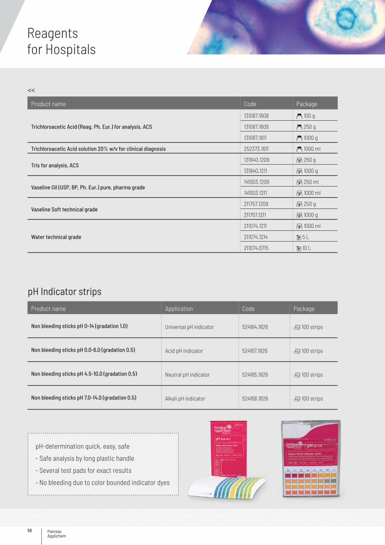
Panreac Applichem
Reagentsfor Hospitals
56
pH Indicator strips
Product name Application Code Package
Non bleeding sticks pH 0-14 (gradation 1.0) Universal pH indicator 524164.1826 v 100 strips
Non bleeding sticks pH 0.0-6.0 (gradation 0.5) Acid pH indicator 524167.1826 v 100 strips
Non bleeding sticks pH 4.5-10.0 (gradation 0.5) Neutral pH indicator 524165.1826 v 100 strips
Non bleeding sticks pH 7.0-14.0 (gradation 0.5) Alkali pH indicator 524168.1826 v 100 strips
pH-determination quick, easy, safe
- Safe analysis by long plastic handle
- Several test pads for exact results
- No bleeding due to color bounded indicator dyes
Product name Code Package
Trichloroacetic Acid (Reag. Ph. Eur.) for analysis, ACS
131067.1608 a 100 g
131067.1609 a 250 g
131067.1611 a 1000 g
Trichloroacetic Acid solution 20% w/v for clinical diagnosis 252373.1611 a 1000 ml
Tris for analysis, ACS131940.1209 b 250 g
131940.1211 b 1000 g
Vaseline Oil (USP, BP, Ph. Eur.) pure, pharma grade141003.1209 b 250 ml
141003.1211 b 1000 ml
Vaseline Soft technical grade211757.1209 b 250 g
211757.1211 b 1000 g
Water technical grade
211074.1211 b 1000 ml
211074.1214 i 5 L
211074.0715 i 10 L
<<
57
Derquim detergents
DERQUIM products have been specially made for cleaning the laboratory equipment. They can be used in the manual cleaning (series LM) or for washing using automatic machines (series LA). The various formulations are specifically adap-ted to any kind of laboratory residue: chemical, biological or clinical.
Product name Application Code Package
DERQUIM + Universal Detergent, LIQUID Universal detergent
503574.1211 b 1000 ml
503574.1246 i 4 L
503574.1315 C 10 L
MACHINE WASHING
DERQUIM LA 11 Slightly alkaline SOLID Removal of residues in laboratories 502603.1245 b 2 kg
502603.0415 G 10 kg
DERQUIM LA 12 Alkaline SOLID Strong cleaning agent, useful for starch and protein residues
502604.1245 b 2 kg
502604.0415 G 10 kg
502604.0416 G 25 kg
DERQUIM LA 13 Alkaline with detergents SOLID Strong cleaning agent specially for fatty acids
502605.1245 b 2 kg
502605.0415 G 10 kg
DERQUIM LA 14 Slightly alkaline LIQUID Good cleaning agent for machines with liquid dosing, indicated for analytical laboratories
502606.1246 i 4 L
502606.0716 i 25 L
DERQUIM LA 15 Alkaline LIQUID Strong cleaning agent 502607.1246 i 4 L
DERQUIM LA 21 Acid, with phosphoric acid LIQUID
Pre-wash for residues of amines, carbonates, hydroxides, proteines and so on; neutralizing effect, prevents calcification
502608.1246 i 4 L
DERQUIM LA 22 Acid, with citric acid LIQUID Pre-wash with neutralizing effects, prevents calcification
502609.1246 i 4 L
MANUAL WASHING
DERQUIM LM 01 Alkaline LIQUID General detergent for very contaminated items, also for bench tops, suitable for ultrasonic cleaning
502600.1246 i 4 L
DERQUIM LM 02 Neutral, phosphates free LIQUID
Special for cleaning of precision equipment made of glass, quartz, and sensitive metals, suitable for ultrasonic cleaning
502601.1246 i 4 L
502601.1315 C 10 L
502601.0716 i 25 L
DERQUIM LM 03 Phosphates free LIQUID General detergent for very contaminated items, suitable for ultrasonic cleaning
502602.1246 i 4 L
DERQUIM MC Chromic Mixture Elimination of organic waste resistant to detergents 502612.2211 2 1000 ml
DERQUIM SALT (Sodium Chloride lumps) For water decalcification503468.0415 G 10 kg
503468.0416 G 25 kg
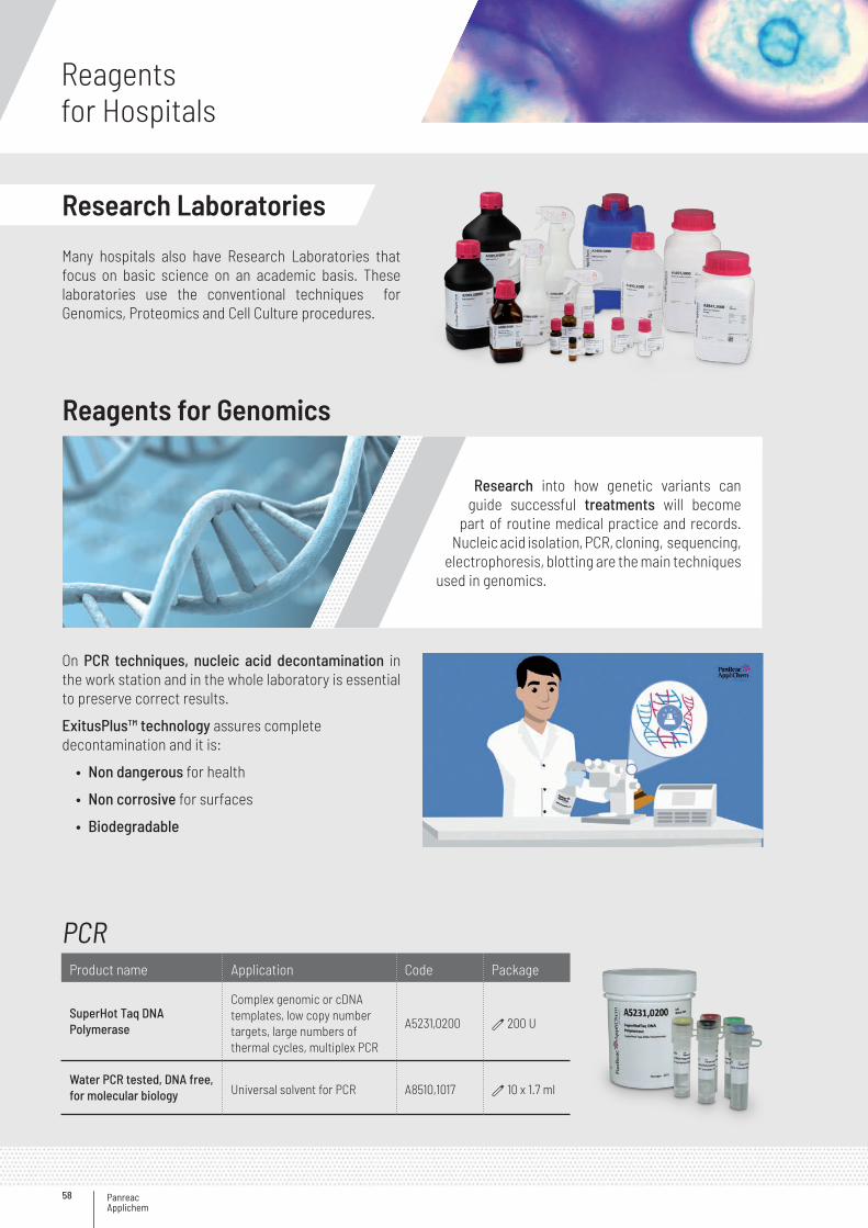
Panreac Applichem
Reagentsfor Hospitals
58
Research Laboratories
Many hospitals also have Research Laboratories that focus on basic science on an academic basis. These laboratories use the conventional techniques for Genomics, Proteomics and Cell Culture procedures.
On PCR techniques, nucleic acid decontamination in the work station and in the whole laboratory is essential to preserve correct results.
ExitusPlus™ technology assures complete decontamination and it is:
• Non dangerous for health
• Non corrosive for surfaces
• Biodegradable
Research into how genetic variants can guide successful treatments will become
part of routine medical practice and records. Nucleic acid isolation, PCR, cloning, sequencing,
electrophoresis, blotting are the main techniques used in genomics.
Reagents for Genomics
PCRProduct name Application Code Package
SuperHot Taq DNAPolymerase
Complex genomic or cDNA templates, low copy number targets, large numbers of thermal cycles, multiplex PCR
A5231,0200 d 200 U
Water PCR tested, DNA free, for molecular biology Universal solvent for PCR A8510,1017 d 10 x 1.7 ml
59
DNA Decontamination
Gel electrophoresis
Nucleic Acid Isolation
Product name Application Code Package
DNA-ExitusPlus™ IF Decontamination solution for the removal of DNA and RNA contaminations
A7409,0100 100 ml
A7409,0250 250 ml
A7409,0500 500 ml
RNase-ExitusPlus™ Decontamination removal solution for RNaseA7153,0500 500 ml
A7153,1000 1000 ml
Product name Application Code Package
DNA-Dye NonTox Ethidium bromide substitute A9555,1000 d 1 ml
Agarose low EEO (Agarose Standard)
Recommended for the preparation of analytical and preparative gels with a very good resolution of nucleic acid fragments with sizes larger than 1000 bp
A2114,0100 b 100 g
A2114,0250 b 250 g
A2114,0500 b 500 g
DNA ladder 100bp (lyophilised) DNA Size Standard for Gel Electrophoresis A3470,0050 d 50 μg
Product name Application Code Package
Proteinase KProteinase K is used to destruct proteins in cell lysates
A3830,0025 a 25 mg
A3830,0100 a 100 mg
A3830,0500 a 500 mg
DNase I
Used in molecular biology techniques like digestion of DNA, in the RNA purification or generating “random nicks” for “nick translation” or ‘footprint’-assays, or investigations on chromatin
A3778,0010 a 10 mg
A3778,0050 a 50 mg
A3778,0100 a 100 mg
A3778,0500 a 500 mg
Lysozyme for molecular biology Used to lyse E. coli for the isolationof plasmid-DNA
A4972,0001 b 1 g
A4972,0010 b 10 g
TRItidy G™ Ready-to-use solution for the simultaneous isolation of RNA, DNA and proteins
A4051,0100 b 100 ml
A4051,0200 b 200 ml
Panreac Applichem
Reagentsfor Hospitals
60
Cloning Assays
Buffers and Solvents
Product name Application Code Package
IPTG for molecularbiology
The most commonly used synthetic inductor of the Lac-operon since it is both active at very low concentrations and not subject to enzymatic degradation
A4773,0005 b 5 g
A4773,0025 b 25 g
X-Gal for molecularbiology
It is used for the identification of lacZ+ bacteria, especially for the assay of β-galactosidase, expressed from recombinant vectors
A4978,0500 b 25 g
A4978,0001 b 1 g
Product name Application Code Package
Tris for molecularbiology
Tris is the most commonly used buffer in biological research, component of TBE, TAEand TE Buffers
A2264,0250 b 250 g
A2264,0500 b 500 g
A2264,1000 b 1000 g
A2264,5000 G 5 kg
EDTA for molecular biologyEDTA is a chelator of calcium, magnesium and zinc ions and therefore may inhibit metallo proteases
A5097,0500 b 500 g
Acetic Acid 100 % for molecular biology Component of TAE Buffer for electrophoresis A3686,1000 b 1000 ml
61
Although genomics has delivered major advances in cancer prognostics, treatment and diagnostics, it still only provides a static image of the situation. To study more dynamic molecular entities, proteomics has been introduced into the cancer research field more than a decade ago. Currently, however, the impact of clinical proteomics on patient management and clinical decision-making is low and the implementations of scientific results in the clinic appear to be scarce.
The search for cancer-related biomarkers with proteomics however, has major potential
to improve risk assessment, early detection, diagnosis, prognosis, treatment selection and
monitoring. Main techniques used in proteomics are electrophoresis and blotting.
Reagents for Proteomics
Products for electrophoresis and blottingProduct name Application Code Package
GEL ELECTROPHORESIS COMPONENTS
Acrylamide 4K solution (30 %) - Mix 37.5 : 1
For most applications in the electrophoresis of nucleic acids or proteins, polyacrylamide gels are prepared from 30 % or 40 % stock solutions with a ratio Acrylamide : Bisacrylamide of 29 : 1 or 37.5 : 1
A1672,0500 b 500 ml
A1672,1000 b 1000 ml
Ammonium Persulfate BioChemica Ammonium persulfate (APS) serves as the initiators of the polymerization of Acrylamide
A1142,0250 b 250 g
Glycine for molecular biologyOne of the most commonly used buffer in the polyacrylamide gel electrophoresis for proteins is based on the work of Laemmli
A1067,0500 b 500 g
A1067,1000 b 1 kg
A1067,5000 G 5 kg
A1067,9010 G 10 kg
SDS BioChemica For SDS polyacrylamide gels
A2572,0250 b 250 g
A2572,0500 b 500 g
A2572,1000 b 1 kg
TEMED Enhancer of the polymerization (cross-linking) of acrylamide and bisacrylamide in gel electrophoresis
A1148,0025 a 25 ml
A1148,0100 a 100 ml
Tris ultrapure
One of its most important applications is the use as an electrophoresis buffer (e.g. TBE, see A1417 and A0972 or TAE, see A1416 and A1691) for polyacrylamide and agarose gel electrophoresis, respectively
A1086,0500 b 500 g
A1086,1000 b 1 kg
A1086,5000 G 5 kg
A1086,9010 G 10 kg
>>
Panreac Applichem
Reagentsfor Hospitals
62
Product name Application Code Package
BLOCKING AGENTS
Albumin Fraction V (pH 7.0)
Applied as a blocking agent for blocking unbound surfaces of blotting membranes in immunoblots or ELISAs, also used for the dilution of antisera and antibody-stock solutions
A1391,0025 b 25 g
A1391,0050 b 50 g
A1391,0100 b 100 g
A1391,0250 b 250 g
A1391,0500 b 500 g
Blocking Buffer I
Saturates free binding capacities on plastic consumables and other surfaces like ELISA plates and blotting membranes, thus a reduction of unspecific binding on surfaces can be achieved
A7099,0125 b 125 ml
A7099,0500 b 500 ml
TRANSFER MEMBRANES
Pure Nitrocellulose unsupported 0.45 μm Transfer Membrane
Used for Southern and Northern blots; Dot/Slot blots, Western blots and immunoblotting
A5239,3030Rv 30 cmx 3 m Roll
PVDF-Star Transfer Membrane 0.45 μm Used for Western Blots, immunoblotting and amino acid and protein analysis
A5243,3030Rv 30 cmx 3 m Roll
PROTEIN DETECTION
CheLuminate-HRP PicoDetect Kit for medium and poorly expressed proteins A3417,1200 v 1 Kit
Coomassie® Brilliant Blue R-250(C.I. 42660)
One of the most commonly used stains for proteins, after their separation by polyacrylamide gel electrophoresis
A1092,0025 b 25 g
A1092,0100 b 100 g
Ponceau S solution
For the staining of proteins immobilized on nitrocellulose filters, it is particularly suitable for reversible staining of proteins on transfer membranes during immunoblotting
A2935,0500 b 500 ml
GENERAL BIOCHEMICALS FOR PROTEIN PURIFICATION, ELECTROPHORESIS AND WESTERN BLOTTING
Protein Marker VI (10 – 245) prestained Protein Gel Electrophoresis Size Marker, Blue-Green-Red Protein Ladder
A8889,0500 d 500 µl
Acetic Acid 100 % BioChemica For protein staining solution preparationA3701,1000PE b 1000 ml
A3701,2500PE b 2.5 L
DTT BioChemica
It may substitute for β-mercaptoethanol in almost all experiments at three to four fold lower concentrations. DTT is less toxic, its odor is less intensive and it doesn’t form mixed disulfides like β-mercaptoethanol
A1101,0005 b 5 g
A1101,0025 b 25 g
A1101,0100 b 100 g
>>
<<
63
Product name Application Code Package
β-Mercaptoethanol Molecular biology gradeReducing agent used for the reduction of proteins during sample preparation, it prevents protein oxidation and acts as a denaturing agent of ribonucleases
A1108,0025 a 25 ml
A1108,0100 a 100 ml
A1108,0500 a 500 ml
Methanol BioChemica For western blottingA3493,1000PE b 1 L
A3493,5000 i 5 L
Tween® 20 for molecular biology For blocking buffers
A4974,0100 b 100 ml
A4974,0250 b 250 ml
A4974,0500 b 500 ml
A4974,1000 b 1 L
Urea BioChemica For Tris Urea gels, for protein staining solution preparation
A1360,5000 G 5 kg
A1360,9010 G 10 kg
PBS tablets pH 7.4 (for 500 ml)Used in a wide range of applications including Tissue culture/ Cell culture; Sample dilution/ Protein dilution; Immunoassays/ Immuno-histochemistry; Microbiology
A9191,0100 b 100 Tabs
<<
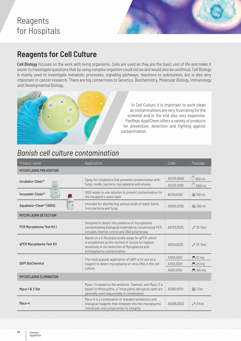
Panreac Applichem
Reagentsfor Hospitals
64
Cell Biology focuses on the work with living organisms. Cells are used as they are the basic unit of life and make it easier to investigate questions that by using complex organism could not be and would also be unethical. Cell Biology is mainly used to investigate metabolic processes, signaling pathways, reactions to substances, but is also very important in cancer research. There are big connections to Genetics, Biochemistry, Molecular Biology, Immunology and Developmental Biology.
In Cell Culture it is important to work clean as contaminations are very frustrating for the
scientist and in the end also very expensive. PanReac AppliChem offers a variety of products
for prevention, detection and fighting against contamination.
Reagents for Cell Culture
Banish cell culture contaminationProduct name Application Code Package
MYCOPLASMA PREVENTION
Incubator-Clean™ Spray for incubators that prevents contamination with fungi, molds, bacteria, mycoplasma and viruses
A5230,0500 500 ml
A5230,1000 1000 ml
Incuwater-Clean™ 100X ready-to-use solution to prevent contamination for the incubator’s water bath
A5219,0100 b 100 ml
Aquabator-Clean™ (100X) Intended for disinfecting various kinds of water baths from bacteria and fungi
A9390,0250 b 250 ml
MYCOPLASMA DETECTION
PCR Mycoplasma Test Kit IDesigned to detect the presence of mycoplasma contaminating biological materials by conventional PCR, includes internal control and DNA polymerase
A9753,0025 d 25 Test
qPCR Mycoplasma Test Kit
Based on a 5-Nuclease probe assay for qPCR, which is established as the method of choice for highest sensitivity in the detection of Mycoplasma and Acholeplasma contamination
A9019,0025 d 25 Test
DAPI BioChemicaThe most popular application of DAPI is its use as a reagent to detect mycoplasma or virus DNA in the cell culture
A1001,0010 a 10 mg
A1001,0025 a 25 mg
A1001,0100 a 100 mg
MYCOPLASMA ELIMINATION
Myco-1 & 2 SetMyco-1 is based on the antibiotic Tiamulin, and Myco-2 is based on Minocycline, a Tetracycline derivative, both are generally used sequentially in combination
A8360,0010 b 1 Set
Myco-4Myco-4 is a combination of standard antibiotics and biological reagents that integrate into the mycoplasma membrane and compromise its integrity
A8366,0002 d 2 Kits
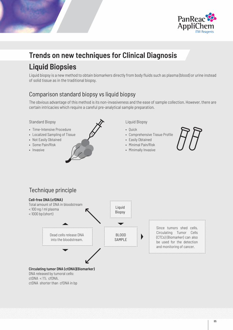
65
Trends on new techniques for Clinical DiagnosisLiquid BiopsiesLiquid biopsy is a new method to obtain biomarkers directly from body fluids such as plasma (blood) or urine instead of solid tissue as in the traditional biopsy.
Comparison standard biopsy vs liquid biopsyThe obvious advantage of this method is its non-invasiveness and the ease of sample collection. However, there are certain intricacies which require a careful pre-analytical sample preparation.
Technique principle
Standard Biopsy
• Time-Intensive Procedure• Localized Sampling of Tissue• Not Easily Obtained• Some Pain/Risk• Invasive
• Quick• Comprehensive Tissue Profile• Easily Obtained• Minimal Pain/Risk• Minimally Invasive
Liquid Biopsy
LiquidBiopsy
BLOOD SAMPLE
Since tumors shed cells, Circulating Tumor Cells (CTCs) (Biomarker) can also be used for the detection and monitoring of cancer.
Dead cells release DNA into the bloodstream.
Cell-free DNA (cfDNA) Total amount of DNA in bloodstream< 100 ng / ml plasma< 1000 bp (short)
Circulating tumor DNA (ctDNA)(Biomarker)DNA released by tumoral cells:ctDNA < 1% cfDNA.ctDNA shorter than cfDNA in bp
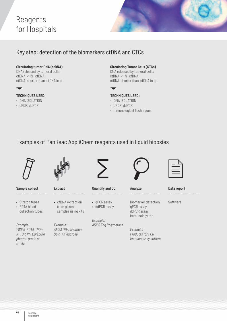
Panreac Applichem
Reagentsfor Hospitals
66
Key step: detection of the biomarkers ctDNA and CTCs
Examples of PanReac AppliChem reagents used in liquid biopsies
Circulating tumor DNA (ctDNA)DNA released by tumoral cells:ctDNA < 1% cfDNA.ctDNA shorter than cfDNA in bp
Sample collect
• Stretch tubes• EDTA blood
collection tubes
Example:141026 EDTA (USP-NF, BP, Ph. Eur) pure, pharma grade or similar
Extract
• cfDNA extraction from plasma samples using kits
Example:A5193 DNA Isolation Spin-Kit Agarose
Quantify and QC
• qPCR assay• ddPCR assay
Example:A5186 Taq Polymerase
Analyze
Biomarker detectionqPCR assayddPCR assayImmunology tec.
Example:Products for PCRImmunoassay buffers
Data report
Software
Circulating Tumor Cells (CTCs)DNA released by tumoral cells:ctDNA < 1% cfDNA.ctDNA shorter than cfDNA in bp
TECHNIQUES USED: • DNA ISOLATION• qPCR, ddPCR
TECHNIQUES USED: • DNA ISOLATION• qPCR, ddPCR• Inmunological Techniques
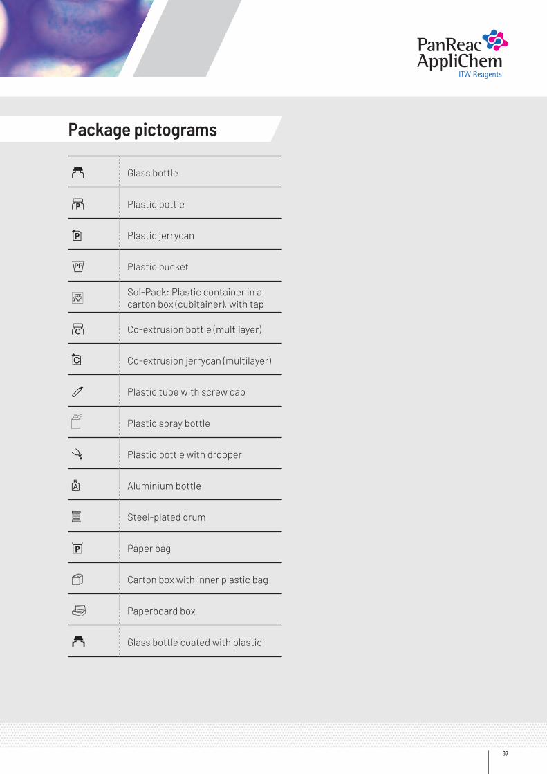
67
a Glass bottle
b Plastic bottle
i Plastic jerrycan
G Plastic bucket
C Sol-Pack: Plastic container in a carton box (cubitainer), with tap
~ Co-extrusion bottle (multilayer)
Co-extrusion jerrycan (multilayer)
d Plastic tube with screw cap
Plastic spray bottle
l Plastic bottle with dropper
z Aluminium bottle
s Steel-plated drum
h Paper bag
1 Carton box with inner plastic bag
v Paperboard box
2 Glass bottle coated with plastic
Package pictograms
www.itwreagents.com
A193,EN;201803
AppliChem GmbHOttoweg 4 · DE-64291 Darmstadt · Germany · Phone +49 6151 9357 0 · Fax +49 6151 9357 [email protected]
Nova Chimica SrlVia G. Galilei, 47 · I-20092 Cinisello Balsamo · (Milano) Italy · Phone +39 02 66045392 · Fax +39 02 [email protected]
PanReac Química SLUC/ Garraf 2, Polígono Pla de la Bruguera · E-08211 Castellar del Vallès · (Barcelona) Spain · Phone +34 937 489 400 · Fax +34 937 489 [email protected]


![Untitled-22 [] · 52 Introduction Acknowledgements Geological setting Systematic part . JAROSLAW STOLARSKI CONTENTS 52 53 55 56 58 58 59 59 59 62 62 Order Scleractinia BOURNE, 1900](https://img.pdfslide.us/doc/110x75/5fc59ec0053cdc0b740b27a7/untitled-22-52-introduction-acknowledgements-geological-setting-systematic-part.jpg)