Embed Size (px)
Citation preview
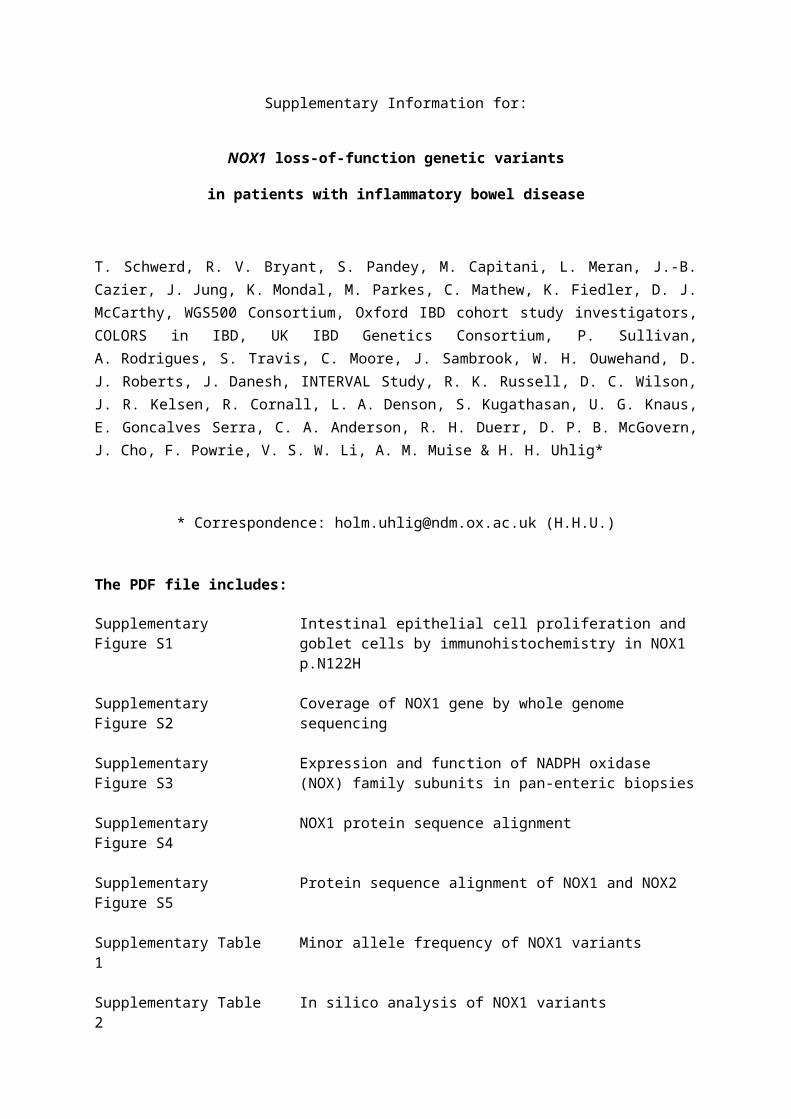
Supplementary Information for:
NOX1 loss-of-function genetic variants
in patients with inflammatory bowel disease
T. Schwerd, R. V. Bryant, S. Pandey, M. Capitani, L. Meran, J.-B. Cazier, J. Jung, K. Mondal, M. Parkes, C. Mathew, K. Fiedler, D. J. McCarthy, WGS500 Consortium, Oxford IBD cohort study investigators, COLORS in IBD, UK IBD Genetics Consortium, P. Sullivan, A. Rodrigues, S. Travis, C. Moore, J. Sambrook, W. H. Ouwehand, D. J. Roberts, J. Danesh, INTERVAL Study, R. K. Russell, D. C. Wilson, J. R. Kelsen, R. Cornall, L. A. Denson, S. Kugathasan, U. G. Knaus, E. Goncalves Serra, C. A. Anderson, R. H. Duerr, D. P. B. McGovern, J. Cho, F. Powrie, V. S. W. Li, A. M. Muise & H. H. Uhlig*
* Correspondence: [email protected] (H.H.U.)
The PDF file includes:
Supplementary Figure S1 Intestinal epithelial cell proliferation and goblet cells by immunohistochemistry in NOX1 p.N122H
Supplementary Figure S2 Coverage of NOX1 gene by whole genome sequencing
Supplementary Figure S3 Expression and function of NADPH oxidase (NOX) family subunits in pan-enteric biopsies
Supplementary Figure S4 NOX1 protein sequence alignment
Supplementary Figure S5 Protein sequence alignment of NOX1 and NOX2
Supplementary Table 1 Minor allele frequency of NOX1 variants
Supplementary Table 2 In silico analysis of NOX1 variants
Supplementary Table 3 Rare hemizygous, homozygous and compound heterozygous variants identified by WGS in P1 (p.N122H)
Supplementary Table 4 Patient characteristics of published hemi- and heterozygous NOX1 variants and splice variants without functional testing
Supplementary Table 5 Genetic association testing in males
Supplementary Table 6 Genetic association testing in females
Supplementary References
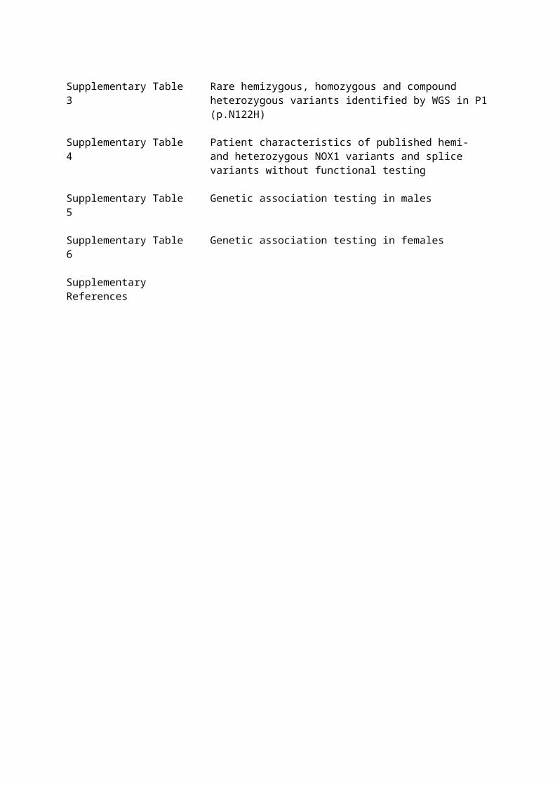
Supplementary Figure S1
Supplementary Figure S1. (a) Representative images of immunohistochemistry staining of Ki67 in colonic
crypts of a healthy control, the NOX1 mutant patient p.N122H and an IBD control patient. Histology for
NOX1 and IBD patient was obtained during disease quiescence. (b) Representative images of goblet cells
in colonic crypts stained with Alcian blue. Healthy control biopsy showing regular, plump goblet cells lining
the colonic epithelial crypt. NOX1 patient p.N122H during active disease showing relative paucity of goblet
cells and retention of mucin within goblet cells. IBD control patient during active disease showing increased
number of goblet cells which are of relatively reduced size having extruded mucin onto the surface
epithelium.
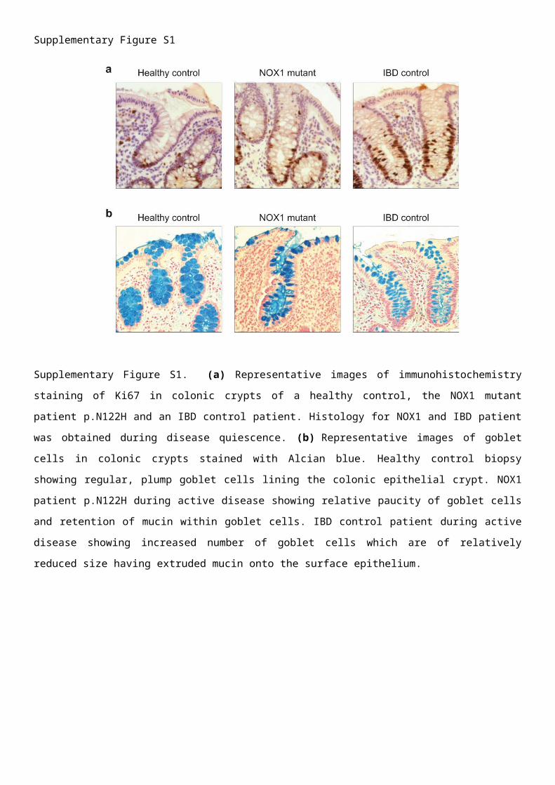
Supplementary Figure S2
Supplementary Figure S2. Coverage of NOX1 gene by whole genome sequencing. The reverse strand is
shown.
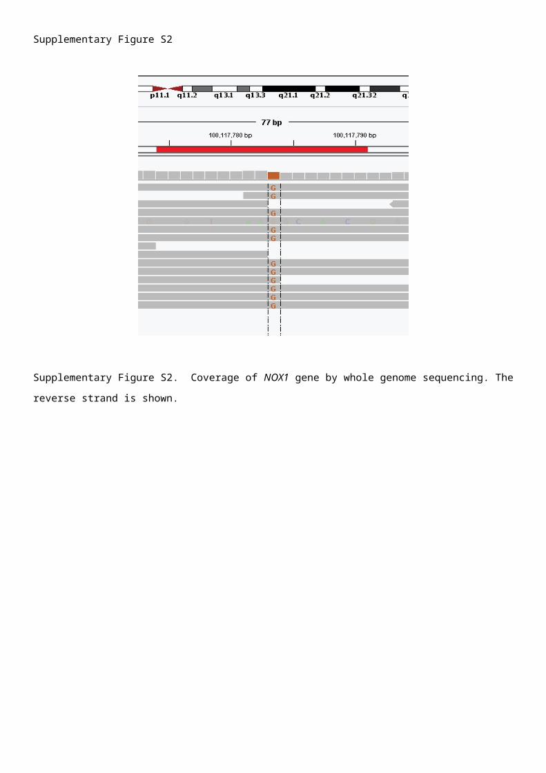
Supplementary Figure S3
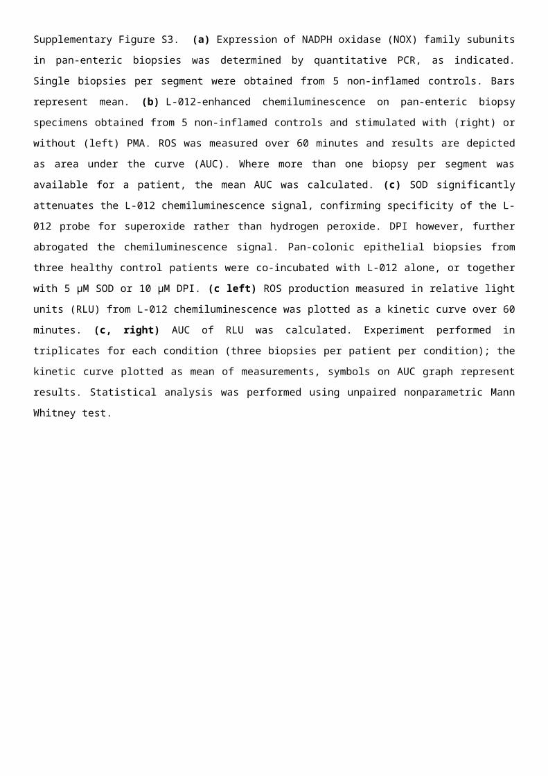
Supplementary Figure S3. (a) Expression of NADPH oxidase (NOX) family subunits in pan-enteric biopsies
was determined by quantitative PCR, as indicated. Single biopsies per segment were obtained from 5 non-
inflamed controls. Bars represent mean. (b) L-012-enhanced chemiluminescence on pan-enteric biopsy
specimens obtained from 5 non-inflamed controls and stimulated with (right) or without (left) PMA. ROS
was measured over 60 minutes and results are depicted as area under the curve (AUC). Where more than
one biopsy per segment was available for a patient, the mean AUC was calculated. (c) SOD significantly
attenuates the L-012 chemiluminescence signal, confirming specificity of the L-012 probe for superoxide
rather than hydrogen peroxide. DPI however, further abrogated the chemiluminescence signal. Pan-colonic
epithelial biopsies from three healthy control patients were co-incubated with L-012 alone, or together with
5 μM SOD or 10 μM DPI. (c left) ROS production measured in relative light units (RLU) from L-012
chemiluminescence was plotted as a kinetic curve over 60 minutes. (c, right) AUC of RLU was calculated.
Experiment performed in triplicates for each condition (three biopsies per patient per condition); the kinetic
curve plotted as mean of measurements, symbols on AUC graph represent results. Statistical analysis was
performed using unpaired nonparametric Mann Whitney test.
Supplementary Figure S4
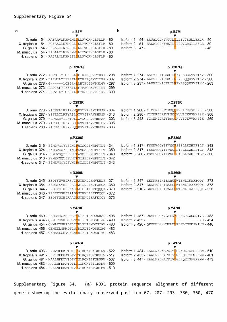
Supplementary Figure S4. (a) NOX1 protein sequence alignment of different genera showing the
evolutionary conserved position 67, 287, 293, 330, 360, 470 and 497. Alignment was performed using
ClustalW. (b) Corresponding amino acid sequence alignment of three NOX1 isoforms using ClustalW.
Supplementary Figure 5
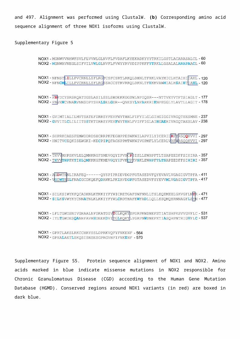
Supplementary Figure S5. Protein sequence alignment of NOX1 and NOX2. Amino acids marked in blue
indicate missense mutations in NOX2 responsible for Chronic Granulomatous Disease (CGD) according to
the Human Gene Mutation Database (HGMD). Conserved regions around NOX1 variants (in red) are
boxed in dark blue.
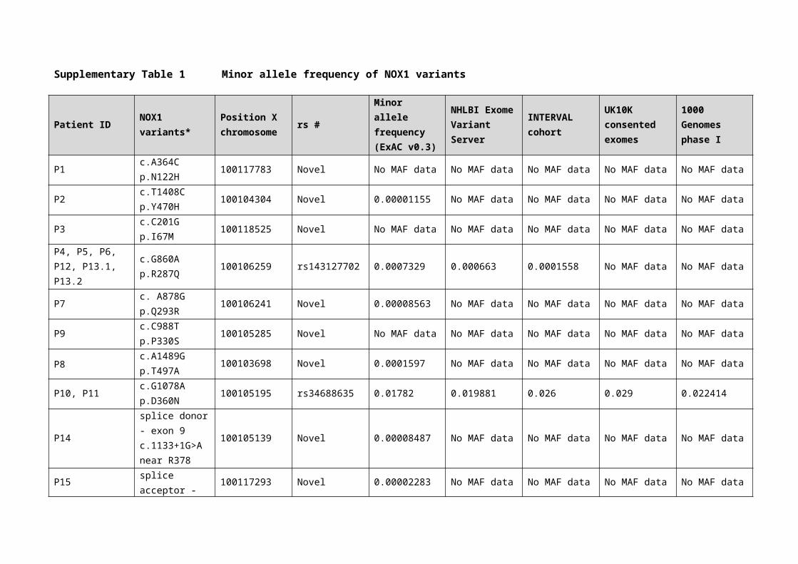
Supplementary Table 1 Minor allele frequency of NOX1 variants
Patient ID NOX1 variants* Position X chromosome rs #
Minor allele frequency (ExAC v0.3)
NHLBI Exome Variant Server
INTERVAL cohort
UK10K consented exomes
1000 Genomes phase I
P1c.A364Cp.N122H
100117783 Novel No MAF data No MAF data No MAF data No MAF data No MAF data
P2c.T1408Cp.Y470H
100104304 Novel 0.00001155 No MAF data No MAF data No MAF data No MAF data
P3c.C201Gp.I67M
100118525 Novel No MAF data No MAF data No MAF data No MAF data No MAF data
P4, P5, P6, P12, P13.1, P13.2
c.G860Ap.R287Q
100106259 rs143127702 0.0007329 0.000663 0.0001558 No MAF data No MAF data
P7c. A878Gp.Q293R
100106241 Novel 0.00008563 No MAF data No MAF data No MAF data No MAF data
P9c.C988Tp.P330S
100105285 Novel No MAF data No MAF data No MAF data No MAF data No MAF data
P8c.A1489Gp.T497A
100103698 Novel 0.0001597 No MAF data No MAF data No MAF data No MAF data
P10, P11c.G1078Ap.D360N
100105195 rs34688635 0.01782 0.019881 0.026 0.029 0.022414
P14
splice donor - exon 9c.1133+1G>Anear R378
100105139 Novel 0.00008487 No MAF data No MAF data No MAF data No MAF data
P15
splice acceptor - exon 7c.672-1G>Cnear G224
100117293 Novel 0.00002283 No MAF data No MAF data No MAF data No MAF data
P16Splice region variant c.142-4G>A
100118588 rs201776721 0.0007563
* Based on NOX1 transcript variant 1.
Supplementary Table 2 In silico analysis of NOX1 variants
Patient ID NOX1 variantsSIFT prediction(cutoff = 0.05)
PolyPhen-2 predictionPROVEAN prediction(cutoff = -2.5)
CADD score
P1 c.A364Cp.N122H
Damaging (0.0) Probably damaging (1.0) Deleterious (-4.68) 25.4
P2c.T1408Cp.Y470H Damaging (0.001) Benign (0.347) Deleterious (-3.79) 25.2
P3c.C201Gp.I67M
Damaging (0.0) Probably damaging (1.0) Neutral (-2.42) 23.8
P4, P5, P6, P12, P13.1, P13.2
c.G860Ap.R287Q Damaging (0.0) Probably damaging (1.0) Deleterious (-3.31) 33
P7c. A878Gp.Q293R
Tolerated (0.144) Possibly damaging (0.635) Deleterious (-2.65) 23.6
P9c.C988Tp.P330S
Tolerated (0.051) Probably damaging (0.995) Deleterious (-6.27) 23.7
P8c.A1489Gp.T497A Damaging (0.019) Probably damaging (1.0) Deleterious (-4.75) 24.7
P10, P11c.G1078Ap.D360N
Damaging (0.042) Benign (0.446) Deleterious (-4.37) 25.1
P14splice donor - exon 9c.1133+1G>Anear R378
NA NA NA 23.4
P15splice acceptor - exon 7c.672-1G>Cnear G224
NA NA NA 24.2
P16 Splice region variant c.142-4G>A
NA NA NA 3.7
Note: The severity of amino acid substitutions was predicted using SIFT (http://sift.jcvi.org/), Polyphen-2 (http://genetics.bwh.harvard.edu/pph2/),
PROVEAN (http://provean.jcvi.org/index.php), or using CADD scores (http://cadd.gs.washington.edu/)1. NA, not applicable

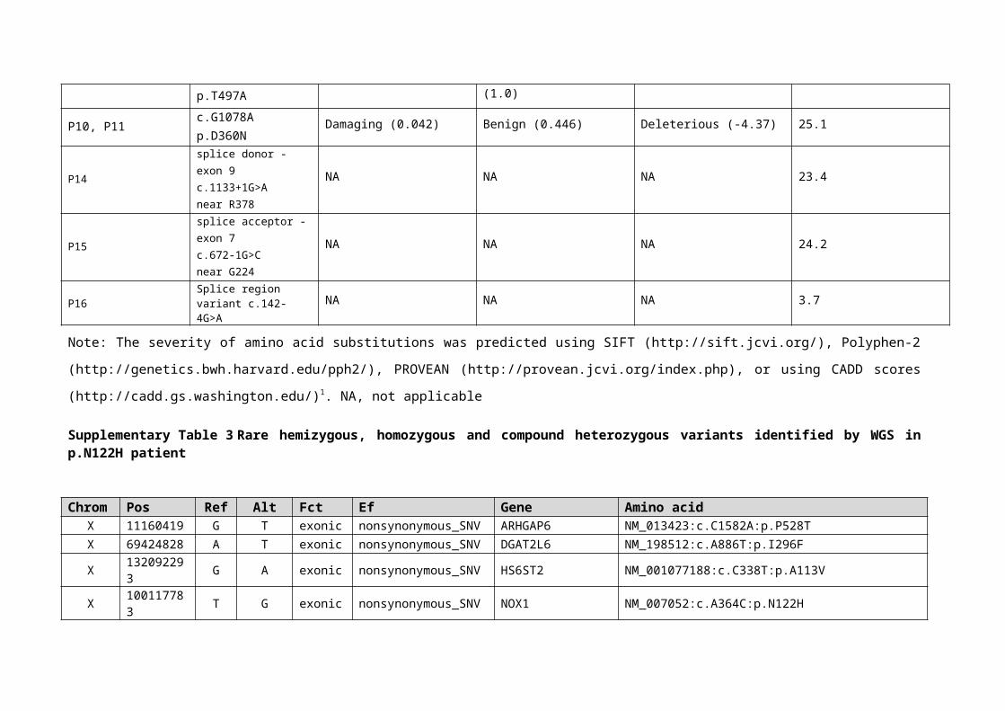
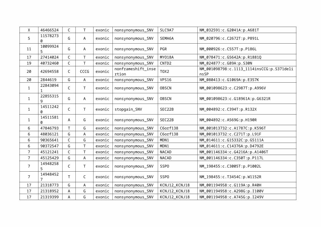
Supplementary Table 3 Rare hemizygous, homozygous and compound heterozygous variants identified by WGS in p.N122H patient
Chrom Pos Ref Alt Fct Ef Gene Amino acidX 11160419 G T exonic nonsynonymous_SNV ARHGAP6 NM_013423:c.C1582A:p.P528TX 69424828 A T exonic nonsynonymous_SNV DGAT2L6 NM_198512:c.A886T:p.I296FX 132092293 G A exonic nonsynonymous_SNV HS6ST2 NM_001077188:c.C338T:p.A113VX 100117783 T G exonic nonsynonymous_SNV NOX1 NM_007052:c.A364C:p.N122HX 46466524 C T exonic nonsynonymous_SNV SLC9A7 NM_032591:c.G2041A:p.A681T5 115782730 G A exonic nonsynonymous_SNV SEMA6A NM_020796:c.C2672T:p.P891L
11 100999245 G A exonic nonsynonymous_SNV PGR NM_000926:c.C557T:p.P186L17 27414024 C T exonic nonsynonymous_SNV MYO18A NM_078471:c.G5642A:p.R1881Q19 40732460 C T exonic nonsynonymous_SNV CNTD2 NM_024877:c.G89A:p.S30N20 42694558 C CCCG exonic nonframeshift_insertion TOX2 NM_001098798:c.1113_1114insCCG:p.S371delinsSP20 2844619 G A exonic nonsynonymous_SNV VPS16 NM_080413:c.G1069A:p.E357K1 228430941 C T exonic nonsynonymous_SNV OBSCN NM_001098623:c.C2987T:p.A996V1 228553159 G A exonic nonsynonymous_SNV OBSCN NM_001098623:c.G18961A:p.G6321R1 145112420 C T exonic stopgain_SNV SEC22B NM_004892:c.C394T:p.R132X1 145115810 A G exonic nonsynonymous_SNV SEC22B NM_004892:c.A569G:p.H190R6 47846793 T G exonic nonsynonymous_SNV C6orf138 NM_001013732:c.A1787C:p.K596T6 48036121 G A exonic nonsynonymous_SNV C6orf138 NM_001013732:c.C271T:p.L91F6 90365641 C G exonic nonsynonymous_SNV MDN1 NM_014611:c.G15332C:p.G5111A6 90372547 G T exonic nonsynonymous_SNV MDN1 NM_014611:c.C14376A:p.D4792E7 45121241 C T exonic nonsynonymous_SNV NACAD NM_001146334:c.G4216A:p.A1406T7 45125429 G A exonic nonsynonymous_SNV NACAD NM_001146334:c.C350T:p.P117L7 149482585 C T exonic nonsynonymous_SNV SSPO NM_198455:c.C3005T:p.P1002L7 149484527 T C exonic nonsynonymous_SNV SSPO NM_198455:c.T3454C:p.W1152R
17 21318773 G A exonic nonsynonymous_SNV KCNJ12_KCNJ18 NM_001194958:c.G119A:p.R40H17 21318952 A G exonic nonsynonymous_SNV KCNJ12_KCNJ18 NM_001194958:c.A298G:p.I100V
17 21319399 A G exonic nonsynonymous_SNV KCNJ12_KCNJ18 NM_001194958:c.A745G:p.I249V
Note: Filtering revealed 277 rare variants (non-synonymous AA variants/potential splice sites/ncRNA-exonic/rare <0.001 MAF variants). This
included six genes with homozygous variants (SEMA6A, PGR, MYO18A, CNTD2, TOX2, VPS16), five genes with hemizygous variants
(ARHGAP6, DGAT2L6, HS6ST2, NOX1, SLC9A7) and seven genes with potentially compound heterozygous variants (OBSCN, SEC22B,
C6orf138, MDN1, NACAD, SSPO, KCNJ12_KCNJ18). None of these variants have been described as causative for IBD or EBV-induced
hemophagocytosis. No variants in the Crohn’s disease associated gene NOD2 were identified.
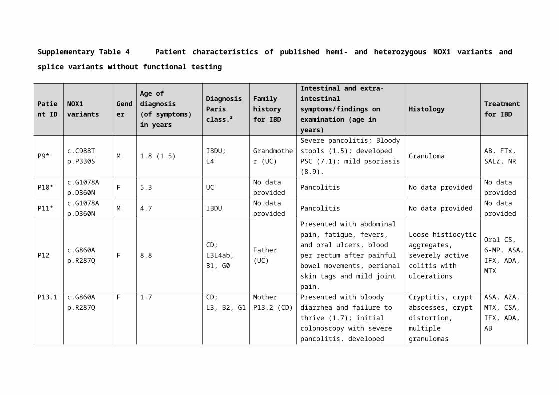
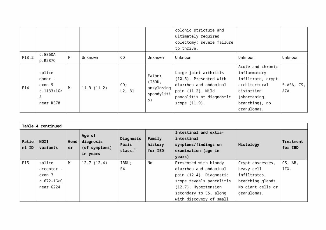
Supplementary Table 4 Patient characteristics of published hemi- and heterozygous NOX1 variants and splice variants without functional testing
Patient ID
NOX1 variants
Gender
Age of diagnosis(of symptoms) in years
DiagnosisParis class.2
Family history for IBD
Intestinal and extra-intestinal symptoms/findings on examination (age in years)
Histology Treatment for IBD
P9*c.C988Tp.P330S
M 1.8 (1.5)IBDU;E4
Grandmother (UC)
Severe pancolitis; Bloody stools (1.5); developed PSC (7.1); mild psoriasis (8.9).
GranulomaAB, FTx,SALZ, NR
P10*c.G1078Ap.D360N
F 5.3 UCNo data provided
Pancolitis No data providedNo data provided
P11*c.G1078Ap.D360N
M 4.7 IBDUNo data provided
Pancolitis No data providedNo data provided
P12c.G860Ap.R287Q
F 8.8CD;L3L4ab, B1, G0
Father (UC)
Presented with abdominal pain, fatigue, fevers, and oral ulcers, blood per rectum after painful bowel movements, perianal skin tags and mild joint pain.
Loose histiocytic aggregates, severely active colitis with ulcerations
Oral CS, 6-MP, ASA, IFX, ADA, MTX
P13.1c.G860Ap.R287Q
F 1.7CD;L3, B2, G1
Mother P13.2 (CD)
Presented with bloody diarrhea and failure to thrive (1.7); initial colonoscopy with severe pancolitis, developed colonic stricture and ultimately required colectomy; severe failure to thrive.
Cryptitis, crypt abscesses, crypt distortion, multiple granulomas
ASA, AZA, MTX, CSA, IFX, ADA, AB
P13.2c.G860Ap.R287Q
F Unknown CD Unknown Unknown Unknown Unknown
P14
splice donor - exon 9 c.1133+1G>Anear R378
M 11.9 (11.2)CD;L2, B1
Father (IBDU, ankylosing spondylitis)
Large joint arthritis (10.6). Presented with diarrhea and abdominal pain (11.2). Mild pancolitis at diagnostic scope (11.9).
Acute and chronic inflammatory infiltrate, crypt architectural distortion (shortening, branching), no granulomas.
5-ASA, CS, AZA
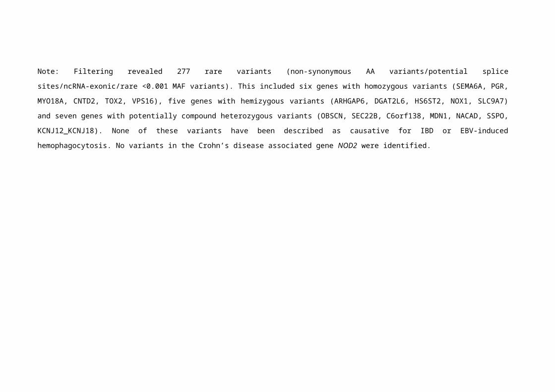
Table 4 continued
Patient ID
NOX1 variants
Gender
Age of diagnosis(of symptoms) in years
DiagnosisParis class.2
Family history for IBD
Intestinal and extra-intestinal symptoms/findings on examination (age in years)
Histology Treatment for IBD
P15
splice acceptor - exon 7c.672-1G>Cnear G224
M 12.7 (12.4)IBDU;E4
No
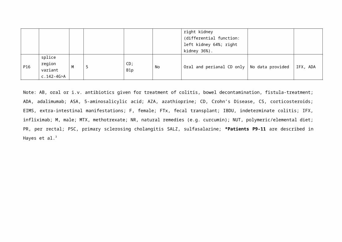
Presented with bloody diarrhea and abdominal pain (12.4). Diagnostic scope reveals pancolitis (12.7). Hypertension secondary to CS, along with discovery of small right kidney (differential function: left kidney 64%; right kidney 36%).
Crypt abscesses, heavy cell infiltrates, branching glands. No giant cells or granulomas.
CS, AB, IFX.
P16splice region variantc.142-4G>A
M 5CD;B1p
No Oral and perianal CD only No data provided IFX, ADA
Note: AB, oral or i.v. antibiotics given for treatment of colitis, bowel decontamination, fistula-treatment; ADA, adalimumab; ASA, 5-aminosalicylic
acid; AZA, azathioprine; CD, Crohn’s Disease, CS, corticosteroids; EIMS, extra-intestinal manifestations; F, female; FTx, fecal transplant; IBDU,
indeterminate colitis; IFX, infliximab; M, male; MTX, methotrexate; NR, natural remedies (e.g. curcumin); NUT, polymeric/elemental diet; PR, per
rectal; PSC, primary sclerosing cholangitis SALZ, sulfasalazine; *Patients P9-11 are described in Hayes et al.3
Supplementary table 5 Genetic association testing in males
Males control C(HOM A1) C(HET) C(HOM A2) C(HAP A1) C(HAP A2) C(MISSING) MAF Allele numbers p-value ALLELIC Test
European ancestry exome chip dataset 0 0 0 71 2741 0 0.0252 2812
INTERVAL cohort 0 0 0 55 1877 0 0.0285 1932
Summed counts 0 0 0 126 4618 0 0.0266 4744
Males IBD C(HOM A1) C(HET) C(HOM A2) C(HAP A1) C(HAP A2) C(MISSING)
European ancestry exome chip dataset 0 0 0 147 5093 3 0.0281 5240 0.5047
Oxford IBD study 0 0 0 4 244 0 0.0161 248
COLORS in IBD 0 0 0 1 60 0 0.0164 61
Summed counts 0 0 0 152 5397 3 0.0274 5549 0.8425
Males UC C(HOM A1) C(HET) C(HOM A2) C(HAP A1) C(HAP A2) C(MISSING)
European ancestry exome chip dataset 0 0 0 84 2299 3 0.0352 2383 0.04243
Oxford IBD study 0 0 0 1 60 0 0.0164 61
COLORS in IBD 0 0 0 0 18 0 0.0000 18
Summed counts 0 0 0 85 2377 3 0.0345 2462 0.0675
Males CD C(HOM A1) C(HET) C(HOM A2) C(HAP A1) C(HAP A2) C(MISSING)
European ancestry exome chip dataset 0 0 0 61 2691 3 0.0222 2752 0.5045
Oxford IBD study 0 0 0 1 114 0 0.0087 115
COLORS in IBD 0 0 0 1 27 0 0.0357 28
Summed counts 0 0 0 63 2832 3 0.0218 2895 0.2173
Supplementary table 6 Genetic association testing in females
Female controls C(HOM A1)C(HET) C(HOM A2) C(HAP A1) C(HAP A2) C(MISSING) MAF Allele numbers p-value ALLELIC Test
European ancestry exome chip dataset 2 137 2775 0 0 0 0.0242 5828
INTERVAL cohort 1 90 1764 0 0 0 0.0248 3710
Summed counts 3 227 4539 0 0 0 0.0244 9538
Female IBD C(HOM A1)C(HET) C(HOM A2) C(HAP A1) C(HAP A2) C(MISSING)
European ancestry exome chip dataset 7 271 5001 0 0 1 0.0270 10560 0.3044
Oxford IBD study 2 15 248 0 0 0 0.0358 530
COLORS in IBD 0 6 37 0 0 0 0.0698 86
Summed counts 9 292 5286 0 0 1 0.0277 11176 0.1486
Female UC C(HOM A1)C(HET) C(HOM A2) C(HAP A1) C(HAP A2) C(MISSING)
European ancestry exome chip dataset 1 131 2067 0 0 1 0.0302 4400 0.06982
Oxford IBD study 0 5 43 0 0 0 0.0521 96
COLORS in IBD 0 0 16 0 0 0 0.0000 32
Summed counts 1 136 2126 0 0 1 0.0305 4528 0.04142
Female CD C(HOM A1)C(HET) C(HOM A2) C(HAP A1) C(HAP A2) C(MISSING)
European ancestry exome chip dataset 5 135 2847 0 0 0 0.0243 5974 1
Oxford IBD study 1 5 127 0 0 0 0.0263 266
COLORS in IBD 0 5 13 0 0 0 0.1389 36
Summed counts 6 145 2987 0 0 0 0.0250 6276 0.8567
Supplementary References
1. Kircher M, Witten DM, Jain P, O'Roak BJ, Cooper GM, Shendure J. A general framework for estimating the relative pathogenicity of human genetic variants. Nature genetics 2014; 46(3): 310-315.
2. Levine A, Griffiths A, Markowitz J, Wilson DC, Turner D, Russell RK et al. Pediatric modification of the Montreal classification for inflammatory bowel disease: the Paris classification. Inflammatory bowel diseases 2011; 17(6): 1314-1321.
3. Hayes P, Dhillon S, O'Neill K, Thoeni C, Hui KY, Elkadri A et al. Defects in NADPH Oxidase Genes and in Very Early Onset Inflammatory Bowel Disease. Cellular and molecular gastroenterology and hepatology 2015; 1(5): 489-502.