Embed Size (px)
Citation preview

MEDIA GUIDE ON BLOOD CLOTSVOLUME II: BLOOD CLOTS IN THE ARTERIES
VOLUME II: BLOOD CLOTS IN THE ARTERIES
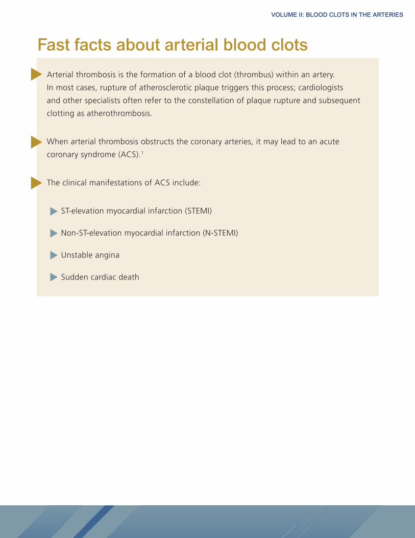
Arterial thrombosis is the formation of a blood clot (thrombus) within an artery.
In most cases, rupture of atherosclerotic plaque triggers this process; cardiologists
and other specialists often refer to the constellation of plaque rupture and subsequent
clotting as atherothrombosis.
When arterial thrombosis obstructs the coronary arteries, it may lead to an acute
coronary syndrome (ACS).1
The clinical manifestations of ACS include:
ST-elevation myocardial infarction (STEMI)
Non-ST-elevation myocardial infarction (N-STEMI)
Unstable angina
Sudden cardiac death
Fast facts about arterial blood clots
VOLUME II: BLOOD CLOTS IN THE ARTERIES
What is an arterial blood clot?The formation of a blood clot within an artery is referred to as arterial thrombosis. In most cases, arterial thrombosis
follows the rupture of an atheroma, a fatty plaque in the artery, resulting from atherosclerosis.2
Arterial blood clots in cardiovascular diseaseArterial blood clots are associated with a number of chronic cardiovascular diseases. In many cases, blood clots
can lead to serious complications, such as heart attack or stroke.
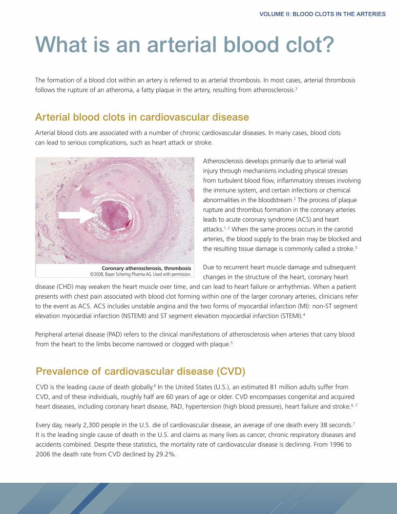
Atherosclerosis develops primarily due to arterial wall
injury through mechanisms including physical stresses
from turbulent blood flow, inflammatory stresses involving
the immune system, and certain infections or chemical
abnormalities in the bloodstream.2 The process of plaque
rupture and thrombus formation in the coronary arteries
leads to acute coronary syndrome (ACS) and heart
attacks.1, 2 When the same process occurs in the carotid
arteries, the blood supply to the brain may be blocked and
the resulting tissue damage is commonly called a stroke.3
Due to recurrent heart muscle damage and subsequent
changes in the structure of the heart, coronary heart
disease (CHD) may weaken the heart muscle over time, and can lead to heart failure or arrhythmias. When a patient
presents with chest pain associated with blood clot forming within one of the larger coronary arteries, clinicians refer
to the event as ACS. ACS includes unstable angina and the two forms of myocardial infarction (MI): non-ST segment
elevation myocardial infarction (NSTEMI) and ST segment elevation myocardial infarction (STEMI).4
Peripheral arterial disease (PAD) refers to the clinical manifestations of atherosclerosis when arteries that carry blood
from the heart to the limbs become narrowed or clogged with plaque.5
Prevalence of cardiovascular disease (CVD)CVD is the leading cause of death globally.6 In the United States (U.S.), an estimated 81 million adults suffer from
CVD, and of these individuals, roughly half are 60 years of age or older. CVD encompasses congenital and acquired
heart diseases, including coronary heart disease, PAD, hypertension (high blood pressure), heart failure and stroke.6, 7
Every day, nearly 2,300 people in the U.S. die of cardiovascular disease, an average of one death every 38 seconds.7
It is the leading single cause of death in the U.S. and claims as many lives as cancer, chronic respiratory diseases and
accidents combined. Despite these statistics, the mortality rate of cardiovascular disease is declining. From 1996 to
2006 the death rate from CVD declined by 29.2%.
Coronary atherosclerosis, thrombosis ©2008, Bayer Schering Pharma AG. Used with permission.
VOLUME II: BLOOD CLOTS IN THE ARTERIES
Risk factors for CVDThere are several identifiable factors that increase the risk of a person developing CVD. Some are considered major risk
factors, while others play a lesser role. Some risk factors can be modified, treated or controlled, and others cannot. The
major risk factors that cannot be changed include age, gender and family history; some risk factors can be favorably
modified with an associated decrease in risk, including tobacco use, physical inactivity and high blood pressure.8
Risk factors that cannot be modified:
Age: CVD, a chronic condition that often develops slowly, is more common in older individuals. More than 40% of
deaths for people aged 65 to 74 are from CVD, increasing to 60% for those over 80.9
Gender: On average, the annual rates of first major cardiovascular events are three per 1,000 in men ages 35-44.
This number rises to 74 per 1,000 men at ages 85-94. For women, comparable rates occur 10 years later in life and
the gap narrows with advancing age.7
Family history: Family history also plays an important role in the risk of developing heart disease. Children of parents
who developed CVD are more likely to develop coronary or vascular disease, or to be at risk for stroke, than children
from unaffected families. Prevalence of specific types of CVD differs among population subgroups. For example,
African Americans are more likely to have hypertensive heart disease (high blood pressure) when compared to
Caucasians. American Heart Association (AHA) data also show that Mexican Americans, Native Americans, Hawaiians
and some Asian Americans are at a heightened risk for cardiovascular disease, which is partly due to higher rates of
obesity and diabetes.7
Risk factors that can be managed:
Individuals can modify or manage several important risk factors to reduce their risk of CHD/CVD.
These include: 8
• tobacco use
• high blood pressure
• high cholesterol levels
• physical inactivity
• diet
• diabetes
Acute coronary syndrome (ACS)ACS is a term used for a clinical constellation of findings brought on by sudden reduction in blood flow to the
heart.10 The subtypes of ACS include: unstable angina and two types of myocardial infarction (MI), based on EKG
findings when the person presents to the hospital—Non-ST-Segment Elevation Myocardial Infarction (NSTEMI) or
ST-Segment Elevation Myocardial Infarction (STEMI).1
Myocardial infarction (MI)
The heart muscle requires a constant supply of nutrients and oxygen to maintain function. The heart receives
oxygen-rich blood from the coronary arteries.
VOLUME II: BLOOD CLOTS IN THE ARTERIES
There are two main coronary arteries, the left and right. They stem from the aorta, the large artery that leaves the
heart carrying oxygenated blood, and branch into smaller arteries that supply all parts of the heart muscle with
blood. MI occurs when these coronary arteries are blocked and cannot supply blood to the working heart muscle.
Heart muscle deprived of blood flow is irreversibly damaged within a few hours.11
Cardiologists diagnose MI initially based on the changes in the electrocardiogram (ECG) that occur in response to
reduction of coronary blood flow. These EKG changes reflect the amount of heart muscle at risk of dying:
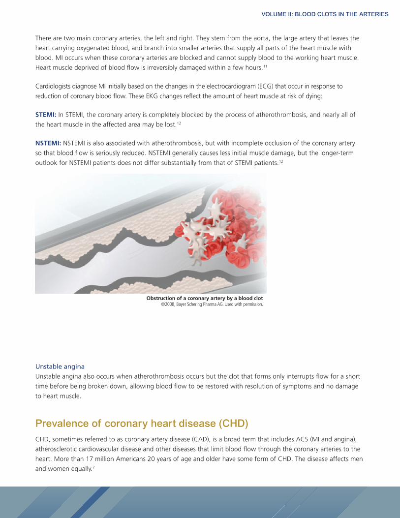
STEMI: In STEMI, the coronary artery is completely blocked by the process of atherothrombosis, and nearly all of
the heart muscle in the affected area may be lost.12
NSTEMI: NSTEMI is also associated with atherothrombosis, but with incomplete occlusion of the coronary artery
so that blood flow is seriously reduced. NSTEMI generally causes less initial muscle damage, but the longer-term
outlook for NSTEMI patients does not differ substantially from that of STEMI patients.12
Unstable angina
Unstable angina also occurs when atherothrombosis occurs but the clot that forms only interrupts flow for a short
time before being broken down, allowing blood flow to be restored with resolution of symptoms and no damage
to heart muscle.
Prevalence of coronary heart disease (CHD)CHD, sometimes referred to as coronary artery disease (CAD), is a broad term that includes ACS (MI and angina),
atherosclerotic cardiovascular disease and other diseases that limit blood flow through the coronary arteries to the
heart. More than 17 million Americans 20 years of age and older have some form of CHD. The disease affects men
and women equally.7
Obstruction of a coronary artery by a blood clot ©2008, Bayer Schering Pharma AG. Used with permission.
VOLUME II: BLOOD CLOTS IN THE ARTERIES
Each year, 785,000 Americans will have a new coronary attack, and nearly half a million will have a recurrent attack.
Of these new events each year, the most common coronary attack is an MI. There are more than 600,000 first heart
attacks in the U.S. each year, while another 470,000 people suffer a second, third, or fourth myocardial event.7
AHA figures indicate that CHD is responsible for one of every six deaths in the U.S. each year, making it the leading
cause of death among Americans. In 2006, approximately 425,000 U.S. adults died from CHD, and of these, more
than 141,000 died from an MI. Every 25 seconds someone in the U.S. suffers a coronary event and every minute
somebody dies from one. Every 34 seconds, somebody in the U.S. suffers a MI.7
According to the World Health Organization (WHO), 7.2 million people worldwide die from CHD each year.13
Symptoms of ACSThe symptoms of ACS vary from one patient to another, and may reflect the extent and location of obstruction of
coronary flow, as well as other conditions. Symptoms may include neck, jaw, shoulder, upper back or abdominal
discomfort, shortness of breath, nausea, sweating, lightheadedness or unusual or unexplained fatigue.14
Women are more likely than men to have unusual symptoms of a MI such as heartburn, loss of appetite, weakness,
coughing and heart flutters.14
Diagnosing ACSHealthcare professionals use diagnostic tests in addition to the patient’s history and physical examination to
diagnose ACS. The ECG records the electrical activity of the heart via electrodes attached to the skin and can show
that a heart attack has occurred or is in progress. Additionally, blood tests are done to see whether the levels of
cardiac enzymes are elevated, usually a sign that the heart has been damaged. Based on the results of the ECG
and blood tests, cardiologists may recommend more invasive studies, such as a coronary angiogram (cardiac
catheterization) or echocardiogram.15
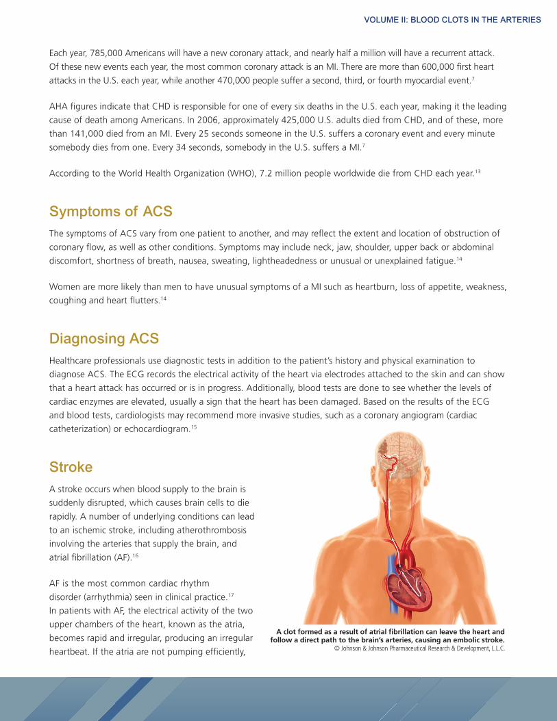
StrokeA stroke occurs when blood supply to the brain is
suddenly disrupted, which causes brain cells to die
rapidly. A number of underlying conditions can lead
to an ischemic stroke, including atherothrombosis
involving the arteries that supply the brain, and
atrial fibrillation (AF).16
AF is the most common cardiac rhythm
disorder (arrhythmia) seen in clinical practice.17
In patients with AF, the electrical activity of the two
upper chambers of the heart, known as the atria,
becomes rapid and irregular, producing an irregular
heartbeat. If the atria are not pumping efficiently,
A clot formed as a result of atrial fibrillation can leave the heart and follow a direct path to the brain’s arteries, causing an embolic stroke.
© Johnson & Johnson Pharmaceutical Research & Development, L.L.C.
VOLUME II: BLOOD CLOTS IN THE ARTERIES
this causes blood to pool in the atria, which may lead to the formation of a clot. If a blood clot that has formed in
the left atrium dislodges and is carried into the systemic circulation, it can become lodged in and occlude an artery
(this clot that travels from its point of origin is known as an embolus). Cardioembolic strokes occur when atrial clots
from the heart are carried to the brain. AF increases the risk for stroke five-fold.18
A stroke interrupts blood flow to the brain, interrupting the supply of oxygenated blood. This lack of oxygen
causes brain cells to die in the affected area. This can result in loss of functions, including impairment of speech,
movement or memory. The extent of the impairment depends on where the arterial occlusion occurred and how
much of the brain tissue is damaged.19
An individual’s risk of stroke depends on the types of comorbidities. For patients with AF, risk stratification may help
with the choice of preventive strategies. There are more than a dozen published stroke risk stratification schemas;
the most commonly used schema is CHADS2, which assigns points for each stroke-associated risk factor including
prior stroke, age of more than 75 years, hypertension, diabetes or recent clinical heart failure.20 New practice
guidelines issued by the European Society of Cardiology, known as CHA2DS
2-VASc, include additional score points
for specific age categories and for the presence of vascular disease and female gender.21
Prevalence of stroke
Each year, nearly 610,000 individuals experience a first stroke and 185,000 individuals have a recurrent stroke in
the U.S.7 Worldwide, stroke is responsible for five million deaths each year.22
Stroke accounted for one of every 18 deaths in the U.S. in 2006. When considered separately from other
cardiovascular diseases, stroke is the third leading cause of death in the U.S., behind heart disease and cancer. On
average, every 40 seconds someone in the U.S. has a stroke and every four minutes someone dies of a stroke.7
An ischemic stroke is caused by either atherothrombosis or cardioembolism that results in loss of arterial blood
supply to a limited area of the brain. Ischemic strokes are the most prevalent, accounting for 87% of all strokes.7
Symptoms of a strokeStrokes typically occur without warning. The symptoms of a stroke may include trouble walking, speaking or
seeing. Speech also might be slurred or patients might experience trouble understanding. Individuals suffering a
stroke may also have blurred or double vision.23
Other warning signs and symptoms of a stroke include sudden paralysis or numbness of the face, arm or leg,
limited to one side of the body.23
Diagnosing stroke
When an individual shows signs and symptoms that may indicate a stroke has occurred, the physician will obtain a
personal and medical history and perform a physical and neurological examination. In order to determine the type
and severity of stroke, imaging tests like head CT or MRI will be used.
VOLUME II: BLOOD CLOTS IN THE ARTERIES
Preventing and treating arterial blood clotsBecause arterial blood clots are rich in platelets and fibrin, medications such as antiplatelets and anticoagulants are
often used. Agents for the prevention and treatment of arterial blood clots include oral antiplatelet drugs, such as
aspirin and clopidogrel, intravenous antiplatelet agents, anticoagulant and thrombolytic agents that help to break
down clots.24
Interventional treatments such as balloon angioplasty or stenting may be used to restore blood flow in the event of
complete or incomplete atherothrombotic arterial occlusion.
Antiplatelet agents: Platelets are important components of the atherothrombotic clot, and as a result, treatments
for these events have focused on antiplatelet medications to prevent this process from occurring. Aspirin
substantially reduces platelet function, and is an established treatment in heart attack patients. Another antiplatelet
agent, clopidogrel, also may be given in conjunction with aspirin. In the emergency setting, intravenous antiplatelet
agents might also be prescribed to treat unstable angina or certain types of heart attacks.24
Anticoagulants: Anticoagulants are frequently prescribed to treat and prevent different forms of cardiovascular
disease. Anticoagulants interfere with the formation of the fibrin components of the atherothrombotic clot. They
affect the various clotting factors in the coagulation cascade. There are a number of anticoagulant therapies
approved for the treatment and prevention of arterial blood clots, including vitamin K antagonists such as warfarin,
heparin and low-molecular-weight heparin.24
Thrombolytic agents: Thrombolysis is the breakdown of blood clots by pharmacological means. For individuals
having a heart attack, thrombolytic agents should be used within a few hours of symptom onset.25 Thrombolytic
therapy has been shown to benefit select patients with acute brain ischemia, with evidence showing a benefit with
various protocols for administering thrombolytic agents.25
Direct thrombin inhibitors: Direct thrombin inhibitors (DTIs) are another class of medication for the prevention
and treatment of unwanted blood clots. DTIs directly inhibit the enzyme thrombin, which plays an important role in
coagulation.26
New oral therapeutic options in development
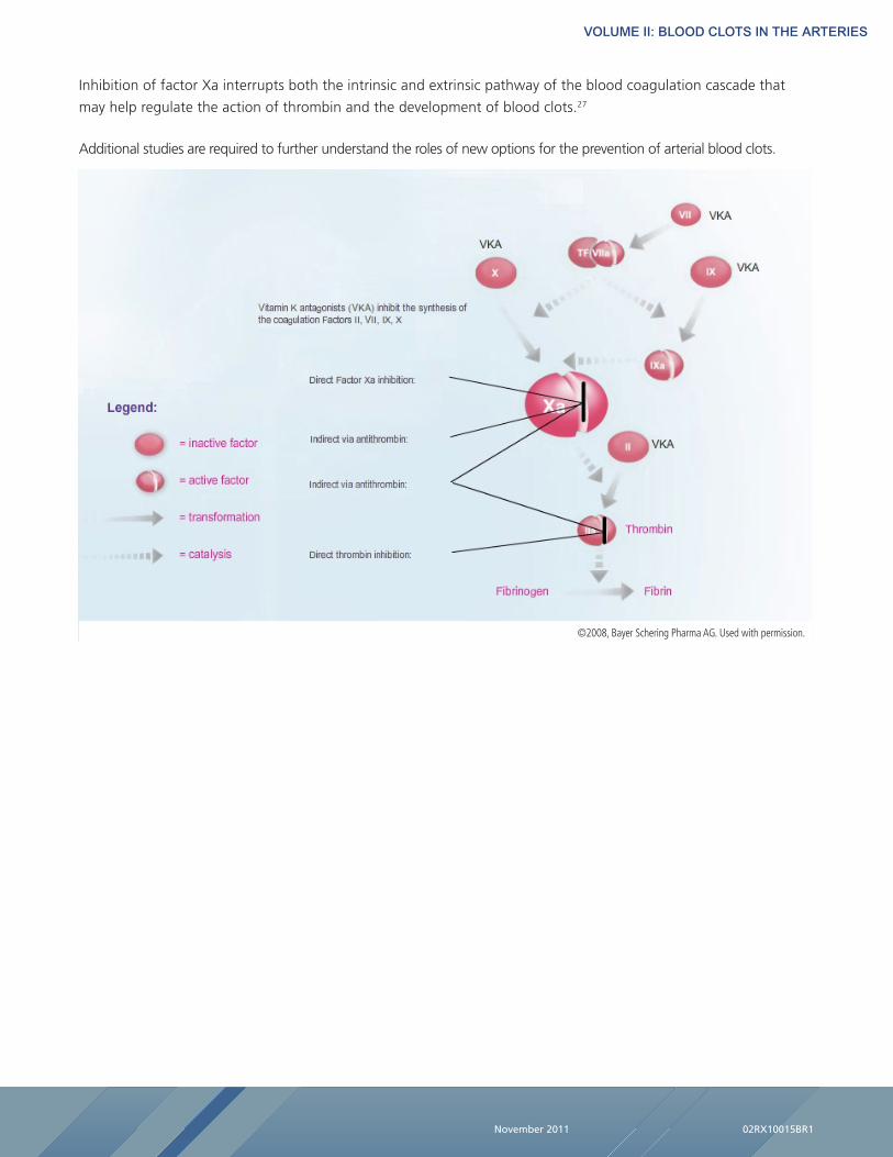
Inhibiting factor Xa
Factor Xa has emerged as a target for potential new anticoagulant agents due to its pivotal role in the coagulation
cascade, where it stimulates the production of thrombin, the enzyme that promotes clot formation.27
VOLUME II: BLOOD CLOTS IN THE ARTERIES
Inhibition of factor Xa interrupts both the intrinsic and extrinsic pathway of the blood coagulation cascade that
may help regulate the action of thrombin and the development of blood clots.27
Additional studies are required to further understand the roles of new options for the prevention of arterial blood clots.
November 2011 02RX10015BR1
©2008, Bayer Schering Pharma AG. Used with permission.
VOLUME II: BLOOD CLOTS IN THE ARTERIES
1 Merck Manual. Acute Coronary Syndromes. http://www.merckmanuals.com/professional/cardiovascular_disorders/
coronary_artery_disease/acute_coronary_syndromes_acs.html. Accessed October 1, 2011.
2 Merck Manual. Atherosclerosis. http://www.merckmanuals.com/professional/cardiovascular_disorders/
arteriosclerosis/atherosclerosis.html. Accessed October 1, 2011.
3 Merck Manual. Stroke. http://www.merckmanuals.com/professional/neurologic_disorders/stroke_cva/overview_of_
stroke.html. Accessed October 1, 2011.
4 American College of Cardiology. Cardiosmart: Guideline: Non-ST-Elevation Myocardial Infarction (NSTEMI) -
Unstable Angina. http://www.cardiosmart.org/ManageCondition/Default.aspx?id=852. Accessed October 1, 2011.
5 American College of Cardiology. Cardiosmart: Peripheral Arterial Disease (PAD). http://www.cardiosmart.org/
HeartDisease/CTT.aspx?id=134. Accessed October 1, 2011.
6 World Health Organization. Cardiovascular diseases fact sheet. http://www.who.int/mediacentre/factsheets/fs317/
en/index.html. Accessed October 1, 2011.
7 American Heart Association. Heart Disease and Stroke Statistics 2010 Update. http://circ.ahajournals.org/cgi/reprint/
CIRCULATIONAHA.109.192667. Accessed October 1, 2011.
8 American Heart Association. Risk Factors and Coronary Heart Disease. http://www.heart.org/HEARTORG/Conditions/
HeartAttack/UnderstandYourRiskofHeartAttack/Understand-Your-Risk-of-Heart-Attack_UCM_002040_Article.jsp.
Accessed October 1, 2011.
9 Centers for Disease Control and Prevention National Institutes of Health. Healthy People 2010: Cardiovascular
Disease and Stroke. http://healthypeople.gov/2020/topicsobjectives2020/overview.aspx?topicid=21. Accessed
October 1, 2011.
10 Mayo Clinic. Acute Coronary Syndrome. http://www.mayoclinic.com/health/acute-coronary-syndrome/DS01061/
DSECTION=tests-and-diagnosis. Accessed October 1, 2011.
11 National Heart, Lung, and Blood Institute. Disease and condition index: What Is a Heart Attack? http://www.nhlbi.
nih.gov/health/dci/Diseases/HeartAttack/HeartAttack_WhatIs.html. Accessed October 1, 2011.
12 American Heart Association. Mission Lifeline Glossary. http://www.heart.org/HEARTORG/HealthcareResearch/
MissionLifelineHomePage/Mission-Lifeline-Glossary_UCM_308046_Article.jsp#stemi. Accessed October 1, 2011.
13 Mackay J, Mensah GA. The Atlas of Heart Disease and Stroke. World Health Organization, Geneva, Switzerland.
14 Women’s Health. Heart Disease. http://womenshealth.gov/faq/heart-disease.cfm. Accessed October 1, 2011.
15 Mayo Clinic. Acute Coronary Syndrome. http://www.mayoclinic.com/health/acute-coronary-syndrome/DS01061/
DSECTION=tests-and-diagnosis. Accessed October 1, 2011.
16 Mayo Clinic. Stroke: Causes. http://www.mayoclinic.com/health/stroke/DS00150/DSECTION=causes. Accessed
October 1, 2011.
17 Fuster V, Lars ER, Cannom DS, et al. 2011 ACCF/AHA/HRS Focused Updates Incorporated Into the ACC/AHA/ESC
Practice Guidelines for the Management of Patients with Atrial Fibrillation. Circulation. 2011; 123:e269-e367.
VOLUME II: BLOOD CLOTS IN THE ARTERIES
18 American College of Cardiology. CardioSmart: Atrial Fibrillation. http://www.cardiosmart.org/HeartDisease/CTT
aspx?id=222. Accessed October 1, 2011.
19 American Stroke Association. How Stroke Effects the Brain. http://www.strokeassociation.org/STROKEORG/
AboutStroke/EffectsofStroke/Effects-of-Stroke_UCM_308534_SubHomePage.jsp. Accessed October 1, 2011.
20 Lip GY. Am J Med. 2010 Jun;123(6):484-48.
21 Camm JA. Eur Heart J. 2010 Oct;31(19):2369-429. Epub 2010 Aug 29.
22 World Health Organization. The Atlas of Heart Disease and Stroke: Global Burden of Stroke. http://www.who.int/
cardiovascular_diseases/en/cvd_atlas_15_burden_stroke.pdf. Accessed October 1, 2011.
23 Mayo Clinic. Stroke. http://www.mayoclinic.com/health/stroke/DS00150/DSECTION=symptoms. Accessed October
1, 2011.
24 Goldhaber SZ, Grasso-Correnti N. Circulation. 2002;106:e138-e140.
25 American Heart Association. Thrombolytics. http://www.medmovie.com/mmdatabase/MediaPlayer.
aspx?ClientID=65&TopicID=582 and http://www.medmovie.com/mmdatabase/MediaPlayer.
aspx?ClientID=65&TopicID=582. Accessed October 1, 2011.
26 Di Nisio M. N Engl J Med. 2005 Sep 8;353(10):1028-1040.
27 Turpie AGG. Arterioscler Thromb Vasc Biol. 2007;27:1238-1247.