Embed Size (px)
Citation preview

This is an Accepted Manuscript, which has been through the Royal Society of Chemistry peer review process and has been accepted for publication.
Accepted Manuscripts are published online shortly after acceptance, before technical editing, formatting and proof reading. Using this free service, authors can make their results available to the community, in citable form, before we publish the edited article. We will replace this Accepted Manuscript with the edited and formatted Advance Article as soon as it is available.
You can find more information about Accepted Manuscripts in the author guidelines.
Please note that technical editing may introduce minor changes to the text and/or graphics, which may alter content. The journal’s standard Terms & Conditions and the ethical guidelines, outlined in our author and reviewer resource centre, still apply. In no event shall the Royal Society of Chemistry be held responsible for any errors or omissions in this Accepted Manuscript or any consequences arising from the use of any information it contains.
Accepted Manuscript
rsc.li/medchemcomm
www.rsc.org/medchemcomm
ISSN 2040-2503
MedChemComm Broadening the field of opportunity for medicinal chemists
Themed issue: Antibiotic Resistance
Volume 7 Number 1 January 2016 Pages 1–204
MedChemComm Broadening the field of opportunity for medicinal chemists
View Article OnlineView Journal
This article can be cited before page numbers have been issued, to do this please use: G. Calabrese, A.
Daou, A. Rova , E. D. Tseligka, I. S. Vizirianakis, D. Fatouros and J. Tsibouklis, Med. Chem. Commun., 2016,
DOI: 10.1039/C6MD00383D.
Boron-containing delocalised lipophilic cations for the
selective targeting of cancer cells†
Calabrese Gianpieroa*, Daou Anis
a, Rova Aikaterini
b, Tseligka Eirini
b, Vizirianakis S. Ioannis
b,
Fatouros G. Dimitriosc, and Tsibouklis John
d
a School of Life Science, Pharmacy and Chemistry, Kingston University London, Penrhyn
Road, Kingston-upon-Thames, Surrey, KT1 2EE, UK.
b Department of Pharmacology, School of Pharmacy, Aristotle University of Thessaloniki, GR-
54124 Thessaloniki, Greece
c Department of Pharmaceutical Technology, School of Pharmacy, Aristotle University of
Thessaloniki, GR-54124 Thessaloniki, Greece
d School of Pharmacy and Biomedical Sciences, University of Portsmouth, Portsmouth, PO1
2DT, UK
*corresponding author: [email protected]
†The authors declare no compe5ng interests.
To limit the incidence of relapse, cancer treatments must not promote the emergence of
drug resistance in tumour and cancer stem cells. Under the proviso that a therapeutic
amount of boron is selectively delivered to cancer cells, Boron Neutron Capture Therapy
(BNCT) may represent one approach that meets this requirement. To this end, we report on
the synthesis and pharmacology of several chemical entities, based on boron-rich carborane
moieties that are functionalised with Delocalized Lipophilic Cations (DLCs), which target
selectively the mitochondria of tumour cells. The treatment of tumour and cancer stem cells
(CSCs) with such DLC-functionalized carboranes (DLC-carboranes) induces cell growth arrest
that is both highly cancer-cell selective and permanent. Experiments involving cultures of
normal and cancer cells show that only normal cells exhibit recapitulation of their
proliferation potential upon removal of the DLC-carborane treatment. At the molecular
level, the pharmacological effect of DLC-carboranes is exerted through activation of the
p53/p21 axis.
Introduction
Page 1 of 16 MedChemComm
Med
Che
mC
omm
Acc
epte
dM
anus
crip
t
Publ
ishe
d on
28
Oct
ober
201
6. D
ownl
oade
d by
Kin
gsto
n U
nive
rsity
on
28/1
0/20
16 1
5:38
:46.
View Article OnlineDOI: 10.1039/C6MD00383D

It is predicted that by the end of 2016 there will be 78,000 new cases of primary brain
tumours; this figure includes nearly 25,000 primary malignant and 53,000 non-malignant
brain tumours.1 Consequent to these levels of incidence, around 17,000 people will lose
their battle against primary malignant and central nervous system (CNS) brain tumours.2
Glioblastomas, which are categorised by the World Health Organization according to
increasingly unfavourable prognosis as grades I to IV, account for 55% of all gliomas and for
15% of all primary brain tumours.3
Cancer cell initiation and progression represent complex multifactorial processes that
impede the efficacy and safe clinical outcome of pharmacological interventions. Tumour cell
heterogeneity and microenvironment present major challenges at the clinical setting.
Cancer treatment protocols are determined by location, type and stage. Most commonly,
preliminary surgery is employed to remove as much tumour tissues as possible, followed by
radiotherapy and/or chemotherapy.4 Since glioblastomas possess a high metastasising
capability, relapse rates are very high.5 Recent experimental evidence has identified cancer
stem cells (CSCs) as a subpopulation that exhibits distinct cellular and genomic
characteristics that render these cells capable of escaping radiotherapy- and chemotherapy-
induced cell death by staying arrested over time at the G1/G0 cell growth phase, which in
turn allows them to re-enter the cell cycle under appropriate microenvironment conditions.
This implies that research efforts have to be focused on the design of anti-cancer molecules,
the therapeutic range of which includes CSCs.6,7,8
Owing to the capability of recently
developed BNCT agents to target cancer cells, including CSCs, both efficiently and
selectively, the clinical use of these agents may be characterized by low incidence drug
resistance profiles.9,10
Apart from the phenotypic distinctions, several differences between mitochondria in normal
and in tumour cells have been observed, and some have been rationalized at the genetic,
molecular and biochemical level.4,11,12
ATP synthesis via oxidative phosphorylation displays
an electrochemical gradient that incorporates contributions from the pH gradient and the
potential difference across the inner membrane of mitochondria.13,14
The in vitro
mitochondrial trans-membrane potential of a healthy cell is in the range 180-200 mV
whereas the corresponding in vivo potential is of the order of 130-150 mV.15
The
mitochondrial trans-membrane potential of carcinoma cells is ca. 60 mV higher than that of
epithelial cell.16
This difference renders mitochondria promising targets for selective drug
delivery. In addition, the mitochondria of CSCs or tumour initiating cells (TICs) exhibit
features - e.g. increased glycolytic metabolism; different redox state regulation
(mitochondrial membrane potential, expression of apoptosis related proteins) - that
distinguish them from the ordinary, more differentiated, tumour cells. These features may
be exploited to guide the design of therapeutics that target cancer cells with a high degree
of specificity.17,18
Page 2 of 16MedChemComm
Med
Che
mC
omm
Acc
epte
dM
anus
crip
t
Publ
ishe
d on
28
Oct
ober
201
6. D
ownl
oade
d by
Kin
gsto
n U
nive
rsity
on
28/1
0/20
16 1
5:38
:46.
View Article OnlineDOI: 10.1039/C6MD00383D

Boron Neutron Capture Therapy (BNCT)
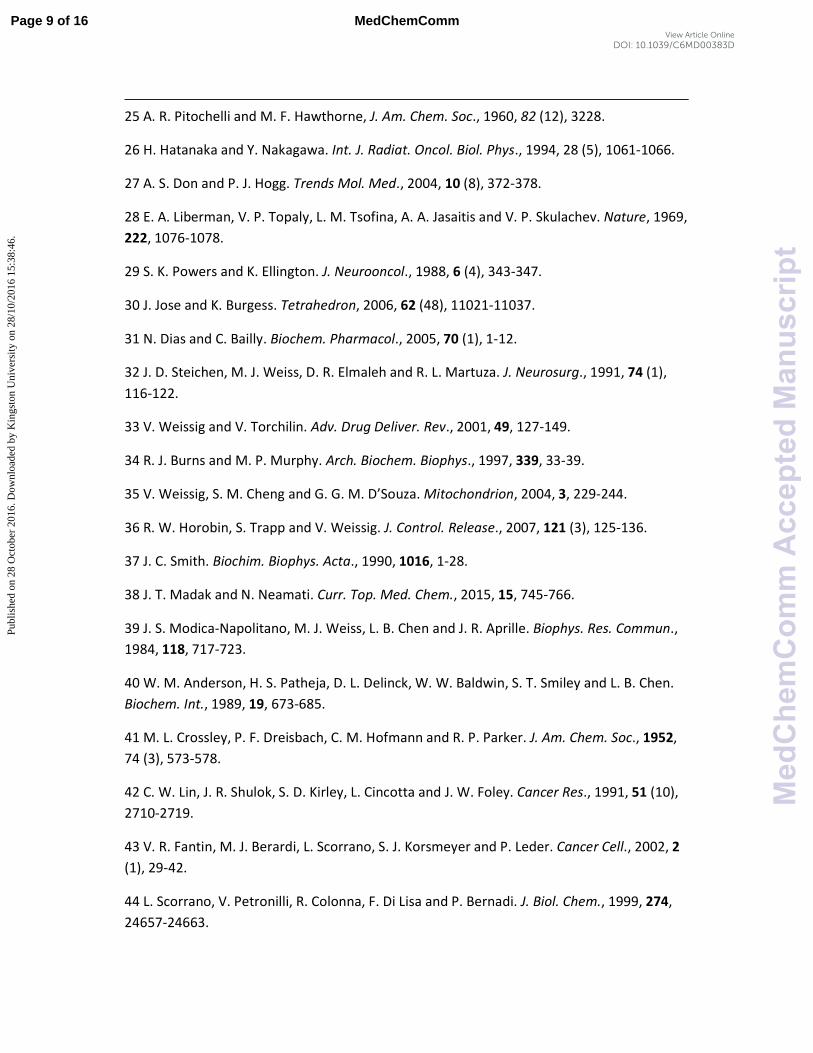
BNCT is a two-step chemo-radiotherapeutic technique (Fig. 1) that involves the selective
delivery of 10
B-rich agents to tumours and their subsequent irradiation with low-energy
neutrons.
Figure 1.
The interaction of 10
B with low energy neutrons (i.e. the capture reaction) results in a
nuclear fission that effects the selective destruction of the host tumour cells. During this
reaction, energetic alpha particles possessing high linear energy transfer (LET), low oxygen
enhancement ratio and high relative biological effectiveness are produced. LET particles are
lethal but – because of their size, energy and short path lengths (4.5 - 10 µm) – the effect is
confined to the host cell.19
The predominant products of the 10
B neutron capture reaction
are recoiling 7Li nuclei and α particles. It takes only a few α particles, which are as lethal to
hypoxic and oxygenated cells as they are to non-proliferating cells, to kill a malignant cell.20
If 10
B is accumulated selectively in cancer cells and assuming that in surrounding healthy
tissue its concentration does not exceed a certain critical value, the cytocidal effects of the
capture reaction are limited to malignant cells. Inevitable capture reactions involving 1H and
14N from normal tissue, which respectively produce γ rays and protons, are of little
relevance since the corresponding thermal-neutron-capture cross sections for these nuclei
are too small to induce marked complications during BNCT.21
BNCT has been investigated for use in the treatment of glioblastoma multiforme (GBM);22
a
malignancy with a current mean survival time of <12 months.23
BNCT offers promise in
GBM, but significant research effort is required before the many performance requirement
for successful treatment are met,24
including the: synthesis of 10
B-enriched compounds with
very low inherent toxicity; and, integration of the selective targeting strategy into the
molecular design, such that therapeutically useful concentrations (>109) of
10B
atoms/tumour cell are achieved while a tumour/blood concentration ratio >5:1 and a
tumour/normal tissue concentration ratio over >3:1 are maintained throughout the neutron
capture stage of the treatment.
The multitude of performance demands that 10
B-containing drugs need to satisfy before
they can be used in the clinic is reflected by the very small number of 10
B compounds that
have reached this stage: para-borophenylalanine (BPA) and sodium
mercaptoundecahydrododecaborate (BSH), which respectively incorporate 1 and 12 boron
atoms in their molecular structures. Following an early attempt by Pitochelli and
Hawthorne25
and fuelled by the data of clinical trials conducted by Hatanaka26
in Japan,
polyhedral boranes have become the materials of choice for the delivery of 10
B-rich moieties
Page 3 of 16 MedChemComm
Med
Che
mC
omm
Acc
epte
dM
anus
crip
t
Publ
ishe
d on
28
Oct
ober
201
6. D
ownl
oade
d by
Kin
gsto
n U
nive
rsity
on
28/1
0/20
16 1
5:38
:46.
View Article OnlineDOI: 10.1039/C6MD00383D

to cancer cells. Among the common polyhedral boron agents, the neutral lipophilic
icosahedral dicarba-closo-dodecarboranes (C2B10H12; commonly referred to as carboranes),
which also exist as ortho-, meta- and para-isomers, are of particular interest not only
because of their high 10
B content, good catabolic stability and low toxicity, but also because
of their amenability to chemical functionalisation.21
The base-induced removal of one
boron atom from the icosahedral cage transforms the lipophilic closo-carborane into the
corresponding nido-carborane, which is more hydrophilic, hence more compatible with
water-based systems.
Mitochondrial targeting
Integral to the performance requirements for BNCT is the molecular design of boron
compounds that are capable of targeting intra-cellular organelles. Neoplastic cells are
characterized by high metabolic activity, which renders possible the design of therapeutic
agents that target the cellular organelles, of both dividing and non-dividing cancer cells.27
Since the negative-inside transmembrane potential of mitochondria (130-150 mV) is far
great than that of any other organelle, functionalisation with DLC moieties represents a
means of imparting mitochondrial targeting specificity to carboranes.28
Some
examples29,30,31,32
of DLCs that have been shown to accumulate preferentially in malignant
cells are presented in Figure 2.
Figure 2.
As the name implies, all DLCs are amphiphilic cationic compounds in which the positive
charge is delocalized over an extensive π electron system. Their lipophilic nature coupled
with the delocalization of the positive charge over a large area act co-operatively to reduce
the free energy change when DLCs diffuse through lipid membranes, such as those of
mitochondria. Driven by the mitochondrial membrane potential, the accumulation of DLCs
may be described by the Nernst equation:
ΔΨ (mV) = 61.5 log10 {[cation]in / [cation]out}
Since the mitochondrial membrane potential of carcinoma cells is ca. 60 mV greater than
that of normal epithelial cells,16
one consequence of the Nernstian relationship is that there
is a ten-fold increase in the propensity of DLCs to accumulate within the mitochondria of
such cells.33
This process is further assisted by the plasma membrane potential (typically 30-
60 mV; negative inside), which encourages the increased accumulation of cations into
carcinoma cells prior to their localisation into mitochondria. The synergistic effect results in
90-95 % of the available intracellular cations becoming localised at mitochondrial sites.34
DLCs, often termed mitochondriotropics,35,36
are used extensively in the visualisation of
mitochondria and also in the estimation of mitochondrial activity.37,38
The use of these
Page 4 of 16MedChemComm
Med
Che
mC
omm
Acc
epte
dM
anus
crip
t
Publ
ishe
d on
28
Oct
ober
201
6. D
ownl
oade
d by
Kin
gsto
n U
nive
rsity
on
28/1
0/20
16 1
5:38
:46.
View Article OnlineDOI: 10.1039/C6MD00383D
materials in diagnosis is constrained only by their concentration-dependent toxicity to
mitochondria. The toxic effects of rhodamine-123 and of dequalinium chloride, two of the
most commonly used DLCs, have been linked to their capacity to inhibit the enzymes F0F1
ATPase39
and NADH-ubiquinone reductase.40
Oral administration to mice bearing
transplanted tumours, has shown that substances containing a benzo[a]phenoxazine
nucleus, such as Nile Blue chloride, exhibit a tumour-staining and growth-retarding action.41
Studies have shown that the subcellular localisation of Nile Blue chloride is highly selective
to lysosomal targets and also that cellular uptake in tumours involves an ion-trapping
mechanism.42
Also, DLCs have been shown to cause mitochondrial depolarisation, which
leads to the opening of mPTPC and the consequent loss of pyridine nucleotides from the
mitochondrial matrix. This effects a further increase in mitochondrial membrane potential,43
which promotes the influx of DLCs into cancer cell. Such cells exhibit mitochondrial
abnormalities that include progressive swelling, disruption of the mitochondrial cristae,
concomitant mitochondrial outer membrane rupture and multiple mitochondrial lesions,
which ultimately result in cell death.44
Boronated DLCs
Although DLCs are promising carriers for the selective tagging of 10
B to mitochondria of
cancer cells, there are very few literature examples of synthetic compounds that combine
boronated entities with DLC moieties such that their corresponding therapeutic effect and
targeting specificity are combined in a single molecular structure.
Adams et al.45
synthesised a carboranyl derivative of dequalinium (DEQ-B), which has been
shown by in vitro evaluations to exhibit tumour uptake and toxicology that are similar to
those characterising its non-boronated analogue. In vitro, DEQ-B was seen to be taken up
and retained by KB, F98, and C6 tumour cell lines, but not by the normal epithelial cell line
CV1. At low concentrations, DEQ-B was shown to be less toxic towards the latter cell line.
The uptake, retention and toxicity of DEQ-B were found to be comparable with those of
other non-boronated DLCs, such as dequalinium chloride, rhodamine 123 and
tetraphenylphosphonium chloride.45
Another example of a boronated DLC derivative is
rhodamine B-phenyl boronic acid (containing a single 10
B atom per molecule), which has
been employed as a fluorescent marker and which has been shown to accumulate
selectively in colon carcinoma cells at a cancer/normal cells ratio of 5.5.46
Calabrese et al.47
reported the synthesis of a series of compounds in which carboranes are combined with
DLCs. A preliminary in vitro evaluation study, utilising human prostate carcinoma (PC3) and
normal (PNT2) epithelial cell lines, indicated the combined propensity of these agents to
target tumour cells and to deliver therapeutically relevant quantities of boron.47
Consistent
with the increased mitochondrial membrane potential of carcinoma cells, the percentage of 10
B taken up by PC3 cells was higher than that determined in parallel experiments involving
PNT2 cells. The data indicated that, with the exception of one Nile Blue derivative, the
synthesized compounds exhibited the features of uptake efficiency, tumour selectivity and
Page 5 of 16 MedChemComm
Med
Che
mC
omm
Acc
epte
dM
anus
crip
t
Publ
ishe
d on
28
Oct
ober
201
6. D
ownl
oade
d by
Kin
gsto
n U
nive
rsity
on
28/1
0/20
16 1
5:38
:46.
View Article OnlineDOI: 10.1039/C6MD00383D
capability to deliver therapeutically relevant amounts of boron that are pre-requisite to
candidate materials for the BNCT therapy of cancer. Figure 3 presents the structures of
compounds selected for further evaluation.10
Rendina et al.48
reported the synthesis of
closo-carborane phosphonium salts derivatives and commented on the absence of a
covalent bond between the anionic boron entity and the cation in the compounds reported
by Calabrese et al47
. In vitro boron uptake studies have shown47
that advantageous
cancer/healthy cells distribution ratios and overall levels of boron accumulation can be
achieved irrespective of the presence or absence of a covalent bond between the carborane
and DLC moieties. Hence, it appears that a covalent link between the DLC and the boron
moiety may not be necessary for the selective delivery of boron to tumour sites.48
Figure 3.
Towards a preliminary assessment of the effects of DLC-carboranes on cell growth,49
U-87
MG cells were incubated over a specified time period with systematically varied
concentrations of bis-nido-carborane dequalinium salt (BOR4). Cell viability data (Figure 4)
demonstrated a concentration-dependent toxicity effect and allowed the estimation of IC50
of this molecule at 0.832 μM. Analogous experiments with carborane-loaded PC and DMPC
liposomes over the boron concentration range 0.2–4.0 μM indicated the biocompatibility of
these formulations at boron concentrations of the order of ≤2.0 μM of boron. A recent
study has investigated the pharmacological behavior of BOR, BOR2, BOR3, BOR4 and BOR5
in malignant, cancer stem and normal cell lines. The work identified selective cytotoxic
behavior towards tumor cells and towards cancer stem cells, while normal cells were seen
to recapitulate their physiological proliferation rate upon removal of the DLC-carborane
from cultures.10
Tested against the criterion of selective cytotoxicity behavior, the same
study identified BOR2 and BOR3 as the most promising candidate materials for further
investigation.10
Figure 4.
Discussion and conclusions
While limited, studies to date show the promise of DLC-carboranes to act as BNCT agents
that target cancer and primary GBM CSCs in the presence of normal cells with a high degree
of selectivity.10,47
Complementary work10
has further revealed the capability of DLC-
carboranes to effect growth inhibition in primary GBM CSCs (Figure 5).
Figure 5.
Underpinned by the principle that the elimination of CSCs may lead to positive long-term
clinical outcomes,50,51
DLC-carboranes offer the potential for selective cytotoxicity within
Page 6 of 16MedChemComm
Med
Che
mC
omm
Acc
epte
dM
anus
crip
t
Publ
ishe
d on
28
Oct
ober
201
6. D
ownl
oade
d by
Kin
gsto
n U
nive
rsity
on
28/1
0/20
16 1
5:38
:46.
View Article OnlineDOI: 10.1039/C6MD00383D

heterogeneous tumour cell populations. It has been shown that the mechanism of action of
DLC-carboranes involves activation of the p53/p21 gene axis. It appears that the selective
accumulation of DLC-carboranes to the mitochondria of tumour cells provides the stimulus
that ultimately activates the tumour suppressor protein p53, which mediates the DNA
damage-induced checkpoint mechanism through the trans-activation of growth inhibitory
genes, such as p21. This in turn leads to permanently malignant cells to enter a phase of
p53-dependent G1 growth arrest, and prevents cell cycling and division. Notably, it has been
shown that malignant cells exposed to DLC-carboranes activate simultaneously the
expression of genes that are linked to apoptosis (e.g. bax, bad, caspases 3 and 9), to survival
(e.g. bcl-2) and to mitogenesis (e.g. c-myc, cyclin D1, cdk4).10
The opposing cellular response
functions in the observed gene expression profile may be explained in terms of an attempt
by cells to overcome the DLC-carboranes-triggered cell-cycle arrest mediated by p53/p21
through genetic manipulation.
Evidence to date suggests that DLC-carboranes may be of value not only to BNCT but also as
stand-alone anticancer drugs. Consequently, DLC-carboranes represent a new class of
anticancer agents, the pharmacological efficacy and safety profiles of which merit
investigation with reference to chemical structure such that the most promising compounds
are identified for clinical verification. In terms of chemistry, the significance of the nature of
the bond that connects the DLC and carborane moieties needs to be understood in order to
allow the research focus to shift to ionic or covalent structures according to their promise to
meet the performance requirements imposed by the BNCT protocol.
References
1 Q. T. Ostrom, H. Gittleman, P. M. de Blank, J. L. Finlay, J. G. Gurney, R. McKean-Cowdin, D.
S. Stearns, J. E. Wolff, M. Liu, Y. Wolinsky, C. Kruchko and J. S. Barnholtz-Sloan, Neuro.
Oncol., 2016, 18 (suppl 1), i1-i50.
2 J. Ferlay, E. Steliarova-Foucher, J. Lortet-Tieulent, S. Rosso, J.W.W. Coebergh, H. Comber,
D. Forman and F. Bray. Eur. J. Cancer, 2013, 49 (6), 1374-1403.
3 O. Visser, E. Ardanaz, L. Botta, M. Sant, A. Tavilla and P. Minicozzi. Eur. J. Cancer, 2015, 51,
2231-2241.
4 J. S. Modica-Napolitano and K. K. Singh. (2004 Sep). Mitochondrion. 2004, 4 (5-6), 755-62.
5 Treating a malignant brain tumour (2015). Available:
http://www.nhs.uk/Conditions/brain-tumour-malignant/Pages/Treatment.aspx. [Last
accessed 28 September 2015].
Page 7 of 16 MedChemComm
Med
Che
mC
omm
Acc
epte
dM
anus
crip
t
Publ
ishe
d on
28
Oct
ober
201
6. D
ownl
oade
d by
Kin
gsto
n U
nive
rsity
on
28/1
0/20
16 1
5:38
:46.
View Article OnlineDOI: 10.1039/C6MD00383D

6 P. A. Sotiropoulou , M. S. Christodoulou , A. Silvani , C. Herold-Mende and D. Passarella.
Drug Discov. Today, 2014, 19, 1547-1562.
7 C. Alifieris and D. T. Trafalis. Pharmacol Ther., 2015, 152, 63–82.
8 J. D. Lathia, S. C. Mack, E. E. Mulkearns-Hubert, C. L. Valentim and J. N. Rich. Genes Dev.,
2015, 29, 1203–1217.
9 T. Sun, Y. Li, Y. Huang, Z. Zhang, W. Yang, Z. Du and Y. Zhou. Oncotarget. 2016, , doi:
10.18632/oncotarget.9355. [Epub ahead of print]
10 E. D. Tseligka, A. Rova, E. P. Amanatiadou, G. Calabrese, J. Tsibouklis, D. G. Fatouros and I.
S. Vizirianakis. Pharm Res., 2016, http://dx.doi.org/10.1007/s11095-016-1930-4 [Epub
ahead of print]
11 E. Carafoli and I. Roman. Mol. Aspects Med., 1980, 3 (5), 295-429.
12 L. O. Chang, C. A. Schnaitman and H. P. Morris. Cancer Res., 1971, 31 (2), 108-113.
13 L. B. Chen. Annu Rev Cell Biol., 1988, 4, 155-181.
14 M. Breunig, S. Bauer and A. Goepferich. Eur. J. Pharm. Biopharm., 2008, 68 (1), 112-128.
15 M. P. Murphy. Biochim. Biophys. Acta., 2008, 1777 (7-8), 1028-1031.
16 J. S. Modica-Napolitano, J. R. Aprille. Cancer Res., 1987, 47 (16), 4361-4365.
17 D. H. Margineantu and D. M. Hockenbery. Curr. Opin. Genet. Dev., 2016, 38, 110-117.
18 B. Yan, L. Dong, J. Neuzil. Mitochondrion. 2016,26, 86-93.
19 D. Schiffer, P. Cavalla, G. J. Pilkington. Brain Tumor Invasion: Biological, Clinical And
Therapeutic Considerations. Mikkelsen/Liss, editors. 1998; pp. 161-84.
20 M. A. Davis and J. B. Little. Radiat. Res., 1970, 43, 534-553.
21 G. Calabrese, J. J. Nesnas, E. Barbu, D. Fatouros and J. Tsibouklis. Drug Discov. Today.,
2012, 17 (3–4), 153-159.
22 T. Yamamoto, K. Nakai, T. Kageji, H. Kumada, K. Endo, M. Matsuda, Y. Shibata and A.
Matsumura. Radiother. Oncol., 2009, 91 (1), 80-84.
23 M. G. Castro, R. Cowen, I. K. Williamson, A. David, M. J. Jimenez- Dalmaroni, X. Yuan, A.
Bigliari, J. C. Williams, J. Hu and P. R. Lowenstein. Pharmacol. Therapeut., 2003, 98 (1), 71-
108.
24 R. F. Barth. Appl. Radiat. Isotopes, 2003, 67 (7-8), S3-S6.
Page 8 of 16MedChemComm
Med
Che
mC
omm
Acc
epte
dM
anus
crip
t
Publ
ishe
d on
28
Oct
ober
201
6. D
ownl
oade
d by
Kin
gsto
n U
nive
rsity
on
28/1
0/20
16 1
5:38
:46.
View Article OnlineDOI: 10.1039/C6MD00383D
25 A. R. Pitochelli and M. F. Hawthorne, J. Am. Chem. Soc., 1960, 82 (12), 3228.
26 H. Hatanaka and Y. Nakagawa. Int. J. Radiat. Oncol. Biol. Phys., 1994, 28 (5), 1061-1066.
27 A. S. Don and P. J. Hogg. Trends Mol. Med., 2004, 10 (8), 372-378.
28 E. A. Liberman, V. P. Topaly, L. M. Tsofina, A. A. Jasaitis and V. P. Skulachev. Nature, 1969,
222, 1076-1078.
29 S. K. Powers and K. Ellington. J. Neurooncol., 1988, 6 (4), 343-347.
30 J. Jose and K. Burgess. Tetrahedron, 2006, 62 (48), 11021-11037.
31 N. Dias and C. Bailly. Biochem. Pharmacol., 2005, 70 (1), 1-12.
32 J. D. Steichen, M. J. Weiss, D. R. Elmaleh and R. L. Martuza. J. Neurosurg., 1991, 74 (1),
116-122.
33 V. Weissig and V. Torchilin. Adv. Drug Deliver. Rev., 2001, 49, 127-149.
34 R. J. Burns and M. P. Murphy. Arch. Biochem. Biophys., 1997, 339, 33-39.
35 V. Weissig, S. M. Cheng and G. G. M. D’Souza. Mitochondrion, 2004, 3, 229-244.
36 R. W. Horobin, S. Trapp and V. Weissig. J. Control. Release., 2007, 121 (3), 125-136.
37 J. C. Smith. Biochim. Biophys. Acta., 1990, 1016, 1-28.
38 J. T. Madak and N. Neamati. Curr. Top. Med. Chem., 2015, 15, 745-766.
39 J. S. Modica-Napolitano, M. J. Weiss, L. B. Chen and J. R. Aprille. Biophys. Res. Commun.,
1984, 118, 717-723.
40 W. M. Anderson, H. S. Patheja, D. L. Delinck, W. W. Baldwin, S. T. Smiley and L. B. Chen.
Biochem. Int., 1989, 19, 673-685.
41 M. L. Crossley, P. F. Dreisbach, C. M. Hofmann and R. P. Parker. J. Am. Chem. Soc., 1952,
74 (3), 573-578.
42 C. W. Lin, J. R. Shulok, S. D. Kirley, L. Cincotta and J. W. Foley. Cancer Res., 1991, 51 (10),
2710-2719.
43 V. R. Fantin, M. J. Berardi, L. Scorrano, S. J. Korsmeyer and P. Leder. Cancer Cell., 2002, 2
(1), 29-42.
44 L. Scorrano, V. Petronilli, R. Colonna, F. Di Lisa and P. Bernadi. J. Biol. Chem., 1999, 274,
24657-24663.
Page 9 of 16 MedChemComm
Med
Che
mC
omm
Acc
epte
dM
anus
crip
t
Publ
ishe
d on
28
Oct
ober
201
6. D
ownl
oade
d by
Kin
gsto
n U
nive
rsity
on
28/1
0/20
16 1
5:38
:46.
View Article OnlineDOI: 10.1039/C6MD00383D

45 D. M. Adams, W. Ji, R. F. Barth and W. Tjarks. Anticancer Res., 2000, 20, 3395-3402.
46 D. Yova, V. Atlamazoglou, N. Kavantzas and S. Loukas. Lasers Med. Sci., 2000, 15, 140–
147.
47 G. Calabrese, A. C. N. M. Gomes, E. Barbu, T. G. Nevell and J. Tsibouklis. J. Mater. Chem.,
2008, 18 (40), 4864–4871.
48 J. A. Ioppolo, M. Kassiou and L. M. Rendina. Tetrahedron Lett., 2009, 50 (47), 6457-6461.
49 D. Theodoropoulos, A. Rova, J. R. Smith, E. Barbu, G. Calabrese, I. S. Vizirianakis, J.
Tsibouklis and D. G. Fatouros. Bioorgan. Med. Chem. Lett., 2013, 23, 6161–6166.
50 F. Ismail and D. A. Winkler. Chem. Med. Chem., 2014, 9, 885-898.
51 R. Würth, F. Barbieri and T. Florio. BioMed. Res. Int., 2014, Article ID126586, 11 pages.
Page 10 of 16MedChemComm
Med
Che
mC
omm
Acc
epte
dM
anus
crip
t
Publ
ishe
d on
28
Oct
ober
201
6. D
ownl
oade
d by
Kin
gsto
n U
nive
rsity
on
28/1
0/20
16 1
5:38
:46.
View Article OnlineDOI: 10.1039/C6MD00383D
Figure 1. BNCT steps: the selective delivery of 10B-containing drugs to tumour cells is followed by irradiation with thermal neutrons (1n) to initiate the destruction of cancer cells and to allow tissue repair.
Figure 1. BNCT steps: the sele 13x4mm (300 x 300 DPI)
Page 11 of 16 MedChemComm
Med
Che
mC
omm
Acc
epte
dM
anus
crip
t
Publ
ishe
d on
28
Oct
ober
201
6. D
ownl
oade
d by
Kin
gsto
n U
nive
rsity
on
28/1
0/20
16 1
5:38
:46.
View Article OnlineDOI: 10.1039/C6MD00383D

Figure 2. Typical Delocalized Lipophilic Cations (DLCs): rhodamine-123; Nile Blue chloride; dequalinium
chloride; and, tetraphenylphosphonium chloride.
Figure 2. Typical Delocalized
22x14mm (300 x 300 DPI)
Page 12 of 16MedChemComm
Med
Che
mC
omm
Acc
epte
dM
anus
crip
t
Publ
ishe
d on
28
Oct
ober
201
6. D
ownl
oade
d by
Kin
gsto
n U
nive
rsity
on
28/1
0/20
16 1
5:38
:46.
View Article OnlineDOI: 10.1039/C6MD00383D

Figure 3. DLC-functionalised carboranes: triphenyl, methylenecarboranyl phosphonium bromide (BOR2); and, nido-carborane salts of tetraphenyl phosphonium (BOR3), dequalinium (BOR4) and rhodamine-B
(BOR5). Figure 3. DLC-functionalised c 17x14mm (300 x 300 DPI)
Page 13 of 16 MedChemComm
Med
Che
mC
omm
Acc
epte
dM
anus
crip
t
Publ
ishe
d on
28
Oct
ober
201
6. D
ownl
oade
d by
Kin
gsto
n U
nive
rsity
on
28/1
0/20
16 1
5:38
:46.
View Article OnlineDOI: 10.1039/C6MD00383D
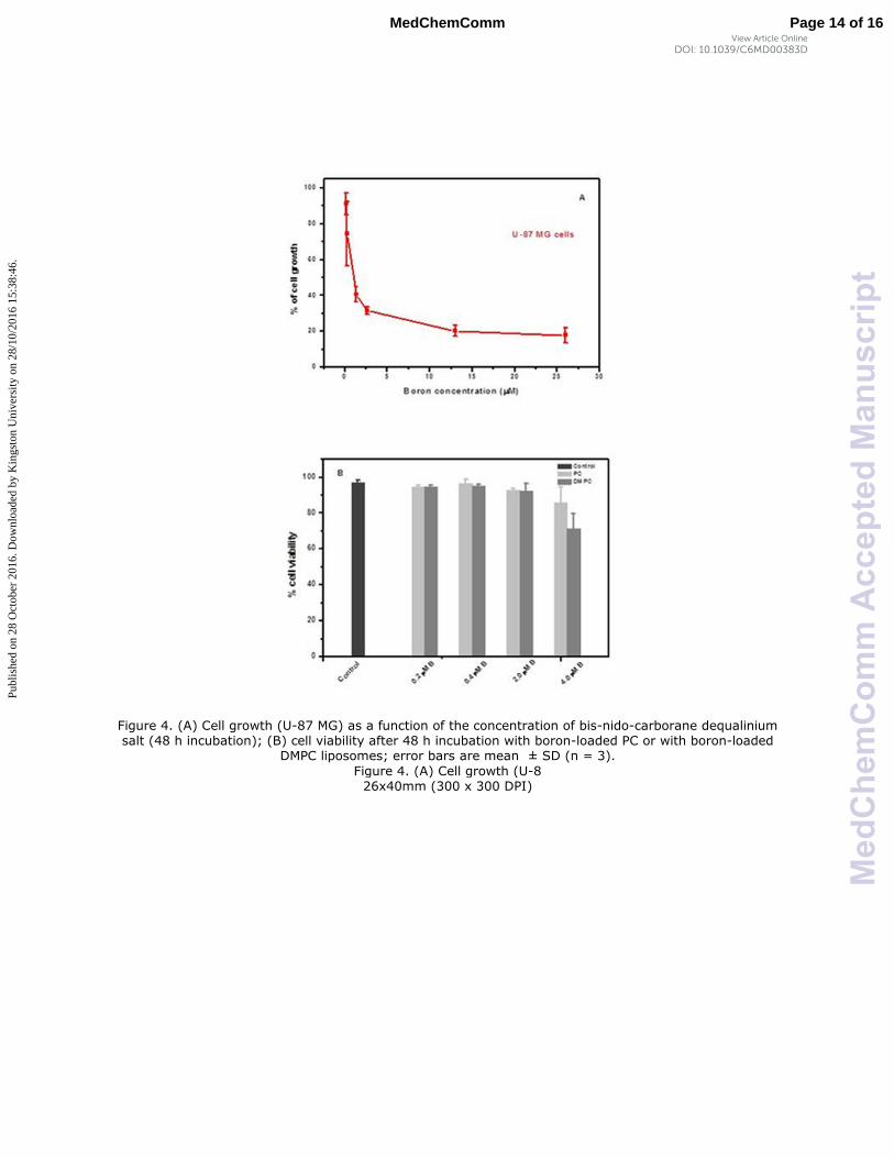
Figure 4. (A) Cell growth (U-87 MG) as a function of the concentration of bis-nido-carborane dequalinium salt (48 h incubation); (B) cell viability after 48 h incubation with boron-loaded PC or with boron-loaded
DMPC liposomes; error bars are mean ± SD (n = 3).
Figure 4. (A) Cell growth (U-8 26x40mm (300 x 300 DPI)
Page 14 of 16MedChemComm
Med
Che
mC
omm
Acc
epte
dM
anus
crip
t
Publ
ishe
d on
28
Oct
ober
201
6. D
ownl
oade
d by
Kin
gsto
n U
nive
rsity
on
28/1
0/20
16 1
5:38
:46.
View Article OnlineDOI: 10.1039/C6MD00383D
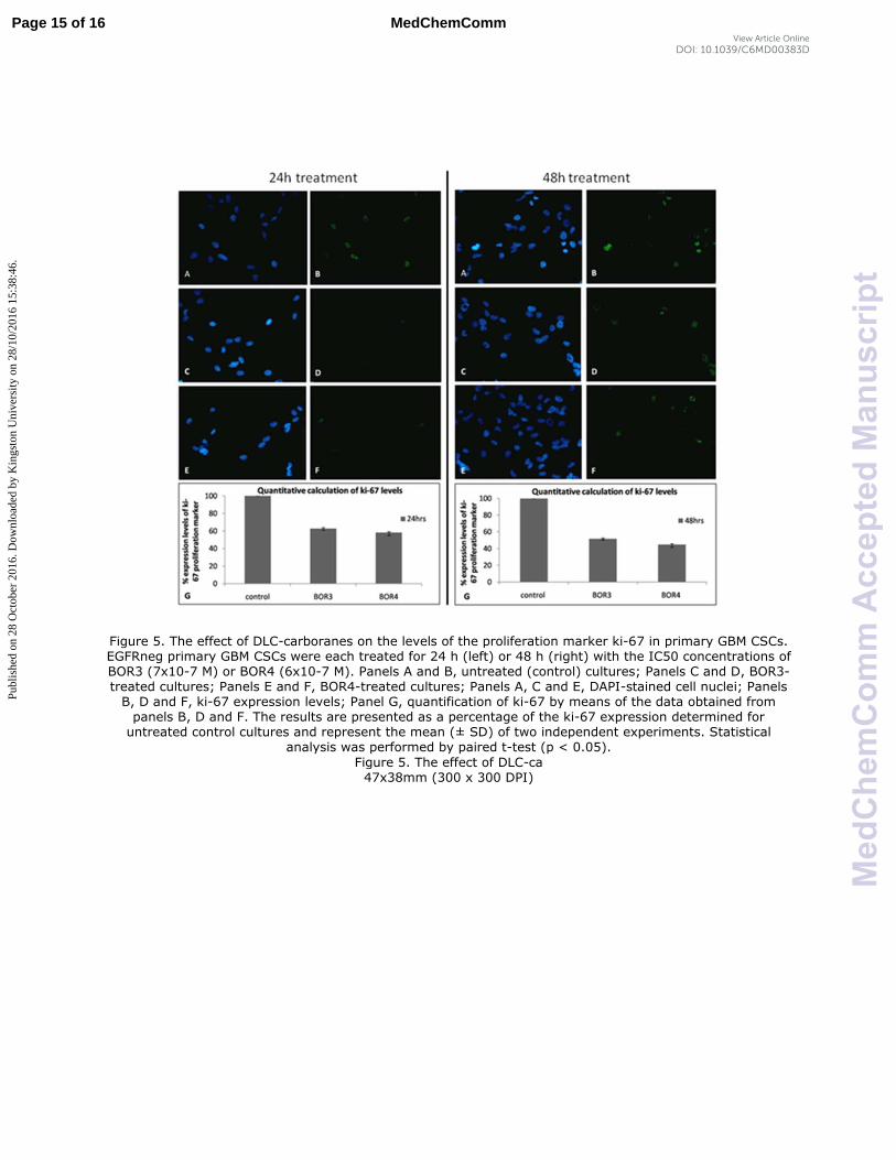
Figure 5. The effect of DLC-carboranes on the levels of the proliferation marker ki-67 in primary GBM CSCs. EGFRneg primary GBM CSCs were each treated for 24 h (left) or 48 h (right) with the IC50 concentrations of BOR3 (7x10⁻⁷ M) or BOR4 (6x10⁻⁷ M). Panels A and B, untreated (control) cultures; Panels C and D, BOR3-treated cultures; Panels E and F, BOR4-treated cultures; Panels A, C and E, DAPI-stained cell nuclei; Panels B, D and F, ki-67 expression levels; Panel G, quantification of ki-67 by means of the data obtained from panels B, D and F. The results are presented as a percentage of the ki-67 expression determined for untreated control cultures and represent the mean (± SD) of two independent experiments. Statistical
analysis was performed by paired t-test (p < 0.05).
Figure 5. The effect of DLC-ca 47x38mm (300 x 300 DPI)
Page 15 of 16 MedChemComm
Med
Che
mC
omm
Acc
epte
dM
anus
crip
t
Publ
ishe
d on
28
Oct
ober
201
6. D
ownl
oade
d by
Kin
gsto
n U
nive
rsity
on
28/1
0/20
16 1
5:38
:46.
View Article OnlineDOI: 10.1039/C6MD00383D

254x190mm (96 x 96 DPI)
Page 16 of 16MedChemComm
Med
Che
mC
omm
Acc
epte
dM
anus
crip
t
Publ
ishe
d on
28
Oct
ober
201
6. D
ownl
oade
d by
Kin
gsto
n U
nive
rsity
on
28/1
0/20
16 1
5:38
:46.
View Article OnlineDOI: 10.1039/C6MD00383D