Embed Size (px)
Citation preview
Electro
nic Cop
y

Electro
nic Cop
y

Electro
nic Cop
y
Electro
nic Cop
y
Med. Forum, Vol. 25, No.12 December, 2014 ISSN 1029-385-X
Recognized by PMDC CONTENTS Recognized by HEC
Editorial
1. Eradicating Polio - Our National Duty 1
Mohsin Masud Jan
Original Articles
2. Assessment of Circulating Biochemical, Heamatological and Oxidative Stress Markers in
Females Using Contraceptives from Punjab, Pakistan 2-6
1. Waheed Jamil Ch. 2. Muhammad Binyameen Chishti 3. Madiha Ashraf 4. Arif Malik
5. Mahmood Husain Qazi
3. A Study of Finger Prints Pattern in Relation to ABO, RH Blood Groups among Medical Students
of AJK Medical College, Muzaffarabad (AJK) 7-10
1. Rameez Iqbal Hashme 2. Naveed Ahmed Khan
4. Placebo- Controlled Trial of Pharmaceutical Optimized Lisinopril 10mg (F-5) in Patients with
Essential Hypertension for Efficacy & Biochemical Evaluation 11-14
1. Asnad 2. Mohammad Tariq Ijaz Afridi 3. Munir Tahir 4. Mohammad Adnan Shereen
5. Diagnostic Accuracy of IgA Anti-Tissue Transglutaminase Antibodies in Comparison with
Histopathological Findings in Celiac Disease in Pakistan 15-19
1. Arslaan Javaeed 2. Walayat Shah 3. Rizwan Akhtar 4. Sanniya Khan Ghauri 5. Shafqat Husnain
Khan 6. Aftab Haider Alvi
6. Shouldice Versus Bassini’S Procedures for Inguinal Hernial Repair 20-22
1. Gul Sher Khan 2. Ishfaq Ali Shah Bukhari 3. Musarrat 4. Israr Ahmed 5. Muhammad Ishaq
7. Oral Hygiene Habits Among 6-12 Year Religious School Students 23-25
1. Mohammad Yaqoob Memon 2. Feroze Ali Kalhoro 3. Abdul Jabbar 4. Abdul Bari Memon
5. Irfan Ahmed Shaikh
8. Reversal of Loperamide Induced Intestinal Smooth Muscle Relaxation by Glibenclamide and
Repaglinide in Vitro 26-30
1. Javaria Arshad 2. Naila Abrar 3. Munir Ahmad Khan 4. Sarwat Jahan
9. Experimental Study of Antimony Induced Hepato Toxicity in Rabbits 31-34
1. Khawaja Usman Masud 2. A.H. Nagi
10. Major Determinants of Anemia in Pregnant Women Residing in the Urban Slums of Taluka
Qasimabad, District Hyderabad 35-38
1. Rehana Siddiqui 2. Muhammad Muqeem Mangi 3. Azhar Ali Shah 4. Rafique Ahmed Soomro
5. Khalida Naz Memon
11. Frequency of Surgical Intervention Due to Cord Around the Neck 39-41
1. Shazia Shaikh 2. Fozia Shaikh 3. Shazia Jatoi
12. Trend of Poisoning in Muzaffarabad (AJK) 42-45
1. Naveed Ahmed Khan 2. Rameez Iqbal Hashme 3. Azhar Masud Bhatti
13. Protective Effect of Vitamin C on Diameter of Seminiferous Tubules and Spermatogenesis of
Albino Rats with Lead Toxicity 46-51
1. Mujahid Akbar Mamoun 2. Syed Tariq Ali Rizvi 3. H. Mohammad Fareedullah Alvi
14. Anxiety in Dental Patients 52-56
Marium Iqbal
Electro
nic Cop
y
Med. Forum, Vol. 25, No.12 December, 2014 ISSN 1029-385-X
15. To Evaluate the Outcome of Sacrococcygeal Pilonidal Sinus Excision Using Karydakis
Technique 57-59
1. Muhammad Iqbal Khan 2. Muhammad Jawed 3. Sajeer Bhura 4. Ubedullah Shaikh 5. Anum Arif
16. The Complex Regional Pain Syndrome after Fractures of Distal Radius 60-64
1. Madan Lal Jesswani 2. Syed Imaduddin 3. Muhammad Asif Memon 4. Rahila Razzak
17. Eclampsia and its Association with Seasonal Variations and other External Factors 65-67
1. Shazia Jatoi 2. Shazia Shaikh 3. Fozia Shaikh
18. Comparison of Efficacy of Topical Ofloxacin and Gentamycin in Tubotympanic Type of
Chronic Suppurative Otitis Media 68-71
1. Asmatullah 2. Qaisar Khan 3. Ghareeb Nawaz 4. Gohar Ullah 5. Johar Iqbal 6. Munib Khan
7. Raza Muhammad
19. Outcome of Internal Fixation of Fractures in a Tertiary Care Hospital in Peshawar 72-75
1. Mohammad Tariq Ijaz Afridi 2. Iftikhar Ahmad Chaudary 3. Mohammad Irfan Shereen 4. Asnad
5. Muhammad Adnan Shereen
Corrigendum
20. Corrigendum: (i) Inter-Relationship of Circulating Biochemical Markers of Oxidative Stress
and Thyroid Hormones in newly Diagnosed Schizophrenics: Perspective study from Local
Population of Punjab Pakistan (ii) Significance of Hepatic Profile and Malondialdehyde as
Marker of Lipid Peroxidation in HCV Patients: A Perspective Study from Local Population of
Punjab-Pakistan (iii) Response of Antiretroviral Therapy (Ziduvodine, Lamivudine and
Niverapine) in Patients Suffering from Acquired Immuno Deficiency Syndrome (Aids)
(iv) Inter-Relationship of Viral Load and CD4+
Cells in Patients Suffering from Acquired
Immuno Deficiency Syndrome (Aids): Update from Punjab-Pakistan 75
21. Author Index January to December 2014 76-79
22. Subject Index January to December 2014 80-89
Guidelines and Instructions to Authors i-ii
Electro
nic Cop
y
Med. Forum, Vol. 25, No.12 December, 2014 1
Editorial Eradicating Polio - Our National Duty
MohsinMasud Jan Editor
Polio currently remains endemic in only three countries
- Afghanistan, Nigeria and Pakistan.
The Polio virus has struck again with vengeance, as
confirmed by the sources at The National Institute of
Health, Pakistan. The national polio count so far this
year has risen to 291 cases.
The visibly rattled Pakistani authorities should take a
little heart from the fact that in 1952, over 58,000
Americans-including former US President Franklin D.
Roosevelt and the country’s Supreme Court Justice
William Douglas - were suffering from this disease,
research reveals.
Of the nearly 58,000 cases of this epidemic reported in
the United States of America in 1952, 3,145 people
infected with the disease died and 21,269 were left with
mild to disabling paralysis. This was the time when the
peak age of incidence of Polio in the United States
shifted from infants to children aged five to nine years,
when the risk of paralysis is greater; and about one-
third of the cases were reported in Americans over the
age of 15 years.
These three nations should also bear in mind that in
1916, not less than 27,363 Polio cases were reported in
20 American states.
New York alone had 9,023 cases, of which 2,448 (28
per cent) resulted in death, and a larger number in
paralysis.
This number rested at 37,476 in 1954. In 1977, there
were 254,000 people living in the United States who
had been paralysed by polio.
Moreover, some 40,000 polio survivors with varying
degrees of paralysis still live in Germany, 30,000 in
Japan, 24,000 in France, 16,000 in Australia, 12,000 in
Canada and 12,000 in the United Kingdom.
Polio epidemics began to appear in Europe and the
United States around 1900, spreading all over Europe,
North America, Australia, and New Zealand during the
first half of the 20th century.
In 1960, Czechoslovakia became the first country in the
world to scientifically demonstrate nationwide
eradication of polio.
According to WHO, Europe was declared polio-free
only on June 21, 2002!
It was on March 24, 2014 that the WHO announced the
eradication of Poliomyelitis in 11 countries of the
South-East Asia region.These countries included
Bangladesh, Bhutan, India, Indonesia, Nepal,
Myanmar, Maldives, North Korea, Sri Lanka, Thailand
and Timor-Leste.
The last case of wild Polio in the South-East Asia
Region was reported in India on 13 January 2011.
The Global Polio Eradication Initiative, financed by a
wide range of public and private donors, has estimated
that the financial requirements for the eradication of
Polio from the world would be approximately US$ 5.5
billion for the 2013-18.
Polio was first recognized by a German
OrthopaedistJakob Heine (1800-1879), who had
authored the first medical report on the disease.
Austrian biologist Karl Landsteiner (1868-1943) first
discovered the Polio Virus in 1909, making him eligible
for the 1930 Nobel Prize in Physiology.Jonas Edward
Salk (1914-1995) developed the first successful
inactivated Polio vaccine in the 1950s.
On July 2, 1952, assisted by the staff at New York’s
D.T. Watson Home for Crippled Children, Jonas Salk
had injected 43 children with his killed-virus vaccine.
The Jonas Salk vaccine was first introduced in 1957
and an immediate vaccination rush commenced with
this medicine in countries including Canada, Sweden,
Denmark, Norway, West Germany, Netherlands,
Switzerland and Belgium etc.
By 1962, Polio had become almost extinct in United
States.
All of that being history, it should be used as inspiration
by the administrators of our country. As the Chief
Secretary, Punjab has stated, ZERO tolerance will be
shown against all districts’ administration of the
province if they fail in achieving targets pertaining to
the polio eradication, primary healthcare. Strict action
would be taken if targets were not.
About the polio eradication campaign, DCOs were
directed that movement of IDPs from FATA should be
monitored, especially in Lahore and Rawalpindi
divisions, to check the transfer of polio virus. Besides
this, he directed DCOs to ensure persistent positive
environmental samples which reflect the increase in
internal virus circulation. The DCOs were also directed
to ensure monitoring of permanent tehsil posts (PTP) at
provincial borders as well as monitoring of vaccinators
through Android phones.
The eradication of Polio is not an impossible task.
Border area at provincial, divisional, district and tehsil
levels should be completely monitored during polio
campaigns and measures should be adopted to
safeguard the same.
We still have an opportunity to reverse this trend of crippling
our future generation and join the rest of the world on the
finish line on eradication. Let us make eradicating polio our
national duty.
Electro
nic Cop
y
Med. Forum, Vol. 25, No. 12 December, 2014 2
Assessment of Circulating
Biochemical, Heamatological and
Oxidative Stress Markers in Females Using
Contraceptives from Punjab, Pakistan 1. Waheed Jamil Ch. 2. Muhammad Binyameen Chishti 3. Madiha Ashraf 4. Arif Malik
5. Mahmood Husain Qazi 1. Asstt. Prof. of Physiology, AIMC, Lahore 2. Assoc. Prof. of Physiology, AIMC, Lahore 3. Asstt. Prof.
Physiology, AIMC, Lahore 4. Assoc. Prof. of Biochemistry, The University of Lahore 5. Director, Center for
Research and Molecular Medicine, The University of Lahore
ABSTRACT
Objective: To Assess the circulating Biochemical, Heamatological and Oxidative Stress Markers in Females using
Contraceptives from Punjab, Pakistan.
Study Design: Case Control Study
Place and Duration of Study: This study was carried out at the Gynae Units of Jinnah Hospital Lahore from
January, 2011 to December 2011.
Materials and Methods: Two injectable contraceptives (Depo-Medroxyprogesterone & NorethisteroneEnantate)
and Oral contraceptive pills (COCs) were administered in females of reproductive age, the oxidative and nitrosative
stress biomarkers (Malondialdehyde, Superoxide Dismutase, Catalase, Nitic oxide and Glutathione) were analyzed
in these subjects.
Results: DMPA, NET-EN & COCs treatment could induce oxidative stress with a significant change in lipid profile
and other biochemical markers thus affecting the normal biological system. These effects after prolonged use can
generate pathological events leading to disease pattern.
Conclusion: These biomarkers can become diagnostic tools to evaluate the health of a woman using these methods
as preventive measure in future
Key Words: Contraceptives, Estrogen, Progestin, Stress Biomarkers, Catalase, Glutathione, MDA, SOD
INTRODUCTION
Hormonal contraceptive methods for prevention of
unplanned pregnancy have been very popular among
females since the beginning of modern era. In
developing countries like Pakistan, governments and
organizations are running campaigns to increase the use
of these methods in order to space pregnancies 1(Emokpaeet al., 2010). These hormonal preparations
can be taken orally (taken by mouth), injected beneath
the skin, implanted in the body tissues, absorbed from a
patch on the skin, or placed inside the vagina.Oral
contraceptive pills are mainly of two types, the
combined pills and the progestin only pills. Combined
pill is a combination of synthetic estrogens and
progestin. Mestranol, a synthetic estrogen is the
inactive form which is converted to ethinyl estradiol
(E2) to cause the action in the body.Effect of injectable
contraceptive lasts for two or three months after a
single dose.Even most effective and well tolerated
contraception is not 100% effective; chances of
pregnancy are still 3%.DMPA acts by inhibition of
pituitary gonadotropin secretions which results in loss
of ovulation, amenorrhea and decline in estrogen
production leading to prevention of pregnancy 2(Mia et
al, 2005).Level of steroids measured in the blood
reflects the difference in formulations of these
injectable preparations. Blood levels of NET-EN
increase rapidly attaining peak levels within 5 days. In
contrast to this, DMPA gains peak levels in ten days 3(Draper et al., 2007).
Ovary is the major synthesizer of estrogen using
cholesterol as raw material under the influence of
pituitary hormonal secretion. They are also produced by
the aromatization of androgens in fat cells, skin, bone,
and other tissues. There is production of “good
estrogens” and their function is to act as antioxidants
exerting their effect as eliminators of damaged or
cancerous cells all over the body. “Bad estrogen” is the
result of inefficient estrogen metabolism.They act
negatively to cause oxidation, damage to DNA, and
play a role in promotion of cancer. Body produces
many antioxidants which are helpful in detoxifying
cancer-causing estrogens, example is glutathione. For
excretion of many toxins glutathione levels are
important 4(Dalessandri et al., 2004).Endogenous
estrogens and exogenous other hormones can act as
potential modulators in oxidative stress in otherwise
healthy female, it is necessary to clarify their roles in
oxidative stress.
The main action of progesterone is to strengthen as well
as check the actions of estrogen. Estrogen gives the
Original Article Changes after using
Contraceptives
Electro
nic Cop
y
Med. Forum, Vol. 25, No. 12 December, 2014 3
message while progesterone controls and modifies that
message. Progestins are synthetic progesterone.
Chemically similar to progesterone but their action is
different.Their contraceptive action is brought about by
decreasing gonadotropin releasing hormone 5(Lobo and
Stanczyk, 1994)causing thick cervical mucus and
renders the endometrium unresponsive to implantation 6(Loose-Mitchell and Stancel, 2001). Progestin only
contraceptives are the method of choice in women
during lactation and the females in which estrogen is
contraindicated 7(Affandi, 1998).
Oxidative stress is referred to imbalance between
antioxidants and reactive oxygen species. Reactive
oxygen species are highly reactive and unstable due to
impaired electrons in their outer shell. In physiological
concentrations they are beneficial while their excess
can lead to damage to structures of cell. Free radicals
are neutralized by antioxidants 8(Palmieri and
Sblendorio, 2007). These antioxidants are of two types:
enzymatic and non-enzymatic. Enzymatic antioxidants
are produced in the body and are proteins i.e. enzymes.
Non-enzymatic antioxidants are supplied to the body by
dietary intake as vitamins and minerals 9(Agarwal et al.,
2005). Free radicals are continuously produced in low
concentrations and perform certain important tasks such
as regulation of apoptosis, modulation of gene
expression related to immune response as well as
activating transcriptional factors 10
(Pincemail et al.,
2007).Estrogens have antioxidant property and this
property is exhibited even at low plasma concentrations
in vascular systems and lipid metabolism (Palmieri and
Sblendorio, 2007).Evidence suggests that as oxidative
stress increases, it leads to endothelial dysfunction and
ultimately atherosclerosis 11
(Cai and Harrison, 2000).
During oral contraceptive use, Ethinyl Estradiol and
progestin levels in plasma increase leading to oxidative
stress. Several studies conducted to demonstrate the
effect of oral contraceptives on erythrocytes for
evaluation of antioxidant markers such as glutathione
peroxidase (GSH-PX), catalase and superoxide
dismutase activities 12
(Massafra et al., 1993;13
Subakir et
al., 2000) which showed increase in activity of two of
these markers (Massafra et al., 1993; Pincemail et al.,
2007). The objective of this study was to assess the
circulating biochemical markers, antioxidative capacity
and lipid peroxidation in premenopausal women due to
hormonal contraceptive therapy.
MATERIALS AND METHODS
Study Design: It was a case control study.
Target Population: Healthy women aged 25-40 years
in the periphery of city of district Kasur.
Study settings: This study was carried out on women
who were attending Jinnah Hospital Lahore and Basic
Health Units (BHU) in Kasur.
Sample size and Specifications: 51 women were
chosen for this study. 32 of them were using hormonal
contraceptives preparations. 19 healthy women of the
same age group were taken as control that had not used
any method for the last 9 months. Among these 32
subjects, 22 of them were using injectable. Two
different preparations of hormonal injectable were used.
17 of them were usinginjection Depo-medroxy-
progesterone Acetate (DMPA) while remaining 5 were
on injection Norethisterone Enantate (NET-EN).10
subjects had been using Combined Oral Contraceptives
pills for the last 9 cycles.
Sampling: 5ml of blood was collected as a sample for
study from each woman. Out of that 3ml was separated
in a tube without anticoagulant. This portion was
preceded by centrifugation at 3000 rpm for 20 minutes
to obtain serum samples. These serum samples were
stored at 2-5°C for hematological parameter analysis.
The remaining 2ml was added to Ethyline Diamine
Tatric Acid coated tube for performing Complete Blood
Count.
Inclusion Criteria: Subjects selected were of the
average age of 30 years. The range of their age was 21-
41 years. They had been using these hormonal
contraceptive methods for at least 15 months. As for
control group they were not using any of these methods
for the last 10-12 months. The mean weight and height
were 55kg (range) and1.66m (1.6-1.75). Blood pressure
range was less than 140/100 and more than
90/60 mm Hg.
Exclusion Criteria: All those women were excluded
who had had hypertension, thyroid disease, breast
feeding, obesity, diabetes, vascular disease, epilepsy,
any pelvic pathology, myomas and polyps. Subjects
were fully explained about study and their written
consent was taken.
Following parameters were estimated: I-Stress
biomarkers estimatedincluding malondialdehyde
(MDA) 14
(Ohkawa et al., 1979), superoxide dismutase
(SOD)15
(Kakkar et al., 1984), catalase 16
(Aebi, 1974),
reduced glutathione (GSH) 17
(Moron et al., 1979) and
Nitric oxide (NO) 18
(Moshage et al., 1995).
II- Haematological parameters were analyzed by
Hemolytic Seismic Analyzer.
RESULTS
The level of bio-markers in oxidative stress is different
when compared to each other in different formulations
of hormonal contraceptives during 1st year use.
Malondialdehyde (MDA) levels are important markers
of lipid per-oxidation product. Our study has shown
MDA levels to be increased in all subjects of hormonal
contraceptive users significantly. Highest increase was
noted in the group using Injection 1, the mean was
6.840nmol/mLwhen compared with control group
which was 1.345 nmol/mL. Group using Pills showed
mean 4.604μmol/ml, while those using Injection 2
mean 3.692 nmol/mL. Injectable 1 group has shown
508% increase, Pills group 342% and least increase was
Electro
nic Cop
y
Med. Forum, Vol. 25, No. 12 December, 2014 4
noted as 274% in injectable 2 group. Therefore, the
result coincides with the statement that lipid
peroxidation product MDA shows significant increase
during first year of administration of hormonal
contraceptives 19
(Faddah et al., 2005).
Superoxide Dismutase (SOD) level of control group,
non-users of hormonal contraceptives, was
0.473ng/mLand in injectable 1 group 0.118ng/mL.
While the comparative mean levels of Pills group was
0.198μ mol/ml and injectable 2 group was 0.276μ
mol/ml. The noted levels indicate that there is an
overall decrease in SOD in HC user groups in
comparison with control group. The result is in
accordance with the statement that long term use of
DMPA causes oxidative stress 20
(Bakry et al., 2011).
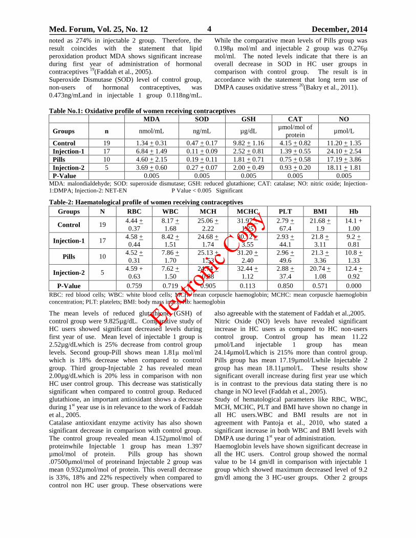
Table No.1: Oxidative profile of women receiving contraceptives
MDA SOD GSH CAT NO
Groups n nmol/mL ng/mL µg/dL µmol/mol of
protein µmol/L
Control 19 1.34 + 0.31 0.47 + 0.17 9.82 + 1.16 4.15 + 0.82 11.20 + 1.35
Injection-1 17 6.84 + 1.49 0.11 + 0.09 2.52 + 0.81 1.39 + 0.55 24.10 + 2.54
Pills 10 4.60 + 2.15 0.19 + 0.11 1.81 + 0.71 0.75 + 0.58 17.19 + 3.86
Injection-2 5 3.69 + 0.60 0.27 + 0.07 2.00 + 0.49 0.93 + 0.20 18.11 + 1.81
P-Value
0.005 0.005 0.005 0.005 0.005 MDA: malondialdehyde; SOD: superoxide dismutase; GSH: reduced glutathione; CAT: catalase; NO: nitric oxide; Injection-
1:DMPA; Injection-2: NET-EN P Value < 0.005 Significant
Table-2: Haematological profile of women receiving contraceptives
Groups N RBC WBC MCH MCHC PLT BMI Hb
Control 19 4.44 +
0.37
8.17 +
1.68
25.06 +
2.22
31.97 +
1.25
2.79 +
67.4
21.68 +
1.9
14.1 +
1.00
Injection-1 17 4.58 +
0.44
8.42 +
1.51
24.68 +
1.74
30.12 +
3.55
2.93 +
44.1
21.8 +
3.11
9.2 +
0.81
Pills 10 4.52 +
0.31
7.86 +
1.70
25.13 +
1.53
31.20 +
2.40
2.96 +
49.6
21.3 +
3.36
10.8 +
1.33
Injection-2 5 4.59 +
0.63
7.62 +
1.50
24.74 +
0.88
32.44 +
1.12
2.88 +
37.4
20.74 +
1.08
12.4 +
0.92
P-Value
0.759 0.719 0.905 0.113 0.850 0.571 0.000
RBC: red blood cells; WBC: white blood cells; MCH: mean corpuscle haemoglobin; MCHC: mean corpuscle haemoglobin
concentration; PLT: platelets; BMI: body mass index; Hb: haemoglobin
The mean levels of reduced glutathione (GSH) of
control group were 9.825µg/dL. Comparative study of
HC users showed significant decreased levels during
first year of use. Mean level of injectable 1 group is
2.52µg/dLwhich is 25% decrease from control group
levels. Second group-Pill shows mean 1.81μ mol/ml
which is 18% decrease when compared to control
group. Third group-Injectable 2 has revealed mean
2.00µg/dLwhich is 20% less in comparison with non
HC user control group. This decrease was statistically
significant when compared to control group. Reduced
glutathione, an important antioxidant shows a decrease
during 1st year use is in relevance to the work of Faddah
et al., 2005.
Catalase antioxidant enzyme activity has also shown
significant decrease in comparison with control group.
The control group revealed mean 4.152µmol/mol of
proteinwhile Injectable 1 group has mean 1.397
µmol/mol of protein. Pills group has shown
.07500µmol/mol of proteinand Injectable 2 group was
mean 0.932µmol/mol of protein. This overall decrease
is 33%, 18% and 22% respectively when compared to
control non HC user group. These observations were
also agreeable with the statement of Faddah et al.,2005.
Nitric Oxide (NO) levels have revealed significant
increase in HC users as compared to HC non-users
control group. Control group has mean 11.22
µmol/Land injectable 1 group has mean
24.14µmol/Lwhich is 215% more than control group.
Pills group has mean 17.19µmol/Lwhile Injectable 2
group has mean 18.11µmol/L. These results show
significant overall increase during first year use which
is in contrast to the previous data stating there is no
change in NO level (Faddah et al., 2005).
Study of hematological parameters like RBC, WBC,
MCH, MCHC, PLT and BMI have shown no change in
all HC users.WBC and BMI results are not in
agreement with Pantoja et al., 2010, who stated a
significant increase in both WBC and BMI levels with
DMPA use during 1st year of administration.
Haemoglobin levels have shown significant decrease in
all the HC users. Control group showed the normal
value to be 14 gm/dl in comparison with injectable 1
group which showed maximum decreased level of 9.2
gm/dl among the 3 HC-user groups. Other 2 groups
Electro
nic Cop
y
Med. Forum, Vol. 25, No. 12 December, 2014 5
had the values of 10 gm/dl in Pills group and 12 gm/dl
in Injectable 2 group.
DISCUSSION
Hormonal Contraceptives whether progesterone only
injections or in combination as Pills has gained
popularity all over the world. Whether these are long
term reversible DMPA and NET-EN injections or
combined oral contraceptive pills, their efficacy has
been under observation year after year. The purpose of
our study was to explore the effects of different
hormonal contraceptives on some biochemical markers
related to oxidative stress among users.
Our study confirmed certain oxidative stress markers to
be raised and others to be decreased as compared to
normal control group. Some haematological parameters
were also checked and most of them were found
unchanged. Previous studies data has been considered
and is the basis of this study.
Oxygen metabolism generates molecules which are
highly reactive, unstable as well as short lived. These
are known as free radicals. Free Radicals, if not
balanced with anti-oxidants, lead to cell injury and
ultimately cell death. To prevent this damage, nature
has provided balance between production and
elimination of these free radicals endogenous defence
system which, if failed, promotes oxidative stress.
Although when existing in low concentrations, these
free radical reactions are beneficial to the body.
Measurement of ROS as well as antioxidants in
cells/tissues or body fluids can lead us to information
about the health of the subject. Stable metabolites are
formed as a result of these molecular reactions which
are directly or indirectly measured. The quantitative
measurements of oxidative damaging end products are
a proof of ROS existence in the cell.
Natural and synthetic oestrogens both act as
antioxidants attributed to their phenolic group. Thus
oestrogens along with their metabolites show pro-
oxidants or antioxidants properties which are dependent
upon how many metal ions are available or what is the
dose or formulation in which they are used. Decrease
in antioxidant defences have been reported by oral
contraceptive use (Palmieri and Sblendorio, 2007).
Increased plasma lipid peroxidation levels are one of
the reasons of oxidative stress. As reactive oxygen
species are too reactive, and short lived, direct
measurements are difficult in body fluids. For this
reason their metabolic or end products are usually
measured. In cell damage MDA levels are usually the
most important clue to confirm the free radical
involvement.
Elevation of lipid per-oxidation products (MDA level)
during first year of administration of hormonal
contraceptive, are in agreement with 21
Kose et al.,1993.
It stated that use of DMPA shifted the plasma redox
towards the oxidative side, with elevated lipid
peroxidation and decrease in antioxidant levels.
Oxidative stress was indicated by the results which
showed depletion of GSH, CAT, SOD levels. This is in
accordance with Massafra et al., 1993.All the enzymes
which catalyse the breakdown as well as destruction of
free radical are decreased leading to imbalance and
resultant oxidative stress.To clear intermediate toxic
products of the cell GSH dependent enzymes like
glutathione peroxides and glutathione transferase are
important. As GSH is an important factor in the
metabolism of free radicals as well as numerous
intermediate metabolites. The depletion of GSH is an
indicator of oxidative stress.
Along with elevated MDA, NO levels have also been
increased. Vascular endothelial cells synthesize NO
from L-arginine. It is a chemical messenger and act as
a potent vasodilator. It also act as potent free radical
scavenger 22
(Choudhariet al., 2013).When the levels of
NO increase pathologically it plays a different role. It
becomes a free radical which is highly reactive and
causes inflammation leading to injury of vascular cells
(Choudhariet al., 2013). So our results show increase in
NO levels which is indicative of oxidative stress.
There was no effect on blood parameters, except
haemoglobin levels which were significantly decreased.
This result was in contrast to the observations of
Anonymous 1998, which showed no change in
haemoglobin levels in OC users. This can be correlated
with the fact that different concentrations of OC have
different effects on blood parameters. It also depends
on the regularity and occasional use.
CONCLUSION
These biomarkers can become diagnostic tools to
evaluate the health of a woman using these methods as
preventive measure in future
REFERENCES
1. Emokpae MA, Uadia PO, Osadolor HB. Effect of
duration of use of hormonal contraceptive pills on
total lipid and lipoproteins in Nigerian women. Int
J on Pharma and Bio Sci 2010; 1(3):1-5.
2. Mia AR, Siddqui NI, Islam MN, Khan MR,
Shampa SS, Rukunuzzaman M. Effects of
prolonged use of injectable hormonal contraceptive
on serum lipid profile. Mymensingh Med J 2005;
14(1):19-21.
3. Draper BH, Morroni C, Hoffman M, Smit J,
Bekinska M, Hapgood J, et al. Depot
medroxyprogesterone versus Norethisterone
enanthate for long-acting progestogenic
contraception. Cochrane Database Syst Rev 2006;
3:CD005214.
4. Dalessandri KM, Firestone GL, Fitch MD,
Bradlow HL, Bjeldanes LF. Pilot study: effect of 3,
3'-diindolylmethane supplements on urinary
hormone metabolites in postmenopausal women
Electro
nic Cop
y
Med. Forum, Vol. 25, No. 12 December, 2014 6
with a history of early-stage breast cancer.
Nutrition and Cancer 2004; 50(2):161-167.
5. Lobo RA, Stanczyk FZ. New knowledge in the
physiology of hormonal contraceptives. Am J
Obstet Gynecol 1994;170:1499-1507.
6. Loose-Mitchell DS, Stancel GM. Estrogens and
Progestins. Goodman & Gilman’s Pharmacological
Basis of Therapy. 12th
ed. 2001.
7. Affandi B. An integrated analysis of vaginal
bleeding patterns in clinical trials of Implanon.
Contraception 1998; 58:99-107.
8. Palmieri B, Sblendorio V. Oxidative stress
detection: what for? Part II. European Rev for Med
and Pharma 2007; 11:27-54.
9. Agarwal A, Gupta S, R Sharma. Role of Oxidative
stress in female reproduction Reprod Bio and Endo
2005; 28(3):1477-7827.
10. Pincemail J, S Vanbelle , Gaspard U, Collette G,
Haleng, Cheramy-Bien JP, et al. Effect of different
contraceptive methods on the oxidative stress
status in women aged 40-48 years from the ELAN
study in the province of Lie`ge. Belgium. Human
Reprod 2007; 22(8): 2335-2343.
11. Cai H, Harrison DG. Endothelial dysfunction in
cardiovascular diseases the role of oxidant stress.
Circ Res 2000; 87:840-844.
12. Massafra C, Buonocore G, Berni S, Gioia D,
Giuliani A, Vezzosi P. Antioxidant erythrocyte
enzyme activities during oral contraception.
Contraception 1993; 47:591-596.
13. Subakir SB, Abdul Madjid O, Sabariah S and
Affandi B. Oxidative stress, vitamin E and
progestin breakthrough bleeding. Hum Reprod
2000; 15(3):18-23.
14. Ohkawa H, Onishi N, Yagi K. Assay for lipid
peroxidation in animal tissue by thiobarbituric acid
reaction. Anal Biochem 1979; 95: 351-358.
15. Kakkar P, Das B, Viswanathan PN.A modified
spectrophotometric assay of superoxide dismutase.
Ind J Biochem Biophys 1984;21: 131-132.
16. Aebi H. Methods in enzymatic analysis. New
York: Academic Press; 1974.p.674-684.
17. Moron MS, Depierre J, Mannervik B. Levels of
glutathione, glutathione reductase and glutathione -
S- transferase activities in rat lung and liver.
Biochem Biophys Acta 1979;582: 67-78.
18. Moshage H, Kok B, Huizenga JR, Jansen PL.
Nitrite and nitrate determinations in plasma: a
critical evaluation. Clin Chem 1995;41(6):892-896.
19. Faddah LM, Al-Rehany MA, Abdel-Hamid NM,
Bakeet AA. Oxidative Stress, Lipid Profile and
Liver Functions in Average Egyptian Long Term
Depo Medroxy Progesterone Acetate (DMPA)
Users. Molecules 2005; 10:1145-1152.
20. Bakry S, Khattab E, Mansour A, Abu-Amer W and
Abu-Shaeir W. Parentral toxicity of Medroxy-
progestrone Acetate. Aus J Basic & App Sci 2011;
5(3):420-429.
21. Kose K, Dogan P and Ozesmi C. Contraceptive
steroids increase erythrocyte lipid peroxidation in
female rats. Contraception 1993; 47:421-425.
22. Choudhari SK, Chaudhary M, Bagde S, Gadbail
AR, Joshi V. Nitric oxide and cancer: a review.
World J Surg Oncol 2013; 11:118.
Address for Corresponding Author:
Waheed Jamil Ch.
Asstt. Prof. of Physiology,
AIMC, Lahore E-mail :[email protected]
Phone No: +923004256730-1
Elec
tronic
Copy
Med. Forum, Vol. 25, No. 12 December, 2014 7
A Study of Finger Prints Pattern
in Relation to ABO, RH Blood Groups
among Medical Students of AJK Medical College,
Muzaffarabad (AJK) 1. Rameez Iqbal Hashme 2. Naveed Ahmed Khan
1. Prof. of Anatomy, Mohi-ud-Din Islamic Medical College, Mirpur AJK 2. Assoc. Prof of Forensic Medicine &
Toxicology, Islamabad Medical & Dental College Islamabad
ABSTRACT
Objective: To find out the relation of pattern of finger prints with blood groups among medical students.
Study Design: Cross sectional study.
Place and Duration of Study: This study was conducted at Azad Jammu Kashmir Medical College Muzaffarabad
(AJK) in the department of Forensic Medicine & Toxicology from Feb, 2013 to March, 2013.
Materials and Method: A total of 200 medical students of 1st year and 2
nd year MBBS of AJK Medical College
Muzaffarabad with known blood groups were included in the study. Finger prints were taken by stamp pad method.
Results: Loops are the most common while arches are the least common occurring finger prints. Loops are
predominant in blood group B and lowest in blood group AB and percentage of loops were highest in Rh-positive
individuals and lowest in Rh- negative individuals.
Conclusion: There is an association between distribution of finger print pattern and blood groups.
Key words: Finger prints, Blood groups, Whorls, Loops, Arches.
INTRODUCTION
Identification means determination of individuality of a
person. It may be complete (Absolute) or incomplete
(partial).1
There are different parameters of identification both in
living as well as in dead that defines the individual.
Such as speech, Gait, Handwriting, iririscolors, finger
prints, DNA profiling etc. Biometric technologies that
is based on one’s individual for human identification
has gained a key role now a days. Fingerprints have
been found on ancient Babylonian clay tablets, seals,
and pottery.2,3,4,5
They have also been found on the
walls of Egyptian tombs and on Minoan, Greek, and
Chinesepottery, as well as on bricks and tiles from
ancient Babylon and Rome. Some of these fingerprints
were deposited unintentionally by the potters and
masons as a natural consequence of their work, and
others were made in the process of adding decoration.
However, on some pottery, fingerprints have been
impressed so deeply into the clay that they were
possibly intended to serve as an identifying mark by the
maker. Fingerprints were used as signatures in ancient
Babylon in the second millennium BCE. In order to
protect against forgery, parties to a legal contract would
impress their fingerprints into a clay tablet on which the
contract had been written.
The skin of the balls of finger and thumbs is covered
with charactertic ridges. The ridges pattern depends
upon cornified layer of epidermis as well as dermal
papillae. The characteristic pattern of ridges are
differenced in their primitive forms during third &
Fourth months of fetal life.6
Finger prints are constant and individualistic and forms
the more reliable criteria for identification7.Even the
finger prints of twins are not similar .Finger prints are
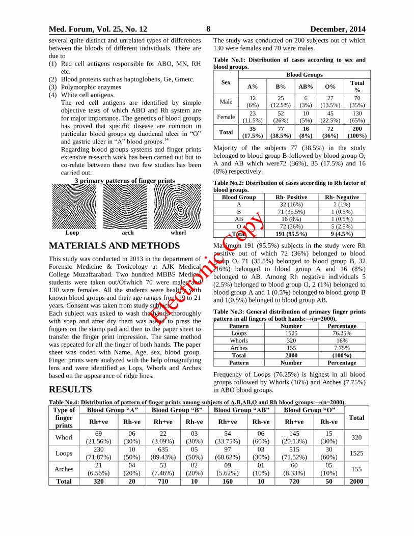
classified into three primary patterns.8
i. Loops. ii. Whorls. Iii. Arches.
Finger prints follow the Locard’s principle of exchange.
The secretions in the finger prints contain residues,
various chemicals and their metabolites which can be
detected and used for Forensic purpose. It they are
found on scene of occurrence, then the suspects, can be
easily identified. Human fingerprints are detailed,
unique, difficult to alter, and durable over the life of an
individual making them suitable as long-term markers
of human identity and may be employed by police or
other authorities to identify individuals who wish to
conceal their identity, or to identify people who are
incapacitated or deceased and thus unable to identify
themselves, as in the aftermath of a natural disaster.
Fingerprint analysis, in use since the early 20th century,
has led to many crimes being solved.9 This means that
many criminals consider gloves essential.10,11
Deliberate impressions of fingerprints may be formed
by ink or other substances transferred from the peaks of
friction ridges on the skin to a relatively smooth surface
such as a fingerprint card.12
Blood itself is an extremely important entity in the
medico legal practice which alone or along with other
trace evidences can play a clinching role to unfold
different chemical problems. Blood groups system was
discovered by Karl landsterer in 1901.13
.There are
Original Article Finger Prints Relation
Blood Groups
Electro
nic Cop
y
Med. Forum, Vol. 25, No. 12 December, 2014 8
several quite distinct and unrelated types of differences
between the bloods of different individuals. There are
due to
(1) Red cell antigens responsible for ABO, MN, RH
etc.
(2) Blood proteins such as haptoglobens, Ge, Gmetc.
(3) Polymorphic enzymes
(4) White cell antigens.
The red cell antigens are identified by simple
objective tests of which ABO and Rh system are
for major importance. The genetics of blood groups
has proved that specific disease are common in
particular blood groups eg duodenal ulcer in “O”
and gastric ulcer in “A” blood groups.14
Regarding blood groups systems and finger prints
extensive research work has been carried out but to
co-relate between these two few studies has been
carried out.
3 primary patterns of finger prints
Loop arch whorl
MATERIALS AND METHODS
This study was conducted in 2013 in the department of
Forensic Medicine & Toxicology at AJK Medical
College Muzaffarabad. Two hundred MBBS Medical
students were taken out/Ofwhich 70 were males and
130 were females. All the students were healthy with
known blood groups and their age ranges from 19 to 21
years. Consent was taken from study subjects.
Each subject was asked to wash the hands thoroughly
with soap and after dry them was asked to press the
fingers on the stamp pad and then to the paper sheet to
transfer the finger print impression. The same method
was repeated for all the finger of both hands. The paper
sheet was coded with Name, Age, sex, blood group.
Finger prints were analyzed with the help ofmagnifying
lens and were identified as Lops, Whorls and Arches
based on the appearance of ridge lines.
RESULTS
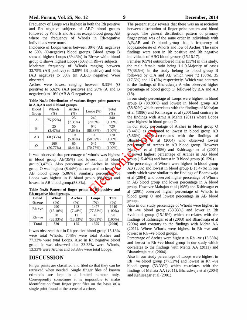
The study was conducted on 200 subjects out of which
130 were females and 70 were males.
Table No.1: Distribution of cases according to sex and
blood groups.
Sex
Blood Groups
A% B% AB% O% Total
%
Male 12
(6%)
25
(12.5%)
6
(3%)
27
(13.5%)
70
(35%)
Female 23
(11.5%)
52
(26%)
10
(5%)
45
(22.5%)
130
(65%)
Total 35
(17.5%)
77
(38.5%)
16
(8%)
72
(36%)
200
(100%)
Majority of the subjects 77 (38.5%) in the study
belonged to blood group B followed by blood group O,
A and AB which were72 (36%), 35 (17.5%) and 16
(8%) respectively.
Table No.2: Distribution of cases according to Rh factor of
blood groups.
Blood Group Rh- Positive Rh- Negative
A 32 (16%) 2 (1%)
B 71 (35.5%) 1 (0.5%)
AB 16 (8%) 1 (0.5%)
O 72 (36%) 5 (2.5%)
Total 191 (95.5%) 9 (4.5%)
Maximum 191 (95.5%) subjects in the study were Rh
positive out of which 72 (36%) belonged to blood
group O, 71 (35.5%) belonged to blood group B, 32
(16%) belonged to blood group A and 16 (8%)
belonged to AB. Among Rh negative individuals 5
(2.5%) belonged to blood group O, 2 (1%) belonged to
blood group A and 1 (0.5%) belonged to blood group B
and 1(0.5%) belonged to blood group AB.
Table No.3: General distribution of primary finger prints
pattern in all fingers of both hands:→(n=2000).
Pattern Number Percentage
Loops 1525 76.25%
Whorls 320 16%
Arches 155 7.75%
Total 2000 (100%)
Pattern Number Percentage
Frequency of Loops (76.25%) is highest in all blood
groups followed by Whorls (16%) and Arches (7.75%)
in ABO blood groups.
Table No.4: Distribution of pattern of finger prints among subjects of A,B,AB,O and Rh blood groups:→(n=2000).
Type of
finger
prints
Blood Group “A” Blood Group “B” Blood Group “AB” Blood Group “O”
Total Rh+ve Rh-ve Rh+ve Rh-ve Rh+ve Rh-ve Rh+ve Rh-ve
Whorl 69
(21.56%)
06
(30%)
22
(3.09%)
03
(30%)
54
(33.75%)
06
(60%)
145
(20.13%)
15
(30%) 320
Loops 230
(71.87%)
10
(50%)
635
(89.43%)
05
(50%)
97
(60.62%)
03
(30%)
515
(71.52%)
30
(60%) 1525
Arches 21
(6.56%)
04
(20%)
53
(7.46%)
02
(20%)
09
(5.62%)
01
(10%)
60
(8.33%)
05
(10%) 155
Total 320 20 710 10 160 10 720 50 2000
Electro
nic Cop
y
Med. Forum, Vol. 25, No. 12 December, 2014 9
Frequency of Loops was highest in both the Rh positive
and Rh negative subjects of ABO blood groups
followed by Whorls and Arches except blood group AB
where the frequency of Whorls in Rh-negative
individuals were more.
Incidence of Loops varies between 30% (AB negative)
to 60% (O-negative) blood groups. Blood group B
showed highest Loops (89.43%) in Rh+ve while blood
group O shows highest Loops (60%) in Rh-ve subjects.
Moderate frequency of Whorls ranging between
33.75% (AB positive) to 3.09% (B positive) and 60%
(AB negative) to 30% (in A,B,O negative) Were
observed.
Arches were lowest ranging between 8.33% (O
positive) to 5.62% (AB positive) and 20% (A and B
negatives) to 10% (AB & O negatives)
Table No.5: Distribution of various finger print patterns
in A,B,AB and O blood groups.
Blood
Group
Whorls
(%)
Arches
(%) Loops (%)
Total
(%)
A 75 (22%) 25
(7.35%)
240
(70.5%)
340
(100%)
B 25
(3.47%)
55
(7.63%)
640
(88.88%)
720
(100%)
AB 60 (35%) 10
(5.86%)
100
(58.82%)
170
(100%)
O 160
(20.77%)
65
(8.44%)
545
(70.77%)
770
(100%)
It was observed that percentage of whorls was highest
in blood group AB(35%) and lowest in B blood
group(3,47%). Also percentage of Arches in blood
group O was highest (8.44%) as compared to lowest in
AB blood group (5.86%). Similarly percentage of
Loops was highest in B blood group (88.88%) and
lowest in AB blood group.(58.8%).
Table No.6: Pattern of finger prints in Rh-positive and
Rh-negative blood groups. Blood
Group
Whorl
(%)
Arches
(%)
Loops
(%)
Total
(%)
Rh +ve 290
(15.18%)
143
(7.48%)
1477
(77.32%)
1910
(100%)
Rh –ve 30
(33.33%)
12
(13.33%)
48
(53.33%)
90
(100%)
Total 320 155 1525 (2000)
It was observed that in Rh positive blood group 15.18%
were total Whorls, 7.48% were total Arches and
77.32% were total Loops. Also in Rh negative blood
group it was observed that 33.33% were Whorls,
13.33% were Arches and 53.33% were total Loops.
DISCUSSION
Finger prints are classified and filed so that they can be
retrieved when needed. Single finger files of known
criminals are kept in a limited number only.
Consequently sometimes it is impossible to make
identification from finger print files on the basis of a
single print found at the scene of a crime.
The present study reveals that there was an association
between distribution of finger print pattern and blood
groups. The general distribution pattern of primary
finger prints was of the same order in individuals with
A,B,AB and O blood group that is frequency of
loops,moderate of Whorls and low of Arches. The same
findings were seen in Rh positive and Rh negative
individuals of ABO blood groups (15,16,17).
Females (65%) outnumbered males (35%) in this study,
the male female ratio being 1:1.9.Majority of cases
77(38.5%) in the study belong to blood group B
followed by O,A and AB which were 72 (36%), 35
(17.5%) and 16 (8%) respectively. Which was contrary
to the findings of Bharadwaja A who observed higher
percentage of blood group O, followed by B,A and AB
(18).
In our study percentage of Loops were highest in blood
group B (88.88%) and lowest in blood group AB
(58.82%) which correlates with the findings of Mahajan
et al (1986) and Kshirsagar et al (2001)and contrary to
the findings with Amit A Mehta (2011) where Loops
were highest in blood group O.
In our study percentage of Arches in blood group O
(8.44%) as compared to lowest in blood group AB
(5.86%) which co-relates with the findings of
Bharadwaja et al (2004) who observed lowest
percentage of Arches in AB blood group. However
Mahajan et al (1986) and Kshirsagar et al (2001)
observed highest percentage of Arches in AB blood
group (15.46%) and lowest in B blood group (6.15%).
The percentage of Whorls were highest in blood group
AB (35%) and lowest in blood group B (3.47%) in our
study which were similar to the findings of Bharadwaja
et al (2004) who observed higher percentage of Whorls
in AB blood group and lower percentage in A blood
group. However Mahajan et al (1986) and Kshirsagar et
al (2001) observed higher percentage of Whorls in
blood group O and lowest percentage in AB blood
groups.
Also in our study percentage of Whorls were highest in
Rh –ve blood group (33.33%) and lower in Rh
+veblood group (15.18%) which co-relates with the
findings of Kshirsagar et al (2003) and Bhardwaja et al
(2004) and contrary to the findings with Mehta AA
(2011). Where Whorls were highest in Rh +ve and
lowest in Rh –ve blood groups.
Percentage of Arches were highest in Rh –ve (13.33%)
and lowest in Rh +ve blood group in our study which
co-relates to the findings with Mehta AA (2011) and
Bharadwaja et al (2004).
Also in our study percentage of Loops were highest in
Rh +ve blood group (77.32%) and lowest in Rh –ve
blood group (53.33%) which co-relates with the
findings of Mehata AA (2011), Bharadwaja et al (2004)
and Kshirsagar et al (2001).
Electro
nic Cop
y
Med. Forum, Vol. 25, No. 12 December, 2014 10
CONCLUSION
- Whorls were highest in blood group AB and the
difference was significant with blood group B.
- Arches were highest in blood group O and the
difference was significant with blood group AB.
- Loops were highest in blood group B and the
difference was significant with AB blood group.
- Loops were highest in Rh +ve blood groups as
compared to in Rh –ve blood groups and the
difference was statistically significant.
REFERENCES
1. Vig K. Text Book of Forensic Medicine&
Toxicology. 4th
ed. New Delhi: Elseveir;2005.
2. Laufer B, History of the finger-print system,
Smithsonian Institution Annual Report 1912.
Reprinted in "The Print [newsletter of South
California Association of Fingerprint Officers]",
2000.p.1–13.
3. Ashbaugh D. Quantitative-Qualitative Friction
Ridge Analysis; An Introduction to Basic and
Advanced Ridgeology, Florida: CRC Press;1999.
p.11–19.
4. Åström P. The study of ancient fingerprints. J of
Ancient Fingerprints 2007;1(1):2–3.
5. Åström P, Eriksson SA. Fingerprints and
Archaeology. Studies in Mediterranean
Archaeology series, Sweden:Paul Åströms Förlag;
1980.
6. Pillay VV. Textbook of Forensic Medicine &
Toxicology, 15th
ed. Hyderabad Paras Medical
Publishers;2009.p.53-94.
7. Galton F. Finger prints. London:Macmillan and
CO;1892.
8. Awan NR. Principles and practice of forensic
medicine. Lahore:Sublime Arts;2002.p.32.
9. Hueske E. Firearms and Fingerprints . New York:
Facts on File/Infobase Publishing ;2009 .
10. http://en.wikipedia.org/wiki/Glove
11. Hall A. The Crime Busters. United
Kingdom/United States: Book Sales;1989.
12. Olsen, Robert D. The Chemical Composition of
Palmar Sweat. Fingerprint and Identification
Magazine. Sr (1972);53(10):4.
13. Bijlani RL. Textbook of Physiology. 2nd
ed. New
Delhi:Jaypee;1995.
14. Aird J, Bentall HH. A relation between cancer of
stomach and ABO blood groups. Br Med J 1953;
1(4814):799-801.
15. Kshersagar SV, et al. Study of finger print pattern
in ABO blood group. J Anat SOC India 2003;
52(1):82-115.
16. Bhavana D, Ruchi J, Prakash T. study of finger
print patterns in relationship with blood group and
gender. Res J Forensic Sci 2013;1(1):15-17.
17. Mehta AA, Sonar V. Digital dermatoglyphis in
ABO, Rh blood groups. J Ind Acad Forensic Med
2011;33(4):349-351.
18. Bharadwaja A, Saraswat PK, Aggarwal SK. Pattern
of finger prints in different ABO blood groups. J I
A F M 2004;26(1):6- 9.
Address for Corresponding Author:
Dr. Naveed Ahmed Khan,
Associate Prof. & Head of Department.
Islamabad Medical & Dental College Islamabad
Main Murree Road, Bhara Kahu,
Islamabad.
Mobile #: 0332-5104544
E-Mail Address: [email protected]
Electro
nic Cop
y
Med. Forum, Vol. 25, No. 12 December, 2014 11
Placebo- Controlled Trial of
Pharmaceutical Optimized Lisinopril
10mg (F-5) in Patients with Essential Hypertension for
Efficacy & Biochemical Evaluation 1. Asnad 2. Mohammad Tariq Ijaz Afridi 3. Munir Tahir 4. Mohammad Adnan Shereen
1. Asstt. Prof. of Biochemistry, MBBS Medical College, Mirpur AJ&K 2. Asstt. Prof. of Medicine, JMC, Pesharar
3. Assoc. Prof. of Biochemistry, MBBS MC, Mirpur AJ&K 4. Demonstrator, of Microbiology, KMU, Peshawar
ABSTRACT
Objective: The objective of this double-blind, randomized placebo-controlled trial study evaluating efficacy and
biochemical effects of optimized lisinopril 10mg (F-5) as compared to placebo in adult patients with essential
hypertension.
Study Design. Double-blind, randomized placebo-controlled trial
Place and Duration of Study: This study was conducted at the Department of Biochemistry, University of Karachi
from October 20 11 to January 2012.
Materials and Methods: Patients were randomized to receive once optimized lisinopril 10mg (F-5) daily and
Placebo once daily for 8 weeks and at the end of study efficacy and biochemical evaluation was done.
Result: The patients treated with optimized lisinopril 10mg (F-5) alone, blood pressure reduction was lower,
although significant; reaching values of 140.1 ± 11.4/ 87.7 ± 5.4 mmHg (p < 0.05 versus Placebo) by the end of
eight weeks of treatment. .No significant variation of blood glucose was observed and different parameters of lipid
profile were also observed during the eight weeks of treatment with antihypertensive regimen used. Thus, the drug
regimens used may be considered neutral as regards glucose and plasma lipid metabolism profile because drug
used at low doses.
Conclusion: We can suggest that the high antihypertensive efficacy, good tolerability and no biochemical effects of
the optimized Lisinopril 10mg (F-5) it is an excellent option for the treatment of hypertension in a wide range of
hypertensive patients, with a high potential to reduce cardiovascular risks.
Key Words: Hypertension, Lisinopril, Biochemical Effects
INTRODUCTION
Adequate blood pressure is a treatment of hypertension
and it is the risk of cardiovascular morbidity and
mortality so proper therapy is essential. And the
reduction of blood pressure lower than 130/85 mmHg
provides additional benefits regarding both protection
of organs and cardiovascular mortality. Guidelines of
World Health Organization for the treatment of
hypertension that is, 130/85 mmHg which is lower than
the previous limit of 140/90 mmHg.1-6
Blood pressure is an important modifiable risk factor
for the progression of renal disease.7
of all
antihypertensive agents; inhibitors of angiotensin-
converting enzyme (ACE) are regarded as particularly
effective in limiting renal-disease progression , because
of possible beneficial influences on kidney function,
which are separate from the effects on systemic blood
pressure.ACE inhibitors significantly limit the
progression of renal disease in patients with
macroalbuminuria,7 and, at the time our trial was
designed, there were indications that this beneficial
effect also occurred in patients with micro-
albuminuria.8,9
If ACE inhibitors can slow the relentless
decline of renal function in patients with
microalbuminuria, it is reasonable to investigate
whether use of ACE inhibitors in patients with
normoalbuminuria may also be beneficial. However,
previous trials of ACE inhibitors in normoalbuminuric
patients are few, 10
and have either lacked power or
have not been designed as randomized and controlled. 10, 11
consequently, the degree of albuminuria at which
treatment with ACE inhibitors should start is unclear.
Lisinopril is one of the most widely used angiotensin-
converting enzyme (ACE) inhibitors in adult medicine,
and ACE inhibitors (ACE-Is) are a major component of
cardiovascular therapy because of their beneficial
effects on cardiac function in heart failure and
myocardial infarction.12,13
ACE-Is are particularly
effective antihypertensive agents. In most hypertensive
pediatric patients, especially younger patients,
hypertension is secondary to renal disease and isrenin-
mediated with activation of the renin- angiotensin
system (RAS). Therapy with an ACE-I is therefore the
first choice of drug in the pediatric population. The
ability of ACE-Is to block the renin-angiotensin-
aldosterone system (RAAS) accounts for their effect in
reducing blood pressure (BP) but also prevents the
deleterious effects of Ang II on endothelial function.
Original Article Effect of
Lisinopril in
Hypertension
Electro
nic Cop
y
Med. Forum, Vol. 25, No. 12 December, 2014 12
Lisinopril has been shown to decrease urinary protein
excretion in adults with diabetes mellitus.
14
Comparative safety and efficacy trials indicate that
angiotensin receptor blockers like olmesartan
medoxomil have superior tolerability and
antihypertensive efficacy15
. Similar investigation using
olmesartan, medoxomil and amlodipine besylate
showed great effectiveness and tolerance in patient with
hypertension16
. Combination therapies reduced B.P to a
greater extent than with amlodipine besylate alone as
indicated with benazepril hydrochloride with valsartan
and with perindopril17, 18
Therefore, the objective of this comparative study
evaluating the efficacy and biochemical effects of
optimized Lisinopril 10mg (F-5) with placebo in the
treatment of patients with essential hypertension.
MATERIALS AND METHODS
This was multicenter, randomized, placebo-controlled,
comparative study. Patient was randomized to receive
optimized Lisinopril 10mg (F-5) once daily and
Placebo once daily for 8 weeks. The study was
conducted in Department of Biochemistry, University
of Karachi from October 20 11 to January 2012.
Patients were selected from four different hospitals of
orange Town and 80 patients were selected for the
study. Therefore 80 patients were effectively analyzed
for efficacy and tolerability the analysis of
antihypertensive efficacy and biochemical effects of a
therapeutic regimen in the long term becomes
important. The primary efficacy variable was change
from baseline in MSDP at the end of study. Secondary
variable was change in mean sitting systolic blood
pressure from baseline. Safety biochemical parameters
(complete blood count, renal function, liver function,
electrolytes, protein profile, and enzymes) and
electrocardiogram at rest were also determined in all
patients at the baseline (week O) and at the 8th week of
antihypertensive treatment. At the same time points,
glucose metabolism parameter values and plasma lipids
(total cholesterol, HDL-cholesterol, LDL-cholesterol,
and triglycerides) were also recorded. Biochemical
parameters were determined using an automated
method.
RESULTS
The patients treated with optimized Lisinopril 10mg (F-
5) alone, blood pressure reduction was lower, although
significant; reaching values of 140.1 ± 11.4 / 87.7 ± 5.4
mmHg (p < 0.05 versus Placebo) by the end of eight
weeks of treatment. Variations in blood pressure
measurement in the standing position during treatment
were similar to those recorded in the sitting position,
and no episode of orthostatic hypotension was reported
in either of the therapeutic regimen. No significant
variation in leg volume measurement was observed
among the both groups studied during the eight weeks
of treatment. No significant variations of blood glucose
were observed and different parameters of lipid profile
were also observed during the eight weeks of treatment
with antihypertensive regimen used. Thus, the drug
regimens used may be considered neutral as regards
glucose and plasma lipid metabolism profile because
drug used at low doses.
Table No.1: Baseline characteristics
Lisinopril
10mg(F-5)
(n=60)
Placebo
(n=20)
Age (years) 51.2 + 9.4 51.5 + 9.8
Male / Female (%) 40.4 / 59.6 35.0 / 65.0
Body weight (Kg) 69.9 + 13.5 70.2 + 12.2
BMI (kg/m2) 27.4 + 3.6 27.8 + 3.4
SBP sitting (mmHg) 149.9 + 11.2 148.7 + 10.7
DBP sitting (mmHg) 96.7 + 7.3 95.9 + 7.5
Table No.2: Ambulatory blood pressure monitoring.
Mean values of blood pressure
Lisinopril 10mg
(F-5) (n=60)
Placebo
(n=20)
P-value
Systolic BP - 24
hours (mmHg)
Baseline 149.9 + 11.2 148.7+ 10.7 NS
Week 8 140.1 ± 11.4 148.9± 11.3 0.0037
Diastolic BP - 24
hours (mmHg)
Baseline 96.7 + 7.3 95.9 + 7.5 NS
Week 8 87.7 ± 5.4 94.9 ± 7.8 0.0001 NS: Non significant, p: probability
Table No.3: Baseline Biochemical characteristics
Lisinopril 10mg (F-5) (n=60) Placebo
(n=20)
Fasting Blood Glucose(mg/dl)
Baseline 99.4 ± 11.3 98.1 ± 8.7
Week 8 98.5 ± 11.7 97.9 ± 9.5
Total Cholesterol (mg/dl)
Baseline
197.9 ± 43.2 194.2 ± 33.4
Week 8 198.2 ± 43.4 193.9 ± 34.2
LDL - Cholesterol (mg\dl)
Baseline 114.4 ± 33.2 118.3± 25.8
Week 8 114.9 ± 33.5 117.8 + 24.7
HDL - Cholesterol (mg\dl)
Baseline 53.9 ± 13.2 47.9 ± 11.6
Week 8 52.8 ± 12.8 47.7 ± 11.5
Triglycerides (mg\dl)
Baseline 137.8 ± 88.7 145.6 ± 88.2
Week 8 137.1 ± 89.2 144.2 ± 88.9
DISCUSSION
Hypertension is a major risk factor for stroke. In
relation to other stroke-specific factors, brain tissue loss
as a consequence of stroke has been associated with
cognitive impairment; these strokes may be isolated or
Electro
nic Cop
y
Med. Forum, Vol. 25, No. 12 December, 2014 13
strategically located ones (e.g. in the thalamus, angular
gyrus, frontal white matter).19
Also, because
hypertension often does not exist as a solitary factor but
occurs in the presence of other metabolic risks, other
stroke-related factors such as inflammation or abnormal
insulin signaling in the brain, or the presence of
metabolic syndrome could exist and underlie cognitive
impairment or dementia in persons with
hypertension.20,
21
The baseline characteristics of the population included
in the study are shown in Table no1. We can observe
that the groups were not different in relation to age,
body mass index and weight, heart rate, and systolic
and diastolic pressure values. The results of this study
showed that the optimized product Lisinopril 10mg (F-
5) as a high antihypertensive efficacy that is sustained
in the long term with a quite reduced percentage of loss
of blood pressure control in table No.2 We observed
that more than 69.2% of the patients treated with
optimized product of Lisinopril 10mg (F-5) remained
with diastolic blood pressure levels equal to or lower
than 90 mmHg, thus achieving the goals for the
treatment of hypertension. The difficulty to achieve the
goal of controlling systolic blood pressure explains why
the international guidelines for studies on
antihypertensive drugs still use criteria based on
diastolic blood pressure to describe the antihypertensive
efficacy of a drug, in spite of the fact that guidelines
indicate the real need to control systolic blood pressure
as well. It is important to point out that blood pressure
reduction provided by the treatment with optimized
product of Lisinopril 10mg (F-5) did not cause any
secondary Increase in sympathetic activity, since no
significant variations of heart rate occurred. Our results
showed that the optimized product of Lisinopril 10mg
(F-5) at low doses has a very good biochemical profile
with a low incidence of adverse events. The good
biochemical profile of the optimized Lisinopril 10mg
(F-5) may be explained by the use of lower doses of
each of the hypotensive drugs, since the existence of a
strong relation between the dose of the hypotensive
drug and the frequency of adverse events is known.
However, some drugs used in the treatment of
hypertension, such as diuretics and beta-blockers, are
known to be able to promote harmful alterations in lipid
metabolism, especially in glucose metabolism. In our
study we observed that the use of the optimized
Lisinopril 10mg (F-5) did not change parameters of
either glucose metabolism or plasma lipids, thus having
a neutral biochemical profile even when used for 8
weeks. Table.No.3 Based on these results we can
suggest that the optimized product Lisinopril 10mg
(F-5) is safe and adequate for the treatment of
hypertension in patients with metabolic syndrome,
diabetes mellitus and dyslipidemias .Incidentally,
hypertension is frequently associated to the metabolic
syndrome; also, the frequency of this association
increases with age.
CONCLUSION
In brief, the results of this multicenter study
demonstrated that the optimized Lisinopril 10mg (F-5)
has a high antihypertensive efficacy, allowing
approximately 69.2% of the patients treated to achieve
and maintain for eight weeks. We can suggest that the
high antihypertensive efficacy, good tolerability and no
biochemical effects of the optimized Lisinopril 10mg
(F-5) it is an excellent option for the treatment of
hypertension.
REFERENCES
1. Sykowsky PA, D’Agostino RB, Belanger AJ,
Kannel WB. Secular Trends in Long Term
Sustained Hypertension, Long Term Treatment and
Cardiovascular Morbidity. The Framingham Heart
Study 1950-1990. Circulation 1996; 93: 697-703.
2. MacMahon S, Peto R, Cutler J, et al. Blood
Pressure, stroke, and coronary heart disease. Part 1,
prolonged differences in blood pressure:
prospective observational studies corrected for
regression dilution bias. Lancet 1990; 335:765-77.
3. IV Diretrizes Brasileiras de Hipertensão Arterial -
Sociedade Brasileira de Hipertensão;Sociedade
Brasileira de Cardiologia e Sociedade Brasileira de
Nefrologia. Hipertensão 2003; 5(4): 126-63.
4. Chobaniam AV, Bakris GL, Black HR, et al.
Seventh report of the Joint National Committee on
prevention, detection, evaluation and treatment of
high blood pressure- JNC 7. Hypertension 2003;
42:1206-52.
5. 2003 European Society of Hypertension -
European Society of Cardiology guidelines for
management of arterial hypertension. J Hypertens
2003;21: 1011-53.
6. Hansson L, Zanchetti A, Carruthers SG, Dahlof B,
et al on behalf of HOT Study group. Effects of
intensive blood-pressure lowering and low-dose
aspirin in patients with hypertension: principal
results of the Hypertension Optimal Treatment
(HOT) randomized trial. Lancet 1998;351:1755-62.
7. Lewis EJ, Hunsicker LG, Bain RP, Rohde RD. The
effect of angiotensin converting enzyme inhibition
on diabetic nephropathy. N Engl J Med 1993; 329:
1456–62.
8. Marre M, Chatellier G, Leblanc H, Guyene TT,
Menard J, Passa P. Prevention of diabetic
nephropathy with enalapril in normotensive
diabetics with microalbuminuria. BMJ 1988;297:
1092–95.
9. Mathiesen ER, Hommel E, Giese J, Parving H-H.
Efficacy of captopril in postponing nephropathy in
normotensive insulindependent diabetic patients
with microalbuminuria. BMJ 1991;303:81–87.
Electro
nic Cop
y
Med. Forum, Vol. 25, No. 12 December, 2014 14
10. Passa P, Leblanc H, Marre M. Effects of enalapril
in insulin dependent diabetic subjects with mild to
moderate uncomplicated hypertension. Diabetes
Care 1987;10:200–04.
11. Pedersen MM, Schmitz A, Pedersen EB, Danielsen
H, Christiansen JS. Acute and long-term renal
effects of angiotensin converting enzyme inhibition
in normotensive, normoalbuminuric insulin-
dependent diabetic patients. Diabet Med 1988;5:
562–69.
12. Garg R, Yusuf S. Overview of randomized trials of
angiotensin-converting enzyme inhibitors on
mortality and morbidity in patients with heart
failure. Collaborative Group on ACE Inhibitor
Trials. JAMA 1995;273:1450-6.
13. Indications for ACE inhibitors in the early
treatment of acute myocardial infarction:
systematic overview of individual data from
100,000 patients in randomized trials. ACE
Inhibitor Myocardial Infarction Collaborative
Group. Circulation 1998;97:2202-12.
14. Randomised placebo-controlled trial of lisinopril in
normotensive patients with insulin-dependent
diabetes and normoalbuminuria or
microalbuminuria. The EUCLID Study Group.
Lancet 1997;349:1787-92.
15. Bernard RC, Carl JP , John OP , Jaroslav Sl, Galina
C, Jerzy K, et al. Effects of ranolazine with
atenolol, amlodipine, or diltiazem on exercise
tolerance and angina frequency in patients with
severe chronic angina. JAMA 2004; 291: 309-316.
16. Khalida B, Najaf AG, Naheed A. Comparative
studies of cimetidine derivative “temalastine” for
potential energy calculation by Kitaigorodskii and
lennard-jones functions. Pak J Biochem Mol Biol
2010; 43: 81-86.
17. Afshan N, Naheed A, Khalida B, Najaf AG, Farhat
B. Conformational analysis geometry optimization
of nucleosidic antitumor antibiotic showdomycin
by Arguslab 4 software. Pak J Pharmacol 2009;
22:78-82.
18. Weir MR, Crikelair N, Levy D, Rocha R, Kuturu
V, Glazer R. Evaluation of the dose response with
valsartan and valsartan/hydrochlorothiazide in
patients with essential hypertension. J Clin
Hypertens (Greenwich) 2007;9(2):103–112.
19. Gorelick PB. Status of risk factors for dementia
associated with stroke. Stroke 1997;28:459–463.
20. Gorelick PB. Role of inflammation in cognitive
impairment: results of observational epidemio-
logical studies and clinical trials. Ann NY Acad
Sci 2010;1207:155–162.
21. Gorelick PB. William Feinberg Lecture: cognitive
vitality and the role of stroke and cardiovascular
disease risk factors. Stroke 2005; 36: 875–879.
Address for Corresponding Author:
Dr. Asnad,
Assistant Professor
Department of Biochemistry
MBBS Medical College, Mirpur AJ&K
Contact No: 0332-3698204
Electro
nic Cop
y
Med. Forum, Vol. 25, No. 12 December, 2014 15
Diagnostic Accuracy of IgA Anti-
Tissue Transglutaminase Antibodies in
Comparison with Histopathological Findings in Celiac
Disease in Pakistan 1. Arslaan Javaeed 2. Walayat Shah 3. Rizwan Akhtar 4. Sanniya Khan Ghauri 5. Shafqat
Husnain Khan 6. Aftab Haider Alvi 1. Histopathologist, Fatima Memorial Hospital / FMMC, Lahore 2. Asstt. Prof. of Pathology, KMU, Peshawar
3. Prof. of Pathology, FMMC, Lahore 4. Resident of Emergency Medicine, AKUH, Karachi 5. Senior Demonstrator
of Pathology, CMC, Lahore 6. Asstt. Prof. of Medicine, FMMC, Lahore
ABSTRACT
Objective: The objective of this study was to assess the diagnostic accuracy of most widely used serological test for
diagnosis of celiac disease (CD) i.e. anti-tissue transglutaminase antibody (IgA) in comparison to histopathological
lesions in CD.
Study Design: cross sectional study
Place and Duration of Study: This study was carried out at the Departments of Gastroenterology and Pathology of
Fatima Memorial Hospital, Shadman, Lahore from March 2014 to October 2014.
Materials and Methods: 121 patients clinically suspected of celiac disease were included in this cross sectional
study. The biopsy was taken from the second part of duodenum and was evaluated according to Marsh classification
of CD. Blood sample of every patient was obtained to perform anti-tTG antibody test.
Results: The range of the patients included in the study came out to be 18-65 years with 30.24 years as mean age.
Out of all the patients included in this study 34 (28.1%) were males and 87(71.9%) were females. The overall
sensitivity and specificity of anti-tTG were 78.6% and 98.1%.The positive predictive value (PPV) and negative
predictive value (NPV) came out to be 84.6% and 97.2% respectively.
Conclusion: We have come to the conclusion that currently there is no serological test which can be used as a sole
tool for the diagnosis of celiac disease. Relying on serological test will lead to missed diagnosis of CD especially
those patients which have Marsh lesions of lesser degrees.
Key Words: Celiac Disease, Anti-Tissue Transglutaminase Antibody, Sensitivity, Duodenal Biopsy.
INTRODUCTION
Celiac disease (CD) also called as gluten-sensitive
enteropathy is an autoimmune disease triggered by
gluten, affects small intestine in genetically susceptible
children and adults. It is the only immune-mediated
disease which is fully treatable only when a precise
diagnosis is established. Gluten is a protein present in
wheat, barley and rye etc. It is mainly composed of
gliadin and glutenin (Catassi and Fasano, 2010).1
The prevalence of CD is becoming significantly higher
than that recognized 20 years ago. The prevalence of
celiac disease at global level is considered to be 1%
(Mustalhati et al, 2010)2. According to a study the
prevalence of CD varies from 2-13% (van der Windt,
2010)3.
Scientists have found a strong linkage between
presence of human leukocyte antigen (HLA) DQ2 or
DQ8 and celiac disease. HLA-DQ typing can be used in
ruling out the celiac disease. On the other hand
presence of DQ2 or DQ8 does not exhibit the presence
of disease as these genes are present in general
population as well (Kapitani, 2006)4.
The parameters to diagnose CD have significantly
changed over the last 50 years. Diarrhea and
malabsorption once thought to be major mode of
presentation of celiac disease are becoming less
common (Reily, 2012)5. Over time many specific and
sensitive serological tests were introduced to make the
diagnosis of CD less invasive process. Anti-gliadin
antibody (AGA is among the first immunological
assays used for screening CD (Fasano and Carlo,
2001)6.
AGA is also found in diseases like rheumatoid arthritis
and depression among elderly population which adds to
its poor specificity (Anitta and Katri, 2012)7. Later
these serological tests have been replaced by more
sensitive and specific tests including antiendomysial
(EMA), antireticulin (RA) and anti tissue
transglutaminase (tTG) antibodies (Shinjini and Nitya,
2006)8. The major breakthrough in the shape of the
discovery of anti-tTG as the autoantigen recognized by
the EMA led to the development of ELISA based
assays. These assays were projected at the detection of
anti-tissue transglutaminase (anti-tTG) antibody
(Dieterich et al, 1997)9.
Original Article Findings in Celiac
Disease
Electro
nic Cop
y

Med. Forum, Vol. 25, No. 12 December, 2014 16
The discrepancy associated with anti-EMA and anti-
tTG includes their unreliability in children of less than
2 years and IgA dependence (Wang et al, 2014)10
. The
sensitivity of AGA was better than that of anti-EMA
and anti-tTGA in children aged less than 2 years
(Mankai et al, 2005)11
.
Anti-tTG antibody test is routinely used as the first
choice because of its high sensitivity, cost-effectiveness
and easy interpretation. However discrepancy of tTG
assays is their variable accuracy among manufacturers
(Giersiepen et al., 201212
, & Astrid and Juri, 2013)13
.
The role of pathologist in the diagnosis of celiac disease
is of utmost significance. For the histopathological
examination scientists have agreed that biopsy for the
diagnosis of CD should be taken from 2nd
part of
duodenum. Marsh was the first scientist who explained
the broad spectrum of inflammatory and structural
changes which took place in CD. That is why Marsh
classification became very popular. Later on Marsh
classification was modified by Oberhuber (Oberhuber,
1999)14
.
Once a patient is diagnosed with CD, adherence to
gluten free diet (GFD) for life is the only treatment
available currently as it leads to complete recovery of
the patient (Akobeng and Thomas, 2008)15
. The
patients of CD are suffering from the deficiency of
many minerals and vitamins including Iron, copper,
zinc, B12, B 6, folic acid and vitamin D which leads to
increased risk of fractures. (Rubio et al, 2013)16
.
The risk of premature and low weight infant births,
abortions and infertility rises in women suffering from
CD. The treatment of CD decreases these risks (Janet,
2011)17
. Researchers have shown that those patients
who did not get treatment for CD have revealed
considerably lower bone mineral density (Mora,
2001)18
.
The celiac disease patients are rarely diagnosed in
Pakistan because of two main reasons. Firstly
physicians are unaware if its existence and its clinical
presentation. Secondly no proper protocol is present to
successfully diagnose this disease. Mostly anti-tTG
antibody test is used to diagnose or exclude diagnosis
of this disease. In addition to this there is no data
available about prevalence rate of CD in Pakistan and
sensitivity of anti-tTG antibody test. In this study we
evaluated the diagnostic accuracy of anti-tTG antibody
test in comparison to histopathological lesions
according to Marsh classification of CD in order to
draft a proper approach to diagnose this disease.
MATERIALS AND METHODS
This cross sectional study was conducted at the
departments of gastroenterology and pathology of
Fatima Memorial Hospital, Shadman, Lahore, Pakistan
from March 2014 to October 2014. In this research, 121
patients were recruited according to our inclusion and
exclusion criteria. We included patients from both
gender from age 5 to 60 years. These patients were
clinically diagnosed for CD. The clinical diagnosis
included typical clinical presentation including
diarrhea, weight loss, fatigue, iron deficiency anemia
and also atypical clinical presentation including non
specific GI symptoms for a long duration like
abdominal pain, abdominal bloating, short stature and
constipation etc. We excluded patients with any other
known disease (comorbidity).
All the patients after receiving the oral and written
explanation of the whole research study signed the
informed consent form. This study protocol was
approved by ethics committee.
Four to five biopsies were taken with sterilized forceps
from the second part of the duodenum in all patients
through endoscopy for histopathological examination.
At the same time 5ml blood sample was taken from
every patient for serological evaluation. The results of
intestinal biopsy were considered as gold standard of
our research. The age, gender and complete clinical
history of every patient were documented.
RESULTS
Biopsy specimens were kept in labeled and separate
collection jars. After fixation in buffered formalin the
biopsy specimens were embedded in paraffin wax. The
thickness of the sections was kept at standard
3µm.These were stained with hematoxylin and eosin
and slides were prepared. The slides were evaluated by
expert pathologists who were blinded to the serology
results. The number of intraepithelial lymphocytes,
crypt hyperplasia and villous atrophy were documented
according to modified Marsh classification (Table 1).
Table No.1 Modified Marsh Classification
Marsh
Type
*IEL/100
Enterocytes
(Duodenum)
Crypt
Hyperplasia
Atrophy of
Villi
0 <30 Normal Normal
1 >30 Normal Normal
2 >30 Increased Normal
3A >30 Increased Mild
3B >30 Increased Moderate
3C >30 Increased Total (*Intraepithelial Lymphocytes (IELs) Per 100 Enetrocytes)
According to patient’s history and mode of presentation
other diseases which cause duodenal damage e.g.
Giardia lambia infection, food protein hypersensitivity
were also considered.
Serum Analysis: The serum analysis was performed in
a laboratory with hundreds of routine samples. The
laboratory staff neither knew biopsy results nor clinical
presentation of the patient. The serology test was
performed on each blood sample with following
method through commercial kit in accordance with
Electro
nic Cop
y
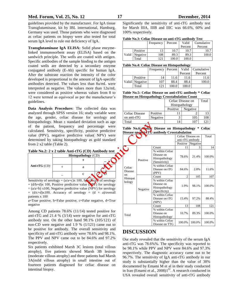
Med. Forum, Vol. 25, No. 12 December, 2014 17
guidelines provided by the manufacturer. For IgA tissue
Transglutaminase, kit by IBL international, Hamburg,
Germany was used. Those patients who were diagnosed
as celiac patients on biopsy were also tested for total
serum IgA level to rule out deficiency of IgA.
Transglutaminase IgA ELISA: Solid phase enzyme-
linked immunosorbent assay (ELISA) based on the
sandwich principle. The wells are coated with antigen.
Specific antibodies of the sample binding to the antigen
coated wells are detected by a secondary enzyme
conjugated antibody (E-Ab) specific for human IgA.
After the substrate reaction the intensity of the color
developed is proportional to the amount of IgA-specific
antibodies detected. The values less than 8u/mL were
interpreted as negative. The values more than 12u/mL
were considered as positive whereas values from 8 to
12 were termed as equivocal as per the manufacturer’s
guidelines.
Data Analysis Procedure: The collected data was
analyzed through SPSS version 16; study variable were
the age, gender, celiac disease for serology and
histopathology. Mean ± standard deviation such as age
of the patient, frequency and percentage were
calculated. Sensitivity, specificity, positive predictive
value (PPV), negative predictive value( NPV) were
determined by taking histopathology as gold standard
from 2 x2 table (Table 2).
Table No.2: 2 x 2 table Anti-tTG (CD) Antibody test
Anti-tTG (CD)
Histopathology (CD)
+ _
+ a B
_ c D
Sensitivity of serology = (a/a+c)x 100, Specificity of serology
= (d/b+d)x 100, Positive predictive value (PPV) for serology
= (a/a=b) x100, Negative predictive value (NPV) for serology
= (d/c+d)x100, Accuracy of serology = (d + a)/overall
patients x 100
a=True positive, b=False positive, c=False negative, d=True
negative
Among CD patients 78.6% (11/14) tested positive for
anti-tTG and 21.4 % (3/14) were negative for anti-tTG
antibody test. On the other hand 98.1% (105/121) of
non-CD were negative and 1.9 % (1/121) came out to
be positive for antibody. The overall sensitivity and
specificity of anti-tTG antibody were 78.6% and 98.1%.
The PPV and NPV came out to be 84.6% and 97.2%
respectively.
Six patients exhibited Marsh 3C lesions (total villous
atrophy), five patients showed Marsh 3B lesions
(moderate villous atrophy) and three patients had Marsh
3A(mild villous atrophy) in small intestine out of
fourteen patients diagnosed for celiac disease on
intestinal biopsy.
Significantly the sensitivity of anti-tTG antibody test
for Marsh IIIA, IIIB and IIIC was 66.6%, 60% and
100% respectively.
Table No.3: Celiac Disease on anti-tTG antibody Test Frequency Percent Valid
Percent
Cumulative
Percent
Valid
Positive 13 10.7 10.7 10.7
Negative 108 89.3 89.3 100.0
Total 121 100.0 100.0
Table No.4: Celiac Disease on Histopathology Frequency Percent Valid
Percent
Cumulative
Percent
Valid
Positive 14 11.6 11.6 11.6
Negative 107 88.4 88.4 100.0
Total 121 100.0 100.0
Table No.5: Celiac Disease on anti-tTG antibody * Celiac
Disease on Histopathology Crosstabulation Count
Celiac Disease on
Histopathology
Total
Positive Negative
Celiac Disease
on anti-tTG
Positive 11 2 13
Negative 3 105 108
Total 14 107 121
Table No.6: Celiac Disease on Histopathology * Celiac
Disease on anti-tTG antibody Crosstabulation Celiac Disease on
anti-tTG antibody Total
Positive Negative
Celiac
Disease on
Histopat
hology
Positive
Count 11 3 14
% within Celiac Disease on
Histopathology
(Sensitivity)
78.6% 21.4% 100.0%
% within Celiac Disease on tTG
(PPV)
84.6% 2.8% 11.6%
Negative
Count 2 105 107
% within Celiac Disease on
Histopathology
(Specificity)
1.9% 98.1% 100.0%
% within Celiac
Disease on tTG
(NPV)
15.4% 97.2% 88.4%
Total
Count 13 108 121
% within Celiac
Disease on
Histopathology
10.7% 89.3% 100.0%
% within Celiac Disease on TTG
100.0% 100.0% 100.0%
DISCUSSION
Our study revealed that the sensitivity of the serum IgA
anti-tTG was 78.6%%. The specificity was reported to
be 98.1% while PPV and NPV were 84.6% and 97.3%
respectively. The diagnostic accuracy came out to be
96.7%. The sensitivity of IgA anti-tTG antibody in our
study is substantially higher than the value of 38%
documented by Emami M et al in their study conducted
in Iran (Emami et al., 2008)19?
. A research conducted in
USA revealed overall sensitivity of anti-tTG antibody
Electro
nic Cop
y
Med. Forum, Vol. 25, No. 12 December, 2014 18
test at 70.6%, whereas over specificity was found to be
65.0 % (Abrams et al., 2006)20
. In our study sensitivity
of anti-tTG antibody test was 60% for partial villous
atrophy which is higher than the 42.3% sensitivity
reported by Abram et al. (2006) and 36.8% documented
by Emami et al (2008)19
. According to our research
sensitivity of anti-tTG antibody test for total villous
atrophy (Mrash III C lesion) was 100% which is again
higher than 90.0% sensitivity reported by Abram J et al.
for patients with total villous atrophy (Abrams et al.,
2006)20
.
Celiac disease can be diagnosed correctly even if the
physicians are aware of the several ways in which it is
presented. Despite the fact that prevalence of CD is
increasing all over the world, still physicians in
Pakistan have not made their minds to include this new
endemic disease even in their differential diagnosis. It
would not be wrong to state that in reality CD is already
wide-spread because of wheat consumption but is either
misdiagnosed or undiagnosed. On one hand it is very
important that physicians learn to recognize signs and
symptoms of CD while on the other hand it is equally
necessary that pathologist must recognize mild
categories of CD including 1, 2 and 3a.
CONCLUSION
We conclude that there is no single serological test with
100% sensitivity and specificity therefore biopsy
remains the gold standard for the diagnosis of this
disease. On this antibody test many patients were
reported as false negatives and also false positives. The
sensitivity of anti-tTG antibody for lesser degree of
Marsh lesion is considerably low at 60% as compared
to 100% for Marsh 3C (total villous atrophy). It reveals
that a significant proportion of patients suffering from
celiac disease having mild to moderate degree of
lesions will remain undiagnosed if only anti-tTG
antibody test is used for diagnosis. Therefore it is
recommended that when there is persistence of signs
and symptoms of celiac disease and patient is reported
seronegative for antibody test, still an intestinal biopsy
is necessary for making or ruling out the diagnosis. The
challenge of diagnosis of celiac disease can be fulfilled
through good communication between the pathologist
and the clinician. The pathologist’s report should
concisely highlight the histopathological findings which
must be correlated with clinical presentation and
serological results.
REFERENCES
1. Catassi C;Fasano A. Celiac disease diagnosis:simple
rules are better than complicated alogrithms. Am J
Med 2010; 123(8):691-3.
2. Mustalahti K, Catassi C, Reunanen A, Fabiani E,
Heier M, McMillan S et al. The prevalence of
celiac disease in Europe: results of a centralized,
international mass screening project. Ann
Med 2010; 42(8): 587-595.
3. van der Windt DA. Diagnostic testing for celiac
disease among patients with abdominal symptoms: a
systematic review. JAMA 2010; 303(17):1738-46.
4. Kapitány A, Tóth L, Tumpek J, Csípo I, Sipos E,
Woolley N, Partanen J et al. Diagnostic significance
of HLA-DQ typing in patients with previous coeliac
disease diagnosis based on histology alone. Aliment
Pharmacol Therap 2006; 24(2): 1395-402.
5. Reilly NR. Epidemiology and clinical presentations
of celiac disease. Semin Immunopathol 2012;
34(4):473-8.
6. Fassano A, Carlo C. Current approaches to
diagnosis and treatment of celiac disease; an
evolving spectrum. Gastroenterology 2001; 120(2):
636-651.
7. Anitta R, Katri K. Positive serum antigliadin
antibodies without celiac disease in the elderly
population: does it matter? Scand J Gastroenterol
2012; 45(10): 1197-1201.
8. Shinjini B, Nitya T. Diagnosis of celiac disease.
Indian J Pediatr 2006; 73(8): 70-709.
9. Dieterich W, Ehnis T, Bauer M. Identification of
tissue transglutaminase as the autoantogen of celiac
disease.Nat Med 1997;3(7):797-801.
10. Wang N, Truedsson L, Elvin K, Andersson B,
Rönnelid J, Mincheva-Nilsson L et al. Serological
assessment for celiac disease in IgA deficient
adults. PLoS One 2014; 9(4):
http://www.plosone.org.
11. Mankai A, Sakly W,Landolsi H. Tissue
transglutaminase antibodies in celiac disease,
comparison of an enzyme linked immunosorbent
assay and a dot blot assay. Pathol Biol 2005; 53(4):
204-209.
12. Giersiepen K, Lelgemann M, Stuhldreher N,
Ronfani L, Husby S, Koletzko S et al. Accuracy of
diagnostic antibody tests for coeliac disease in
children: summary of an evidence report. J Pediatr
Gastroenterol Nutr 2012; 54(2): 229-41.
13. Astrid S, Juri R. Serological testing for celiac
disease in adults. United European Gastroenterol J
2013; 1(5): 319-325.
14. Oberhuber G, Granditsch G, Vogelsang H. The
histopathology of coeliac disease: time for a
standardized report scheme for pathologists. Eur J
Gastroenterol Hepatol 1999; 11(10): 1185-1194.
15. Akobeng A, Thomas A. Systematic review:
tolerable amount of gluten for people with coeliac
disease. Aliment Pharmacol Therap 2008; 27(11):
1044-1052.
16. Rubio A, Hill D, Kelly P, Calderwood H, Murray
A. ACG clinical guidelines: diagnosis and
management of celiac disease. Am J Gastroenterol
2013; 108(5): 656-76.
Electro
nic Cop
y
Med. Forum, Vol. 25, No. 12 December, 2014 19
17. Janet M, Benjamin L, Peter G. Increased
Prevalence of Celiac Disease in Patients with
Unexplained Infertility in the United States: A
Prospective Study. J Reprod Med May 2011;
56(5): 199-203.
18. Mora S, Barera G, Beccio S, Menni L, Proverbio
MC, Bianchi C, Chiumello G. A prospective,
longitudinal study of the long-term effect of
treatment on bone density in children with celiac
disease. J Pediatr 2001; 139(4): 516-521.
19. Emami M,Karimi S,Kouhestani S,Hashemi
M,Taheri H. Diagnostic accuracy of IgA anti-tissue
transglutaminase in patients suspected of having
coeliac disease in Iran. J Gastronintestin Liver
Dis2008; 17(2): 141-146.
20. Abrams J, Brar P, Diamond B, Rotterdam H, Green
H. Utility in clinical practice of immunoglobulin a
anti-tissue transglutaminase antibody for the
diagnosis of celiac disease. Clin Gastroenterol
Hepatol 2006;4(6):726-730.
Address for Corresponding Author:
Arslaan Javaeed Histopathologist, Fatima Memorial Hospital /
FMMC, Lahore
Cell No. 0300-4717057
Electro
nic Cop
y
Med. Forum, Vol. 25, No. 12 December, 2014 20
Shouldice Versus Bassini’S
Procedures for Inguinal Hernial Repair 1. Gul Sher Khan 2. Ishfaq Ali Shah Bukhari 3. Musarrat 4. Israr Ahmed
5. Muhammad Ishaq 1. Asstt. Prof. of Surgery, Khalifa Gul Nawaz Teaching Hospital Bannu 2. Asstt. Prof. of Pharmacology, JMC,
Peshawar 2. Asstt. Prof. of Gynae & Obst., JMC, Peshawar 4. Prof. of Physiology, JMC, Peshawar 5. Prof. of
Surgery, JMC, Peshawar
ABSTRACT
Objective: To evaluate the optimum surgical technique for inguinal hernia repair, Shouldice or Bassini’s ?
Study Design: Retrospective comparative study.
Place and Duration of Study: This study was conducted between 2004 to 2006 in the surgical ward DHQ Hospital
Karak. 200 patients with unilateral & primary inguinal hernia were randomly allotted to either Shouldice or
Bassini’s repair. The cases were collected either as emergencies or electively.
Materials and Methods: All the patients had primary and unilateral inguinal hernia. They were operated
electilively or as emergencies. Patients were randomly allotted to either Shouldice or Bassini’s repair. The Shouldice
was performed with 2/0 prolene in four layers while the bassini’s repair was done with prolene 0 or 1.
Results: The patients operated for inguinal hernia were followed for up to 5 years. The shouldice repair was found
associated with a lowest recurrence rate of 3% and the Bassini’s repair with 5.7%. The difference remains
statistically significant (P<0.001).
Conclusion: The Shouldice repair for inguinal hernia was associated with a recurrence rate of less than 1% in the
Shouldice clinic at TORONTO[1]
. Here in this study the Shouldice resulted in 3% recurrence rate which is nearer to
the international value while the Bassini’s repair was associated with a recurrence rate of 5.7% which is higher than
the Shouldice repair. The Shouldice repair for inguinal hernia should be the Gold Standard and serves as the basis
for comparison for all other techniques, be they prosthetic or laparoscopic [2]
.
Key word: Shouldice, Bassini’s, inguinal hernial repair.
INTRODUCTION
The Shouldice surgical technique for inguinal hernia
repair; The Shouldice is performed in 4 layers
(Glassow) [3-4]
; After herniotomy, the fascia transversals
is cut horizontally, starting at the stretched deep
inguinal ring and proceed medially to the pubic
tubercle, safeguarding the inferior epigastric vessels.
The upper and lower flaps of fascia transversalis are
formed. The lower flap is stitched to the under surface
of the upper flap and the upper flap is stitched to the
upper surface of the lower flap with 2/0 prolene. The
conjoint tendon and the lateral fleshy part of the
internal oblique and transversus abdominus are stitched
to the inguinal ligament in two layers with 2/0 prolene.
Finally the external oblique is stitched over the cord
with catgut 1. Bassini’s Repair for Inguinal hernia;
Bassini’s repair, a massive leap forward and has been
the basis of open repair for over hundred years. After
herniotomy, the conjoint tendon above is sutured with
the lower edge of inguinal ligament with interrupted,
non-absorbable sutures e.g. polypropylene, nylon or
thick black silk. Today, the Basini’s repair is the most
commonly performed procedure for inguinal hernia and
most surgeons use continuous, non-absorbable sutures
which are darned between the conjoint tendon and
inguinal ligament. The Shouldice repair is actually a
development and refinement on the Bassini’s repair [5]
.
The original Shouldice was performed under local
anesthesia. The Shouldice gave excellent results with
2/0 steel wire. Inguinal hernia repair is the second most
commonly performed operation (Appendectomy the 1st
one) in general surgery all over the world [6]
. Shouldice
hernia repair provides the best chances of non-
recurrence regardless of the anatomical type of hernia.
MATERIALS AND METHODS
This study was conducted between 2004 to 2006 in the
surgical ward DHQ Hospital Karak. 200 patients with
unilateral & primary inguinal hernia were randomly
allotted to either Shouldice or Bassini’s repair. The
cases were collected either as emergencies or electively.
All the patients had primary and unilateral inguinal
hernia. They were operated electilively or as
emergencies. Patients were randomly allotted to either
Shouldice or Bassini’s repair. The Shouldice was
performed with 2/0 prolene in four layers while the
bassini’s repair was done with prolene 0 or 1.
RESULTS
All the patients had primary and unilateral inguinal
hernia. They were operated electilively or as
emergencies. Patients were randomly allotted to either
Shouldice or Bassini’s repair. The Shouldice was
performed with 2/0 prolene in four layers while the
Original Article Inguinal Hernial Repair
Electro
nic Cop
y

Med. Forum, Vol. 25, No. 12 December, 2014 21
bassini’s repair was done with prolene 0 or 1. At the
time of induction of anaesthesia, a broad spectrum IV
antaibiotic was given. General anaesthesia was given
for 75% cases, spinal anaesthesia was given for 20%
cases and local anaesthesia was used for 05% cases.
Patients remained in surgical ward for 24 to 48 hours.
In this comparative study, some 200 patients were
randomly allotted to either Shouldice or Bassini’s
repair.
Patients 200
Age 30-65 years
Average 47 years
Gender All male patients
Type of Hernia:
Indirect inguinal Hernia: 160
Direct inguinal hernia 40
Shouldice repairs: 130
Bassini’s repairs: 70
Follow Up: The operated patients were examined
routinely at 1 and 6th month and then every 6 month
for a total of 5 years.
Results: Patient was checked for recurrences.
Recurrence: In Shouldice series, there were
4 recurrences out of 130 operations.
Recurrence rate: 3%
In Bassini’s Repair: There were 4 recurrences out of 70
operations.
Recurrence rate: 5.7%
Relative risk of Recurrence:
Shouldice: 1 (reference value)
Bassini’s: 1.9 (p value = 0.001)
The difference is statistically significant. The Bassini’s
repair resulted in almost twice as recurrences as in the
Shouldice repair.
Follow up: All the patients were seen routinely at 1st
week then after 1 and 6th month and every 6th month
for 3 years and then every year after. The Shouldice
repair was associated with statistically significant fewer
recurrences than that of the Bassini’s repair (P<0.001)
DISCUSSION
In the population under study, the recurrence occurred
less often after the Shouldice repair compared with the
Bassini’s repair. In the original Shouldice, the posterior
inguinal wall was repaired in four layers with running
2/0 stainless steel sutures under local anesthesia. In the
Shouldice hospital at Toronto, the repair gave a
recurrence rate of less than 01 % where patients were
operated by specialists while the recurrence elsewhere
was 6-15 % in non specialized centers on long term
basis (i.e. 10-15 years) [7,8,9]
. Shouldice repair is
relatively more efficient for indirect Inguinal hernia
than direct one [10]
, but the difference is not statistically
significant. In our study 50% recurrences occurred
within 2 years and 75% within 3 years after the
operation which compares favorably with the study
conducted by Panos-et al [11]
. In a controlled trial,
Kingsnort et al and Deysine and Soroff reported 2-3 %
recurrence rate for Shouldice and 4-6% for Bassini’s [12-
13]. Suture line tension(and hence ischemia) gives rise to
post operative pain and recurrence. In Shouldice repair,
the suture line tension is the least while it is high for
Bassini’s repair. So early post operative pain and
numbness below the medial part of Inguinal Ligament
were high for Bassini’s repair [14-15]
. Shouldice gives the
lowest recurrence rate next to Lichtenstein with as:
1. Good quality of life
2. Less post operative pain.
3. With less chance of wound infection.
4. And is cost effective
So Shouldice is superior to Bassini for inguinal
herniorrhaphy [16-17]
.
CONCLUSION
The Shouldice technique should be the 1st choice in
Unilateral and primary inguinal hernia in adult males
with hernial size less than 3cms. The Shouldice repair
for inguinal hernia was associated with a recurrence
rate of less than 1% in the Shouldice clinic at
TORONTO[1]
. Here in this study the Shouldice resulted
in 3% recurrence rate which is nearer to the
international value while the Bassini’s repair was
associated with a recurrence rate of 5.7% which is
higher than the Shouldice repair. The Shouldice repair
for inguinal hernia should be the Gold Standard and
serves as the basis for comparison for all other
techniques, be they prosthetic or laparoscopic[2]
.
REFERENCES
1. Glass F. The Shouldice hospital technique. Int Surg
1986;7:148-153.
2. Hay JM, et al. The Shouldice inguinal hernia repair
in adults, the Gold Standard. Am J Surg 1995;
222:719-727.
3. Glassow F. Inguinal herial repair, a comparison of
the Shouldice and Bassini’s repair. Am J Surg
1976;131:306-311.
4. Schumpelick V, Treutner KH, Arlt G. Inguinal
hernial repair in adults. Lancet 1996;344: 375-379.
5. Jones TI. An experience with the shouldice repair.
Philadelphia: JB Lipponcott, 1978: 174-178.
6. Danielsson P, Isacson S, Hansen MV (1999).
Randomized study of Lichtenstein compared with
shouldice for inguinal hernia repair by surgeons in
training. Eur J Surg 165: 49-53.
7. Arvidsson D et al. (2005). A randomized clinical
trail comparing 5 yr recurrence rate after Shouldice
versus Bassini’s for primary inguinal hernia. Br. J
Surg 92: 1085-1091.
Electro
nic Cop
y
Med. Forum, Vol. 25, No. 12 December, 2014 22
8. Schjoth –Iversen, Nilsen-B,Line(1996). The
recurrence frequency in inguinal hernia.A 10 years
survival material. Tidsski Nor-laegeforen 116;
2774-2775.
9. Beetls GL et al(1977). Long term follow up(12-15
yrs) of a randomized controlled trial comparing
Bassini versus Shouldice. J Am Coll Surg 185:352-
357.
10. Glassow-F. The Shouldice repair for inguinal
hernia. In Nyhus LM,London RE, eds. Hernia 2nd
ed.Philadelphia: JB Lippincott,1978: 163-174.
11. Panos et al(1992). Preliminary results of a
prospective randomized study of Bassini’s versus
Shouldice herniorrhaphy technique. Surg Gynecol
Obstet.175:315-319.
12. Kingsnorth AN et al. A prospective randomized
trial comparing Shouldice and Bassini’s repair. Br
J Surg 1992; 79:1068-70
13. Deysine M. Soroff HS. Must we spechize
herniorrhaphy for better results? Am j Surg 1990;
160:239-241.
14. Paul A et al. A randomized trail of Bassini’s versus
Shouldice in inguinal hernial repair. Br.J Surg
1994; 81:1531-4.
15. Simon MP et al. Role of shouldice technique in
inguinal hernial repair, a systemic review of
controlled trials and meta analysis. Br. J Surg
1996; 83: 784-8.
16. Kus M, Fuchsjager N. Inguinal hernial repair. Br j
surgery 1989; 76:788.
17. Bendavid R. The shouldice technique, a Canon in
hernia repair. Can J Surg 1997; 40: 199-207.
Address for Corresponding Author:
Prof. Dr. Muhammad Ishaq
Chairman & Founder
Jinnah Medical College Warsak Road Peshawar
Ph: 091-5602471-74
Fax: 091-5602475
Email: [email protected]
Electro
nic Cop
y
Med. Forum, Vol. 25, No. 12 December, 2014 23
Oral Hygiene Habits Among
6-12 Year Religious School Students 1. Mohammad Yaqoob Memon 2. Feroze Ali Kalhoro 3. Abdul Jabbar 4. Abdul Bari
Memon 5. Irfan Ahmed Shaikh 1. Senior Dental Surgeon, Health Department, Government of Sindh 2. Assoc. Prof. of Operative Dentistry,
LUMHS, Jamshoro, Sindh 3. Asstt. Prof. of Orthodontics, LUMHS, Jamshoro, Sindh 4. Asstt. Prof. of Community
Dentistry, LUMHS, Medical Research Center, Jamshoro, Sindh 5. Asstt. Prof. of Prostodontics, Isra Dental College,
Hyderabad, Sindh.
ABSTRACT
Objective: To assess the knowledge, attitude and practices for oral hygiene habits among 6-12 years religious
school students
Study Design: Cross sectional study.
Place and Duration of Study: This study was carried out at the Department of Community Dentistry, LUMHS,
Jamshoro from 15th
July to 10th
August 2014.
Materials and Methods: Cross sectional research was conducted among the religious students of Madarsa Jamia
Ghousia Taheria Matiari (Rural Area) and Mumtaz ul Madaris Hirabad Hyderabad (Urban Area). Madrasas were
selected on convenient bases. Religious students between age group 6-12 year male only were included in the study.
All the students were asked the questions from self-administered questionnaire and were ticked the answers. Data
were analyzed in statistical package for social sciences (SPSS) version 16.
Results: Majority of religious students from rural and urban areas were cleaning their teeth once a day. 36% from
rural and 28% from urban areas reported for miswak (chewing stick) followed by tooth brush and tooth powder, no
one was using dental floss. 59% reported for occasionally usage of miswak at the time of ablution (wadoo). Only
10% religious students were rinsing their mouth after meal. 65% religious students were complaining of bad smell.
Conclusion: it is concluded that oral health knowledge, attitude and practices (KAP) among study participants were
poor and needs to be improved.
Key Words: Oral health, Knowledge, Attitude, Religious school children.
INTRODUCTION
Good oral hygiene is the foundation for a healthy
mouth and prevents 80% of all dental problems1,2
. Oral
hygiene levels show an inverse relationship with dental
caries, especially when using fluoridated toothpastes3-6
.
Dentists play an important role in the improvement of
the public’s oral health education. Therefore, acquiring
knowledge and attitudes related to dental health and the
prevention of oral diseases is very important during the
future dentists’ training period7. One of the main
objectives of dental education is to train students who
can motivate patients and communities to adopt good
oral hygiene8, 9
.
The systematic community-oriented oral health
promotion programs are needed to target lifestyles and
the needs of children, particularly for those living in
rural areas. A prevention-oriented oral health care
policy would seem more advantageous than the present
curative approach10
.Literature shows that oral health is
affected by urbanization, gender and important aspects
of tooth brushing e.g. frequency, time spent on and
method of tooth brushing. Several socio-economic and
socio-cultural factors such as religious affiliation,
material living conditions and participation in a social
network were significantly associated with the use of
oral health care services11-15
. It is recommended by
World Health Organization that programs focusing
awareness of oral health among school children should
be planned for prevention and control of oral diseases7.
The aim of this study was to assess knowledge, attitude
and practices (KAP) of oral health among the 6-12 year
religious school students. This study provides baseline
information about children’s knowledge attitudes and
practice about oral health. The results of this study are
aimed to give a wakeup call to stakeholders and to
design an effective programme which will help to
educate the children of madrasas to maintain their oral
hygiene.
MATERIALS AND METHODS
Cross sectional study was done from 15th
July to 10th
August 2014 among the religious students of Madarsa
Jamia Ghousia Taheria Matiari (Rural Area) and
Mumtaz ul Madaris Hirabad Hyderabad (Urban Area).
The permission was obtained from the administrators of
Madarsas and from the Ethical committee of
University. Madrasas were selected on convenient
bases. Administrators were informed about the aims
and objectives of the study. Religious students between
age group 6-12 year male only were included in the
study. Students who were not living in madarsas (day
scholars) were excluded from the study. All the
Original Article Oral Hygiene Habits
Electro
nic Cop
y
Med. Forum, Vol. 25, No. 12 December, 2014 24
students were asked the questions from self-
administered questionnaire and were ticked the
answers.
Data were analyzed in statistical package for social
sciences (SPSS) version 16. Quantitative variables like
type of madarsas, teeth cleaning timings, devices used
for cleaning teeth, miswak used at the time of ablution
(Wadoo), rinsing of mouth, complain of bad smell are
presented in frequencies and percentages.
RESULTS
This study was conducted on rural and urban religious
students of madrsas. 54.5% students from rural area and
45.4% were from urban area. Majority of religious
students from rural and urban areas were cleaning their
teeth once a day (Table-1). When asked about the
device used for cleaning teeth; 36% from rural and 28%
from urban areas reported for miswak (chewing stick)
followed by tooth brush and tooth powder, no one was
using dental floss. (Table-2)
When asked about use of miswak at the time of ablution
(wadoo) 59% reported for occasionally usage of
miswak (Table-3)
Table No.1: Percentage Distribution of Teeth
Cleaning Habits
Choice Rural
n (%)
Urban
n(%)
Total
n(%)
Once a day 51(53.1) 54(67.5) 105(59.6)
Twice a day 16(16.6) 11(13.75) 27(15.3)
Infrequently 02(02,1) 02(02.5) 04(02.2)
Weekly but not
regular
27(28.1) 13(16.2) 40(22.7)
Table No.2: Percentage Distribution of Device used
for Cleaning Teeth
Choices Rural
n (%)
Urban
n(%)
Total
n(%)
Tooth brush 11 (11.4) 17(21.25) 28(15.9)
Miswak(chewing
stick)
35(36.45) 23(28.7) 58(32.9)
Both tooth brush
& Miswak
02(2.0) 05(6.5) 7(3.9)
Tooth powder
(Manjan)
00(00) 01(1.25) 1(0.5)
None 48 (50) 34(42.5) 82(46.5)
Table No.3: Percentage distribution of Miswak used
at the time of ablution (Wudoo)
Rural
n (%)
Urban
n(%)
Total
n(%)
Always 16(16.6) 09(11.2) 25(14.2)
Occasionally 56(58.3) 48(60) 104(59)
Never 24(25) 23(28.7) 47(26.7)
There were few religious students who were always
rinsing their mouth after meal. (Table-4)
When asked about the self-perceived bad smell;
majority of students were complaining of bad smell.
(Table-5)
Table No.4: Percentage distribution of rinsing of
mouth of water
Rural n(%) Urban
n (%)
n(%)
No 81(64.3) 48(60) 129(73.2)
Some time 10(10.4) 19(23.7) 29(16.4)
Always 5(5.2) 13 (16.2) 18(10.2)
Table No.5: Percentage distribution of complain of
self-perceived bad smell
Rural n(%) Urban n(%) Total
n(%)
Yes 68(70.8) 46(57.5) 114(64.7)
No 20(20.8) 28(35) 48(27.2)
Don’t
Know
08(8.3) 06(7.5) 14(7.9)
DISCUSSION
The present study was conducted to look into aspects of
oral hygiene habits among religious school students.
Religious School children were selected in this study
because of the ease of accessibility, deprived and less
aware community. This is embedded by the fact that
since the beginning of the modern day dentistry, a
strong emphasis has been placed on the importance of
oral hygiene and cleaning of teeth16
.
A recent consensus statement on oral hygiene
concluded that bacterial plaque plays an important role
in the etiology of gingivitis and periodontitis that
effective removal of dental plaque can result in the
prevention or reduction of these diseases. It has been
established that mechanical cleaning procedures are
reliable means of controlling plaque, provided cleaning
is sufficiently through and performed at regular
intervals. Oral hygiene is directly linked with teeth
cleaning habit. A quite big proportion of our study i.e.
59.6% of the respondents reported that they clean their
teeth only once daily while another 22.7% reported
irregular habit of teeth cleaning. The prevalence of oral
hygiene habits was interesting as 28.1% of rural and
16.25%of urban students in schools never brushed their
teeth that shows the increase in neglecting oral hygiene.
In contrast to this, one of the studies aimed at knowing
the oral hygiene habits among school children in
Lithuania. Children from urban areas reported a regular
tooth brushing more often than children from rural
areas 17
.
Miswak (chewing stick) was commonly used by
religious students but tooth brush and pastewas less
commonly used. In Pakistan, the miswak is used more
among the rural than the urban population. In contrast
to this, one of the researches aimed at knowing the use
of miswak versus toothbrushes in Jordan. The majority
of the respondents (72%) use the toothbrush and 20.5%
Electro
nic Cop
y
Med. Forum, Vol. 25, No. 12 December, 2014 25
use toothbrush-plus-miswak.18
another study conducted
to knowing the oral health status in Pakistan reveals
that tooth brush was used by majority (51.3%). Miswak
was the second most common (43.1%), while tooth
powder was used by very few (5.5%)19
.
The factor associated with oral hygiene habits,
especially with teeth brushing, was living environment.
Children from rural areas reported higher percentage of
inadequate oral hygiene than children from urban areas.
This result indicates need of health education efforts at
schools located in rural areas. Most of the students of
this study do not rinse the mouth after meal with water.
Only 10.2% of students always rinse the mouth after
meal, this reflects the lack of knowledge about oral
hygiene. 65% students of this study complained of self-
perceived bad breath which is not in agreement with the
study conducted in Thai school children20
; it might be
due to the different study area and sample size.
CONCLUSION
In the light of limitation of this study it is concluded
that the results of this study suggest that oral health
knowledge, attitude and practices (KAP) among study
participants were poor and needs to be improved. Based
upon these findings, systematic community-oriented
oral health promotion programs are needed to target
lifestyles and the needs of school children. Also,
information regarding oral health should be included on
wider basis in the school curriculum in an attempt to
prevent and control dental diseases. Comprehensive
oral health educational programs for both children and
their parents are required to achieve this goal.
REFERENCES
1. Bjertness, E. The importance of oral hygiene on variation in dental caries in adults. Acta Odontol Scand 1991; 39:257-65.
2. Smyth E, Caamano F, Riveiro PF. Oral health knowledge, attitudes and practice in 12-year-old school children. Med Oral Patol Oral Cir Bucal 2007;12(8): E614-20.
3. Kawamura M, Iwamoto Y. The Assessment of Oral Health Status by Factor Analysis. Int Dent J 2005; 55:231-41.
4. Moss. Daily frequency of fluoride dentifrice as a correlate of caries experience in a fluoridated community. Caries Res 1987; 21:190.
5. Rahman B, Kawas SA. The relationship between dental health behavior, oral hygiene and gingival status of dental students in the United Arab Emirates. Eur J Dent 2013;7(1):22-7.
6. Vanobbergen J, Declerck D, Mwalili S, Martens L. The effectiveness of a 6-year oral health education programme for primary schoolchildren. Community Dent Oral Epidemiol 2004;32:173–82.
7. Bakdash, B. Current patterns of oral hygiene product use andpractices.Periodontol 2000;8:11-14.
8. Mirza BAQ, Syed A, Izhar F, Khan AI. Oral Health Attitudes, Knowledge and behavior amongst high and low socioeconomic school going children in Lahore. Pak Oral Dent J 2011;31: 396-401.
9. TS Barbosa, MBD Gaviao. Oral health-related quality of life in children: Part II. Effects of clinical oral health status. A systematic review. J Internat Dent Hygi 2008;6:100–107.
10. Jamjoum, H., Preventive Oral Health Knowledge, Practice and Behavior in Jeddah, Saudi Arabia. Odonto-Stomatologie Tropicale 1997;13-18.
11. Almas K, Al-Hawish A, Al-Khamis W. Oral Hygiene Practices, Smoking Habits, and Self-Perceived Oral Malodor Among Dental Students. J Contemp Dent Pract 2003;15(4):77-90.
12. Kawamura, M., Iwamoto, Y. The Assessment of Oral Health Status by Factor Analysis..Int Dent J 2005;55: 231-41.
13. Farsi J, Farghaly MM, Farsi NJ. Oral health knowledge, attitude and behaviors among Saudi school students in Jeddah city. J Dent 2004;32(1): 47-53.
14. Zhu L, Petersen PE, Wang H, Bian J, Zhang B. Oral health knowledge, attitudes and behaviour of children and adolescents in China. Int Dent J 2003; 53:289-98.
15. V arenne B, Petersen PE, Fournet F, Msellati P, Gary J, Ouattara S, et al. Illness-related behaviour and utilization of oral health services among adult city-dwellers in Burkina Faso: evidence from a household survey. BMC Health Services Research 2006;6:164. Available from: http://www.biomed central.com/1472-6963/6/164.
16. Smyth E, Caamano F, Riveiro PF. Oral health knowledge, attitudes and practice in 12-year-old school children. Med Oral Patol Oral Cir Bucal 2007;12(8): E614-20.
17. Zaborskyte A, Bendoraitiene E. Oral Hygiene Habits and Complaints of Gum BleedingAmong Schoolchildren in Lithuania. Stomatologija 2003; 5:31-36.
18. RS Tubaishat, ML Darby, DB Bauman, CE Box, Use of miswak versus toothbrushes, oral health beliefs and behavior among a sample of Jordanian adults, Intl J Dent Hygiene 2005;3:126.
19. Pervaiz MI. Oral Health Status; very low income strata of population. Profess Med J 2006;13(2); 220-224.
20. Gherunpong S, Tsakos G, Sheiham A. The prevalence and severity of oral impacts on daily performances in Thai primary school children. Health Qual Life Outcomes 2004 Oct 12;2:57.
Address for Corresponding Author:
Dr. Abdul Bari Memon
Cell No.: +92300 2426578
Electro
nic Cop
y
Med. Forum, Vol. 25, No. 12 December, 2014 26
Reversal of Loperamide Induced
Intestinal Smooth Muscle Relaxation by
Glibenclamide and Repaglinide in Vitro 1. Javaria Arshad 2. Naila Abrar 3. Munir Ahmad Khan 4. Sarwat Jahan
1. Asstt. Prof., 2. Asstt. Prof., 3. Prof., 4. Demonstrator,
Deptt. of Pharmacology. Yusra Medical and Dental College, Islamabad
ABSTRACT
Objective: To compare the inhibitory effects of Glibenclamide and Repaglinide on loperamide induced relaxation
of isolated ileum of Rabbit.
Study Design: Comparative controlled in-vitro experimental Study.
Place and Duration of Study: This study was conducted at Department of Pharmacology, Yusra Medical & Dental
College Islamabad from February to April 2014.
Materials and Methods: Isolated pieces of small intestine of rabbits placed in freshly prepared Tyrode nutritional
solution. Six groups were designed. In group I, effect of Acetylcholine on the intestine was observed. In group II
ileum was exposed to serial dilutions of acetyl choline in the presence of fixed concentration of loperamide 10-6
,
dose response curve was plotted. In group III fixed dose of Glibenclamide 10-6
was given and dose response curve
was plotted with Acetylcholine. In group IV fixed dose of Repaglinide 10-6
was given and dose response curve was
plotted with Acetylcholine. Group V was given Loperamide+Glibenclamide and dose responce curve was plotted
with Acetylcholine, while group VI was given Loperamide+Repaglinide and dose response curve was plotted with
Acetylcholine. The effects were observed and recorded on Power lab .
Results: Acetyl choline has produced dose dependent increase in force of contraction from 4.9 to 7.2 mN. In the
presence of glibenclamide the force of intestinal smooth muscle contraction increase from 6.4 to 7.8mN and in the
presence of loperamide the force decreased from 4.8 to 3.03mN. In the end effect observed with acetyl choline in the
presence of loperamide and glibenclamide is 6.5 to 7.7mN. Similarly with repaglinide alone the force of contraction
increased from 5.4 to 9.6mN and with repaglindie + loperamide from 4.3 to 21.5 mN. On statistical analysis ‘t’ test
was applied and P value was found to be significant that is P<0.05.
The dose response curve of acetylcholine on intestinal smooth muscle of rabbit shifted towards left side with
glibenclamide and rapaglinide alone. In the presence of Loperamide the curve shifted towards right side.
Glibenclamide and repaglinide when given together with loperamide respectively lead to leftwards shift of the dose
response curve.
Conclusion: Hence sulfonylurea glibenclamide and repaglinide, the oral anti-diabetics effectively reversed the
relaxation of intestinal smooth muscle by loperamide.
Key Words: Loperamide, Relaxation, Meglitinide, Repaglinide, K+
ATP Channel
INTRODUCTION
Opiate-induced constipation (OIC) is widely observed
among patients receiving chemotherapy1. In the
gastrointestinal system, the opioid peptides are released
and activate opioid receptors, which regulate the enteric
circuitry by controlling motility and secretion. Together
with the inhibition of ion and fluid secretion, these
effects result in constipation, one of the most
troublesome side effects of opiate analgesic treatment2.
The development of a better therapy for treating OIC is
urgent and necessary. Loperamide is widely used
clinically to treat a variety of diarrheal syndromes,
including acute and nonspecific (infectious) diarrhea,.
Loperamide is a peripheral agonist of opioid μ-
receptors with poor ability to penetrate the blood-brain
barrier 3. Opioid μ-receptors are divided into three
subtypes: μ-1, μ-2 and μ-3 . The activation of opioid μ-1
receptors has been reported to be associated primarily
with the phospholipase C (PLC)-protein kinase C
(PKC) pathway4. PLC-PKC signals can increase the
intracellular calcium concentration, inducing
gastrointestinal or bladder contraction 5, Therefore, it is
unlikely that intestinal relaxation is induced by the
activation of opioid μ-1 receptors. ATP-sensitive K+
(KATP) channels are involved in the regulation of
intestinal smooth muscle.6 In addition, the opening of
KATP channels has been reported to reduce intracellular
Ca+ concentration
7. The KATP channel opener diazoxide
has been shown to have the ability to attenuate
indomethacin-induced small intestinal damage in rats 8.
However, the role of KATP channels in loperamide-
induced gastrointestinal transit remains obscure.
Glibenclamide a second generation sulphonylurea,
inhibits an ATP-dependent K+ (KATP) channel on the
cell membrane of pancreatic beta cells. This
depolarization opens voltage-gated Ca2+ channels. The
Original Article Effects of
Glibenclamide
and Repaglinide
Electro
nic Cop
y
Med. Forum, Vol. 25, No. 12 December, 2014 27
rise in intracellular calcium leads to increased release of
insulin9 .
Repaglinide belongs to meglitinide class and is used in
the management of type 2 diabetes mellitus. This
depolarizes the beta cells, opening the cells' calcium
channels, and the resulting calcium influx induces
insulin secretion10
Acetylcholine acts in the gut by stimulation of M3
muscarinic receptors subtypes, and causes increased
contractions of the small intestine11,12
.
In the present study an attempt has been made to cause
reversal of loperamide induced relaxation by
glibenclamide and rapaglinide as they block K+
channel.
MATERIALS AND METHODS
All experimental work was carried out in the
Department of Pharmacology and Therapeutics, Yusra
Medical & Dental College Islamabad. Loperamide
Hydrochloride, Acetylcholine, Glibenclamide and
Repaglinide was supplied by medizan laboratories (pvt)
ltd Pakistan. Serial dilutions of loperamide and
acetylcholine were made from 10-3
to 10-9
gm/ml. The
aerated (oxygenated) and fresh specified Tyrode
physiological nutrient solution was used for the
perfusion of isolated intestinal segments. Healthy
rabbits of both sexes (non pregnant) obtained from
animal house of college. All animal-handling
procedures were performed according to the Guide for
the Care and Use of Laboratory Animals of the
National Institutes of Health, as well as the Guidelines
of the Animal Welfare Act.
The animals were slaughtered (approval for animal
experimentation 26). A segment of about 15 – 20 mm
long was taken from isolated ileum, and mounted
vertically in inner organ bath (which contains 20 ml
aerated Tyrode solution). It was connected to the force
transducer. Preparations were allowed to stabilize in
Tyrode solution for at least 30 – 45 minutes. The drugs
were added in small quantities (1 ml) to inner organ
bath according to experimental protocol. Study samples
were divided in Six groups and six experiments were
performed in each group. In group-I, the tissues were
exposed to serial dilutions of acetylcholine (from10-18
to 10 -3
gm/ml) and standard (control) concentration of
acetylcholine was selected (10 -6
gm/ml), that had
produced maximum stimulation. In group-II, the tissues
were exposed to serial dilutions of acetylcholine in the
presence of fixed concentration 10-6
of glibenclamide..
While in group III, the tissues were exposed to serial
dilutions of acetyl choline in the presence of fixed
concentration 10-6
of loperamide 19
In group IV the
tissues were exposed to serial dilutions of acetylcholine
in the presence of fixed concentration 10-6
of
Rapaglinide. In group V effect of serial dilutions of
acetylcholine were recorded in presence of loperamide
+ glibenclamide. In group VI effect of serial dilutions
of acetylcholine were recorded in presence of
loperamide and rapaglinide. Responses were recorded
on Power Lab machine for 30 sec or each dilution in
each group
Statistical Analysis: Analysis was done on SPSS
version 14 and ‘t’ test was used to evaluate the
significance between groups. P<0.05 was considered to
be a significant
RESULTS
Effect of increasing concentration of acetylcholine in
the presence of Glibenclamide on intestinal smooth
muscle: Intestinal strips were exposed to serial
dilutions of acetyl choline from 10-8
to 10-6
M. and the
force of contraction increased from 6.4 to 7.8 mN as
shown in Table 1
Table No.1: Effect of increasing concentration of
acetylcholine in the presence of Glibenclamide on
intestinal smooth muscle Sr.
No.
Dose
µg
(Ach)
Conc.
(M)
log
dose
conc.
Force
of
Contraction
(mN)
%age
Response
1 0.01 -8 -2.00 6.4 82.05
2 0.05 -8 -1.30 7 89.74
3 0.1 -7 -1.00 7.2 92.31
4 0.5 -7 -0.30 7.5 96.15
5 1 -6 0.00 7.7 98.72
6 5 -6 0.70 7.8 100
Effect of increasing concentration of acetylcholine in
the presence of loperamide on intestinal smooth
muscle: Intestinal strips were exposed to serial
dilutions of acetyl choline from 10-8
to 10-6
M. in the
presence of loperamide and the force of contraction
decreased from 4.85 to 3.85 mN as shown in Table 2
Table No.2: Effect of increasing concentration of
acetylcholine in the presence of loperamide on intestinal
smooth muscle.
Sr.
No.
Dose
µg
(Ach)
Conc.
(M)
log
dose
conc.
Force
of
Contraction
(mN)
%age
Response
1 0.01 -8 -2.00 4.85 160.07
2 0.05 -8 -1.30 4.34 143.23
3 0.1 -7 -1.00 4.04 133.33
4 0.5 -7 -0.30 3.85 127.06
5 1 -6 0.00 3.18 104.95
6 5 -6 0.70 3.03 100
Effect of increasing concentration of acetylcholine in
the presence of loperamide + Glibenclamide on
intestinal smooth muscle: Intestinal strips were
exposed to serial dilutions of acetyl choline from 10-8
to 10-6
M. in the presence of loperamide and
Glibenclamide and the force of contraction increased
from 6.5 to 7.7 mN as shown in Table 3.
Dose Response Curves with the 4 groups of drugs:
The dose response curves of the mean values of all
Electro
nic Cop
y

Med. Forum, Vol. 25, No. 12 December, 2014 28
groups are plotted and are shown in figure I On
comparison between groups the curve with
acetylcholine and loperamide is reversed in the
presence of glibenclamide. On applying ‘t’ test P values
are found to be significant as shown in Table 4 & 5.
Table No.3: Effect of increasing concentration of
acetylcholine in the presence of loperamide +
Glibenclamide on intestinal smooth muscle. Sr.
No
Dose
µg
(Ach)
Conc.
(M)
log
dose
conc.
Force
of
Contraction
(mN)
%age
Response
1 0.01 -8 -2.00 6.5 84.42
2 0.05 -8 -1.30 6.7 87.01
3 0.1 -7 -1.00 7 90.91
4 0.5 -7 -0.30 7.2 93.51
5 1 -6 0.00 7.6 98.70
6 5 -6 0.70 7.7 100
Figure No.1: Dose response curve of acetyl choline in
presence of glibenclamide, loperamide + glibenclamide
Table No.4: Comparison between groups also showing P values
Sr.
No
Dose of acetylc
Acetylcholine
in µg
Conc.
(M)
Effect of
acetylcholine
mN (Ach)
Force
of Contraction (mN) in
the presence of
glibenclamide alone
Force
of Contraction
(mN) in the
presence of
loperamide
Force
of Contraction (mN)
in the presence of
glibenclamide +
loperamide
1 0.01 -8 4.94 6.4 4.85 6.5
2 0.05 -8 5.22 7 4.34 6.7
3 0.1 -7 6.27 7.2 4.04 7
4 0.5 -7 6.88 7.5 3.85 7.2
5 1 -6 7.12 7.7 3.18 7.6
6 5 -6 7.23 7.8 3.03 7.7
P
value
0.002664 0.008227 (Ach)
0.00049
(glibenclamide)
0.006407 (Ach)
0.029958
(glibenclamide)
0.000531
(loperamide)
Table No.5: Effect of increasing concentration of
acetylcholine in the presence of Repaglinide on intestinal
smooth muscle
Sr.
No
Dose
µg
(Ach)
Conc.
(M)
log
dose
conc.
Force
of
Contraction
(mN)
(Repaglinide)
%age
Response
1 0.01 -8 -2.00 5.44 56.67
2 0.05 -8 -1.30 7.33 76.35
3 0.1 -7 -1.00 7.71 80.31
4 0.5 -7 -0.30 7.94 82.71
5 1 -6 0.00 8.12 84.58
6 5 -6 0.70 9.6 100.00
Effect of increasing concentration of acetylcholine in
the presence of loperamide on intestinal smooth
muscle: Intestinal strips were exposed to serial
dilutions of acetyl choline from 10-8
to 10-6
M. and the
force of contraction decreased from 4.8 to 3.0 mN as
shown in Table 6.
Effect of increasing concentration of acetylcholine in
the presence of Rapaglinide + loperamide on
intestinal smooth muscle: Intestinal strips were
exposed to serial dilutions of acetyl choline from 10-8
to 10-6
M. and the force of contraction increased from
4.37 to 21.5 mN
Table No.6: Effect of increasing concentration of
acetylcholine in the presence of loperamide on intestinal
smooth muscle
Sr.
No
Dose
µg
(Ach)
Conc.
(M)
log
dose
conc.
Force
of
Contraction
(mN)
(loperamide)
%age
Response
1 0.01 -8 -2.00 4.85 160.07
2 0.05 -8 -1.30 4.34 143.23
3 0.1 -7 -1.00 4.04 133.33
4 0.5 -7 -0.30 3.85 127.06
5 1 -6 0.00 3.18 104.95
6 5 -6 0.70 3.03 100
Dose Response Curves with the 4 groups of drugs:
The dose response curves of the mean values of all
groups are plotted and are shown in figure II On
comparison between groups the curve with
acetylcholine and loperamide is reversed in the
presence of Repaglinide.
Electro
nic Cop
y
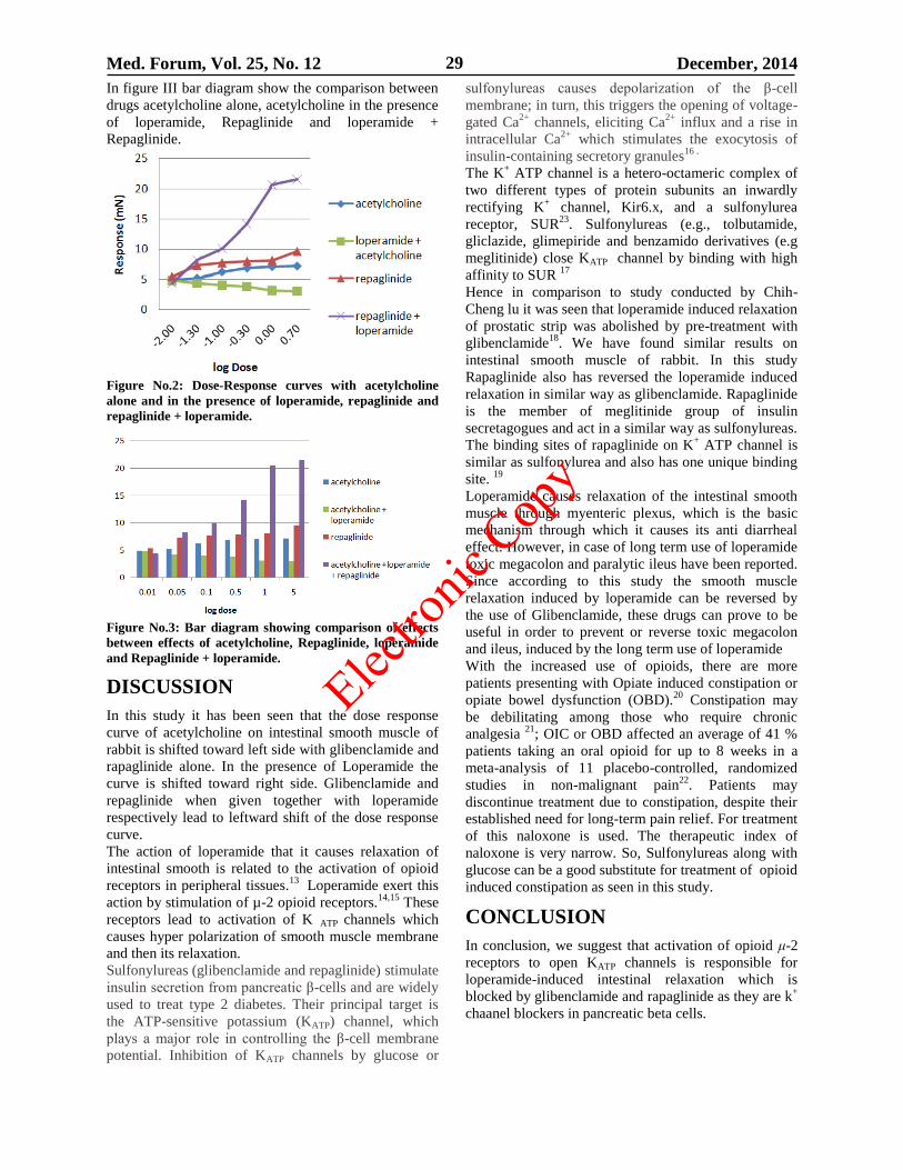
Med. Forum, Vol. 25, No. 12 December, 2014 29
In figure III bar diagram show the comparison between
drugs acetylcholine alone, acetylcholine in the presence
of loperamide, Repaglinide and loperamide +
Repaglinide.
Figure No.2: Dose-Response curves with acetylcholine
alone and in the presence of loperamide, repaglinide and
repaglinide + loperamide.
Figure No.3: Bar diagram showing comparison of effects
between effects of acetylcholine, Repaglinide, loperamide
and Repaglinide + loperamide.
DISCUSSION
In this study it has been seen that the dose response
curve of acetylcholine on intestinal smooth muscle of
rabbit is shifted toward left side with glibenclamide and
rapaglinide alone. In the presence of Loperamide the
curve is shifted toward right side. Glibenclamide and
repaglinide when given together with loperamide
respectively lead to leftward shift of the dose response
curve.
The action of loperamide that it causes relaxation of
intestinal smooth is related to the activation of opioid
receptors in peripheral tissues.13
Loperamide exert this
action by stimulation of µ-2 opioid receptors.14,15
These
receptors lead to activation of K ATP channels which
causes hyper polarization of smooth muscle membrane
and then its relaxation.
Sulfonylureas (glibenclamide and repaglinide) stimulate
insulin secretion from pancreatic β-cells and are widely
used to treat type 2 diabetes. Their principal target is
the ATP-sensitive potassium (KATP) channel, which
plays a major role in controlling the β-cell membrane
potential. Inhibition of KATP channels by glucose or
sulfonylureas causes depolarization of the β-cell
membrane; in turn, this triggers the opening of voltage-
gated Ca2+
channels, eliciting Ca2+
influx and a rise in
intracellular Ca2+
which stimulates the exocytosis of
insulin-containing secretory granules16 .
The K+ ATP channel is a hetero-octameric complex of
two different types of protein subunits an inwardly
rectifying K+ channel, Kir6.x, and a sulfonylurea
receptor, SUR23
. Sulfonylureas (e.g., tolbutamide,
gliclazide, glimepiride and benzamido derivatives (e.g
meglitinide) close KATP channel by binding with high
affinity to SUR 17
Hence in comparison to study conducted by Chih-
Cheng lu it was seen that loperamide induced relaxation
of prostatic strip was abolished by pre-treatment with
glibenclamide18
. We have found similar results on
intestinal smooth muscle of rabbit. In this study
Rapaglinide also has reversed the loperamide induced
relaxation in similar way as glibenclamide. Rapaglinide
is the member of meglitinide group of insulin
secretagogues and act in a similar way as sulfonylureas.
The binding sites of rapaglinide on K+ ATP channel is
similar as sulfonylurea and also has one unique binding
site. 19
Loperamide causes relaxation of the intestinal smooth
muscle through myenteric plexus, which is the basic
mechanism through which it causes its anti diarrheal
effect. However, in case of long term use of loperamide
toxic megacolon and paralytic ileus have been reported.
Since according to this study the smooth muscle
relaxation induced by loperamide can be reversed by
the use of Glibenclamide, these drugs can prove to be
useful in order to prevent or reverse toxic megacolon
and ileus, induced by the long term use of loperamide
With the increased use of opioids, there are more
patients presenting with Opiate induced constipation or
opiate bowel dysfunction (OBD).20
Constipation may
be debilitating among those who require chronic
analgesia 21
; OIC or OBD affected an average of 41 %
patients taking an oral opioid for up to 8 weeks in a
meta-analysis of 11 placebo-controlled, randomized
studies in non-malignant pain22
. Patients may
discontinue treatment due to constipation, despite their
established need for long-term pain relief. For treatment
of this naloxone is used. The therapeutic index of
naloxone is very narrow. So, Sulfonylureas along with
glucose can be a good substitute for treatment of opioid
induced constipation as seen in this study.
CONCLUSION
In conclusion, we suggest that activation of opioid μ-2
receptors to open KATP channels is responsible for
loperamide-induced intestinal relaxation which is
blocked by glibenclamide and rapaglinide as they are k+
chaanel blockers in pancreatic beta cells.
Electro
nic Cop
y

Med. Forum, Vol. 25, No. 12 December, 2014 30
REFERENCES
1. Camilleri M. Opioid-induced constipation:
challenges and therapeutic opportunities. Am J
Gastroenterol 2011;106:835–842; quiz 843.
2. Holzer P. Opioid receptors in the gastrointestinal
tract. Regul Pept 2009;155:11–17.
3. Hanauer SB. The role of loperamide in
gastrointestinal disorders. Rev Gastroenterol
Disord 2008;8:15–20.
4. Liu IM, Liou SS, Chen WC, Chen PF, Cheng JT.
Signals in the activation of opioid mu-receptors by
loperamide to enhance glucose uptake into cultured
C2C12 cells. Horm Metab Res 2004;36:210–214.
5. Cheng TC, Lu CC, Chung HH, Hsu CC, Kakizawa
N, Yamada S, et al. Activation of muscarinic M-1
cholinoceptors by curcumin to increase
contractility in urinary bladder isolated from
Wistar rats. Neurosci Lett 2010;473:107–109.
6. Choi S, Parajuli SP, Yeum CH, Park CG, Kim MY,
Kim YD, et al. Calcitonin gene-related peptide
suppresses pacemaker currents by nitric
oxide/cGMP-dependent activation of ATP-
sensitive K(+) channels in cultured interstitial cells
of Cajal from the mouse small intestine. Mol Cells
2008;26:181–185.
7. Mishra SK, Aaronson PI. A role for a
glibenclamide-sensitive, relatively ATP-insensitive
K+ current in regulating membrane potential and
current in rat aorta. Cardiovasc Res 1999;44:
429–435.
8. Menozzi A, Pozzoli C, Poli E, Passeri B, Gianelli
P, Bertini S. Diazoxide attenuates indomethacin-
induced small intestinal damage in the rat. Eur J
Pharmacol 2011;650:378–383.
9. Held B, Tsvilovskyy V, Meissner M, Kaestner L,
Ludwig A, Mossmang S, et al. Ca2+ channel
currents and contraction in CaVbeta3-deficient
ileum smooth muscle from mouse. Cell Calcium.
2007;42:477–487.
10. Aschcroft FM, Gribble FM: ATP sensitive K+
channels and insulin secretion: their role in health
and disease. Diabetologia 1999;42:903-919.
11. Moazedi AA, Mirzae DN, Seyyednejad SM,
Zadkarami MR, Amirzargar A. Spasmolytic effects
of petroselinum crispum (Parsley) on rat’s ileum at
different calcium chloride concentration. Pak J Biol
Sci 2007; 10(22): 4036-4042.
12. Rohra D.K, Siato S, Ohizumi Y. Functional role of
ryanodine-sensitive Ca2+ stores in acidic pH-
induced contraction in Wister Kyoto Rat aorta. Life
Sci 2003; 72: 1259-1269.
13. Manzoor AU, Gulshan AJ, et al. In vitro effects of
loperamide on spontaneously contracting and
primed ileum of Guinea pig. MC 2011;17(14):
16-21.
14. Nozaki-Taguchi N, Yaksh TL. Characterization of
the anti-hyperalgesic action of a novel peripheral
mu-opioid receptor agonist-loperamide.
Anesthesiol 1999;90:225-234.
15. Matsumoto K, Hatori Y, Murayama T, et al.
Involvement of mu-opioid receptors in
antinociception and inhibition of gastrointestinal
transit induced by 7-hydroxymitragynine, isolated
from Thai herbal medicine Mitragyna speciosa. Eur
J Pharmacol 2006;549:63-70.
16. Gribble FM, Ashcroft FM. Differential sensitivity
of β-cell and extrapancreatic KATP channels to
gliclazide. Diabetologia 1999;42:845–848.
17. Gribble FM, Tucker SJ, Seino S, Ashcroft FM:
Tissue specificity of sulphonylureas: studies on
cloned cardiac and beta-cell KATP channels.
Diabetes 1998;47:1412-1418.
18. Chih-cheng LU, Hsien-hui chung, Juei-Tang
Cheng. Prostatic relaxation induced by loperamide
is mediated through activation of opioid μ-2
receptors in vitro. Exp Ther Med 2011;2(2):
281–285.
19. Bertram G. Katzung. Basic & Clinical
Pharmacology. Pancreatic hormones and
Antidiabetic drugs.12th
ed. 2012.p.756.
20. Kalso E, Edwards JE, Moore A, et al. Opioids in
chronic non-cancer pain: systemic review of
efficacy and safety. Pain 2004;112:372-80.
21. Benyamin R, Trescot AM, Datta S et al. Opioid
complications and side effects. Pain Physician
2008;11(2 Suppl) S105-20.
22. Sykes NP. An investigation of the ability of oral
naloxone to correct opioid related constipation in
patients with advanced cancer. Palliat Med 1996;
10:135-44.
Address for Corresponding Author:
Dr. Javaria Arshad
Asstt. Prof. of Pharmacology & Therapeutics.
Yusra Medical and Dental College,
Islamabad
Electro
nic Cop
y

Med. Forum, Vol. 25, No. 12 December, 2014 31
Experimental Study of Antimony
Induced Hepato Toxicity in Rabbits 1. Khawaja Usman Masud 2. A.H. Nagi
1. Prof. of Histopathology, Bolan Medical College, Quetta, 2. Prof. of Pathology, UHS, Lahore.
ABSTRACT
Objectives: To demonstrate the effects of antimony on hepatic tissues of Rabbits. To correlate the severity of tissue
damage to the dose of antimony and to an immunologically altered state of the animal.
Study Design: Experimental study.
Place and Duration of Study: This study was carried out at PGMI and KEMU, Lahore from January 1988 to
March 1988.
Materials and Methods: This study was carried out on 40 healthy rabbits weighing 1.5kg divided into 4 groups
each group having 10 animals with one control group. Group I animals were injected with antimony sodium
tartrate of ½ MLD 6mg/kg body weight I/V at interval of 2 days for 12 weeks, whereas experimental dose of
1.71mg/kg body weight was injected I/V at interval of 4 days to group II animals. Those of group III were injected
2ml of specific bovine albumin 30%(DADEUSA) followed by schedule of group II animals. Group IV (control
group) animals were injected I/V with distilled water.
Results: Hepatic Enzymes, Serum Alanine Aminotransferase and Serum Gamma Glutamyl transpeptidasc (GT)
levels were estimated at the end of six weeks and twelve weeks. These were found to be raised gradually from 7th
week onwards until the experiment was terminated.
Conclusion: It is concluded from this study that antimony sodium tartrate has toxic effects on liver tissue and can
cause hepatocellular damage (if given for prolong period)
Key Words: Antimony, Hepato Toxicity, Hepatic enzymes, S-ALAT, ɣ GT, H&E, PAS, MSS.
INTRODUCTION
Antimony and its compounds were used medicinally as
early as 4000 B.C. Antimony is used in Alloys, lead,
tin, and copper. Its compounds are used in textile
industry, flame proofing, dies, paints, rubber
compounding, ceramic and glass opicifiers 1
. Human
beings have always been exposed to antimony but its
amount is substantially increased due to industrial
production 2
. As regards the therapeutic applications in
the 19th
century, potassium antimony Tartrate (Tartar
emetic) began to be used for the treatment of
shistosomiasis and leishmaniasis 3. The heavy metals
have their toxic effects on liver kidneys central nervous
system, skin muscles, bone etc. Keeping in mind a
wide industrial use of the metals, we have opted to
study the effects of antimony on liver tissue.
MATERIALS AND METHODS
This study was carried out on 40 healthy rabbits
weighing 1.5kg divided into 4 groups each group
having 10 animals with one control group.
Group I animals were injected with antimony sodium
tartrate of ½ MLD (minimum lethal dose) 6mg/kg body
weight I/V at interval of 2 days for 12 weeks.
Group II where as experimental dose of 1.71mg/kg
body weight was injected I/V at interval of 4 days to
group II animals.
Group III were injected 2ml of specific bovine
albumin 30%(DADEUSA) followed by schedule of
group II animals.
Group IV (control group) animals were injected I/V
with distilled water.
Blood chemistry was done to see the effects of
antimony on liver functions of animals of all groups, to
determine the (S-ALAT) activity sclavo diagnostic
G.P.T kit was used by calorimetric method and Serum
ɣ GT (Gamma Glutamyl transpeptidase) using kinetic
colorimetric method. Blood samples were collected by
using disposable syringes from one of peripheral ear
veins at the end of six and twelve weeks. Five ml of
blood was collected in a test tube, kept for 2 hours to
clot and serum was separated after centrifuging the test
tubes at 3000 RPM.
Tissue Histology of liver was done at the end of
experiment after sacrificing all the animals. Liver
tissues were preserved in 10% formalin saline solution,
to study the different morphological lesions.
Following staining procedures were carried out:-
1. Haemotoxylin and eosin staining (H&E)
2. Periodic Acid Schiff staining (PAS)
3. Methenamine sliver staining (MSS)
4. Reticulin staining (RS)
RESULTS
Laboratory Results: Liver Chemistry:-. Serum
Alanine Aminotranferase (S-ALAT) levels were
estimated in group I using the colorimetric method
(Reithman-Frankel, 1959, 1970) (modified) at the end
of six weeks, and their mean value was found to be
12.3+4.1 IU/L. Their levels rose gradually from 7th
week onwards until the experiment was terminated and
Original Article Antimony Induced
Hepato Toxicity
Electro
nic Cop
y

Med. Forum, Vol. 25, No. 12 December, 2014 32
their mean value was found to be 25.5+9.09IU/L at the
end of experiment (Table 1).
Serum Gamma Glutamyl Transpeptidase (ɣ GT IU/L)
levels were estimated using the Kinetic Colorimetric
method. The mean value of the ɣ GT was 4.6 + 1.91
IU/L at the end of six weeks which rose gradually from
7th
week onwards until the experiment was terminated
and their mean value was found to be 9.3 + 4.22 IU/L at
the end of 12th
week (Table 2).
In animal group II and group III, there was gradual
increase seen in the levels of S-ALAT and ɣ GT
(Gamma Glutamyl Transpeptidasc) after six weeks till
the end of experiment (Tables 3, 4, 5, 6).
Table No.1: Serum Alanine Aminotransferase
(S-Alat) IU/L after IV Dose of Antimony Sodium
Tartrate I in Ten Rabibts of Group I. Duration 12
weeks. Animal
number Weight of
Rabbit K(kg) End of 6th week End of 12th week
IU/L IU/L
1. 1.5 18 40
2 1.5 13 24
3 1.3 12 20
4 1.5 16 38
5 1.5 7 15
6 1.5 4 10
7 1.5 11 20
8 1.0 15 30
9 1.5 11 28
10 1.5 16 30
Mean X 12.3 25.5
Standard deviation (S.D) 4.1 9.09
Table No.2: Serum Gamma Glutamyl
Transpeptidase (ɣ-Gt) IU/L after IV Dose of
Antimonmy Sodium Tartrate in Ten Rabbits of
Group 1. Duration 12 weeks. Animal
Number
Weight of
Rabbit (kg)
End of 6th
Week
End of 12
week.
IU/L IU/L
1. 1.5 4 7
2. 1.5 7 15
3 1.3 1 2
4 1.5 2 4
5 1.5 4 10
6. 1.5 7 15
7. 1.5 6 12
8. 1.5 5 10
9. 1.5 6 12
10. 1.5 4 6. Mean X 4.6 9.3
Standard Deviation (S.D) 1.91 4.22
In group IV (Control group) animals, S-ALAT and
ɣ GT levels were estimated. The S-ALAT was 20.0, +
6.32 IU/L and ɣ -GT was -3.16 + 0.90 at the start,
which remained the same at the end of the experiment.
(Table 7 & 8)
Histological and Microscopic Study:
Group I Gross Examination: Livers of all the animals appeared
normal in size, shape and color, except two animals in
which surface of liver shows slight nodularity. They
were triangular in shape and were covered with thin
capsule.
Microscopic Examination: Eight animals revealed
normal Lobular architecture. Three animals showed
mild dilatation of central veins and sinusoids but no
significant congestion observed.
Microscopic examination of livers revealed severe
degree of lymphocytes infiltration around portal tracts
which were widened . Single cell necrosis along with
focal fatty change.
A moderate amount of fibrosis around the portal tracts
extending to the adjacent portal tracts.
Group II
Gross Examination: Most of the animals revealed
congested spots diffusely scattered on surface of liver.
Microscopic Examination: Liver showed mild to
moderate degree of congestion of central veins and
sinusoids.
Mild to moderate degree of lymphocytic infiltration,
singe cell necrosis seen in six animals. Few animals
revealed fibrosis around the portal tracts..
Group III
Gross Examination: Livers were rectangular in shape
covered with thin capsule.
Microscopic Examination: Revealed maintained
lobular architecture with mild dilatation, congestion of
central vein and sinusoids. Moderate degree of focal
lymphocytic infiltration in portal tracts in few animals .
Single cell necrosis , and focal fatty changes seen in
few animals..
Table No.3: Serum Alanine Aminotransferase
(S-Alat) IU/L after IV Dose of Antimony Sodium
Tartrate I in Ten Rabibts of Group II. Duration 12
weeks. Animal
Number
Weight of
Rabbit (kg)
End of 6th
Week
End of 12
week.
IU/L IU/L
1. 1.5 15 30
2. 1.5 12 15
3 1.3 6 16
4 1.5 5 26
5 1.5 16 32
6. 1.5 15 30
7. 1.5 8 36
8. 1.0 10 28
9. 1.5 10 24
10. 1.0 6 15 Mean X 10.3 25.2 Standard Deviation (S.D) 3.87 7.15
Group IV (Control Group)
Gross Examination: The examination of the livers of
all the animals normal in size, shape and color; covered
Electro
nic Cop
y
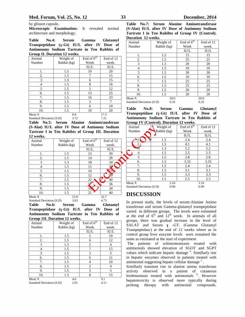
Med. Forum, Vol. 25, No. 12 December, 2014 33
by glisson capsule.
Microscopic Examination: It revealed normal
architecture and morphology.
Table No.4: Serum Gamma Glutamyl
Transpeptidase (ɣ-Gt) IU/L after IV Dose of
Antimonmy Sodium Tartrate in Ten Rabbits of
Group II. Duration 12 weeks. Animal
Number
Weight of
Rabbit (kg)
End of 6th
Week
End of 12
week.
IU/L IU/L
1. 1.5 10 20
2. 1.5 7 15
3 1.3 6 15
4 1.5 8 16
5 1.5 5 12
6. 1.5 13 25
7. 1.5 16 33
8. 1.5 3 7
9. 1.5 6 10
10. 1.5 10 20 Mean X 8.4 17.3
Standard Deviation (S.D) 3.72 7.21
Table No.5: Serum Alanine Aminotransferase
(S-Alat) IU/L after IV Dose of Antimony Sodium
Tartrate I in Ten Rabibts of Group III. Duration
12 weeks. Animal
Number
Weight of
Rabbit (kg)
End of 6th
Week
End of 12
week.
IU/L IU/L
1. 1.5 13 30
2. 1.5 14 28
3 1.3 18 34
4 1.5 15 30
5 1.5 10 25
6. 1.5 12 30
7. 1.5 22 40
8. 1.5 17 36
9. 1.5 16 34
10. 1.5 21 40 Mean X 15.8 32.7
Standard Deviation (S.D) 3.63 4.73
Table No.6: Serum Gamma Glutamyl
Transpeptidase (ɣ-Gt) IU/L after IV Dose of
Antimonmy Sodium Tartrate in Ten Rabbits of
Group 1II. Duration 12 weeks. Animal
Number
Weight of
Rabbit (kg)
End of 6th
Week
End of 12
week.
IU/L IU/L
1. 1.5 5 10
2. 1.5 6 12
3 1.3 2 4
4 1.5 7 15
5 1.5 3 5
6. 1.5 6 11
7. 1.5 4 10
8. 1.5 2 4
9. 1.5 3 5
10. 1.5 8 15 Mean X 4.6 9.1
Standard Deviation (S.D) 2.01 4.11
Table No.7: Serum Alanine Aminotransferase
(S-Alat) IU/L after IV Dose of Antimony Sodium
Tartrate I in Ten Rabibts of Group IV (Control).
Duration 12 weeks. Animal
Number
Weight of
Rabbit (kg)
End of 6th
Week
End of 12
week.
IU/L IU/L
1. 1.5 15 15
2. 1.5 25 25
3 1.3 20 20
4 1.5 10 10
5 1.5 30 30
6. 1.5 10 10
7. 1.5 25 25
8. 1.5 25 25
9. 1.5 20 20
10. 1.5 20 20 Mean X 20.0 20.0 Standard Deviation (S.D) 6.32 6.32
Table No.8: Serum Gamma Glutamyl
Transpeptidase (ɣ-Gt) IU/L after IV Dose of
Antimonmy Sodium Tartrate in Ten Rabbits of
Group 1V (Control). Duration 12 weeks. Animal
Number
Weight of
Rabbit (kg)
End of 6th
Week
End of 12
week.
IU/L IU/L
1. 1.5 2.4 2.4
2. 1.5 4.1 4.1
3 1.3 5.2 5.2
4 1.5 2.5 2.5
5 1.5 2.8 2.8
6. 1.5 3.33 3.33
7. 1.5 2.4 2.4
8. 1.5 3.1 3.1
9. 1.5 3.3 3.3
10. 1.5 2.5 2.5 Mean X 3.16 3.16 Standard Deviation (S.D) 0.90 0.90
DISCUSSION
In present study, the levels of serum-Alanine Amino
transferase and serum Gamma-glutamyl transpeptidase
varied in different groups. The levels were estimated
at the end of 6th
and 12th
week. In animals of all
groups, there was gradual increase in the level of
SALAT and Serum ɣ -GT. (Gamma Glutamyl
Transpeptidasc) at the end of 12 weeks where as in
control group liver enzyme levels were remained the
same as estimated at the start of experiment
The patients of schistomomiasis treated with
antimonials showed elevation of SGOT and SGPT
values which indicate hepatic damage 4. Simillarly rise
in hepatic enzymes observed in patients treated with
antimonial suggesting hepato cellular damage 5 .
Simillarly transient rise in alanine amino transferase
activity observed in a patient of cutaneous
leishmaniasis treated with antimonials 6. However
hepatotoxicity is observed more typically during
prolong therapy with antimonial compounds.
Electro
nic Cop
y
Med. Forum, Vol. 25, No. 12 December, 2014 34
Parenteral treatment with antimony compounds has
caused hepatic necrosis although reversible elevation of
liver enzymes activities are more typical 7.
The above mentioned studies coincides with the
observations made in the present study.
As regards morphological appearance of liver tissue,
trivalent antimony compounds were found in
abnormally high concentration in liver and thyroid 8.
Where as slight to moderate parenchymatous
degeneration of liver was observed by workers 9.
In the present study, most of animals from various
groups revealed a maintained lobular architecture
except few animals showed disorganized architecture.
Lymphocytic infiltration of moderate (++) to severe
degree (+++) was common feature in most of the
animals of various groups. A fatty change was observed
in 30% of the animals from group I and III where as no
change was seen in group II animals.
Single cell necrosis was observed in most of animals of
all groups. A few animals of group I and II, revealed a
moderate amount of fibrosis around the portal tracts.
Fatty degeneration of liver reported in chronically
exposed guinea pigs to antimonial compounds 10
.
Similar fatty degeneration was observed in another
experimental study 11
. These observations coincides
with the findings seen in the present study.
Centrilobular necrosis was reported in occupationally
exposed workers to antimony 12
which is not observed
in the present study. On the other hand a moderate
amount of fibrosis around the portal tract, a
morphological change that has not been demonstrated
in the literature.
CONCLUSION
It is concluded that the morphological lesions in the
liver appear to be dose and time dependent. As regards
biochemical evidence hepatic injury, serum enzyme
(S-ALAT, ɣ - GT. (Gamma Glutamyl Transpeptidasc),
levels were found to be raised significantly.
REFERENCES
1. Koutso PL, Maravelias CF, Koutselinis AJ.
Immunological aspects of the common heavy
metals, amaranth and tartrazine. Hum Toxicol
1998;40:11-24.
2. Reotjman S, Frankel S. Hypthothalamic lesions
after antimony injection in newly born primates.
Science 2001;272:1702-1708.
3. Dickerson OB. Occupational Medicine priniciples
and practical applications, published by year book
Medical 1975;29: 613-614.
4. Blacow NW. Martindale, the extra pharmacopocia.
26th
ed. The pharmaceutical press; 1973.
5. Sampaio RN, Marsden PD. Pentavalent Antimonial
treatment in mucosal Leishmaniasis. The Lancet
1985;2:1097.
6. Herwadlt BL, Kaye ET, Lepore TJ, Berman JD,
Baden HP. Sodium stipogluconate (Pentostam)
over dose during treatment of American
cutanenous leishmainasis. J Inflit Dis 1992;165:
968-71.
7. Winship KA, toxicity of antimnony an its
coumpunds. Adverse drug react acute poisning Rev
1987;2:67-90.
8. Webster LT. The pharmacological Basis of
Therapeutics. 7th
ed. 1985.p.1028-1030.
9. Brieger H, Semisch CW, Stasney J. Industrial
Antimony poisoning. Ind Med Surg 1954;23:521-
523, Cited by Carlzenz (1975). In Occupational
medicine Principles and Practical applications. year
Book Medical Publisher.p.636-644.
10. Stokinger HE. The Metals in Patty Industrial
Hygiene and Toxicology. 2nd
ed. New York: John
Wiley;1963.p.1506-1517.
11. Dernehl CU, Nau GA. Sweets HH. Animal
studies on the toxicity of inhaled Antimony
trioxide, J Ind Hyg Toxicol 1945. Cited by
Carlzenz. Occupational Medicine Principles and
practical applications. 1975;29:636-640.
12. Harrington JM, Gill FS. Occupational Health, 2nd
ed. 1987.p.89.
Address for Corresponding Author:
Prof. Dr. Khawaja Usman Masud,
Doctors Lab, 3-7/2, Faiz Mohammad Road,
Quetta.
Tel: 0812843189. Mob: 03009383100.
Email: [email protected]
Address for Corresponding Author:
Khawaja Usman Masud,
Prof. of Histopathology, Bolan Medical College,
Quetta
Electro
nic Cop
y
Med. Forum, Vol. 25, No. 12 December, 2014 35
Major Determinants of Anemia in
Pregnant Women Residing in the Urban
Slums of Taluka Qasimabad, District Hyderabad 1. Rehana Siddiqui 2. Muhammad Muqeem Mangi 3. Azhar Ali Shah 4. Rafique Ahmed
Soomro 5. Khalida Naz Memon 1. Senior Lecturer of Community Medicine, GMMMC, Sukkur 2. Asstt. Prof. of Physiology, GMMMC, Sukkur
3. Asstt. Prof. of Surgery, GMMMC, Sukkur 4. Prof. of Community Medicine, LNMC Karachi, 5. Asstt. Prof. of
Community Medicine, LUMHS, Jamshoro
ABSTRACT
Objective: To find out major determinants of anemia in pregnant women residing in the urban slums of Taluka
Qasimabad, district Hyderabad.
Study Design: Cross-sectional descriptive study.
Place and Duration of Study: This study was conducted in urban slum areas of Taluka Qasimabad, District
Hyderabad during six months of study period i.e. from 1st March 2011 to 31
st August 2011.
Patients and Methods: The total population residing in the study areas was twelve thousand two hundred and
seven. During the study period of six months, two hundred and fifty pregnant women were enrolled for the study.
Pregnant women during 2nd
and 3rd
trimester of pregnancy were included in the study. The data was collected by
conducting interviews, filling of the pre-tested, structured questionnaire and by assessing anemia by determining the
hemoglobin level in the enrolled pregnant women. The questionnaire was a close-ended one, filled by the principle
researcher herself. It comprised of demographic information about woman. Every woman’s hemoglobin was
determined by using Sahli’s Hemoglobinometer. Anemia in pregnancy according to WHO classified into mild
anemia hemoglobin level in the range of 10.0-10.9 g/dl, moderate anemia hemoglobin level in the range of 7-9.9
g/dl and severe anemia hemoglobin level is <7 g/dl.
Results: The association of various factors (determinants) with anemia was analyzed by applying chi-squared test;
the p-value of <0.05 was taken as the level of significance. Two hundred and thirty three pregnant women were
anemic while only seventeen women (6.8%) were found non-anemic. Majority of the women i.e. 70% presented
with moderate anemia (hemoglobin level 7.0-9.9Gm /dl) while severe anemia (hemoglobin level <7 Gm/dl) was
recorded in 5.2% pregnant women. There was strong statistically significant association seen between parity of
pregnant women and the degree/severity of anemia (p=0.00). There was strong association between socio-economic
status and the severity of anemia (p=0.00). The family type was strongly associated with the severity of anemia
(p=0.01).
Conclusion: Prevalence and severity of anemia in pregnant women residing in urban slum areas of Taluka
Qasimabad, District Hyderabad is high. Current findings highlight the anemia in pregnancy as a priority area of
concern.
Key Words: Anemia, Pregnant women, Urban slums.
INTRODUCTION
Iron deficiency anemia is the most prevalent form of
malnutrition, affecting around 50% of pregnant women
world wide and the eighth leading cause of disease in
girls and women in developing countries.1
The most
common cause of anemia in pregnancy is due to iron
deficiency, reason for anemia in pregnancy would be
lack of nutritious diet.2
Anemia in pregnancy is defined
by the World Health Organization WHO as a
hemoglobin concentration below 11 g /dl. It is most
common cause of anemia in pregnancy worldwide is
iron deficiency. The predisposing factors for it include
grand parity, low socioeconomic status, and inadequate
birth spacing.3
It is estimated that 1,200 million people
are anemic globally. The prevalence of anemia depends
upon socioeconomic status, life style, parity, associated
medical problems and regular antenatal care. Maternal
anemia in pregnancy is commonly considered as risk
factor for poor pregnancy out comes and can threat the
life of mother as well as fetus.4
Risk is also increased
with parity nearly 3-fold higher for women with 2-3
children and nearly 4-fold greater for women with four
or more children, thus implicating pregnancy.5 Iron
deficiency anemia in pregnancy is highly prevalent in
lower southern Thailand where poverty and low levels
of education prevail.6
Iron deficiency is characterized
by deficient hemoglobin level synthesis caused by lack
of iron. 7Anemia is now one of the most frequently
observed nutritional disease in the world due to
inadequate intake8 and malnutrition.
9 In our society
girls are lacking access to balanced diet, adequate
health care and proper education particularly pregnancy
with iron and folate deficiency due to socially
Original Article Anemia in
Pregnant Women
Electro
nic Cop
y
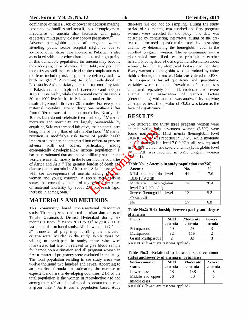
Med. Forum, Vol. 25, No. 12 December, 2014 36
dominance of males, lack of power of decision making,
ignorance by families and herself, lack of employment.
Prevalence of anemia also increases with parity
especially multi parity, closely spaced pregnancy.10
Adverse hemoglobin status of pregnant women
attending public sector hospital might be due to
socioeconomic status, less income in Pakistan is also
associated with poor educational status and high parity.
In this vulnerable population, the anemia may become
the underlying cause of maternal mortality and perinatal
mortality as well as it can lead to the complications to
the fetus including risk of premature delivery and low
birth weights.11
According to safe motherhood in
Pakistan by Sadiqua Jafary, the maternal mortality ratio
in Pakistan remains high in between 350 and 500 per
100,000 live births, while the neonatal mortality ratio is
50 per 1000 live births. In Pakistan a mother dies as a
result of giving birth every 20 minutes. For every one
maternal mortality, around thirty one mothers suffer
from different rates of maternal morbidity. Nearly 1 in
10 new born do not celebrate their birth day.12
Maternal
mortality and morbidity are largely preventable by
acquiring Safe motherhood initiative, the antenatal care
being one of the pillars of safe motherhood.13
Maternal
nutrition is modifiable risk factor of public health
importance that can be integrated into efforts to prevent
adverse birth out comes, particularly among
economically developing/low income population.14
It
has been estimated that around two billion people in the
world are anemic, mostly in the lower income countries
of Africa and Asia.15
The greatest burden of death and
disease due to anemia in Africa and Asia is associated
with the consequences of anemia among pregnant
women and young children. A recent meta-analysis
shows that correcting anemia of any severity decreases
of maternal mortality by about 20% for each 1g/dl
increase in hemoglobin.16
MATERIALS AND METHODS
This community based cross-sectional descriptive
study. The study was conducted in urban slum areas of
Taluka Qasimabad, District Hyderabad during six
months ie from 1st March 2011 to 31
st August 2011. It
was a population based study. All the women in 2nd
and
3rd
trimester of pregnancy fulfilling the inclusion
criteria were included in the study. While those not
willing to participate in study, those who were
interviewed but later on refused to give blood sample
for hemoglobin estimation and all pregnant women in
first trimester of pregnancy were excluded in the study.
The total population residing in the study areas was
twelve thousand two hundred and seven. According to
an empirical formula for estimating the number of
expectant mothers in developing countries, 24% of the
total population is the women in reproductive age and
among them 4% are the estimated expectant mothers at
a given time.17
As it was a population based study
therefore we did not do sampling. During the study
period of six months, two hundred and fifty pregnant
women were enrolled for the study. The data was
collected by conducting interviews, filling of the pre-
tested, structured questionnaire and by assessing
anemia by determining the hemoglobin level in the
enrolled pregnant women. The questionnaire was a
close-ended one, filled by the principle researcher
herself. It comprised of demographic information about
woman, her family, obstetrical history and her diet.
Every woman’s hemoglobin was determined by using
Sahli’s Hemoglobinometer. Data was entered in SPSS-
16. Frequencies for all qualitative and quantitative
variables were computed. Prevalence of anemia was
calculated separately for mild, moderate and severe
anemia. The association of various factors
(determinants) with anemia was analyzed by applying
chi-squared test; the p-value of <0.05 was taken as the
level of significance.
RESULTS
Two hundred and thirty three pregnant women were
anemic while only seventeen women (6.8%) were
found non-anemic. Mild anemia (hemoglobin level
10.0-10.9 g/dl) was reported in 17.6%, while moderate
anemia (hemoglobin level 7.0-9.9Gm /dl) was reported
in 70.45% women and severe anemia (hemoglobin level
<7 Gm/dl) was recorded in 5.2% pregnant women
(Table 1).
Table No.1: Anemia in study population (n=250)
Anemia No. %
Mild (hemoglobin level
10.0-10.9 g/dl)
44 17.6
Moderate (hemoglobin
level 7.0-9.9Gm /dl)
176 70.4
Severe (hemoglobin level
<7 Gm/dl)
13 5.2
No 17 6.8
Table No.2: Relationship between parity and degree
of anemia
Parity Mild
anemia
Moderate
anemia
Severe
anemia
Primiparous 10 29 3
Multiparous 32 115 2
Grand Multiparous 2 32 8
p = 0.00 (Chi-square test was applied)
Table No.3: Relationship between socio-economic
status and severity of anemia in pregnancy
Socioeconomic
status
Mild
anemia
Moderate
anemia
Severe
anemia
Lower class 18 138 8
Middle and upper
middle class
26 38 5
p = 0.00 (Chi-square test was applied)
Electro
nic Cop
y
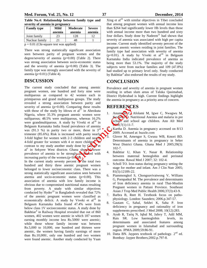
Med. Forum, Vol. 25, No. 12 December, 2014 37
Table No.4: Relationship between family type and
severity of anemia in pregnancy
Family type Mild
anemia
Moderate
anemia
Severe
anemia
Joint family 23 128 12
Nuclear family 21 48 1
p = 0.01 (Chi-square test was applied)
There was strong statistically significant association
seen between parity of pregnant women and the
degree/severity of anemia (p=0.00) (Table 2). There
was strong association between socio-economic status
and the severity of anemia (p=0.00) (Table 3). The
family type was strongly associated with the severity of
anemia (p=0.01) (Table 4).
DISCUSSION
The current study concluded that among anemic
pregnant women, one hundred and forty nine were
multiparous as compared to 42 women each in
primiparous and grand multiparous groups. The study
revealed a strong association between parity and
severity of anemia (p=0.00). Comparing these results
with those of the study by Idowu et al17
in Abeokuta
Nigeria, where 35.3% pregnant anemic women were
nulliparous; 48.5% were multiparous, whereas 16.2%
were grandmultiparous. A study by Viveki et al18
in
Belgaum Karnataka India found prevalence of anemia
was (91.3 %) in parity two or more, those in 3rd
trimester (85.6%). Risk is increased with parity nearly
3-fold higher for women with 2-3 children and nearly
4-fold greater for women with 4 or more children.5 In
contrast to my study another study done by Grover et
al3 in Sekyere West districts Ghana showed lower
prevalence of anemia to be strongly associated with
increasing parity of the women.(p<0.003).
In the current study seventy percent of the total two
hundred and thirty three anemic pregnant women
belonged to lower socioeconomic class. There was a
strong statistically significant association seen between
anemia and socio-economic status (p=0.00). This
association of anemia with low family income is
obvious due to compromised nutritional status resulting
from poverty. A study with similar objectives
conducted by Hyder19
in Bangladesh revealed that 72%
of the anemic pregnant women were classified as
economically deficit. A study by Viveki et al18
in
Belgaum Karnataka India found 47.4% were from
below class 1V socioeconomic status. A study done by
Bakhtiar4 in Railway Hospital revealed that out of 860
women, 402 women were anemic in which 187 women
earning monthly income less Rs.5000/ were anemic;
while those whose family income was between
Rs.5,000 to 10,000, one hundred and thirteen were
anemic, the women having family earnings of more
than Rs.10,000/, only one hundred and two women
were found anemic. Another study conducted by Yuan
Xing et al20
with similar objectives in Tibet concluded
that among pregnant women with annual income less
than $264 had significantly lower Hb levels than those
with annual income more than two hundred and sixty
four dollars. Study done by Nadeem
21 had shown that
severity of anemia was associated with high per capita
income. Current study identified seventy percent of the
pregnant anemic women residing in joint families. The
family type had association with severity of anemia
(p=0.01). A study by Viveki et al18
in Belgaum
Karnataka India indicated prevalence of anemia as
being more than 53.1%. The majority of the study
subjects were from nuclear families and 109 (47.8%)
had studied up to primary level only. Study conducted
by Bakhtiar4 also endorsed the results of my study.
CONCLUSION
Prevalence and severity of anemia in pregnant women
residing in urban slum areas of Taluka Qasimabad,
District Hyderabad is high. Current findings highlight
the anemia in pregnancy as a priority area of concern.
REFERENCES
1. Anumudu C, Afolami M, Igwe C, Nwagwu M,
Keshinro O. Nutritional Anemia and malaria in pre
school and school age children. Ann Afr Med
2008;7(1):11-7.
2. Kartha D. Anemia in pregnancy accessed on 8/13
2009. Accessed at: buzzle.com
3. Glover M, Amengor S, Gwusu WB, Kmori BD.
Determinants of anemia in pregnancy in Seker
West District Ghana. Ghana Med J 2005;39(3):
102-7.
4. Bakhtiar U, Khan Y, Nasar R. Relationship
between maternal hemoglobin and perinatal
outcome. Rawal Med J 2007; 32: 102-4.
5. Scholl TO. Iron status during pregnancy setting the
stage for mother and infant. Am J Clin Nutr 2005;
81(5):1218S-22.
6. Piammongkol S, Chongsuvivatwong V, Willians
G, Pornpatkul M. The prevalence and determinants
of Iron deficiency anemia in rural Thai-Muslim
Pregnant women in Pattani Province. Southeast
Asian J Trop Med Public Health 2006;37(3):43-9.
7. Barbra B, Reet H. Textbook focus on patho-
physiology. London: Saunders; 2006.p.347-57.
8. Gautam C, Sahal, Sekhri K, Saha P. Iron
deficiency in pregnancy and rationality of iron
supplements prescribed. J Med 2008; 10(12):283.
9. Ayub R, Tariq N, Iqbal M, Jafery T, Adil MM,
Rais SR. Low haemoglobin levels, its
determinants and associated features among
pregnant women in Islamabad and surrounding
region. JPMA 2009;59:86-91.
10. Datta BN. Jaypees textbook of pathology. 2nd
ed.
Bombay: Jaypee Brothers;2002.p.797-8.
Electro
nic Cop
y
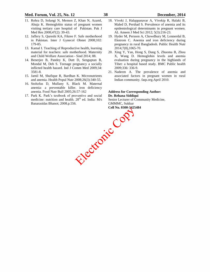
Med. Forum, Vol. 25, No. 12 December, 2014 38
11. Rohra D, Solangi N, Memon Z, Khan N, AzamI,
Ahuja K. Hemoglobin status of pregnant women
visiting tertiary care hospital of Pakistan. Pak J
Med Res 2008;47(2): 39-43.
12. Jaffery S, Qureshi KA, Fikree F. Safe motherhood
in Pakistan. Inter J Gynecol Obstet 2008;102:
179-85.
13. Kamal I. Teaching of Reproductive health, learning
material for teachers: safe motherhood. Maternity
and Child Welfare Association - Sind 2014; 88.
14. Benerjee B, Pandey K, Dutt D, Senguptan B,
Mondal M, Deb S. Teenage pregnancy a socially
inflicted health hazard. Ind J Comm Med 2009;34:
3581-8.
15. Jamil M, Shafique R, Bardhan K. Micronutrients
and anemia. Health Popul Nutr 2008;26(3):340-55.
16. Stoltzfus D, Mullany S, Black M. Maternal
anemia: a preventable killer. iron deficiency
anemia. Food Nutr Bull 2005;26:57-162
17. Park K. Park’s textbook of preventive and social
medicine: nutrition and health. 28th
ed. India: M/s
Banarasidas Bhanot; 2008.p.556.
18. Viveki J, Halappanavar A, Vivekip R, Halaki B,
Maled D, Pershad S. Prevalence of anemia and its
epidemiological determinants in pregnant women.
AL Ameen J Med Sci 2012; 5(3):216-23.
19. Hyder M, Persson A, Chowdhury M, Lonnerdal B,
Ekstrom C. Anemia and iron deficiency during
pregnancy in rural Bangladesh. Public Health Nutr
2014;7(8),1065-70.
20. Xing Y, Yan, Hong S, Dang S, Zhuoma B, Zhou
X, Wang D. Hemoglobin levels and anemia
evaluation during pregnancy in the highlands of
Tibet: a hospital based study. BMC Public health
2009;336: 336-9.
21. Nadeem A. The prevalence of anemia and
associated factors in pregnant women in rural
Indian community. faqs.org.April 2010.
Address for Corresponding Author:
Dr. Rehana Siddiqui
Senior Lecturer of Community Medicine,
GMMMC, Sukkur
Cell No. 0300-3425484
Electro
nic Cop
y
Med. Forum, Vol. 25, No. 12 December, 2014 39
Frequency of Surgical
Intervention Due to Cord Around the Neck 1. Shazia Shaikh 2. Fozia Shaikh 3. Shazia Jatoi
1. Asstt. Prof., Deptt. of Gynae & Obst., 2. Senior Registrar, Deptt. of Gynae & Obst.3. Consultant Gynaecologist,
Deptt. of Gynae & Obst., Shaikh Zaid Women Hospital, CMC & SMBBMU Larkana
ABSTRACT
Objective: To see the frequency of cord around the neck.
Study Design: Retrospective Observational Study.
Place & Duration of Study: This study was carried out at Shaikh Zaid Women Hospital, CMCH, SMBBMU,
Larkana from January 2012 to December 2013.
Materials And Methods: Total patient 8250 taken from Jan 2012 to Dec 2013. All cases were studied in detail with
reference to course of labour, mode of delivery, interference required and maternal and fetal outcome. A detailed
history was taken and general and systemic examination was done. The muller Munro Kerr maneuver was used to
assess the adequacy of the pelvis and diagonal conjugate was accurately measured.
Results: Total patient taken 8250, from Jan 2012 to Dec 2013 among them vaginally delivered 4238(51.3%), 92
have applied forcep 27(0.63%) babies delivered cord around the neck with 30 patient have applied vacuum out of
which 16 (0.37%) cord around the neck. Spontaneously vaginal deliveries without surgical intervention 4116
(31.9%), out of which 1312 (15.9%) have cord around the neck. 2360 (28.6%) patient delivered through emergency
LSCS. 722(17%) babies with cord around the neck and 1560 (18.9%) patient delivered through elective LSCS
among them 759 (17.9%) babies delivered with cord around the neck.
Conclusions: Most of these cases delivered vaginally with minimal maternal and fetal morbidity. Frequency of
surgical intervention in these cases can be reduced by proper antenatal care especially in 3rd
trimester, by plotting
partogram and using oxytocin judiciously during intrapartum period.
Key Words: Surgical intervention, cord around the neck and feto-maternal outcome
INTRODUCTION
Nuchal cord is defined as an umbilical cord that passes
360o
around the fetal neck1. around 25% to 50% of
nuchal cord found at any one time will resolve prior to
delivery2 and upto 60% of fetuses have nuchal cord
present at some time during pregnancy3.
The diagnosis is not made routinely of nuchal cord until
had is not engaged, however, it can be suspected due to
unengaged head prior delivery conduct, with direct
method of diagnosis all cardiotocograph during labour
due to presence of variable deceleration in fetal heart
rate, particularly if there is ‘shouldering’ are double
variable or ‘W’ pattern4. Other indirect methods
describe by fetal heart rate change following
application of manual compression of fetal neck
abdominal5. In response to acoustic vibrity
stimulations6. More recently color Doppler imaging has
been used as an aid sonographic diagnosis7,8,9,10.
The
presence of nuchal has been associated with many
different factors in the mother, fetus, umbilical cord,
placenta, labour and with a less favourable fetal
outcome. The impact of nuchal cord on indication of
labour is unknown and there are no study have
specifically studies this group
MATERIALS AND METHODS
Total patients 8250 taken from jan 2012 to Dec 2013.
All cases were studied in detail with reference of cause
of labor, mode of delivery, interference required and
maternal and fetal outcome. A detailed history was
taken and general and systemic examination was done.
the Muller Munro kerr manoeuver was used to asses the
adequacy of the pelvis and diagonal conjugate was
accurately measured. Also cardiotocography, doppler
ultrasound has been used as an aid to sonographic
diagnosis. The outcome of labor, delivery and neonates
were obtained from the women’s care notes after
delivery.
RESULTS
Total patient taken 8250, from Jan 2012 to Dec 2013.
Among them 3599 (44.112%) were primigravida &
4559 (55.88%) were MultigravidaTable No:01
. Vaginally
delivery accurred in 4238(51.3%), 92 have applied
forcep 27(0.63%) babies delivered cord around the neck
with 30 patient have applied vacuum out of which 16
(0.37%) cord around the neck. Spontaneously vaginal
deliveries without surgical intervention 4116 (31.9%),
out of which 1312 (15.9%) have cord around the neck.
2360 (28.6%) patient delivered through emergency
LSCS. 722(17%) babies with cord around the neck and
1560 (18.9%) patient delivered through elective LSCS
among them 759 (17.9%) babies delivered with cord
around the neck Table No: 02
. APGAR score at the time of
delivery was noticed 7 in 1 & 9 in 5 minutes among
3060 patients, 5 in 1 & 7 in 5 Minutes was noticed
among 4550 patients and 4 in 1 minute & 5 in 5
Original Article Cord Around the Neck
Electro
nic Cop
y
Med. Forum, Vol. 25, No. 12 December, 2014 40
minutes among 140 patients Table No.03
. outcome of fetus
obtained according to apparent aetiology and severity
of cord around the neck at the time of labour. There
were no neonatal death.
Table No.1: Parity
Parity No: Percentage
Primigravida 3599 44.112%
Multigravida 4559 55.88%
Table No.2: Outcome of labour (Mode of delivery)
(n=8158)
Mode of Delivery No: of Delivery Percentage
SVD without
surgical
intervention
4116 31.9%
Forceps vaginal
delivery
92 2.1%
Vacuum vaginal
delivery
30 0.707%
Em-lscs 2360 60.20%
El-lscs 1560 39.795%
Table No.3: Fetal Outcome (Apgar Scores at 1-5
minutes)
Apgar at 1
Minute
Apgar at 5
Minutes
No. of Cases
7 9 3060
5 7 4550
4 5 140
DISCUSSION
This study was carried out on 8158 primigravidas and
multigravidas attending labour room with floating
heads at onset of labor. All the women recruited had an
ultrasound scan performed by one operator in shaikh
zaid women hospital larkana and outcomes were
obtained for all deliveries. Although a nuchal cord has
been associated with several markers of poor neonatal
outcome and some groups have reported an increase in
perinatal mortality11,12
. These studies were
retrospective. Our studies report the feto-maternal
outcome are associated with breech presentation, right
sided fetus position, increased fetal activity, reduced
fetal movement13
, a long length and less vascular
coiling of the cord14,15
, abnormal umbilical artey
Doppler findings16
, abnormal ductus venosus velocity
waveforms17
, a posterior placenta18
, induction of
labor19
, variable decelerations of the fetal heart
rate19,20,21,22
, meconium stained amniotic fluid19,20,21,23
,
shoulder dystocia24
, operative vaginal deliveries20
,
emergency lower segment cesarean section23
,
IUGR25,26
, low apgar scores19,20,12,27
, increase neonatal
unit admission23
, need for resusctation19
, umbilical
artery acedemia19,28
, neonatal hypovolemic shock29
,
neonatal anemia30
, dural sinus dilatation, stillbirth, poor
neural development performance at 1 year, and cerebral
palsy, Despite these reports, cord around the neck is
usually associated with a normal neonatal and maternal
outcome. study was conducted in UK (2005) shows
cord around the neck was present at 18% of delivery31
.
whereas in our studies cord around the neck was
present 18.9% in Em-lscs, 17.9% in El-lscs 0.63% in
forceps vaginal deliveries and 0.37% in vacuum
deliveries.
CONCLUSION
Most of cases of cord around the neck delivered
vaginally with minimal maternal and fetal morbidity.
Frequency of surgical intervention in these cases can be
reduced by proper antenatal care especially in 3rd
trimester, by plotting partogram and using oxytocin
judiciously during intrapartum period. cord around neck
is not preventable so no need to worry because baby is
getting oxygen supply via umbilical cord not from air
going in trachea. so we can take precaution during
delivery and during caesarean section
REFERENCES
1. Shui KP, Eastman NJ. Coiling of the umbilical
cord around the foetal neck. J Obstet Gynaecol Br
Emp 1957; 64: 227–228.
2. Collins JH, Collins CL, Weckwerth SR, De
Angelis L. Nuchal cords: timing of prenatal
diagnosis and duration. Am J Obstet Gynecol
1995; 173: 768.
3. Clapp JF, Stepanchak W, Hashimoto K, Ehrenberg
H, Lopez B. The natural history of antenatal nuchal
cords. Am J Obstet Gynecol 2003; 189: 488–493.
4. Simmons JN, Rufleth P, Lewis PE. Identification
of nuchal cords during nonstress testing. J Reprod
Med 1985; 30: 97–100.
5. Mendez-Bauer C, Troxell RM, Roberts JE, Firman
SM, Dubois JF, Menendez A, Freese UE. A
clinical test for diagnosing nuchal cords. J Reprod
Med 1987; 32: 924–927.
6. Sherer DM, Abramowicz JS, Hearn-Stebbins B,
Woods JR. Sonographic verification of a nuchal
cord following a vibratory acoustic stimulation-
induced severe variable fetal heart rate deceleration
with expedient abdominal delivery. Am J Perinatol
1991; 8: 345–346.
7. Jauniaux E, Mawissa C, Peellaerts C, Rodesch F.
Nuchal cord in normal third-trimester pregnancy: a
color Doppler imaging study. Ultrasound Obstet
Gynecol 1992; 2: 417–419.
8. Qin Y, Wang CC, Lau TK, Rogers MS. Color
ultrasonography: a useful technique in the
identification of nuchal cord during labor.
Ultrasound Obstet Gynecol 2000; 15: 413–417.
9. Schaefer M, Laurichesse-Delmas H, Ville Y. The
effect of nuchal cord on nuchal translucency
measurement at 10–14 weeks. Ultrasound Obstet
Gynecol 1998; 11: 271–273.
Electro
nic Cop
y
Med. Forum, Vol. 25, No. 12 December, 2014 41
10. Hanaoka U, Yanagihara T, Tanaka H, Hata T.
Comparison of three-dimensional, two-dimensional
and color Doppler ultrasound in predicting the
presence of a nuchal cord at birth. Ultrasound
Obstet Gynecol 2002; 19: 471–474.
11. Dhar KK, Ray SN, Dhall GI. Significance of
nuchal cord. J Indian Med Assoc 1995; 93: 451–
453.
12. Adinma JIB. Effect of cord entanglement on
pregnancy outcome. Int J Gynecol Obstet 1990; 32:
15–18.
13. Sadovsky E, Weinstein D, Aboulafia Y, Milwidsky
A, Polishuk WZ. Decreased fetal movements
associated with umbilical cord complications. Isr J
Med Sci 1976; 11: 295–298.
14. Rogers M, Ip Y, Qin Y, Rogers S, Sahota D.
Relationship between umbilical cord morphology
and nuchal cord entanglement. Acta Obstet
Gynecol Scand 2003; 82: 32–37.
15. Strong TH, Manriquez-Gilpin MP, Gilpin BG.
Umbilical vascular coiling and nuchal
entanglement.J Matern Fetal Med 1996;5:359–361.
16. Pilu G, Falco P, Guazzarini M, Sandri F, Bovicelli
L. Sonographic demonstration of nuchal cord and
abnormal umbilical artery waveform heralding
fetal distress. Ultrasound Obstet Gynecol 1998; 12:
125–127.
17. Baz E, Zikulnig L, Hackeloer BJ, Hecher K.
Abnormal ductus venosus blood flow: a clue to
umbilical cord complication. Ultrasound Obstet
Gynecol 1999; 13: 204–206.
18. Collins JH. An association between placental
location and nuchal cord occurrence. Am J Obstet
Gynecol 1992; 167: 570–571.
19. Rhoades DA, Latza U, Mueller BA. Risk factors
and outcomes associated with nuchal cord. A
population-based study. J Reprod Med 1999; 44:
39–45.
20. Larson JD, Rayburn WF, Crosby S, Thurnau GR.
Multiple nuchal cord entanglements and
intrapartum complications. Am J Obstet Gynecol
1995; 173: 1228–1231.
21. Tipton RH, Chang AM. Nuchal encirclement by
the umbilical cord. J Obstet Gynaecol Br
Commonw 1971; 78: 901–903.
22. Strong TH, Sarno AP, Paul RH. Significance of
intrapartum amniotic fluid volume in the presence
of nuchal cords. J Reprod Med 1992; 37: 718–720.
23. Jauniaux E, Ramsey B, Peellaerts C, Scholler Y.
Perinatal features of pregnancies complicated by
nuchal cord. Am J Perinatol 1995; 12: 255–258.
24. Flamm BL. Tight nuchal cord and shoulder
dystocia: a potentially catastrophic combination.
Obstet Gynecol 1999; 94: 853.
25. Sørnes T. Umbilical cord encirclements and fetal
growth restriction. Obstet Gynecol 1995;86:725–
728.
26. Osak R, Webster KM, Bocking AD, Campbell
MK, Richardson BS. Nuchal cord evident at birth
impacts on fetal size relative to that of the placenta.
Early Hum Dev 1997; 49: 193–202.
27. Sornes T. Umbilical cord encirclements and Apgar
scores. Acta Obstet Gynecol Scand 1998;77:
313–316.
28. Hankins GDV, Snyder RR, Hauth JC, Gilstrap LC,
Hammond T. Nuchal cords and neonatal outcome.
Obstet Gynecol 1987; 70: 687–691.
29. Vanhaesebrouck P, Vanneste K, De Praeter C, Van
Trappen Y. Tight nuchal cord and neonatal
hypovolaemic shock. Arch Dis Child 1987; 62:
1276–1277.
30. Shepherd AJ, Richardson CJ, Brown JP. Nuchal
cord as a cause of neonatal anemia. Am J Dis Child
1985; 139: 71–73.
31. E.Peregrine, P.O'Brien and E.Jauniaux, Ultrasound
Obstet Gynecol 2005; 25: 160-164.
Address for Corresponding Author:
Dr.Shazia Shaikh
Cell: 0300-3412707
Banglow # 12 Doctors Colony VIP Road,
Larkana
Electro
nic Cop
y
Med. Forum, Vol. 25, No. 12 December, 2014 42
Trend of Poisoning in
Muzaffarabad (AJK) 1. Naveed Ahmed Khan 2. Rameez Iqbal Hashme 3. Azhar Masud Bhatti
1. Assoc. Prof of Forensic Medicine & Toxicology, Islamabad Medical & Dental College Islamabad 2. Prof. of
Anatomy, Mohi-ud-Din Islamic Medical College, Mirpur AJ 3. Ex-Senior Demonstrator of Forensic Medicine,
KEMU, Lahore /DHS (EPI), Punjab, Lahore.
ABSTRACT
Objective: The objective of this study is to determine the trend of poisoning in Muzaffarabad AJK.
Study Design: Retrospective study.
Place and Duration of Study: This study was conducted at combined Military Hospital (CMH) Muzaffarabad
(AJK) during the period of 1st January 2009 to 31
st December 2010.
Material& Methods: One hundred & forty cases of poisoning was brought in hospital. Information regarding Age,
Gender, demography, manner, time of occurrence, and patient outcome was confirmed from hospital records and
collected data was analyzed.
Results: There were 98 female and 42 mate victims involved in this study and maximum cases belong to second and
third decade of life (23.57% and 48.57% respectively. Most common manner of poisoning was suicidal / attempts.
Most incidences of poisoning occur in month of June and an organophosphorous compounds was leading cause of
poisoning followed by Benzodiazepines.
Conclusion: Organophosphorous compounds are the major chemical agents which pose a health threat particularly
to young people.
Key Words: Suicide, organophosphorous, Benzodiazepines, poisoning, Trends.
INTRODUCTION
Poison is a silent weapon capable of destroying life
mysteriously secretly and without violence. A poison is
a substance which when administered, inhaled or
ingested is capable of acting deleteriously on the human
body. Thus there are no limits between a medicine and
a poison. For a medicine in a toxic dose is a poison and
a poison in small doses may be a medicine. The real
difference between a medicine and a poison is the intent
with which it is given, if the substance is given with the
intention to save life it is a medicine, but if it is given
with the intention to cause body harm it is a poison1
.Throughout human history intentional application of
poison has been used as a method of assassination,
murder, suicide and execution.2 Formerly all savage
tribes used poisoned arrows and historical case like
snake bite death of Cleopatra is also on the record.
Poisoning is usually accidental or non-accidental. The
non-accidental is ether suicidal or homicidal. Suicidal is
either deliberate suicidal attempt or an attempt to gain
sympathy or manipulate the environment called
Parasuicide3. Different poisons in different era,
remained a challenge for medical profession. Pakistan
is developing country of south Asia, rural population of
the country is mostly dependent on agriculture for
living.
Now a day’s insecticides and pesticides are routinely
used for modern cultivation methods and are readily
available at all places easily without any check on their
sale and also incidence of poisoning with them is also
increasing day by day. Previously the cause of
poisoning was mostly accidental but presently poisons
are the commonest mode of committing suicide.
Environmental and cultural factors are known to
influence the rate of self-poisoning4. Nowadays, self-
poisoning has been indicated as major health problems
in many developing countries5. Killing about 3, 00,000
people each year6. However reported incidence of
deliberate self-poisoning in Pakistan is about 8 per 1,
00,000 in men and women7.
MATERIALS AND METHODS
This retrospective study was conducted from 1st
January 2009 to 31st December 2010 of all poisoning
cases admitted in the emergency of CMH
Muzaffarabad. Information regarding Age, gender,
demography, manner, time of occurrence, stay in
hospital and patient outcome was confirmed from the
hospital records to know the current trend of poisoning.
The collected data was analyzed, observations were
discussed and compared with other studies and final
conclusion was drawn.
RESULTS
During the period of study 140 cases of poisoning was
reported. The observation and results of 140 cases
studied are tabulated.
Table 1 depicts more poisoning in the month of June
whereas minimum number of cases seen in the month
of October.
Table 2 depicts poisoning more common among
females (70%) than males (30%) and maximums
Original Article Poisoning
Electro
nic Cop
y
Med. Forum, Vol. 25, No. 12 December, 2014 43
number of cases were from the age group of 21-30
years (48.5%) followed by 11-20 years (23.5%).
Table No.1: Month Wise Distribution.
Months 2009 2010
January 4 2
February 5 3
March 8 6
April 4 4
May 6 8
June 20 18
July 10 10
August 7 6
September 3 2
October 2 1
November 4 3
December 4 1
Total 77 63
Table No.2: Gender & Age Wise Distribution.
Age
Group
(in years)
Male Female Total %age
0-10 02 01 03 (2.14%)
11-20 10 23 33 (23.57%)
21-30 15 53 68 (48.57%)
31-40 04 16 20 (14.28%)
41-50 05 05 10 (7.14%)
51-60 03 00 03 (2.14%)
>60 03 00 03 (2.14%)
Total 42
(30%)
98
(70%) 140 100
Table No.3: Type And Manner of Poisoning.
Poison Acci-
dental
Suicidal
/
Attempts
Homicidal Total
Organo-
phosphorous
compounds
25 45 00 70(50%)
Benzodia-
zepines 02 30 08
40
(28.5%)
Paracetamol 04 06 00 10
(7.14%)
Aluminum
phosphide 00 07 00 07 (5%)
Alcohol 05 00 00 05
(3.5%)
Kerosene oil 05 00 00 05
(3.57%)
CUSO4 00 03 00 03
(2.14%)
Total 41 91 08 140
Table 3 depicts maximum suicidal attempts/suicide
with Organophosphorous compounds followed by
Benzodiazepines and accidental poisoning with Alcohol
followed by kerosene oil.
Table 4 depicts Organophosphorous compounds are
responsible for (50%) of cases followed by
Benzodiazepines (28.5%). The most common cause of
poisoning among males was insecticides followed by
Benzodiazepines and among females was insecticides
followed by Benzodiazepines.
Table No.4: Gender And Type of Poison.
Poison Male Female Total %
Organophosphorous
compounds 20 50 70 (50%)
Benzodiazepines 10 30 40
(28.5%)
Paracetamol 03 07 10
(7.14%)
Aluminum
phosphide 02 05 07 (5%)
Alcohol 05 00 05
(3.5%)
Kerosene oil 02 03 05
(3.5%)
CUSO4 00 03 03
(2.14%)
Total 42 98 140
DISCUSSION
Total 140 patients of acute poisoning were admitted in
CMH Muzaffarabad during the period from January
2009 to December 2010. According to WHO three
million acute poisoning cases with 2, 20,000 deaths
occur annually and of these 90% of fatal poisoning in
developing countries.
Muzaffarabad having a population of 5, 45,817 during
the study period, the rate of poisoning comes out to
12.824 per / 100000 population per year. Maximum
number of poisoning cases occurred in the month of
June whereas minimum number of poisoning was seen
in the month of October. Increased cases in June were
due to increased farming activity like spraying of
pesticides in the season.
The poisoning was common in 21-30 years (48.5%)
followed by 11-20 years (23.5%) as found by others (8,
9, 10, 11). The higher incidence can be explained by the
fact that persons of this age group are suffering from
stress of the modern life style, failure of less percentage
in the exams, scolding from parents or teachers, failure
to love, family problems etc. change over from the
concept of joint family to nuclear family has forced
modern youth to face the problem of day to day living
both at home and outside, on their own, without the
much needed advice from the elders. When their
problems and tensions become unbearable ending one’s
life seems to be the only solution for them.
The number of females was more (70%) as compared to
males (30%). Almost similar pattern of predominance
by females was shown in study by Dr. Afzal Memon
(11) where females outnumbered (55.08%) males
(44.91%). However the previous studies (12,13) shows
male predominance and when compared with current
Electro
nic Cop
y

Med. Forum, Vol. 25, No. 12 December, 2014 44
study it shows shift of trend towards females. Females
being alone at home were more prone to suffer from
loneliness and depression from various psychological
problems. Family problems are also on increase.
The most common poison abused was
organophosphorus compounds (50%) followed by
Benzodiazepines (28.5%), paracetamol and other drugs
(7.14%). Aluminumphosphide (5%). Studies available
from other parts of country also denote Agrochemicals
are the most commonly abused and of the
Agrochemicals organophosphonus compounds are more
commonly encountered agents (11) similar reports were
seen in the study from Asian countries such as Sri
Lanka, Bangladesh(14,15)However some studies from
Pakistan and from Malaysia and Oman shows 11
therapeutic agents mostly responsible for poisoning
(16,17)
Muzaffarabad is hilly area but for common people main
source of bread and butter is farming. These
Agrochemicals are easily available, leading to increased
incidence of poisoning. In Muzaffarabad most drugs
including benzodiazepines are easily available. There
are almost 50 different brands available in the market,
10 of which are of diazepam alone. They are
particularly popular as sleeping pills and tension
relievers. It is extremely easy for someone to walk into
a medical store and ask for a packet of diazepam.
The most common manner of poisoning was suicidal
followed by accidental this may be because of reasons
like economic crises, examination failure, love failure,
quarrels, un employment and chronic illness. Similar
are the findings of various authors. The results of study
showed that trend from accidental poisoning have
shifted in favor of suicidal poisoning. The major cause
of morbidity and motility in United States is self-
poisoning (18). About a million people die by suicide
each year worldwide. Organophosphorous compounds
are the commonest agent not only for accidental
purposes but also for self-poisoning.
We found increase in the use of Organophosphorous
compounds poisoning (50 %) as compared to
Benzodiazepines (28.5 %). This study is opposite to
study by khurramat al(19)that showed that poisoning
with Benzodiazepines is more common than
Organophosphorous compounds. However the studies
done in Karachi Pakistan support our current study (11,
20).
CONCLUSION
Organophosphorous compounds’ are the major
chemical agents which pose a health threat particularly
to young people, our study shows that self-poisoning
was common in females and there is a marked decline
in the use of benzodiazepines and other agents as
compared to Organophosphorous compounds which
shows increase in their usage resulting in changing
trends of poisonous agents.
Effective measures should be adapted by the concerned
authorities to decrease the risk of poisoning and to
improve the outcome which many be as follows.
Continuous Medical Education should be
mandatory for doctors to keep them updated
regarding diagnoses and management of poisoning.
Antidotes should be made available in hospitals.
Storage and sale of insecticides and other drugs
should be controlled and there should be strict
enforcement of law.
Government should take necessary steps to
improve the condition of farmers and for better
employment facilities.
Basic health education during schooling.
Educating the teen age population regarding
handling of stressful situations.
Religious scholars must all assist in preventing
self-poisoning.
We must follow the injunctions of Islam which
clearly condemn suicide
REFERENCES
1. Parikh CK. Textbook of Medical Jurisprudence,
Forensic Medicine &Toxicology, 6th
ed. New
Delhi. CBS Publishers 2007.p.660.
2. Bhugra D, Desai M. Attempted suicide in south
Asian women, Advan Psychiatric Treatment 2002;
8:418-23.
3. Poison CJ, Grebes MA, Lee MR. Clinical
toxicology, 2nd
ed. United States: Peterman
publishing;1984.
4. Kelly C, Galloway R. Deliberate Self Poisoning.
The Ulster Medical Journals. 1992;61(1):12-18.
5. Eddleston M. Pattern and Problems of Deliberate
Self Poisoning in the Developing World. QJM
2000;93:715-31.
6. Eddleston M, Phillips MR. Self Poisoning With
Pesticides. BMJ 2004;328:42-44.
7. Haider SI, HaiderI. Deliberate Self Harm. Pak J
Med Sci 2001;17:151-5.
8. Gorea RK, Dalal JS, Gargi J. Pattern ofPoisoningin
Punjab. J Punjab Acad Forensic Med Toxicol
2001;1:6-8.
9. Islam MN, Islam N. Retrospective study of 273
deaths due to poisoning at Sir Sal mullah Medical
College from 1988 to 1997. Legal Med 2003;5
(1):129-31.
10. Verma SK, Garge V. Trends of poisoning in rural
area of south west Punjab. J Ind Accad Forensic
Med 2010;32(3):189-93.
11. Memon A, Sheikh JM. Changing Trends
InDeliberate Self Poisoning at Hyderabad
.JLUMHS. 2012; 11(3) :124-26.
12. Shaikh JM, Siddiqui FG. Management of Acute OP
Insecticide Poisons. LUMHS 2008:97-101.
Electro
nic Cop
y
Med. Forum, Vol. 25, No. 12 December, 2014 45
13. Vaidaya YP. Study of Trends of Poisoning In The
Cases Reported To Government Hospital.
Chronicles of Young Scientists 2012;3(1):63-67.
14. Vander HW, Konradsen F. Analysis Of8000
Hospital Admissions for Acute Poisoning in a rural
area of Sri Lanka, Clin Toxicol (Phila) 2006; 44:
225-31.
15. Ahmad M, Rahman FN, Ashraf Z.Overview of
Organophosphonus compound Poisoning in
Bangladesh and Medical aspects related to fatal
cases. JAFMC Bangladesh 2009;5:41-5.
16. Raja SR, Awang R, HashimSB. Profile of
Poisoning Admission in Malaysia. Hum Exp
Toxicol 2007;26:73-81.
17. Hanssens Y, Deleu D, Taqi A. Etiologic and
Demographic Characteristics of Poisoning. A
prospective hospital based study in Oman. J
Toxicol ClinToxicol 2001;39:371-80.
18. Thomas AG, Bricked JD. Pesticides Ravan Press
Newyork 1990;8 : 232-235.
19. .Khurram M, Mahmood N, Deliberate self
poisoning. J Pak Med Assoc 2008;55:455-457.
20. Khan MM, Raza H. Methods of Deliberate Self
Harmin Pakistan .Psychiatric bulletin. 1996;20:
367-8.
Address for Corresponding Author:
Dr. Naveed Ahmed Khan,
Associate Prof. & Head of Department.
Islamabad Medical & Dental College Islamabad
Main Murree Road, Bhara Kahu,
Islamabad.
Mobile #: 0332-5104544
E-Mail Address: [email protected]
Electro
nic Cop
y

Med. Forum, Vol. 25, No. 12 December, 2014 46
Protective Effect of Vitamin C on
Diameter of Seminiferous Tubules and
Spermatogenesis of Albino Rats with Lead Toxicity 1. Mujahid Akbar Mamoun 2. Syed Tariq Ali Rizvi 3. H. Mohammad Fareedullah Alvi
1. Asstt. Prof. of Anatomy, 2. Senior Demonstrator of Anatomy, 3. Asstt. Prof. of Anatomy,
Quad-A-Azam Medical College, Bahawalpur
ABSTRACT
Objectives: The study was undertaken to investigate whether lead toxicity can reduce the diameter and
spermatogenesis of testes of albino rats. If the reduction occurs, with what dose the animals can be protected by
vitamin C against lead toxicity.
Study Design: Experimental study.
Place and Duration of Study: This study was carried out at the Department of Anatomy, PGMI, Lahore from
March 2007 to August, 2007.
Materials and Methods: In this study, 90 animals (albino rats) were taken from National Health Institute
Islamabad. These were divided into five groups. Each group has 18 animals as group A,B,C,D and E.
Results: The lead treated animals reduced 16% of diameter in 4 weeks as reported by Harvey. The loss of diameter
and reduction in spermatogenesis was due to lead toxicity. In another study by Biswas and Gosh, the animals gave
same results with lead toxicity in 14 days. In this experiment it has proved that lead toxicity reduced the body
weight of albino rats and this toxicity can be protected with heavy dose of vitamin C.
Conclusion: Vitamin C reduces the toxic effects of lead on diameter of seminiferous tubules and spermatogenesis
of rats, which is shown in this study.
Key Words: Lead toxicity, Seminiferous tubules, Vitamin C
INTRODUCTION
Lead is a heavy metal present in earth crust. It is also
end product of uranium disintegration. Lead is a
common environmental toxic metal used by human
beings for thousands of years. Lead is present as
inorganic metal in lead oxide, lead chloride, lead sulfide
etc and as organic metal in lead tetra ethyl chloride etc.
Lead can replace the trace metals from human body
such as calcium, copper, chromium, manganese and
magnesium. Its absorption enhances from
gastrointestinal tract if these trace elements are
deficient in human beings.1 No organ of the body is
immune for lead poisoning if it is exposed to it
chronically. Lead is continuously emitted from the
industries.2 The mechanism by which blood lead
concentration increases depends upon both ingestion
and inhalation. It can be mobilized from bone where
lead is deposited.3
It can catalyze the oxidative reaction
and produce excessive reactive oxygen species.4
Beverages and acidic foods can dissolve the lead from
improperly glazed containers.5 Heavy metals exert their
toxic effects by combining with more reactive groups
reducing the appetite and body weight.
MATERIALS AND METHODS
For this study, 90 animals (albino rats) were taken from
National Health Institute Islamabad. These were
divided into five groups. Each group has 18 animals as
group A,B,C,D and E . The animals of group A were
given 1 cc normal saline daily intraperitoneally. Group
B animals were given lead acetate 10 mg/kg body
weight daily intraperitoneally. Group C animals were
given lead acetate 10 mg/kg body weight and vitamin C
250 mg/kg body weight daily intraperitoneally. Group
D animals were given lead acetate 10 mg/kg body
weight and vitamin C 500 mg/kg body weight daily
intraperitoneally. Group E animals were given lead
acetate 10 mg/kg body weight and vitamin C 1000
mg/kg body weight daily intraperitoneally.
In the beginning of experiment, Group A was divided
into subgroup A1, A2 and A3. Group B into subgroup
B1, B2 and B3. Group C, D and E were divided into
C1, C2 and C3,D1,D2 and D3 and E1, E2 and E3
respectively. Subgroup 1 was sacrificed after 5th week,
subgroup 2 was sacrificed after 6th week and subgroup
3 was sacrificed after 7th week. Lead acetate was
purchased from Anarkali near King Edward Medical
University, Lahore.
Statistical Analysis: All values were presented by
SPSS version 17. The diameter of seminiferous tubules
of all the same subgroups were taken and compared as
A1, A2 and A3 etc, and with each other as A1, B1 and
C1 etc. The significant and insignificant P values were
calculated by ANOVA. The diameter was measured
under low power microscope with electrometer.
ANOVA was applied and P value was calculated. All
these values are presented in the form of tables and
graphs.
Original Article Effect of Vit.C on
Lead Toxicity
Electro
nic Cop
y
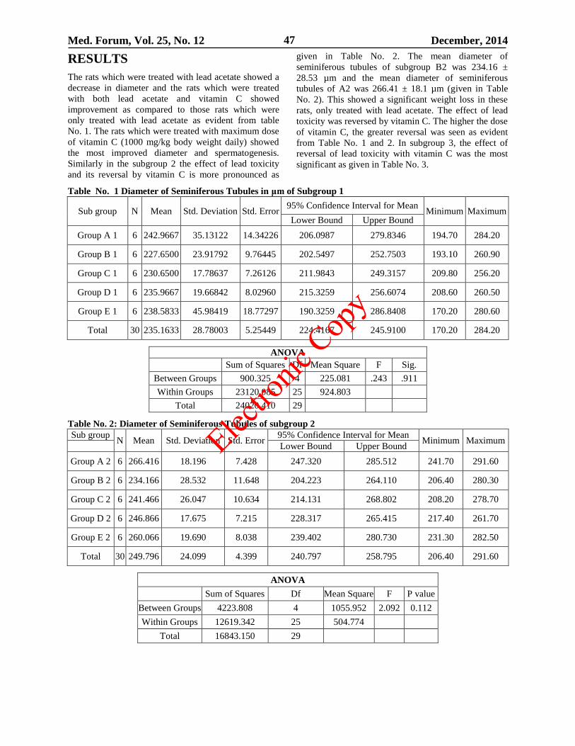
Med. Forum, Vol. 25, No. 12 December, 2014 47
RESULTS
The rats which were treated with lead acetate showed a
decrease in diameter and the rats which were treated
with both lead acetate and vitamin C showed
improvement as compared to those rats which were
only treated with lead acetate as evident from table
No. 1. The rats which were treated with maximum dose
of vitamin C (1000 mg/kg body weight daily) showed
the most improved diameter and spermatogenesis.
Similarly in the subgroup 2 the effect of lead toxicity
and its reversal by vitamin C is more pronounced as
given in Table No. 2. The mean diameter of
seminiferous tubules of subgroup B2 was 234.16 ±
28.53 µm and the mean diameter of seminiferous
tubules of A2 was 266.41 ± 18.1 µm (given in Table
No. 2). This showed a significant weight loss in these
rats, only treated with lead acetate. The effect of lead
toxicity was reversed by vitamin C. The higher the dose
of vitamin C, the greater reversal was seen as evident
from Table No. 1 and 2. In subgroup 3, the effect of
reversal of lead toxicity with vitamin C was the most
significant as given in Table No. 3.
Table No. 1 Diameter of Seminiferous Tubules in μm of Subgroup 1
Sub group N Mean Std. Deviation Std. Error 95% Confidence Interval for Mean
Minimum Maximum Lower Bound Upper Bound
Group A 1 6 242.9667 35.13122 14.34226 206.0987 279.8346 194.70 284.20
Group B 1 6 227.6500 23.91792 9.76445 202.5497 252.7503 193.10 260.90
Group C 1 6 230.6500 17.78637 7.26126 211.9843 249.3157 209.80 256.20
Group D 1 6 235.9667 19.66842 8.02960 215.3259 256.6074 208.60 260.50
Group E 1 6 238.5833 45.98419 18.77297 190.3259 286.8408 170.20 280.60
Total 30 235.1633 28.78003 5.25449 224.4167 245.9100 170.20 284.20
ANOVA
Sum of Squares Df Mean Square F Sig.
Between Groups 900.325 4 225.081 .243 .911
Within Groups 23120.085 25 924.803
Total 24020.410 29
Table No. 2: Diameter of Seminiferous Tubules of subgroup 2
Sub group N Mean Std. Deviation Std. Error
95% Confidence Interval for Mean Minimum Maximum
Lower Bound Upper Bound
Group A 2 6 266.416 18.196 7.428 247.320 285.512 241.70 291.60
Group B 2 6 234.166 28.532 11.648 204.223 264.110 206.40 280.30
Group C 2 6 241.466 26.047 10.634 214.131 268.802 208.20 278.70
Group D 2 6 246.866 17.675 7.215 228.317 265.415 217.40 261.70
Group E 2 6 260.066 19.690 8.038 239.402 280.730 231.30 282.50
Total 30 249.796 24.099 4.399 240.797 258.795 206.40 291.60
ANOVA
Sum of Squares Df Mean Square F P value
Between Groups 4223.808 4 1055.952 2.092 0.112
Within Groups 12619.342 25 504.774
Total 16843.150 29
Electro
nic Cop
y
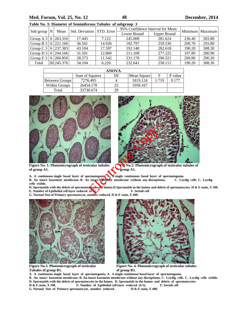
Med. Forum, Vol. 25, No. 12 December, 2014 48
Table No. 3: Diameter of Seminiferous Tubules of subgroup 3
Sub group N Mean Std. Deviation STD. Error 95% Confidence Interval for Mean
Minimum Maximum Lower Bound Upper Bound
Group A 3 6 263.316 17.445 7.122 245.008 281.624 236.40 283.80
Group B 3 6 221.166 36.561 14.926 182.797 259.536 200.70 293.80
Group C 3 6 237.383 43.104 17.597 192.148 282.618 190.20 308.30
Group D 3 6 244.166 31.501 12.860 211.108 277.225 197.80 280.90
Group E 3 6 260.850 28.273 11.542 231.178 290.521 206.80 290.20
Total 30 245.376 34.104 6.226 232.641 258.111 190.20 308.30
ANOVA
Sum of Squares Df Mean Square F P value
Between Groups 7276.495 4 1819.124 1.719 0.177
Within Groups 26454.178 25 1058.167
Total 33730.674 29
Figure No. 1: Photomicrograph of testicular tubules Fig No.2: Photomicrograph of testicular tubules of
of group A1. group A1.
A. A continuous single basal layer of spermatogonia. A. A single continuous basal layer of spermatogonia.
B. An intact basement membrane. B. An intact basement membrane without any disruptionn. C. Leydig cells C. Leydig
cells visible.
D. Spermatids with the debris of spermatocytes in the lumen.D.Spermatids in the lumen and debris of spermatocytes. H & E stain, X 100.
E. Number of Epithelial cell layer reduced (4-5). F. Sertoli cell
G. Normal Size of Primary spermatocyte, number reduced. H & E stain, X 400.
Figure No.3. Photomicrograph of testicular Figure No. 4. Photomicrograph of testicular tubules
Tubules of group B1. of group B1. A. A continuous single basal layer of spermatogonia. A. A single continuous basal layer of spermatogonia.
B. An intact basement membrane. B. An intact basement membrane without any disruptionn. C. Leydig cells C. Leydig cells visible.
D. Spermatids with the debris of spermatocytes in the lumen. D. Spermatids in the lumen and debris of spermatocytes.
H & E stain, X 100. E. Number of Epithelial cell layer reduced (4-5). F. Sertoli cell
G. Normal Size of Primary spermatocyte, number reduced. H & E stain, X 400.
Electro
nic Cop
y
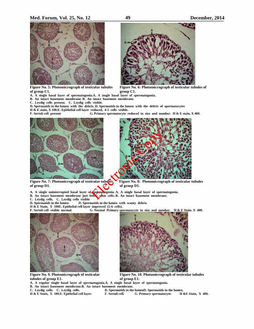
Med. Forum, Vol. 25, No. 12 December, 2014 49
Figure No. 5: Photomicrograph of testicular tubules Figure No. 6: Photomicrograph of testicular tubules of
of group C1. group C1. A. A single basal layer of spermatogonia.A. A single basal layer of spermatogonia. B. An intact basement membrane. B. An intact basement membrane. C. Leydig cells present. C. Leydig cells visible. D. Spermatids in the lumen with the debris. D. Spermatids in the lumen with the debris of spermatocytes H & E stain, X 100.E. Epithelial cell layer reduced, 4-5 cells visible. F. Sertoli cell present G. Primary spermatocyte reduced in size and number. H & E stain, X 400.
Figure No. 7: Photomicrograph of testicular tubules Figure No. 8: Photomicrograph of testicular tubules
of group D1. of group D1.
A. A single uninterrupted basal layer of spermatogonia. A. A single basal layer of spermatogonia. B. An intact basement membrane just below stem cells. B. An intact basement membrane. C. Leydig cells. C. Leydig cells visible D. Spermatids in the lumen D. Spermatids in the lumen with scanty debris. H & E Stain, X 100E. Epithelial cell layer improved (5-6 cells). F. Sertoli cell visible normal. G. Normal Primary spermatocyte in size and number. H & E Stain. X 400.
Figure No. 9. Photomicrograph of testicular Figure No. 10. Photomicrograph of testicular tubules
tubules of group E1. of group E1. A. A regular single basal layer of spermatogonia.A. A single basal layer of spermatogonia. B. An intact basement membrane.B. An intact basement membrane. C. Leydig cells. C. Leydig cells. D. Spermatids in the lumenD. Spermatids in the lumen. H & E Stain, X 100.E. Epithelial cell layer. F. Sertoli cell. G. Primary spermatocyte. H &E Stain, X 400.
Electro
nic Cop
y

Med. Forum, Vol. 25, No. 12 December, 2014 50
Effect of lead toxicity and its reversal by vitamin C on
diameter and spermatogenesis of testes of albino rats
concluded. In table no 1 and 2 the mean diameter of
semniferous tubules of rats of different subgroups
showed that the rats which were treated with lead alone
showed a significant decrease in mean diameter while
those treated with vitamin C along with lead acetate
showed better results. The subgroup C showed less
decline as compared to subgroup B. The subgroup D
showed increased mean diameter while subgroup E
showed more increased mean diameter compared to
subgroup D. This showed that the rats which were
treated with higher dose of vitamin C showed better
response as compared to those which were treated with
lower doses of vitamin C (as evident in Table 1,2 & 3
and photograph 1 to 10).
DISCUSSION
The rats of group B which were treated only with lead
showed a significant decrease in diameter over different
times of scarification. This significant decrease in
diameter and spermatogenesis was due to lead toxicity
which were documented by Harvey.6 According to
another study by Ahmad I et al, the reason for the toxic
effect of lead is accumulation of lead in testes of albino
rats.7 The group C rats which were treated with lead
and vitamin C (250 mg/kg body weight daily) showed
less decrease in diameter and spermatogenesis as
compared to group B rats which were treated with lead
only and showed a significant decrease in diameter and
spermatogenesis. This shows that the toxic effect of
lead is being lessen by vitamin C. The protective action
of vitamin C against lead acetate can be attributed to the
antioxidant action of vitamin C.8
A study conducted by
Bassem M. Raafat et al showed that administration of
vitamin C with lead exposed animals exerts an obvious
ameliorating as well as treatment effects.9 The rats of
group D showed more improvement in diameter and
spermatogenesis as compared to group C animals, the
reason behind this is that the group D rats were treated
with higher dose of vitamin C as compared to rats of
group C. The rats of group E were treated with the
highest dose of vitamin C while all experimental groups
(group B, group C, group D and group E) were given
the same quantity of lead. The rats of group E showed
most improved results among the experimental groups.
A study by Hsu P C et al showed that vitamin C in
considerable concentration showed significant reversal
of lead toxicity in rats.10
Thus greater the amount of
vitamin C the more is the reversal against lead toxicity.
In addition to acting as an antioxidant vitamin C also
has an inhibiting effect on lead uptake on a cellular
level.11
A number of studies showed that lead has no effect on
diameter and spermatogenesis. A study conducted by
Ping-Chi Hsu et al showed that there were no essential
differences among the diameter either taking lead or
not.12
While Beata M. Pace et al showed in their studies
that there were significant changes in pup over the first
3 weeks of lead treatment, when compared with the
control group.13
In another study, a slight reduction of
diameter was observed where lead acetate was given for
14 days.14
Thus, lead has decreased the normal diameter
and spermatogenesis of the rats which is also shown in
this study. The effect of vitamin C on lead levels has
been clarified by studies that showed that ascorbic acid
(vitamin C) decreased intestinal absorption of lead.15
Vitamin C has a significant role in reversing the lead
toxicity which is proved by a number of studies. One
rat pharmacokinetic study found that intravenously
administered vitamin C lowered lead tissue levels in
rats that were continuously administered lead.16
Another study showed that the adults with the highest
ascorbic acid (vitamin C) levels had a 60-80%
decreased prevalence of elevated blood lead.17
CONCLUSION
Vitamin C reduces the toxic effects of lead on diameter
of seminiferous tubules and spermatogenesis of rats,
which is shown in this study.
REFERENCES
1. Abdallah GM, El-sayed EM, Abo-Salem OM.
Effect of lead toxicity on co-enzyme Q levels in rat
tissue, Afri J Pharm and Pharmacol 2009;3(11):
568-572.
2. Suradkar SG, Ghodasara DJ, Vihol P, et al.
Haemato-Biochemical alterations induced by lead
acetate toxicity in wistar rats. Veteriary World.
2009; 2(11): 429-431.
3. Han S, Li W, Jamil U, et al. Effects of weight loss
and exercise on the distribution of lead and
essential trace elements with prior lead exposure.
Environmental Health Perspectives 1999;107(8):
657-661.
4. Patrick L. The role of free radical damage and the
use of antioxidants in the pathology and treatment
of lead toxicity. Alternative medicine review.
2006;11(2): 114-127.
5. Klaassen CD. Heavy metals and heavy metal
antagonists. The pharmacological basis of
therapeutics. 10th
ed. McGraw-Hill Medical
Publishing division: New York; 2001.p.1851-1872.
6. Harvey SC. Heavy metals. The pharmacological
basis of therapeutics. In: Goodman LS, Gillman A,
editors. London MacMillan;1970.p. 976.
7. Ahmad I, Sabir M. Yasin KF. Study of the effects
of lead poisoning on the testes in albino rats. J Med
Res 2003;42:1-9.
8. Dabrowski K, Cicreszko. Ascorbic acid and
reproduction in fish: Endocrine regulation and
gamete quality. Aquaculture Res 2001;32:623-638.
9. Rafaat BM, Shafaa MW, Rizk AR, et al.
Ameliorating effect of vitamin C against acute lead
Electro
nic Cop
y
Med. Forum, Vol. 25, No. 12 December, 2014 51
toxicity in albino rats. Aust J of Basic and Applied
Sci 2009; 3(4): 3597-3608.
10. Hsu PC, Hsu CC, Liu MY, et al. Lead induced
changes in spermatozoa function and metabolism. J
of Toxicol and Environ Health 1998;55:45-64.
11. Lyn Patrick ND. Lead toxicity part II: The role of
free radical damage and the use of antioxidants in
the pathology and treatment of lead toxicity.
Alternative Med Rev 2006; 11: 114-127.
12. Ping-Chi Hsu, Ming-Yie Liu, Chao-Chin Hsu,
et al. Effect of vitamin E and / or C on reactive
oxygen species-related lead toxicity in the rat
sperm. J Toxicity 1998; 128: 169-170.
13. Pace BM, Lawrence DA, Behr MJ, et al. Neonatal
lead exposure changes quality of sperm and
number of macrophages in testes of BALB/c mice.
J of Toxicol 2005;210:247-256.
14. Turner TT, Lysiak JJ. Oxidative stress: A common
factor in testicular dysfunction. J of Androl 2008;
29(3): 488-498.
15. Suzuki T, Yoshida A. Effectiveness of dietary iron
and ascorbic acid in the prevention and cure of
moderately long term lead toxicity in rats. J of
Nutri 1979;109: 1974-1978.
16. Dalley JW, Gupta PK, Hung CT. A physiological
pharmacokinetic model describing the disposition
of lead in the absence and presence of L-ascorbic
acid in rats. J of Toxicol 1990;50:337-348.
17. Simon JA, Hudes ES. Relationship of ascorbic acid
to blood lead levels. JAMA 1990; 281: 2289-2293.
Address for Corresponding Author:
Dr. Mujahid Akbar Mamoun,
Assistant Professor of Anatomy
Quaid-A-Azam Medical College,
Bahawalpur
E-mail mujahidqmc.yahoo.com
Mailing Address;
98-B Model Town B, Bahawalpur.
Electro
nic Cop
y
Med. Forum, Vol. 25, No. 12 December, 2014 52
Anxiety in Dental Patients Marium Iqbal
Assoc. Prof. and HOD, Operative Dentistry, Endodontics and Paedodontics,
Jinnah Medical and Dental College, Karachi
ABSTRACT
Objective: To determine the frequency of self-reported anxiety levels related to dental procedures amongst dental
patients using Revised Corah’s Dental Anxiety Scale, (DAS-R).
Study Design: A cross sectional descriptive study
Place and Duration of Study: This study was carried out at the Medical, Dental and Allied Health Professionals’
College from October 2010 and April 2011.
Materials and Methods: 485 patients comprised the study sample. All those patients who consented to participate
and were 12-65 years of age were included. They were scored for anxiety using revised Corah’s Dental Anxiety
Scale (DAS-R).
Results: The mean DAS score for our study population was 9.91 (SD 3.29), while the modal score was 12. Based on
the DAS Score, distribution of the respondents was: Non anxious - 63.1% (n=304); Anxious - 33.2% (n=160); and
Phobic - 3.7% (n=18). Other findings are detailed subsequently. We found that self-reported-phobic levels were
higher in males compared to females, while the DAS scores on the contrary were higher for females than males.
Conclusion: We found that DAS score were higher (more anxiety) in females than in males. Interestingly, we also
found that self-reported dental phobia was observed more often in males than females.
Key Words: Anxiety, Dental Procedures, Revised Corah’s Dental Anxiety Scale, (Das-R), Dental Phobia, Patients
INTRODUCTION
Anxiety to dental procedures is not uncommon.1 It not
only results in cancelling and delaying of dental
appointments2 but also leads to an experience of
enhanced pain in an anxious patient.3,4
Moreover,
anxiety may lead to systemic complications.5,6
Avoidance of dental treatment and management of
anxious dental patients adds another paradigm of
challenging issues that may have a negative effect on
dental health7 and at large, the general health. Studies
from Europe8,9
America10
and Australia11,12
have shown
the presence of high levels of fear and anxiety in
patients towards dental procedures. A Norwegian study
reported anxiety scores (DAS ≥ 13) in 11.5% males and
23% females.13
Avoidance behavior with resultant
compromise to oral health eventually set up a vicious
cycle which leads to exacerbation of fear for dental
treatments and greater likelihood of avoidance of dental
treatment leading to poorer dental outcomes. The
transfer of this dental anxiety from one generation to
the next, with resultant aggravation of the problem,
raises further challenge.14
Many stimuli related to
dental settings can evoke dental anxiety.15
Boyle,
Newton and Milgrom have particularly addressed the
issue of receiving injections and its relation to
anxiety.16
When compared with ten other common fears, among
Dutch adults Oosternik et al observed that dental fear
was fourth in ranking.17
Muppa and co-workers
suggested that noise produced in the dental clinic
setting was the third most important reason of ignoring
a dental appointment.18
Evidence suggests that young females are more
commonly afflicted with dental fear and anxiety than
young males and that a reduction in these issues was
noted with advancement of age of those affected.19
In a study conducted at Isfahan University on children’s
dental fear, the parents blamed traumatic dental
experiences amongst other external factors, as the
cause.20
Cases with dental fear require careful
management to alleviate anxiety.21
To manage dental anxiety, the logical first step is to
measure its burden. An objective study is needed to
assess dental anxiety, beyond subjective perception of
individual clinicians and using pre-validated tools.
Corah’s Revised-Dental Anxiety Score test, commonly
known as DAS, is a well-recognized, widely used, and
pre-validated tool for assessment of dental anxiety and
phobia.
It has also shown high internal consistency22,23
and
test‐retest reliability.24
To our knowledge, no literature was available on
prevalence of dental anxiety in Pakistan until 2011
when a short communication was published. It
highlighted a study conducted in Islamabad,
Rawalpindi and Multan. We conducted our study to
determine the frequency of dental anxiety in the
patients presenting to dental OPDs at two centers, one
each in Karachi and Hyderabad.
MATERIALS AND METHODS
Self –responding questionnaires were administered in
this cross sectional study to a convenient sample of
about 550 patients of Jinnah Medical and Dental
College, Karachi, and, Isra University, Hyderabad. We
Original Article Anxiety in Dental Patients
Electro
nic Cop
y
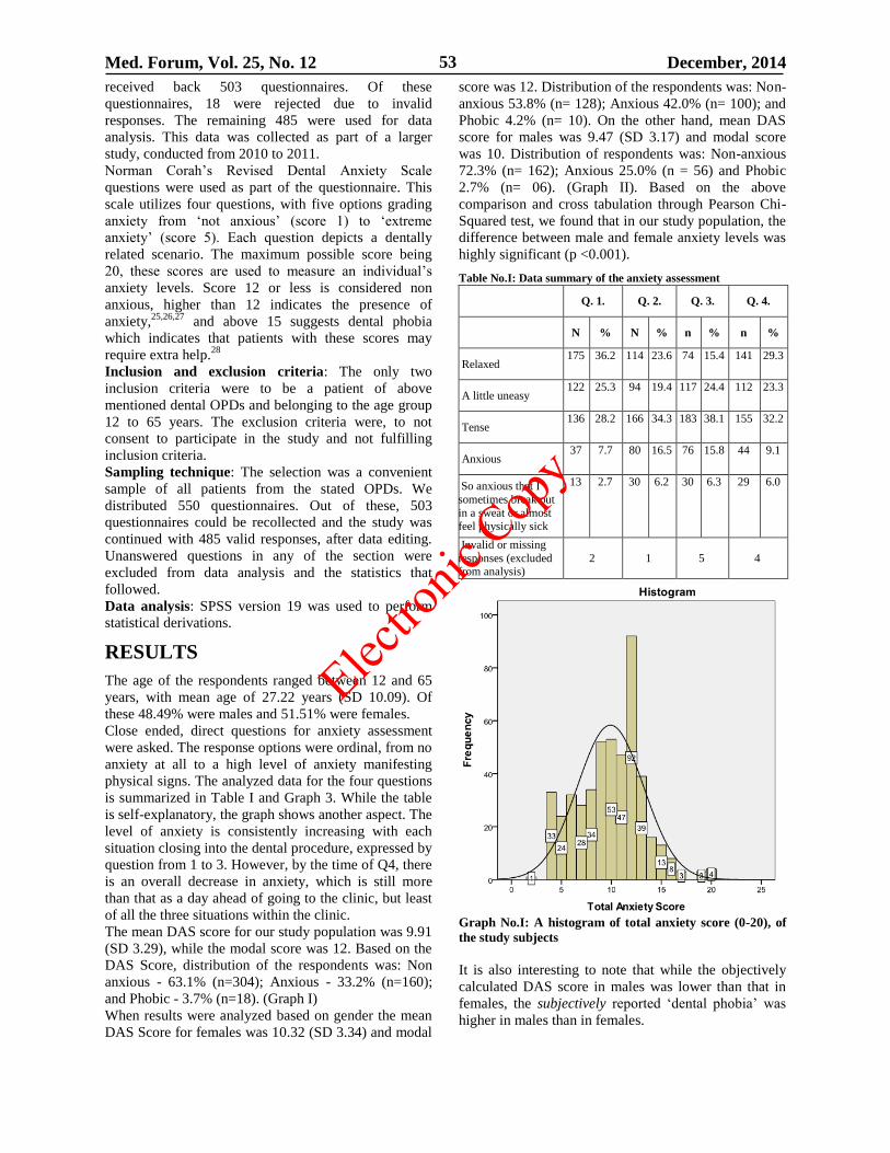
Med. Forum, Vol. 25, No. 12 December, 2014 53
received back 503 questionnaires. Of these
questionnaires, 18 were rejected due to invalid
responses. The remaining 485 were used for data
analysis. This data was collected as part of a larger
study, conducted from 2010 to 2011.
Norman Corah’s Revised Dental Anxiety Scale
questions were used as part of the questionnaire. This
scale utilizes four questions, with five options grading
anxiety from ‘not anxious’ (score 1) to ‘extreme
anxiety’ (score 5). Each question depicts a dentally
related scenario. The maximum possible score being
20, these scores are used to measure an individual’s
anxiety levels. Score 12 or less is considered non
anxious, higher than 12 indicates the presence of
anxiety,25,26,27
and above 15 suggests dental phobia
which indicates that patients with these scores may
require extra help.28
Inclusion and exclusion criteria: The only two
inclusion criteria were to be a patient of above
mentioned dental OPDs and belonging to the age group
12 to 65 years. The exclusion criteria were, to not
consent to participate in the study and not fulfilling
inclusion criteria.
Sampling technique: The selection was a convenient
sample of all patients from the stated OPDs. We
distributed 550 questionnaires. Out of these, 503
questionnaires could be recollected and the study was
continued with 485 valid responses, after data editing.
Unanswered questions in any of the section were
excluded from data analysis and the statistics that
followed.
Data analysis: SPSS version 19 was used to perform
statistical derivations.
RESULTS
The age of the respondents ranged between 12 and 65
years, with mean age of 27.22 years (SD 10.09). Of
these 48.49% were males and 51.51% were females.
Close ended, direct questions for anxiety assessment
were asked. The response options were ordinal, from no
anxiety at all to a high level of anxiety manifesting
physical signs. The analyzed data for the four questions
is summarized in Table I and Graph 3. While the table
is self-explanatory, the graph shows another aspect. The
level of anxiety is consistently increasing with each
situation closing into the dental procedure, expressed by
question from 1 to 3. However, by the time of Q4, there
is an overall decrease in anxiety, which is still more
than that as a day ahead of going to the clinic, but least
of all the three situations within the clinic.
The mean DAS score for our study population was 9.91
(SD 3.29), while the modal score was 12. Based on the
DAS Score, distribution of the respondents was: Non
anxious - 63.1% (n=304); Anxious - 33.2% (n=160);
and Phobic - 3.7% (n=18). (Graph I)
When results were analyzed based on gender the mean
DAS Score for females was 10.32 (SD 3.34) and modal
score was 12. Distribution of the respondents was: Non-
anxious 53.8% (n= 128); Anxious 42.0% (n= 100); and
Phobic 4.2% (n= 10). On the other hand, mean DAS
score for males was 9.47 (SD 3.17) and modal score
was 10. Distribution of respondents was: Non-anxious
72.3% (n= 162); Anxious 25.0% (n = 56) and Phobic
2.7% (n= 06). (Graph II). Based on the above
comparison and cross tabulation through Pearson Chi-
Squared test, we found that in our study population, the
difference between male and female anxiety levels was
highly significant (p <0.001).
Table No.I: Data summary of the anxiety assessment
Q. 1. Q. 2. Q. 3. Q. 4.
N % N % n % n %
Relaxed 175 36.2 114 23.6 74 15.4 141 29.3
A little uneasy 122 25.3 94 19.4 117 24.4 112 23.3
Tense 136 28.2 166 34.3 183 38.1 155 32.2
Anxious 37 7.7 80 16.5 76 15.8 44 9.1
So anxious that I
sometimes break out
in a sweat or almost feel physically sick
13 2.7 30 6.2 30 6.3 29 6.0
Invalid or missing
responses (excluded from analysis)
2 1 5 4
Graph No.I: A histogram of total anxiety score (0-20), of
the study subjects
It is also interesting to note that while the objectively
calculated DAS score in males was lower than that in
females, the subjectively reported ‘dental phobia’ was
higher in males than in females.
Electro
nic Cop
y
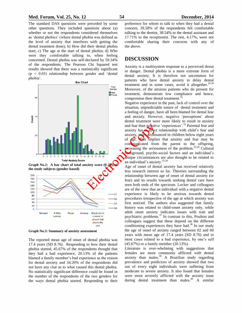
Med. Forum, Vol. 25, No. 12 December, 2014 54
The standard DAS questions were preceded by some
other questions. They included questions about (a)
whether or not the respondents considered themselves
as ‘dental phobics’ (where dental phobia was defined as
the level of anxiety that interferes with getting the
dental treatment done); b) How did their dental phobia
start; c) The age at the start of dental phobia; d) Who
were they comfortable talking to, when feeling
concerned. Dental phobia was self-declared by 59.34%
of the respondents. The Pearson Chi Squared test
results showed that there was a statistically significant
(p < 0.03) relationship between gender and ‘dental
phobia’.
Graph No.2: A bar chart of total anxiety score (0-20), of
the study subjects (gender-based)
Graph No.3: Summary of anxiety assessment
The reported mean age of onset of dental phobia was
17.4 years (SD 8.76). Responding to how their dental
phobia started, 45.67% of the respondents thought that
they had a bad experience, 20.13% of the patients
blamed a family member’s bad experience as the reason
for dental anxiety and 34.20% of the respondents did
not have any clue as to what caused this dental phobia.
No statistically significant difference could be found in
the number of the respondents of the two genders for
the ways dental phobia started. Responding to their
preference for whom to talk to when they had a dental
concern, 39.58% of the respondents felt comfortable
talking to the dentist, 38.54% to the dental assistant and
17.71% to the receptionist. The rest, 4.17%, were not
comfortable sharing their concerns with any of
the above.
DISCUSSION
Anxiety is a multisystem response to a perceived threat
or danger. Dental phobia is a more extreme form of
dental anxiety. It is therefore not uncommon for
patients who have dental anxiety to delay dental
treatment and in some cases, avoid it altogether.29.30
Moreover, of the anxious patients who do present for
treatment, demonstrate low compliance and hence,
compromise their dental treatment.31
Negative experience in the past, lack of control over the
situation, unpredictable nature of dental treatment and
a feeling of danger, have all been blamed for dental fear
and anxiety. However, negative ‘perceptions’ about
dental treatment were more likely to result in anxiety
and fear than negative ‘experiences’.32
Parental fear and
anxiety has a direct relationship with child’s fear and
anxiety, more pronounced in children below eight years
of age. This implies that anxiety and fear may be
communicated from the parent to the offspring,
increasing the seriousness of the problem.33,34
Cultural
background, psycho-social factors and an individual’s
unique circumstances are also thought to be related to
an individual’s anxiety.35,36
Age of onset of dental anxiety has received relatively
less research interest so far. Theories surrounding the
relationship between age of onset of dental anxiety (or
fear) and its results towards seeking dental care have
seen both ends of the spectrum. Locker and colleagues
are of the view that an individual with a negative dental
experience is likely to be anxious towards dental
procedures irrespective of the age at which anxiety was
first noticed. The authors also suggested that family
history was related to child-onset anxiety only, while
adult onset anxiety indicates issues with trait and
psychiatric problems.37
In contrast to this, Poulton and
colleagues suggest that these depend on the different
conditioning experiences they have had.38
In our study
the age of onset of anxiety ranged between 02 and 60
years with mean age of 17.4 years (SD 8.76) and in
most cases related to a bad experience, by one’s self
(45.67%) or a family member (20.13%).
Literature is over-whelming with suggestions that
females are more commonly afflicted with dental
anxiety than males.39
A Brazilian study regarding
prevalence and predictors of anxiety showed that two
out of every eight individuals were suffering from
moderate to severe anxiety. It also found that females
were more severely afflicted with the anxiety issue
during dental treatment than males.40
A similar
Electro
nic Cop
y
Med. Forum, Vol. 25, No. 12 December, 2014 55
Brazilian research done on adolescents found that 18%
manifested moderate to severe anxiety towards dental
treatment and that females were more commonly
affected.41
Our study result was also aligned to the
above international finding that the average DAS score
for females was higher than that for males and this
difference was statistically significant.
A Norwegian study observed 25-year old, randomly
selected individuals, grouped in 2 separate cohorts of
1997 and 2007. Their findings were also suggestive of
the fact that females (23%) have a higher prevalence of
dental anxiety than males (11%).Error! Bookmark not
defined. Another observation in the same study was
that rising education level
from 1997 to 2007 had reduced the anxiety level in the
latter cohort.
CONCLUSION
We found that the R-DAS scores based on self-reported
anxiety levels related to dental procedures, were higher
in females compared to males and the difference was
statistically significant. We also found that conversely,
self-reported-phobic levels were higher in males
compared to females.
REFERENCES
1. Karst M, Winterhalter M, Munte S, et al. Auricular
acupuncture for dental anxiety: A randomized
controlled trial Anesth Analg 2007; 104: 295-300.
2. Shaikh MA, Kamal A. Over dental anxiety
problems among university students: Perspective
from Pakistan. J Coll Physicians Surg Pak 2011;
21(4):237-8
3. Weisensee W, Scheer M, Muller L, et al. Impact of
anxiety parameters on prospective and experienced
pain intensity in implant surgery. Implant Dentistry
2012;21:502-506.
4. Eli I, Schwartz-Arad D, Baht R, Ben-Tuvim H.
Effect of anxiety on the experience of pain in
implant insertion. Clin Oral Implants Res 2003;14:
115-118.
5. Fardal O, McCulloch CA. Impact of anxiety on
pain perception associated with periodontal and
implant surgery in a private practice. J Periodontol
2012;83:1079-1085.
6. Okawa K, Ichinohe T, Kaneko Y. Anxiety may
enhance pain during dental treatment. Bull Tokyo
Dent Coll 2005; 46 (3):51-58.
7. McGrath C, Bedi R. The association between
dental anxiety and oral health‐related quality of life
in Britain. Comm Dent and Oral Epidemiol 32 (1),
67-72.
8. Pohjola V, Lahti S, Vehkalahti MM, Tolvanen M,
Hausen H. Association between dental fear and
dental attendance among adults in Finland. Acta
Odontol Scand 2007;65:224-30.
9. Enkling N, Marwinski G, Jöhren P. Dental anxiety
in a representative sample of residents of a large
German city. Clin Oral Investig 2006;10(1):84-91.
10. Rayman S, Dincer E, Almas K. Managing dental
fear and anxiety. N Y State Dent J 2013;79(6):
25-9.
11. Armfield JM, Slade GD, Spencer AJ. Dental fear
and adult oral health in Australia. Community Dent
Oral Epidemiol 2009;37(3):220-30.
12. Armfield JM, Stewart JF, Spencer AJ. The vicious
cycle of dental fear: exploring the interplay
between oral health, service utilization and dental
fear. BMC Oral Health 2007;7:1.
13. Åstrøm AN, Skaret E, Haugejorden O. Dental
anxiety and dental attendance among 25-year-olds
in Norway: time trends from 1997 to 2007. BMC
Oral Health 2011;11:10
14. Nuttall NM, Gilbert A, Morris J. Children's dental
anxiety in the United Kingdom in 2003. Dentist
2008;36 (11):857-860.
15. Vermaire JH, Ad De Jongh, Irene H, Aartman A.
Dental anxiety and quality of life: the effect of
dental treatment. Community Dent and Oral
Epidemiol 2008;36 (5):409–416.
16. Milgrom P, Newton JT, Boyle C, Heaton LJ,
Donaldson N. The effects of dental anxiety and
irregular attendance on referral for dental treatment
under sedation within the National Health Service
in London. Community Dent Oral Epidemiol 2010;
38:453–459.
17. Oosterink FM, de Jongh A, Hoogstraten J.
Prevalence of dental fear and phobia relative to
other fear and phobia subtypes. Eur J Oral Sci
2009;117(2):135-43.
18. Muppa R, Bhupatiraju P, Duddu M, Penumatsa
NV, Dandempally A, Panthula P. Comparison of
anxiety levels associated with noise in the dental
clinic among children of age group 6-15 years.
Noise Health 2013;15:190-3.
19. Klingberg G, Broberg AG. Dental fear/anxiety and
dental behaviour management problems in children
and adolescents: a review of prevalence and
concomitant psychological factors. Int J Paediatr
Dent 2007;17(6):391-406.
20. Jafarzadeh M, Keshani F, Ghazavi Z, Keshani F.
Reviewing the parental standpoint about origin of
the dental fear in children referred to dentistry
centers of Isfahan University of Medical Sciences.
Iran J Nurs Midwifery Res 2011; 16(1):133–139.
21. Armfield JM, Heaton LJ. Management of fear and
anxiety in the dental clinic: A review. Aust Dent J
2013;58(4):390-407.
22. Facco E, Zanette G, Manani G. Italian version of
Corah's Dental Anxiety Scale: Normative data in
patients undergoing oral surgery and relationship
with the ASA physical status classification. Anesth
Prog 2008;55:109–115.
Electro
nic Cop
y
Med. Forum, Vol. 25, No. 12 December, 2014 56
23. Eli I, Baht R, Kozlovsky A, Simon H. Effect of
gender on acute pain prediction and memory in
periodontal surgery. Eur J Oral Sci 2000;108:
99–103.
24. Halvari A, Halvari H, Bjørnebekk G. Motivation
and anxiety for dental treatment: Testing a self-
determination theory model of oral self-care
behaviour and dental clinic attendance. Motiv
Emot 2010; 34:15–33
25. Sohn W, Ismail AI. Regular dental visits and dental
anxiety in an adult dentate population. J Am Dent
Assoc 2005;136:58–66.
26. Ekanayake L, Dharmawardena D. Dental anxiety
in patients seeking care at the University Dental
Hospital in Sri Lanka. Community Dent Health
2003;20:112–116.
27. Bedi R, McGrath C. Factors associated with dental
anxiety among older people in Britain. Gerodontol
2000;17:97–103.
28. Newton JT, Buck DJ. Anxiety and pain measures
in dentistry: a guide to their quality and
application. J Am Dent Assoc 2000;131:1449–57.
29. Skaret E, Raadal M, Berg E, Kvale G. Dental
anxiety and dental avoidance among 12-18-year
olds in Norway. Eur J Oral Sci 1999;107:422–8.
30. Hägglin C, Hakeberg M, Ahlqwist M, Sullivan M,
Berggren U. Factors associated with dental anxiety
and attendance in middle-aged and elderly women.
Community Dent Oral Epidemiol 2000;28:451–60.
31. Scheutz F, Heidmann J. Determinants of utilization
of dental services among 20- to 34-year-old Danes.
Acta Odontol Scand 2001;59:201–11.
32. Armfield JM. Towards a better understanding of
dental anxiety and fear: cognitions vs. experiences.
Eur J Oral Sci 2010;118: 259–264.
33. Themessl-Huber M, Freeman R, Humphris G,
MacGillivray S, Terzi N. Empirical evidence of the
relationship between parental and child dental fear:
a structured review and meta-analysis. Int J
Paediatr Dent 2010;20(2):83-101.
34. Lahti S, Luoto A. Significant relationship between
parental and child dental fear. Evid Based Dent
2010;11(3):77.
35. Berggren U, Pierce CJ, Eli I. Characteristics of
adult dentally fearful individuals. A cross-cultural
study. Eur J Oral Sci 2000;108:268–74.
36. Hittner JB, Hemmo R. Psychosocial predictors of
dental anxiety. J Health Psychol 2009;14:53–9.
37. Locker D, Liddell A, Dempster L,Shapiro D. Age
of Onset of Dental Anxiety. JDR 1999;78(3):
790-96.
38. Poulton R, Karen EW, Thomson WM, Locker D.
Determinants of early- vs late-onset dental fear in a
longitudinal-epidemiological study. Behaviour
Research and Therapy 2001;39:777–85.
39. Schuller AA, Willumsen T, Holst D. Are there
differences in oral health and oral health behavior
between individuals with high and low dental fear?
Comm Dent Oral Epidemiol 2003;31:116–21.
40. de Carvalho RW, Wathson F, et al. Anxiety
regarding dental treatment: prevalence and
predictors among Brazilians. Ciênc. Saúde
Coletiva 2012;17(7):1915-1922.
41. de Carvalho RW, de Carvalho Bezerra Falcão PG,
de Luna Campos GJ, de Souza Andrade ES, do
Egito Vasconcelos BC, da Silva Pereira MA.
Prevalence and predictive factors of dental anxiety
in Brazilian adolescents. J Dent Child (Chic) 2013;
80(1):41-6.
Address for Corresponding Author:
Dr. Marium Iqbal,
Associate Professor and HOD,
Operative Dentistry, Endodontics and Paedodontics,
Jinnah Medical and Dental College
22-23, Shaheed-e-Millat Road, Karachi
Electro
nic Cop
y
Med. Forum, Vol. 25, No. 12 December, 2014 57
To Evaluate the Outcome of
Sacrococcygeal Pilonidal Sinus
Excision Using Karydakis Technique 1. Muhammad Iqbal Khan 2. Muhammad Jawed 3. Sajeer Bhura 4. Ubedullah Shaikh
5. Anum Arif 1. Senior Registrar of Surgery, JPMC, Karachi 2. Asstt. Prof. of Surgery & Bariatric Surgeon, DUHS, Karachi
3. PGR of Surgery, JPMC, Karachi 4. SMO of Surgery, DUHS, Karachi 5. PGR of Surgery, DUHS, Karachi.
ABSTRACT
Objective: To evaluate the outcome of sacrococcygeal pilonidal sinus excision using karydakis technique.
Study Design: Prospective case series study.
Place and Duration of Study: This study was carried out in the Department of General Surgery Unit III, Ward 26,
Jinnah Post graduate Medical Centre, Karachi form March 2005 to Feb 2012.
Materials and Methods: The study included 85 consecutive patients who underwent pilonidal sinus excision by
karydakis technique fulfilling the inclusion criteria. We excluded the cases of pilonidal abscess and the cases which
came with acute infections. Patients under 12 years were also excluded. A prospective method of data collection was
utilizes by filling in the proforma designed for the study. A complete record of the procedure, follow up was done
initially on weekly basis for one month and then fortnightly for 6 months and subsequently monthly for 30 months.
Results: Total of 85 patents were included in our study in which 68 (80%) were male and 17 (20%) were female.
The mean age of the group was 30.56 years. All patients were followed postoperatively for 30 months. Mean
hospital stay was 2.5 days. Majority 63(74.1%) of the patients underwent smoothly without major complication. In
all 85 patients wound closed with prolene 2/0 interrupted sutures. In 9(10.6%) patients developed minor wound
infection while 4(4.7%) patients develop wound dehiscence and 3(3.52%) patients develop recurrence. In all 85
patients prophylactic antibiotics amoxicillin + clavulanic acid 1.2gm was used. In infected patients accounting to a
total of 13(15.3%), both major and minor infections were included and appropriate antibiotic was used as indicated
in the culture and sensitivity report.
Conclusion: Karydakis technique is superior to midline excision surgery. It is associated with significantly shorter
complication rate, shorter hospital length of duration, rapid healing, cost effective, good cosmetic satisfaction, a high
patient satisfaction rate and low rate of recurrence.
Key Words: Sacrococcygeal Pilonidal, Karydakis Technique, Complication.
INTRODUCTION
Pilonidal means pertaining to a nest of hair’
(latin:pilus=hair, nidus=nest).Initially thought to be
congenital in origin, pilonidal sinus disease is now
thought to be an acquired disease.1,2
The mode of origin
of a pilonidal sinus is now believed to be that: on
sitting, the buttocks take the weight of the body, and
move independently, or together. Hairs broken off by
friction against clothing, and shed short hairs, from the
nape of the neck, back, or buttocks, tend to collect in
the cleft of the nates and/or a post anal dimple.
Furthermore, it is suggested that the use of toilet paper
may contribute to hair entangled in fecal matter being
swept into the cleft; pilonidal sinus is extremely rare in
those races that employ ablution after defecation.3
According to Hodges in 1880, pilonidal sinus disease, is
an epithelial tract at the natal cleft.4
Herbert Mayo in
1883, describe a disease that involve a hair atthe base of
the coccyx.5
Pilonidal sinus is common disease that
affect young people.6
There are various surgical options
for the treatment of pilonidal sinus disease, Controversy
still exists as to the best technique, however Karydakis
technique proves to be with lower post operative
complication rate, shorter hospital stay and good
cosmetic satisfaction.7
MATERIALS AND METHODS
This study was carried out in the department of general
surgery unit III ward 26, Jinnah Post graduate Medical
Centre, Karachi, form March 2005 to Feb 2012.
The study included 85 consecutive patients who
underwent pilonidal sinus excision by karidakis
technique fulfilling the inclusion criteria. We excluded
the cases of pilonidal abscess and the cases which came
with acute infections. Patients under 12 years were also
excluded. A prospective method of data collection was
utilizes by filling in the proforma designed for the
study. A complete record of the procedure, follow up
was done initially on weekly basis for one month and
then fortnightly for 6 months and subsequently monthly
for 30 months.
Original Article Pilonidal Sinus
Excision
Electro
nic Cop
y
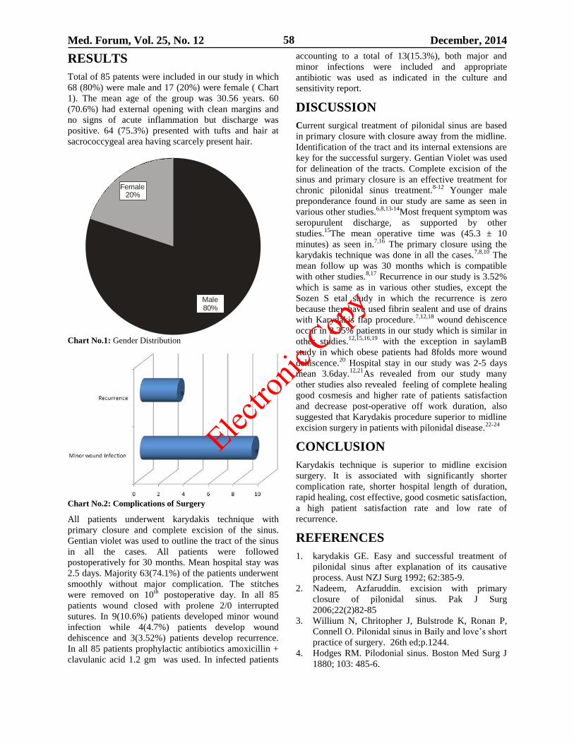
Med. Forum, Vol. 25, No. 12 December, 2014 58
RESULTS
Total of 85 patents were included in our study in which
68 (80%) were male and 17 (20%) were female ( Chart
1). The mean age of the group was 30.56 years. 60
(70.6%) had external opening with clean margins and
no signs of acute inflammation but discharge was
positive. 64 (75.3%) presented with tufts and hair at
sacrococcygeal area having scarcely present hair.
Female20%
Male80%
Chart No.1: Gender Distribution
Chart No.2: Complications of Surgery
All patients underwent karydakis technique with
primary closure and complete excision of the sinus.
Gentian violet was used to outline the tract of the sinus
in all the cases. All patients were followed
postoperatively for 30 months. Mean hospital stay was
2.5 days. Majority 63(74.1%) of the patients underwent
smoothly without major complication. The stitches
were removed on 10th
postoperative day. In all 85
patients wound closed with prolene 2/0 interrupted
sutures. In 9(10.6%) patients developed minor wound
infection while 4(4.7%) patients develop wound
dehiscence and 3(3.52%) patients develop recurrence.
In all 85 patients prophylactic antibiotics amoxicillin +
clavulanic acid 1.2 gm was used. In infected patients
accounting to a total of 13(15.3%), both major and
minor infections were included and appropriate
antibiotic was used as indicated in the culture and
sensitivity report.
DISCUSSION
Current surgical treatment of pilonidal sinus are based
in primary closure with closure away from the midline.
Identification of the tract and its internal extensions are
key for the successful surgery. Gentian Violet was used
for delineation of the tracts. Complete excision of the
sinus and primary closure is an effective treatment for
chronic pilonidal sinus treatment.8-12
Younger male
preponderance found in our study are same as seen in
various other studies.6,8,13-14
Most frequent symptom was
seropurulent discharge, as supported by other
studies.15
The mean operative time was (45.3 ± 10
minutes) as seen in.7,16
The primary closure using the
karydakis technique was done in all the cases.7,8,10
The
mean follow up was 30 months which is compatible
with other studies.8,17
Recurrence in our study is 3.52%
which is same as in various other studies, except the
Sozen S etal study in which the recurrence is zero
because they have used fibrin sealent and use of drains
with Karydakis flap procedure.7,12,18
wound dehiscence
occur in 2.35% patients in our study which is similar in
other studies.12,15,16,19
with the exception in saylamB
study in which obese patients had 8folds more wound
dehiscence.20
Hospital stay in our study was 2-5 days
mean 3.6day.12,21
As revealed from our study many
other studies also revealed feeling of complete healing
good cosmesis and higher rate of patients satisfaction
and decrease post-operative off work duration, also
suggested that Karydakis procedure superior to midline
excision surgery in patients with pilonidal disease.22-24
CONCLUSION
Karydakis technique is superior to midline excision
surgery. It is associated with significantly shorter
complication rate, shorter hospital length of duration,
rapid healing, cost effective, good cosmetic satisfaction,
a high patient satisfaction rate and low rate of
recurrence.
REFERENCES
1. karydakis GE. Easy and successful treatment of
pilonidal sinus after explanation of its causative
process. Aust NZJ Surg 1992; 62:385-9.
2. Nadeem, Azfaruddin. excision with primary
closure of pilonidal sinus. Pak J Surg
2006;22(2)82-85
3. Willium N, Chritopher J, Bulstrode K, Ronan P,
Connell O. Pilonidal sinus in Baily and love’s short
practice of surgery. 26th ed;p.1244.
4. Hodges RM. Pilodonial sinus. Boston Med Surg J
1880; 103: 485-6.
Electro
nic Cop
y
Med. Forum, Vol. 25, No. 12 December, 2014 59
5. Mayo OH. Observations on injuries and diseases of
the rectum London: Burgess and Hill; 1833.p.
45-46.
6. Horwood J, Hanratty D, Chandran, Billings P,
Primary closure or rhomboid excision and Limberg
flap for the management of primary sacroccygeal
pilonidal disease? A meta-analyisis of randomized
controlled trials.colorectal Dis 2012;14(2):143-51.
7. Ates M, Dirican A Sarac, M Aslan A, Colak C.
Short and Long-term results of Karydakis flap
Versus the limberg for treating pilonidal sinus
disease;A prospective randomized study. Am J
Surg 2011;202(5);568-73.
8. Moran DC , Kavanagh DO, Ahmed I, Regan MC.
Excision and primary closure using the Karydakis
flap for the treatment of pilonidal disease:
outcomes from a single institution. World J Surg
2011;35(8):1803-8.
9. Khanna A, Rombeau JL. Clincolan rectal Surg
2011;24(1):46-53.
10. Can MF, Sevinc MM, Hancerliogullario O, Yilmaz
M. Yagicig. AM J Surg 2010;200(3):318-27.
11. Khanzada TW, Samad A. Recurrence after
excision and primary closune by pilonidalsinus.
Pak J Med Sci 2007;23(3);375-9.
12. Saleh M, Alsalamah, Husain MI, Mirza SM.
Excision with or without primary closure for
pilonid al sinus disease. J Pak Med Assoe 2007;57
(8):388-91.
13. mohamed HA ,Kadry I ,AalyS.Comparison
between three therapentic modalities for non-
complicated pilonidal sinus disease surgeon
2005,3;73-7.
14. Dalenback J, MgnessonO,wedelN,Rimback G.
Prospective follow up after ambulatory plain
midline excision of pilonidal sinus and primary
closure under local anthesia–efficient, sufficient,
and persistent. Colorectal Dis 2004;6:488-93.
15. Ahmed S, Pervaiz N, Baloch N.Primaryclosure of
Pilondalsinus: Our experience. Pak J Surg 2010;
26(1):36-40.
16. Sheikh R, Malik KA, Rehman S. Out come of
surgery for pilonidal sinus:Karidakis versus open
procedure. Pak J Surg 2007;23(3):202-4.
17. Arsalan K, SaidKokcam S, Kosal H, Turan E, Atay
A, Dogruo. Which flap method should be
preferredfor the treatment of Pilonidal sinus? A
prospective randomized study. Coloproctol 2013.
18. Sozen S, Emir S, Guzel K, Ozdemir CS. Are
postoperativedrains necessary with the Karidakis
flap for treatment of Pilonidal sinus? (can fibrin
glue be replaced to drains?) A prospective
randomized trial. Ir J Med Sci 2011;180(2):479-82.
19. Bukhari AJ, Bhutta A, Khan AZ, Abid KJ.
Treatment of Pilonidal sinus:Comparsion of
Pilonidal sinus. Ann King Edward Med Uni 2003:
9(2):74-6.
20. Saylam B, Balli DN, Duz GUN PA, Ozer MV,
Coskun F. Which surgical procedure offers the best
treatment for Pilonidal disease? Langenbecks Arch
Surg 2011;396(5):651-8.
21. Khatoon S, Junejo A, Memon M, Arif M.
Pilonidalsinus: Excision with Primary midline
closure versus open method. J Liaquat Univ Med
Health Sci 2010,9(1):09-11.
22. Malik GA, Chaudhry TH, Wahab A.
Pilonidalsinus. Professionel Med J 2009;16(1):
17-23.
23. Lesalnieks l, Deimel S, Schlitt HJ. Karydakis flap
for recurrent pilonidal disease.World J Surg 2013;
37(5):1115-20.
24. Bessa SS. Comparision of short- term results
between the modified Kaydakis flap and the
modified Limberg flap in the managrment of
pilonidal sinus disease:a randomized
contolledstudy. Dis Colon Rectum 2013;56(4):
491-8.
Address for Corresponding Author:
Dr. Muhammad Iqbal Khan,
C-111, Block – 2, Clifton, Karachi.
Email: [email protected]
Cell No. 03322514095
Electro
nic Cop
y
Med. Forum, Vol. 25, No. 12 December, 2014 60
The Complex Regional Pain
Syndrome after Fractures of Distal Radius 1. Madan Lal Jesswani 2. Syed Imaduddin 3. Muhammad Asif Memon 4. Rahila Razzak 1. Specialist Orthopedic Surgery, Sheikh Khalifa Hospital, Ajman, UAE 2. Asstt. Prof. of Orthopaedic Surgery,
United Medical & Dental College and Hospitals, Karachi 3. Consultant, Sulaiman Al Rajhi Colleges, Bukayria,
Saudi Arabia 4. Consultant, Ziauddin University, Karachi
ABSTRACT
Objectives: The present study aims to determine frequency of Complex regional pain syndrome (CRPS) after distal
radius fractures on the basis of clinical examination findings and classify into stages and to find out association of
CPRS with age, genders, and risk factors.
Study Design: Prospective cross sectional study
Place and Duration of Study: This research work was conducted at the Department of Orthopaedics, Creek
General Hospital Korangi Karachi, Pakistan from January 2013 to April 2014.
Materials and Methods: This is a prospective cross sectional study of 150 cases. Follow up of patients is
undertaken in out- patient department for a minimum 4 months period following injury. The percentage of patients
with CRPS was identified according to IASP Diagnostic Criteria based on history and physical examination.1,2,3,4
Patients were studied prospectively to ascertain the incidence, natural history and the degree of morbidity induced
by CRPS.
Results: Mean± SD age was 45.6± 14.2 years (Range = 18 – 75 years). There were 88 (58.7%) males and 62
(41.3%) females with Male: Female = 1: 0.7. CPRS was found in 20 (13.3%) cases. Out of 20 CRPS patients 12
(60%) were female, out of 20 CRPS patients 12 (60%) had age 40 – 59 years and 8 (40%) had age >59 years. Out of
20 CRPS patients 14 (70%) were diagnosed in stage 1.
Conclusion: The incidence of CRPS after distal radius fracture in this study is 13.3%.Proportion of CRPS was high
in females and in old patients. Most of the patients of CRPS were diagnosed in stage 1. Diabetes mellitus,
hypertension, stroke, carpal tunnel syndrome and myocardial infarction were the risk factors found in patients
diagnosed with CRPS. CRPS is an under diagnosed entity. More work needs to be done on CRPS as many areas of
research remains.
Key Words: Complex regional pain syndrome; Sudeck’s dystrophy; Reflex sympathetic dystrophy; IASP.
INTRODUCTION
Complex regional pain syndrome (CRPS), formerly
known as Sudeck’s dystrophy or reflex sympathetic
dystrophy, is a painful disorder with clinical features
that include pain, sensory and vasomotor disturbances,
trophic changes and impaired motor function.1
The
disease course varies from relatively mild and self-
limiting to chronic disease with a high impact on daily
functioning and quality of life.2 Usually, symptoms
appear in one extremity after even a relatively mild
trauma, for example a fracture, contusion or surgery.3
Complex regional pain syndrome (CRPS) is a
perplexing condition characterized by local neurogenic
inflammation out of proportion to injury affecting the
limbs, without nerve injury (CRPS I or reflex
sympathetic dystrophy [RSD]) or with obvious nerve
lesions (CRPS II or causalgia).4-9
It consists of pain and
related sensory abnormalities, abnormal blood flow and
sweating, abnormalities in the motor system and
changes in the structure of both superficial and deep
tissues (trophic changes).7–12
Psychological and social
problems such as anxiety, depression, fear avoidance
(of painful movements) and loss of employment may
develop. Both the affected body part and the patient as
a whole may become “dysfunctional” in CRPS. Other
terms commonly used to describe CRPS are
algodystrophy, shoulder-hand syndrome and sudeck's
atrophy. 5,6,8,10
The inciting injury may be a sprain,
dislocation, fracture or post-surgery. 4,7,13,14
The
syndrome may also be associated with medical
conditions such as diabetic neuropathy, multiple
sclerosis, stroke, myocardial infarction and cancerous
infiltration of a nerve plexus.7,9
The reported incidence
of CRPS after wrist fractures is 7-35%.4,7,15,16
CRPS
appeared equally distributed in every age group, except
in children under 10 as widely reported in literature. It
is not a diagnosis of exclusion, because International
Association for the Study of Pain (IASP) has
formulated a Diagnostic Criteria for CRPS.4,5,6
The
diagnosis of CRPS is possible as soon as one week after
fracture by history and physical exam.
Treatment is crucial within the first three to six months
when the disease responds best. Otherwise the
syndrome may progress producing a lifetime chronic
pain, permanent functional impairment, resistance to
treatment and resultant emotional distress.8
Management of CRPS is based on the bio-psycho-
social model of pain and should involve a
multidisciplinary team. Keystones include the provision
Original Article CRPS after Distal
Radius Fractures
Electro
nic Cop
y
Med. Forum, Vol. 25, No. 12 December, 2014 61
of effective analgesia, allowing the affected region to
be mobilized (physical therapy) and returned to normal
function as soon as possible, the “use it or lose it
principle”. Psychological, social and occupational
rehabilitation should also be provided. The study will
be helpful in identifying early screening of CRPS
patients to avoid delayed complications involving no
risk and benefit to patient.
MATERIALS AND METHODS
This research work was conducted at the department of
Orthopaedic surgery, Creek General Hospital Korangi,
Karachi from January 2013 to April 2014. This is a
cross sectional done on 150 patients sustained close
distal radius fractures. All patients with fractures of
distal radius were clerked at Orthopedics clinic and
Accident & Emergency, a full history and physical
examination was done. Patients were recruited after
taking the informed consent. The patients who were
excluded from the study included presentation after one
week of fracture, multiple fractures, disability of
affected limb prior to fracture, pathological fracture and
patient suffering from psychiatric illness. Pre-reduction
and post-reduction roentgenograms of affected wrist
were taken. Fracture patterns were categorized
according to Fernandez classification. 17
All relevant
features including patient’s bio data, clinical and
radiological findings at presentation and follow up
visits were recorded on proforma. Follow up of patients
was undertaken in out patient department for a
minimum of 4 months period following injury. The
percentage of patients with CRPS was identified
according to IASP Diagnostic Criteria based on history
and physical examination.1,2,3,4
Patients were studied
prospectively to ascertain the incidence, natural history
and the degree of morbidity induced by CRPS.
The data was entered and analyzed by SPSS 12.
Frequencies and percentages were calculated for all
qualitative/categorical data including sex, age groups,
mechanism of injury, risk factors, types of fracture,
history of surgery, sign and symptoms of CPRS, pain
visual analog scale (PVAS) and the ratio of CRPS
among the total screened patients was presented by
their frequency and percentage. Chi-square test for
proportions was used for compare the proportion of
sign and symptoms (between day-1 and 16 weeks).
Mean± SD was computed for age and PVAS. Student’s
t-test was used to compare the mean of PVAS (between
day-1 and 16 weeks). The results were considered
statistically significant at p < 0.05.
RESULTS
This study was conducted on 150 patients with fractures
of distal radius. Mean± SD age was 45.6± 14.2 years
(Range = 18 – 75 years), age of 68(48) cases was 40 –
59 years, 48 (33.3%) cases had age 20 - 39 years while
30 (16%) cases had age > 59 years.
2 (1.3)8 (5.3)
30 (20)
110 (73.3)
0
20
40
60
80
100
120
Type-I Type-II Type-III Type-IV
Fracture Types
Nu
mb
er
of p
atie
nts
(%
)
Figure No.1: Types of Fracture n = 150 Keys:
Type-I = Metaphyseal bending fracture
Type-II = Shearing fracture Type-III = Compression of the articular surface without
fragmentation
Type-IV = Avulsion fracture or radiocarpal fracture dislocation Type-V= Combined injury with significant soft tissue involvement
CPRS
20 (13.3% )
Normal
130 (86.7% )
Figure No.2: Proportion of Patients with Complex
Regional Pain Syndrome (CRPS) n = 150 Key: CRPS = Complex Regional Pain Syndrome
Among these 150 patients, 88 (58.7%) were males and
62 (41.3%) were females with Male: Female = 1: 0.7
According to the presenting complaint, all patients
came with Pain and swelling.
Out of 150 patients, 88 (58.7%) patients had history of
fall in different modes while 62 (41.3%) came with
history of road traffic accident.
According to Fernandez classification of fracture
patterns, there were 110 (73.3%) patients of type-I
fracture, followed by 30 (20%) patients of type-II, 8
(5.3%) patients of type-III and 2 (1.3%) patients of type
IV. (Figure-1)
Out of 150 patients, only 12 (8%) patients had a history
of previous surgery.
The percentage of patients with Complex Regional Pain
Syndrome (CRPS) was identified according to IASP
diagnostic criteria based on history and physical
examination. CPRS was found in 20 (13.3%) cases.
(Figure-2)
Swelling was the most common finding in all 150
(100%) patients at day-one, followed by altered
temperature of limb skin in 136 (90.7%) patients, color
changes of limb in 132 (88%) patients. All these
symptoms were decreased significantly after sixteen
weeks, found in only 18 (12%) patients (P-value <
Electro
nic Cop
y
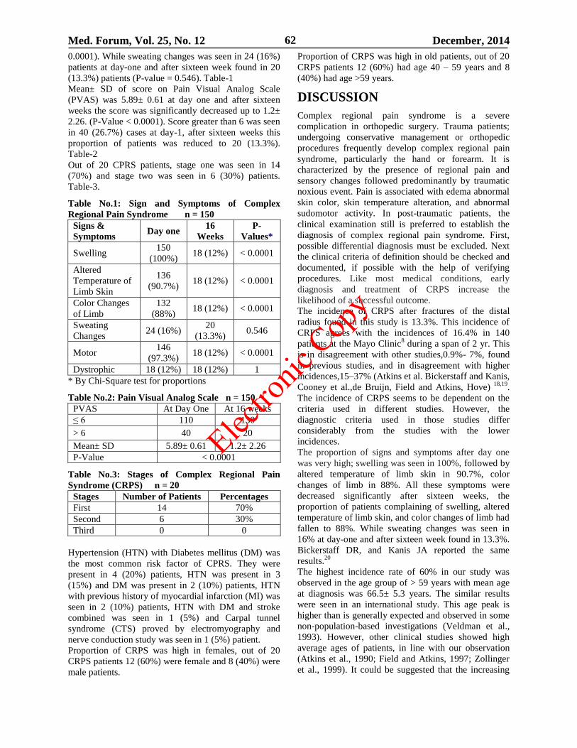
Med. Forum, Vol. 25, No. 12 December, 2014 62
0.0001). While sweating changes was seen in 24 (16%)
patients at day-one and after sixteen week found in 20
(13.3%) patients (P-value = 0.546). Table-1
Mean± SD of score on Pain Visual Analog Scale
(PVAS) was 5.89± 0.61 at day one and after sixteen
weeks the score was significantly decreased up to 1.2±
2.26. (P-Value < 0.0001). Score greater than 6 was seen
in 40 (26.7%) cases at day-1, after sixteen weeks this
proportion of patients was reduced to 20 (13.3%).
Table-2
Out of 20 CPRS patients, stage one was seen in 14
(70%) and stage two was seen in 6 (30%) patients.
Table-3.
Table No.1: Sign and Symptoms of Complex
Regional Pain Syndrome n = 150
Signs &
Symptoms Day one
16
Weeks
P-
Values*
Swelling 150
(100%) 18 (12%) < 0.0001
Altered
Temperature of
Limb Skin
136
(90.7%) 18 (12%) < 0.0001
Color Changes
of Limb
132
(88%) 18 (12%) < 0.0001
Sweating
Changes 24 (16%)
20
(13.3%) 0.546
Motor 146
(97.3%) 18 (12%) < 0.0001
Dystrophic 18 (12%) 18 (12%) 1
* By Chi-Square test for proportions
Table No.2: Pain Visual Analog Scale n = 150
PVAS At Day One At 16 weeks
≤ 6 110 130
> 6 40 20
Mean± SD 5.89± 0.61 1.2± 2.26
P-Value < 0.0001
Table No.3: Stages of Complex Regional Pain
Syndrome (CRPS) n = 20
Stages Number of Patients Percentages
First 14 70%
Second 6 30%
Third 0 0
Hypertension (HTN) with Diabetes mellitus (DM) was
the most common risk factor of CPRS. They were
present in 4 (20%) patients, HTN was present in 3
(15%) and DM was present in 2 (10%) patients, HTN
with previous history of myocardial infarction (MI) was
seen in 2 (10%) patients, HTN with DM and stroke
combined was seen in 1 (5%) and Carpal tunnel
syndrome (CTS) proved by electromyography and
nerve conduction study was seen in 1 (5%) patient.
Proportion of CRPS was high in females, out of 20
CRPS patients 12 (60%) were female and 8 (40%) were
male patients.
Proportion of CRPS was high in old patients, out of 20
CRPS patients 12 (60%) had age 40 – 59 years and 8
(40%) had age >59 years.
DISCUSSION
Complex regional pain syndrome is a severe
complication in orthopedic surgery. Trauma patients;
undergoing conservative management or orthopedic
procedures frequently develop complex regional pain
syndrome, particularly the hand or forearm. It is
characterized by the presence of regional pain and
sensory changes followed predominantly by traumatic
noxious event. Pain is associated with edema abnormal
skin color, skin temperature alteration, and abnormal
sudomotor activity. In post-traumatic patients, the
clinical examination still is preferred to establish the
diagnosis of complex regional pain syndrome. First,
possible differential diagnosis must be excluded. Next
the clinical criteria of definition should be checked and
documented, if possible with the help of verifying
procedures. Like most medical conditions, early
diagnosis and treatment of CRPS increase the
likelihood of a successful outcome.
The incidence of CRPS after fractures of the distal
radius found in this study is 13.3%. This incidence of
CRPS agrees with the incidences of 16.4% in 140
patients at the Mayo Clinic8 during a span of 2 yr. This
is in disagreement with other studies,0.9%- 7%, found
in previous studies, and in disagreement with higher
incidences,15–37% (Atkins et al. Bickerstaff and Kanis,
Cooney et al.,de Bruijn, Field and Atkins, Hove) 18,19
.
The incidence of CRPS seems to be dependent on the
criteria used in different studies. However, the
diagnostic criteria used in those studies differ
considerably from the studies with the lower
incidences.
The proportion of signs and symptoms after day one
was very high; swelling was seen in 100%, followed by
altered temperature of limb skin in 90.7%, color
changes of limb in 88%. All these symptoms were
decreased significantly after sixteen weeks, the
proportion of patients complaining of swelling, altered
temperature of limb skin, and color changes of limb had
fallen to 88%. While sweating changes was seen in
16% at day-one and after sixteen week found in 13.3%.
Bickerstaff DR, and Kanis JA reported the same
results.20
The highest incidence rate of 60% in our study was
observed in the age group of > 59 years with mean age
at diagnosis was 66.5± 5.3 years. The similar results
were seen in an international study. This age peak is
higher than is generally expected and observed in some
non-population-based investigations (Veldman et al.,
1993). However, other clinical studies showed high
average ages of patients, in line with our observation
(Atkins et al., 1990; Field and Atkins, 1997; Zollinger
et al., 1999). It could be suggested that the increasing
Electro
nic Cop
y
Med. Forum, Vol. 25, No. 12 December, 2014 63
incidence of CRPS with age is due to a higher
occurrence of fractures at older age. However, the same
age distribution pattern was observed in the group of
patients with another precipitating event other than a
fracture.
Out of 20 CRPS patients 14 were diagnosed in stage 1
and 6 were diagnosed in stage 2. Most of the cases were
picked early.
Hypertension, diabetes mellitus, stroke, history of
myocardial infarction and carpal tunnel syndrome were
the risk factors found in patients diagnosed with CRPS.
Many of these have been associated with CRPS in
previous studies.
From our findings, it can be concluded that the majority
of the CRPS cases in females occur in the
postmenopausal stage of life. This was noted before by
Zollinger and colleagues (Zollinger et al., 1999).
The age and sex distribution pattern suggests that
hormonal etiological factors may be involved in the
pathogenesis of CRPS.
CONCLUSION
Incidence of CRPS after distal radius fracture found in
this study is13.3%.At day one swelling was the most
common symptom followed by altered temperature of
limb skin and color changes. After 16 weeks sweating
changes was in 13.3% patients followed by swelling,
altered temperature of limb skin, motor changes and
dystrophic changes at 12%.Hypertension, stroke,
previous history of myocardial infarction, diabetes
mellitus and carpal tunnel syndrome were the risk
factors found in patients diagnosed with CRPS.
Proportion of CRPS was high in females and in old
patients. Most of the cases diagnosed with CRPS were
in stage 1. CRPS is a neglected disorder and is far more
common than appreciated. The problem may be an
under recognized one. We observed that persons with
persistent post traumatic pain and swelling eventually
diagnosed with CRPS undergo unnecessary test
resulting in inappropriate or delayed treatment. Early
diagnosis of CRPS is essential as treatment in early
stage is beneficial whereas, if the disease progresses to
an advanced stage treatment is mainly ineffective.
Awareness about the condition is the only way it can be
diagnosed early. Diagnosis of CRPS raises many
questions regarding its etiology, underlying
mechanisms, contributing and perpetuating factors.
More studies should be done on CRPS as several
areas of research remain the risk factors, the
clinical management of CRPS, the difference in
patient’s susceptibility to develop CRPS, the criteria
themselves, etc.
REFERENCES
1. Bruehl S, Harden RN, Galer BS, Saltz S, Backonja
M, Stanton-Hicks M. Complex regional pain
syndrome: are there distinct subtypes and
sequential stages of the syndrome? Pain 2002; 95:
119-24.
2. Galer BS, Henderson J, Perander J, Jensen MP.
Course of symptoms and quality of life
measurement in Complex Regional Pain
Syndrome: a pilot survey. J Pain Symptom Manage
2000;20:286-92.
3. Merritt WH. The challenge to manage reflex
sympathetic dystrophy/complex regional pain
syndrome. Clin Plast Surg 2005;32:575-604.
4. Reuben SS. Preventing the development of
complex regional pain syndrome after surgery.
Anesthesiol 2004;101:1215-24.
5. Quisel A, Gill JM, Witherell P. Complex regional
pain syndrome underdiagnosed. J Fam Pract 2005;
54:524-32.
6. Baron R, Fields HL, Jänig W, Kitt C, Levine JD.
National Institutes of Health Workshop: reflex
sympathetic dystrophy/complex regional pain
syndromes--state-of-the-science. Anesth Analg
2002; 95:1812-6.
7. Singh MK, Patel J, Grothusen J, Foye PM.
Complex regional pain syndromes. [online] 2006
[cited 2007 May 21]. Available from: URL:
http://www.emedicine.com/PMR/topic123.htm.
8. Rho RH, Brewer RP, Lamer TJ, Wilson PR.
Complex regional pain syndrome. Mayo Clin Proc
2002; 77:174-80.
9. Baron R, Binder A, Ulrich W, Maier C. Complex
regional pain syndrome. Reflex sympathetic
dystrophy and causalgia. Nervenarzt 2002;73:|
305-18.
10. Burton AW, Bruehl S, Harden RN. Current
diagnosis and therapy of complex regional pain
syndrome: refining diagnostic criteria and
therapeutic options. Expert Rev Neurother 2005; 5:
643-51.
11. Köck FX, Borisch N, Koester B, Grifka J.
Complex regional pain syndrome type I (CRPS I).
Pathophysiology, diagnostics, and therapy.
Orthopade 2003; 32:418-31.
12. Weber M, Neundörfer B, Birklein F. Sudeck's
atrophy: pathophysiology and treatment of a
complex pain syndrome. Dtsch Med Wochenschr
2002;127:384-9.
13. Rustam Z, Niazi PHK, Ahmad M, Khan M,
Mumtaz N. Frequency of carpal tunnel syndrome
(CTS) after conservatively managed colles’
fracture. Pak Armed Forces Med J 2004;54:151– 4.
Electro
nic Cop
y
Med. Forum, Vol. 25, No. 12 December, 2014 64
14. Baig MA, Qureshi MA. Colle’s fracture; fixation
by percutaneous wire.Prof Med J 2005; 12:99–104.
15. Raja SN, Grabow TS. Complex regional pain
syndrome I (reflex sympathetic dystrophy).
Anesthesiol 2002; 96:1254-60.
16. Dai LY, Jiang LS. Loss of bone mass after Colles'
fracture: a follow-up study. Chin Med J (Engl)
2004;117:327-30.
17. Jupiter JB, Fernandez DL. Comparative
classification for fractures of the distal end of the
radius. J Hand Surg [Am] 1997; 22:563-71.
18. Cooney WP, Dobyns JH, Linscheid RL.
Complications of Colles' fractures. J Bone Joint
Surg Am 1980;62:613-9.
19. De Bruijn HP. Functional treatment of Colles
fracture. Acta Orthop Scand Suppl 1987; 223:1-95.
20. Bickerstaff DR, Kanis JA. The use of nasal
calcitonin in the treatment of post-traumatic
algodystrophy. Br J Rheumatol 1991;30(4):291.
Address for Corresponding Author:
Dr. Madan Lal Jesswani
Specialist Orthopedic Surgery
Sheikh Khalifa Hospital, Ajman, UAE
Electro
nic Cop
y
Med. Forum, Vol. 25, No. 12 December, 2014 65
Eclampsia and its Association with
Seasonal Variations and other External Factors 1. Shazia Jatoi 2. Shazia Shaikh 3. Fozia Shaikh
1. Consultant Gynaecologist, Deptt. of Gynae & Obst. 2. Asstt. Prof., Deptt. of Gynae & Obst., 3. Senior Registrar,
Deptt. of Gynae & Obst., Shaikh Zaid Women Hospital, CMC & SMBBMU Larkana
ABSTRACT
Objectives: This study was carried out to evaluated the epidemiological aspects of patients presenting with
eclampsia in the Shaikh Zaid Women Hospital, Larkana.
Study Design: Descriptive retrospective observational study
Place and Duration of Study: This study was carried out in the Department of Obstetric & Gynaecology SZWH,
CMC & SMBBMU, Larkana from Jan 2012 to Dec 2013.
Materials and Methods: All the patient admitted in eclmpasia were included in the study. All the patients were
managing according to basic protocol for eclampsia. The data were compiled for frequency distribution of eclmapsia
according to gage, parity, socioeconomic status of the patients and seasonal variation. Pregnant patient with other
convulsive disorder and more than 7 days post-partum were excluded.
Results: In the duration of study of 24 months 50.8% cases of Eclampsia occur in Summer, 25% in Autumn, 12.9%
in Winter and 11.2% in Spring Season. During this period of 24 months > 10977 patients were admitted in SZWH,
Larkana out of them 108( 0.98%)were cases of eclampsia.
Conclusion: Pre eclampsia and eclampsia are major obstructed complication with unclear etiologies. Understanding
the exist association with different weather patterns may help us in understanding what factors may be involved in
triggering these events.
Key Words: Eclampsia, Pregnancy, Primigravdia, Seasonal variations.
INTRODUCTION
Eclampsia is pregnancy specific disease characterized
by convulsions associated with preeclmapsia some
times progressing into a multiorgan cluster of varying
clinical features conversion way occur antepartum
(34%), intrapartum(18%) and postpartum(2%).
Primigravida is at highest risk.
Eclampsia remains a leading cause of maternal
morbidity and mortality as well as perinatal mortality.
Eclmapsia can be diagnosis very easily on the basis of
history and typical clinical features that is proteinurea,
Oedema, High Blood Pressure with fits it can be very
well prevented by proper antenatal checkup, and
manage if the cases are refer to tertiary care hospital at
an early state hence reducing the maternal and fetal
mortality and morbidity1,2
. The purpose of study was to
report the frequency associated disorders in terms of
age, parity, socioeconomic status and seasonal versions
in interior of Sindh.
MATERIALS AND METHODS
This was a descriptive and non interventional study
conducted in the Department of Obstetrics &
Gynaecology SZWH, CMC & SMBBMU, Larkana
during the period 24 months from Jan2012 Dec 2013.
All the patients admitted with eclampsia were included
in the study. Inclusion criteria were patients > 20 weeks
gestation with history of preeclmpaisa & convulsions
patients of all reproductive age group and parity
ranging from teenagers, primigravida to multigravida
were included.
Pregnant patient with convulsive disorder and > 7 days
postpartum were excluded All the patients were
managed according to basic protocol for eclmapsia. The
data were compiled for frequency distribution of
eclmapsia according to age, parity, socioeconomic
status of the patients and seasonal variations.
RESULTS
In the duration of study of 24 months 50.8% cases of
Eclampsia occur in Summer, 25% in Autumn, 12.9% in
Winter and 11.2% in Spring Season.
During this period of 24 months > 10977 patients were
admitted in SZWH, Larkana out of them 108(
0.98%)were cases of eclampsia.
Patients within age group 14 to 19 years were having
maximum incidence (48%) incidence of the disease
(Table No.1).
Table No.1: Age and year wise Number of
Eclampsia Patient
Age
(years)
Years No. of
Patients
% of
Total 2012 2013
14-19 30 30 60 48%
20-30 12 35 47 37%
>30 10 7 17 13.7%
Total 52 72 124 100%
Incidences of cases seem to increase with decreasing
gestation (Table No.2). Cases of eclampsia were
Original Article Eclampsia
Electro
nic Cop
y
Med. Forum, Vol. 25, No. 12 December, 2014 66
maximum (50.8%) in summer season (Table No.3).
Majority (71.7%) of the patients belong to poor
socioeconomic class living in rural areas and never
seeking proper antenatal advise even if living in the
areas near Larkana City (Table No.4) (40%) of
multigravida had previous history of hypertension and
non of the primigravida had such history.
Table No.2: Year wise No. of Primi and
Multigravida
Year Primi patients Multi patients
2012 40 12
2013 35 37
Total 75 49
% 60% 39.51%
Table No.3: Seasonal Variation
Seasonal Year No. of
patients
% of
Total 2012 2013
Summer 26 37 63 50.8%
Autumn 12 19 31 25%
Winter 10 6 16 12.9%
Spring 4 10 14 11.2%
Total 52 72 124 100%
Table No.4: Socio-ecnomic status of eclmpasia
patients
Year Primigravida Multigravida
Middle Poor Middle Poor
2012 12 20 6 14
2013 15 35 2 20
Total 27 55 8 34
DISCUSSION
Studies coming from different parts of the world
frequently give opposing results. There are two studies
which demonstrate no relationship of meteorological
factors on the incidence of eclampsia3,4
.Most data
however tends to suggest that eclampsia is associated
with cooler temperatures or winter or with increased
humidity or rainfall5-6
. On the other hand, Griswold et
al in their study from Florida, USA suggest higher
incidence of eclampsia in the hurricane weather, which
is characterized by higher temperatures rather than
lower, increased humidity and reduced barometric
pressures7. Available studies on the association of
preeclampsia with various weather patterns are also
divided in their conclusions. Majority of published
studies conclude that preeclampsia occurs more
frequently in winter8-10
. Conversely, Tan et al have
suggested that preeclampsia is common in summer11
.
A number of hypotheses for the aetiology of pre-
eclampsia and eclampsia have been put forward13-14
.
Our observations and the seasonal trends reported from
other countries15-18.
point towards environmental and
socioecnomic factors. Could cold weather lead to the
kind of vasospasm that is a part of the pathogenesis of
preeclampsia? An analogy between ischaemic heart
disease and eclampsia could be postulated like Rose18
.
Who assumed that the effect of cold weather on
ischemia is the basis of the relatively strong association
between outdoor temperature and the occurrence of
myocardial infarction. Like myocardial infarction, pre-
eclampsia can be thought of as having predisposing (the
feto-maternal genes), contributing (infections, diet, and
smoking) and precipitating causes (cold weather) 12
.
In our study we included the patient over 20 week’s
gestation with history of pre-eclmpasia and convulsions
patients of all reproductive age group and party,
ranging from teenagers primigravida to multigravida.
In our study patient frequency of eclmpasia was heights
during summer than in winter, but on the other studies
done over seasonal variations showed increased
frequency during winter as in one of the study done in
Abbotabad frequency of cases was seen to be increasing
during Winter (34-25%), compared to Spring (26.85%),
Summer (21.29%) and Autumn (17.59%) 1.
CONCLUSION
Pre-eclampsia and eclampsia are major obstructed
complication with unclear etiologies. Understanding the
exist association with different weather patterns may
help us in understanding what factors may be involved
in triggering these events.
REFERENCES
1. Halimi S, Ashad HSM. Eclampsia and its
association with External Factors. Department of
Obstetrics & Gynaecology, Saidu Medical College,
Swat, *Department of Pharmacy University of
Peshawar, Pakistan. J Ayub Med Coll Abbottabad
2010;22(3).
2. Piercy CN. In: Handbook of Obstetric medicine.
Isis Medical Media, Oxford, 1997.
3. Alderman BW, Boyko EJ, Loy GL, Jones RH,
Keane EM, Daling JR. Weather and occurrence of
eclampsia. Int J Epidemiol 1988;17(3):582-8.
4. Magann EF, Perry KG, Morrison JC, Martin JN:
Climatic factors and preeclampsia-related
hypertensive disorders of pregnancy. Am J Obstet
Gynecol 1995;172(1 Pt 1):204-5.
5. Neela J, Raman L: Seasonal trends in the
occurrence of eclampsia. Natl Med J India 1993;
6(1):17-8.
6. Neutra R. Meteorological factors and eclampsia. J
Obstet Gynaecol Br Common 1974;81(11):833-40.
7. Agobe JT, Good W, Hancock KW: Meteorological
relations of eclampsia in Lagos, Nigeria. Br J
Obstet Gynaecol 1981;88(7):706-10.
8. Griswold DM, Cavanagh D. Eclampsia and the
weather. Am J Obstet Gynecol 1965;91:847-51.
9. Bider D, Sivan E, Seidman DS, Dulitzky M,
Mashiach S, Serr DM, et al. Meteorological factors
Electro
nic Cop
y
Med. Forum, Vol. 25, No. 12 December, 2014 67
in hypertensive disorders, vaginal bleeding and
premature rupture of membranes during pregnancy.
Gynecol Obstet Invest 1991;32(2):88-90.
10. Makhseed M, Musini VM, Ahmed MA, Monem
RA: Influence of seasonal variation on pregnancy-
induced hypertension and/or preeclampsia. Aust N
Z J Obstet Gynaecol 1999;39(2):196-9.
11. Magnus P, Eskild A. Seasonal variation in the
occurrence of preeclampsia. BJOG 2001; 108
(11):1116-9.
12. Ros HS, Cnattingius S, Lipworth L. Comparison of
risk factors for preeclampsia and gestational
hypertension in a population based cohort study.
Am J Epidemiol 1998;147(11):1062-70.
13. Tan GW, Salmon YM. Meteorological factors and
pre-eclampsia. Singapore Med J 1988;29(2):133-7.
14. Magnus P, Eskild A. Seasonal variation in
occurrence of preeclampsia. Br J Obstet Gynaecol
2001;108:1116–9.
15. Zhang J, Zeisler J, Hatch MC, Berkowitz G.
Epidemiology of pregnancy-induced hypertension.
Epidemiol Rev 1997;19:218–32.
16. Dekker GA, Sibai BM. Etiology and pathogenesis
of preeclampsia: current concepts. Am J Obstet
Gynecol 1998;179:1359–75.
17. Obed SA, Wilson JB, Elkins TE. Eclampsia: 134
consecutive cases. Int J Gynecol Obstet 1994;45:
97–103.
18. Rose G. Cold Weather and Ischemic heart disease.
Br J Prev Soc Med 1996;20:97-100.
Address for Corresponding Author:
Dr. Shazia Ahmed,
Woman Medical Officer,
Shaikh Zaid Women Hospital,
CMC & SMBBMU, Larkana.
Electro
nic Cop
y
Med. Forum, Vol. 25, No. 12 December, 2014 68
Comparison of Efficacy of Topical
Ofloxacin and Gentamycin in Tubotympanic
Type of Chronic Suppurative Otitis Media 1. Asmatullah 2. Qaisar Khan 3. Ghareeb Nawaz 4. Gohar Ullah 5. Johar Iqbal 6. Munib
Khan 7. Raza Muhammad 1. Assoc. Prof. of ENT, 2. Senior Registrar of ENT, 3. Senior Registrar of ENT, 4. MO of ENT, 5. MO of ENT, 6.
MO of ENT, Postgraduate Medical Institute, Hayatabad Medical Complex Peshawar 7. Senior Registrar of ENT,
Ayub Medical Complex, Abbot Abad.
ABSTRACT
Objectives: To compare the efficacy of 0.3% topical ofloxacin 4 drops thrice daily with topical gentamycin 0.3%, 4
drops thrice daily in patients with active tubotympanic type of CSOM.
Study Design: Randomized controlled trial study.
Place and Duration of Study: This study was carried out in the Department of ENT and Head and Neck Surgery,
HMC, Peshawar from Jan 2012 to July 2012.
Materials and Methods: This Randomized controlled trial was conducted, consisting of 134 patients with ear
discharge for more than three months which were randomly allocated to two groups each consisting of 67 patients.
Patients in group A received gentamycin 0.3% in a dosage of four drops thrice daily, while patients in group B
received 0.3 % ofloxacin four drops thrice daily for ten days. Patients were followed for two weeks after therapy for
ear symptoms assessment, otoscopy and examination under microscopy.
Results: A total of 134 patients of chronic suppurative otitis media were included in the study. Results showed that
the rate of resolution of ear discharge (otorrhea) is significantly higher in patients treated with topical Ofloxacin than
gentamycin (98.5% vs 89.6%). (P<0.05)
Conclusion: Topical Ofloxocin is a better choice in management of CSOM than topical Gentamycin in terms of
resolution of ear discharge.
Key Words: CSOM, Tubotypmanic otitis media, Topical Ofloxocin, Topical Gentamycin
INTRODUCTION
Chronic suppurative otitis media (CSOM) is a
persistent infection of middle ear cavity presenting as
purulent ear discharge extending over a period of 3
months. Chronic Suppurative otitis media is one of the
most common ear diseases in South East Asia having a
prevalence of approximately 5.2 % in the general
population.1
The disease is slow and insidious that often
leads to destructive changes in the middle ear mucosa,
ossicles and underlying bone. This local destruction
sometimes leads to serious extracranial and intracranial
complications.2
In CSOM the most common bacterial
isolates are Pseudomonas aeruginosa followed by S.
aureus, Proteus, Klebsiella pneumoniae, diphtheroides,
fungal isolates such as Aspergillus species and Candida
albicans as well as Mycobecterium tuberculosis.3
A
variety of treatment modalities, both medical and
surgical have been tried for the management of CSOM.
More recently there is growing trend in using topical
aural therapy as an isolated modality or in conjunction
with systemic therapy due its documented advantages.
Topical medications are delivered directly to the
infected area bypassing the untoward side effects of
systemic therapy. The topical antibiotics are less likely
to cause microbial resistance than systemic ones.4
The
antibiotic should have an appropriate spectrum of
activity that includes gram –ve organisms especially
pseudomonas and gram positive organisms especially
Staph aureus. The antibiotics that meet this criterion are
aminoglycosides and flouroquinolones and both are
available for topical use.5
Considering the significant
advantages and the popular use of available antibiotic
ear drops, it is worth mentioning that the quinolones
have been proved to be superior to aminoglycosides in
terms of overall cure rate, relief of otalgia and
otorrhea.6,7
There is an intense need to have a modality
of treatment which is not only cheaper, easily
administrable but also effective in all age groups.1,7
The
success rate of Ofloxacin in reducing ear discharge has
been found to be 90% and that of gentamycin 70%.6
While another study shows that Ofloxacin application
resulted in dry ear in 80% of cases as compared to 50%
in gentamycin group.7
MATERIALS AND METHODS
This randomized control study was conducted at the
ENT Department, Hayatabad Medical complex (HMC)
Peshawar from Jan 2012 to July 2012. All patients who
presented to OPD meeting the inclusion criteria were
included in the study. The inclusion criteria were
patients above 16 years of any gender having active
tubotympanic type of chronic suppurative otitis media.
Patients who had taken antibiotics in last 2 weeks and
Original Article Effects of Drugs
on CSOM
Electro
nic Cop
y
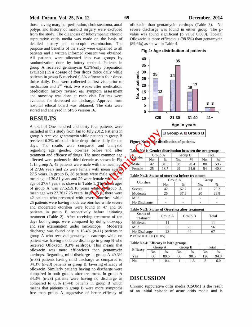
Med. Forum, Vol. 25, No. 12 December, 2014 69
those having marginal perforation, cholesteatoma, aural
polyps and history of mastoid surgery were excluded
from the study. The diagnosis of tubotympanic chronic
suppurative otitis media was made on the basis of
detailed history and otoscopic examination. The
purpose and benefits of the study were explained to all
patients and a written informed consent was obtained.
All patients were allocated into two groups by
randomization done by lottery method. Patients in
group A received gentamycin 0.3%(only preparation
available) in a dosage of four drops thrice daily while
patients in group B received 0.3% ofloxacin four drops
thrice daily. Data were collected at first visit prior to
medication and 2nd
visit, two weeks after medication.
Medication history review, ear symptom assessment
and otoscopy was done at each visit. Patients were
evaluated for decreased ear discharge. Approval from
hospital ethical board was obtained. The data were
stored and analyzed in SPSS version 16.
RESULTS
A total of One hundred and thirty four patients were
included in this study from Jan to July 2012. Patients in
group A received gentamycin while patients in group B
received 0.3% ofloxacin four drops thrice daily for ten
days. The results were compared and analyzed
regarding age, gender, otorrhea before and after
treatment and efficacy of drugs. The most common age
affected were patients in third decade as shown in Fig
1. In group A, 42 patients were male with the mean age
of 27.66 years and 25 were female with mean age of
27.5 years. In group B, 38 patients were male with the
mean age of 30.81 years and 29 were female with mean
age of 27.67 years as shown in Table 1. The mean ages
of group A was 27.52±9.16 years while in group B,
mean age was 27.76±7.25 years. In group A, there were
42 patients who presented with severe otorrhea, while
25 patients were having moderate otorrhea while severe
and moderated otorrhea were found in 47 and 20
patients in group B respectively before initiating
treatment (Table 2). After receiving treatment of ten
days both groups were compared by doing otoscopy
and rear examination under microscope. Moderate
discharge was found only in 16.4% (n-11) patients in
group A who received gentamycin eardrops while no
patient was having moderate discharge in group B who
received Ofloxacin 0.3% eardrops. This means that
ofloxacin was more efficacious than gentamycin
eardrops. Regarding mild discharge in group A 49.3%
(n-33) patients having mild discharge as compared to
34.3% (n-23) patients in group B, favoring efficacy of
ofloxacin. Similarly patients having no discharge were
compared in both groups after treatment. In group A
34.3% (n-23) patients were having no discharge as
compared to 65% (n-44) patients in group B which
means that patients in group B were more symptoms
free than group A suggestive of better efficacy of
ofloxacin than gentamycin eardrops (Table 3). No
severe discharge was found in either group. The p-
value was found significant (p value 0.000). Topical
Ofloxacin is more efficacious (98.5%) than gentamycin
(89.6%) as shown in Table 4.
Fig.1: Age distribution of patients
25 24
15
3
12
35
17
3
0
5
10
15
20
25
30
35
40
≤20 21-30 31-40 41+
Age in years
No
. o
f p
ati
en
tsGroup A Group B
Figure No.1: Age distribution of patients.
Table No.1: Gender distribution between the two groups
Gender Group A Group B Total
No. % No. % No. %
Male 42 31.3 38 28.4 80 59.7
Female 25 18.7 29 21.6 54 40.3
Table No.2: Status of otorrhea before treatment
Otorrhea Group A Group
No. % No. %
Severe 42 62.7 47 70.2
Moderate 25 37.3 20 29.8
Mild - - - -
No Discharge - - - -
Table No.3: Status of Otorrhea after treatment
Status of
treatment Group A Group B Total
Moderate 11 - 11
Mild 33 23 56
No Discharge 23 44 67
P value = 0.000 (<0.05)
Table No.4: Efficacy in both groups
Efficacy Group A Group B Total
No. % No. % No. %
Yes 60 89.6 66 98.5 126 94.0
No 7 10.4 1 1.5 8 6.0
DISCUSSION
Chronic suppurative otitis media (CSOM) is the result
of an initial episode of acute otitis media and is
Electro
nic Cop
y

Med. Forum, Vol. 25, No. 12 December, 2014 70
characterized by a persistent discharge from the middle
ear through a tympanic perforation. Among such
patients, medical treatment can be aimed at control of
infection and elimination of ear discharge as short-term
goals and eventual healing of the tympanic perforation
and improvement of hearing as ultimate goals. Medical
treatment options include aural toilet, use of antibiotics
(topical, systemic, parentral), topical antiseptics and
combination of these. In our study 134 patients were
recruited after informed consent from every patient.
Results showed that patients who underwent treatment
with ofloxacin (group B) showed better results in terms
of resolution of the ear discharge after the treatment as
compared to those treated with Gentamycin (group A).
Results of this study were quite similar to the study of
Kadar et al6, who recruited 120 patients in a
comparative clinical trial and divided into two groups:
aminoglycoside group of 60 patients were treated with
gentamycin hydrocortisone ear drops and quinolone
group of 60 patients were prescribed norfloxacin topical
solution with a dose of 6 drops in the affected ear twice
daily for two weeks. They concluded that in the medical
management of chronic suppurative otitis media, the
topical quinolones should be considered first line of
treatment as there are no ototoxic effects of quinolones,
which can be used safely in the presence of tympanic
membrane perforation. Tong et al8 conducted a double-
blind study compared two antibiotics, namely ofloxacin
and neomycin-polymyxin B, with similar in vitro
sensitivities to Gram positive and Gram negative
organisms. Fifty-two patients were selected randomly
and the results show that ofloxacin eardrops have
marginal benefits in symptomatic improvement (89 per
cent versus 79 per cent, p = 0.27) and bacterial
eradication (81 per cent versus 75 per cent, p = 0.81) in
active chronic suppurative otitis media. Significantly
fewer patients (seven per cent versus 29 per cent, p =
0.04) in the ofloxacin group had active disease at the
end of the two-week treatment. They recommended the
use of ofloxacin eardrops in managing active chronic
suppurative otitis media since it has high clinical
efficacy, contains no steroid component and has no
demonstrated risk of ototoxicity.
Lorente et al9
conducted a multicentric double-blind
randomized study to compare topical ciprofloxacin and
topical gentamycin in the treatment of simple non-
cholesteatomatous purulent chronic otitis media. Three
hundred and eight patients were included in the study,
159 treated with ciprofloxacin and 149 treated with
gentamycin. The percentage of clinical success
(elimination of otorrhea) was 95% with ciprofloxacin
and 94% with gentamycin. Likewise, the percentage of
bacteriological eradication was 96% with ciprofloxacin
and 93% with Gentamycin.In these patients, topical
ciprofloxacin shows the same efficacy as topical
Gentamycin without any potential ototoxic effects.
Tutkun et al10
determined and compared the therapeutic
efficiency of ciprofloxacin hydrochloride and
Gentamycin sulfate in the treatment of chronic ear
disease. They recruited 44 patients with chronic
suppurative otitis media randomized into two groups.
Ciprofloxacin hydrochloride (200 mg/mL) was
administered to the first group (composed of 24
patients), while the second group (composed of 20
patients) received Gentamycin sulfate (5 mg/mL)
locally, five drops three times a day for 10 days. In the
ciprofloxacin group, 21 (88%) of the 24 patients with
suppurative chronic otitis media were cured. On the
other hand, only six (30%) of the patients in the
Gentamycin group were cured. The rest of the patients
showed no clinical or bacteriological improvement.
They concluded that topical ciprofloxacin preparation is
more efficacious and efficient than topical Gentamycin
for the treatment of chronic otitis media in the acute
stage. Miro et al.11
administered otic drops of either
ciprofloxacin 0.2% solution or a combination of
polymyxin B, neomycin, and hydrocortisone
suspension (PNH) for 6 to 12 days to patients (14-71
years old) with chronic suppurative otitis media in a
randomized, non-blinded, multicenter clinical trial. Two
hundred thirty-two enrolled patients were analyzed for
efficacy on a per protocol basis. Clinical success was
observed in 91% and 87% of the ciprofloxacin and
PNH-treated patients, respectively. In the comparative
study of Supiyaphun et al12
reported that the efficacy
and safety of 0.3 per cent Ofloxacin otic solution 6
drops twice daily with those of oral Amoxycillin 500
mg three times daily plus 1 per cent Chloramphenicol
ear drop at 3 drops three times daily were compared in
a two-week treatment of chronic suppurative otitis
media with acute exacerbation. The susceptibility of all
the pathogenic isolates to ofloxacin, amoxycillin and
chloramphenicol were 96.4, 57.1 and 51.8 per cent
respectively.
Cure rate was significantly better in Ofloxacin treated
group than in AMOX + CRP-treated groups in terms of
painless (p = 0.05) and dry (p < 0.001) ears. Similarly
De Miguel Martínez et al13
conducted a randomized
study of 125 patients with chronic middle ear infection.
In order to study the effectiveness of ciprofloxacin in
chronic otitis media, they selected four different
treatment groups: oral ciprofloxacin (500 mg/12 h); 0.5
and 0.2% topical solutions of ciprofloxacin (3 drops/8
h), and oral ciprofloxacin plus 0.2% topical solution.
Topical polymyxin and neomycin were used as
controls. They found that topical ciprofloxacin (0.2%)
was the most effective regimen of those tested for the
treatment of chronic otitis media. In summary, there are
different treatment options for the management of
chronic suppurative otitis media. Variety of topical
antibiotics has been used and results of their efficacy
are reported in the literature. Topical Quiolones have
also been used in the treatment. In this study, 0.3%
Electro
nic Cop
y
Med. Forum, Vol. 25, No. 12 December, 2014 71
topical ofloxacin, a flouroquinolone, 4 drops thrice
daily was compared with 0.3% topical Gentamycin, an
aminoglycoside given in a dosage of 4 drops thrice
daily in the management of CSOM. It was found that
ofloxacin is a better choice than Gentamycin in terms of
resolution of ear discharge (otorrhea).
CONCLUSION
0.3% topical ofloxacin is a better choice in medical
management of CSOM than 0.3% topical gentamycin in
terms of resolution of ear discharge (otorrhea)
REFERENCES
1. Uddin W, Hussain A, Khan A, Ahmad F, Ullah S.
Prevalence and comparison of chronic suppurative
otitis media in government and private schools.
Ann Pak Inst Med Sci 2009; 5(3):141-4.
2. Aziz N, Preciado D. Middle ear, inflammatory
diseases. [online] 2007 [Cited2008April].
Availablefrom:(URL:www.http://emedicine.medsc
ape.com/article/860227-overview)
3. Iqbal SM, Udaipurwala IH, Hasan A, Shafiq M,
Mughal S. Chronic suppurative otitis media:disease
pattern and drug sensitivity. J Surg Pak 2006;
11:17-9.
4. Pappas S, Nikoloppulos TP, Korres S,
Papacharalampous G, Tzangarulakis A, Ferekidis
E. Topical antibiotic ear drops are they safe? Int J
Clin Pract 2006 60; 1115-9.
5. Parry D, Roland PS. Middle Ear, Chronic
Suppurative Otitis, Medical Treatment: Treatment
& Medication. [cited December 2008]. Available
http://emedicine.medscape.com
6. Kadar AA, Usman M, Tirmizi S, Topical
quinolones versus topical aminoglycosides in the
medical management of chronic suppurative otitis
media, a comparative trial. J Coll Physicians Surg
Pak 2008; 8:6-9.
7. Fareed G, Manzoor T, Ahmad S. Comparison of
Ciprofloxacin Versus Gentamycin Ear Drops in
Chronic Suppurative Otitis Media (Tubo-
tympanicType). Pak J Otolaryngol 2009;25:63-5.
8. Tong MC, Woo JK, van Hasselt CA. A double-
blind comparative study of ofloxacin otic drops
versus neomycin-polymyxin B-hydrocortisone otic
drops in the medicaltreatment of chronic
suppurative otitis media. J Laryngol Otol
1996;110:309-14.
9. Lorente J, Sabater F, Maristany M, Jimenez R,
Menem J, Vinas J, et al. Estudio multicentrico
comparativo de la eficacia y tolerancia de
ciprofloxacino topico (0.3%) versus gentamicina
topica (0.3%) en el tratamiento de la otitis media
cronica simple no colesteatomatosa en fase
supurativa. Anales Otorrinolaringologicos
Iberoamericanos 1995; 5:521-33.
10. Tutkun A, Ozagar A, Koc A, Batman C, Uneri C,
Sehitoglu MA.Treatment of chronic ear disease –
Topical ciprofloxacin vs topical gentamicin. Arch
Otolaryngol Head Neck Surg 1995; 121:1414-16.
11. Miro N. The Spanish ENT Study Group.
Controlled multicenter study on chronic
suppurative otitis media treated with topical
applications of ciprofloxacin 0.2% solution in
single-dose containers or combination of
polymyxin-neomycin, and hydrocortisone
suspension. Otolaryngol Head Neck Surg 2000;
123:617-23.
12. Supiyaphun P, Kerekhanjanarong V,
Koranasophonepun J, Sastarasadhit V. Comparison
of ofloxacin otic solution with oral amoxicillin plus
chloramphenicol ear drop in the treatment of
chronic suppurative otitis media with acute
exacerbation. J Med Assoc Thai 2009; 83:61-68.
13. De Miguel Martinez I, Vasallo Morillas J, Ramos
Macias A. Terapeutica antimicrobiana en otitis
media cronica supurada. Acta Otorrinolaring Esp
1999; 50:15-19.
Address for Corresponding Author:
Dr. Asmatullah
Assoc. Prof. of ENT, Head and Neck Surgery, Postgraduate
Medical Institute, Hayatabad Medical Complex Peshawar.
Email: [email protected]
Electro
nic Cop
y
Med. Forum, Vol. 25, No. 12 December, 2014 172
Outcome of Internal Fixation of
Fractures in a Tertiary Care Hospital in Peshawar 1. Mohammad Tariq Ijaz Afridi 2. Iftikhar Ahmad Chaudary 3. Mohammad Irfan Shereen
4. Asnad 5. Muhammad Adnan Shereen 1. Asstt. Prof. of Medicine, JMC, Pesharar 2. Asstt. Prof. of Community Medicine, MBBS MC, Mirpur AJ&K
3. Demonstrator of Physiology, KMU, Peshawar 4. Asstt. Prof. of Biochemistry, MBBS MC, Mirpur AJ&K
5. Demonstrator of Microbiology , KMU, Peshawar
ABSTRACT
Objective: This study was aimed at reviewing internal fixation in our hospital and attendant complications with a
view to identifying measures necessary to improve outcome.
Study Design: Retrospective study
Place and Duration of Study: This study was conducted at orthopedic department of Lady Reading Hospital,
Peshawar from March 2012to February 2014.
Materials and Methods: The operation register was used to identify patients who had undergone internal fixation
in the main theatre of the hospital over a Three-year period were collected and their case notes were subsequently
retrieved from the medical records unit of the hospital. Data pertinent to study interests were extracted using a
questionnaire
Results: One hundred and fifteen patients had internal fixation during the study period but case notes of only 100
patients could be retrieved. Most patients were males with male to female ratio of 2.3:1. The mean age of patients
was 32.87±15.2 years and the mean duration of surgery was 2±0.56 hours. Plate and screws constituted the most
commonly used implants. Interval between surgery and fracture union was increased by long operation time (> 2.
1hrs) and occurrence of post operative complications.
Conclusion: Improvement in operating facilities and choice of implants would reduce operation time and post
operative complications thereby impacting positively on fracture union time.
Key Words: infection, internal fixation
INTRODUCTION
Open reduction and internal fixation (ORIF) is a
commonly used treatment for fractures throughout the
body, including the distal femur. Supracondylar
fractures of the femur account for approximately 7% of
all femur fractures.1 They occur just proximal to the
knee joint, in the terminal 9 cm of the femur between
the metaphyseal-diaphyseal junction and the femoral
condyles.2
Fractures of the horizontal surface of the distal tibia are
known commonly as pilon or plafond fractures. They
represent 1–5% of lower extremity fractures and 7–10%
of all tibial fractures 3.
Operative fixation of pilon fractures has presented a
significant challenge to the orthopaedic surgeon as the
extensive soft tissue damage associated with
such injuries makes surgical intervention hazardous.
The traditional approach advocated by Rüedi and
Allgöwer 4, 5
involves an extensive dissection to the
distal tibia and has been associated with significant
rates of infection and wound dehiscence, ranging from
0–55%. 6, 7
Minimal disturbance of the soft tissue envelope is key
to the prevention of the common wound problems of
dehiscence and infection. The vascularity of the soft
tissue sleeve surrounding the distal tibia is tenuous 8–12
and aggressive handling with extensive periosteal
stripping will disturb the nutrition to the myocutaneous
tissue and underlying bone.
Internal fixation is a common method of treatment of
skeletal injuries. 13
The choice of internal fixation as a
method of fracture treatment is determined by a number
of factors including type of injury, facilities available
and expertise of the attending surgeon. 14
Internal
fixations like any other modality of fracture treatment
can be attended by complications such as implant
failure, infection, non-union among others. These are
often related to certain factors such as the operation
time, operating room conditions and availability of
appropriate skills and facilities.14
This study is aimed at reviewing the internal fixations
done in the Lady Reading Hospital, Peshawar with a
view to determining the methods of internal fixation
used, time taken for fractures to heal, factors
influencing this, complications as well as duration of
hospital stay post operatively. It is believed that the
information so obtained will positively influence the
institution of appropriate measures to improve quality
of practice with ultimate benefit to the patients.
MATERIALS AND METHODS
This study was conducted at orthopedic department of
Lady Reading Hospital, Peshawar from March 2012 to
February 2014.The operation register was used to
Original Article Fixation of Fractures
Electro
nic Cop
y
Med. Forum, Vol. 25, No. 12 December, 2014 173
identify patients who had undergone internal fixation in
the main theatre of the hospital over a three-year period
were collected and their case notes were subsequently
retrieved from the medical records unit of the hospital.
Data pertinent to study interests were extracted using a
questionnaire. Data obtained from the case notes
included patient’s demographics, type of injuries, pre-
operative prophylactic antibiotics and implants used,
rank of surgeon, duration of surgery, type of
anaesthesia, post operative complications and
treatment, duration of hospital admission post
operatively. The data obtained analysis was done using
SPSS version 16. Statistical methods such as correlation
and regression analyses, and non-parametric test for
comparison of means were used to explore relationships
between variables. A 0.05 significance level was used.
Results are expressed as means, frequencies and tables.
RESULTS
Hundred patients were recorded in the operation
register as having had internal fixation over the period
of the study. However, case notes of hundred patients
could be traced and retrieved for analysis representing a
retrieval rate of 77.2%. There were 70 males and 30
females, with a male to female ration of 2.3:1. Most of
the patients (98.1%) had some form of formal education
to varying levels. The mean age of patient’s was
32.87±15.2 years; mean duration of surgery was 2±0.56
hours; mean pre-operative Packed Cell Volume (PCV)
was 36.79±5.2%; mean post-operative PCV was
30±5.7%; mean number of units of blood transfused
was 1.57±0.8. 0.022).In one hundred and one cases
(89.0 %), surgery was done by a Consultant while 11
cases (11.0%) were done by resident doctors. Table No.
1. The main indication for internal fixation was fracture
(66.1%).The femur was the most operated bone
constituting 53.4% of cases followed by the tibia
(23.0%). Plate and Screws (59.1%) and Interlocking
intramedullary Nail (31.3%) were the most commonly
used implants. Table No. 2. Sub-Arachnoid block was
the most common anaesthetic technique used
(62.6%).The duration of surgery was found to be a
statistically significant predictor of the interval between
surgery and union of fracture, accounting for 15% of
the interval. The mean interval between surgery and
union was 10.81±2.66 weeks for upper limb fractures
and 18.11±2.11 weeks for lower limb fractures. Mean
duration of post operative admission was 18.91±13.98
days. Table No. 3. Thirty eight patients had one or more
complications. These were wound infection – 13,
osteomyelitis – 13, implant loosening – 3, implant
breakage – 6, knee stiffness – 5, non-union – 4,
shoulder stiffness – 2, wound dehiscence – 1, limb
shortening – 1, wrist drop – 1, exposed implant – 1, pin
track infection – 1, common peroneal nerve injury – 1,
faulty screw placement – 1.Twelve of the patients
(12.0%) had post-operative infection and 88 patients
had not post-operative infection. This constituted the
most common complication. Organisms isolated were
Staphylococcus aureus 7(58.33%), Pseudomonas
aeruginosa 3(25.0%), Coliforms 1(8.3%), Entero-
bacteria 1(8.3%). Table No. 4. Mixed infections were
found in 6patients (50.0%); 5 (41.66%) of which were
mixed Staph. aureus and Pseudomonas infections.
The complications were treated by administration of
antibiotics in 17 cases (17.0%), wound dressing
13(13.0%), and debridement 1(1.0%).
Table No.1: Percentage Distribution between
consultant and resident Doctor in Orthopaedic
Department of LRH Peshawar
Operated by % Cases
Consultant 89%
Resident Doctor 11%
Table No.2: Percentage cases of Surgery in
Orthopaedic Department of LRH Peshawar
Surgery % Cases
Femur 53.1%
Tibia 23%
Plate and screw 59.1%
Intramedullary Nail 31.3%
Table No.3: Mean Interval between Surgery and
Union in Orthopaedic Department of LRH
Peshawar
Surgery Mean Duration
Upper Limb Fracture 10.81 ± 2.66 weeks
Lower Limb fracture 18.10 ± 2.11 weeks
Post operative Admission 18.9 ± 13.98 days
Table No.4: Post Operative Infections
Microorganism % Infected % Non-
Infected
12% 88
Staphylococcus aureus 7 (58.3%)
Pseudomonas aeruginosa 3 (25%)
Coliforms 1 (8.3%)
Enterobacteria 1 (8.3%)
DISCUSSION
Internal fixation is the preferred method of fracture treatment in many cases. Operative fracture treatment results in early mobilization, short hospital stay, and early return to productive economic activities and less social dislocations.
13-15 The cases of non-union and
mal-union treated were mostly in patients who had previously received treatment from the traditional bone setters (TBS) which is a rather common practice in our society.
16-18 This study shows that fractures of
the long bones of the lower limb are more frequently treated by internally fixation compared to the upper limb fractures - based on absolute numbers (6.4:1). The finding agrees with that of another study.
19 This
Electro
nic Cop
y
Med. Forum, Vol. 25, No. 12 December, 2014 174
may be due to the need to mobilize the patient early and prevent further complications. The more frequent fixation of femoral fractures (53.4% ) compared to the tibia (23.0%) in this study is consistent with results of another local study
19 and may be due to the more
common occurrence of severe open fractures in the tibia which makes them unsuitable for internal fixation. Furthermore, non operative method such as casting is widely used in treatment of tibial fractures. This practice is encouraged by the aversion of patients to operative treatment in our society.
16
Even though the trend is changing in recent years, plate and screws was the most common implant used for internal fixation in this study as in many local studies.
19,20 This may be due to easy availability of the
implant and instrumentation as well as lower cost. All the interlocking intramedullary nails (IM Nails) inserted in these patients were done open due to the non-availability of intra-operative imaging facilities. This has greatly limited the use of the IM nails even in the treatment of femoral and tibial shaft fractures where it is the preferred method. Post operative infection was 13%. Even though very high compared to results from developed societies
, 21
the figure is consistent with finding of another local study in which a rate of 12% was recorded following internal fixation.
20 This may be partially attributed to
extensive soft tissue dissection and periosteal stripping involved in insertion of plate and screws
13, 14 the most
common method of internal fixation in our centre. This study shows that the union time of the fractures was directly proportional to the operation time and occurrence of post operative complications. In other words, it would take much longer time for a fracture treated by internal fixation to heal if the operation time is long (>2.1hrs) and there being associated post operative complication. A long operation time may actually reflect difficulty at surgery as a result of the complex nature of the fracture being treated. Even though it would be desirable to drastically reduce operation time without compromising accuracy and safety, it is also expedient that measures to prevent post operative complications are instituted
CONCLUSION
Complications following internal fixation especially
infection, as shown by this study, is still unacceptably
high and limits the benefits derivable from internal
fixation. This may be attributed to many factors
including inadequate operating environment and
facilities as well as the lack of choice of appropriate
implants. It is therefore imperative that measures be
taken by relevant authorities to improve on the facilities
in our hospitals and ensure access to appropriate
implants for more effective and result-oriented
treatment of fractures in our society. This calls for a
paradigm shift in allocation of health resources
REFERENCES
1. Bucholz R, Heckman J, Court-brown C. Rockwood and Green's fractures in adults. Vol. 1. 6
th ed.
Philadelphia: Lippincott Williams & Wilkins; 2009.
2. Albert MJ. Supracondylar Fractures of the Femur. J Am Acad Orthop Surg 1997;5(3):163-171.
3. Egol KA, Wolinsky P, Koval KJ. Open reduction and internal fixation of tibial pilon fractures. Foot Ankle Clin 2000;5(4):873–885.
4. Rüedi TP, Allgöwer M. Fractures of the lower end of the tibia into the ankle joint. Injury 1969;2: 92–99.
5. Rüedi TP, Allgöwer M. The operative treatment of intra-articular fractures of the lower end of the tibia.Clin Orthop Relat Res 1979;138:105–110.
6. Patterson MJ, Cole JD. Two-staged delayed open reduction and internal fixation of severe pilon fractures. J Orthop Trauma 1999;13(2):85–91.
7. Dillin L, Slabaugh P. Delayed wound healing, infection, and nonunion following open reduction and internal fixation of tibial plafond fractures. J Trauma 1986;26(12):1116–1119.
8. Borrelli J, Prickett W, Song E, Becker D, Ricci W. Extraosseous blood supply of the tibia and the effects of different plating techniques: a human cadaveric study. J Orthop Trauma 2002; 16(10):691–695.
9. Macnab I, Haas WG. The role of periosteal blood supply in the healing of fractures of the tibia. Clin Orthop Relat Res 1974;105:27–33.
10. Zhang L, Bail H, Mittlmeier T, Haas NP, Schaser KD. Immediate microcirculatory derangements in skeletal muscle and periosteum after closed tibial fracture. J Trauma. 2003;54(5):979–985.
11. Dickson KF, Katzman S, Paieent G. The importance of the blood supply in the healing of tibial fractures. Contemp Orthop 1995;30(6): 489–493.
12. Menck J, Bertram C, Lierse W. Sectorial angioarchitecture of the human tibia. Acta Anat (Basel) 1992;143(1):67–73.
13. Gerber C, Mast JW, Ganz R. Biological internal fixation of fractures. Archives of Orthopaedic and Trauma Surgery 1990;109(6):295-303.
14. Solomon L, Warwick D, Nayagam S. Apley’s System of Orthopaedics and Fractures, 8tj ed. Arnold (Publishers) Ltd;2001.p.553–555.
15. Reeves RB, Ballard RI, Hughes JL. Internal fixation versus traction and casting of adolescent femoral shaft fractures. J Pediatric Orthopedics 1990;10(5):592-
16. Ngim NE, Udosen AM, Ikpeme IA, Ngim OE. Prospective Study of Limb Injuries in Calabar. The Int J Orthopedic Surg 2008;2(2).
Electro
nic Cop
y

Med. Forum, Vol. 25, No. 12 December, 2014 175
17. Udosen AM, Otei OO, Onuba O. The role of traditional bonesetters in Africa: Experience in Calabar, Nigeria. Annals of Afr. Med 2006; 5(4): 170 – 173.
18. Ogunlusi DO, Ikem IC, Oginni. Why Patients Patronisew Traditional Bone Setters. The Internet J Orthopaedic Surg 2007;4(2).
19. Thanni Lateef OA, Aigoro Nofiu O. Surgical site infection complicating internal fixation of fractures: incidence and risk factors. J Natl Med Assoc 2004;96(8):1070–1072.
20. Solomon L, Warwick D, Nayagam S. Apley’s System of Orthopaedics and Fractures, 8th ed. Arnold (Publishers) Ltd;2001.p.35.
21. Kasatpibal N, Jamulitrat S, Chongsuvivatwong V. Standardized incidence rates of surgical site infection: a multicenter study in Thailand. Am J Infect Control 2005;33(10):587-94.
Address for Corresponding Author:
Dr. Asnad,
Assistant Professor of Biochemistry
MBBS Medical College, Mirpur AJ&K
Contact No: 0332-3698204
Corrigendum
Name and designations of the authors in the articles published in Med Forum Vol. 25 No.11 at pages mentioned against each, may be read as follows: 1. Inter-Relationship of Circulating
Biochemical Markers of Oxidative Stress and Thyroid Hormones in newly Diagnosed Schizophrenics: Perspective study from Local Population of Punjab Pakistan (Pages 19-22)
1. Waheed Jamil 2. Shumaila Saleem 3. Arif Malik 4. Naveed Shuja 5. Abdul Manan 6. Sumaira Shaheen 7. Mahmood Husain Qazi 1. Asstt. Prof. of Physiology, AIMC, Lahore 2. Asstt. Prof. of Physiology, UCMD, The University of Lahore, Lahore-Pakistan 3. Assoc. Prof. of Biochemistry, Institute of Molecular Biology and Biotechnology (IMBB), The University of Lahore, Lahore-Pakistan 4. Lecturer of Biochemistry, LMDC, Lahore 5. Demonstrator of Biochemistry, Institute of Molecular Biology and Biotechnology (IMBB), The University of Lahore, Lahore, Pakistan 6. Lecturer, CRiMM, The University of Lahore, Lahore-Pakistan 7. Director, Centre for research in molecular Medicine (CRiMM), The University of Lahore, Lahore-Pakistan
2. Significance of Hepatic Profile and
Malondialdehyde as Marker of Lipid Peroxidation in HCV Patients: A Perspective Study from Local Population of Punjab-Pakistan (Pages 31-34 )
1. Ghazal Mansur 2. Nusrat Tariq 3. Mahwish Arooj 4. Arif Malik 5. Hafiz Muhammad Arsalan 6. Mahmood Husain Qazi 1, 2. Assist. Prof. of Physiology, Sharif Medical and Dental College, Lahore-Pakistan, 3. Assoc. Prof. of Physiology, UCMD, The University of Lahore, Lahore- Pakistan 4. Assoc. Prof. of Biochemistry, Institute of Molecular Biology and Biotechnology (IMBB), The University of Lahore, Lahore- Pakistan 5. Demonstrator of Biochemistry, Institute of Molecular Biology and Biotechnology (IMBB), The University of Lahore, Lahore-Pakistan 6. Director, Centre for research in
molecular Medicine (CRiMM), The University of Lahore, Lahore-Pakistan 3. Response of Antiretroviral Therapy
(Ziduvodine, Lamivudine and Niverapine) in Patients Suffering from Acquired Immuno Deficiency Syndrome (Aids) (Pages 56-59)
1. Humaira Shoukat 2. Ghazal Mansur 3. Nusrat Tariq 4. Arif Malik 5. Abdul Manan 6. Mahmood Husain Qazi 1. Assist. Prof. of Physiology, Akhtar Saeed Medical and Dental College, Lahore-Pakistan 2, 3. Assist. Prof. of Physiology, Sharif Medical and Dental College, Lahore-Pakistan 4. Assoc. Prof. of Biochemistry, Institute of Molecular Biology and Biotechnology (IMBB), The University of Lahore, Lahore-Pakistan 5. Demonstrator of Biochemistry, Institute of Molecular Biology and Biotechnology (IMBB), The University of Lahore, Lahore-Pakistan 6. Director, Centre for research in molecular Medicine (CRiMM), The University of Lahore, Lahore-Pakistan 4. Inter-Relationship of Viral Load and CD
4+
Cells in Patients Suffering from Acquired Immuno Deficiency Syndrome (Aids): Update from Punjab-Pakistan (Pages 80-82)
1. Nusrat Tariq 2. Ghazal Mansur 3. Humaira Shoukat 4. Arif Malik 5. Abdul Manan 6. Mahmood Husain Qazi 1, 2. Assist. Prof. of Physiology, Sharif Medical and Dental College, Lahore-Pakistan 3. Assist. Prof. of Physiology, Akhtar Saeed Medical and Dental College, Lahore-Pakistan 4. Assoc. Prof. of Biochemistry, Institute of Molecular Biology and Biotechnology (IMBB), The University of Lahore, Lahore-Pakistan 5. Demonstrator of Biochemistry, Institute of Molecular Biology and Biotechnology (IMBB), The University of Lahore, Lahore-Pakistan 6. Director, Centre for research in molecular Medicine (CRiMM), The University of Lahore, Lahore-Pakistan Editor in Chief
Electro
nic Cop
y
Med. Forum, Vol. 25, No. 12 December, 2014 76
Author Index January to December 2014 Azhar Masud Bhatti
Editor in Chief
Vol. 25, No. 1, January, 2014
Author (s) Page No.
1. Jan MM 1
2. Memon ZA, Pandhiani MB, Shaikh AA,
Khoharo HK 2
3. Samina, Kalim D, Zahoor A, Akhund IA,
Ishaq M 6
4. Bhatti K, Mahar T, Hafeez R, Nisa S 10
5. Sohail MMA, Sahito RA, Mahar FA 13
6. Asad F, Rizvi F, Iqbal J 18
7. Yasmeen T, Siddiqui IA, Amjad Z,
Shoro AA, Mirza T 22
8. Ibran E, Rao MH, Khan M, Hasan F,
Khaliq RA, Zehra S 27
9. Soomro MA, Ansari IA, Abro GY,
Shah SAA, Shah SSR 31
10. Alam N, Najam R 36
11. Anwer J, Babar MI, Niazi K,
Mahmood Z 40
12. Rehman M, Imran M, Kang TA 44
13. Imran M, Rehman M, Kang TA 49
14. Pandhiani MB, Kazi M, Rehman S,
Kazmi N, Kazi SAF, Khoharo HK 53
15. Kazi M, Pandhiani MB, Kazi SAF,
Rehman S, Khoharo HK 57
16. Khokhar GN, Ashfaq M, Khan IU,
Ishaq M, Ahmed I 62
17. Nazar A, Ahmed R, Bano S, Ashraf M,
Altaf I, Javeed A 66
18. Qasmi SA, Khan AA, Askari H,
Pirzada R 71
19. Shaikh AA, Pandhiani MB, Memon ZA,
Khoharo HK 76
20. Mehmood Z, Mengal MH, Rasheed H,
Ahmed HKN 80
21. Jaffery SA 85
22. Siddiqui QA, Zaidi SIH, Hussain A,
Shamim MO, Hussain N, Naqvi SNH,
Arshad M, Tariq RM, Hijazi M 90
23. Bhatti AM 95
24. Bhatti AM 104
Vol. 25, No. 2, February, 2014
Author (s) Page No.
1. Jan MM 1
2. Imran M, Rana ZA, Rabbani MW,
Iqbal I, Raza H 2
3. Sheikh MR, Rana A, Bhatti AM 7
4. Salam J, Baloch AA, Siddiqui MH,
Bashir B, Nisa Q, Qasim A, Masroor M 11
5. Younas M 15
6. Rathore MI, Memon S, Pushpa 18
7. Fida R, Muhammad S, Ashfaq M,
Asnad, Ishaq M 23
8. Kumar A, Shahani RA, Baloch S,
Bajarani SA 27
9. Siddiqui MH, Baloch AA, Qasim A,
Bashir B, Salam J 30
10. Mehdi H, Girach MM, Lakhani MJ 33
11. Anwar A, Anwar S 37
12. Akhtar S, Jan R, Askar G 40
13. Chughtai BR, Iqbal M, Bhatti AM 44
14. Das D, Rai P, Dodani AL, Shaikh RA,
Perkash J 47
15. Laghari AH, Memon AN, Samoo AH,
Qurashi KA, Shah AM 51
16. Saleem M, Ahmad M, Siddiqui BA,
Ijaz S 55
17. Chang F, Sahito MM, Naz R, Shams R,
Sahito RA 59
18. Jan R, Akhtar S 64
19. Soomro RA, Memon KN, Ali SM 68
20. Nazar A, Ahmed R, Bano S, Ashraf M,
Altaf I, Javeed A 72
21. Khan K, Khan M, Qazi WE 78
22. Imran M, Khan AU, Rehman M 83
23. Laghari M, Khand F, Channa NA,
Khoharo HK 88
Vol. 25, No. 3, March, 2014
Author (s) Page No.
1. Jan MM 1
2. Jat N, Bano F, Memon IA, Khokhar PB,
Azmi MA 2
3. Lashari MN, Alam MT, Ashraf T,
Pathan MA 6
4. Siddique M, Achakzai A, Ahamad I,
Tareen S 10
5. Sheikh MR, Bhatti AM 14
6. Nawaz U, Ilyas N, Jehangir A, Sadiq S 20
7. Khan FA, Ashrafi KA, Jawaid I,
Wadood, Abbasi A 24
8. Ejaz A, Khan AA, Haq QM 27
9. Rathore JA, Saleem M, Trumbu BA 31
10. Ahmad I, Siddique M, Kakar A,
Tareen S 35
11. Nadeem MA, Shakaib M, Iqbal H,
Farooqi TH 38
Author Index
Electro
nic Cop
y
Med. Forum, Vol. 25, No. 12 December, 2014 77
12. Naeem S, Najam R, Alam N 41
13. Razzaq Q, Aziz S, Adil R 46
14. Qureshi KA, Laghari AH, Samoo AH 50
15. Mubeen S, Rafique M, Ilyas A 54
16. Sahito MM, Chang F, Sheikh N,
Mughal MI, Mir A, Iqbal F 59
17. Pathan N, Fahim A, Khan A, Qureshi A,
Khoja S 63
18. Naz R, Afzal I, Zain Z, Kamran A,
Mughal MI 67
19. Lashari AA, Bhatti K, Mahar T,
Hafeez R 71
20. Khan MI, Asad K, Khattak RA 74
21. Jan R, Askar Z 78
22. Akhtar S, Gillani S 82
23. Baloch AA, Siddiqui MH, Bashir B,
Majid U, Qasim A, Aurangzaib,
Salam J, Masroor M, Nisa Q 86
24. Lakhani MJ, Girach M, Kadri W,
Hasan U, Sheikh S, Sultan H,
Khan H, Abro MI 89
Vol. 25, No. 4, April, 2014
Author (s) Page No.
1. Jan MM 1 2. Malik A, Shaukat MS, Qureshi A,
Rasheed A 3 3. Lashari AA, Bhatti K, Mahar T,
Hafeez R 7 4. Chaudhry AG, Ahmed A, Nasir A,
Sohail M, Zeeshan M 10 5. Chughtai BR, Iqbal M, Bhatti AM 15 6. Qureshi RN, Khalid E, Hameed N 19 7. Imran M, Adnan M, Ali Z 23 8. Taj Y, Soomro AA, Ali SW, Kazmi SU 28 9. Nadeem MA, Iqbal H, Adnan A,
Farooqi TH 33 10. Sair A, Haq A, Tariq A 37 11. Hanif M, Din SZ, Bhatti MA 41 12. Ahmad RI, Shaukat MS, Shaheen A,
Rahim A 44 13. Bux M, Latif A, Mohammad S,
Asnad, Ishaq M 49 14. Mal K, Shaikh BA, Khalid S.
Jalbani A 52 15. Ahmed A, Chaudhry AG, Bhatti AG,
Zeeshan M, Dinar H 55 16. Rathore JA, Saleem M, Ahmed M 60 17. Ata MA, Atif H, Shaikh SS, Ata MA 63 18. Shaukat F, Tassaduq I, Irfan A 66 19. Khalid E, Hameed N 70 20. Ahmad S, Ahmad M, Hussain N,
Khan TR 75 21. Ali Z, Imran M, Khan WI, Sheikh MA,
Rabbani MW 78 22. Rehman H, Sarwar U, Majeed M 83 23. Shah M, Shah S, Shah M 88
Vol. 25, No. 5, May, 2014
Author (s) Page No.
1. Jan MM 1
2. Chang F, Sahito MM, Qazi RA 2
3. Jabeen S, Qureshi GS, Rafique M 7
4. Rathore MI, Khooharo Y, Balouch AH 11
5. Ahmed H 16
6. Shaheen S, Khattak NN, Parveen T 20
7. Arain SN, Mehmood Z, Shaikh SS 23
8. Pahore MK, Laghari MY, Niaz A,
Bhand SA, Saeed M 26
9. Jamali SN, Turab SM, Siddiqui MH,
Aurangzeb M, Salam J, Qasim A,
Bashir B, Balouch AA 30
10. Rathore JA, Kango ZA, Saleem M 33
11. Kazi SN, Fahim A, Qureshi A,
Sheikh KR, Kazi N, Khan A 37
12. Saeed TB, Ashfaq M, Ahmed S,
Qureshi R, Hussain SS, Pervez S 42
13. Chaudhry AG, Ahmed A, Farooq H,
Bhatti AG, Zeeshan M 46
14. Qasim AP, Tariq SA, Naeem M 51
15. Shakoor I, Ghazal R, Fareeduddin M,
Noor M 55
16. Girach M, Lakhani MJ, Kadri W,
Ijaz Y, Griffin M 59
17. Sohail S, Rehman K, Usman T62
18. Samina, Nabi I, Khokhar GN,
Akhund IA, Ishaq M 66
19. Khan S, Badshah N, Samina, Asnad,
Ishaq M 70
20. Ali Z, Shafique S, Sheikh AA,
Hussain SS 73
21. Bhatti AM, Chughtai BR, Iqbal M 77
22. Ahsen W, Meraj B, Waseem M,
Ahmed S 81
23. Shakoor S, Shaikh HA, Khan FR,
Shad KF 85
24. Jamshed F, Ahmad W, Saleem M,
Sarwar J, Gul N 90
25. Mengal MH, Mehmood Z, Kapoor C,
Iqbal M, Mengal MA 95
Vol. 25, No. 6, June, 2014
Author (s) Page No.
1. Jan MM 1
2. Khan MW, Chaudhry RA, Sadiq M 2
3. Siddique M, Kakar AL, Ahmad S,
Tareen S 6
4. Rathore JA, Saleem M, Khan AG 9
5. Chaudhry AG, Ahmed A, Zeeshan M,
Mehmood R 13
6. Gulnaz H, Khalid S 18
7. Jawed M, Shaikh AA, Khan MI 22
Electro
nic Cop
y
Med. Forum, Vol. 25, No. 12 December, 2014 78
8. Bashir U, Butt AM, Hassan SG,
Haq M, Shams S 26
9. Arain B, Punjabi SK, Hassan Q,
Shaikh AA 29
10. Latif A, Farhan MA, Waliullah K,
Hamid A 33
11. Nazish Z, Inayatullah M, Mustafa F 38
12. Yousaf R, Jawed S, Yasmin T,
Rashid A 43
13. Shafi S 47
14. Bukhari AAS, Khokhar GN, Lakho GR,
Shah M 50
15. Jan R, Akhtar S 54
Vol. 25, No. 7, July, 2014
Author (s) Page No.
1. Jan MM 1
2. Rathore JA, Kango ZA, Saleem M 2
3. Ahmed H 6
4. Sharafat Z 9
5. Jawed M, Jamali KS, Sana S,
Shaikh U, Shaikh SU 14
6. Waliullah K, Farhan MA, Latif A,
Hamid A 18
7. Ahmed A, Chaudhry AG, Farooq H 22
8. Zulqarnain A, Iqbal I, Haq MA 27
9. Asad, Rehman H, Yasmin, Hamid A 32
10. Qazi AQS, Irfan N, Shaikh SS 36
11. Khan KH, Adil S, Khan BH, Khan Z,
Sheikh AH 39
12. Pechuho MA, Kumar K, Ahmad R 43
13. Shafi S 47
14. Khan MS, Fahim A, Kazi SA, Qureshi A.
Azmi MA, Kashif S, Zulfiqar S 53
15. Hameed A, Sadozai SK, Rahim F,
Ullah R, Aurakzai R 56
Vol. 25, No. 8, August, 2014
Author (s) Page No.
1. Jan MM 1
2. Anjum MU, Shah SH 2
3. Chaudhry RA, Khan AM, Mahmood A 6
4. Memon MH, Hanif S, Javed M,
Ahmed MM 10
5. Ahmed A, Chaudhry AG, Farooq H 14
6. Ahmad N, Shahzad MN, Ahmad S 19
7. Karim F, Mehsar AL, Bux M, Ishaq M,
Ahmed I 24
8. Waheed A, Nawaz U, Miana GA 27
9. Aziz S, Razzaq Q, Adil R 32
10. Afzal I, Naz R, Afzal MK 36
11. Ahmad S, Ahmad N, Shahzad MN 42
12. Nazish Z, Mustafa F, Inayatullah M 46
Vol. 25, No. 9, September, 2014
Author (s) Page No.
1. Jan MM 1
2. Afzal I, Naz R, Afzal MK, Mughal MI 2
3. Memon S, Shahani SB, Bano U,
Shahani MY 6
4. Khurshid A, Khan MA, Fatima F 10
5. Nisa Z, Hassan Q, Hassan SG, Shams S,
Khan MSU 13
6. Khan MA, Fatima F, Khurshid A 16
7. Shehzad M, Nisa Z, Hassan Q, Habib G 18
8. Shafi S 21
9. Kalhoro FA, Rehman M, Rajput R,
Shaikh MA, Karim K 24
10. Ahmed T, Ahmed S, Rahman AS,
Rahman A, Husain T, Ahmed L 28
11. Nadar S, Iqbal M, Raja KS,
Rana PA, Khokhar JI 32
12. Chang F, Pirzado MS, Qazi RA,
Sahito RA 36
13. Chohan AM, Kadri IA, Haider G,
Memon I 41
Vol. 25, No. 10, October, 2014
Author (s) Page No.
1. Jan MM 1
2. Habib G, Nisa Z, Memon QN, Hassan Q,
Shams S 3
3. Channa SR, Anjum F, Haider G 6
4. Qureshi MA, Bhurgri GH, Yousfani GM 10
5. Khan MA, Sheikh TH, Naeem A 15
6. Parveen S, Latif A, Ashraf M 18
7. Khan MW, Sadiq MT, Rafique K,
Mahmood S 21
8. Iltaf S, Siraj M 26
9. Siddiqui R, Mangi MM, Shah AA,
Bhutto SA 31
10. Memon A, Pirwani M, Memon SA 35
11. Imran M, Ali Z, Khan WI, Raza H 40
12. Arif M, Arif MK, Pervaiz NA, Ahmad Z 46
13. Rehman H, Mahesar AL, Khalid SN,
Ishaq M 50
14. Mahesar AL, Rehman H, Nabi I,
Ishaq M 53
15. Nisa Z, Hassan Q, Memon QN,
Habib G, Shams S 56
16. Gul P, Gul Z 59
Vol. 25, No. 11, November, 2014
Author (s) Page No.
1. Jan MM 1
2. Parveen S, Latif A, Ashraf M 2
3. Rabia M, Rasheed T, Farooq U,
Ahmed I 5
Electro
nic Cop
y
Med. Forum, Vol. 25, No. 12 December, 2014 79
4. Zulqarnain A, Maqbool B, Iqbal I 9
5. Abbasi MA, Farid J, Sahito RA,
Saeed M 12
6. Sheikh S 15
7. Jamil W, Saleem S, Malik A, Shuja N,
Manan A, Shaheen S, Qazi MH 19
8. Lakhani MJ, Girach M, Qadri W,
Malik S, Riaz S, Qayum KA 23
9. Ansari SA, Syed SA, Aslam K 26
10. Mansur G, Tariq N, Arooj M, Malik A,
Arsalan HM, Qazi MH 31
11. Maqbool A, Athar Z, Hameed O 35
12. Farooq MS, Asif M, Shaheen B,
Manzoor Z 40
13. Jaffery MF, Alam MT, Aurangzeb M,
Rasheed T, Masroor M, Nawaz Z,
Khero S 45
14. Memon S, Shahani MY, Bano U,
Shahani SB 48
15. Khan I, Khan MI, Jawed M, Shaikh U,
Ahmed S, Arif A 52
16. Shoukat H, Mansur G, Tariq N,
Malik A, Manan A, Qazi H 56
17. Shah SA, Laghari MA, Shah KA,
khan MP, Shah S 60
18. Abbasi F, Ahmed S, Jawed M, Khan MI,
Aurangzeb M, Anwer Z 64
19. Khan AM, Mughal TM, Khan AM,
Haq E, Khan M 68
20. Pervaiz AA, Razzak NA, Aziz A,
Sultan N 72
21. Mughal TM, Khan AM, Khan AM,
Haq E, Khan M 76
22. Tariq N, Mansur G, Shoukat H, Malik A,
Manan A, Qazi MH 80
Vol. 25, No. 12, December, 2014
Author (s) Page No.
1. Jan MM 1
2. Ch WJ, Chishti MB, Ashraf M, Malik A,
Qazi MH 2
3. Hashme RI, Khan NA 7
4. Asnad, Afridi MTI, Tahir M, Shereen
MA 11
5. Javaeed A, Shah W, Akhtar R, Ghauri
SK, Khan SH, Alvi AH 15 6. Khan GS, Bukhari IAS, Musarrat,
Ahmed I, Ishaq M 20
7. Memon MY, Kalhoro FA, Jabbar A,
Memon AB, Shaikh IA 23
8. Arshad J, Abrar N, Khan MA, Jahan S 26
9. Masud KU, Nagi AH 31
10. Siddiqui R, Mangi MM, Shah AA, Soomro
RA, Memon KN 35
11. Shaikh S, Shaikh F, Jatoi S 39
12. Khan NA, Hashme RI, Bhatti AM 42
13. Mamoun MA, Rizvi STI, Alvi HMF 46
14. Iqbal M 52
15. Khan MI, Jawed M, Bhura S, Shaikh U,
Arif A 57
16. Jesswani ML, Imaduddin S, Memon MA,
Razzak R 60
17. Jatoi S, Shaikh S, Shaikh F 65
18. Asmatullah, Khan Q, Nawaz G, Ullah G,
Iqbal J, Khan M, Muhammad R 68
19. Afridi MTI, Chaudary IA, Shereen MI,
Asnad, Shereen MA 72
Electro
nic Cop
y
Med. Forum, Vol. 25, No. 12 December, 2014 80
Subject Index January to December 2014 Azhar Masud Bhatti
Editor in Chief
Vol. 25, No. 1, January, 2014
Subject Page No.
ANATOMY
Hepatoprotective Effects of Curcuma Longa
against Carbon Tetrachloride Induced Liver Injury
in Rats (Memon ZA, et al) 2
Alterations in Mitochondria of Kidney Tubules
by Different Doses of Diclofenac Sodium in
Rabbits (Yasmeen T, et al) 22
UTEROVAGINAL PROLAPSE
Proportion of Urinary Symptoms in Pre and
Post-Menopausal Women with Uterovaginal
Prolapse (Samina, et al) 6
POSTPARTUM HAEMORRHAGE
A Randomized Controlled Trial on Prevention of
Postpartum Haemorrhage with Sublingual
Misoprostol or Oxytocin (Bhatti K, et al) 10
PERITONITIS
Causes and Management Out Come of Peritonitis
(Sohail MA, et al) 13
PERMETHRIN IN SCABIES
Beneficial Effect and Safety of 5% Permethrin
Cream in Scabies Patients (Asad F, et al) 18
DRESSING ON BURN WOUND
Efficacy of Platelet Rich Plasma Application in
Comparison to Conventional Dressing Therapy in
Partial Thickness Burn Wound (Ibran E, et al) 27
ESOPHAGEAL VARICES
Prediction of Large Esophageal Varices in
Patients with Decompensated Cirrhosis by
ChildPugh Score, in Medical Unit-II, Chandka
Medical College Hospital, Larkana (Soomro MA,
et al) 31
PHARMACOLOGY
Cognition-Enhancing Effect of Oral Therapeutic
Doses of Methylphenidate in Rats (Alam N,
et al) 36
Validity of Pleural Fluid Protein in
Differentiating Tuberculouse from Malignant
Pleural Effusion (Khokhar GN, et al) 62
To Assess the In vitro Genotoxicity of Metformin and
Aspartame alone & In Combination (Nazar A,
et al) 66
The Effects of Monosodium Glutamate on the
Histology of Fallopian Tube in Female Rats
(Shaikh AA, et al) 76
Comparative Study on Neem Leaf Extract and
Nimolicin (NC) on Gastric Mucosa of Albino Rats
(Sidiqui QA, et al) 90
EPI STATUS
EPI Status under One Year Children and their
Mothers attending Paediatric Department Sheikh
Zayed Medical College & Hospital, Rahim Yar
Khan (Anwer J, et al) 40
INTRACAPSULAR HIP FRACTURES
Functional Outcome of Cemented Versus
Uncemented Hemiarthroplasty for Intracapsular
Hip Fractures (Rehman, et al) 44
OSTEOARTHRITIS
Efficacy of Intra Articular Injections in Different
Grades of Osteoarthritis of Knee (Imran M) 49
THIAMINE ON GLYCEMIC CONTROL
Effect of Thiamine on Glycemic Control in
Induced Diabetic Rat Model (Pandhiani MB,
et al) 53
PENTOXIFYLLINE ON LIVER
Pentoxifylline Protects against Carbon
Tetrachloride Induced Liver Injury in Adult Male
Wistar Rat Model (Kazi M, et al) 57
DIABETES MELLITUS
Study of Type 2 Diabetes Mellitus and its
Correalation with Blood Groups (Qasmi SA,
et al) 71
MALIGNANT LYMPHOMAS
Incidence of Malignant Lymphomas in Balochistan
(Mehmood Z, et al) 80
GLIOBLASTOMA
Adjuvant Therapy for Old Age Glioblastoma
Patients (Jaffery SA) 85
Subject Index
Electro
nic Cop
y
Med. Forum, Vol. 25, No. 12 December, 2014 81
Vol. 25, No. 2, February, 2014
Subject Page No.
NEPHROTIC SYNDROME
Histopathological Patterns in Childhood Steroid
Resistant Nephrotic Syndrome (Imran M, et al) 2
ARTERIAL SUPPLY OF BRAIN
Variable Arterial Supply of Motor Areas of
Human Brain (Sheikh MR, et al) 7
MYASTHENIA GRAVIS
Repetitive Nerve Stimulation Test, An
Investigation that helps in Confirming Diagnosis
in Seronegative Myasthenia Gravis (Salam J,
et al) 11
LIGNOCAINE IN NASAL PACKS
Efficacy of 5% Lignocaine Ointment in
Reducing the Post-Operative Pain due to Intra-
Nasal Packs (Younas M) 15
ROLE OF VIT. E ON KIDNEY
Effects of Gentamicin on Renal Parenchyma
and Prevention by Vitamin E in Young Albino
Rats (Rathore MI, et al) 18
EFFECTS OF ANTI-HYPERTENSION DRUG
Efficacy & Biochemical Evaluation of
Pharmaceutical Optimized Valsartan 80mg (F-3)
with Essential Hypertension (Fida R, et al) 23
HEPATITIS E
Frequency of Hepatitis E among Patients Visiting
Rural Health Centre Ranipur Sindh (Kumar A,
et al) 27
EOSIN COUNT IN ASTHMATIC
Correlation Between Clinical Severity and
Eosinophillic Count in Asthmatic Patients
(Siddiqui MH, et al) 30
ORAL MAXILLOFACIAL SURGERY
Anxiety Levels in Dental Patients (Mehdi H,
et al) 33
LEARNING VIA MCQs
Effective learning through Multiple Choice
Questions against short Essay type of assessment in
Medical Physiology students of Lahore (Anwar A,
et al) 37
LOW BACKACHE IN PREGNANCY
Prevention of Low Backache in Pregnancy (Akhtar
S, et al) 40
FINGERPRINT PATTERN
Fingerprint Pattern in the population of Wah Cantt
(Chughtai BR, et al) 44
EFFECTS OF DRUGS IN KERATOCON-
JUNCTIVITIS
Efficacy of Lodoxamide Versus Sodium
Cromoglycate in Vernal Keratoconjunctivitis (Das
D, et al) 47
SYPHILIS IN PREGNANCY
Analysis of Syphilis and Associated Risk Factors
in Pregnant Women Belongs to Remote Areas of
Sukkur (Laghari AH, et al) 51
SUICIDE
Epidemiology of Suicide in Multan(Saleem M,
et al) 55
ANAEMIA IN PREGNANCY
Prevalence and Proportion of Anemia in Pregnant
Women Suffering from Malaria (Chang F, et al) 59
FEMORAL FRACTURE DUE TO BIRTH
TRAUMA
Risk Factors and Management of Birth Related
Femoral Fracture (Jan R, et al) 64
TETANUS TOXOID
Tetanus Toxoid (TT-2) Coverage & its Associated
Socio-demographic Factors among Married
Women of Reproductive Age in Urban & Slum
Areas of Hyderabad (Soomro RA, et al) 68
COMBINATION THERAPY OF DM
In vitro Evaluation of Mutagenicity of Metformin
and Aspartame alone and in Combination (Nazar
A, et al) 72
PIVD
The use of Mechanical and Manual lumbar
Traction in the Management of Prolapsed Inter-
vertebral Disc (PIVD). A Survey of Physical
Therapists in Pakistan (Khan K, et al) 78
ARTHROSCOPY
Correlation of the Findings of Clinical
Examination and Arthroscopy of Knee Joint under
Local Anesthesia (Imran M, et al) 83
BLADDER STONES
Evaluation of Dietary and Biochemical Risk
Factors Involved in the Pathogenesis of Bladder
Stones in Children of Below Ten Years Age at
Hyderabad Sindh (Laghari M, et al) 88
Electro
nic Cop
y
Med. Forum, Vol. 25, No. 12 December, 2014 82
Vol. 25, No. 3, March, 2014
Subject Page No.
VISCERAL INJURIES
Hollow Abdominal Visceral Injuries following
Minimally Invasive Gynecological Procedure:
A 16 Year Experience (Jat N, et al) 2
ACUTE CORONARY SYNDROME
Efficacy of Intracoronary Bolus administration of
Tirofiban in Acute Coronary Syndrome
Patients with No-Reflow Phenomenon during
Percutaneous Coronary intervention (PCI) (Lashari
MN, et al) 6
ANGIOFIBROMA
Clinical Profile, Conventional Surgical Approaches
and the Outcome of the Surgery in Juvenile
Nasopharyngeal Angiofibroma (Siddique M,
et al) 10
MUSCLE INJURIES
Histological Study of Muscles Injuries to Observe
the Effects of Environmental Pollutants on its
Recovery and Regeneration (Sheikh MR, et al) 14
ANTIHYPERLIPIDEMIC EFFECT OF CASSIA
FISTULA
Assessment of Antihyperlipidemic Properties of
Aqueous Extract of Cassia fistula Leaves (Nawaz
U, et al) 20
CUT THROAT
Cut Throat Injury: One Year Study (Khan FA,
et al) 24
CHOLESTEATOMA
Cholesteatoma Clinical Outcome and
Complications : A Study on Patients with Chronic
Ear Disease (Ejaz A, et al) 27
HYPERTENSION WITH STROKE
Hypertension as Independent Risk Factors for
Acute Stroke (Rathore JA, et al) 31
POLYPECTOMY
Role of Oral and Topical Nasal Steroids in
Prevention of Ethmoidal Nasal Polyp Recurrence
after Intranasal Polypectomy: A Comparative
Study of 64 Cases (Ahmad I, et al) 35
FOREIGN BODY ESOPHAGUS
Age and Sex Distribution and Types of Foreign
Body esophagus -- Mode of Presentation, Risk
Factors Involved, Management and Complications
(Nadeem MA, et al) 38
PHARMACOLOGY
Comparison on Hepatotoxicity Profile of
Diclofenac Sodium & Diclofenac Potassium on
Rabbits (Naeem S, et al) 41
CEASAREAN SECTION
Outcome of Trial of Labour with Previous One
Ceasarean Section (Razzaq Q, et al) 46
PREVALENCE OF MALARIA
Study to Estimate the Prevalence of Malaria
Infection in Sukkur (Qureshi KA, et al) 50
INSULIN REDUCED LIVER
The Effects of Insulin on the Volume, Absolute
and Relative Weight PF Liver in HFD/Streptozocin
Induced Diabetic Rats (Mubeen S, et al) 54
VICTIMS OF VITRIOLAGE
Socio-Demographic Profile of Female Victims of
Vitriolage in Interior Sindh Pakistan (Sahito MM,
et al) 59
NON HODGKIN’S LYMPHOMA
Significance of CD Markers in the Classification,
Patterns and Sub Typing of Non Hodgkin’s
Lymphoma (Pathan N, et al) 63
ROAD ACCIDENT
Fatal Road Traffic Accidents in Karachi (Naz R,
et al) 67
ACUTE APPENDICITIS
Fetomaternal Outcome of Pregnancies
Complicated by Acute Appendicitis (Lashari AA,
et al) 71
TURBINECTOMY
Comparison of the Efficacy of Partial Inferior
Turbinectomy and Submucosal Diathermy on
Nasal Obstruction in Allergic Rhinitis (Khan MI,
et al) 74
SEPTIC ARTHRITIS
Acute Septic Arthritis: Open Drainage Versus
Needle Aspiration (Jan R, et al) 78
DELIVERY IN SHORT-STATURED PRIMI-
GARAVIDAE
To Determine the Frequency of Modes of Delivery
in Short-Statured Primigaravidae at Term (Akhtar
S, et al) 82
VIT. D DEFICIENCY IN FIBROMYALGIA
To Assess Vitamin D Levels in Patients Diagnosed
as Fibromyalgia in Patients Attending Dow
University Hospital (Baloch AA, et al) 86
Electro
nic Cop
y
Med. Forum, Vol. 25, No. 12 December, 2014 83
ORAL HEALTH
Attitude and Perception of Oral Health Problems in
Pregnant Women (Lakhani MJ, et al) 89
Vol. 25, No. 4, April, 2014
Subject Page No.
DENTAL CARIES
Reliability of Cast Index for Dental Caries
Detection (Malik A, et al) 3
HEPATITIS C
Frequency of Hepatitis C in General Surgical
Patients at Teaching Hospital Khairpur Sindh
(Lashari AA, et al) 7
OLDER PERSONS HEALTH
Older Persons, Familial Care and Psychological
Stresses: An Anthropo-Gerontological Approach
on Health (Chaudhry AG, et al) 10
HOMICIDAL DEATHS
Weaponry Pattern & Incidence of Homicidal
Deaths - 5 Year Study (Ghughtari BR, et al) 15
VAG. BIRTH AFTER CS
Vaginal Birth after Cesarean Section - A
Continuing Challenge (Qureshi RN, et al) 19
ATYPICAL NEPHROTIC SYNDROME
Histopathologic and Clinicopathologic
Correlations in Children with Atypical
Nephrotic Syndrome (Imran M, et al) 23
METHICILLIN-RESISTANT STAPHY.AUREUS
Study on Alternative Therapeutic Agents on
Methicillin-Resistant Staphylococcus Aureus from
Clinical and Environmental Isolates (Taj Y,
et al) 28
TRACHIOBRONCHIAL FOREIGN BODIES
Types, Management and Complications of
Trachio-bronchial Foreign Bodies (Nadeem MA,
et al) 33
FUNGAL SINUSITIS
Role of MDCT in Diagnosis of Fungal Sinusitis
(Sair A, et al) 37
RAPE WITH FEMALES
Increasing Tendency of Rape with Females in
Pakistan (Hanif M, et al) 41
BIO-SAFETY, BIOHAZARDS OF HT. STAFF
Evaluation of Knowledge Attitude and Practice
of Health Care Staff on Bio-Safety and
Biohazards, District Pakpattan – Punjab (Ahmad
RI, et al) 44
EFFECT OF ATENOLOL IN BP
Efficacy & Biochemical Evaluation of
Pharmaceutical Optimized Atenolol 50mg (F-9)
with Essential Hypertension (Bux M, et al) 49
VIRAL HEPATITIS
Prevalence of Viral Hepatitis in Patients attending
Medical Camp (Mal K, et al) 52
BREASTFEEDING
Prevalence of Ghutti and Breastfeeding: An
Ethnographic Study of Lactating Women of
Khewayaali, Wazirabad (Ahmed A, et al) 55
ACUTE MYOCARDIAL INFARCTION
Risk Factors for Acute Myocardial Infarction
(Rathore JA, et al) 60
HELICOBACTER PYLOR
Helicobacter Pylori Infection may be a New
Risk Factor in Developing Acute Myocardial
Infarction (Ata MA, et al) 63
ELECTROMAGNETIC RADIATIONS ON
THYROID
Effects of Electromagnetic Radiations on Thyroid
Follicles of Mice (Shaukat F, et al) 66
OBSTETRIC FISTULA
Knowledge of Medical Students About Obstetric
Fistula in Pakistan - Still A Tragedy (Khalid E,
et al) 70
EFFECT OF CEFIXIME IN TYPHOID
Efficacy of Cefixime in the Treatment of
Uncomplicated Typhoid (Ahmad S, et al) 75
HODGKIN LYMPHOMA
Treatment Outcome of Childhood Hodgkin
Lymphoma after Chemotherapy Alone (Ali Z,
et al) 78
EFFECT OF ISOTRETINION IN ACNE
VULGARIS
Efficacy of Low Dose Isotretinoin (20 Mg) for
the Treatment of Mild to Moderate Acne
Vulgaris (Rehman H, et al) 83
GLYCOGEN STORAGE
Glycogen Storage Disorder (Shah M, et al) 88
Vol. 25, No. 5, May, 2014
Subject Page No.
BETA THALASSAEMIA
Evaluation of Nacked Eye Single Tube Red Cell
Osmotic Fragility Test for Screening of Beta
Thalassaemia Trait (Chang F, et al) 2
Electro
nic Cop
y
Med. Forum, Vol. 25, No. 12 December, 2014 84
EFFECT OF RADIATIONS ON CELL PHONES
Effects of Electromagnetic Radiations from Cell
Phones on the Concentration of Sperm in Albino
Rats (Jabeen S, et al) 7
EFFECT OF VITAMIN E
Protective Effect of Vitamin E (α Tocopherol
Acetate) on Diclofenac-Induced Nephrotoxicity in
Young Albino Rats (Rathore MI, et al) 11
ROOT CANAL TECHNIQUE
A Comparative Assessment of Root Canal
Preparation, Employing Manual and Rotary
Instrumentation Technique - An in Vitro Study
(Ahmed H) 16
VAGINAL MISOPROSTOL
The Use of Vaginal Misoprostol to terminate the
Pregnancy in Second Trimester (Shaheen S,
et al) 20
ADIPOSE TISSUE IN WOMEN
Quantitative and Morphometric Study of Adipose
Tissue from Abdomen in Adult Women of
Hyderabad (Arain SN, et al) 23
MANAGEMENT OF CLUB FOOT
Outcome of Early Management of Club Foot by
Ponseti Technique (Pahore MK, et al) 26
TREATING PREHYPERTENSION
To Observe the Role of Angiotension Receptor
Blocker Losartan in Treating Prehypertension
(Jamali SN, et al) 30
TUBERCULOSIS TREATMENT DEFAULT
Factors Associated with Tuberculosis Treatment
Default (Rathore JA, et al) 33
CORONARY ARTERY DISEASE
Estimation of Monocytes in Patients with Coronary
Artery Disease (Kazi, SA, et al) 37
HYPODONTIA
Frequency of Hypodontia in a Tertiary Care
Hospital of Karachi (Saeed TB, et al) 42
ANTHROPOLOGICAL STUDY OF AGEING
Health, Marital Status and Mode of Living: An
Anthropological Study of Ageing Community in
Rawalpindi City (Chaudhry AG, et al) 46
UNNATURAL DEATHS
Profile of Unnatural Deaths; in Faisalabad (Qasim
AP, et al) 51
MORTALITY IN CHILDREN
Mortality Pattern in Children in General Pediatric
Ward of Abbasi Shaheed Hospital Karachi
(Shakoor IS, et al) 55
DENTAL PROCEDURES ON BLOOD THINNERS
Practices and Perceptions of Dental Surgeons to
Patients on Blood Thinners (Girach M, et al) 59
H PYLORI ON IRON & FERRITIN
Effect of H Pylori on Iron and Serum Ferritin
(Sohail S, et al) 62
UTEROVAGINAL PROLAPSE
Frequency of Urinary Symptoms in Post
Menopausal Women With Uerovaginal Prolapse
(Samina, et al) 66
EFFECT OF FELODIPINE ON BP
Efficacy & Biochemical Evaluation of
Pharmaceutical Optimized Felodipine 10mg (F-7)
with Essential Hypertension (Khan S, et al) 70
MAXILLOFACIAL TRAUMA
Three Years Audit of Maxillofacial Trauma at
Abbasi Shaheed Hospital, Karachi (Ali Z, et al) 73
TARGETED BODY AREA IN MURDERS
Head: The Most Common Targeted Area in
Murders (Bhatti AM, et al) 77
DIABETES & HEPATITIS
Incidence of Gestation Diabetes and Viral Hepatitis
in Pregnant Women (Ahsen W, et al) 81
EFFECT OF CARNITINE
Therapeutic Effect of Carnitine on Atorvastatin-
induced Mechanical Myotoxicity of Gastro-
cnemius Muscles of Rats (Shakoor S, et al) 85
TUBERCULOSIS
Frequency of Category 1 and Category 2
Tuberculosis in District Kotli Azad Kashmir
(Jamshed F, et al) 90
NON HODGKIN’S LYMPHOMA
Frequency of Chemotherapy Induced Neutropenia
in Patients of Non Hodgkin’s Lymphoma (Mengal
MH, et al) 95
Vol. 25, No. 6, June, 2014
Subject Page No.
BRAIN INJURY
Traumatic Brain Injury: Experience at Divisional
Headquarter Teaching Hospital, Mirpur, AJK
(Khan MW, et al) 2
Electro
nic Cop
y
Med. Forum, Vol. 25, No. 12 December, 2014 85
EPISTAXIS
Aetiology of Epistaxis: A Retrospective Study of
87 cases at Bolan Medical Complex Hospital,
Quetta (Siddique M, et al) 6
HEPATITIS C
Response Rate of Standard Interferon Therapy in
Chronic Hepatitis C (Rathore JA, et al) 9
ANTHROPOLOGY OF AGEING
Income Status and Medical History of Older
Persons in Rawalpindi: Anthropology of Ageing
(Chaudhry AG, et al) 13
FACIAL CLEFTS
Familial Predisposition and Gender
Discrimination in Patients with Facial Clefts in
Local Population (Gulnaz H, et al) 18
THYROID CANCER
Frequency of Thyroid Cancer at King Fahad
Hospital, Madinah (Jawed M, et al) 22
PROSTHODONTICs
Effect of Relining Methods on Dimensional
Accuracy and Stability of Conventional Complete
Dentures - A Literature Review (Bashir U,
et al) 26
DRY SOCKET
Clinical Presentation of Dry Socket at Teaching
Hospital of Hyderabad City (Arain B, et al) 29
VARICOSE VEINS
Treatment and Incidence of Recurrence of
Varicose Veins of Lower Limbs (Latif A, et al) 33
HYPOGLYCEMIA
Risk Factors of Hypoglycemia in Diabetics (Nazish
Z, et al) 38
CAESAREAN BIRTH
An Analysis of Caesarean Birth in A Private
Teaching Hospital (Yousaf R, et al) 43
TYPE 2 DIABETES
Awareness Regarding Early Initiation of Insulin
in Type 2 Diabetes Mellitus in Family
Physicians (Shafi S) 47
ACUTE URTICARIA
Comparative Efficacy of H1 blocker, H2 blocker,
and Corticosteroid Individually and in
Combination in Resolution of Sign and Symptoms
of Acute Urticaria (Bukhari AAS, et al) 50
BREECH NEONATE
The Relationship between Developmental
Dysplasia of Hip and Mode of Delivery in Term-
breech neonate (Jan R, et al) 54
Vol. 25, No. 7, July, 2014
Subject Page No.
BMI – CHOLESTROL & SUGAR
Correlation of BMI with Cholesterol and Sugar in
Adults (Rathore JA, et al) 2
OPERATIVE DENTISTRY
Infection Control Practices Across Karachi:
Do Dentists follow the Recommendations?
(Ahmed H) 6
ANTEPARTUM HAEMORRHAGE
A Survey of Pregnancies Complicated by
Antepartum Haemorrhage (Sharafat Z) 9
APPENDECTOMY
Compare the Complications of Laparoscopic
versus Open Appendectomy (Jawed M, et al) 14
NASAL SYNECHIA
Use of Intranasal Splints to Prevent Post Operative
Nasal Synechia Formation (Waliullah K, et al) 18
TB STIGMA
TB Stigma, Attitude and Practices among Urban
Dwellers. A Descriptive Study on TB (Ahmed A,
et al) 22
DIAGNOSTIC TESTS OF TB
Immune Response of Tuberculin test and
Diagnostic BCG Test in Children Suffering from
Tuberculosis (Zulqarnain A, et al) 27
EXHUMATION
The Causes of Death on Exhumation in Pakistan
(Asad, et al) 32
PERIODONTAL HEALTH STATUS
Periodontal Health Status of Patients attending
Isra Dental College OPD using CPI Index (Qazi
AQS, et al) 36
IMMUNOLOGICAL STUDY OF TYPE 2
DIABETES
Immunological Study of Type 2 Diabetic Patients
with Periodontal Disease (Khan KH, et al) 39
OCULAR INJURIES
Ocular Injuries - An Experience at Anwar Paracha
Eye Hospital Sukkur (Pechuho MA, et al) 43
Electro
nic Cop
y
Med. Forum, Vol. 25, No. 12 December, 2014 86
DIABETES MELLITUS
Incidence of Orthostatic Hypotension and Postural
Dizziness in Patients with Type II / Non-
Insulin-Dependent Diabetes Mellitus (Shafi S) 47
COMPARISON OF ORAL HYPOGLYCEMIC
Anti Diabetic Effects of Cinnamon Extract in
Albino Rats with Effects on the Serum Insulin
Levels (Khan MS, et al) 53
DRUGS EFFECT ON DIFFERENT STRAINS
Study to Determine the Antimicrobial Sensitivity
and Resistance pattern of Various Strains against
Commonly prescribed Antibiotics (Hameed A,
et al) 56
Vol. 25, No. 8, August, 2014
Subject Page No.
HEMATOLOGICAL DISORDERS
Pattern of Hematological Disorders in Abbottabad
(Anjum MU, et al) 2
DIABETES WITH DISEASES
Diabetics and their diseases, what do they
know? Assessing knowledge level among Diabetic
Patients (Chaudhry RA, et al) 6
MATERNAL RISK FACTORS
Maternal Risk Factors in Preterm Neonates
(Memon MH, et al) 10
T.B.
TB Entropy among Urban Inhabitants: A Study
of Community Perceived Opinion about
Tuberculosis (Ahmed A, et al) 14
STERNAL INFECTION AFTER CABG
Sternal Wound Infection following CABG: A
Review of 1121 Patients (Ahmad N, et al) 19
URINARY TRACT STONE
Risk Factors in the Upper Urinary Tract Stone
Disease in Peshawar and Charsadda (Karim F,
et al) 24
GARLIC IN DIABETES
Antidiabetic Actions of Powdered Plant and
Aqueous Extract of Allium Sativum (Garlic) Bulbs
in Type-II Diabetic Patients (Waheed A, et al) 27
GESTATIONAL DIABETES
Gestational Diabetes in Patients with Obesity (Aziz
S, et al) 32
BURNS
Epidemiology and Mortality of Burns in Karachi
(Afzal I, et al) 36
MRSA
Prevalence of Methicillin-Resistant Staphylococcus
Aureus (MRSA) in Intensive Care Unit of CPEIC,
Multan (Ahmad S, et al) 42
RENAL FAILURE
Causes of Acute Renal Failure in Nishtar Hospital
Multan (Nazish Z, et al) 46
Vol. 25, No. 9, September, 2014
Subject Page No.
HEAD INJURY
Head Injury and its Consequences – a One Year
Study in Karachi (Afzal I, et al) 2
NIGELLA SATIVA ROLE IN LIVER
Role of Nigella Sativa L. Seeds against Carbon
Tetrachloride Induced Liver Injury in Rabbit –
An Experimental Study (Memon S, et al) 6
MENINGITIS IN NEWBORNS
Frequency of Meningitis in Newborns Presenting
with Sepsis to Nishtar Hospital, Multan (Khurshid
A, et al) 10
TREATMENT OF MANDIBULAR FRACTURE
Comparison Between Extraoral and Intraoral
Surgical Procedures for the Treatment of
Mandibular Angle Fractures Using Semirigid
Fixation or Rigid Fixation (Nisa Z, et al) 13
CAUSES OF UTI IN CHILDREN
Clinical Presentation and Aetiological Agents
of Urinary Tract Infection in Children (Khan MA,
et al) 16
HEP. B&C IN ORAL SURGERY
Prevalence of Hepatitis B and C in Oral &
Maxillofacial Surgery reported at Liaquat
University Hospital Hyderabad (Shehzad M,
et al) 18
DIABETES AND CENTRAL OBESITY Page No.
Relationship between Type 2 Diabetes Mellitus
and Central Obesity (Shafi S) 21
DENTISTRY
Frequency of C-Shaped Canals in Mandibular
Permanent Second Molar among Hyderabad
Population (Kalhoro FA, et al) 24
PAD IN DIABETIC
Frequency of Peripheral Arterial Disease in
Diabetic Patients by Ankle Brachial Index (Ahmed
T, et al) 28
Electro
nic Cop
y
Med. Forum, Vol. 25, No. 12 December, 2014 87
HANGING AND STRANGULATION
Position of the Knot in Hanging and Strangulation
in Asphyxial Deaths in Medico-Legal Autopsies in
Lahore (Nadar S, et al) 32
SCREENING OF BETA THALASSAEMIA
The Prevalence and Antenatal Screening of Beta
Thalassaemia Trait in Pregnancy by naked Eye
Single Tube Red Cell Osmotic Fragility Test
(Ghang F, et al) 36
ELECTIVE SURGERY
Preoperative Information & informed consent in
Patients undergoing Elective Surgery at Isra
University Hospital, Hyderabad (Chohan AM,
et al) 41
Vol. 25, No. 10, October, 2014
Subject Page No.
MANDIBULAR FRACTURE
Mandibular Fracture Fixation with Miniplate
and MMF for up to two weeks. A prospective
study (Habib G, et al) 3
INSTRUMENTAL VAGINAL DELIVERY
Complications of Instrumental Vaginal Delivery in
Perimiparus Patients and Foetomaternal Outcome
(Channa SR, et al) 6
HYPERTENSIVE PREGNANCY
Morphological and Histological Changes in
Placenta of Hypertensive and Gestational Diabetic
Women (Qureshi MA, et al) 10
ANESTHESIA FOR C-SECTION
Priority of Spinal versus General Anesthesia for
Caesarian-Section in the Eyes of Gynaecologists
and Patients (Khan MA, et al) 15
HIV
Seroprevalence of Human Immunodeficiency
Virus (HIV) In Southern Punjab Parveen S,
et al) 18
MEDULLOBLASTOMAS
Comparison Between Signs and Symptoms of
Medulloblastomas and Desmoblastic Medullo-
blastomas (Khan MW, et al) 21
JOB TRENDS
Contemporary Job Trends Among Medical
Students in Pakistan (Iltaf S, et al) 26
ANEMIA WITH PREGNANCY
Relationship of Anemia During Pregnancy With
Education and Trimester of Pregnancy (Siddiqui R,
et al) 31
TIBIAL FRACTURE TREATMENT
Functional Outcome of Open Diaphyseal Tibial
Fracture Treated By A.O Fixation VS N.A Fixation
(Memon A, et al) 35
NEPHROTIC SYNDROME
Treatment Outcome in Childhood Steroid Resistant
Nephrotic Syndrome with Different Therapeutic
Regimens (Imran M, et al) 40
RENAL BIOPSIES
Histopthological Spectrum of Renal Biopsies in
Pediatric Population: An Update (Arif M, et al) 46
MEASLES IMMUNIZATION
Assessment of Measles Immunization in Children
1-2 Year Age in District Peshawar, Khyber
Pakhtunkhwa Pakistan (Rehman H, et al) 50
SCHIZOPHRENIA
Knowledge of Physicians of Tertiary Care
Hospitals of Peshawar About Schizophrenia
(Mahesar AL, et al) 53
ORAL CANCER
Histological Types and Common Sites of Oral
Cancer in Patients Presenting at Liaquat University
Hospital Jamshoro/Hyderabad Sindh (Nisa Z,
et al) 56
PYELONEPHRITIS
Successful Conservative Treatment of Emphy-
sematous Pyelonephritis in a Diabetic Patient (Gul
P, et al) 59
Vol. 25, No. 11, November, 2014
Subject Page No.
HCV
Seroprevalence of Hepatitis C Virus (HCV) in
Southern Punjab (Parveen S, et al) 2
Significance of Hepatic Profile and Malondial-
dehyde as Marker of Lipid Peroxidation in HCV
Patients: A Perspective Study from Local
Population of Punjab (Mansur G, et al) 31
BURN INJURIES
Pattern and Mortality of Burns Injuries in Children
(Rabia M, et al) 5
Electro
nic Cop
y
Med. Forum, Vol. 25, No. 12 December, 2014 88
CHILDREN INFECTED WITH STAPHYLO-
COCCUS
Comparison of Duration of Hospitalization and
Clinical Outcome in Children Infected With
Methicillin Resistant Staphylococcus Aureus and
Methicillin Sensitive Staphylococcus Aureus
(Zulqarnain A, et al) 9
SMALL BOWEL OBSTRUCTION
Current Pattern and Diagnosis of Small Bowel
Obstruction in the Patients of Rural Areas (Abbasi
MA, et al) 12
PERINATAL MORTALITY
Perinatal Mortality: A Mirror Image of Maternal
Health (Sheikh S) 15
BIOCHEMICAL CHANGES IN SCHIZO-
PHRENICS
Inter-Relationship of Circulating Biochemical
Markers of Oxidative Stress and Thyroid
Hormones in newly Diagnosed Schizophrenics:
Perspective study from Local Population of Punjab
Pakistan (Jamil W et al) 19
DISORDERS OF MUSCLES IN DENTISTS
Disorders of the Musculoskeletal System amongst
Practicing Dentists (Lakhani MJ, et al) 23
INFECTION CONTROL PROTOCOLS
Practice of Universal Infection Control Protocols
(Ansari SA, et al) 26
MORPHOMETRY OF ARCUATE FORAMEN IN
ATLAS VERTEBRAE
Prevalence and Morphometry of Arcuate Foramen
in Atlas Vertebrae in Pakistanis (Maqbool A,
et al) 35
THALASSEMIA
Serum Ferritin Level in Thalassemic Patients of
10-15 Years and its Relationship with Thyroid
Function Tests (Farooq MS, et al) 40
POTT’S DISEASE
To Determine the Frequency of Pott’s Disease in
Patient of Parapresis Presenting to Medical Wards
of Civil Hospital Karachi (Jaffery MF, et al) 45
TERATOGENIC EFFECTS OF RETINOIC ACID
Teratogenic Effects of Different Concentrations of
Retinoic Acid on Chick Embryonic Heart Cells
Cultured in Vitro (Memon S, et al) 48
LAP. VS OPEN APPENDECTOMY
To Compare the Frequency of Superficial Surgical
Site Infection After Laparoscopic Versus Open
Appendectomy (Khan I, et al) 52
HIV AIDS
Response of Antiretroviral Therapy (Ziduvidune,
Lamivudine, Niverapine) in Patients Suffering
from Acquired Immuno Deficiency Syndrome
(AIDS) (Shoukat H, et al) 56
Inter-Relationship of Viral Load and CD4+ Cells in
Patients Suffering from Acquired Immuno
Deficiency Syndrome (AIDS): Update from
Punjab, Pakistan (Tariq N, et al) 80
TREATMENT OF HUMERAL FRACTURE
Current Pattern and Outcome of Closed Diaphyseal
Humeral Fracture Treated With Intra-medullary
Interlocking Nail (Shah SA, et al) 60
PANCREATITIS
To Determine the Frequency of Raised C-Reactive
Protein in Patients of Acute Pancreatitis (Abbasi F,
et al) 64
GBS AND AFP
GBS and AFP (Acute flaccid Paralysis) System in
AJK (Khan AM, et al) 68
SENSITIVITY OF MICRO-ORGANISM
Frequency and Sensitivity of Micro-Organism in
Post-Operative Wound Infections: A Quest for
Microbes (Pervaiz AA, et al) 72
AFP SURVEILLANCE
Evaluation of AFP Surveillance Sensitivity in AJK
(Mughal TM, et al) 76
Vol. 25, No. 12, December, 2014
Subject Page No.
CHANGES AFTER USING CONTRACEPTIVES
Assessment of Circulating Biochemical,
Heamatological and Oxidative Stress Markers in
Females Using Contraceptives from Punjab,
Pakistan (Ch. WJ, et al) 2
FINGER PRINTS RELATION BLOOD GROUPS
A Study of Finger Prints Pattern in Relation
to ABO, RH Blood Groups among Medical
Students of AJK Medical College, Muzaffarabad
(AJK) (Hashme RI, et al) 7
EFFECT OF LISINOPRIL IN HYPERTENSION
Placebo- Controlled Trial of Pharmaceutical
Optimized Lisinopril 10mg (F-5) in Patients with
Essential Hypertension for Efficacy & Biochemical
Evaluation (Asnad, et al) 11
FINDINGS IN CELIAC DISEASE
Diagnostic Accuracy of IgA Anti-Tissue
Transglutaminase Antibodies in Comparison with
Electro
nic Cop
y
Med. Forum, Vol. 25, No. 12 December, 2014 89
Histopathological Findings in Celiac Disease in
Pakistan (Javaeed A, et al) 15
INGUINAL HERNIAL REPAIR
Shouldice Versus Bassini’S Procedures for
Inguinal Hernial Repair (Khan GS, et al) 20
ORAL HYGIENE HABITS
Oral Hygiene Habits Among 6-12 Year Religious
School Students (Memon MY, et al) 23
EFFECTS OF GLIBENCLAMIDE AND
REPAGLINIDE
Reversal of Loperamide Induced Intestinal Smooth
Muscle Relaxation by Glibenclamide and
Repaglinide in Vitro (Arshad J, et al 26
ANTIMONY INDUCED HEPATO TOXICITY
Experimental Study of Antimony Induced Hepato
Toxicity in Rabbits (Masud KU, et al) 31
ANEMIA IN PREGNANT WOMEN
Major Determinants of Anemia in Pregnant
Women Residing in the Urban Slums of
Taluka Qasimabad, District Hyderabad (Siddiqui
R, et al) 35
CORD AROUND THE NECK
Frequency of Surgical Intervention Due to Cord
Around the Neck (Shaikh S, et al) 39
POISONING
Trend of Poisoning in Muzaffarabad (AJK) (Khan
NA, et al) 42
EFFECT OF VIT.C ON LEAD TOXICITY
Protective Effect of Vitamin C on Diameter of
Seminiferous Tubules and Spermatogenesis of
Albino Rats with Lead Toxicity (Mamoun MA,
et al) 46
ANXIETY IN DENTAL PATIENTS
Anxiety in Dental Patients (Iqbal M) 52
PILONIDAL SINUS EXCISION
To Evaluate the Outcome of Sacrococcygeal
Pilonidal Sinus Excision Using Karydakis
Technique (Khan MI, et al) 57
CRPS AFTER DISTAL RADIUS FRACTURES
The Complex Regional Pain Syndrome after
Fractures of Distal Radius (Jesswani ML,
et al) 60
ECLAMPSIA
Eclampsia and its Association with Seasonal
Variations and other External Factors (Jatoi S,
et al) 65
EFFECTS OF DRUGS ON CSOM
Comparison of Efficacy of Topical Ofloxacin and
Gentamycin in Tubotympanic Type of Chronic
Suppurative Otitis Media (Asmatullah, et al) 68
FIXATION OF FRACTURES
Outcome of Internal Fixation of Fractures in a
Tertiary Care Hospital in Peshawar (Afridi MTI,
et al) 72
Electro
nic Cop
y

Med. Forum, Vol. 25, No.12 December, 2014 i
Guidelines & Instructions Guidelines and Instructions to Authors The Journal MEDICAL FORUM agrees to accept
manuscripts prepared in accordance with the Uniform
Requirements submitted to the Biomedical Journals
published in the British Medical Journal 1991;302:334-
41.Revised in February 2006.
Medical forum is a Peer Reviewed Journal of all
Specialities. Recognized by PMDC, HEC and Indexed
by WHO, EXCERPTA MEDICA, SCOPUS Database,
Pakmedinet, National Liabrary of Pakistan, Medlip of
CPSP and registered with International serials data
system of France.
Requirement for Submission of
Manuscripts The material submitted for publication may be Original
research, Review article, Evidence based reports,
Special article, Commentary, Short Communication,
Case report, Recent advances, New technique, View
points on Clinical/Medical education, Adverse drug
reports, Letter to Editor and Guest Editorials.
1) 3 Hard copies of Laser Print.
2) 1 Soft copy on a CD.
3) Letter of Undertaking in which Authors Name,
Address, Mobile no, Degrees, Designations,
Department of Posting and Name of Institution.
4) All Manuscript typed in MS Word and Figures,
Graphs and Charts in Corel, JPG or BMP.
The manuscript should be typed in double spacing.
Begin each section or component on a new page.
Review the sequence: Title Page, Abstract, Key Words,
Text, Acknowledgement, References, Tables (each on
separate page). Illustrations, Uncounted prints, should
not be larger than 8 x 10 inches.
ORIGINAL ARTICLE
Original Article should be of 2000 Words and not more
than 3000 Words, not more than 6 Tables or Figures
and at least 20 References but not more than 40.
REVIEW ARTICLE
Review Article should be of 3000 Words with at least
40 References but not more than 60.
SHORT COMMUNICATIONS OR
CASE REPORTS
It should be 600 Words with one Table or Figure and 5
References.
LETTER TO EDITOR
It should be 400 Words with 5 References.
TITLE OF THE ARTICLE
It should be Accurate, Effective and Represent the main
message of Article.
ABSTRACT
In Original Article, It should consist of the following
subheadings: Objective, Design, Place & Duration,
Materials & Methods, Results, Discussion, Conclusion
& Key Words. In Original Article, the abstract should
not more than 250 Words.
Review Article, Case Report and other require a short
unstructured abstract. Short Communications &
Commentaries do not require abstract.
INTRODUCTION
The start of the introduction should be Relevant.
Reasons and Importance of the study should be clear. In
the subject of the paper Significant findings may be
elaborated. Previous 10 years National & International
literature may be reviewed and recorded in the
introduction. State the purpose of the Article and
summarize the rationale for the study or observation.
Give only strictly pertinent References and do not
include data or conclusions from the work being
reported.
MATERIALS & METHODS
The Population taken for the study should be uniform
and Sample selection criteria should be reliable.
Inclusion & Exclusion criteria should be clearly
specified. Control within the study or literature may be
given. Important variable measurement criteria should
be mentioned. Investigation, Procedure & Technique
should be clearly described.
RESULTS
Present yours results in a logical sequence in the Text,
Tables, Illustrations. Do not repeat in the text all the
data in the tables or illustrations. Emphasize or
Summarize only important observations. Do not
duplicate data in Graphs & Tables.
DISCUSSION
Emphasize the new and important aspects of the study
and conclusions that follow from them. Do not repeat
in detail data or other material given in the Introduction
or Results Section. Include in the Discussion Section
the implications of the findings and their limitations,
including implications for future research. Relate the
observations to other relevant studies.
Electro
nic Cop
y
Med. Forum, Vol. 25, No.12 December, 2014 ii
CONCLUSION
In this link write the goals of the study but avoid
unqualified statements and conclusions not completely
supported by data.
RECOMMENDATIONS
When appropriate, may be included.
ACKNOWLEDGMENTS
List of all contributors who do not meet the criteria for
Authorship, such as a person who provided purely
technical help, writing assistance or department chair
who provided only general support. Financial &
Material support should be acknowledged.
REFERENCES
It should be in the Vancouver style. References should
be numbered in the order in which they are cited in the
text. At the end of the article, the full list of references
should give the names and initials of all the authors. (if
the authors are more than 6, then et al should be
followed after the 6th
name). The author (s) names are
followed by the title of the article; title of the journal
abbreviated according to the style of the Index Medicus
(see “List of Journals Indexed.” Printed yearly in the
January issue of Index Medicus); year volume and page
number; e.g: Hall RR. The healing of tissues by C02
laser. Br J Surg: 1971;58:222-5. (Vancover Style).
Note to the Authors Before Submitt-
ing of Manuscript a) Redundant or Duplicate Publications.
Redundant or Duplicate Publications are
publications which overlap substantially with one
already published. If such publication is attempted
without proper notification, author should expect
editorial action to be taken. At the very least,
prompt rejection of the manuscript will occur.
b) Acceptable Secondary Publication.
Secondary publication in the same or another
language, especially in other countries, is
justifiable and can be beneficial, provided all our
conditions are met.
c) Protection of Patient's Rights to Privacy.
Patients have a right to privacy, which is not to be
infringed. Proper informed consent should be
attained from all patients in a study.
Note regarding Peer Review Policy Every article will be read by the Editorial Staff & Board
first. After this every article will be sent to one or more
external reviewers. If statistical analysis is included
further examination by a statistician will be carried out.
COPYRIGHT
Material printed in this journal is the copyright of the journal “MEDICAL FORUM” and can not be reproduced
without the permission of the editors or publishers. Instructions to authors appear on the last page of each issue.
Prospective authors should consult them before sending their articles and other material for publication with the
understanding that except for abstract, no part of the data has been published or will be submitted for publication
elsewhere before appearing in this journal.
The Editorial Board makes every effort to ensure the accuracy and authenticity of material printed in the journal.
However, conclusions and statements expressed are views of the authors and do not necessarily reflect the opinions
of the Editorial Board or the journal “MEDICAL FORUM”. Publishing of advertising material does not imply an
endorsement by the journal “MEDICAL FORUM”
Azhar Masud Bhatti,
Editor in Chief
ADDRESS FOR SUBMISSION OF ARTICLES:
66-R, Phase-VIII, Defence Housing Authority, Lahore.
Mob. 0331-6361436, 0300-4869016, 0345-4221303, 0345-4221323
E-mail. [email protected]
Website: www.medforum.pk
Electro
nic Cop
y

Electro
nic Cop
y