Embed Size (px)
Citation preview
Mechanistic studies on the developmental
neurotoxicity of polybrominated diphenyl
ethers (PBDEs) in human and murine 3D
in vitro models
Dissertation to obtain the degree
Doctor Rerum Naturalium (Dr. rer. nat.)
at the Heinrich-Heine University Düsseldorf
submitted by
Katharina Dach
from Bad Hersfeld
Düsseldorf, November 2015

Mechanistische Studien zur
Entwicklungsneurotoxizität Polybromierter
Diphenylether (PBDE) in 3D Modellen von
Maus und Mensch in vitro
Inaugural-Dissertation
zur Erlangung des Doktorgrades
der Mathematisch-Naturwissenschaftlichen Fakultät
der Heinrich-Heine-Universität Düsseldorf
vorgelegt von
Katharina Dach
aus Bad Hersfeld
Düsseldorf, November 2015
Table of Contents
1. Introduction .................................................................................................................. 1
1.1 Brain development ................................................................................................... 1
1.1.1 Species-specific brain development. ................................................................ 2
1.1.2 Role of thyroid hormones in brain development. .............................................. 3
1.2 Developmental Neurotoxicity (DNT) ......................................................................... 5
1.2.1 Developmental neurotoxicity testing ................................................................. 5
1.2.2 Paradigm shift in toxicology ............................................................................. 6
1.2.3 Primary neural progenitor cells as in vitro models for studying DNT................. 8
1.3 Polybrominated diphenyl ethers (PBDEs) .............................................................. 12
1.3.1 Chemical properties of PBDEs ....................................................................... 12
1.3.2 PBDEs in the environment and human exposure ........................................... 13
1.3.3 Developmental neurotoxicity of PBDEs .......................................................... 13
1.4 Aim of this thesis .................................................................................................... 15
2. Manuscripts ................................................................................................................ 16
2.1 Application of the Neurosphere Assay for DNT Hazard Assessment: Challenges
and Limitations....................................................................................................... 17
2.2 BDE-99 impairs human and mouse oligodendrogenesis by species-specific modes
of action ................................................................................................................. 19
3. Discussion .................................................................................................................. 25
3.1 The ‘Neurosphere Assay’ as an in vitro model for studying species-specific DNT
mechanisms .......................................................................................................... 25
3.2 Species differences in susceptibility towards PBDEs ............................................. 28
3.3 Characterization of the TH responses in the ‘Neurosphere Assay’ and
establishment of the oligodendrocyte maturation assay for studying TH disruption 29
3.4 Development of AOPs for thyroid hormone disruption ............................................ 35
3.5 Risk assessment of PBDEs ................................................................................... 37
4. Abstract ...................................................................................................................... 39
5. Zusammenfassung ..................................................................................................... 40
Abbreviations ..................................................................................................................... 41
References ......................................................................................................................... 43
Acknowledgements ........................................................................................................... 57
Eidesstattliche Erklärung/Declaration .............................................................................. 58

1. Introduction
1
1. Introduction
1.1 Brain development
Brain development involves multiple complex processes including proliferation of neural
progenitor cells (NPCs) and their maturation to neuronal and glial precursor cells. Precursor
cells then differentiate into neurons or astrocytes and oligodendrocytes, the major cell types
of the brain, while they migrate to their final position. After neurons reach their final position
they form synapses and build neuronal networks. Insufficiently connected neurons undergo
apoptosis. Oligodendrocytes and astrocytes are formed after neuronal differentiation and
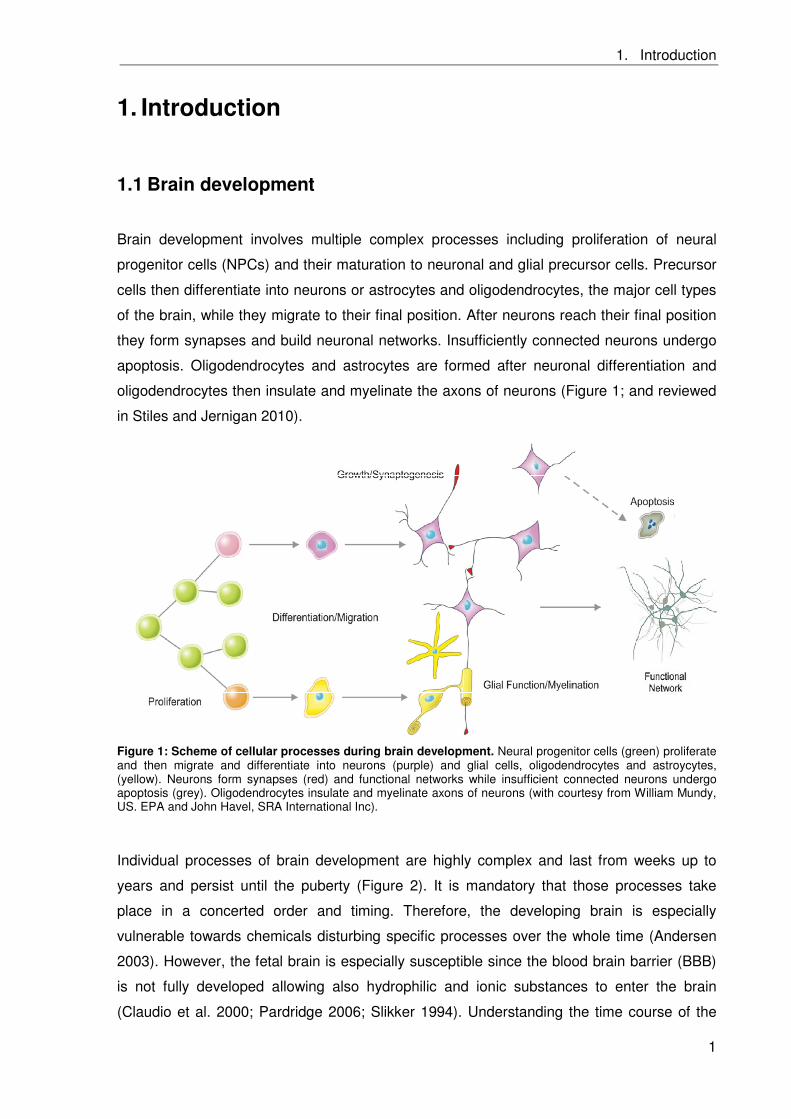
oligodendrocytes then insulate and myelinate the axons of neurons (Figure 1; and reviewed
in Stiles and Jernigan 2010).
Figure 1: Scheme of cellular processes during brain development. Neural progenitor cells (green) proliferate and then migrate and differentiate into neurons (purple) and glial cells, oligodendrocytes and astroycytes, (yellow). Neurons form synapses (red) and functional networks while insufficient connected neurons undergo apoptosis (grey). Oligodendrocytes insulate and myelinate axons of neurons (with courtesy from William Mundy, US. EPA and John Havel, SRA International Inc).
Individual processes of brain development are highly complex and last from weeks up to
years and persist until the puberty (Figure 2). It is mandatory that those processes take
place in a concerted order and timing. Therefore, the developing brain is especially
vulnerable towards chemicals disturbing specific processes over the whole time (Andersen
2003). However, the fetal brain is especially susceptible since the blood brain barrier (BBB)
is not fully developed allowing also hydrophilic and ionic substances to enter the brain
(Claudio et al. 2000; Pardridge 2006; Slikker 1994). Understanding the time course of the
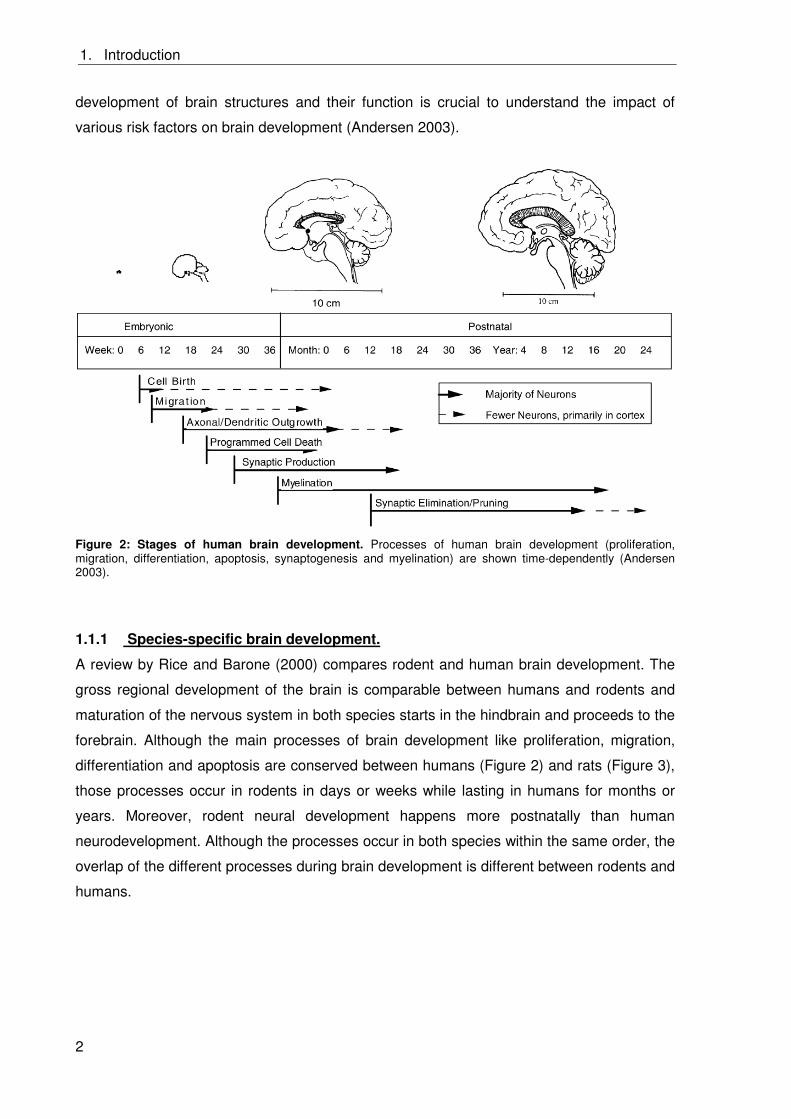
1. Introduction
2
development of brain structures and their function is crucial to understand the impact of
various risk factors on brain development (Andersen 2003).
Figure 2: Stages of human brain development. Processes of human brain development (proliferation, migration, differentiation, apoptosis, synaptogenesis and myelination) are shown time-dependently (Andersen 2003).
1.1.1 Species-specific brain development.
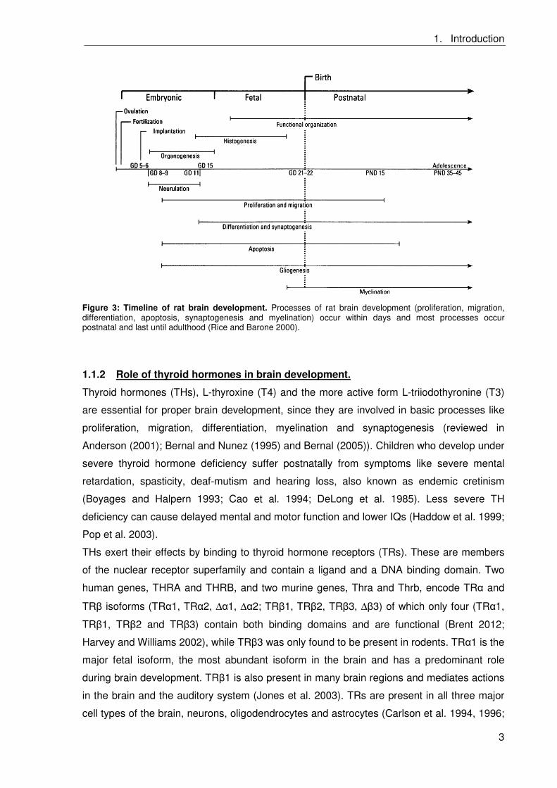
A review by Rice and Barone (2000) compares rodent and human brain development. The
gross regional development of the brain is comparable between humans and rodents and
maturation of the nervous system in both species starts in the hindbrain and proceeds to the
forebrain. Although the main processes of brain development like proliferation, migration,
differentiation and apoptosis are conserved between humans (Figure 2) and rats (Figure 3),
those processes occur in rodents in days or weeks while lasting in humans for months or
years. Moreover, rodent neural development happens more postnatally than human
neurodevelopment. Although the processes occur in both species within the same order, the
overlap of the different processes during brain development is different between rodents and
humans.
1. Introduction
3
Figure 3: Timeline of rat brain development. Processes of rat brain development (proliferation, migration, differentiation, apoptosis, synaptogenesis and myelination) occur within days and most processes occur postnatal and last until adulthood (Rice and Barone 2000).
1.1.2 Role of thyroid hormones in brain development.
Thyroid hormones (THs), L-thyroxine (T4) and the more active form L-triiodothyronine (T3)
are essential for proper brain development, since they are involved in basic processes like
proliferation, migration, differentiation, myelination and synaptogenesis (reviewed in
Anderson (2001); Bernal and Nunez (1995) and Bernal (2005)). Children who develop under
severe thyroid hormone deficiency suffer postnatally from symptoms like severe mental
retardation, spasticity, deaf-mutism and hearing loss, also known as endemic cretinism
(Boyages and Halpern 1993; Cao et al. 1994; DeLong et al. 1985). Less severe TH
deficiency can cause delayed mental and motor function and lower IQs (Haddow et al. 1999;
Pop et al. 2003).
THs exert their effects by binding to thyroid hormone receptors (TRs). These are members
of the nuclear receptor superfamily and contain a ligand and a DNA binding domain. Two
human genes, THRA and THRB, and two murine genes, Thra and Thrb, encode TR and
TR isoforms (TR 1, TR 2, Δ 1, Δ 2; TR 1, TR 2, TR 3, Δ 3) of which only four (TR 1,
TR 1, TR 2 and TR 3) contain both binding domains and are functional (Brent 2012;
Harvey and Williams 2002), while TR 3 was only found to be present in rodents. TR 1 is the
major fetal isoform, the most abundant isoform in the brain and has a predominant role
during brain development. TR 1 is also present in many brain regions and mediates actions
in the brain and the auditory system (Jones et al. 2003). TRs are present in all three major
cell types of the brain, neurons, oligodendrocytes and astrocytes (Carlson et al. 1994, 1996;
1. Introduction
4
Carre et al. 1998; Lebel et al. 1993; Puymirat et al. 1992; Strait et al. 1991). However, there
is evidence that TR is involved in earlier functions of brain development than TR (Jones et
al. 2003). This observation is supported by the fact that TR is present in both,
oligodendrocyte progenitor cells and differentiated oligodendrocytes, and is involved in
mediating the starting of oligodendrocyte differentiation while TR is only present in
differentiated oligodendrocytes (Billon et al. 2002; Carre et al. 1998).
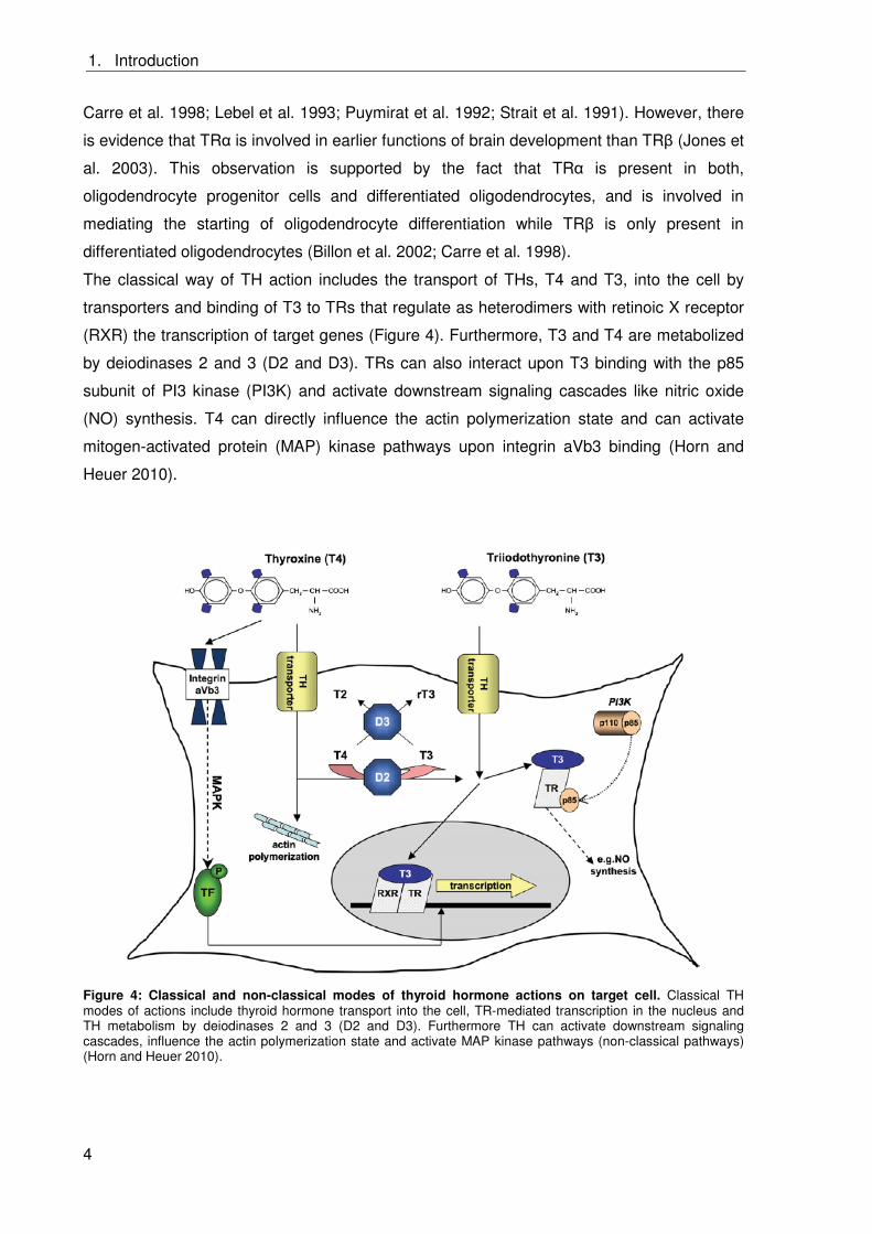
The classical way of TH action includes the transport of THs, T4 and T3, into the cell by
transporters and binding of T3 to TRs that regulate as heterodimers with retinoic X receptor
(RXR) the transcription of target genes (Figure 4). Furthermore, T3 and T4 are metabolized
by deiodinases 2 and 3 (D2 and D3). TRs can also interact upon T3 binding with the p85
subunit of PI3 kinase (PI3K) and activate downstream signaling cascades like nitric oxide
(NO) synthesis. T4 can directly influence the actin polymerization state and can activate
mitogen-activated protein (MAP) kinase pathways upon integrin aVb3 binding (Horn and
Heuer 2010).
Figure 4: Classical and non-classical modes of thyroid hormone actions on target cell. Classical TH modes of actions include thyroid hormone transport into the cell, TR-mediated transcription in the nucleus and TH metabolism by deiodinases 2 and 3 (D2 and D3). Furthermore TH can activate downstream signaling cascades, influence the actin polymerization state and activate MAP kinase pathways (non-classical pathways) (Horn and Heuer 2010).
1. Introduction
5
Although it is known that TH influences basic processes of brain development and
considerable progress in the understanding of TH signaling pathways was achieved, the
underlying molecular mechanisms of THs’ effects on different processes of brain
development remain elusive.
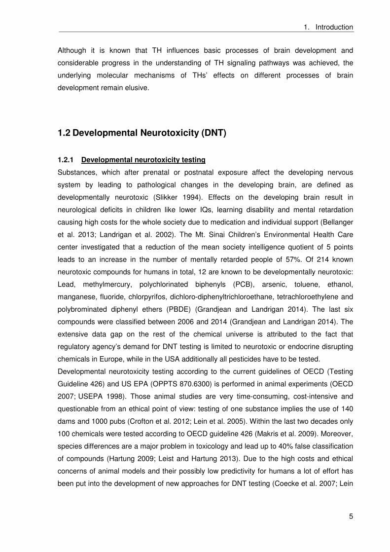
1.2 Developmental Neurotoxicity (DNT)
1.2.1 Developmental neurotoxicity testing
Substances, which after prenatal or postnatal exposure affect the developing nervous
system by leading to pathological changes in the developing brain, are defined as
developmentally neurotoxic (Slikker 1994). Effects on the developing brain result in
neurological deficits in children like lower IQs, learning disability and mental retardation
causing high costs for the whole society due to medication and individual support (Bellanger
et al. 2013; Landrigan et al. 2002). The Mt. Sinai Children’s Environmental Health Care
center investigated that a reduction of the mean society intelligence quotient of 5 points
leads to an increase in the number of mentally retarded people of 57%. Of 214 known
neurotoxic compounds for humans in total, 12 are known to be developmentally neurotoxic:
Lead, methylmercury, polychlorinated biphenyls (PCB), arsenic, toluene, ethanol,
manganese, fluoride, chlorpyrifos, dichloro-diphenyltrichloroethane, tetrachloroethylene and
polybrominated diphenyl ethers (PBDE) (Grandjean and Landrigan 2014). The last six
compounds were classified between 2006 and 2014 (Grandjean and Landrigan 2014). The
extensive data gap on the rest of the chemical universe is attributed to the fact that
regulatory agency’s demand for DNT testing is limited to neurotoxic or endocrine disrupting
chemicals in Europe, while in the USA additionally all pesticides have to be tested.
Developmental neurotoxicity testing according to the current guidelines of OECD (Testing
Guideline 426) and US EPA (OPPTS 870.6300) is performed in animal experiments (OECD
2007;)USEPA 1998). Those animal studies are very time-consuming, cost-intensive and
questionable from an ethical point of view: testing of one substance implies the use of 140
dams and 1000 pubs (Crofton et al. 2012; Lein et al. 2005). Within the last two decades only
100 chemicals were tested according to OECD guideline 426 (Makris et al. 2009). Moreover,
species differences are a major problem in toxicology and lead up to 40% false classification
of compounds (Hartung 2009; Leist and Hartung 2013). Due to the high costs and ethical
concerns of animal models and their possibly low predictivity for humans a lot of effort has
been put into the development of new approaches for DNT testing (Coecke et al. 2007; Lein
1. Introduction
6
et al. 2005, 2007), which imply refinement, reduction and replacement (3Rs) of animal
experiments. The 3R principle was developed by Russel and Burch (Russel et al. 1959).
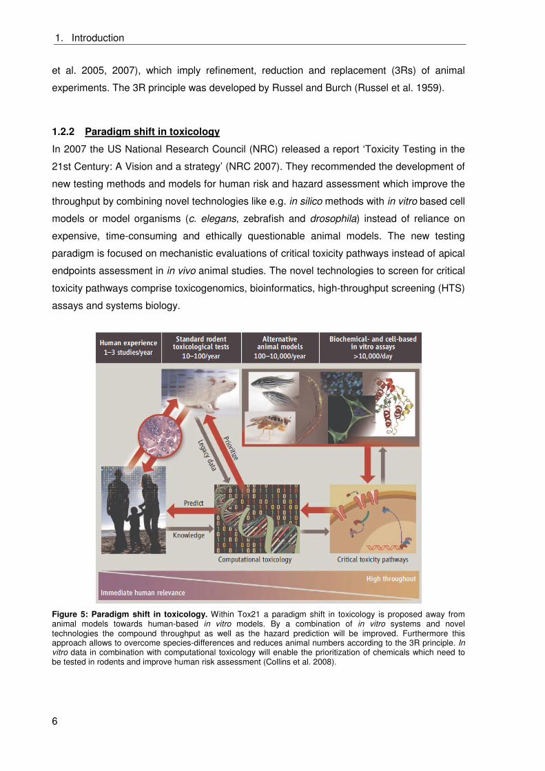
1.2.2 Paradigm shift in toxicology
In 2007 the US National Research Council (NRC) released a report ‘Toxicity Testing in the
21st Century: A Vision and a strategy’ (NRC 2007). They recommended the development of
new testing methods and models for human risk and hazard assessment which improve the
throughput by combining novel technologies like e.g. in silico methods with in vitro based cell
models or model organisms (c. elegans, zebrafish and drosophila) instead of reliance on
expensive, time-consuming and ethically questionable animal models. The new testing
paradigm is focused on mechanistic evaluations of critical toxicity pathways instead of apical
endpoints assessment in in vivo animal studies. The novel technologies to screen for critical
toxicity pathways comprise toxicogenomics, bioinformatics, high-throughput screening (HTS)
assays and systems biology.
Figure 5: Paradigm shift in toxicology. Within Tox21 a paradigm shift in toxicology is proposed away from animal models towards human-based in vitro models. By a combination of in vitro systems and novel technologies the compound throughput as well as the hazard prediction will be improved. Furthermore this approach allows to overcome species-differences and reduces animal numbers according to the 3R principle. In vitro data in combination with computational toxicology will enable the prioritization of chemicals which need to be tested in rodents and improve human risk assessment (Collins et al. 2008).
1. Introduction
7
Obtained data is analyzed using in silico methods like (quantitative) structure relationships
((Q)SAR) and read across for substance priorization for further animal experiments and to
elucidate the underlying mode of action (MOA). Furthermore, the comparability of the human
and rodent situation is addressed by comparing human in vitro data with legacy in vivo data
from rodents (Collins et al. 2008; Crofton et al. 2012; Gibb 2008;and NRC 2007). Figure 5
visualizes this new toxicity testing approach.
In this context the US EPA started the ToxCast21 program, in which molecular and pathway
perturbations caused by environmental chemicals are evaluated by utilizing new techniques
like computational chemistry and HTS in combination with in vitro models. The aim is to test
chemicals with methods, which allow hazard identification to human health and further
prioritization for in vivo testing (Dix et al. 2007; Judson et al. 2010).
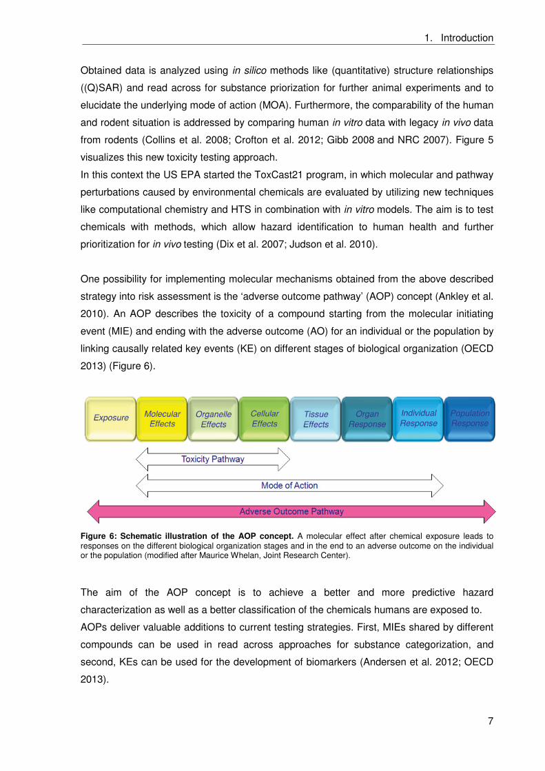
One possibility for implementing molecular mechanisms obtained from the above described
strategy into risk assessment is the ‘adverse outcome pathway’ (AOP) concept (Ankley et al.
2010). An AOP describes the toxicity of a compound starting from the molecular initiating
event (MIE) and ending with the adverse outcome (AO) for an individual or the population by
linking causally related key events (KE) on different stages of biological organization (OECD
2013) (Figure 6).
Figure 6: Schematic illustration of the AOP concept. A molecular effect after chemical exposure leads to responses on the different biological organization stages and in the end to an adverse outcome on the individual or the population (modified after Maurice Whelan, Joint Research Center).
The aim of the AOP concept is to achieve a better and more predictive hazard
characterization as well as a better classification of the chemicals humans are exposed to.
AOPs deliver valuable additions to current testing strategies. First, MIEs shared by different
compounds can be used in read across approaches for substance categorization, and
second, KEs can be used for the development of biomarkers (Andersen et al. 2012; OECD
2013).
1. Introduction
8
In order to obtain physiologically relevant data, based on a KE defined within an AOP, the
applied in vitro system has to reflect the human in vivo situation as precisely as possible.
Furthermore, a comparison of human and rodents within the AOP allows identification of
species differences in toxicologically relevant pathways. Species-specific based elucidation
of involved toxicity pathways should contribute to reduce uncertainty in hazard assessment
(Bal-Price et al. 2015a).
Development of AOPs for DNT is extremely challenging since many complex processes are
involved in brain development, which last over a long time period (see Chapter 1.1). This
implies that timing and duration of exposure determines the effects of chemicals on brain
development. Although a large number of cellular and molecular processes, which are
important for brain development, are known, there are only few examples of well-
documented pathways that include causally linked MIEs and KEs that result in an observed
AO (Bal-Price et al. 2015b).
1.2.3 Primary neural progenitor cells as in vitro models for studying DNT
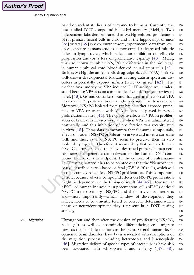
To date, the mouse embryonic stem cell test (EST) is the only in vitro test based on
mammalian cells successfully validated as an alternative for in vivo DNT testing (Coecke et
al. 2007; Genschow et al. 2002). Other rodent based in vitro models, which are considered
as models for testing DNT are organotypic cultures (embryonic brain or spinal cord tissue),
rotating brain cell cultures and primary dissociated cultures. The detailed advantages and
disadvantages of each system are summarized in(Coecke et al. (2007). The advantage of
primary cells isolated from rodent brain tissue and cultured in vitro is the maintenance of the
signaling functions and responses towards xenobiotics (Burke et al. 2006; Foti et al. 2013;
Go et al. 2012; He et al. 2010; Simpson et al. 2011). However, the main limitation of all
those models is their rodent origin in regard to species differences towards humans.
Furthermore, human immortalized cells, tumor cell lines or transformed cells, which
differentiate into neuronal cells, were used to test developmental neurotoxicity (Abdulla et al.
1995; Hong et al. 2003; Påhlman et al. 1990; Scholz et al. 2011; Stern et al. 2014). Those
cells are of human origin, but the main concern about those immortalized cells is that they
are not comparable to “normal” cells and results are therefore difficult to interpret (Coecke et
al. 2007). Primary stem or progenitor cells, which can be generated from human tissue,
become more prominent in the field of DNT testing (Baumann et al. 2014; Fritsche 2014;
Hayess et al. 2013).
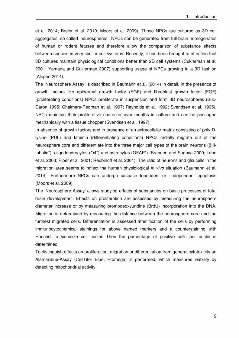
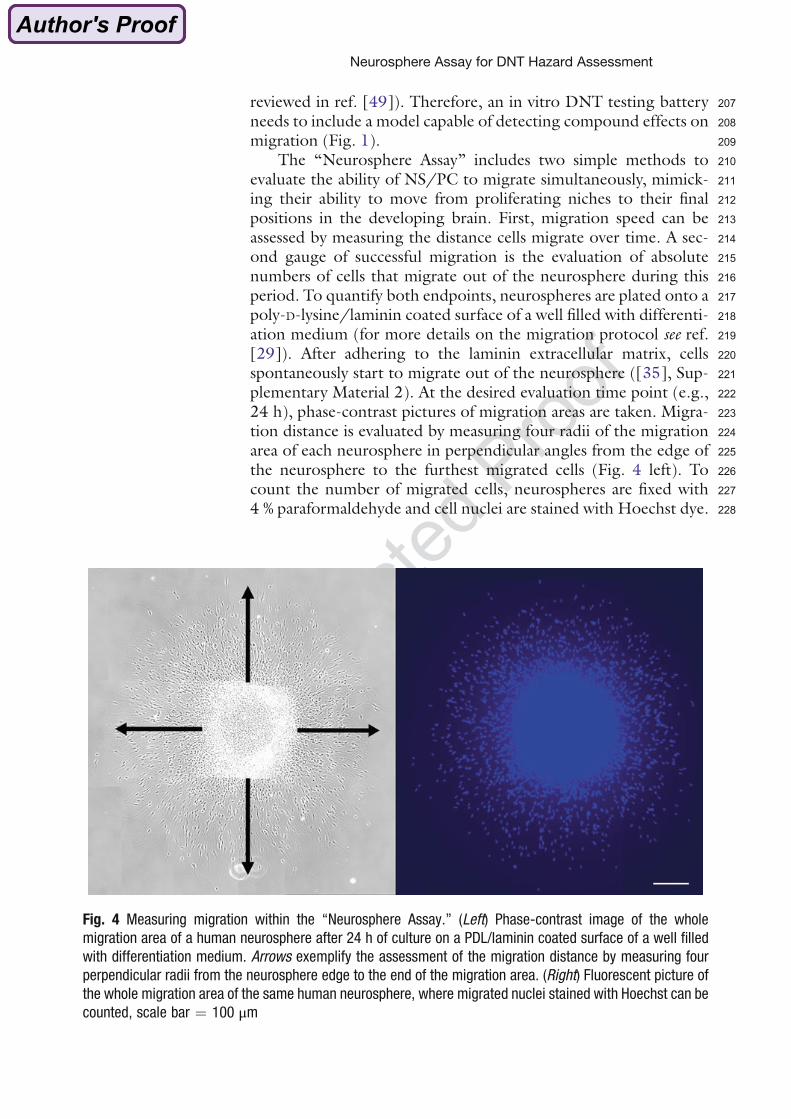
The ‘Neurosphere Assay’ (Figure 7) is a three-dimensional (3D) primary in vitro cell culture
model based on normal neural progenitor cells (NPCs), which mimics basic processes of
fetal brain development like proliferation, differentiation, migration and apoptosis (Baumann
1. Introduction
9
et al. 2014; Breier et al. 2010; Moors et al. 2009). Those NPCs are cultured as 3D cell
aggregates, so called ‘neurospheres’. NPCs can be generated from full brain homogenates
of human or rodent fetuses and therefore allow the comparison of substance effects
between species in very similar cell systems. Recently, it has been brought to attention that
3D cultures maintain physiological conditions better than 2D cell systems (Cukierman et al.
2001; Yamada and Cukierman 2007) supporting usage of NPCs growing in a 3D fashion
(Alépée 2014).
The ‘Neurosphere Assay’ is described in(Baumann et al. (2014) in detail. In the presence of
growth factors like epidermal growth factor (EGF) and fibroblast growth factor (FGF)
(proliferating conditions) NPCs proliferate in suspension and form 3D neurospheres (Buc-
Caron 1995; Chalmers-Redman et al. 1997; Reynolds et al. 1992; Svendsen et al. 1995).
NPCs maintain their proliferative character over months in culture and can be passaged
mechanically with a tissue chopper (Svendsen et al. 1997).
In absence of growth factors and in presence of an extracellular matrix consisting of poly-D-
lysine (PDL) and laminin (differentiating conditions) NPCs radially migrate out of the
neurosphere core and differentiate into the three major cell types of the brain neurons ( III-
tubulin+), oligodendrocytes (O4+) and astrocytes (GFAP+) (Brannen and Sugaya 2000; Lobo
et al. 2003; Piper et al. 2001; Reubinoff et al. 2001). The ratio of neurons and glia cells in the
migration area seems to reflect the human physiological in vivo situation (Baumann et al.
2014). Furthermore NPCs can undergo caspase-dependent or -independent apoptosis
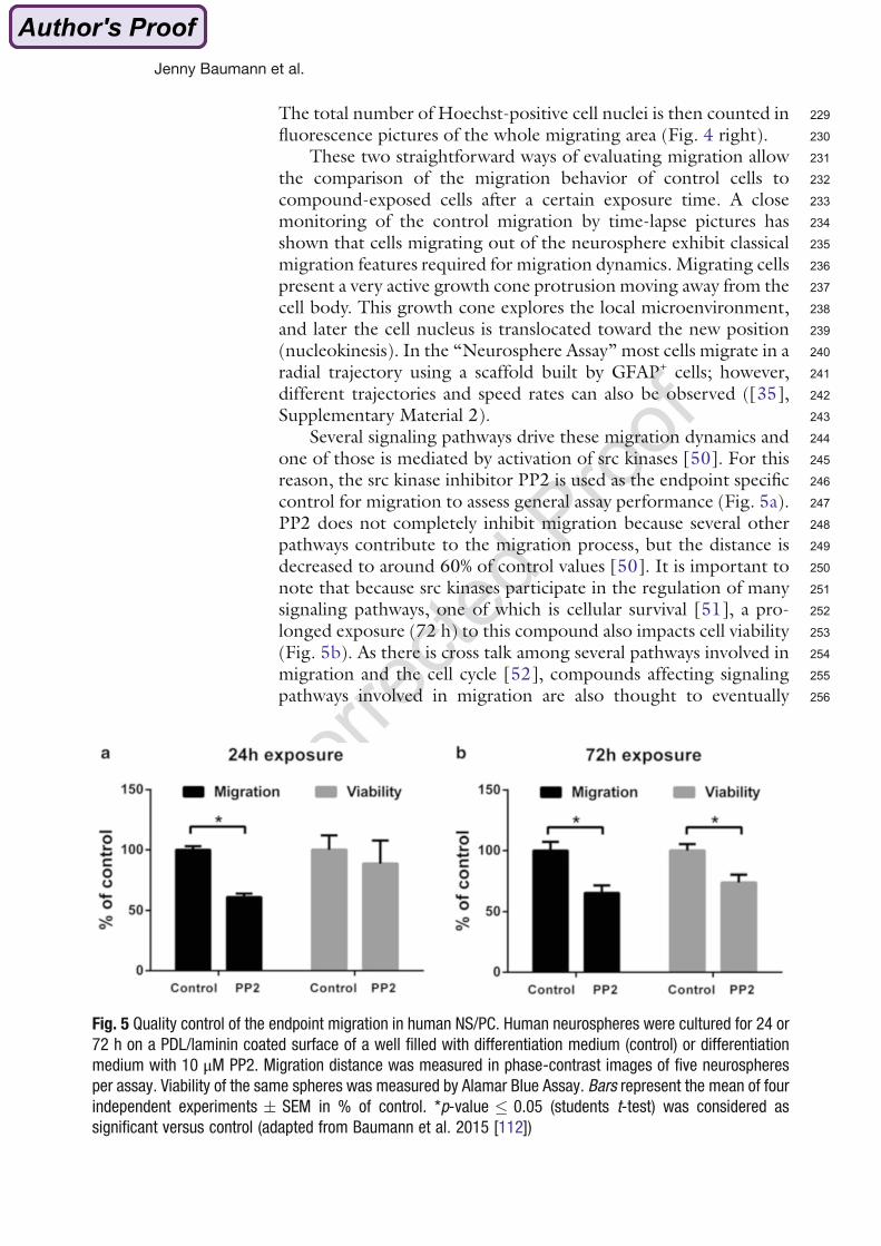
(Moors et al. 2009).
The ‘Neurosphere Assay’ allows studying effects of substances on basic processes of fetal
brain development. Effects on proliferation are assessed by measuring the neurosphere
diameter increase or by measuring bromodeoxyuridine (BrdU) incorporation into the DNA.
Migration is determined by measuring the distance between the neurosphere core and the
furthest migrated cells. Differentiation is assessed after fixation of the cells by performing
immunocytochemical stainings for above named markers and a counterstaining with
Hoechst to visualize cell nuclei. Then the percentage of positive cells per nuclei is
determined.
To distinguish effects on proliferation, migration or differentiation from general cytotoxicity an
AlamarBlue-Assay (CellTiter Blue, Promega) is performed, which measures viability by
detecting mitochondrial activity.
1. Introduction
10
Figure 7: The ‘Neurosphere Assay’. (modified from Breier et al. (2010)). Neural progenitor cells (NPCs) are cultured as floating neurospheres. They mimic basic processes of brain development like proliferation, migration and differentiation into the three main brain cell types: neurons, oligodendrocytes and astrocytes. In presence of growth factors NPCs proliferate and proliferation is assessed by measuring the diameter increase or BrdU incorporation. In absence of growth factors neurospheres settle down on PDL/laminin coated surfaces and cells start to radially migrate out and differentiate. Migration is determined by measuring the distance between the neurosphere core and the furthest migrated cells. Neurons, oligodendrocytes and astrocytes can be visualized by immunocytochemical staining with III-tubulin, O4 or GFAP antibodies and a counterstaining with Hoechst for nuclei.
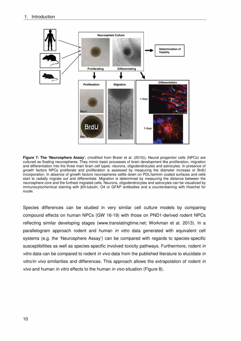
Species differences can be studied in very similar cell culture models by comparing
compound effects on human NPCs (GW 16-19) with those on PND1-derived rodent NPCs
reflecting similar developing stages (www.translatingtime.net; Workman et al. 2013). In a
parallelogram approach rodent and human in vitro data generated with equivalent cell
systems (e.g. the ‘Neurosphere Assay’) can be compared with regards to species-specific
susceptibilities as well as species-specific involved toxicity pathways. Furthermore, rodent in
vitro data can be compared to rodent in vivo data from the published literature to elucidate in
vitro/in vivo similarities and differences. This approach allows the extrapolation of rodent in
vivo and human in vitro effects to the human in vivo situation (Figure 8).
1. Introduction
11
Figure 8: Parallelogram approach for extrapolation to the human in vivo situation. Rodent and human in vitro data generated in an equivalent cell system is compared to determine species differences in susceptibility as well as in involved toxicity pathways. Rodent in vitro data is compared to rodent in vivo data to elucidate in vitro/in vivo differences or similarities. This approach allows the extrapolation to the human in vivo situation.
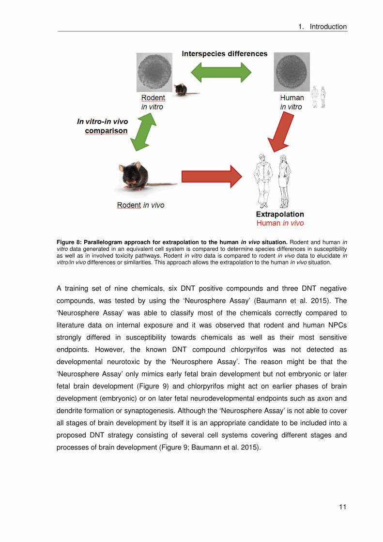
A training set of nine chemicals, six DNT positive compounds and three DNT negative
compounds, was tested by using the ‘Neurosphere Assay’ (Baumann et al. 2015). The
‘Neurosphere Assay’ was able to classify most of the chemicals correctly compared to
literature data on internal exposure and it was observed that rodent and human NPCs
strongly differed in susceptibility towards chemicals as well as their most sensitive
endpoints. However, the known DNT compound chlorpyrifos was not detected as
developmental neurotoxic by the ‘Neurosphere Assay’. The reason might be that the
‘Neurosphere Assay’ only mimics early fetal brain development but not embryonic or later
fetal brain development (Figure 9) and chlorpyrifos might act on earlier phases of brain
development (embryonic) or on later fetal neurodevelopmental endpoints such as axon and
dendrite formation or synaptogenesis. Although the ‘Neurosphere Assay’ is not able to cover
all stages of brain development by itself it is an appropriate candidate to be included into a
proposed DNT strategy consisting of several cell systems covering different stages and
processes of brain development (Figure 9; Baumann et al. 2015).
1. Introduction
12
Figure 9: Testing strategy for in vitro DNT testing. The endpoints evaluated within the ‘Neurosphere Assay’ cover early fetal brain development. For assessment of all brain development stages a combination of different cell systems is needed. Embryonic brain development can be evaluated with embryonic stem cells (ESCs), neural crest cells (NCCs) and neuroepithelial precursors (NEPs) and later fetal brain by measuring neuronal activity on multielectrode arrays (MEAs) (Baumann et al. 2015).
1.3 Polybrominated diphenyl ethers (PBDEs)
Of the 214 known neurotoxic compounds for humans only 12 are known be developmentally
neurotoxic (Grandjean and Landrigan 2014). Those include polybrominated diphenyl ethers
(PBDEs), which were lately classified as human DNT compounds (Grandjean and Landrigan
2014). PBDEs are brominated flame retardants and were widely used in a lot of consumer
products like electronics, plastics and textiles (Alaee et al. 2003).
1.3.1 Chemical properties of PBDEs
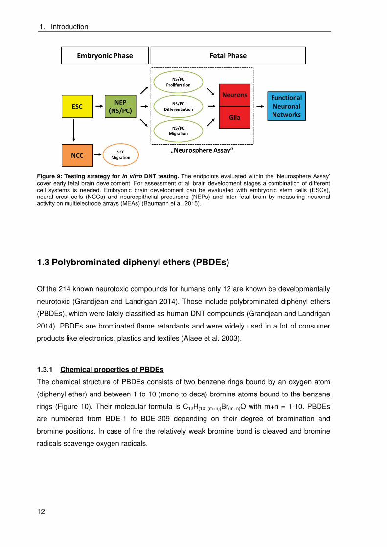
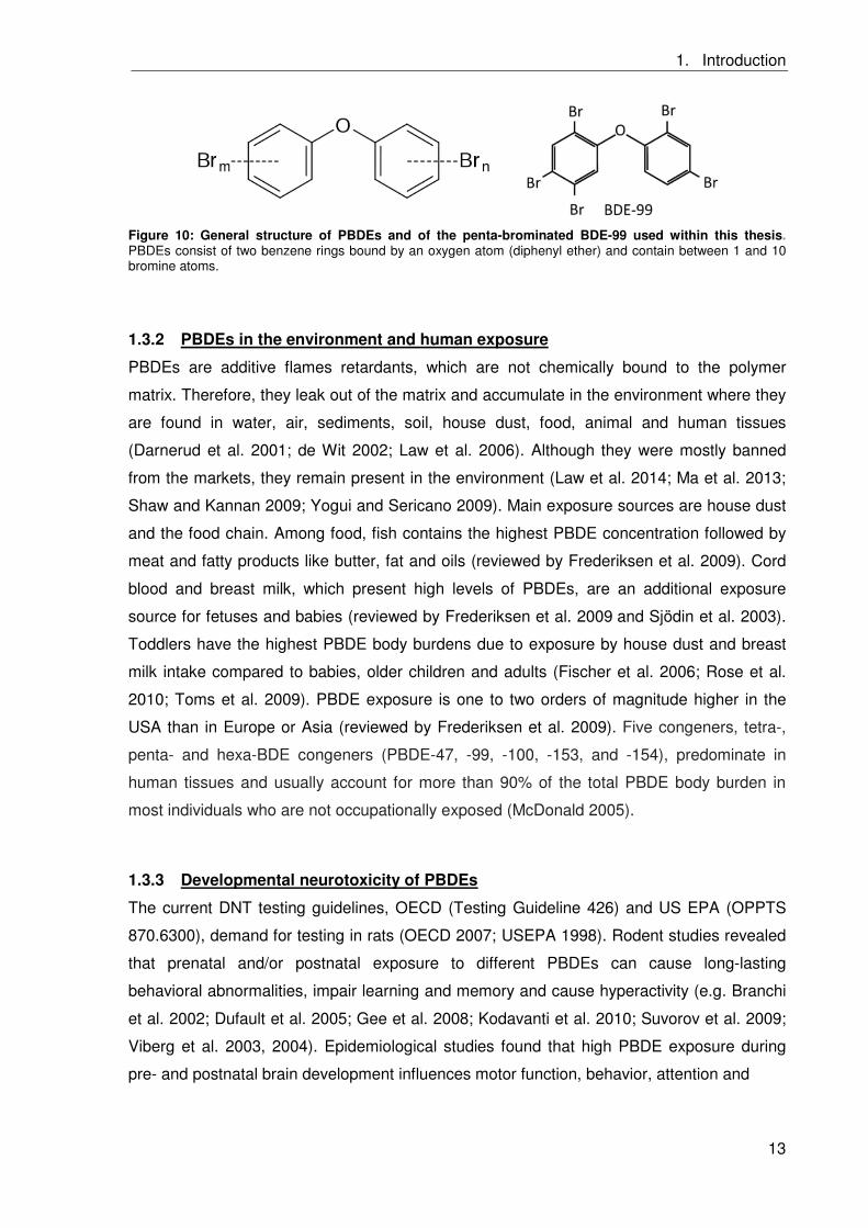
The chemical structure of PBDEs consists of two benzene rings bound by an oxygen atom
(diphenyl ether) and between 1 to 10 (mono to deca) bromine atoms bound to the benzene
rings (Figure 10). Their molecular formula is C12H(10−(m+n))Br(m+n)O with m+n = 1-10. PBDEs
are numbered from BDE-1 to BDE-209 depending on their degree of bromination and
bromine positions. In case of fire the relatively weak bromine bond is cleaved and bromine
radicals scavenge oxygen radicals.
1. Introduction
13
Figure 10: General structure of PBDEs and of the penta-brominated BDE-99 used within this thesis. PBDEs consist of two benzene rings bound by an oxygen atom (diphenyl ether) and contain between 1 and 10 bromine atoms.
1.3.2 PBDEs in the environment and human exposure
PBDEs are additive flames retardants, which are not chemically bound to the polymer
matrix. Therefore, they leak out of the matrix and accumulate in the environment where they
are found in water, air, sediments, soil, house dust, food, animal and human tissues
(Darnerud et al. 2001; de Wit 2002; Law et al. 2006). Although they were mostly banned
from the markets, they remain present in the environment (Law et al. 2014; Ma et al. 2013;
Shaw and Kannan 2009; Yogui and Sericano 2009). Main exposure sources are house dust
and the food chain. Among food, fish contains the highest PBDE concentration followed by
meat and fatty products like butter, fat and oils (reviewed by Frederiksen et al. 2009). Cord
blood and breast milk, which present high levels of PBDEs, are an additional exposure
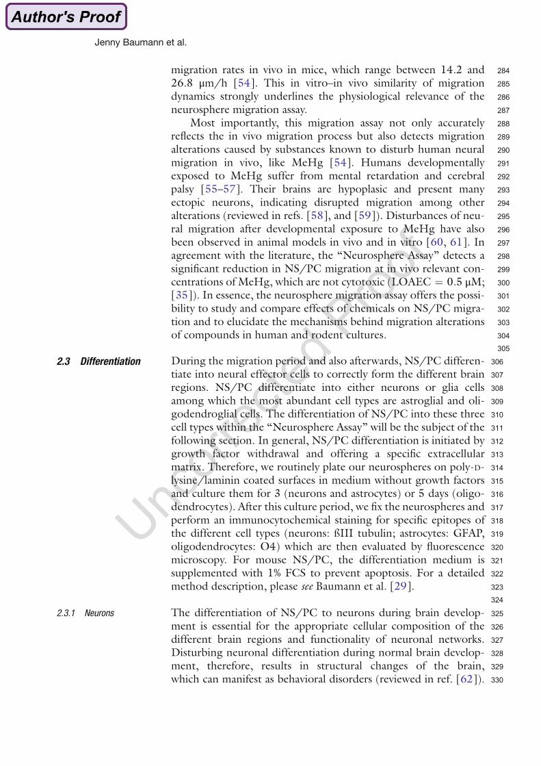
source for fetuses and babies (reviewed by Frederiksen et al. 2009;and Sjödin et al. 2003).
Toddlers have the highest PBDE body burdens due to exposure by house dust and breast
milk intake compared to babies, older children and adults (Fischer et al. 2006; Rose et al.
2010; Toms et al. 2009). PBDE exposure is one to two orders of magnitude higher in the
USA than in Europe or Asia (reviewed by Frederiksen et al. 2009). Five congeners, tetra-,
penta- and hexa-BDE congeners (PBDE-47, -99, -100, -153, and -154), predominate in
human tissues and usually account for more than 90% of the total PBDE body burden in
most individuals who are not occupationally exposed (McDonald 2005).
1.3.3 Developmental neurotoxicity of PBDEs
The current DNT testing guidelines, OECD (Testing Guideline 426) and US EPA (OPPTS
870.6300), demand for testing in rats (OECD 2007;)USEPA 1998). Rodent studies revealed
that prenatal and/or postnatal exposure to different PBDEs can cause long-lasting
behavioral abnormalities, impair learning and memory and cause hyperactivity (e.g. Branchi
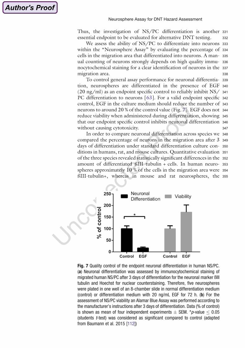
et al. 2002; Dufault et al. 2005; Gee et al. 2008; Kodavanti et al. 2010; Suvorov et al. 2009;
Viberg et al. 2003, 2004). Epidemiological studies found that high PBDE exposure during
pre- and postnatal brain development influences motor function, behavior, attention and
1. Introduction
14
cognition in children at school age and correlated high PBDE exposure with hyperactivity
and lower IQs (Chao et al. 2007; Eskenazi et al. 2013; Roze et al. 2009; Shy et al. 2011).
Although several studies have shown the DNT potential of PBDEs, the mechanisms of
PBDE-induced DNT remain elusive (reviewed by Costa et al. (2014)). It was observed that
PBDE treated animals show the same behavioral abnormalities than animals suffering from
hypothyroidism during brain development (Negishi et al. 2005). Costa et al. (2014) therefore
discussed two MOAs for PBDE-induced DNT: first, interference of PBDEs with THs/TH
signaling and second non TH-related effects on brain cells (reactive oxygen species (ROS)
formation, DNA damage, apoptosis, interference with Ca2+-signaling and neurotransmitter
systems) .
Many studies reported decreased T4 and increased thyroid-stimulating hormone (TSH)
levels in rodents after developmental exposure to different PBDEs (reviewed by Costa et al.
(2014)) and also for humans PBDE exposure was correlated with changes in TH and TSH
levels. However, human data were inconsistent: decreased, increased or unchanged T4
levels and increased or decreased TSH blood levels were reported (Chevrier et al. 2011;
Herbstman et al. 2008; Stapleton et al. 2011; Zota et al. 2011). Moreover, it was shown that
hydroxylated but not parent PBDEs can interfere with TH transport by binding to the TH
transport protein transthyretin (TTR; Hamers et al. 2006; Meerts et al. 2000). Additionally,
hydroxylated, but not parent PBDEs, can directly bind to TRs (Hamers et al. 2006; Kojima et
al. 2009; Li et al. 2010; Ren et al. 2013; Suvorov et al. 2011). Lower brominated PBDEs are
TR agonists while higher brominated congeners are TR antagonists (Ren et al. 2013).
Furthermore, PBDEs deregulate the expression of genes encoding TR isoforms (Blanco et
al. 2011; Lema et al. 2008) and interfere with T(H,R)-mediated transcription (Blanco et al.
2011; Ibhazehiebo et al. 2011; Lema et al. 2008). However, here the results were
contradictory dependent on the studied congener, the cell system and the used method.
1. Introduction
15
1.4 Aim of this thesis
DNT studies are routinely performed in rats, which are cost and time intensive, ethically
questionable and often not predictive for humans. Stakeholders agreed on the need to move
away from those animal models towards in vitro models, which also allow the identification of
toxicity pathways and consider species differences. The ‘Neurosphere Assay’ is a promising
tool for in vitro DNT evaluation since it mimics several basic processes of brain development
and allows studying cultures generated from different species. PBDEs were classified to be
developmental neurotoxic in humans and rodents. However, the mechanisms how PBDEs
induce DNT are poorly understood. Since THs are involved in many processes of brain
development and PBDEs alter TH levels in rodents and humans, also interaction with TH
signaling is discussed as a mechanism for PBDE-induced DNT. Therefore, the overall aim of
this thesis was to investigate whether TH disruption is involved in PBDE effects on basic
processes of fetal brain development. Human and murine NPCs were used to compare the
molecular mechanisms of PBDE-induced DNT between species. BDE-99 was chosen since
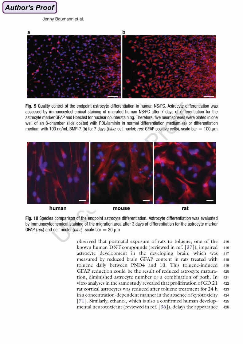
it is one of the most abundant congeners found in human tissue. The following questions
were addressed:
1. Establishment of the mouse neurosphere culture.
2. Investigation of the involvement of THs in basic processes of human and murine
brain development and characterization of neurospheres as appropriate models to
study TH signaling.
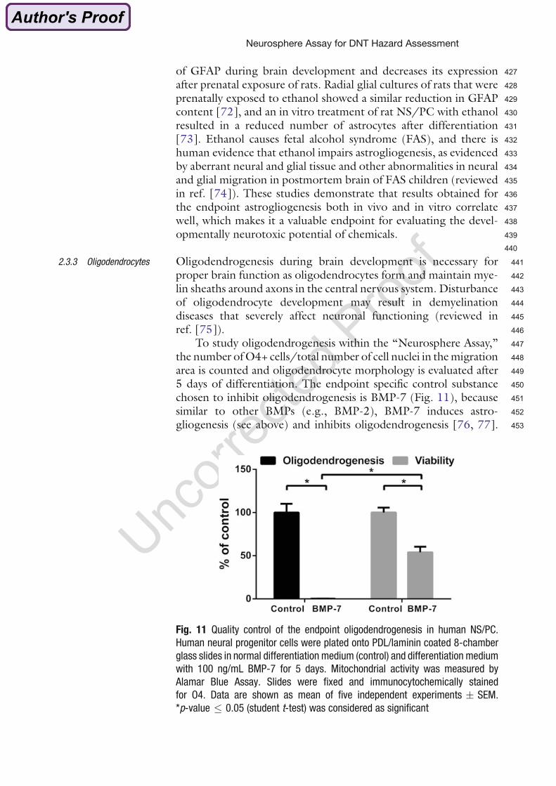
3. Elucidation of the influence of BDE-99 on basic processes of brain development in
human and murine neurospheres and comparison of the sensitivity towards BDE-99
between species.
4. Establishment of a test assay to identify human- and mouse-specific TH disruptors
and application of this assay to assess TH-disruption potential of BDE-99 on human
and mouse neurospheres.
2. Manuscripts
16
2. Manuscripts
The publications, which emerged from this thesis, are attached below.
The first publication ‘Application of the Neurosphere Assay for DNT Hazard Assessment:
Challenges and Limitations’ (manuscript 2.1) is a book chapter comparing basic fetal
processes of brain development (proliferation, migration and differentiation) between human
and rodent (rat, mouse) neural progenitor cells (NPCs). Within this publication for the first
time the murine NPC system was characterized by employing data generated within this
thesis. The publication explains how those processes of brain development are analyzed
within the ‘Neurosphere Assay’ for developmental neurotoxicity (DNT). Furthermore, the
relevance of this cell model for modeling in vivo brain development is demonstrated and the
strengths and limitations of the ‘Neurosphere Assay’ as a DNT testing model versus existing
approaches are critically discussed.
The second publication ‘BDE-99 impairs human and mouse oligodendrogenesis by species-
specific modes of action’ (manuscript 2.2; original work) describes BDE-99 effects on
oligodendrocyte formation and maturation of human and murine NPCs and emphasizes
species differences. This publication is based on the newly established murine NPC system
characterized in manuscript 2.1. The focus lies on the investigation whether BDE-99 affects
oligodendrogenesis by TH disruption. Susceptibility towards BDE-99-impaired oligodendro-
genesis as well as its mode of action differed between both species.
2. Manuscripts
17
2.1 Application of the Neurosphere Assay for DNT Hazard
Assessment: Challenges and Limitations
Jenny Baumann*, Katharina Dach*, Marta Barenys, Susanne Giersiefer, Janette
Goniwiecha, Pamela J. Lein, and Ellen Fritsche
* shared first-authorship
Mechanisms and Predictive Modeling with In vitro and In vivo Approaches
(book chapter) (in press, corrected author’s proof was sent back in August)
Die Gehirnentwicklung ist durch ein Zusammenspiel von komplexen Prozessen, die in einer
geregelten zeitlichen Abfolge geschehen, gekennzeichnet. Diese Prozesse werden in frühe
(embryonale) und späte (fetale) Prozesse unterteilt.
Während der embryonalen Organentwicklung werden durch Differenzierung von
embryonalen Stammzellen zu neuroepithelialen Vorläuferzellen, die die neurale Stamm-
/Progenitorpopulation bilden, die Neuralplatte und das Neuralrohr gebildet. Wir verwenden
neurale Stamm-/Progenitorzellen vom Menschen und vom Nager, die als Neurosphären
kultiviert werden, um Prozesse der fetalen Gehirnentwicklung (Proliferation, Migration und
Differenzierung) zwischen den Spezies zu vergleichen und um adverse Effekte von
Chemikalien auf diese Prozesse zu untersuchen. Wir beschreiben, wie diese
neuroentwicklungsrelevanten Prozesse im „Neurosphären Assay“ für die in vitro
Entwicklungsneurotoxizitätstestung untersucht werden. Dabei betonen wir endpunkt-
spezifische Kontrollen für die einzelnen Prozesse und zeigen durch deren Vergleich mit der
bekannten in vivo Situation, dass in vitro Ergebnisse die in vivo Situation widerspiegeln.
Außerdem beschreiben wir drei Methoden, um transgene humane Neurosphären für
biomolekulare Grundlagenforschung zu generieren. Die Daten werden kritisch diskutiert,
indem die Stärken und Grenzen des „Neurosphären Assays“ für die Beurteilung des
Gefährdungspotentials von Chemikalien für die Gehirnentwicklung hervorgehoben werden.
Metadata of the chapter that will be visualized online
Chapter Title Application of the Neurosphere Assay for DNT Hazard Assessment: Chal-lenges and Limitations
Copyright Year 2015
Copyright Holder Springer Science+Business Media New York
Author Family Name Baumann*Particle
Given Name JennySuffix
Organization IUF—Leibniz Research Institute of Environmental Medi-cine
Address Auf’m Hennekamp 50, D-40225 Duesseldorf, Germany
Author Family Name Dach*Particle
Given Name KatharinaSuffix
Organization IUF—Leibniz Research Institute of Environmental Medi-cine
Address Auf’m Hennekamp 50, D-40225 Duesseldorf, Germany
Author Family Name BarenysParticle
Given Name MartaSuffix
Organization IUF—Leibniz Research Institute of Environmental Medi-cine
Address Auf’m Hennekamp 50, D-40225 Duesseldorf, Germany
Author Family Name GiersieferParticle
Given Name SusanneSuffix
Organization IUF—Leibniz Research Institute of Environmental Medi-cine
Address Auf’m Hennekamp 50, D-40225 Duesseldorf, Germany
Author Family Name GoniwiechaParticle
Given Name JanetteSuffix
Organization IUF—Leibniz Research Institute of Environmental Medi-cine
Address Auf’m Hennekamp 50, D-40225 Duesseldorf, Germany
Author Family Name LeinParticle
Given Name Pamela J.Suffix
Division Department of Molecular Biosciences, School of VeterinaryMedicine
Organization University of California at Davis
Address Davis, CA 95616, USA
Corresponding Author Family Name FritscheParticle
Given Name EllenSuffix
Organization IUF—Leibniz Research Institute of Environmental Medi-cine
Address Auf’m Hennekamp 50, D-40225 Duesseldorf, Germany
Email [email protected]
Abstract Brain development is an integration of developmental processes that takeplace in a time-dependent manner. These processes can be divided intoearly (embryonic) and late (fetal) neurodevelopmental events. Duringembryonic organogenesis, the neural plate and neural tube are formedby embryonic stem cell differentiation into neuroepithelial precursors,which represent the neural stem/progenitor cell population of thedeveloping brain. We use neural stem/progenitor cells of humans androdents growing as neurospheres to investigate fetal neurodevelopmentalevents (proliferation, migration, and differentiation) across species and toassess adverse effects of chemicals on these processes. We describe howthese specific neurodevelopmental processes are analyzed within the“Neurosphere Assay” for developmental neurotoxicity testing in vitro.Thereby, we emphasize endpoint-specific controls for these processes andrelate those to the in vivo situation to demonstrate how in vitro outcomesreflect the actual in vivo situation for developmental neurotoxicity. Inaddition, we describe three methods for creating transgenic humanneurospheres for basic biomolecular research. These data are criticallydiscussed by pointing out strengths and limitations of the “NeurosphereAssay” for developmental neurotoxicity hazard assessment.
Keywords (separatedby ‘-’)
Developmental neurotoxicity - Neural progenitor cell - Brain development -Species differences - Human - Mouse - Rat - In vitro
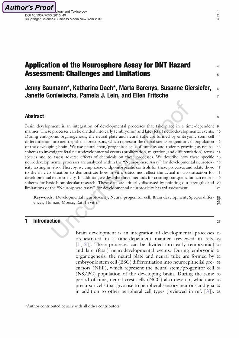
1Methods in Pharmacology and Toxicology2DOI 10.1007/7653_2015_493© Springer Science+Business Media New York 2015
4Application of the Neurosphere Assay for DNT Hazard5Assessment: Challenges and Limitations
6Jenny Baumann*, Katharina Dach*, Marta Barenys, Susanne Giersiefer,
7Janette Goniwiecha, Pamela J. Lein, and Ellen Fritsche
8Abstract
9Brain development is an integration of developmental processes that take place in a time-dependent10manner. These processes can be divided into early (embryonic) and late (fetal) neurodevelopmental events.11During embryonic organogenesis, the neural plate and neural tube are formed by embryonic stem cell12differentiation into neuroepithelial precursors, which represent the neural stem/progenitor cell population13of the developing brain. We use neural stem/progenitor cells of humans and rodents growing as neuro-14spheres to investigate fetal neurodevelopmental events (proliferation, migration, and differentiation) across15species and to assess adverse effects of chemicals on these processes. We describe how these specific16neurodevelopmental processes are analyzed within the “Neurosphere Assay” for developmental neurotox-17icity testing in vitro. Thereby, we emphasize endpoint-specific controls for these processes and relate those18to the in vivo situation to demonstrate how in vitro outcomes reflect the actual in vivo situation for19developmental neurotoxicity. In addition, we describe three methods for creating transgenic human neuro-20spheres for basic biomolecular research. These data are critically discussed by pointing out strengths and21limitations of the “Neurosphere Assay” for developmental neurotoxicity hazard assessment.
22Keywords: Developmental neurotoxicity, Neural progenitor cell, Brain development, Species differ-23ences, Human, Mouse, Rat, In vitro 2425
26
271 Introduction
28Brain development is an integration of developmental processes29orchestrated in a time-dependent manner (reviewed in refs.30[1, 2]). These processes can be divided into early (embryonic)31and late (fetal) neurodevelopmental events. During embryonic32organogenesis, the neural plate and neural tube are formed by33embryonic stem cell (ESC) differentiation into neuroepithelial pre-34cursors (NEP), which represent the neural stem/progenitor cell35(NS/PC) population of the developing brain. During the same36period of time, neural crest cells (NCC) also develop, which are37precursor cells that give rise to peripheral sensory neurons and glia38in addition to other peripheral cell types (reviewed in ref. [3]).
*Author contributed equally with all other contributors.
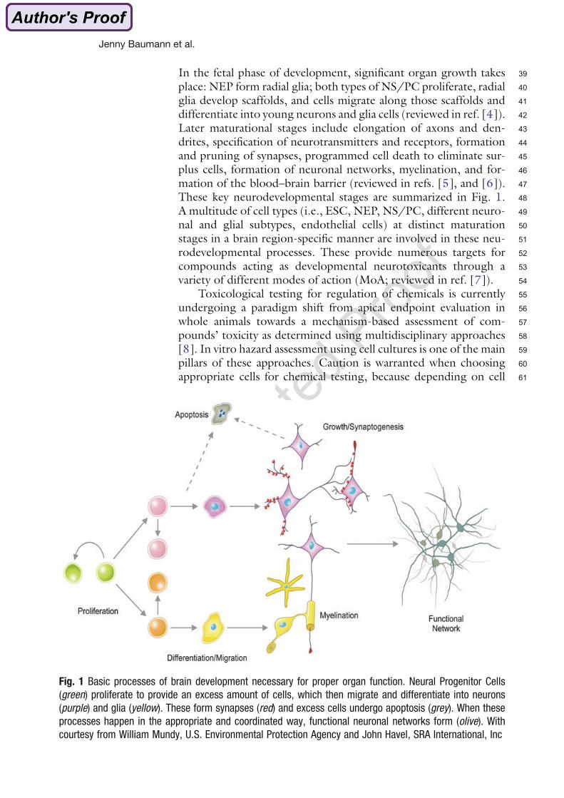
39In the fetal phase of development, significant organ growth takes40place: NEP form radial glia; both types of NS/PC proliferate, radial41glia develop scaffolds, and cells migrate along those scaffolds and42differentiate into young neurons and glia cells (reviewed in ref. [4]).43Later maturational stages include elongation of axons and den-44drites, specification of neurotransmitters and receptors, formation45and pruning of synapses, programmed cell death to eliminate sur-46plus cells, formation of neuronal networks, myelination, and for-47mation of the blood–brain barrier (reviewed in refs. [5], and [6]).48These key neurodevelopmental stages are summarized in Fig. 1.49A multitude of cell types (i.e., ESC, NEP, NS/PC, different neuro-50nal and glial subtypes, endothelial cells) at distinct maturation51stages in a brain region-specific manner are involved in these neu-52rodevelopmental processes. These provide numerous targets for53compounds acting as developmental neurotoxicants through a54variety of different modes of action (MoA; reviewed in ref. [7]).55Toxicological testing for regulation of chemicals is currently56undergoing a paradigm shift from apical endpoint evaluation in57whole animals towards a mechanism-based assessment of com-58pounds’ toxicity as determined using multidisciplinary approaches59[8]. In vitro hazard assessment using cell cultures is one of the main60pillars of these approaches. Caution is warranted when choosing61appropriate cells for chemical testing, because depending on cell
Fig. 1 Basic processes of brain development necessary for proper organ function. Neural Progenitor Cells
(green) proliferate to provide an excess amount of cells, which then migrate and differentiate into neurons
(purple) and glia (yellow). These form synapses (red) and excess cells undergo apoptosis (grey). When these
processes happen in the appropriate and coordinated way, functional neuronal networks form (olive). With
courtesy from William Mundy, U.S. Environmental Protection Agency and John Havel, SRA International, Inc
Jenny Baumann et al.
62type and culture conditions, results from in vitro testing can differ63tremendously for the same compounds. Using the examples of64in vitro testing for developmental neurotoxicity (DNT) endpoints,65species [9–11] and cell type (tumor versus primary cell; [12]) might66influence testing outcome. Moreover, recent advances in tissue67engineering have clearly indicated that cells cultured in a conven-68tional two-dimensional (2D) fashion can differ in their cell physiol-69ogy from their counterparts growing in 3D ([13], reviewed in refs.70[14, 15]). This observation, which was initially based on data from71fibroblasts, also seems to hold true for neural cells [16].72Current international guidelines for DNT testing (OECD73Testing Guideline 426, U.S. EPA OPPTS 870.6300) are very74resource-intensive when it comes to the number of animals used75and the time and costs required ([17, 18], reviewed in refs. [19],76and [20]). In concert with the knowledge on species-specificities of77cell and organ responses [21, 22], there is international consensus78on the need for an alternative strategy for DNT testing with regu-79latory acceptance ([23, 24], reviewed in refs. [25, 26]). The “Neu-80rosphere Assay” presented herein is regarded as one tool for such an81alternative testing strategy because it mimics specific neurodevelop-82mental processes (key events)—NS/PC proliferation, migration,83and differentiation into neural effector cells (neurons, astrocytes,84and oligodendrocytes)—in vitro (Fig. 1). In the following sections,85we describe how these individual processes are assessed within the86“Neurosphere Assay” for in vitro DNT testing. In our discussion,87we emphasize endpoint-specific controls for these key events and88relate those to the in vivo situation.
892 The Neurosphere Assay: How to Evaluate Different Processes of Brain90Development In Vitro
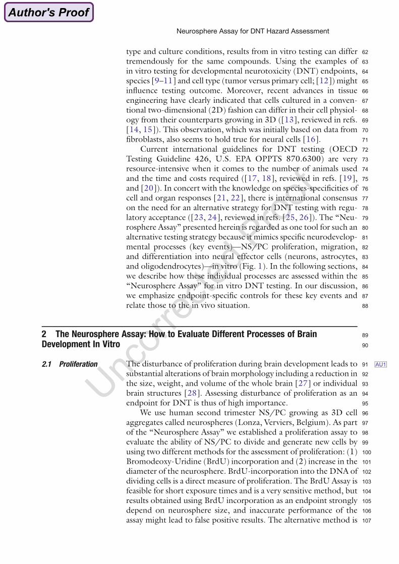
2.1 Proliferation 91 AU1The disturbance of proliferation during brain development leads to92substantial alterations of brain morphology including a reduction in93the size, weight, and volume of the whole brain [27] or individual94brain structures [28]. Assessing disturbance of proliferation as an95endpoint for DNT is thus of high importance.96We use human second trimester NS/PC growing as 3D cell97aggregates called neurospheres (Lonza, Verviers, Belgium). As part98of the “Neurosphere Assay” we established a proliferation assay to99evaluate the ability of NS/PC to divide and generate new cells by100using two different methods for the assessment of proliferation: (1)101Bromodeoxy-Uridine (BrdU) incorporation and (2) increase in the102diameter of the neurosphere. BrdU-incorporation into the DNA of103dividing cells is a direct measure of proliferation. The BrdU Assay is104feasible for short exposure times and is a very sensitive method, but105results obtained using BrdU incorporation as an endpoint strongly106depend on neurosphere size, and inaccurate performance of the107assay might lead to false positive results. The alternative method is
Neurosphere Assay for DNT Hazard Assessment
108simpler and assesses proliferation indirectly by measuring the109increase in the diameter of floating neurospheres over time. How-110ever, this method is less sensitive and only robust changes in prolif-111eration are detected. For this reason, exposure times of at least 1112week are recommended for this assay (for detailed information on113these two methods see ref. [29]).114When performing DNT testing in an in vitro model it is essen-115tial to guarantee that the basic biology of the tested endpoint is116functional and the endpoints can be modulated in vitro by factors117known to modulate them in vivo. Therefore, the use of endpoint118specific controls for quality assurance is necessary. The selected119endpoint specific control for proliferation is growth factor (GF;120epidermal growth factor (EGF) and fibroblast growth factor121(FGF-2)) withdrawal. NS/PC proliferation is tested both in prolif-122eration medium (B27) including GF and in GF-free medium (B27123w/o EGF and FGF-2). For both methods, GF withdrawal causes at124least 60–70 % reduction in proliferation relative to controls (Fig. 2).125NS/PC proliferation is well known to be dependent on EGF and126FGF [30–33]. EGF-receptor (EGF-R)-deficient mice develop127smaller brains with smaller germinal zones, yet on E17, prolifera-128tion in these proliferative areas was not reduced. This might be due129to inhibition of proliferation earlier during development and/or
Fig. 2 Quality control of the endpoint proliferation in human NS/PC. Proliferation
was assessed by performing a BrdU Cell Proliferation ELISA (Roche, black bars)
or measuring the diameter increase (grey bars). Therefore, six neurospheres
were plated in a 96-Well plate (one sphere per well) in normal proliferation
medium with (control) or without growth factors (w/o GF) for 72 h. For
assessment of BrdU incorporation, 16 h prior to the end of the experiment
BrdU was added to the spheres. The assay was performed according to the
manufacturer’s instructions. For the assessment of the diameter increase, the
diameter was measured on day 0 and day 3. Data (% of control) is shown
as mean of 3–4 independent experiments � SEM. *p-value � 0.05 (students t-
test) was considered as significant compared to control (adapted from Baumann
et al. 2015 [112])
Jenny Baumann et al.
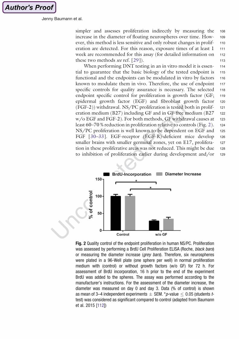
130compensatory effects of FGF [34]. Yet, EGF-R-deficient animals131die within the first 14 days postnatally.132It is essential that an endpoint specific control is specifically133acting on the neurodevelopmental process of interest rather than134influencing the general viability of the neurospheres. An option for135assessing viability is to perform an Alamar Blue Assay, which moni-136tors mitochondrial reductase activity. However, for the endpoint of137proliferation, the information obtained by this assay does not accu-138rately reflect cell viability, as sphere diameter and mitochondrial139activity are directly correlates because of different cell numbers in140different sized spheres [35]. For this reason, e.g., the lactate dehy-141drogenase (LDH) assay, which measures cellular membrane integ-142rity, is the preferred method for monitoring cytotoxic effects of143compounds in proliferating spheres.144One benefit of the neurosphere model is the opportunity to145compare neurodevelopmental processes of different species in par-146allel in vitro, which facilitates extrapolation between species and147enables a direct in vivo–in vitro correlation for rodents. For prolif-148eration, the BrdU and the neurosphere size assay indicated that149human, mouse, and rat NS/PC proliferate at different rates in150culture, and thus present different BrdU incorporation levels after1513 days (Fig. 3a) or different diameter increases after 7 days (Fig. 3b)152of proliferation.153How does NS/PC proliferation respond to DNT compounds154in vitro and in vivo? For the majority of DNTcompounds (reviewed155in refs. [36], and [37]) there is no mechanistic data available for156their MoA in humans; therefore, the rodent model is of great value.157Comparative human–rodent in vitro studies are therefore extremely158useful in assessing whether the proposed mechanism of action
Fig. 3 Species comparison of the endpoint proliferation. (a) Proliferation was assessed by performing a BrdU
Cell Proliferation ELISA (Roche). Control data (RLU) of 3–5 independent experiments is shown as mean �
SEM. *p-value � 0.05 (students t-test) was considered as significant between the species. (b) Phase-contrast
images of human, mouse, and rat neurospheres taken after 0 days (0 d) or 7 days (7 d) of proliferation, scale
bar ¼ 100 μm (adapted from Baumann et al. 2015 [112])
Neurosphere Assay for DNT Hazard Assessment
159based on rodent studies is of relevance to humans. Currently, the160best-studied DNT compound is methyl mercury (MeHg). Two161independent labs demonstrated that MeHg reduced proliferation162of rat primary neural cells in vitro and in the hippocampi of mice163[38] or rats [39] in vivo. Furthermore, experimental data from low-164dose exposure humans studies demonstrated a decreased mitotic165index in lymphocytes, which reflects an inhibition of cell-cycle166progression and/or a loss of proliferative capacity [40]. MeHg167was also shown to inhibit NS/PC proliferation in the nM range168in human umbilical cord blood-derived neural stem cells [41].169Besides MeHg, the antiepileptic drug valproic acid (VPA) is also a170well-known developmental toxicant causing autism spectrum dis-171orders in prenatally exposed infants (reviewed in ref. [42]). The172mechanisms underlying VPA-induced DNT are not well under-173stood because VPA acts on a multitude of cellular targets (reviewed174in ref. [43]). Go and coworkers found that after an injection of VPA175in rats at E12, postnatal brain weight was significantly increased.176Moreover, NS/PC isolated from rat brains either exposed prena-177tally to VPA or treated with VPA in vitro displayed increased178proliferation in vitro [44]. The opposite effects of VPA on prolifer-179ation of brain cells in vivo were seen when VPA was administered180postnatally, and this inhibition of proliferation was recapitulated181in vitro [45]. These data demonstrate that for some compounds,182effects on rodent NS/PC proliferation in vivo and in vitro correlate183well, and thus, ex vivo NS/PC seem to preserve their in vivo184molecular program. Therefore, it seems likely that primary human185NS/PC cultures, such as the above-described primary human neu-186rospheres, will generate data relevant to the human DNT com-187pound hazard on this endpoint. In the context of an alternative188DNT testing battery it has to be pointed out that the “Neurosphere189Assay” described here is based on fetal (GW 16-20) cells, which will190most accurately reflect fetal NS/PC proliferation. This is important191to note, because adverse compound effects on NS/PC proliferation192might be dependent on the timing of insult [44, 45]. How similar193hESC- or human induced pluripotent stem cell (hiPSC)-derived194NS/PC are to primary hNS/PC and their in vivo counterparts195and—most importantly—which window of development they196reflect, needs to be urgently tested to correctly determine which197phase of neurodevelopment they represent in a DNT testing198strategy.199
2.2 Migration 200Throughout and then after the division of proliferating NS/PC,201radial glia as well as postmitotic differentiating cells migrate202towards their final destinations in the brain. Several human devel-203opmental brain disorders have been associated with disruptions of204the migration process, including heterotopia and lissencephaly205[46]. Migration defects of specific types of interneurons have also206been associated with schizophrenia and epilepsy ([47, 48],
Jenny Baumann et al.
207reviewed in ref. [49]). Therefore, an in vitro DNT testing battery208needs to include a model capable of detecting compound effects on209migration (Fig. 1).210The “Neurosphere Assay” includes two simple methods to211evaluate the ability of NS/PC to migrate simultaneously, mimick-212ing their ability to move from proliferating niches to their final213positions in the developing brain. First, migration speed can be214assessed by measuring the distance cells migrate over time. A sec-215ond gauge of successful migration is the evaluation of absolute216numbers of cells that migrate out of the neurosphere during this217period. To quantify both endpoints, neurospheres are plated onto a218poly-D-lysine/laminin coated surface of a well filled with differenti-219ation medium (for more details on the migration protocol see ref.220[29]). After adhering to the laminin extracellular matrix, cells221spontaneously start to migrate out of the neurosphere ([35], Sup-222plementary Material 2). At the desired evaluation time point (e.g.,22324 h), phase-contrast pictures of migration areas are taken. Migra-224tion distance is evaluated by measuring four radii of the migration225area of each neurosphere in perpendicular angles from the edge of226the neurosphere to the furthest migrated cells (Fig. 4 left). To227count the number of migrated cells, neurospheres are fixed with2284 % paraformaldehyde and cell nuclei are stained with Hoechst dye.
Fig. 4 Measuring migration within the “Neurosphere Assay.” (Left) Phase-contrast image of the whole
migration area of a human neurosphere after 24 h of culture on a PDL/laminin coated surface of a well filled
with differentiation medium. Arrows exemplify the assessment of the migration distance by measuring four
perpendicular radii from the neurosphere edge to the end of the migration area. (Right) Fluorescent picture of
the whole migration area of the same human neurosphere, where migrated nuclei stained with Hoechst can be
counted, scale bar ¼ 100 μm
Neurosphere Assay for DNT Hazard Assessment
229The total number of Hoechst-positive cell nuclei is then counted in230fluorescence pictures of the whole migrating area (Fig. 4 right).231These two straightforward ways of evaluating migration allow232the comparison of the migration behavior of control cells to233compound-exposed cells after a certain exposure time. A close234monitoring of the control migration by time-lapse pictures has235shown that cells migrating out of the neurosphere exhibit classical236migration features required for migration dynamics. Migrating cells237present a very active growth cone protrusion moving away from the238cell body. This growth cone explores the local microenvironment,239and later the cell nucleus is translocated toward the new position240(nucleokinesis). In the “Neurosphere Assay” most cells migrate in a241radial trajectory using a scaffold built by GFAP+ cells; however,242different trajectories and speed rates can also be observed ([35],243Supplementary Material 2).244Several signaling pathways drive these migration dynamics and245one of those is mediated by activation of src kinases [50]. For this246reason, the src kinase inhibitor PP2 is used as the endpoint specific247control for migration to assess general assay performance (Fig. 5a).248PP2 does not completely inhibit migration because several other249pathways contribute to the migration process, but the distance is250decreased to around 60% of control values [50]. It is important to251note that because src kinases participate in the regulation of many252signaling pathways, one of which is cellular survival [51], a pro-253longed exposure (72 h) to this compound also impacts cell viability254(Fig. 5b). As there is cross talk among several pathways involved in255migration and the cell cycle [52], compounds affecting signaling256pathways involved in migration are also thought to eventually
Fig. 5 Quality control of the endpoint migration in human NS/PC. Human neurospheres were cultured for 24 or
72 h on a PDL/laminin coated surface of a well filled with differentiation medium (control) or differentiation
medium with 10 μM PP2. Migration distance was measured in phase-contrast images of five neurospheres
per assay. Viability of the same spheres was measured by Alamar Blue Assay. Bars represent the mean of four
independent experiments � SEM in % of control. *p-value � 0.05 (students t-test) was considered as
significant versus control (adapted from Baumann et al. 2015 [112])
Jenny Baumann et al.
257influence cell viability. Therefore, it is important to measure cell258viability as early as migration effects are observed when testing the259effects of unknown compounds. If migration is reduced as a result260of general cytotoxicity, both migration and viability will be reduced,261while in the case of specific migration pathway interference, migra-262tion will be altered first without showing signs of cytotoxicity. In263our experience, this timing effect in vitro is true for all studied264endpoints, yet migration seems to be very sensitive. In addition to265detecting decreases in migration distance, the assay can identify266increased migration rates relative to untreated controls [53]. In267these cases, general viability pathways are commonly not blocked,268and therefore cytotoxic effects are generally not an issue.269Evaluating migration after 24 h is also a convenient time point270when comparing the effects of compounds on migration of NS/PC271of different species, because neurospheres from humans, rats, and272mice exhibit similar migration dynamics in culture during the first27324 h (Fig. 6), with a migration distance covering between 400 and274500 μm during this period (around 20 μm/h). In contrast, during275the second 24 h (24–48 h of the migration assay) the migration276rates between species differ with 17.2, 9.6, and 1.9 μm/h for277human, mouse, and rat, respectively. Within this time frame, the278migration rate of rodent cultures is significantly slower than the279migration speed of the human culture. During the next 24 h280(48–72 h of the migration assay) human migration pace further281decreases to 7.6 μm/h, while rodent speeds stay about the same282(Fig. 6). It is noteworthy that migration speed during the first 24 h283in all species is in agreement with real-time observed granule cell
Fig. 6 Species comparison on NS/PC migration distance over time. Human, mouse, and rat neurospheres were
cultured for 72 h in differentiation medium on PDL/laminin coated wells. Phase-contrast images were taken
every 24 h and migration distance was measured in five neurospheres per assay. Bars represent the mean of
four independent experiments � SEM. Statistical analysis was performed by multiple t-test with Holm–Sidak
multiple comparison with the GraphPrism 6 program, and significant threshold was established at p < 0.05.*
was considered statistically significant different versus human value of the same time-point# [54]
Neurosphere Assay for DNT Hazard Assessment
284migration rates in vivo in mice, which range between 14.2 and28526.8 μm/h [54]. This in vitro–in vivo similarity of migration286dynamics strongly underlines the physiological relevance of the287neurosphere migration assay.288Most importantly, this migration assay not only accurately289reflects the in vivo migration process but also detects migration290alterations caused by substances known to disturb human neural291migration in vivo, like MeHg [54]. Humans developmentally292exposed to MeHg suffer from mental retardation and cerebral293palsy [55–57]. Their brains are hypoplasic and present many294ectopic neurons, indicating disrupted migration among other295alterations (reviewed in refs. [58], and [59]). Disturbances of neu-296ral migration after developmental exposure to MeHg have also297been observed in animal models in vivo and in vitro [60, 61]. In298agreement with the literature, the “Neurosphere Assay” detects a299significant reduction in NS/PC migration at in vivo relevant con-300centrations of MeHg, which are not cytotoxic (LOAEC ¼ 0.5 μM;301[35]). In essence, the neurosphere migration assay offers the possi-302bility to study and compare effects of chemicals on NS/PC migra-303tion and to elucidate the mechanisms behind migration alterations304of compounds in human and rodent cultures.305
2.3 Differentiation 306During the migration period and also afterwards, NS/PC differen-307tiate into neural effector cells to correctly form the different brain308regions. NS/PC differentiate into either neurons or glia cells309among which the most abundant cell types are astroglial and oli-310godendroglial cells. The differentiation of NS/PC into these three311cell types within the “Neurosphere Assay” will be the subject of the312following section. In general, NS/PC differentiation is initiated by313growth factor withdrawal and offering a specific extracellular314matrix. Therefore, we routinely plate our neurospheres on poly-D-315lysine/laminin coated surfaces in medium without growth factors316and culture them for 3 (neurons and astrocytes) or 5 days (oligo-317dendrocytes). After this culture period, we fix the neurospheres and318perform an immunocytochemical staining for specific epitopes of319the different cell types (neurons: ßIII tubulin; astrocytes: GFAP,320oligodendrocytes: O4) which are then evaluated by fluorescence321microscopy. For mouse NS/PC, the differentiation medium is322supplemented with 1% FCS to prevent apoptosis. For a detailed323method description, please see Baumann et al. [29].324
2.3.1 Neurons 325The differentiation of NS/PC to neurons during brain develop-326ment is essential for the appropriate cellular composition of the327different brain regions and functionality of neuronal networks.328Disturbing neuronal differentiation during normal brain develop-329ment, therefore, results in structural changes of the brain,330which can manifest as behavioral disorders (reviewed in ref. [62]).
Jenny Baumann et al.
331Thus, the investigation of NS/PC differentiation is another332essential endpoint to be evaluated for alternative DNT testing.333We assess the ability of NS/PC to differentiate into neurons334within the “Neurosphere Assay” by evaluating the percentage of335cells in the migration area that differentiated into neurons. A man-336ual counting of neurons strongly depends on high quality immu-337nocytochemical staining for a clear identification of neurons in the338migration area.339To control general assay performance for neuronal differentia-340tion, neurospheres are differentiated in the presence of EGF341(20 ng/ml) as an endpoint specific control to reliably inhibit NS/342PC differentiation to neurons [63]. For a valid endpoint specific343control, EGF in the culture medium should reduce the number of344neurons to around 20 % of the control value (Fig. 7). EGF does not345reduce viability when administered during differentiation, showing346that our endpoint specific control inhibits neuronal differentiation347without causing cytotoxicity.348In order to compare neuronal differentiation across species we349compared the percentage of neurons in the migration area after 3350days of differentiation under standard differentiation culture con-351ditions in humans, rat, and mouse cultures. Quantitative evaluation352of the three species revealed statistically significant differences in the353amount of differentiated ßIII tubulin + cells. In human neuro-354spheres approximately 10 % of the cells in the migration area were355ßIII-tubulin+, whereas in mouse and rat neurospheres, the
Fig. 7 Quality control of the endpoint neuronal differentiation in human NS/PC.
(a) Neuronal differentiation was assessed by immunocytochemical staining of
migrated human NS/PC after 3 days of differentiation for the neuronal marker ßIII
tubulin and Hoechst for nuclear counterstaining. Therefore, five neurospheres
were plated in one well of an 8-chamber slide in normal differentiation medium
(control) or differentiation medium with 20 ng/mL EGF for 72 h. (b) For the
assessment of NS/PC viability an Alamar Blue Assay was performed according to
the manufacturer’s instructions after 3 days of differentiation. Data (% of control)
is shown as mean of four independent experiments � SEM. *p-value � 0.05
(students t-test) was considered as significant compared to control (adapted
from Baumann et al. 2015 [112])
Neurosphere Assay for DNT Hazard Assessment
356percentage of ßIII-tubulin + cells was approximately 5 and 15 %,357respectively (Fig. 8).358When testing chemicals for their DNT potential, how does the359endpoint neuronal differentiation measured in in vitro reflect360in vivo data? Kim and coworkers (2009) showed that Bisphenol A361(BPA), an endocrine disruptor that potentially disrupts neuronal362development (reviewed in ref. [64]), promoted neuronal differen-363tiation in the murine immortalized C17.2 progenitor cell line and364in primary embryonic hippocampal neurons. These in vitro data365demonstrating accelerated neurogenesis were reflected by an366enhanced dentate gyrus formation in mice on PND1 after
Fig. 8 Species comparison of the endpoint neuronal differentiation. (a) Neuronal differentiation was assessed by
determining the percentage of neurons in the migration area after immunocytochemical staining for ßIII tubulin
and cell nuclei. Control data (% Neurons of total nuclei) of four independent experiments is shown as mean �
SEM. *p-value � 0.05 (students t-test) was considered as significantly different between the species. (b)
Fluorescent images of human, mouse, and rat neurospheres taken after 3 days of differentiation (blue: cell
nuclei; red: ßIII tubulin positive cells), scale bar ¼ 100 μm (adapted from Baumann et al. 2015 [112])
Jenny Baumann et al.
367administering BPA during pregnancy between E14.5 and 18.5368[65]. Hence, in this study BPA affects murine brain development369by altering neuronal differentiation both in vivo and in vitro.370Another study from Go and coworkers [44] demonstrated that371VPA induced neuronal differentiation of NS/PC generated from372E14 rat cortex after exposure in vitro and in vivo after a single373injection of rat dams with VPA at E12. Studies like these clearly374show that an affected neuronal differentiation in vitro in primary375cells correlates with corresponding in vivo data, illustrating that the376endpoint of neuronal differentiation is a suitable and important377endpoint for in vitro DNT testing using NS/PC. Similarly as dis-378cussed in the proliferation part of this chapter, we expect each379species to be representative of its respective molecular profile, and380thus, human primary cells as described above resemble human381physiology. And again, application of hESC/hiPSC methods for382assessing the endpoint neuronal differentiation, especially have to383be characterized with regard to developmental time points.384
2.3.2 Astrocytes 385Differentiation of NS/PC to astroglial cells during brain develop-386ment is essential for processes like neuronal migration, neurite387guidance, and the formation of functioning synapses (reviewed in388ref. [66]). Disturbing astroglial cells or neuronal–astroglial389interactions during critical periods of brain development (e.g.,390during neuronal migration) can induce substantial deficits in the391function of the central nervous system (reviewed in ref. [67]).392Therefore, the assessment of NS/PC differentiation to astrocytes393is thought to serve as an important functional endpoint during394DNT testing.395We assess the ability of hNS/PC to differentiate into astrocytes396by evaluating quantitative and qualitative changes in astrocytes397differentiation and maturation using immunocytochemical staining398of migrated cells for GFAP. Bonemorphogenetic protein (BMP)-7-399dependent astroglial differentiation serves as an endpoint specific400control [68]. Figure 9 clearly demonstrates that human NS/PC401differentiated in the presence of BMP-7 generated more astrocytes402with a higher GFAP content and a more mature phenotype with403shorter processes and a more stellate-like phenotype.404Comparing the morphology of astrocytes differentiated for 3405days from human, rat, and mouse NS/PC reveals similar astroglial406morphologies in mouse and rat NS/PC with stellate astrocytes,407whereas human NS/PC mainly show elongated astrocytes with408more radial glia-like structures (Fig. 10). This might indicate that409astrocytes differentiated from rodent NS/PC are already more410mature after 3 days of differentiation when compared to human411NS/PC, which again indicates that human and rodent NS/PC412differ in their rate of differentiation and maturation [69, 70].413The endpoint of astroglial differentiation for DNT testing has414been investigated in vivo and in vitro earlier. Burry and coworkers
Neurosphere Assay for DNT Hazard Assessment
415observed that postnatal exposure of rats to toluene, one of the416known human DNT compounds (reviewed in ref. [37]), impaired417astrocyte development in the developing brain, which was418measured by reduced brain GFAP content in rats treated with419toluene daily between PND4 and 10. This toluene-induced420GFAP reduction could be the result of reduced astrocyte matura-421tion, diminished astrocyte number or a combination of both. In422vitro analyses in the same study revealed that proliferation of GD 21423rat cortical astrocytes was reduced after toluene treatment for 24 h424in a concentration-dependent manner in the absence of cytotoxicity425[71]. Similarly, ethanol, which is also a confirmed human develop-426mental neurotoxicant (reviewed in ref. [36]), delays the appearance
Fig. 9 Quality control of the endpoint astrocyte differentiation in human NS/PC. Astrocyte differentiation was
assessed by immunocytochemical staining of migrated human NS/PC after 7 days of differentiation for the
astrocyte marker GFAP and Hoechst for nuclear counterstaining. Therefore, five neurospheres were plated in one
well of an 8-chamber slide coated with PDL/laminin in normal differentiation medium (a) or differentiation
medium with 100 ng/mL BMP-7 (b) for 7 days (blue: cell nuclei; red: GFAP positive cells), scale bar ¼ 100 μm
Fig. 10 Species comparison of the endpoint astrocyte differentiation. Astrocyte differentiation was evaluated
by immunocytochemical staining of the migration area after 3 days of differentiation for the astrocyte marker
GFAP (red) and cell nuclei (blue), scale bar ¼ 20 μm
Jenny Baumann et al.
427of GFAP during brain development and decreases its expression428after prenatal exposure of rats. Radial glial cultures of rats that were429prenatally exposed to ethanol showed a similar reduction in GFAP430content [72], and an in vitro treatment of rat NS/PC with ethanol431resulted in a reduced number of astrocytes after differentiation432[73]. Ethanol causes fetal alcohol syndrome (FAS), and there is433human evidence that ethanol impairs astrogliogenesis, as evidenced434by aberrant neural and glial tissue and other abnormalities in neural435and glial migration in postmortem brain of FAS children (reviewed436in ref. [74]). These studies demonstrate that results obtained for437the endpoint astrogliogenesis both in vivo and in vitro correlate438well, which makes it a valuable endpoint for evaluating the devel-439opmentally neurotoxic potential of chemicals.440
2.3.3 Oligodendrocytes 441Oligodendrogenesis during brain development is necessary for442proper brain function as oligodendrocytes form and maintain mye-443lin sheaths around axons in the central nervous system. Disturbance444of oligodendrocyte development may result in demyelination445diseases that severely affect neuronal functioning (reviewed in446ref. [75]).447To study oligodendrogenesis within the “Neurosphere Assay,”448the number ofO4+ cells/total number of cell nuclei in themigration449area is counted and oligodendrocyte morphology is evaluated after4505 days of differentiation. The endpoint specific control substance451chosen to inhibit oligodendrogenesis is BMP-7 (Fig. 11), because452similar to other BMPs (e.g., BMP-2), BMP-7 induces astro-453gliogenesis (see above) and inhibits oligodendrogenesis [76, 77].
Fig. 11 Quality control of the endpoint oligodendrogenesis in human NS/PC.
Human neural progenitor cells were plated onto PDL/laminin coated 8-chamber
glass slides in normal differentiation medium (control) and differentiation medium
with 100 ng/mL BMP-7 for 5 days. Mitochondrial activity was measured by
Alamar Blue Assay. Slides were fixed and immunocytochemically stained
for O4. Data are shown as mean of five independent experiments � SEM.
*p-value � 0.05 (student t-test) was considered as significant
Neurosphere Assay for DNT Hazard Assessment
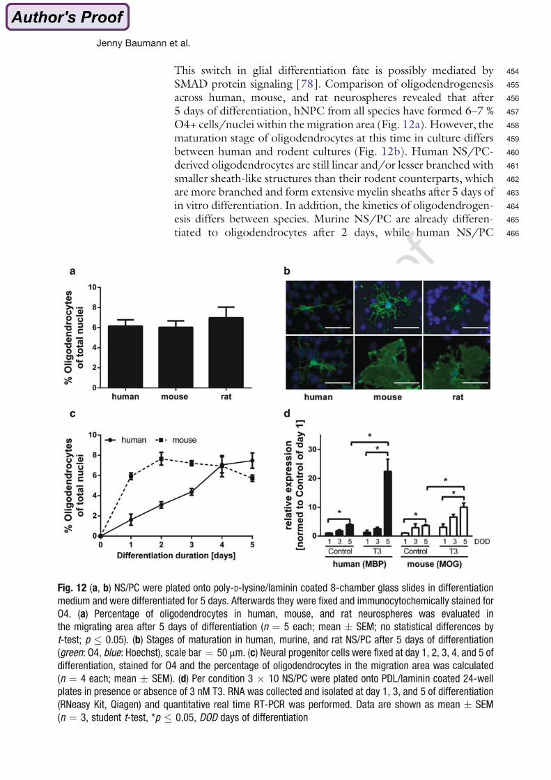
454This switch in glial differentiation fate is possibly mediated by455SMAD protein signaling [78]. Comparison of oligodendrogenesis456across human, mouse, and rat neurospheres revealed that after4575 days of differentiation, hNPC from all species have formed 6–7 %458O4+ cells/nuclei within themigration area (Fig. 12a). However, the459maturation stage of oligodendrocytes at this time in culture differs460between human and rodent cultures (Fig. 12b). Human NS/PC-461derived oligodendrocytes are still linear and/or lesser branched with462smaller sheath-like structures than their rodent counterparts, which463are more branched and form extensive myelin sheaths after 5 days of464in vitro differentiation. In addition, the kinetics of oligodendrogen-465esis differs between species. Murine NS/PC are already differen-466tiated to oligodendrocytes after 2 days, while human NS/PC
Fig. 12 (a, b) NS/PC were plated onto poly-D-lysine/laminin coated 8-chamber glass slides in differentiation
medium and were differentiated for 5 days. Afterwards they were fixed and immunocytochemically stained for
O4. (a) Percentage of oligodendrocytes in human, mouse, and rat neurospheres was evaluated in
the migrating area after 5 days of differentiation (n ¼ 5 each; mean � SEM; no statistical differences by
t-test; p � 0.05). (b) Stages of maturation in human, murine, and rat NS/PC after 5 days of differentiation
(green: O4, blue: Hoechst), scale bar ¼ 50 μm. (c) Neural progenitor cells were fixed at day 1, 2, 3, 4, and 5 of
differentiation, stained for O4 and the percentage of oligodendrocytes in the migration area was calculated
(n ¼ 4 each; mean � SEM). (d) Per condition 3 � 10 NS/PC were plated onto PDL/laminin coated 24-well
plates in presence or absence of 3 nM T3. RNA was collected and isolated at day 1, 3, and 5 of differentiation
(RNeasy Kit, Qiagen) and quantitative real time RT-PCR was performed. Data are shown as mean � SEM
(n ¼ 3, student t-test, *p � 0.05, DOD days of differentiation
Jenny Baumann et al.

467differentiate to O4+ cells progressively over 5 days of differentiation468(Fig. 12c). This characterization of oligodendrocyte maturation by469morphological appearance (branches/sheath formation) over time470demonstrates thatmurine oligodendrocytes differentiate andmature471faster than their human counterparts. These in vitro observations472reflect what is known in vivo: the time span between pre-473oligodendrocyte formation and emergence of the first mature oligo-474dendrocytes is 11–12 weeks in humans but only 5 days in rodents475(reviewed in ref. [79]).476Molecular analyses of myelin basic protein (MBP, for hNS/PC)477or myelin oligodendrocyte glycoprotein (MOG, for mNS/PC)478mRNA expression support this observation (Fig. 12d). Expression479of these maturation markers increases over differentiation time.480Human oligodendrocytes mature between day 3 and 5, while481murine oligodendrocytes mature between day 1 and 3. The482in vivo relevance of oligodendrocyte differentiation from NS/PC483is displayed by studying the consequence of the thyroid hormone484T3 on oligodendrocyte maturation in vitro. Thyroid hormone485drives oligodendrocyte maturation in vivo since hyperthyroid486rodents express higher and hypothyroid rodents lower levels of487oligodendrocyte maturation genes [80–82]. Similarly, T3 induces488MBP and MOG in differentiated human and mouse NS/PC,489respectively, with higher potency in human cultures (Fig. 12d).490This stronger T3-dependent increase of oligodendrocyte matura-491tion markers observed in human compared to murine cultures may492be due to a higher T3-responsiveness of human oligodendrocyte493maturation genes or an already higher maturation grade of mouse494oligodendrocytes at the beginning of the differentiation process495with thus limited acceleration of the process.496Besides physiological, hormonal effects on oligodendrocyte497maturation (Fig. 12d) the literature provides us with the indication498that in vitro studies concerning oligodendrocyte toxicity have499in vivo relevance. Eschenroeder and coworkers observed that low500doses of the opioid buprenorphine accelerated and increased, but501high doses delayed and decreased MBP expression in oligodendro-502cyte cultures isolated from rat brains [83]. The same dose-503dependent effects of buprenorphin on MBP expression were seen504in vivo in brain lysates of pups treated on embryonic day 7 and505sacrificed at PND 12, 19, or 26 [84]. A second compound affecting506oligodendrocytes during the perinatal period is vanadium. This507metal produces hypomyelination during the second postnatal508week in rat in vivo, a period of the most intense oligodendrocyte509development and myelination in this species. This hypomyelination510is thought to be caused by oligodendrocyte precursor cell (OPC)511apoptosis due to vanadium-induced oxidative stress because
Neurosphere Assay for DNT Hazard Assessment
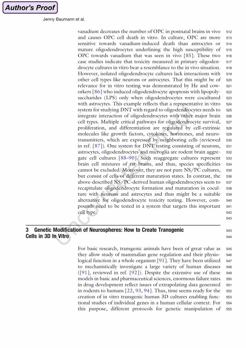
512vanadium decreases the number of OPC in postnatal brains in vivo513and causes OPC cell death in vitro. In culture, OPC are more514sensitive towards vanadium-induced death than astrocytes or515mature oligodendrocytes underlining the high susceptibility of516OPC towards vanadium that was seen in vivo [85]. These two517case studies indicate that toxicity measured in primary oligoden-518drocyte cultures in vitro bear a resemblance to the in vivo situation.519However, isolated oligodendrocyte cultures lack interactions with520other cell types like neurons or astrocytes. That this might be of521relevance for in vitro testing was demonstrated by He and cow-522orkers [86] who induced oligodendrocyte apoptosis with lipopoly-523saccharides (LPS) only when oligodendrocytes were cocultured524with astrocytes. This example reflects that a representative in vitro525system for studying DNT with regard to oligodendrocytes needs to526integrate interaction of oligodendrocytes with other major brain527cell types. Multiple critical pathways for oligodendrocyte survival,528proliferation, and differentiation are regulated by cell-extrinsic529molecules like growth factors, cytokines, hormones, and neuro-530transmitters, which are expressed by neighboring cells (reviewed531in ref. [87]). One system for DNT testing consisting of neurons,532astrocytes, oligodendrocytes and microglia are rodent brain aggre-533gate cell cultures [88–90]. Such reaggregate cultures represent534brain cell mixtures of rat brains, and thus, species specificities535cannot be excluded. Moreover, they are not pure NS/PC cultures,536but consist of cells of different maturation states. In contrast, the537above-described NS/PC-derived human oligodendrocytes seem to538recapitulate oligodendrocyte formation and maturation in cocul-539ture with neurons and astrocytes and thus might be a suitable540alternative for oligodendrocyte toxicity testing. However, com-541pounds need to be tested in a system that targets this important542cell type.543
5433 Genetic Modification of Neurospheres: How to Create Transgenic544Cells in 3D In Vitro
545For basic research, transgenic animals have been of great value as546they allow study of mammalian gene regulation and their physio-547logical function in a whole organism [91]. They have been utilized548to mechanistically investigate a large variety of human diseases549([91], reviewed in ref. [92]). Despite the extensive use of these550models in basic and pharmaceutical sciences, enormous failure rates551in drug development reflect issues of extrapolating data generated552in rodents to humans [22, 93, 94]. Thus, time seems ready for the553creation of in vitro transgenic human 3D cultures enabling func-554tional studies of individual genes in a human cellular context. For555this purpose, different protocols for genetic manipulation of
Jenny Baumann et al.
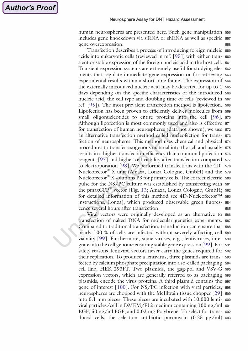
556human neurospheres are presented here. Such gene manipulation557includes gene knockdown via siRNA or shRNA as well as specific558gene overexpression.559Transfection describes a process of introducing foreign nucleic560acids into eukaryotic cells (reviewed in ref. [95]) with either tran-561sient or stable expression of the foreign nucleic acid in the host cell.562Transient expression systems are extremely useful for studying ele-563ments that regulate immediate gene expression or for retrieving564experimental results within a short time frame. The expression of565the externally introduced nucleic acid may be detected for up to 4566days depending on the specific characteristics of the introduced567nucleic acid, the cell type and doubling time of cells (reviewed in568ref. [95]). The most prevalent transfection method is lipofection.569Lipofection has been proven to efficiently deliver molecules from570small oligonucleotides to entire proteins into the cell [96].571Although lipofection is most commonly used and also is effective572for transfection of human neurospheres (data not shown), we use573an alternative transfection method called nucleofection for trans-574fection of neurospheres. This method uses chemical and physical575procedures to transfer exogenous material into the cell and usually576results in a higher transfection efficiency than common lipofection577reagents [97] and higher cell viability after transfection compared578to electroporation [98]. We performed transfections with the 4D-579Nucleofector® X unit (Amaxa, Lonza Cologne, GmbH) and the580Nucleofector® X solutions P3 for primary cells. The correct electric581pulse for the NS/PC culture was established by transfecting with582the pmaxGFP® vector (Fig. 13; Amaxa, Lonza Cologne, GmbH;583for detailed information of this method see 4D-Nucleofector™584instructions, Lonza), which produced observable green fluores-585cence several hours after transfection.586Viral vectors were originally developed as an alternative to587transfection of naked DNA for molecular genetics experiments.588Compared to traditional transfection, transduction can ensure that589nearly 100 % of cells are infected without severely affecting cell590viability [99]. Furthermore, some viruses, e.g., lentiviruses, inte-591grate into the cell genome ensuring stable gene expression [99]. For592safety reasons, lentiviral vectors never carry the genes required for593their replication. To produce a lentivirus, three plasmids are trans-594fected by calcium phosphate precipitation into a so-called packaging595cell line, HEK 293FT. Two plasmids, the gag-pol and VSV-G596expression vectors, which are generally referred to as packaging597plasmids, encode the virus proteins. A third plasmid contains the598gene of interest [100]. For NS/PC infection with viral particles,599neurospheres are chopped with the McIllwain tissue chopper [29]600into 0.1 mm pieces. These pieces are incubated with 10,000 lenti-601viral particles/cell in DMEM/F12 medium containing 100 ng/ml602EGF, 50 ng/ml FGF, and 0.02 mg Polybrene. To select for trans-603duced cells, the selection antibiotic puromycin (0.25 μg/ml)
Neurosphere Assay for DNT Hazard Assessment
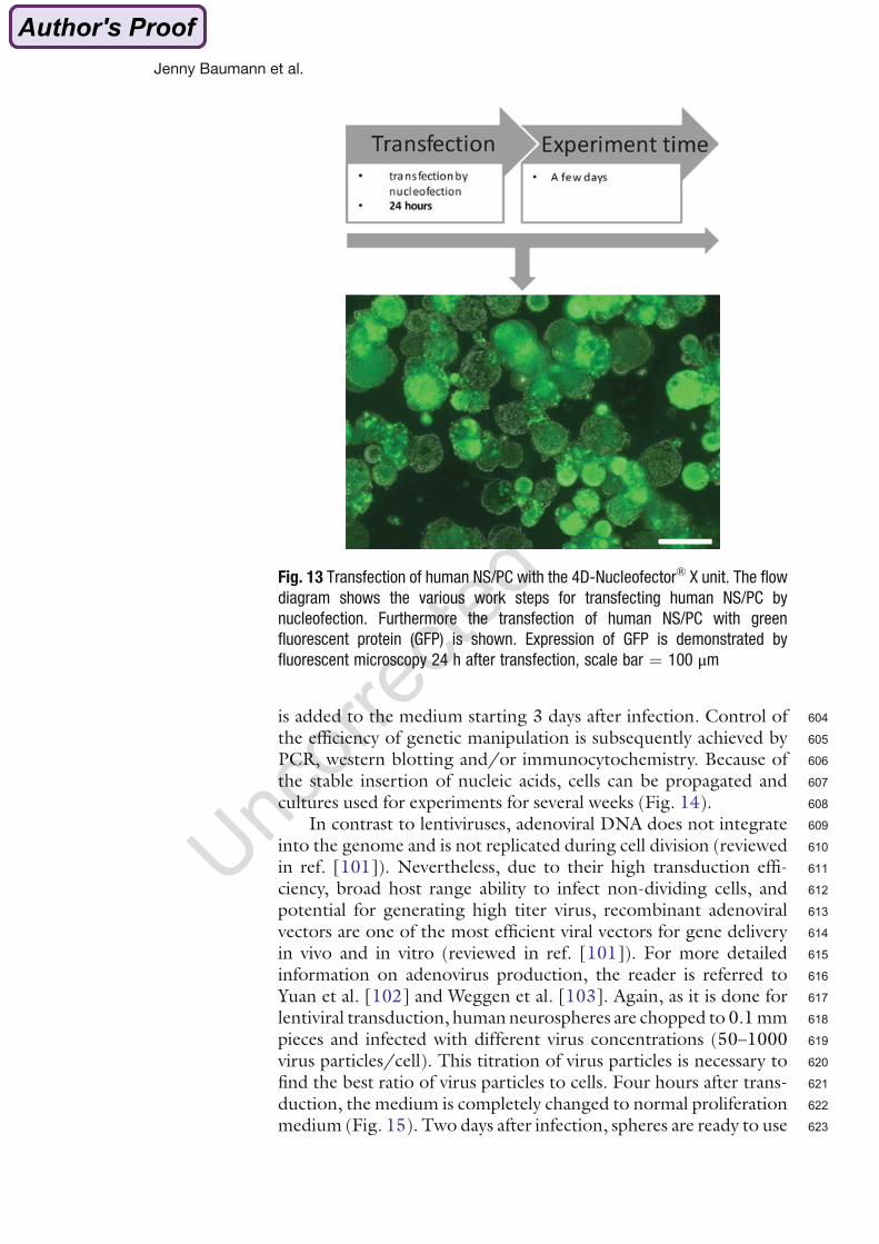
604is added to the medium starting 3 days after infection. Control of605the efficiency of genetic manipulation is subsequently achieved by606PCR, western blotting and/or immunocytochemistry. Because of607the stable insertion of nucleic acids, cells can be propagated and608cultures used for experiments for several weeks (Fig. 14).609In contrast to lentiviruses, adenoviral DNA does not integrate610into the genome and is not replicated during cell division (reviewed611in ref. [101]). Nevertheless, due to their high transduction effi-612ciency, broad host range ability to infect non-dividing cells, and613potential for generating high titer virus, recombinant adenoviral614vectors are one of the most efficient viral vectors for gene delivery615in vivo and in vitro (reviewed in ref. [101]). For more detailed616information on adenovirus production, the reader is referred to617Yuan et al. [102] and Weggen et al. [103]. Again, as it is done for618lentiviral transduction, human neurospheres are chopped to 0.1mm619pieces and infected with different virus concentrations (50–1000620virus particles/cell). This titration of virus particles is necessary to621find the best ratio of virus particles to cells. Four hours after trans-622duction, the medium is completely changed to normal proliferation623medium (Fig. 15). Two days after infection, spheres are ready to use
Fig. 13 Transfection of human NS/PC with the 4D-Nucleofector® X unit. The flow
diagram shows the various work steps for transfecting human NS/PC by
nucleofection. Furthermore the transfection of human NS/PC with green
fluorescent protein (GFP) is shown. Expression of GFP is demonstrated by
fluorescent microscopy 24 h after transfection, scale bar ¼ 100 μm
Jenny Baumann et al.
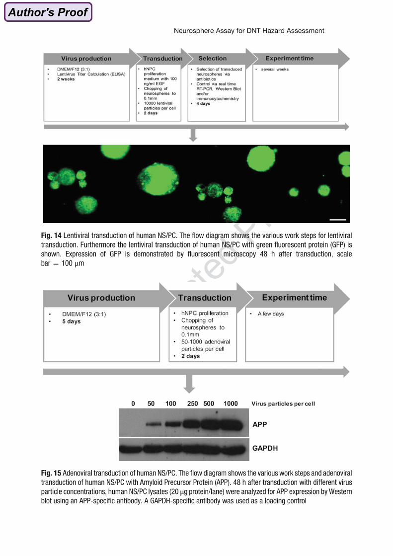
Fig. 14 Lentiviral transduction of human NS/PC. The flow diagram shows the various work steps for lentiviral
transduction. Furthermore the lentiviral transduction of human NS/PC with green fluorescent protein (GFP) is
shown. Expression of GFP is demonstrated by fluorescent microscopy 48 h after transduction, scale
bar ¼ 100 μm
Fig. 15 Adenoviral transduction of human NS/PC. The flow diagram shows the various work steps and adenoviral
transduction of human NS/PC with Amyloid Precursor Protein (APP). 48 h after transduction with different virus
particle concentrations, human NS/PC lysates (20 μg protein/lane) were analyzed for APP expression by Western
blot using an APP-specific antibody. A GAPDH-specific antibody was used as a loading control
Neurosphere Assay for DNT Hazard Assessment
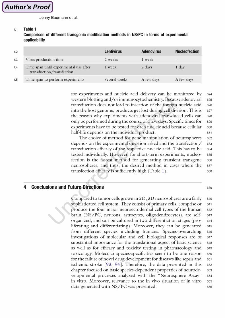
624for experiments and nucleic acid delivery can be monitored by625western blotting and/or immunocytochemistry. Because adenoviral626transduction does not lead to insertion of the foreign nucleic acid627into the host genome, products get lost during cell division. This is628the reason why experiments with adenoviral transduced cells can629only be performed during the course of a few days. Specific times for630experiments have to be tested for each nucleic acid because cellular631half-life depends on the individual product.632The choice of method for gene manipulation of neurospheres633depends on the experimental question asked and the transfection/634transduction efficacy of the respective nucleic acid. This has to be635tested individually. However, for short-term experiments, nucleo-636fection is the fastest method for generating transient transgene637neurospheres, and thus, the desired method in cases where the638transfection efficacy is sufficiently high (Table 1).
6394 Conclusions and Future Directions
640Compared to tumor cells grown in 2D, 3D neurospheres are a fairly641sophisticated cell system. They consist of primary cells, comprise or642produce the four major neuroectodermal cell types of the human643brain (NS/PC, neurons, astrocytes, oligodendrocytes), are self-644organized, and can be cultured in two differentiation stages (pro-645liferating and differentiating). Moreover, they can be generated646from different species including humans. Species-overarching647investigations of molecular and cell biological responses are of648substantial importance for the translational aspect of basic science649as well as for efficacy and toxicity testing in pharmacology and650toxicology. Molecular species-specificities seem to be one reason651for the failure of novel drug development for diseases like sepsis and652ischemic stroke [93, 94]. Therefore, the data presented in this653chapter focused on basic species-dependent properties of neurode-654velopmental processes analyzed with the “Neurosphere Assay”655in vitro. Moreover, relevance to the in vivo situation of in vitro656data generated with NS/PC was presented.
t:1 Table 1
Comparison of different transgenic modification methods in NS/PC in terms of experimental
applicability
Lentivirus Adenovirus Nucleofectiont:2
Virus production time 2 weeks 1 week –t:3
Time span until experimental use aftertransduction/transfection
1 week 2 days 1 dayt:4
Time span to perform experiments Several weeks A few days A few dayst:5
Jenny Baumann et al.
657With regard to the developing brain, primary human cultures658are currently the closest model to the human in vivo situation. This659is supported by the observations that rodent brain cells taken out of660the in vivo situation placed into a tissue culture dish maintain their661signaling functions or their responses towards xenobiotics (e.g.,662[39, 45, 44, 104, 105]), and thus, primary human cells are thought663to behave similarly when removed from the in vivo to the in vitro664situation. Although NS/PC generated from human embryonic665(hESC) or human induced pluripotent stem cells (hiPSC) are also666promising methods for human cell-based DNT testing, the in vivo-667relevance of these models for neurodevelopment, especially with668regard to developmental timing and cell type differentiation, i.e.,669formation of oligodendrocytes, remains an outstanding question.670Nevertheless, despite the advantages of such 3D in vitro sys-671tems, one still has to be aware that these are models—as are animals672and other cell-based assays. Every model has its strengths and673limitations. The limitations of in vitro models in conventional674culture are mainly their lack of physiological cell and organ context675as this creates artificial pharmacokinetics/toxicokinetics. Thus, the676whole issue of absorption/distribution/metabolism/excretion677(ADME) is not considered in in vitro testing situations. This can678be circumvented when human in vivo target organ concentrations679of compounds as well as their in vitro kinetics are known then680providing the opportunity to model realistic exposure scenarios681in vitro. Another approach to implement kinetics in vitro is682the “Organ-on-a-Chip” technology ([106], reviewed in refs.683[107, 108]). The use of human cells in this approach enables better684realization of human ADME. Another limitation of the neuro-685sphere system is the lack of some cell types. Neurospheres do not686contain microglia or endothelial cells. The latter is an especially687crucial factor because in conjunction with astrocytes, endothelial688cells form the blood–brain barrier. This important barrier deter-689mines internal brain exposure towards compounds as well as690towards endogenous mediators like hormones or cytokines.691Although in these self-organized coculture models one does have692interactions between different cell types—neurons, astrocytes, and693oligodendrocytes—the deficiency in microglia and endothelial cells694has to be taken into account. In addition, there is evidence that695identical brain cell types differ in their function from one brain696region to another (e.g., reviewed in ref. [109]). These brain region-697specific differences can easily be studied by cell preparations from698different brain areas in rodents. Such studies have identified region-699dependent susceptibility of developmental astrocyte proliferation700towards MeHgCl: while this compound inhibits hippocampal701astrocyte proliferation, cortical, cerebellar, and brain stem astro-702cytes proliferation is not specifically inhibited by MeHgCl [110].703Modeling human brain region-specific cell characteristics in vitro is704not possible based on current knowledge. In the future, with more
Neurosphere Assay for DNT Hazard Assessment
705information on the physiological factors responsible for brain706region-specific cell development and function, it might be possible707to also model this aspect in a dish by manipulating hESC or hiPSC708accordingly. Last, these cultures cannot think, feel, and associate, so709they can only mimic neurodevelopmental processes. However, with710additional, complementary assays, filling the biological application711domains not covered by the “Neurosphere Assay,” i.e., neuronal712network formation and electrical network activity measurements, a713battery of tests can be created enveloping the major key events of714human brain development in a dish and thus—although still not715thinking—generate an in vitro scenario that is able to give a certain716mechanism-based level of certainty for DNT assessment. Ideally,717such a battery would be embedded into the theoretical frame of the718“Adverse Outcome Pathway” (AOP) concept [111], which cur-719rently is receiving significant attention from regulatory agencies720worldwide. AOPs for DNT are still sparse and thus urgently721needed. The cross-species evaluation of signaling pathways, which722is possible with the neurospheres presented in this chapter, is highly723suited to fill the data gap on translation from rodents to humans, an724important aspect within the AOP concept.725Also for basic biomedical research these cultures can be of great726value, especially with the option of creating transgenic neuro-727spheres as described above. This technique is (a) much faster and728more cost-effective than first creating a transgenic animal and sub-729sequently preparing neurospheres from the pups; and (b) is based730on the species typically of greatest regulatory concern—humans.731The obvious limitation of this approach is, however, that in vitro732gene silencing never produces a complete knockout as in an animal733model, but is restricted to a knock-down with various degrees of734gene repression.735This chapter discusses the strengths and limitations of the736“Neurosphere Assay” for DNT hazard assessment. In the future,737this assay will be further characterized for its biological application
738 domain with regard to human in vivo brain development. Such739 knowledge will provide confidence for its position in a DNT in vitro740 testing battery that covers major key events in neurodevelopment741 for regulatory implementation.
742 References
744 1. Albright TD, Jessell TM, Kandel ER, Posner745 MI (2000) Neural science: a century of prog-746 ress and the mysteries that remain. Neuron 25747 (1):S1–S55
748 2. Kandel ER, Schwartz, JH, Jessell TM (2000)749 The development of the nervous system. In:750 Kandel ER, Schwartz JH, Jessell TM (ed)751 Principles of neural science. New York, pp752 1019–1114
7533. Knecht AK, Bronner-Fraser M (2002) Induc-754tion of the neural crest: a multigene process.755Nat Rev Genet 3(6):453–461
7564. Alvarez-Buylla A, Garcıa-Verdugo JM, Tra-757montin AD (2001) A unified hypothesis on758the lineage of neural stem cells. Nat Rev Neu-759rosci 2(4):287–293
7605. Andersen SL (2003) Trajectories of brain761development: point of vulnerability or
Jenny Baumann et al.
762 window of opportunity? Neurosci Biobehav763 Rev 27(1):3–18
764 6. Ek CJ, Dziegielewska KM, Habgood MD,765 Saunders NR (2012) Barriers in the develop-766 ing brain and Neurotoxicology. Neurotoxi-767 cology 33(3):586–604
768 7. Rodier PM (1995) Developing brain as a tar-769 get of toxicity. Environ Health Perspect 103770 (Suppl 6):73–76
771 8. Collins FS, Gray GM, Bucher JR (2008)772 Transforming environmental health protec-773 tion. Science 319(5865):906–907
774 9. Gassmann K, Baumann J, Giersiefer S, Schu-775 wald J, Schreiber T, Merk H, Fritsche E776 (2012) Automated neurosphere sorting and777 plating by the COPAS large particle sorter is a778 suitable method for high-throughput 3D779 in vitro applications. Toxicol in Vitro 26780 (6):993–1000
781 10. Culbreth ME, Harrill JA, Freudenrich TM,782 Mundy WR, Shafer TJ (2012) Comparison783 of chemical-induced changes in proliferation784 and apoptosis in human and mouse neuropro-785 genitor cells. Neurotoxicology 33786 (6):1499–1510
787 11. Harrill JA, Freudenrich TM, Robinette BL,788 Mundy WR (2011) Comparative sensitivity789 of human and rat neural cultures to790 chemical-induced inhibition of neurite out-791 growth. Toxicol Appl Pharmacol 256792 (3):268–280
793 12. Radio NM, Freudenrich TM, Robinette BL,794 Crofton KM, Mundy WR (2010) Compari-795 son of PC12 and cerebellar granule cell cul-796 tures for evaluating neurite outgrowth using797 high content analysis. Neurotoxicol Teratol798 32(1):25–35
799 13. Cukierman E, Pankov R, Stevens DR, Yamada800 KM (2001) Taking cell-matrix adhesions to801 the third dimension. Science 294802 (5547):1708–1712
803 14. Yamada KM, Cukierman E (2007) Modeling804 tissue morphogenesis and cancer in 3D. Cell805 130(4):601–610
806 15. Alepee N, Bahinski T, Daneshian M, De807 Wever B, Fritsche E, Goldberg A, Hansmann808 J, Hartung T, Haycock J, Hogberg H (2013)809 State-of-the-art of 3D cultures (organs-on-a-810 chip) in safety testing and pathophysiology.811 ALTEX 31(4):441–477
812 16. Lai Y, Asthana A, Cheng K, Kisaalita WS813 (2011) Neural cell 3D microtissue formation814 is marked by cytokines’ up-regulation. PLoS815 One 6(10):e26821
816 17. Tilson HA (1992) Study design considera-817 tions in developmental neurotoxicology. Neu-818 rotoxicol Teratol 14(3):199–203
81918. Mileson BE, Ferenc SA (2001) Methods to820identify and characterize developmental neu-821rotoxicity for human health risk assessment:822overview. Environ Health Perspect 109(Suppl8231):77–78
82419. Hass U (2003) Current status of developmen-825tal neurotoxicity: regulatory view. Toxicol826Lett 140:155–159
82720. Kaufmann W (2003) Current status of devel-828opmental neurotoxicity: an industry perspec-829tive. Toxicol Lett 140:161–169
83021. Hanke T (2006) Lessons from TGN1412.831Lancet 368(9547):1569–1570
83222. Leist M, Hartung T (2013) Inflammatory833findings on species extrapolations: humans834are definitely no 70-kg mice. Arch Toxicol83587(4):563–567
83623. Bal-Price A, Crofton, Kevin M., Shafer, Tim-837othy, Sachana, Magdalini, Behl, Mamta, For-838sby, Anna, Hargreaves, Alan, Landesmann,839Brigitte, Lein, Pamela J., Louisse, Jochem,840Monnet-Tschudi, Florianne, Paini, Alicia,841Rolaki, Alexandra, Schrattenholz, Andre,842Sunol, Cristina, van Thriel, Christoph, Whe-843lan, Maurice, Fritsche, Ellen (2014) Work-844shop report: Adverse Outcome Pathways845(AOP) relevant to neurotoxicity. (in press)
24. AU2Crofton K, Fritsche E, Ylikomi T, Bal-Price A846(2014) International STakeholder NETwork847(ISTNET) for Creating a Developmental848Neurotoxicity Testing (DNT) roadmap for849regulatory purposes. Altex 31(2/85014):223–224
85125. Crofton KM, Mundy WR, Shafer TJ (2012)852Developmental neurotoxicity testing: a path853forward. Congenit Anom (Kyoto) 52854(3):140–146
85526. EFSA (2013) Scientific opinion on the identi-856fication of pesticides to be included in cumu-857lative assessment groups on the basis of their858toxicological profile. EFSA J 11(7)
85927. De Groot DM, Hartgring S, Van de Horst L,860Moerkens M, Otto M, Bos-Kuijpers MH,861Kaufmann WS, Lammers JH, O’Callaghan862JP, Waalkens-Berendsen ID (2005) 2D and8633D assessment of neuropathology in rat864brain after prenatal exposure to methylazoxy-865methanol, a model for developmental neuro-866toxicity. Reprod Toxicol 20(3):417–432
86728. Moore H, Jentsch JD, Ghajarnia M, Geyer868MA, Grace AA (2006) A neurobehavioral sys-869tems analysis of adult rats exposed to methy-870lazoxymethanol acetate on E17: implications871for the neuropathology of schizophrenia. Biol872Psychiatry 60(3):253–264
87329. Baumann J, Barenys M, Gassmann K, Fritsche874E (2014) Comparative human and rat
Neurosphere Assay for DNT Hazard Assessment
875 “neurosphere assay” for developmental neu-876 rotoxicity testing. Curr Protoc Toxicol877 59:12.21.1–12.21.24
878 30. Reynolds BA, Tetzlaff W, Weiss S (1992) A879 multipotent EGF-responsive striatal embry-880 onic progenitor cell produces neurons and881 astrocytes. J Neurosci 12(11):4565–4574
882 31. Reynolds BA, Weiss S (1996) Clonal and pop-883 ulation analyses demonstrate that an EGF-884 responsive mammalian embryonic CNS pre-885 cursor is a stem cell. Dev Biol 175(1):1–13
886 32. Svendsen C, Fawcett J, Bentlage C, Dunnett S887 (1995) Increased survival of rat EGF-888 generated CNS precursor cells using B27 sup-889 plemented medium. Exp Brain Res 102890 (3):407–414
891 33. Gritti A, Parati E, Cova L, Frolichsthal P, Galli892 R, Wanke E, Faravelli L, Morassutti D, Roisen893 F, Nickel D (1996) Multipotential stem cells894 from the adult mouse brain proliferate and895 self-renew in response to basic fibroblast896 growth factor. J Neurosci 16(3):1091–1100
897 34. Kornblum HI, Hussain R, Wiesen J, Mietti-898 nen P, Zurcher SD, Chow K, Derynck R,899 Werb Z (1998) Abnormal astrocyte develop-900 ment and neuronal death in mice lacking the901 epidermal growth factor receptor. J Neurosci902 Res 53(6):697–717
903 35. Moors M, Rockel TD, Abel J, Cline JE, Gass-904 mann K, Schreiber T, Schuwald J, Weinmann905 N, Fritsche E (2009) Human neurospheres as906 three-dimensional cellular systems for devel-907 opmental neurotoxicity testing. Environ908 Health Perspect 117(7):1131–1138
909 36. Grandjean P, Landrigan PJ (2006) Develop-910 mental neurotoxicity of industrial chemicals.911 Lancet 368(9553):2167–2178
912 37. Grandjean P, Landrigan PJ (2014) Neurobe-913 havioural effects of developmental toxicity.914 Lancet Neurol 13(3):330–338
915 38. Bose R, Onishchenko N, Edoff K, Lang AMJ,916 Ceccatelli S (2012) Inherited effects of low-917 dose exposure to methylmercury in neural918 stem cells. Toxicol Sci 130:383–390
919 39. Burke K, Cheng Y, Li B, Petrov A, Joshi P,920 Berman RF, Reuhl KR, DiCicco-Bloom E921 (2006) Methylmercury elicits rapid inhibition922 of cell proliferation in the developing brain923 and decreases cell cycle regulator, cyclin E.924 Neurotoxicology 27(6):970–981
925 40. Amorim MI, Mergler D, Bahia MO, Dubeau926 H, Miranda D, Lebel J, Burbano RR, Lucotte927 M (2000) Cytogenetic damage related to low928 levels of methyl mercury contamination in the929 Brazilian Amazon. An Acad Bras Cienc 72930 (4):497–507
93141. Buzanska L, Sypecka J, Nerini‐Molteni S,932Compagnoni A, Hogberg HT, del Torchio933R, Domanska‐Janik K, Zimmer J, Coecke S934(2009) A human stem cell‐based model for935identifying adverse effects of organic and inor-936ganic chemicals on the developing nervous937system. Stem Cells 27(10):2591–2601
93842. Ornoy A (2009) Valproic acid in pregnancy:939how much are we endangering the embryo940and fetus? Reprod Toxicol 28(1):1–10
94143. Rosenberg G (2007) The mechanisms of942action of valproate in neuropsychiatric disor-943ders: can we see the forest for the trees? Cell944Mol Life Sci 64(16):2090–2103
94544. Go HS, Kim KC, Choi CS, Jeon SJ, Kwon KJ,946Han SH, Lee J, Cheong JH, Ryu JH, Kim947CH, Ko KH, Shin CY (2012) Prenatal expo-948sure to valproic acid increases the neural pro-949genitor cell pool and induces macrocephaly in950rat brain via a mechanism involving the GSK-9513beta/beta-catenin pathway. Neuropharma-952cology 63(6):1028–1041
95345. Foti SB, Chou A, Moll AD, Roskams AJ954(2013) HDAC inhibitors dysregulate neural955stem cell activity in the postnatal mouse brain.956Int J Dev Neurosci 31(6):434–447
95746. Barkovich A, Kuzniecky R, Jackson G, Guer-958rini R, Dobyns W (2005) A developmental959and genetic classification for malformations960of cortical development. Neurology 65961(12):1873–1887
96247. Yang Y, Fung SJ, Rothwell A, Tianmei S,963Weickert CS (2011) Increased interstitial964white matter neuron density in the dorsolat-965eral prefrontal cortex of people with schizo-966phrenia. Biol Psychiatry 69(1):63–70
96748. Volk DW, Matsubara T, Li S, Sengupta EJ,968Georgiev D, Minabe Y, Sampson A, Hashi-969moto T, Lewis DA (2012) Deficits in tran-970scriptional regulators of cortical parvalbumin971neurons in schizophrenia. Am J Psychiatr 169972(10):1082–1091
97349. Bozzi Y, Casarosa S, Caleo M (2012) Epilepsy974as a neurodevelopmental disorder. Front Psy-975chiatr 3:19
97650. Moors M, Cline JE, Abel J, Fritsche E (2007)977ERK-dependent and-independent pathways978trigger human neural progenitor cell migra-979tion. Toxicol Appl Pharmacol 221(1):57–67
98051. Parsons SJ, Parsons JT (2004) Src family981kinases, key regulators of signal transduction.982Oncogene 23(48):7906–7909
98352. Frank CL, Tsai L-H (2009) Alternative func-984tions of core cell cycle regulators in neuronal985migration, neuronal maturation, and synaptic986plasticity. Neuron 62(3):312–326
Jenny Baumann et al.
987 53. Tegenge MA, Rockel TD, Fritsche E, Bicker988 G (2011) Nitric oxide stimulates human neu-989 ral progenitor cell migration via cGMP-990 mediated signal transduction. Cell Mol Life991 Sci 68(12):2089–2099
992 54. Fahrion JK, Komuro Y, Li Y, Ohno N, Littner993 Y, Raoult E, Galas L, Vaudry D, Komuro H994 (2012) Rescue of neuronal migration deficits995 in a mouse model of fetal Minamata disease by996 increasing neuronal Ca2+ spike frequency.997 Proc Natl Acad Sci 109(13):5057–5062
998 55. Matsomoto H, Koya G, Takeuchi T (1965)999 Fetal Minamata disease: a neuropathological1000 study of two cases of intrauterine intoxication1001 by a methyl mercury compound. J Neuro-1002 pathol Exp Neurol 24(4):563–574
1003 56. Amin-Zaki L, Majeed MA, Elhassani SB,1004 Clarkson TW, Greenwood MR, Doherty RA1005 (1979) Prenatal methylmercury poisoning:1006 clinical observations over five years. Am J Dis1007 Child 133(2):172–177
1008 57. Eto K (1997) Review article: pathology of1009 Minamata disease. Toxicol Pathol 251010 (6):614–623
1011 58. Schettler T (2001) Toxic threats to neuro-1012 logic development of children. Environ1013 Health Perspect 109(Suppl 6):813–816
1014 59. Clarkson TW (2002) The three modern faces1015 of mercury. Environ Health Perspect 1101016 (Suppl 1):11–23
1017 60. Kakita A, Inenaga C, Sakamoto M, Takahashi1018 H (2002) Neuronal migration disturbance1019 and consequent cytoarchitecture in the cere-1020 bral cortex following transplacental adminis-1021 tration of methylmercury. Acta Neuropathol1022 104(4):409–417
1023 61. Mancini JD, Autio DM, Atchison WD (2009)1024 Continuous exposure to low concentrations1025 of methylmercury impairs cerebellar granule1026 cell migration in organotypic slice culture.1027 Neurotoxicology 30(2):203–208
1028 62. Rice D, Barone S Jr (2000) Critical periods of1029 vulnerability for the developing nervous sys-1030 tem: evidence from humans and animal mod-1031 els. Environ Health Perspect 108(Suppl1032 3):511–533
1033 63. Ayuso-Sacido A, Moliterno JA, Kratovac S,1034 Kapoor GS, O’Rourke DM, Holland EC,1035 Garcia-Verdugo JM, Roy NS, Boockvar JA1036 (2010) Activated EGFR signaling increases1037 proliferation, survival, and migration and1038 blocks neuronal differentiation in post-natal1039 neural stem cells. J Neuro-Oncol 971040 (3):323–337
1041 64. Masuo Y, Ishido M (2011) Neurotoxicity of1042 endocrine disruptors: possible involvement in1043 brain development and neurodegeneration. J
1044Toxicol Environ Health B Crit Rev 14(5-10457):346–369
104665. Kim K, Son TG, Park HR, Kim SJ, Kim HS,1047Kim HS, Kim TS, Jung KK, Han SY, Lee J1048(2009) Potencies of bisphenol A on the neu-1049ronal differentiation and hippocampal neuro-1050genesis. J Toxic Environ Health A 72(21-105122):1343–1351
105266. Guerri C, Ma P, Renau-Piqueras J (2001) Glia1053and fetal alcohol syndrome. Neurotoxicology105422(5):593–599
105567. Hatten ME (1999) Central nervous system1056neuronal migration. Annu Rev Neurosci 221057(1):511–539
105868. Ortega JA, Alcantara S (2010) BDNF/1059MAPK/ERK-induced BMP7 expression in1060the developing cerebral cortex induces prema-1061ture radial glia differentiation and impairs1062neuronal migration. Cereb Cortex 201063(9):2132–2144
106469. Semple BD, Blomgren K, Gimlin K, Ferriero1065DM, Noble-Haeusslein LJ (2013) Brain1066development in rodents and humans: identi-1067fying benchmarks of maturation and vulnera-1068bility to injury across species. Prog Neurobiol1069106–107:1–16
107070. Workman AD, Charvet CJ, Clancy B, Dar-1071lington RB, Finlay BL (2013) Modeling1072transformations of neurodevelopmental1073sequences across mammalian species. J Neu-1074rosci 33(17):7368–7383
107571. Burry M, Guizzetti M, Oberdoerster J, Costa1076LG (2003) Developmental neurotoxicity of1077toluene: in vivo and in vitro effects on astro-1078glial cells. Dev Neurosci 25(1):14–19
107972. Valles S, Sancho‐Tello M, Minana R, Climent1080E, Renau‐Piqueras J, Guerri C (1996) Glial1081fibrillary acidic protein expression in rat brain1082and in radial glia culture is delayed by prenatal1083ethanol exposure. J Neurochem 671084(6):2425–2433
108573. Vemuri MC, Chetty CS (2005) Alcohol1086impairs astrogliogenesis by stem cells in1087rodent neurospheres. Neurochem Int 47(1-10882):129–135
108974. Clarren SK, Alvord EC Jr, Sumi SM, Streiss-1090guth AP, Smith DW (1978) Brain malforma-1091tions related to prenatal exposure to ethanol. J1092Pediatr 92(1):64–67
109375. Baumann N, Pham-Dinh D (2001) Biology1094of oligodendrocyte and myelin in the mam-1095malian central nervous system. Physiol Rev 811096(2):871–927
109776. Gross RE, Mehler MF, Mabie PC, Zang Z,1098Santschi L, Kessler JA (1996) Bone morpho-1099genetic proteins promote astroglial lineage1100commitment by mammalian subventricular
Neurosphere Assay for DNT Hazard Assessment
1101 zone progenitor cells. Neuron 171102 (4):595–606
1103 77. Mabie P, Mehler M, Marmur R, Papavasiliou1104 A, Song Q, Kessler J (1997) Bone morphoge-1105 netic proteins induce astroglial differentiation1106 of oligodendroglial–astroglial progenitor1107 cells. J Neurosci 17(11):4112–4120
1108 78. Bond AM, Bhalala OG, Kessler JA (2012)1109 The dynamic role of bone morphogenetic1110 proteins in neural stem cell fate and matura-1111 tion. Dev Neurobiol 72(7):1068–1084
1112 79. Barateiro A, Fernandes A (2014) Temporal1113 oligodendrocyte lineage progression: in vitro1114 models of proliferation, differentiation and1115 myelination. Biochim Biophys Acta 18431116 (9):1917–1929
1117 80. Ibarrola N, Rodrıguez-Pena A (1997) Hypo-1118 thyroidism coordinately and transiently affects1119 myelin protein gene expression in most rat1120 brain regions during postnatal development.1121 Brain Res 752(1):285–293
1122 81. Marta CB, Adamo AM, Soto EF, Pasquini JM1123 (1998) Sustained neonatal hyperthyroidism1124 in the rat affects myelination in the central1125 nervous system. J Neurosci Res 531126 (2):251–259
1127 82. Rodriguez-Pena A, Ibarrola N, Iniguez MA,1128 Munoz A, Bernal J (1993) Neonatal hypothy-1129 roidism affects the timely expression of1130 myelin-associated glycoprotein in the rat1131 brain. J Clin Investig 91(3):812–818
1132 83. Eschenroeder AC, Vestal‐Laborde AA, San-1133 chez ES, Robinson SE, Sato‐Bigbee C1134 (2012) Oligodendrocyte responses to bupre-1135 norphine uncover novel and opposing roles of1136 μ‐opioid‐and nociceptin/orphanin FQ recep-1137 tors in cell development: implications for drug1138 addiction treatment during pregnancy. Glia1139 60(1):125–136
1140 84. Sanchez ES, Bigbee JW, Fobbs W, Robinson1141 SE, Sato‐Bigbee C (2008) Opioid addiction1142 and pregnancy: perinatal exposure to bupre-1143 norphine affects myelination in the develop-1144 ing brain. Glia 56(9):1017–1027
1145 85. Todorich B, Olopade JO, Surguladze N,1146 Zhang X, Neely E, Connor JR (2011) The1147 mechanism of vanadium-mediated develop-1148 mental hypomyelination is related to destruc-1149 tion of oligodendrocyte progenitors through1150 a relationship with ferritin and iron. Neurotox1151 Res 19(3):361–373
1152 86. He L-f, H-j C, L-h Q, G-y C, Buzby JS1153 (2010) Curcumin protects pre-1154 oligodendrocytes from activated microglia1155 in vitro and in vivo. Brain Res 1339:60–69
115687. Deng W, Poretz RD (2003) Oligodendroglia1157in developmental neurotoxicity. Neurotoxi-1158cology 24(2):161–178
115988. Eskes C, Honegger P, Jones-Lepp T, Varner1160K, Matthieu J-M, Monnet-Tschudi F (1999)1161Neurotoxicity of dibutyltin in aggregating1162brain cell cultures. Toxicol In Vitro 131163(4):555–560
116489. Honegger P, Richelson E (1976) Biochemical1165differentiation of mechanically dissociated1166mammalian brain in aggregating cell culture.1167Brain Res 109(2):335–354
116890. Monnet-Tschudi F, ZurichM-G, Honegger P1169(1996) Comparison of the developmental1170effects of two mercury compounds on glial1171cells and neurons in aggregate cultures of rat1172telencephalon. Brain Res 741(1):52–59
91. AU3Strachan T, Read AP (1999) Genetic manipu-1173lation of animals (Chapter 21). In: Human1174molecular genetics, 2nd edn. New York:1175Wiley-Liss
117692. Bockamp E, Maringer M, Spangenberg C,1177Fees S, Fraser S, Eshkind L, Oesch F, Zabel1178B (2002) Of mice and models: improved ani-1179mal models for biomedical research. Physiol1180Genomics 11(3):115–132
118193. Seok J, Warren HS, Cuenca AG, Mindrinos1182MN, Baker HV, Xu W, Richards DR,1183McDonald-Smith GP, Gao H, Hennessy L1184(2013) Genomic responses in mouse models1185poorly mimic human inflammatory diseases.1186Proc Natl Acad Sci 110(9):3507–3512
118794. van der Worp HB, Howells DW, Sena ES,1188Porritt MJ, Rewell S, O’Collins V, Macleod1189MR (2010) Can animal models of disease1190reliably inform human studies? PLoS Med 71191(3):e1000245
119295. Colosimo A, Goncz K, Holmes A, Kunzel-1193mann K, Novelli G, Malone R, Bennett M,1194Gruenert D (2000) Transfer and expression of1195foreign genes in mammalian cells. Biotechni-1196ques 29(2):314–331
119796. Dean DA, Gasiorowski JZ (2011) Liposome-1198mediated transfection. Cold Spring Harb Pro-1199toc 2011(3):prot5583
120097. Jacobsen F, Mertens-Rill J, Beller J, Hirsch T,1201Daigeler A, Langer S, Lehnhardt M, Steinau1202H-U, Steinstraesser L (2006) Nucleofection:1203a new method for cutaneous gene transfer?1204BioMed Res Int 2006(5):26060
120598. Maasho K, Marusina A, Reynolds NM, Coli-1206gan JE, Borrego F (2004) Efficient gene trans-1207fer into the human natural killer cell line,NKL,1208using the Amaxa nucleofection system™.1209J Immunol Methods 284(1):133–140
Jenny Baumann et al.
1210 99. Coura R (2012) Viral vectors in neurobiol-1211 ogy: therapeutic and research applications.1212 Molecular virology, Chapter 5. pp 75–92
1213 100. Tiscornia G, Singer O, Verma IM (2006)1214 Production and purification of lentiviral vec-1215 tors. Nat Protoc 1(1):241–245
1216 101. Slack RS, Miller FD (1996) Viral vectors for1217 modulating gene expression in neurons. Curr1218 Opin Neurobiol 6(5):576–583
1219 102. Yuan H, Zhai P, Anderson LM, Pan J, Thim-1220 mapaya B, Koo EH, Marquez-Sterling NR1221 (1999) Recombinant adenovirus is an appro-1222 priate vector for endocytotic protein traffick-1223 ing studies in cultured neurons. J Neurosci1224 Methods 88(1):45–54
1225 103. Weggen S, Eriksen JL, Das P, Sagi SA, Wang1226 R, Pietrzik CU, Findlay KA, Smith TE, Mur-1227 phy MP, Bulter T (2001) A subset of NSAIDs1228 lower amyloidogenic Aβ42 independently of1229 cyclooxygenase activity. Nature 4141230 (6860):212–216
1231 104. L’Episcopo F, Tirolo C, Testa N, Caniglia S,1232 Morale MC, Impagnatiello F, Pluchino S,1233 Marchetti B (2013) Aging-induced Nrf2-1234 ARE pathway disruption in the subventricular1235 zone drives neurogenic impairment in parkin-1236 sonian mice via PI3K-Wnt/β-catenin dysre-1237 gulation. J Neurosci 33(4):1462–1485
1238 105. Simpson KL, Weaver KJ, de Villers-Sidani E,1239 Lu JY-F, Cai Z, Pang Y, Rodriguez-Porcel F,1240 Paul IA, Merzenich M, Lin RC (2011) Peri-1241 natal antidepressant exposure alters cortical1242 network function in rodents. Proc Natl Acad1243 Sci 108(45):18465–18470
1244 106. EschMB, SmithAS, Prot J-M,OleagaC,Hick-1245 man JJ, Shuler ML (2014) How multi-organ1246 microdevices can help foster drug develop-1247 ment. Adv Drug Deliv Rev 69:158–169
1248107. Lee JB, Sung JH (2013) Organ‐on‐a‐chip1249technology and microfluidic whole‐body1250models for pharmacokinetic drug1251toxicity screening. Biotechnol J 81252(11):1258–1266
1253108. Schimek K, Busek M, Brincker S, Groth B,1254Hoffmann S, Lauster R, Lindner G, Lorenz1255A, Menzel U, Sonntag F (2013)1256Integrating biological vasculature into a1257multi-organ-chip microsystem. Lab Chip 131258(18):3588–3598
1259109. Hewett JA (2009) Determinants of regional1260and local diversity within the astroglial lineage1261of the normal central nervous system. J Neu-1262rochem 110(6):1717–1736
1263110. Costa LG, Fattori V, Giordano G, Vitalone A1264(2007) An in vitro approach to assess the1265toxicity of certain food contaminants: meth-1266ylmercury and polychlorinated biphenyls.1267Toxicology 237(1):65–76
1268111. Bal-Price A, Crofton KM, Leist M, Allen S,1269Arand M, Buetler T, Delrue N, FitzGerald R,1270Hartung T, Heinonen T, Hogberg H, Hou-1271gaard S, Lichtensteiger W, Oggier D, Papar-1272ella M, Petersen MA, Piersma A, Rached E,1273Schilter B, Schmuck G, Stoppini L, Tongiorgi1274E, Tiramani M, Tschudi-Monnet F, Wilks M,1275Ylikomi T, Fritsche E (2014) International1276STakeholder NETwork (ISTNET) for creat-1277ing a Developmental Neurotoxicity Testing1278(DNT) roadmap for regulatory purposes.1279Arch Toxicol Rep. In press
1280112. Baumann J, Gassmann K, Masjosthusmann S,1281DeBoer D, Bendt F, Giersiefer S, Fritsche E1282(2015) Comparative human and rat neuro-1283spheres reveal species differences in chemical1284effects on neurodevelopmental key events.1285Arch Toxicol. Submitted
Neurosphere Assay for DNT Hazard Assessment

Author QueriesChapter No.: 49
Query Refs. Details Required Author’s response
AU1 Please check whether the hierarchy of section
headings are presented appropriately.
AU2 Please check the Journal title in ref. [24] if
appropriate.
AU3 Please check whether the presentation of biblio-
graphic details in ref. [91] is appropriate, also
provide editors name(s) in the same.
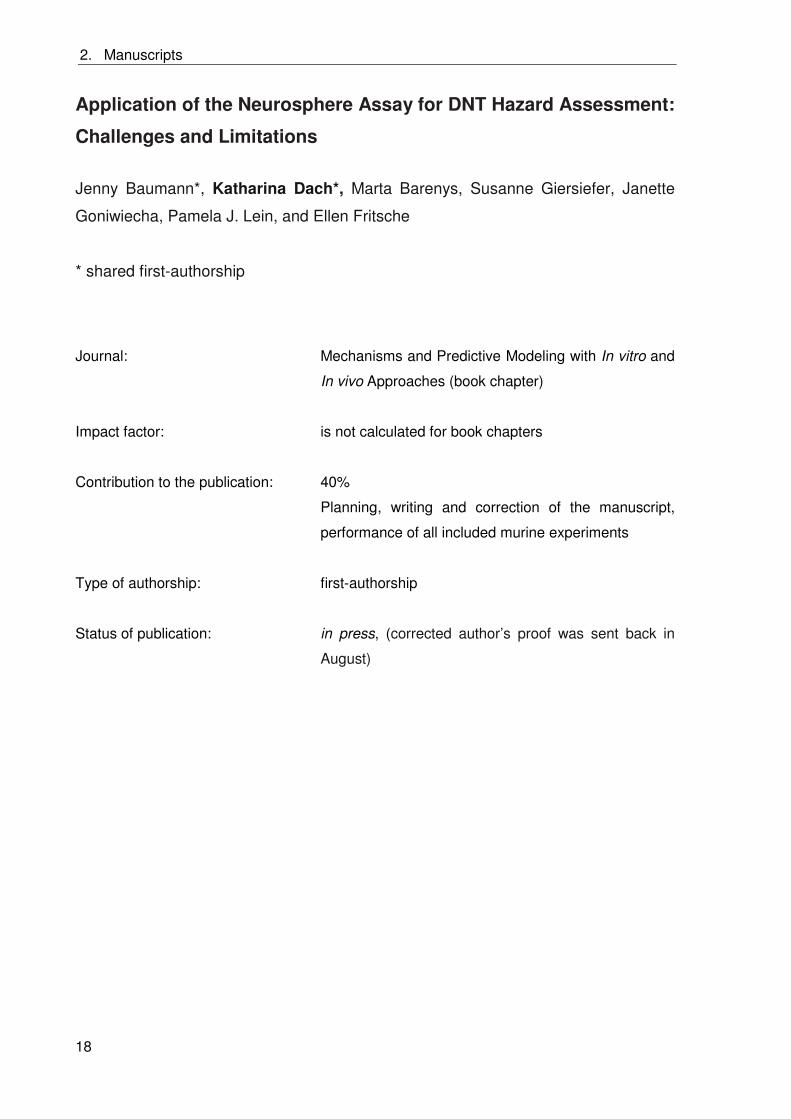
2. Manuscripts
18
Application of the Neurosphere Assay for DNT Hazard Assessment:
Challenges and Limitations
Jenny Baumann*, Katharina Dach*, Marta Barenys, Susanne Giersiefer, Janette
Goniwiecha, Pamela J. Lein, and Ellen Fritsche
* shared first-authorship
Journal: Mechanisms and Predictive Modeling with In vitro and
In vivo Approaches (book chapter)
Impact factor: is not calculated for book chapters
Contribution to the publication: 40%
Planning, writing and correction of the manuscript,
performance of all included murine experiments
Type of authorship: first-authorship
Status of publication: in press, (corrected author’s proof was sent back in
August)
2. Manuscripts
19
2.2 BDE-99 impairs human and mouse oligodendrogenesis by
species-specific modes of action
Katharina Dach, Farina Bendt, Ulrike Huebenthal, Susanne Giersiefer, Heike Heuer
and Ellen Fritsche
Environmental Health Perspectives (submitted on November 9th, 2015)
Hintergrund: Polybromierte Diphenylether (PBDEs) sind bioakkumulierende
Flammschutzmittel, die Entwicklungsneurotoxizität in Menschen und in Nagern hervorrufen.
Zusätzlich wird vermutet, dass sie Thyroidhormon (TH) Disruptoren sind. Die
Oligodendrogenese ist ein thyroidhormonabhängiger Prozess während der Entwicklung des
zentralen Nervensystems, welcher empfindlich gegenüber PBDE Exposition ist.
Ziele: Unser Ziel war die Etablierung humaner und muriner in vitro Methoden, um TH
Disruption auf Oligodendrozytenbildung und –reifung zu untersuchen. Weiterhin
untersuchten wir, ob BDE-99 diese Prozesse durch TH Disruption stört und verglichen die
Sensitivitäten sowie die involvierten Wirkmechanismen zwischen den beiden Spezies.
Methoden: Wir verwendeten humane und murine neurale Progenitorzellen (h,mNPCs), die
als 3D Neurosphären kultiviert wurden, um die Effekte von TH und BDE-99 auf die spezies-
spezifische Oligodendrozytenbildung und –reifung zu untersuchen.
Ergebnisse: TH beschleunigte die humane, aber induzierte die murine,
Oligodendrozytenbildung, während es die Reifung von Oligodendrozyten in beiden Spezies
beschleunigte. BDE-99 inhibierte die Oligodendrozytenbildung in hNPCs bereits in
niedrigeren Konzentrationen als in mNPCs, wobei TH Disruption nur im murinen System
involviert war. Gleichermaßen inhibierte BDE-99 die Reifung muriner Oligodendrozyten
durch TH Disruption, während die humane Oligodendrozytenreifung nicht beeinflusst war.
Die TH Disruption der murinen Oligodendrozytenbildung und –reifung wurde nicht durch
Thyroidhormonrezeptoren (TRs) vermittelt. Während BDE-99 Effekte auf die humane
Oligodendrozytenbildung keine Blockade von TH Signalwegen beinhalteten, war ein
Wirkmechanismus involviert, der durch Vitamin C antagonisiert werden konnte.
Schlussfolgerung: Humane und murine Oligodendrogenese werden durch BDE-99 mit
unterschiedlicher Sensitivität inhibiert und die zugrundeliegenden Wirkmechanismen sind
Spezies-spezifisch. Diese Daten unterstützen das Konzept, dass in vitro Zellsysteme
humanen Ursprungs für die Abschätzung des Gefährdungspotentials von Chemikalien
wichtig sind.
Page 1 of 38
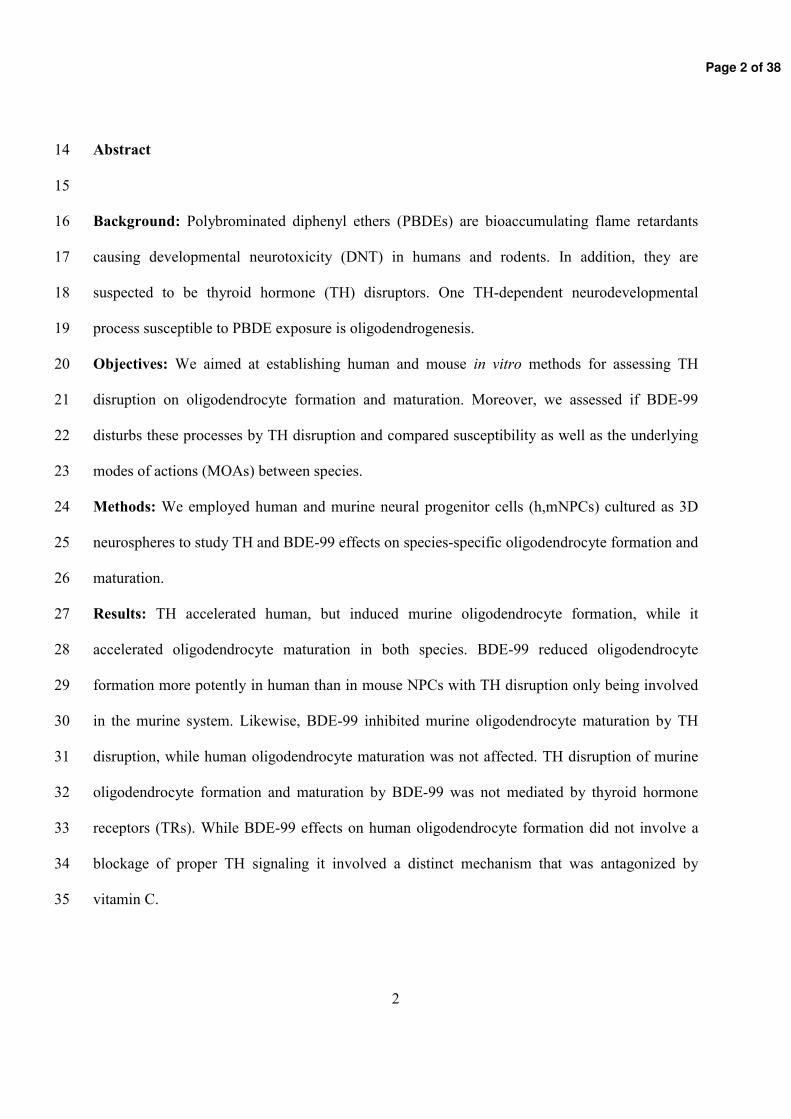
Page 2 of 38
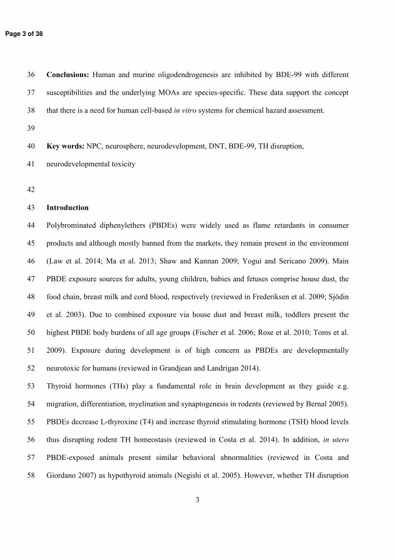
Page 3 of 38
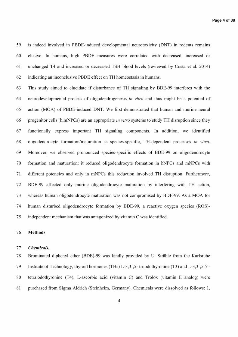
Page 4 of 38
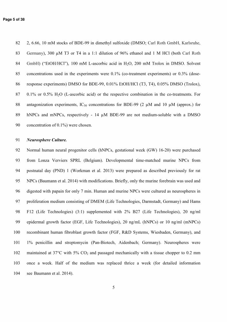
Page 5 of 38
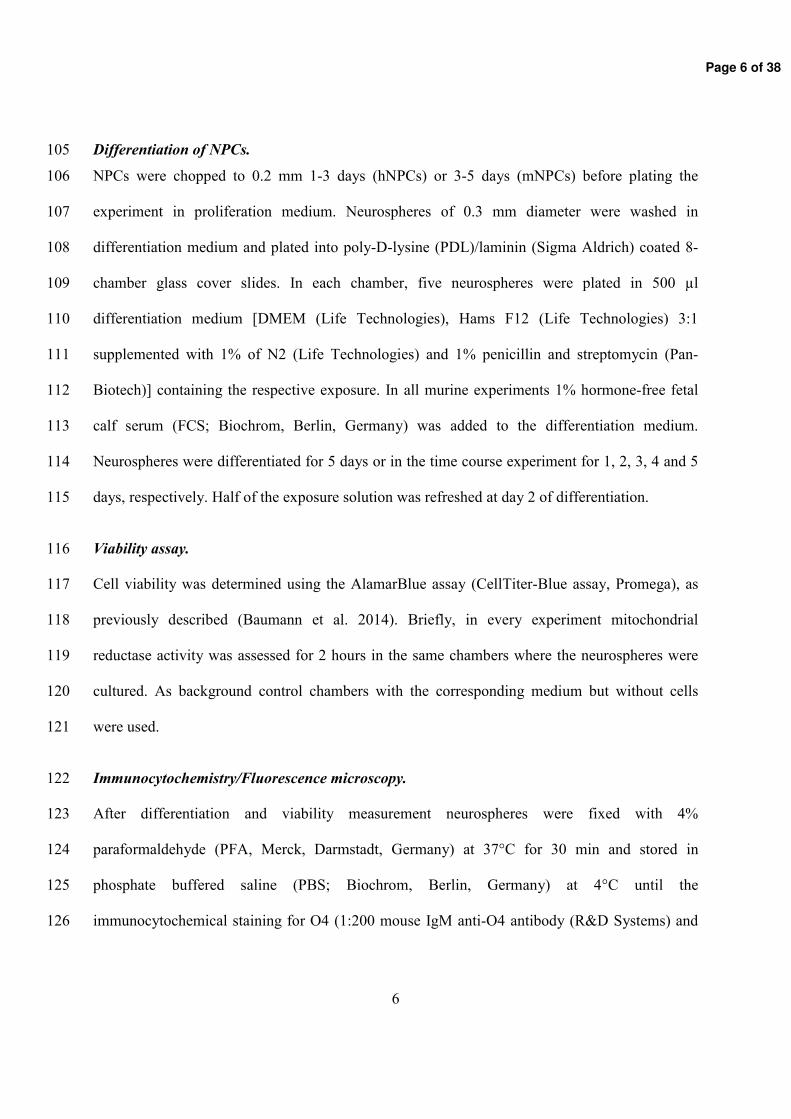
Page 6 of 38
Page 7 of 38
Page 8 of 38
Page 9 of 38
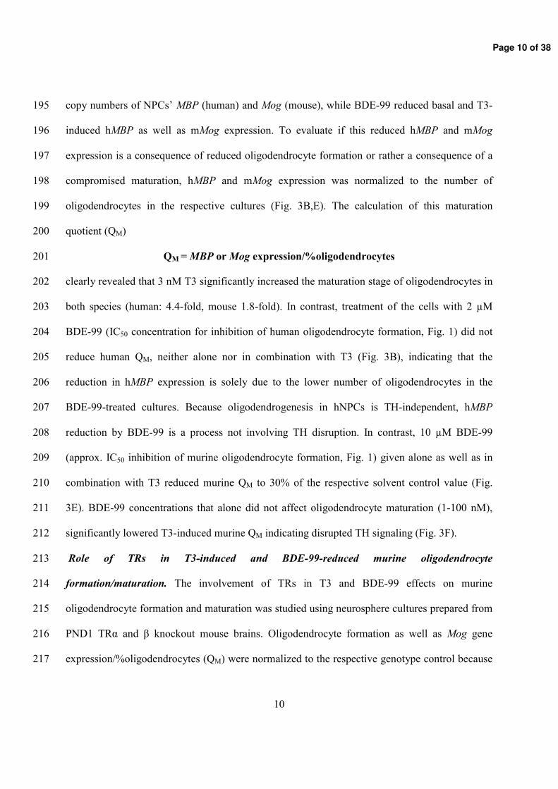
Page 10 of 38
Page 11 of 38
Page 12 of 38
Page 13 of 38
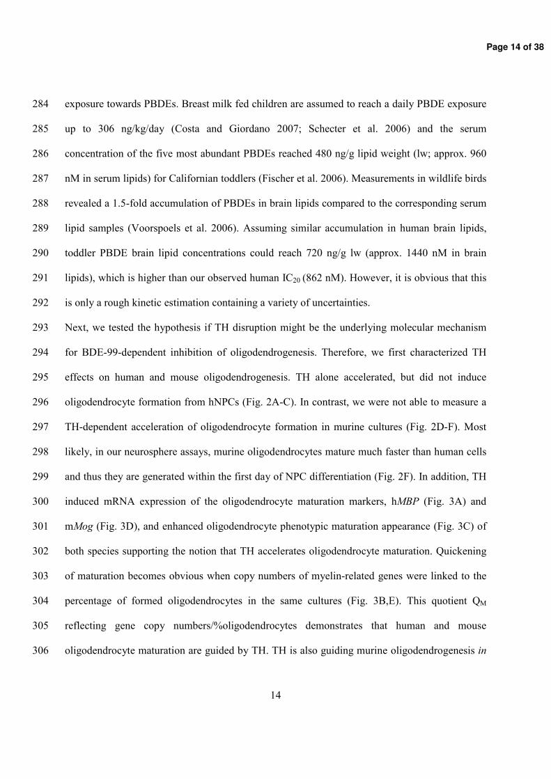
Page 14 of 38
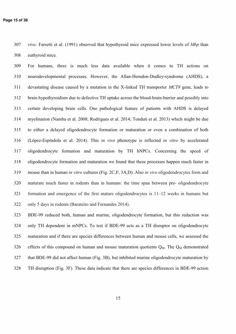
Page 15 of 38
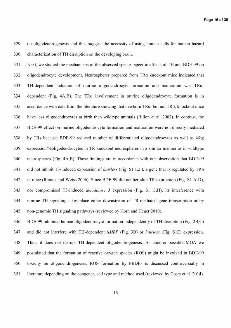
Page 16 of 38
Page 17 of 38
Page 18 of 38
Page 19 of 38
Page 20 of 38
Page 21 of 38

Page 22 of 38
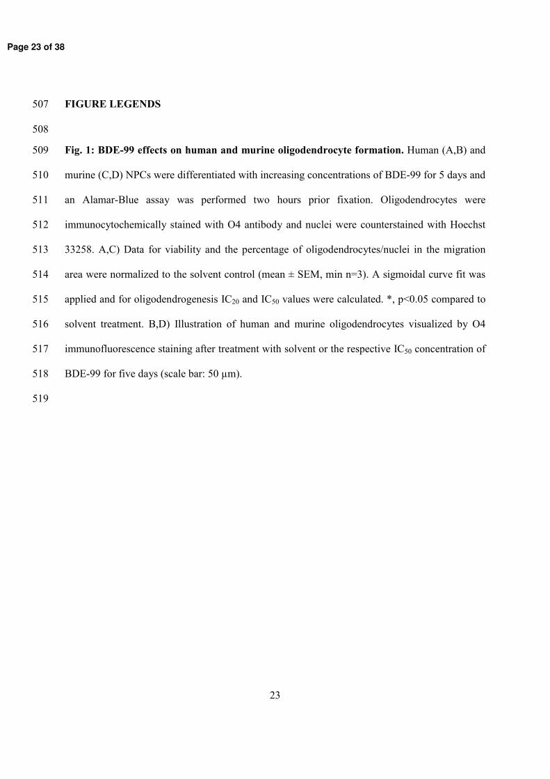
Page 23 of 38
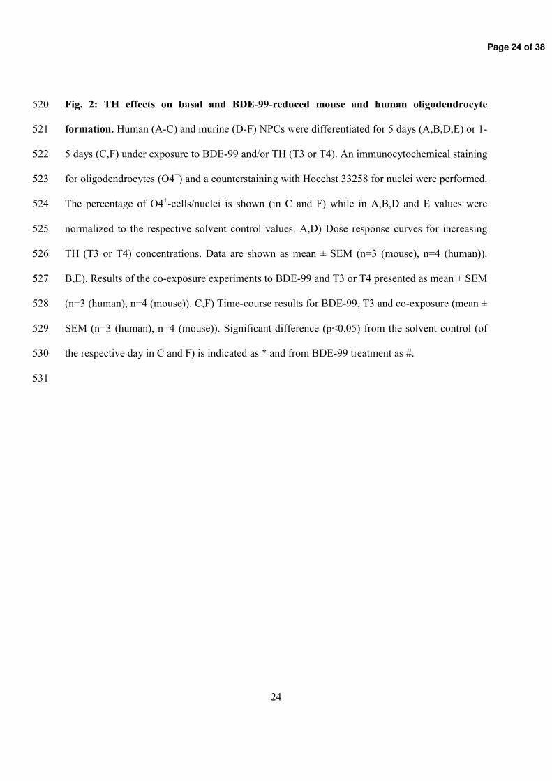
Page 24 of 38
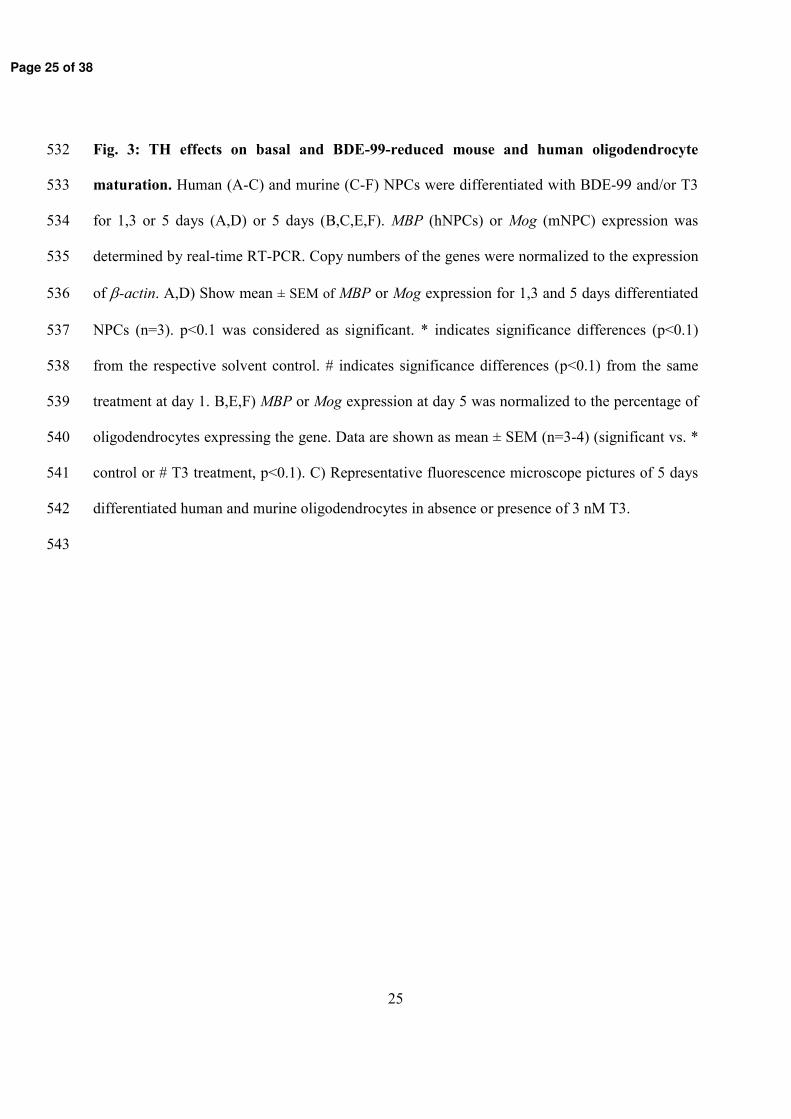
Page 25 of 38
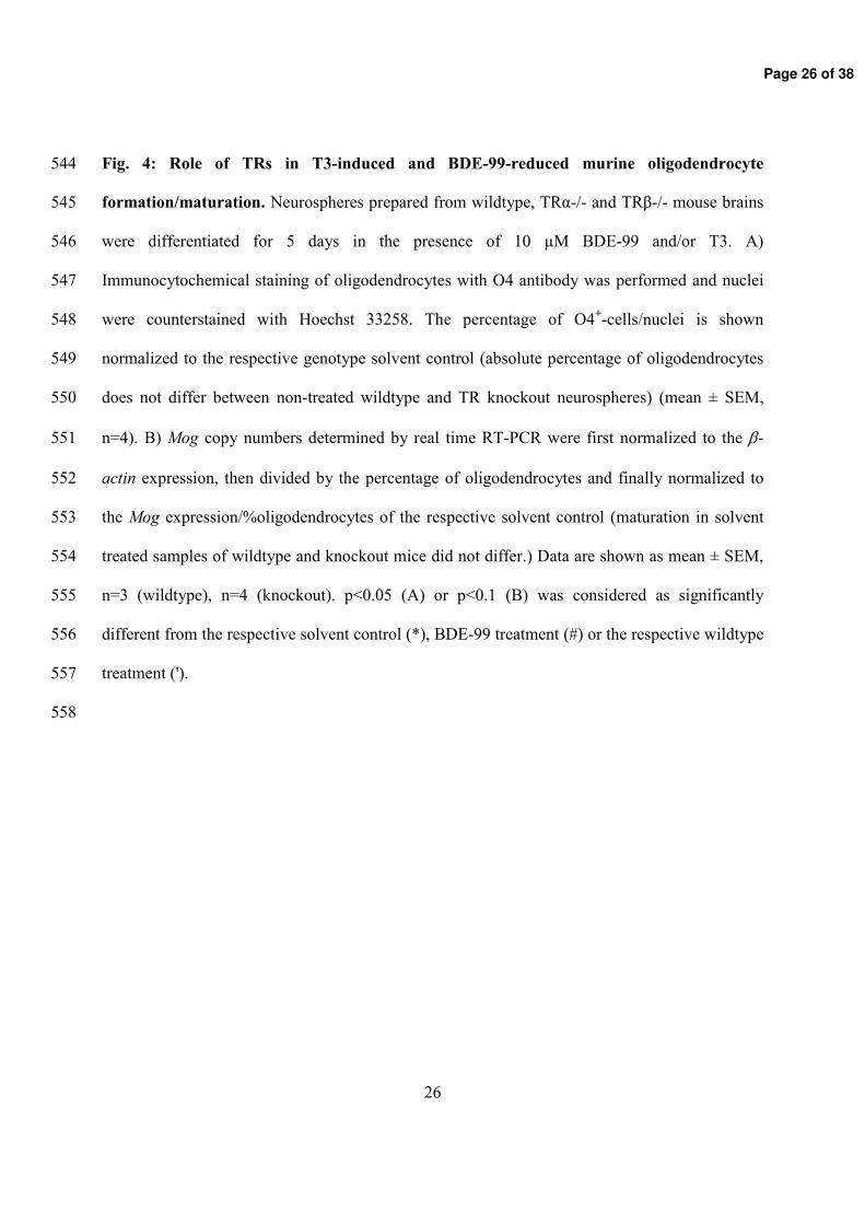
Page 26 of 38
Page 27 of 38
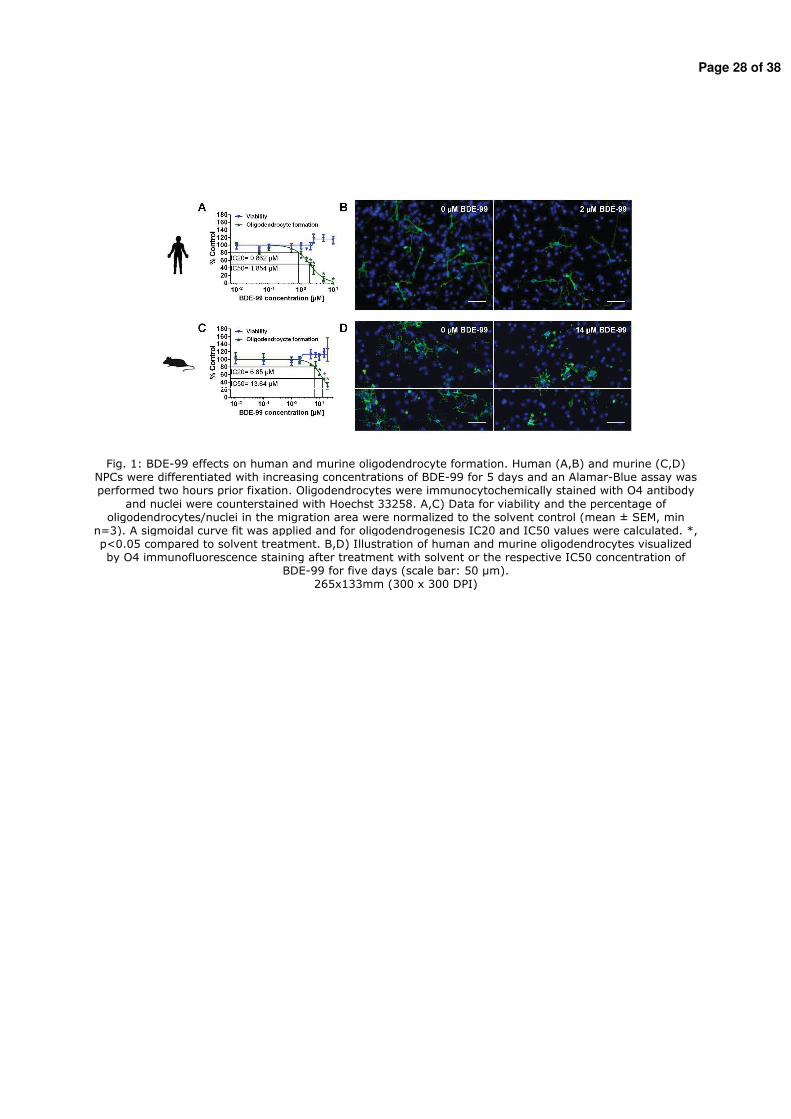
Page 28 of 38
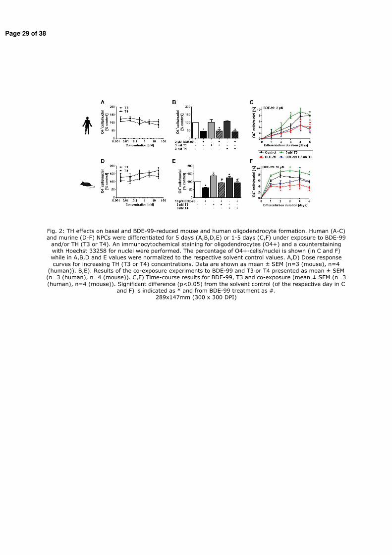
Page 29 of 38
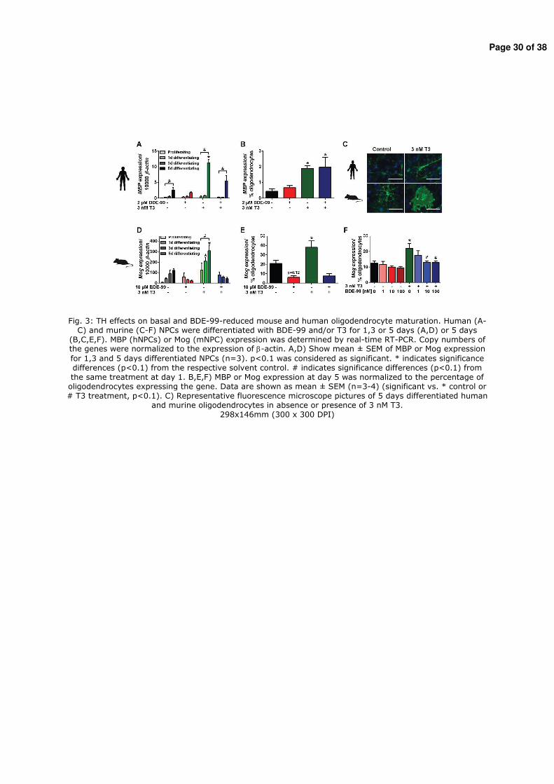
Page 30 of 38
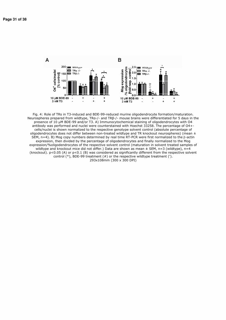
Page 31 of 38
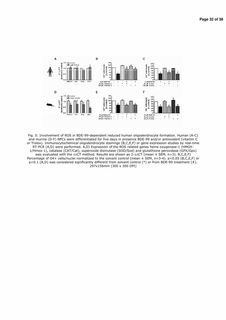
Page 32 of 38
Page 33 of 38
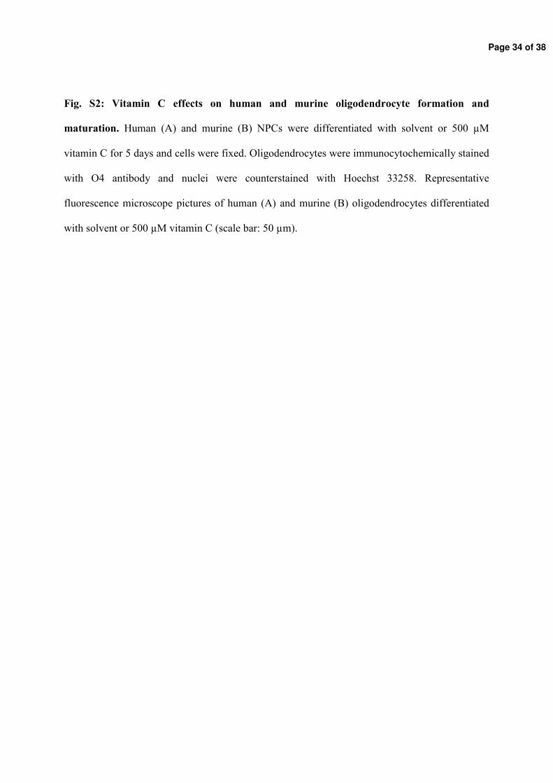
Page 34 of 38
Page 35 of 38
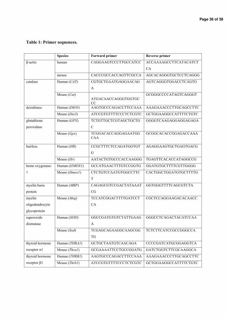
Page 36 of 38
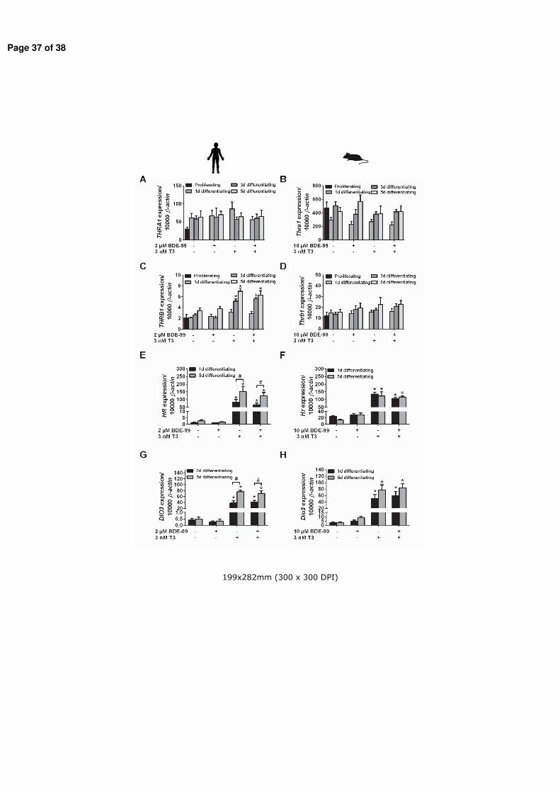
Page 37 of 38
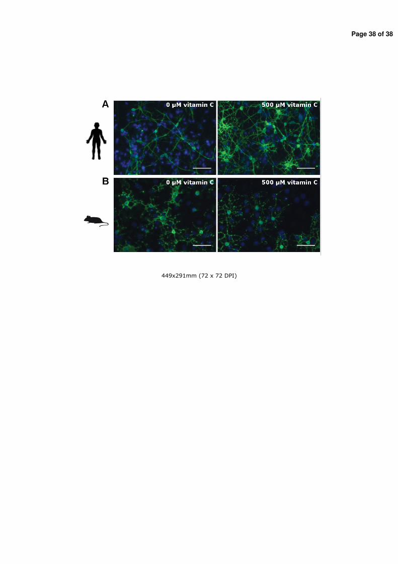
Page 38 of 38
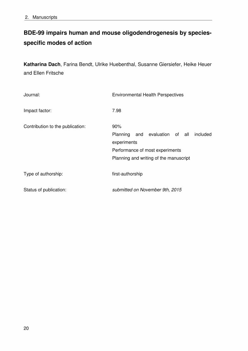
2. Manuscripts
20
BDE-99 impairs human and mouse oligodendrogenesis by species-
specific modes of action
Katharina Dach, Farina Bendt, Ulrike Huebenthal, Susanne Giersiefer, Heike Heuer
and Ellen Fritsche
Journal: Environmental Health Perspectives
Impact factor: 7.98
Contribution to the publication: 90%
Planning and evaluation of all included
experiments
Performance of most experiments
Planning and writing of the manuscript
Type of authorship: first-authorship
Status of publication: submitted on November 9th, 2015
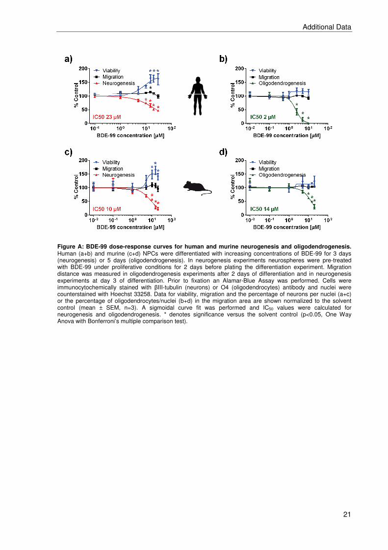
Additional Data
21
Figure A: BDE-99 dose-response curves for human and murine neurogenesis and oligodendrogenesis. Human (a+b) and murine (c+d) NPCs were differentiated with increasing concentrations of BDE-99 for 3 days (neurogenesis) or 5 days (oligodendrogenesis). In neurogenesis experiments neurospheres were pre-treated with BDE-99 under proliferative conditions for 2 days before plating the differentiation experiment. Migration distance was measured in oligodendrogenesis experiments after 2 days of differentiation and in neurogenesis experiments at day 3 of differentiation. Prior to fixation an Alamar-Blue Assay was performed. Cells were immunocytochemically stained with III-tubulin (neurons) or O4 (oligodendrocytes) antibody and nuclei were counterstained with Hoechst 33258. Data for viability, migration and the percentage of neurons per nuclei (a+c) or the percentage of oligodendrocytes/nuclei (b+d) in the migration area are shown normalized to the solvent control (mean ± SEM, n=3). A sigmoidal curve fit was performed and IC50 values were calculated for neurogenesis and oligodendrogenesis. * denotes significance versus the solvent control (p<0.05, One Way Anova with Bonferroni’s multiple comparison test).
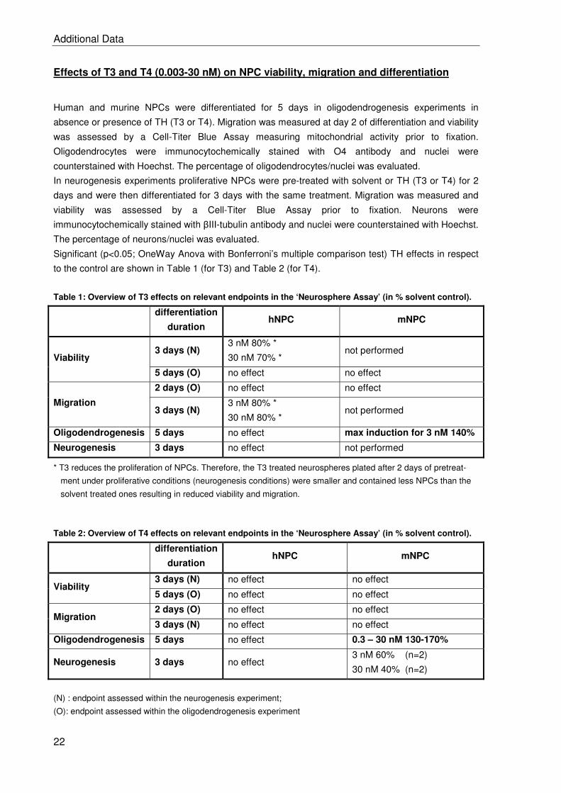
Additional Data
22
Effects of T3 and T4 (0.003-30 nM) on NPC viability, migration and differentiation
Human and murine NPCs were differentiated for 5 days in oligodendrogenesis experiments in
absence or presence of TH (T3 or T4). Migration was measured at day 2 of differentiation and viability
was assessed by a Cell-Titer Blue Assay measuring mitochondrial activity prior to fixation.
Oligodendrocytes were immunocytochemically stained with O4 antibody and nuclei were
counterstained with Hoechst. The percentage of oligodendrocytes/nuclei was evaluated.
In neurogenesis experiments proliferative NPCs were pre-treated with solvent or TH (T3 or T4) for 2
days and were then differentiated for 3 days with the same treatment. Migration was measured and
viability was assessed by a Cell-Titer Blue Assay prior to fixation. Neurons were
immunocytochemically stained with III-tubulin antibody and nuclei were counterstained with Hoechst.
The percentage of neurons/nuclei was evaluated.
Significant (p<0.05; OneWay Anova with Bonferroni’s multiple comparison test) TH effects in respect
to the control are shown in Table 1 (for T3) and Table 2 (for T4).
Table 1: Overview of T3 effects on relevant endpoints in the ‘Neurosphere Assay’ (in % solvent control).
differentiation
duration hNPC mNPC
Viability 3 days (N)
3 nM 80% *
30 nM 70% * not performed
5 days (O) no effect no effect
Migration
2 days (O) no effect no effect
3 days (N) 3 nM 80% *
30 nM 80% * not performed
Oligodendrogenesis 5 days no effect max induction for 3 nM 140%
Neurogenesis 3 days no effect not performed
* T3 reduces the proliferation of NPCs. Therefore, the T3 treated neurospheres plated after 2 days of pretreat-
ment under proliferative conditions (neurogenesis conditions) were smaller and contained less NPCs than the
solvent treated ones resulting in reduced viability and migration.
Table 2: Overview of T4 effects on relevant endpoints in the ‘Neurosphere Assay’ (in % solvent control).
differentiation
duration hNPC mNPC
Viability 3 days (N) no effect no effect
5 days (O) no effect no effect
Migration 2 days (O) no effect no effect
3 days (N) no effect no effect
Oligodendrogenesis 5 days no effect 0.3 – 30 nM 130-170%
Neurogenesis 3 days no effect 3 nM 60% (n=2)
30 nM 40% (n=2)
(N) : endpoint assessed within the neurogenesis experiment;
(O): endpoint assessed within the oligodendrogenesis experiment
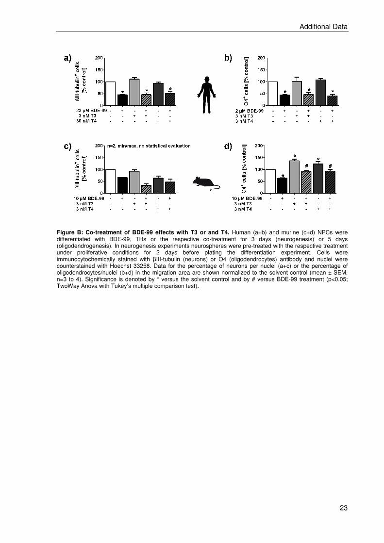
Additional Data
23
Figure B: Co-treatment of BDE-99 effects with T3 or and T4. Human (a+b) and murine (c+d) NPCs were differentiated with BDE-99, THs or the respective co-treatment for 3 days (neurogenesis) or 5 days (oligodendrogenesis). In neurogenesis experiments neurospheres were pre-treated with the respective treatment under proliferative conditions for 2 days before plating the differentiation experiment. Cells were immunocytochemically stained with III-tubulin (neurons) or O4 (oligodendrocytes) antibody and nuclei were counterstained with Hoechst 33258. Data for the percentage of neurons per nuclei (a+c) or the percentage of oligodendrocytes/nuclei (b+d) in the migration area are shown normalized to the solvent control (mean ± SEM, n=3 to 4). Significance is denoted by * versus the solvent control and by # versus BDE-99 treatment (p<0.05; TwoWay Anova with Tukey’s multiple comparison test).
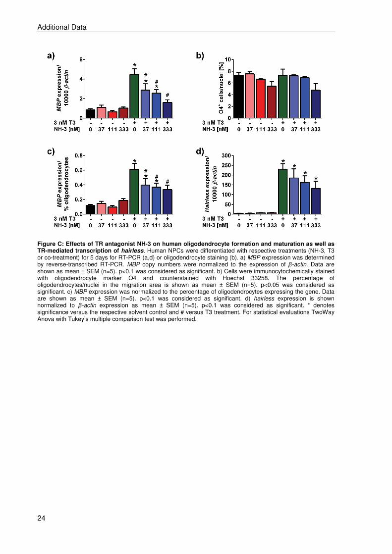
Additional Data
24
Figure C: Effects of TR antagonist NH-3 on human oligodendrocyte formation and maturation as well as TR-mediated transcription of hairless. Human NPCs were differentiated with respective treatments (NH-3, T3 or co-treatment) for 5 days for RT-PCR (a,d) or oligodendrocyte staining (b). a) MBP expression was determined by reverse-transcribed RT-PCR. MBP copy numbers were normalized to the expression of -actin. Data are shown as mean ± SEM (n=5). p<0.1 was considered as significant. b) Cells were immunocytochemically stained with oligodendrocyte marker O4 and counterstained with Hoechst 33258. The percentage of oligodendrocytes/nuclei in the migration area is shown as mean ± SEM (n=5). p<0.05 was considered as significant. c) MBP expression was normalized to the percentage of oligodendrocytes expressing the gene. Data are shown as mean ± SEM (n=5). p<0.1 was considered as significant. d) hairless expression is shown normalized to -actin expression as mean ± SEM (n=5). p<0.1 was considered as significant. * denotes significance versus the respective solvent control and # versus T3 treatment. For statistical evaluations TwoWay Anova with Tukey’s multiple comparison test was performed.
3. Discussion
25
3. Discussion
3.1 The ‘Neurosphere Assay’ as an in vitro model for studying
species-specific DNT mechanisms
Developmentally neurotoxic compounds cause neurological deficits like lower IQs, learning
disability and mental retardation in children resulting in high costs for society (Bellanger et al.
2013; Landrigan et al. 2002). Current DNT testing is performed in animal studies, which are
on the one hand extremely time- and cost-intensive (Crofton et al. 2012) and on the other
hand are not necessarily predictive for human risk assessment (Leist and Hartung 2013).
Therefore, stakeholders agreed on the demand to integrate in vitro models into DNT testing
strategies, which consider not only species differences but also allow to study critical toxicity
pathways (Bal-Price et al. 2015a; Coecke et al. 2007; Crofton et al. 2012). A promising in
vitro model in this regard is the ‘Neurosphere Assay’ described in paragraph 1.2.3.
Over the last decade human NPCs were intensively characterized and interference of
exogenous noxae with relevant endpoints was described by our group (Fritsche et al. 2011;
Gassmann et al. 2010, 2014; Moors et al. 2007, 2009; Schreiber et al. 2010). To enable a
species comparison within the ‘Neurosphere Assay’ a rat neurosphere culture was
established. The rat is the most commonly used animal in animal models and this approach
allows comparing generated rat in vitro data with existing in vivo data from literature. The
book chapter(Baumann et al. (2014) describes in detail how the ‘Neurosphere Assay’ for
human and rat NPCs is performed. The relevance of studying species differences was
demonstrated in (Baumann et al. (2015) for the first time comparing substance effects in rat
and human NPCs: The effects of six positive and three negative DNT compounds on
viability, migration and neuronal differentiation were tested in human and rat NPCs and the
‘Neurosphere Assay’ grouped most of the chemicals correctly. Within this study species
differences in susceptibility and within the most sensitive endpoint towards a certain
chemical were observed between human and rat demonstrating the need of species
comparisons.
The involvement of signaling pathways in vivo is currently studied in knockout mice. This is
attributed to the fact that a higher number of knockout mouse models exists in comparison to
rat models, that knockout mice are easier to generate and that the mouse models are far
better understood (Jacob et al. 2010; Peters et al. 2007;)Nobel Prize 2007 for the generation
3. Discussion
26
of knockout mice). Therefore, in order to investigate signaling pathways, also in regard to
AOP development, one goal of this thesis was to establish a murine neurosphere culture
and to implement the use of neurospheres prepared from knockout mice. TR knockout
murine neurospheres were used in mechanistic studies included in manuscript 2.2 to
investigate whether TRs are involved in BDE-99 or T3 effects on oligodendrogenesis.
In general, primary murine cultures need more complex culture conditions (more medium
supplements and fetal calf serum for differentiation) compared to primary human or rat
cultures (Rogers 2003). Using human/rat culture conditions for the murine culture led to a
high sphere-to-sphere variability in the percentage of neurons under control conditions (0 -
30% neurons/nuclei). This made a statistical evaluation in neuron change after chemical
treatment impossible. The rodent neurosphere preparation protocol routinely used for rat
preparations had to be optimized for murine neurosphere preparation by only using forebrain
and a shorter digestion time. Using only forebrain for the murine neurosphere preparation
lowered the sphere-to sphere variability in neurons significantly compared to using whole
brains for the preparations. There is evidence in the literature, that neural stem cells derived
from different brain regions retain their regional character and that cells from different brain
regions might inhibit each other in the ability to form neurons (Klein et al. 2005). We found
no neurons in murine neurospheres prepared from only cerebellum under standard
differentiation conditions, what might explain the high variability in murine whole brain
neurosphere cultures. Furthermore, the FGF concentration in the proliferating murine culture
was halved compared to the human culture, because murine neurospheres recovered better
from passaging with the lower FGF concentration. During differentiation, 1% fetal calf serum
(FCS) is now added to prevent high apoptosis rates and thereby prolong the possible
differentiation duration. After establishment of the mouse neurosphere culture it was
characterized (manuscript 2.1) and used within this project for investigation of TH signaling
and PBDE effects.
The manuscript 2.1 “Application of the Neurosphere Assay for DNT Hazard Assessment:
Challenges and Limitations” is the first publication of our group characterizing the endpoints
of the ‘Neurosphere Assay’ for murine NPCs. The included murine data were generated
within this PhD project. The manuscript describes how neurodevelopmental processes are
analyzed within the ‘Neurosphere Assay’ for in vitro DNT testing. We compared proliferation,
migration and differentiation into neurons, oligodendrocytes and astrocytes between all three
species and observed that NPCs from the three species distinguish in their proliferation
capacity, migration speed and the amount of formed neurons. Although all three species
showed the same percentage of oligodendrocytes after 5 days of differentiation, murine
oligodendrocytes formed much faster than human ones and were more mature. These in
3. Discussion
27
vitro observations are in accordance with in vivo literature: the time span between pre-
oligodendrocyte formation and emergence of the first mature oligodendrocytes is 11–12
weeks in humans but only 5 days in rodents (Barateiro and Fernandes 2014). We included
endpoint specific positive controls because showing that substances modulating an endpoint
in vivo also modulate it in the used in vitro system is crucial to guarantee that the in vitro
system reflects the basic biology of the endpoint. As endpoint specific control for proliferation
withdrawal of growth factors EGF and FGF is used. For migration src kinase inhibitor PP2 is
used, which reduces migration to 60% (Moors et al. 2007). EGF treatment reduces
differentiation into neurons (Ayuso-Sacido et al. 2010) and bone morphogenetic protein
(BMP)-7, like other BMPs, induces astrocyte and inhibits oligodendrocyte differentiation
(Gross et al. 1996). Furthermore, for each endpoint we compared substance effects on
primary rodent cells with rodent in vivo literature and present the relevance of in vitro neural
stem/progenitor cells for the in vivo situation.
As explained before, the neurosphere model mimics many relevant processes of early fetal
brain processes and has clear advantages towards other in vitro models (human origin,
culture as 3D aggregates, presence of the three main cell types of the brain and therefore
cross-talk between different cell types, species-comparison within the same in vitro system),
it enables to study toxicity pathways and contributes to the creation of adverse outcome
pathways (AOPs).
But the ‘Neurosphere Assay’ as well as all other in vitro systems has its limitations. It lacks
physiological cell and organ surrounding, which is necessary for pharmaco- and
toxicokinetics. Therefore absorption/distribution/metabolism and excretion (ADME) cannot
be studied in vitro. Furthermore, the neurosphere system lacks brain cell types like microglia
and endothelial cells and there is evidence that the function of identical cell types varies
between brain regions (Hewett 2009). Those region-specific differences can be studied with
rodent stem/progenitor cells isolated from different brain regions but not with human whole
brain homogenates.
Although the ‘Neurosphere Assay’ only covers early fetal brain development and has its
limitations, it is an appropriate candidate to be included into DNT strategies consisting of
several cell systems covering different stages of development (see Introduction Figure 9).
3. Discussion
28
3.2 Species differences in susceptibility towards PBDEs
First, BDE-99 effects on viability, migration and differentiation into neurons and
oligodendrocytes were studied performing concentration-response experiments with human
and murine neural progenitor cells (additional data Figure A). The used BDE-99
concentrations did not inhibit human or murine NPCs’ viability or migration. However, BDE-
99 reduced human and murine neuro- and oligodendrogenesis concentration-dependently
with human oligodendrogenesis as the most sensitive endpoint. Human oligodendrogenesis
was 10x more sensitive than human neurogenesis as the least sensitive endpoint (IC50: 2
μM and 23 μM). Sensitivity of murine neurogenesis and oligodendrogenesis was in the same
range (IC50: 10 μM and 14 μM). These data show that human NPCs are more sensitive
towards BDE-99 exposure than the mouse for the endpoint oligodendrogenesis, while the
susceptibility for neurogenesis is vice-versa.
Studies on PBDE effects in neural stem/progenitor cells are rare and focus on proteome
analyses or gene expression studies (Du et al. 2015; Song et al. 2014) as well as Ca2+
signaling (Gassmann et al. 2014), but not on the investigation of individual processes of
brain development. Another study found reduced viability and differentiation into neurons
and astrocytes, but induced apoptosis in in vitro neurosphere cultures derived from embryos
of in vivo BDE-209 treated rats (Chen et al. 2014). However, the rat neurospheres in this
study were prepared from embryos at gestational day (GD) 14 and thereby do not resemble
the developmental stage of NPCs used in our study. Furthermore, repeated application was
performed each day over 14 days (GD1-GD14) resulting in an accumulation of BDE-209.
Moreover, the applied concentrations, ranging from 10-50 mg/kg/day (10-50 μM per day), by
far exceed the exposure of breast fed children (306 ng/kg/day) (Costa and Giordano 2007;
Schecter et al. 2006). Additionally, in those studies different PBDE congeners were used
and effects vary for different congeners (Hamers et al. 2006).
The used BDE-99 concentrations (additional data Figure A; human: up to 30 μM, mouse: up
to 20 μM) did not reduce NPC viability. (Schreiber et al. (2010) reported that up to 10 μM
BDE-99 did not affect viability of hNPCs over 14 days. In opposite to(Tagliaferri et al. (2010)
who reported that 33 μM BDE-99 reduced viability to 50% of control after 24 h in human
neuroblastoma cells, no effect on hNPC viability after 30 μM treatment for 3 days was
observed (additional data Figure A). While(Huang et al. (2010) found that 24 h treatment
with 10 μM BDE-99 reduced viability to 50% of the control in mouse cerebellar granule
neurons, up to 20 μM BDE did not reduce mNPC viability after 5 days of differentiation
(additional data Figure A). These data suggest that BDE effects on viability depend on the
applied cell system and the exposure duration. That neuroblastoma cells are more sensitive
3. Discussion
29
towards apoptosis-inducing agents than primary hNPC was shown earlier (Moors et al.
2009).
PBDE effects on migration have not been studied in vitro so far and the ‘Neurosphere Model’
is unique in assessing this endpoint. While BDE-99 did not affect human or murine NPC
migration (additional data Figure A), in vivo proteomic analysis suggest that BDE-99
exposure might cause disturbances in cell motility in mouse cerebral cortex (Alm et al.
2008). Although seen on the proteome level, no histopathological evaluation of PBDEs on
migrational processes in vivo have been performed that could confirm the proteome data on
the organ level.
BDE-99 reduced human and murine NPCs’ differentiation into neurons and oligodendrocytes
(additional data Figure A). Similar to PBDE effects on neural migration, effects of PBDE
exposure on differentiation are rarely studied in vitro (Li et al. 2013; Schreiber et al. 2010).
However, several studies in vivo suggest that developmental BDE-99 exposure might
interfere with neuritogenesis/synaptogenesis in mice and rats by altering expression of
proteins involved in those processes like GAP-43, stathmin and neuromodulin (Alm et al.
2006, 2010). Our calculated IC50 concentration for BDE-99 inhibiting human oligodendrocyte
formation (2 μM) and the IC20 concentration for BDE-99 inhibiting murine oligodendrocyte
formation (7 μM) are in accordance with the other two publications studying PBDE effects on
oligodendrogenesis where an IC50 BDE-99 concentration of around 1 μM was found for
human oligodendrocyte formation (Schreiber et al. 2010) and an IC25 BDE-47 concentration
of 10 μM for inhibiting murine NSC differentiation into oligodendrocytes (Li et al. 2013).
Since human oligodendrogenesis is the most sensitive endpoint BDE-99, effects on
oligodendrocyte formation and maturation in human and murine NPCs were studied in more
detail (manuscript 2.2).
3.3 Characterization of the TH responses in the ‘Neurosphere
Assay’ and establishment of the oligodendrocyte maturation
assay for studying TH disruption
THs, T3 and T4, are known to play a crucial role in many processes of brain development
like proliferation, migration, differentiation, myelination and synaptogenesis (reviewed in
Anderson (2001); Bernal and Nunez (1995) and Bernal (2005)) and TH deficiency causes
mental retardation (Boyages and Halpern 1993; Cao et al. 1994; DeLong et al. 1985).
Because PBDE-treated animals show similar behavioral abnormalities (Costa and Giordano
2007) than animals, which suffer from hypothyroidism during brain development (Negishi et
3. Discussion
31
brains from children which this devastating disease is delayed myelination (Namba et al.
2008; Rodrigues et al. 2014; Tonduti et al. 2013).
Proliferating and differentiating human and murine NPCs express genes encoding the TH
metabolizing enzyme deiodinase 3 (D3) and TRs, TR 1 and 1. (Fig. S1, manuscript 2.2).
Expression levels of thyroid hormone receptors were higher in murine than in human NPCs.
Human TR 1 was the only receptor which expression was induced by T3 treatment.
However, the low copy number change from 3 to 7 copies/10,000 -actin makes a biological
significance highly unlikely. The functionality of TRs was shown in both species since T3
induced the transcription of the gene hairless (Fig. S1, manuscript 2.2). Hairless is a T3-
inducible in fetal brains in vivo (Potter et al. 2002; Thompson and Potter 2000) which T3
induction is TR -mediated (Ramos and Weiss 2006). Furthermore, T3 probably induces its
own metabolism as it strongly stimulated deiodinase 3 expression in both species (Fig. S1,
manuscript 2.2). TH transport into the cell must be present since THs triggered TR-
dependent gene expression (Fig. S1, manuscript 2.2).
Next, the ability of BDE-99 to disturb NPC development by TH disruption was studied.
Therefore, time-course experiments for oligodendrocyte formation, the most sensitive
endpoint for BDE-99 treatment, were performed in presence of T3, BDE-99 or a combination
of both (Fig. 2, manuscript 2). In addition, gene expression of oligodendrocyte maturation
markers were studied over time (Fig. 3, manuscript 2.2). T3 accelerated human
oligodendrocyte formation after 3 days of differentiation, but after 5 days of differentiation T3
treated NPCs had the same percentage of oligodendrocytes in the migration area than
untreated NPCs. In contrast, T3 induced oligodendrocyte formation of mNPCs over the
whole duration of 5 days. The observations in the mouse cultures are in accordance with
previously published work that T3 is important for the timing of oligodendrocyte formation
and the amount of formed oligodendrocytes in vitro (Barres BA, Lazar MA 1994; Billon et al.
2001; Ibarrola et al. 1996; Koper et al. 1986). To the best of my knowledge, oligodendrocyte
formation has not been studied for humans so far.
In addition to oligodendrocyte formation, their maturation was studied by quantification of
oligodendrocyte maturation markers via real time RT-PCR (Fig. 3, manuscript 2.2). This
added a new endpoint to the ‘Neurosphere Assay’ (manuscript 2.2). Oligodendrocyte
maturation was assessed by normalizing the gene expression (derived from real time RT-
PCR experiments) of oligodendrocyte maturation markers human myelin basic protein
(MBP) or murine myelin oligodendrocyte glycoprotein (Mog) to the percentage of
oligodendrocytes in the migration area resulting in the maturation quotient QM (QM = MBP or
Mog expression/ %oligodendrocytes). The use of the different markers is attributed to the
different maturation stages of oligodendrocytes in the two species. Human and murine
oligodendrocytes matured over a differentiation period of 5 days. Opposite to murine
3. Discussion
32
oligodendroyctes, human oligodendrocytes did not mature between differentiation day 1 and
3, but between day 3 and 5. Furthermore, murine oligodendrocytes were more mature after
5 days of differentiation than human ones: they were already branched and displayed
sheets, while human ones where still predominantly linear (Fig. 3, manuscript 2.2).
Moreover, murine NPCs expressed Mog, a gene which is only expressed in myelinating
oligodendrocytes (Baumann and Pham-Dinh 2001), which was not expressed by hNPCs.
Mbp, an earlier marker, which is already expressed by non-myelinating oligodendrocytes
(Baumann and Pham-Dinh 2001), was expressed by murine oligodendrocytes from the
beginning and its expression did not increase over differentiation time. These observations
are in accordance with in vivo literature that human oligodendrocytes mature slower than
rodent ones (Barateiro and Fernandes 2014).
T3 accelerated human and murine oligodendrocyte maturation (Fig. 3, manuscript 2.2) what
is in accordance with rodent in vitro (Bhat et al. 1979; Shanker et al. 1985) and in vivo
literature (Farsetti et al. 1991; Ibarrola and Rodríguez-Peña 1997; Marta et al. 1998;
Rodriguez-Peña et al. 1993). In humans the AHDS leading to a hypothyroid situation in the
brain causes delayed myelination (Namba et al. 2008; Rodrigues et al. 2014; Tonduti et al.
2013) which can be due to less formed oligodendrocytes or their delayed maturation (López-
Espíndola et al. 2014). This in vivo phenotype is reflected in the ‘Neurosphere Assay’ by
accelerated oligodendrocyte formation and maturation with T3.
To shed light on the mechanism how T3 acts on murine oligodendrogenesis, NPCs
generated from TR and TR were included in the study. These cells revealed that the T3
induction of murine oligodendrocyte formation and maturation was mediated by TR , but not
TR (Fig. 4, manuscript 2.2). This TR involvement in murine oligodendrocyte formation is in
accordance with the literature showing that TR mediates the effect on the timing of
oligodendrocyte formation as newborn TR , but not TR knockout mice display less
oligodendrocytes at birth than wildtype animals (Billon et al. 2002). In further studies we plan
to knockdown TR receptors in hNPCs to elucidate whether TR is also mediating the T3
induced maturation in hNPCs.
These results show that neurospheres express important TH signaling components and that
TH effects on endpoints of the ‘Neurosphere Assay’ are in accordance with the in vitro and
in vivo literature, at least to the extent of information published. This makes the
‘Neurosphere Assay’ an appropriate tool for studying TH signaling and TH disruption.
BDE-99 and TH co-treatment experiments for human and murine neuron and
oligodendrocyte formation revealed that TH only antagonizes the BDE-99-dependent
decrease in murine oligodendrocyte formation. The reduction of mouse neuron as well as
human neuron and oligodendrocyte formation by BDE-99 remained unaffected by THs
3. Discussion
33
indicating that BDE-99 distrurbed these endpoints by mechanism(s) other than TH disruption
(additional data Figure B).
Time course experiments over 5 days clarified that in both species BDE-99 treatment
inhibited oligodendrocyte formation rather than inducing oligodendrocyte-specific cell death
(Fig. 2, manuscript 2.2). The BDE-99 effect on murine oligodendrocyte formation was
antagonized by T3 over the whole differentiation period. On the contrary, the BDE-99 effect
on human oligodendrocyte formation was not antagonized by T3 at any time point, even not
at those days when T3 accelerated this process.
After oligodendrocyte maturation was found to be the only endpoint, which was induced by
T3 in both species, this endpoint was chosen to discriminate species-specific TH disruptors.
BDE-99 inhibited oligodendrocyte maturation in murine NPCs by endocrine disruption since
low concentrations not affecting maturation alone, concentration-dependently reduced
oligodendrocyte maturation in combination with T3. In contrast, BDE-99 was not an
endocrine disruptor of human oligodendrocyte maturation since it did not reduce human
oligodendrocyte maturation, neither alone nor in combination with T3 (Fig. 3, manuscript
2.2).
For both species interference of BDE-99 with TH signaling was investigated. By using
murine neurospheres prepared from TR or TR knockout mice it was shown that neither of
both receptors was involved in the BDE-99 effect on murine oligodendrocyte formation or
maturation (Fig. 4, manuscript 2.2). Furthermore, BDE-99 did not change expression of
genes encoding TR 1 or TR 1 in human or murine differentiated neurospheres and did not
interfere with T3-induced TR -mediated transcription of hairless (Fig. S1, manuscript 2.2), a
gene which is induced by TH in fetal brains (Potter et al. 2002; Thompson and Potter 2000)
by a TR -dependent mechanism (Ramos and Weiss 2006). In the literature contradictory
results were found for BDE-99 interference with TR-mediated transcription: Ibhazehiebo et
al. (2011) observed no suppression of TR-mediated transcription by BDE-99, but Blanco et
al. (2011) reported that BDE-99 deregulated TH-mediated transcription of important genes
for brain development as well as of TR isoforms in rat cerebellar granule neurons. In our
experiments BDE-99 did not affect T3-induced expression of deiodinase 3, a thyroid
hormone metabolizing enzyme. A study dealing with PBDE effects on brain cells reported
that BDE-99 reduced deiodinase 2 activity in human glioma cells (Roberts et al. 2015), yet
not on the transcriptional level.
Because BDE-99 effects on human oligodendrocyte formation were TH independent,
involvement of reactive oxygen species (ROS) was investigated (Fig. 5, manuscript 2.2). In
literature ROS formation by PBDEs is discussed controversially depending on the used
congener, cell system and method (reviewed in Costa et al. 2014). BDE-99 did not up-
regulate ROS related genes in human and murine NPCs and the antioxidant Trolox did not
3. Discussion
34
antagonize the BDE-99 caused reduction of human oligodendrocyte formation. Interestingly,
vitamin C (50 and 100 μM) rescued BDE-99-reduced oligodendrocyte formation in human,
but not murine NPCs. A high vitamin C concentration (500 μM) strongly induced human, but
not murine, oligodendrocyte formation and maturation (Fig. S2, manuscript 2.2). Vitamin C
enables hydroxylation of proline to hydroxyproline (Pinnell 1985), a major component of the
extracellular matrix protein collagen promoting rodent myelination of dorsal root ganglion
neurons by Schwann cells in the peripheral nervous system (Carey and Todd 1987; Eldridge
et al. 1987; Podratz et al. 2004). The effects of vitamin C on central nervous system
oligodendrogenesis are not known. Therefore, the mechanism by which vitamin C rescues
BDE-99-dependent reduction of oligodendrocyte formation in hNPCs remains enigmatic, but
might involve collagen production.
In summary, BDE-99 inhibited mouse oligodendrocyte formation and maturation via TH
disruption, yet independent of TRs. In contrast, BDE-99 effects on human oligodendrocyte
formation were not TH dependent and BDE-99 did not inhibit human oligodendrocyte
maturation. ROS formation is not involved in the BDE-99 effect on human oligodendrocyte
formation either, but vitamin C is able to antagonize this BDE-99 effect – only in human, not
in mouse NPCs. This study shows the need for taking species-specificities into consideration
when studying toxicity pathways.
To test the functionality of the oligodendrocyte maturation assay also for human NPCs the
TR antagonist NH-3 was used (Nguyen et al. 2002; additional data Figure C). As expected,
NH-3 reduced T3-induced TR -mediated transcription of hairless and inhibited T3-induced,
but not basal human oligodendrocyte maturation, in a concentration-dependent manner.
These data indicate that the oligodendrocyte maturation assay developed within this thesis
can detect TH disruptors. For a proper assay validation, however, more potential endocrine
disruptors like polychlorinated biphenyls (PCBs) (Fritsche et al. 2005; Zoeller 2000), OH-
PCBs (Iwasaki et al. 2002), OH-PBDEs (Butt et al. 2011), bisphenol A (BPA) (Moriyama et
al. 2002), tetrabromo- and tetrachloro BPA (TBBPA,TCBPA) (Butt et al. 2011; Kitamura et
al. 2002), cocaine (Kovalevich et al. 2012) or perfluoroalkyl compounds as perfluorohexane
sulfonates (PFHXS) (Vongphachan et al. 2011) need to be tested in this assay.
3. Discussion
35
3.4 Development of AOPs for thyroid hormone disruption
The AOP concept integrates molecular mechanisms into risk assessment (Ankley et al.
2010). Thereby, an AOP describes the adverse outcome (AO) of toxicants on individuals or
the population starting from a single molecular initiating event (MIE) by causally linking
related key events (KEs) on different stages of biological organization (OECD 2013; section
1.2.2, Figure 6).
The aim of the implementation of AOPs into current testing strategies is to achieve a more
predictive hazard characterization and better chemical classification. Within the AOP
species-specific toxicity pathways are considered. Thus, this translational aspect from
rodents to humans is supposed to contribute to reducing uncertainty in hazard assessment
(Bal-Price et al. 2015a). For the description of full AOPs the MIE, the linked KEs and the AO
need to be known. Each AOP is independent of the chemical, but starts with a single,
defined MIE. Different AOPs can cross and overlap when they share one or more KE(s) or
have the same AO leading to the formation of complex AOP networks. Thus, one single MIE
can cause multiple AOs and the same AO can be caused by more than one MIE.
In case of DNT the AO in many cases is known, but there is especially a lack of knowledge
on MIEs. Cellular KE for DNT include neural stem/progenitor cell proliferation, apoptosis,
migration, differentiation into neurons and glia cells, neurite outgrowth, myelination and
dendrite and axon formation (Bal-Price et al. 2015a). Those endpoints can be assessed with
in vitro models like the ‘Neurosphere Assay’.
Within this thesis an AOP scheme was developed how in general a TH disrupting chemical
might lead to the adverse outcome of mental retardation and it was focused on studying the
cellular KE oligodendrocyte formation and maturation in human and murine NPCs (Figure
11). As putative TH disruptors BDE-99 in both species (manuscript 2.2) as well as the TR
antagonist NH-3 (Nguyen et al. 2002) in hNPCs (additional data Figure C) were tested.
MIEs for TH disruption can cover a brought spectrum due to the complexity of TH signaling
(reviewed by Horn and Heuer 2010) and will lead to multiple AOPs for TH disruption.
Putative molecular initiating events for the AO mental retardation are shown in the AOP
scheme (Figure 11). Those are based on interference of compounds with classical TH
signaling like decreased TH transport across the membranes, the inhibition of TH
metabolism by deiodinases or the blockade of the TR binding site for THs by binding of the
compound to TRs. In addition, MIEs might also happen on targets of non-classical TH
signaling (section 1.1.2, Figure 4).
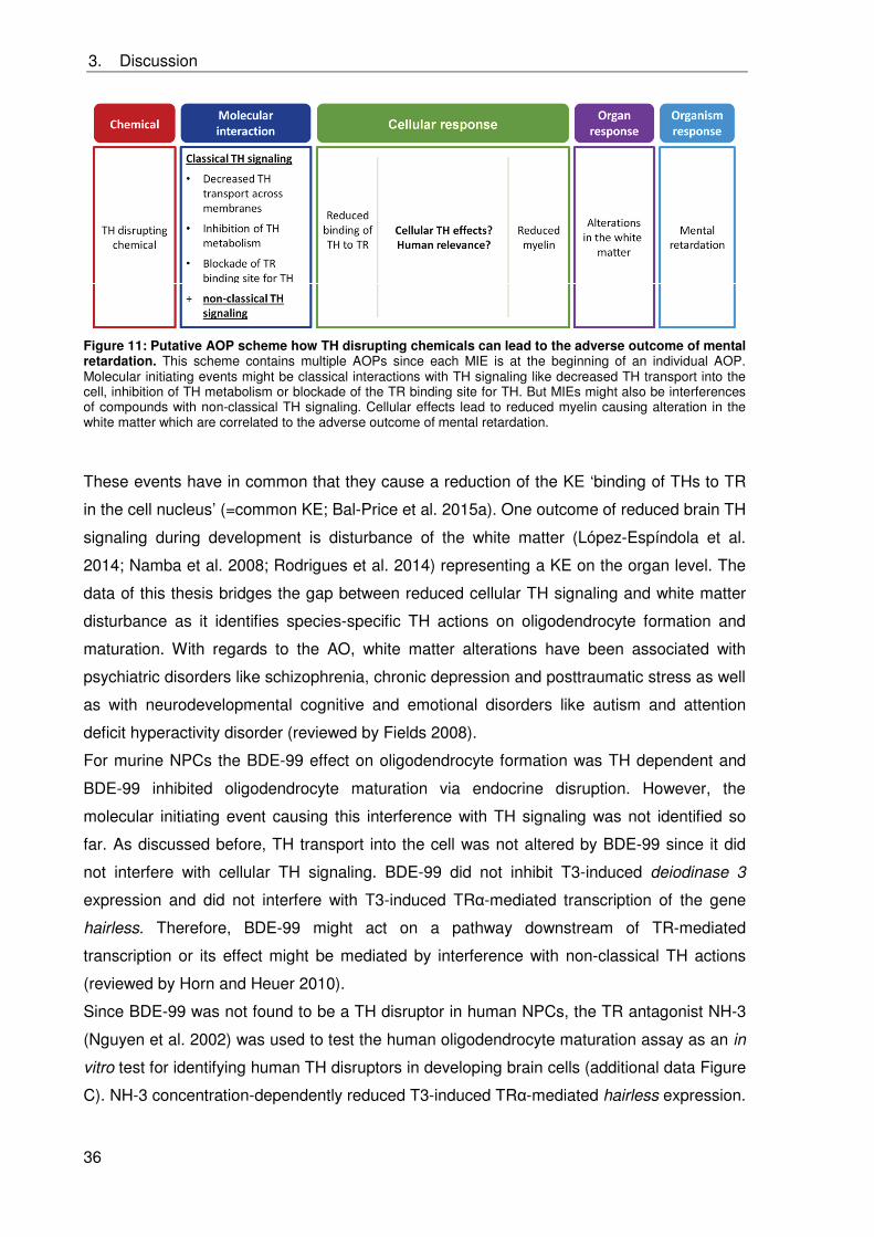
3. Discussion
36
Figure 11: Putative AOP scheme how TH disrupting chemicals can lead to the adverse outcome of mental retardation. This scheme contains multiple AOPs since each MIE is at the beginning of an individual AOP. Molecular initiating events might be classical interactions with TH signaling like decreased TH transport into the cell, inhibition of TH metabolism or blockade of the TR binding site for TH. But MIEs might also be interferences of compounds with non-classical TH signaling. Cellular effects lead to reduced myelin causing alteration in the white matter which are correlated to the adverse outcome of mental retardation.
These events have in common that they cause a reduction of the KE ‘binding of THs to TR
in the cell nucleus’ (=common KE; Bal-Price et al. 2015a). One outcome of reduced brain TH
signaling during development is disturbance of the white matter (López-Espíndola et al.
2014; Namba et al. 2008; Rodrigues et al. 2014) representing a KE on the organ level. The
data of this thesis bridges the gap between reduced cellular TH signaling and white matter
disturbance as it identifies species-specific TH actions on oligodendrocyte formation and
maturation. With regards to the AO, white matter alterations have been associated with
psychiatric disorders like schizophrenia, chronic depression and posttraumatic stress as well
as with neurodevelopmental cognitive and emotional disorders like autism and attention
deficit hyperactivity disorder (reviewed by Fields 2008).
For murine NPCs the BDE-99 effect on oligodendrocyte formation was TH dependent and
BDE-99 inhibited oligodendrocyte maturation via endocrine disruption. However, the
molecular initiating event causing this interference with TH signaling was not identified so
far. As discussed before, TH transport into the cell was not altered by BDE-99 since it did
not interfere with cellular TH signaling. BDE-99 did not inhibit T3-induced deiodinase 3
expression and did not interfere with T3-induced TR -mediated transcription of the gene
hairless. Therefore, BDE-99 might act on a pathway downstream of TR-mediated
transcription or its effect might be mediated by interference with non-classical TH actions
(reviewed by Horn and Heuer 2010).
Since BDE-99 was not found to be a TH disruptor in human NPCs, the TR antagonist NH-3
(Nguyen et al. 2002) was used to test the human oligodendrocyte maturation assay as an in
vitro test for identifying human TH disruptors in developing brain cells (additional data Figure
C). NH-3 concentration-dependently reduced T3-induced TR -mediated hairless expression.
3. Discussion
37
In addition, it disrupted T3-dependent oligodendrocyte maturation showing that the human
oligodendrocyte maturation assay indeed detects disturbances of TH signaling.
The oligodendrocyte maturation assay was established to identify TH disruptors in both
species. Testing of BDE-99 in these assays identified this flame retardant as a TH disruptor
in mouse, but not human NPCs. These data clearly demonstrate the need to implement
species-specific pathway analyses in AOP development and supports the previous notion
that human cells should be used for hazard assessment of TH disruption in the developing
brain.
3.5 Risk assessment of PBDEs
It was clearly shown that PBDEs are developmental neurotoxic for humans reducing the IQ
and inducing hyperactivity (Chao et al. 2007; Eskenazi et al. 2013; Roze et al. 2009; Shy et
al. 2011). In rodents they also exert DNT potential by inducing behavioral abnormalities,
learning and memory deficits and hyperactivity (e.g. Branchi et al. 2002; Dufault et al. 2005;
Gee et al. 2008; Kodavanti et al. 2010; Suvorov et al. 2009; Viberg et al. 2003, 2004).
However, the mechanisms of PBDE-induced DNT still remain elusive (reviewed by Costa et
al. (2014)).
Human oligodendrogenesis was the most sensitive endpoint towards BDE-99 within the
endpoints studied (cell death, NPC proliferation, migration, neurogenesis,
oligodendrogenesis and oligodendrocyte maturation). It was shown to be 7-times more
sensitive than murine oligodendrogenesis (IC50: 2 μM and 14 μM, respectively). In contrast,
human neurogenesis was significantly less sensitive than murine neurogenesis (IC50: 23 μM
and 10 μM, respectively). NPC viability, proliferation and migration were not affected by
BDE-99 at all. These data show that species do not only differ in their susceptibility, but also
in their most sensitive endpoint (MSE) towards BDE-99. These observations were recently
made for a variety of other compounds tested in the neurosphere assay including arsenic,
valproic acid and methylazoxymethanol (Baumann et al. 2015). For DNT hazard assessment
purposes, more KEs have to be tested to identify the MSE. In addition to the ‘Neurosphere
Assay’ endpoints specific for neurodevelopmental processes within the embryonic stage of
development as well as more complex processes like neuronal subtype specification,
synapse and network formation need to be included in a proposed DNT testing strategy
(Baumann et al. 2015). After identification of the MSE, the data can then be evaluated in the
parallelogram approach. For BDE-99, however, no in vivo data on rodent oligodendrocyte
3. Discussion
38
toxicity is available. Such data gaps limit AOP building which is needed for novel concepts of
risk assessment.
Despite the lack of data on BDE-99 toxicodynamics and toxicokinetics (mouse versus
human and in vivo versus in vitro) a risk analysis is attempted that might allow interpretation
of human neurosphere in vitro results. The IC20 value for inhibiting human
oligodendrogenesis lies in the upper nanomolar range (862 nM, corresponding to
approximately 430 ng/ml). Breast milk fed children are assumed to reach a daily total PBDE
exposure up to 306 ng/kg/day (Costa and Giordano 2007; Schecter et al. 2006) and in
serum lipids the total concentration of the five most abundant congeners reaches 480 ng/g
lipid weight (lw; corresponding to approximately 960 nM in serum lipids) for Californian
toddlers (Fischer et al. 2006). In wildlife birds the sum of PBDE congener concentrations in
brain lipids were observed to be 1.5-times higher than in the corresponding serum lipids
(Voorspoels et al. 2006). This demonstrates that PBDEs enter the blood brain barrier (BBB)
and are able to accumulate in fatty tissues like the brain. Assuming a similar accumulation in
human as in bird brain lipids, toddlers’ PBDE brain lipid concentrations might reach 720 ng/g
lw (approximately 1440 nM in brain lipids), which is higher than our observed human IC20
(862 nM). However, this is only a rough kinetic estimation with a variety of uncertainties, e.g.
this study only dealt with BDE-99 and not with a congener mixture and no kinetic in vivo-in
vitro extrapolation or human-specific kinetics were taken into consideration.
This case study with BDE-99 demonstrated that a substance can be an endocrine disruptor
in rodent, but not in human NPCs and thus illustrates the need to consider species
differences in hazard and risk assessment not only in the susceptibility to compounds, but
also in the involved toxicity pathways.
4. Abstract
39
4. Abstract
Polybrominated diphenyl ethers (PBDEs) were widely used as flame retardants in consumer
products. Due to their lipophilicity they rapidly accumulate in the environment and in human
tissues. This is of high concern because PBDEs were classified as developmentally
neurotoxic compounds for humans. Thyroid hormones (THs) play a crucial role in many
processes of brain development and TH deficiency during development is thus one cause
for mental retardation in humans. Since PBDE-treated animals show behavioral
abnormalities comparable to animals suffering from hypothyroidism during brain
development, TH disruption is discussed as a mechanism for PBDE-induced developmental
neurotoxicity (DNT). However, the molecular mechanisms of PBDE-induced DNT are so far
elusive.
The major aim of this thesis was to evaluate whether BDE-99, one of the most abundant
congeners in human tissue, interferes with basic processes of fetal brain development like
migration or differentiation into neurons and oligodendrocytes by TH disruption. This aim
was pursued by using human and murine neural progenitor cells (h,mNPCs) grown as
neurospheres. To reach this goal, first, the mouse neurosphere culture was instituted and
characterized. Second, TH signaling was characterized in hNPCs and mNPCs. Third, the
oligodendrocyte maturation assay as a test for studying TH disruption in NPCs was
developed. Based on these establishments, species-specific modes of action of BDE-99 on
oligodendrogenesis were studied.
BDE-99 treatment did not reduce NPC migration, but concentration-dependently inhibited
human and mouse neuron and oligodendrocyte formation with human oligodendrocyte
formation as the most sensitive endpoint. Murine oligodendrocyte formation was induced by
TH via thyroid hormone receptor (TR)α and BDE-99 disturbed this endpoint by TH disruption
independently of TRs. In addition, oligodendrocyte maturation was induced by TH in both
species and was therefore chosen as the endpoint for studying TH disruption. BDE-99
interfered with oligodendrocyte maturation by disrupting TH signaling in murine NPCs, but
did not affect human oligodendrocyte maturation.
In summary, within this thesis the mouse neurosphere culture was established, TH-
dependent endpoints were identified in hNPCs and mNPCs and the oligodendrocyte
maturation assay was established as a test for studying TH disruption in NPCs. The results
of the mechanistic study with BDE-99 clearly indicate species-specific modes of action for
this compound acting as a TH disruptor only in mouse, but not in human NPCs. Thus, these
data support the notion that human in vitro models should be chosen for chemical hazard
assessment and mode of action analyses.
5. Zusammenfassung
40
5. Zusammenfassung
Polybromierte Diphenylether (PBDEs) fanden breite Verwendung als Flammschutzmittel in
verschiedensten Haushaltsprodukten. Aufgrund ihrer Lipophilie akkumulieren sie schnell in
der Umwelt und in menschlichen Geweben. Dies ist von hoher Relevanz, weil PBDEs als
entwicklungsneurotoxische Substanzen für den Menschen klassifiziert wurden.
Thyroidhormone (THs) spielen eine wichtige Rolle bei einigen Prozessen der
Gehirnentwicklung und daher führt TH Mangel während der Entwicklung zu mentaler
Retardierung. Da PBDE behandelte Tiere ähnliche Verhaltensauffälligkeiten zeigen wie
Tiere, die während der Gehirnentwicklung unter Hypothyreose leiden, wird TH Disruption als
ein Mechanismus für die von PBDE hervorgerufene Entwicklungsneurotoxizität (ENT)
diskutiert. Die molekularen Mechanismen, durch welche PBDEs ENT hervorgerufenen, sind
jedoch weitestgehend unbekannt.
Das Hauptziel dieser Arbeit war aufzuklären, ob BDE-99, eines der am häufigsten in
menschlichem Gewebe vorkommenden PBDE Kongenere, grundlegende Prozesse der
fetalen Gehirnentwicklung wie Migration oder Differenzierung in Neurone und
Oligodendrozyten durch TH Disruption stört. Hierfür wurden humane und murine neurale
Progenitorzellen (h,mNPCs) verwendet, welche als Neurosphären kultiviert wurden. Zum
Erreichen dieses Ziels wurde erstens die Mausneurosphärenkultur etabliert und
charakterisiert, zweitens wurden die zellulären TH Signalwege in hNPCs und mNPCs
charakterisiert und drittens wurde ein Test zur Untersuchung von TH Disruption basierend
auf NPCs entwickelt. Aufbauend auf diesen Etablierungsarbeiten wurden die Mechanismen
untersucht, die der toxischen Wirkung von BDE-99 auf die Oligodendrogenese zugrunde
liegen.
Eine Behandlung von NPCs mit BDE-99 beeinflusste die Migration von NPCs nicht,
inhibierte jedoch konzentrationsabhängig die humane und murine Bildung von Neuronen
und Oligodendrozyten. Dabei stellte die humane Oligodendrozytenbildung den sensitivsten
Endpunkt dar. TH induzierte die murine Oligodendrozytenbildung mittels Thyroid-
hormonrezeptor (TR) und BDE-99 störte diesen Endpunkt durch TH Disruption, jedoch
unabhängig von TRs. Zudem induzierte TH die Oligodendrozytenreifung in beiden Spezies,
welche deshalb als Endpunkt für die Untersuchung von TH Disruption gewählt wurde. BDE-
99 inhibierte die Oligodendrozytenreifung in mNPCs durch TH Disruption, während es die
Reifung von humanen Oligodendrozyten nicht beeinträchtigte.
Zusammengefasst wurde in dieser Arbeit die Mausneurosphärenkultur etabliert, wurden TH-
abhängige Endpunkte in hNPCs und mNPCs identifiziert und der „Oligo-
dendrozytenreifungsassay“ wurde etabliert, um TH Disruption in NPCs zu untersuchen. Die
Ergebnisse der anschließenden mechanistischen Studie zeigen Spezies-spezifische
Wirkmechanismen für BDE-99, welcher nur in murinen, aber nicht in humanen NPCs als TH
Disruptor wirkt. Daher unterstützen diese Ergebnisse die Ansicht, dass humane in vitro
Systeme für die Abschätzung des Gefährdungspotentials von Chemikalien sowie die
Aufklärung von Wirkmechanismen genutzt werden sollten.
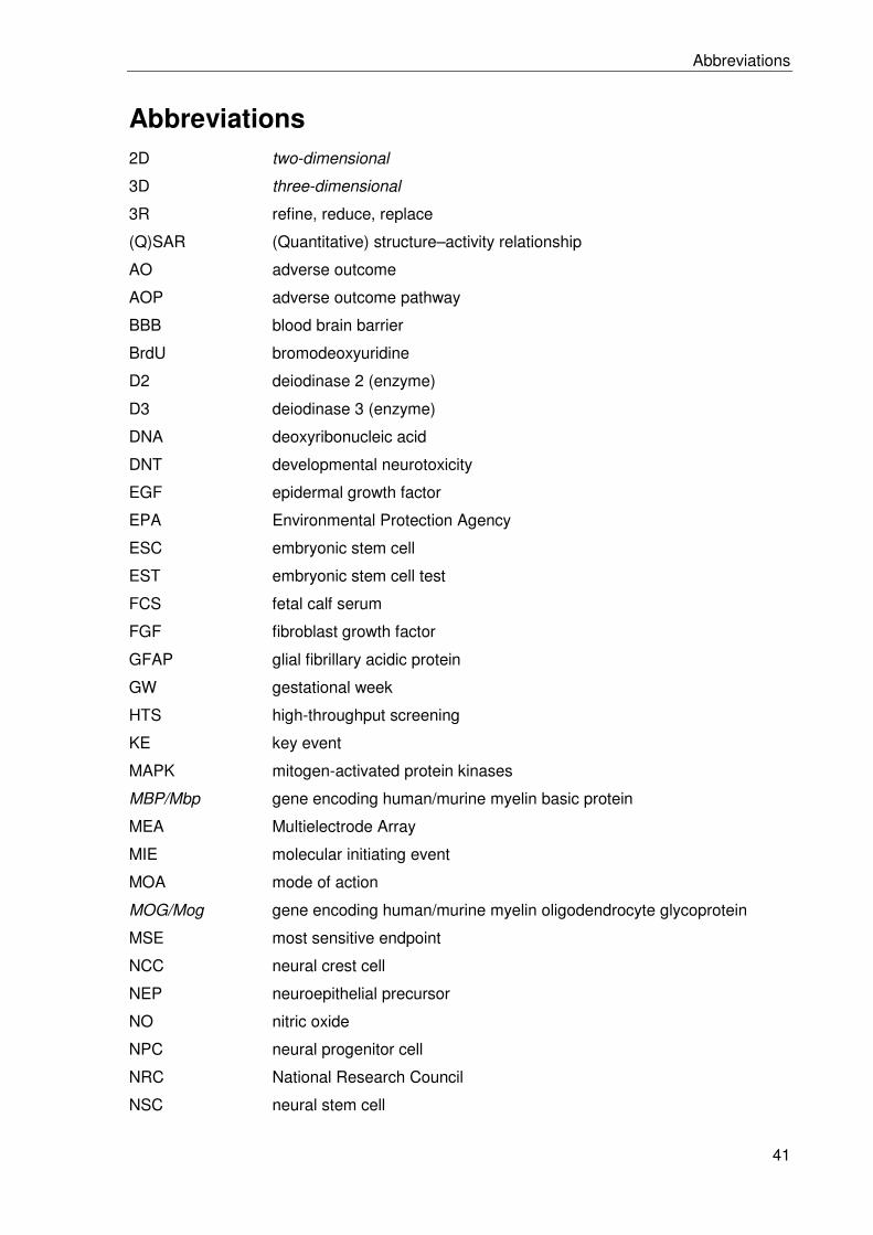
Abbreviations
41
Abbreviations 2D two-dimensional
3D three-dimensional
3R refine, reduce, replace
(Q)SAR (Quantitative) structure–activity relationship
AO adverse outcome
AOP adverse outcome pathway
BBB blood brain barrier
BrdU bromodeoxyuridine
D2 deiodinase 2 (enzyme)
D3 deiodinase 3 (enzyme)
DNA deoxyribonucleic acid
DNT developmental neurotoxicity
EGF epidermal growth factor
EPA Environmental Protection Agency
ESC embryonic stem cell
EST embryonic stem cell test
FCS fetal calf serum
FGF fibroblast growth factor
GFAP glial fibrillary acidic protein
GW gestational week
HTS high-throughput screening
KE key event
MAPK mitogen-activated protein kinases
MBP/Mbp gene encoding human/murine myelin basic protein
MEA Multielectrode Array
MIE molecular initiating event
MOA mode of action
MOG/Mog gene encoding human/murine myelin oligodendrocyte glycoprotein
MSE most sensitive endpoint
NCC neural crest cell
NEP neuroepithelial precursor
NO nitric oxide
NPC neural progenitor cell
NRC National Research Council
NSC neural stem cell
Abbreviations
42
OECD Organization for Economic Co-operation and Development
PBDE polybrominiated diphenyl ether
PDL poly-L-lysine
PI3K phosphatidylinositol-4,5-bisphosphate 3-kinase
PND postnatal day
RXR retinoid X receptor
SRA Systems Research and Applications Corporation
T3 L-triiodothyronine
T4 L-thyroxine
TH thyroid hormone
THRA/Thra gene encoding human/mouse TR isoforms
THRB/Thrb gene encoding human/mouse TR isoforms
TR thyroid hormone receptor (protein)
TSH thyroid-stimulating hormone
TTR transthyretin
US(A) United States (of America)
References
43
References
Abdulla EM, Calaminici M, Campbell IC. 1995. Comparison of neurite outgrowth with neurofilament protein subunit levels in neuroblastoma cells following mercuric oxide exposure. Clin. Exp. Pharmacol. Physiol. 22:362–363; doi:10.1111/j.1440-1681.1995.tb02017.x.
Alaee M, Arias P, Sjödin A, Bergman A. 2003. An overview of commercially used brominated flame retardants, their applications, their use patterns in different countries/regions and possible modes of release. Environ. Int. 29:683–9; doi:10.1016/S0160-4120(03)00121-1.
Alépée N. 2014. State-of-the-art of 3D cultures (organs-on-a-chip) in safety testing and pathophysiology. ALTEX 31:441–77; doi:10.14573/altex1406111.
Alm H, Kultima K, Scholz B, Nilsson A, Andrén PE, Fex-Svenningsen A, et al. 2008. Exposure to brominated flame retardant PBDE-99 affects cytoskeletal protein expression in the neonatal mouse cerebral cortex. Neurotoxicology 29:628–37; doi:10.1016/j.neuro.2008.04.021.
Alm H, Scholz B, Fischer C, Kultima K, Viberg H, Eriksson P, et al. 2006. Proteomic Evaluation of Neonatal Exposure to 2,2 ,4,4 ,5-Pentabromodiphenyl Ether. Environ. Health Perspect. 114:254–259; doi:10.1289/ehp.8419.
Alm H, Scholz B, Kultima K, Nilsson A, Andrén PE, Savitski MM, et al. 2010. In vitro neurotoxicity of PBDE-99: immediate and concentration-dependent effects on protein expression in cerebral cortex cells. J. Proteome Res. 9:1226–35; doi:10.1021/pr900723c.
Anckarsäter R, Zetterberg H, Blennow K, Anckarsäter H. 2007. Association between thyroid hormone levels and monoaminergic neurotransmission during surgery. Psychoneuroendocrinology 32:1138–43; doi:10.1016/j.psyneuen.2007.07.007.
Andersen M, Clewell R, Bhattacharya S. 2012. Developing in vitro Tools Sufficient by themselves for 21st Century Risk Assessment. SEURAT-1 2: 347–60.
Andersen SL. 2003. Trajectories of brain development: point of vulnerability or window of opportunity? Neurosci. Biobehav. Rev. 27:3–18; doi:10.1016/S0149-7634(03)00005-8.
Anderson GW. 2001. Thyroid hormones and the brain. Front. Neuroendocrinol. 22:1–17; doi:10.1006/frne.2000.0208.
Ankley GT, Bennett RS, Erickson RJ, Hoff DJ, Hornung MW, Johnson RD, et al. 2010. Adverse outcome pathways: a conceptual framework to support ecotoxicology research and risk assessment. Environ. Toxicol. Chem. 29:730–41; doi:10.1002/etc.34.
Ayuso-Sacido A, Moliterno JA, Kratovac S, Kapoor GS, O’Rourke DM, Holland EC, et al. 2010. Activated EGFR signaling increases proliferation, survival, and migration and blocks neuronal differentiation in post-natal neural stem cells. J. Neurooncol. 97:323–37; doi:10.1007/s11060-009-0035-x.
References
44
Bal-Price A, Crofton KM, Leist M, Allen S, Arand M, Buetler T, et al. 2015a. International STakeholder NETwork (ISTNET): creating a developmental neurotoxicity (DNT) testing road map for regulatory purposes. Arch. Toxicol. 89:269–87; doi:10.1007/s00204-015-1464-2.
Bal-Price A, Crofton KM, Sachana M, Shafer TJ, Behl M, Forsby A, et al. 2015b. Putative adverse outcome pathways relevant to neurotoxicity. Crit. Rev. Toxicol. 45:83–91; doi:10.3109/10408444.2014.981331.
Barateiro A, Fernandes A. 2014. Temporal oligodendrocyte lineage progression: in vitro models of proliferation, differentiation and myelination. Biochim. Biophys. Acta 1843:1917–29; doi:10.1016/j.bbamcr.2014.04.018.
Barres BA, Lazar MA RM. 1994. A novel role for thyroid hormone, glucocorticoids and retinoic acid in timing oligodendrocyte development. Development 120: 1097–1108.
Baumann J, Barenys M, Gassmann K, Fritsche E. 2014. Comparative human and rat “neurosphere assay” for developmental neurotoxicity testing. Curr. Protoc. Toxicol. 59:12.21.1–12.21.24; doi:10.1002/0471140856.tx1221s59.
Baumann J, Gassmann K, Masjosthusmann S, DeBoer D, Bendt F, Giersiefer S, et al. 2015. Comparative human and rat neurospheres reveal species differences in chemical effects on neurodevelopmental key events. Arch. Toxicol.; doi:10.1007/s00204-015-1568-8.
Baumann N, Pham-Dinh D. 2001. Biology of oligodendrocyte and myelin in the mammalian central nervous system. Physiol. Rev. 81: 871–927.
Bellanger M, Pichery C, Aerts D, Berglund M, Castaño A, Cejchanová M, et al. 2013. Economic benefits of methylmercury exposure control in Europe: monetary value of neurotoxicity prevention. Environ. Health 12:3; doi:10.1186/1476-069X-12-3.
Bernal J. 2005. Thyroid hormones and brain development. Vitam. Horm. 71:95–122; doi:10.1016/S0083-6729(05)71004-9.
Bernal J, Nunez J. 1995. Thyroid hormones and brain development. Vitam. Horm. 133:390–398; doi:10.1530/eje.0.1330390.
Bhat NR, Sarlieve LL, Rao GS, Pieringer RA. 1979. Investigations on myelination in vitro. Regulation by thyroid hormone in cultures of dissociated brain cells from embryonic mice. J. Biol. Chem. 254: 9342–4.
Billon N, Jolicoeur C, Tokumoto Y, Vennström B, Raff M. 2002. Normal timing of oligodendrocyte development depends on thyroid hormone receptor alpha 1 (TRalpha1). EMBO J. 21:6452–60; doi:10.1093/emboj/cdf662.
Billon N, Tokumoto Y, Forrest D, Raff M. 2001. Role of thyroid hormone receptors in timing oligodendrocyte differentiation. Dev. Biol. 235:110–20; doi:10.1006/dbio.2001.0293.
Blanco J, Mulero M, López M, Domingo JL, Sánchez DJ. 2011. BDE-99 deregulates BDNF, Bcl-2 and the mRNA expression of thyroid receptor isoforms in rat cerebellar granular neurons. Toxicology 290:305–11; doi:10.1016/j.tox.2011.10.010.
References
45
Boyages SC, Halpern JP. 1993. Endemic cretinism: toward a unifying hypothesis. Thyroid 3:59–69; doi:10.1089/thy.1993.3.59.
Branchi I, Alleva E, Costa LG. 2002. Effects of perinatal exposure to a polybrominated diphenyl ether (PBDE 99) on mouse neurobehavioural development. Neurotoxicology 23:375–84; doi:10.1016/s0161-813x(02)00078-5.
Brannen CL, Sugaya K. 2000. In vitro differentiation of multipotent human neural progenitors in serum-free medium. Neuroreport 11:1123–8; doi:10.1097/00001756-200004070-00042.
Breier JM, Gassmann K, Kayser R, Stegeman H, De Groot D, Fritsche E, et al. 2010. Neural progenitor cells as models for high-throughput screens of developmental neurotoxicity: state of the science. Neurotoxicol. Teratol. 32:4–15; doi:10.1016/j.ntt.2009.06.005.
Brent GA. 2012. Mechanisms of thyroid hormone action. J. Clin. Invest. 122:3035–3043; doi:10.1172/JCI60047.three.
Buc-Caron MH. 1995. Neuroepithelial progenitor cells explanted from human fetal brain proliferate and differentiate in vitro. Neurobiol. Dis. 2:37–47; doi:10.1006/nbdi.1995.0004.
Burke K, Cheng Y, Li B, Petrov A, Joshi P, Berman RF, et al. 2006. Methylmercury elicits rapid inhibition of cell proliferation in the developing brain and decreases cell cycle regulator, cyclin E. Neurotoxicology 27:970–81; doi:10.1016/j.neuro.2006.09.001.
Butt CM, Wang D, Stapleton HM. 2011. Halogenated Phenolic Contaminants Inhibit the In Vitro Activity of the Thyroid-Regulating Deiodinases in Human Liver. Toxicol. Sci. 124:339–347; doi:10.1093/toxsci/kfr117.
Cao X-Y, Jiang X-M, Dou Z-H, Rakeman MA, Zhang M-L, O’Donnell K, et al. 1994. Timing of Vulnerability of the Brain to Iodine Deficiency in Endemic Cretinism. N. Engl. J. Med. 331:1739–1744; doi:10.1056/NEJM199412293312603.
Carey DJ, Todd MS. 1987. Schwann cell myelination in a chemically defined medium " demonstration of a requirement for additives that promote Schwann cell extracellular matrix formation. Dev. Brain Res. 32:95–102; doi:10.1016/0165-3806(87)90142-8.
Carlson DJ, Strait KA, Schwartz HL, Oppenheimer JH. 1994. Immunofluorescent localization of thyroid hormone receptor isoforms in glial cells of rat brain. Endocrinology 135:1831–6; doi:10.1210/endo.135.5.7525253.
Carlson DJ, Strait KA, Schwartz HL, Oppenheimer JH. 1996. Thyroid hormone receptor isoform content in cultured type 1 and type 2 astrocytes. Endocrinology 137:911–917; doi:10.1210/endo.137.3.8603603.
Carre J-L, Demerens C, Rodriguez-Pena A, Floch HH, Vincendon G, Sarlieve LL. 1998. Thyroid hormone receptor isoforms are sequentially expressed in oligodendrocyte lineage cells during rat cerebral development. J. Neurosci. Res. 54:584–594; doi:10.1002/(SICI)1097-4547(19981201)54:5<584::AID-JNR3>3.0.CO;2-X.
Chalmers-Redman RM., Priestley T, Kemp J., Fine A. 1997. In vitro propagation and inducible differentiation of multipotential progenitor cells from human fetal brain. Neuroscience 76:1121–1128; doi:10.1016/S0306-4522(96)00386-7.
References
46
Chao H-R, Wang S-L, Lee W-J, Wang Y-F, Päpke O. 2007. Levels of polybrominated diphenyl ethers (PBDEs) in breast milk from central Taiwan and their relation to infant birth outcome and maternal menstruation effects. Environ. Int. 33:239–45; doi:10.1016/j.envint.2006.09.013.
Chen Y-H, Li Z-H, Tan Y, Zhang C-F, Chen J-S, He F, et al. 2014. Prenatal exposure to decabrominated diphenyl ether impairs learning ability by altering neural stem cell viability, apoptosis, and differentiation in rat hippocampus. Hum. Exp. Toxicol.; doi:10.1177/0960327113509661.
Chevrier J, Harley KG, Bradman A, Sjödin A, Eskenazi B. 2011. Prenatal exposure to polybrominated diphenyl ether flame retardants and neonatal thyroid-stimulating hormone levels in the CHAMACOS study. Am. J. Epidemiol. 174:1166–74; doi:10.1093/aje/kwr223.
Claudio L, Kwa WC, Russell AL, Wallinga D. 2000. Testing methods for developmental neurotoxicity of environmental chemicals. Toxicol. Appl. Pharmacol. 164:1–14; doi:10.1006/taap.2000.8890.
Coecke S, Goldberg AM, Allen S, Buzanska L, Calamandrei G, Crofton K, et al. 2007. Workgroup Report: Incorporating In Vitro Alternative Methods for Developmental Neurotoxicity into International Hazard and Risk Assessment Strategies. Environ. Health Perspect. 115:924–931; doi:10.1289/ehp.9427.
Collins FS, Gray GM, Bucher JR. 2008. TOXICOLOGY: Transforming Environmental Health Protection. Science (80-. ). 319:906–907; doi:10.1126/science.1154619.
Costa LG, de Laat R, Tagliaferri S, Pellacani C. 2014. A mechanistic view of polybrominated diphenyl ether (PBDE) developmental neurotoxicity. Toxicol. Lett. 282–294; doi:10.1016/j.toxlet.2013.11.011.
Costa LG, Giordano G. 2007. Developmental neurotoxicity of polybrominated diphenyl ether (PBDE) flame retardants. Neurotoxicology 28:1047–67; doi:10.1016/j.neuro.2007.08.007.
Crofton KM, Mundy WR, Shafer TJ. 2012. Developmental neurotoxicity testing: A path forward. Congenit. Anom. (Kyoto). 52:140–146; doi:10.1111/j.1741-4520.2012.00377.x.
Cukierman E, Pankov R, Stevens DR, Yamada KM. 2001. Taking cell-matrix adhesions to the third dimension. Science 294:1708–12; doi:10.1126/science.1064829.
Darnerud PO, Eriksen GS, Jóhannesson T, Larsen PB, Viluksela M. 2001. Polybrominated diphenyl ethers: occurrence, dietary exposure, and toxicology. Env. Heal. Perspect 109:49–68; doi:10.1289/ehp.01109s149.
De Wit CA. 2002. An overview of brominated flame retardants in the environment. Chemosphere 46:583–624; doi:10.1016/s0045-6535(01)00225-9.
DeLong GR, Stanbury JB, Fierro-Benitez R. 1985. Neurological signs in congenital iodine-deficiency disorder (endemic cretinism). Dev. Med. Child Neurol. 27:317–24; doi:10.1111/j.1469-8749.1985.tb04542.x.
References
47
Dix DJ, Houck KA, Martin MT, Richard AM, Setzer RW, Kavlock RJ. 2007. The ToxCast program for prioritizing toxicity testing of environmental chemicals. Toxicol. Sci. 95:5–12; doi:10.1093/toxsci/kfl103.
Du L, Sun W, Zhang H, Chen D. 2015. BDE-209 inhibits pluripotent genes expression and induces apoptosis in human embryonic stem cells. J. Appl. Toxicol.; doi:10.1002/jat.3195.
Dufault C, Poles G, Driscoll LL. 2005. Brief postnatal PBDE exposure alters learning and the cholinergic modulation of attention in rats. Toxicol. Sci. 88:172–80; doi:10.1093/toxsci/kfi285.
Eldridge CF, Bunge MB, Bunge RP, Wood PM. 1987. Differentiation of Axon-related Schwann Cells In Vitro. I. Ascorbic Acid Regulates Basal Lamina Assembly and Myelin Formation. J. Cell Biol. 105:1023–1034; doi:10.1083/jcb.105.2.1023.
Eskenazi B, Chevrier J, Rauch SA, Kogut K, Harley KG, Johnson C, et al. 2013. In Utero and Childhood Polybrominated Diphenyl Ether ( PBDE ) Exposures and Neurodevelopment in the CHAMACOS Study. Env. Heal. Perspect 121:257–262; doi:10.1289/ehp.1205597.
Farsetti A, Mitsuhashi T, Desvergne B, Robbins J, Nikodem VM. 1991. Molecular basis of thyroid hormone regulation of myelin basic protein gene expression in rodent brain. J. Biol. Chem. 266: 23226–32.
Fields RD. 2008. White matter in learning, cognition and psychiatric disorders. Trends Neurosci. 31:361–70; doi:10.1016/j.tins.2008.04.001.
Fischer D, Hooper K, Athanasiadou M, Athanassiadis I, Bergman Å. 2006. Children Show Highest Levels of Polybrominated Diphenyl Ethers in a California Family of Four: A Case Study. Environ. Health Perspect. 114:1581–1584; doi:10.1289/ehp.8554.
Foti SB, Chou A, Moll AD, Roskams AJ. 2013. HDAC inhibitors dysregulate neural stem cell activity in the postnatal mouse brain. Int. J. Dev. Neurosci. 31:434–47; doi:10.1016/j.ijdevneu.2013.03.008.
Frederiksen M, Vorkamp K, Thomsen M, Knudsen LE. 2009. Human internal and external exposure to PBDEs--a review of levels and sources. Int. J. Hyg. Environ. Health 212:109–34; doi:10.1016/j.ijheh.2008.04.005.
Fritsche E. 2014. Human Stem/Progenitor Cell-Based Assays for Neurodevelopmental Toxicity Testing. In In Vitro Toxicology Systems (A. Bal-Price and P. Jenningseds. ), Methods in Pharmacology and Toxicology, pp. 351–73, Springer New York.
Fritsche E, Cline JE, Nguyen N-H, Scanlan TS, Abel J. 2005. Polychlorinated Biphenyls Disturb Differentiation of Normal Human Neural Progenitor Cells: Clue for Involvement of Thyroid Hormone Receptors. Environ. Health Perspect. 113:871–876; doi:10.1289/ehp.7793.
Fritsche E, Gassmann K, Schreiber T. 2011. Neurospheres as a model for developmental neurotoxicity testing. L.G. Costa, G. Giordano, and M. Guizzettieds. Methods Mol Biol. 758:99–114; doi:10.1007/978-1-61779-170-3.
References
48
Gassmann K, Abel J, Bothe H, Haarmann-Stemmann T, Merk HF, Quasthoff KN, et al. 2010. Species-specific differential AhR expression protects human neural progenitor cells against developmental neurotoxicity of PAHs. Environ. Health Perspect. 118:1571–7; doi:10.1289/ehp.0901545.
Gassmann K, Schreiber T, Dingemans MML, Krause G, Roderigo C, Giersiefer S, et al. 2014. BDE-47 and 6-OH-BDE-47 modulate calcium homeostasis in primary fetal human neural progenitor cells via ryanodine receptor-independent mechanisms. Arch. Toxicol.; doi:10.1007/s00204-014-1217-7.
Gee JR, Hedge JM, Moser VC. 2008. Lack of alterations in thyroid hormones following exposure to polybrominated diphenyl ether 47 during a period of rapid brain development in mice. Drug Chem. Toxicol. 31:245–54; doi:10.1080/01480540701873194.
Genschow E, Spielmann H, Scholz G, Seiler A, Brown N, Piersma A, et al. 2002. The ECVAM international validation study on in vitro embryotoxicity tests: results of the definitive phase and evaluation of prediction models. European Centre for the Validation of Alternative Methods. Altern. Lab. Anim. 30: 151–76.
Go HS, Kim KC, Choi CS, Jeon SJ, Kwon KJ, Han S-H, et al. 2012. Prenatal exposure to valproic acid increases the neural progenitor cell pool and induces macrocephaly in rat brain via a mechanism involving the GSK-3 / -catenin pathway. Neuropharmacology 63:1028–1041; doi:10.1016/j.neuropharm.2012.07.028.
Grandjean P, Landrigan PJ. 2014. Neurobehavioural effects of developmental toxicity. Lancet Neurol. 13:330–8; doi:10.1016/S1474-4422(13)70278-3.
Gross RE, Mehler MF, Mabie PC, Zang Z, Santschi L, Kessler JA. 1996. Bone morphogenetic proteins promote astroglial lineage commitment by mammalian subventricular zone progenitor cells. Neuron 17:595–606; doi:10.1016/s0896-6273(00)80193-2.
Haddow J, Palomaki G, Allan W, Williams J, Knight G, Gagnon J, et al. 1999. Maternal thyroid deficiency during pregnancy and subsequent neuropsychological development of the child. N. Engl. J. Med. 341:549–555; doi:10.1056/nejm199908193410801.
Hagen GA, Elliott WJ. 1973. Transport of thyroid hormones in serum and cerebrospinal fluid. J. Clin. Endocrinol. Metab. 37:415–22; doi:10.1210/jcem-37-3-415.
Hamers T, Kamstra JH, Sonneveld E, Murk AJ, Kester MH a, Andersson PL, et al. 2006. In vitro profiling of the endocrine-disrupting potency of brominated flame retardants. Toxicol. Sci. 92:157–73; doi:10.1093/toxsci/kfj187.
Hartung T. 2009. Toxicology for the twenty-first century. Nature 460:208–212; doi:10.1038/460208a.
Harvey CB, Williams GR. 2002. Mechanism of thyroid hormone action. Thyroid 12:441–6; doi:10.1089/105072502760143791.
Hayess K, Riebeling C, Pirow R, Steinfath M, Sittner D, Slawik B, et al. 2013. The DNT-EST: A predictive embryonic stem cell-based assay for developmental neurotoxicity testing in vitro. Toxicology 314:135–147; doi:10.1016/j.tox.2013.09.012.
References
49
He L, Chen H-J, Qian L, Chen G, Buzby JS. 2010. Curcumin protects pre-oligodendrocytes from activated microglia in vitro and in vivo. Brain Res. 1339:60–9; doi:10.1016/j.brainres.2010.04.014.
Herbstman JB, Sjödin A, Apelberg BJ, Witter FR, Halden RU, Patterson DG, et al. 2008. Birth Delivery Mode Modifies the Associations between Prenatal Polychlorinated Biphenyl (PCB) and Polybrominated Diphenyl Ether (PBDE) and Neonatal Thyroid Hormone Levels. Environ. Health Perspect. 116:1376–1382; doi:10.1289/ehp.11379.
Hewett JA. 2009. Determinants of regional and local diversity within the astroglial lineage of the normal central nervous system. J. Neurochem. 110:1717–36; doi:10.1111/j.1471-4159.2009.06288.x.
Hong MS, Hong SJ, Barhoumi R, Burghardt RC, Donnelly K., Wild JR, et al. 2003. Neurotoxicity induced in differentiated SK-N-SH-SY5Y human neuroblastoma cells by organophosphorus compounds. Toxicol. Appl. Pharmacol. 186:110–118; doi:10.1016/S0041-008X(02)00016-9.
Horn S, Heuer H. 2010. Thyroid hormone action during brain development: more questions than answers. Mol. Cell. Endocrinol. 315:19–26; doi:10.1016/j.mce.2009.09.008.
Huang SC, Giordano G, Costa LG. 2010. Comparative cytotoxicity and intracellular accumulation of five polybrominated diphenyl ether congeners in mouse cerebellar granule neurons. Toxicol. Sci. 114:124–32; doi:10.1093/toxsci/kfp296.
Ibarrola N, Mayer-Pröschel M, Rodriguez-Peña A, Noble M. 1996. Evidence for the existence of at least two timing mechanisms that contribute to oligodendrocyte generation in vitro. Dev. Biol. 180:1–21; doi:10.1006/dbio.1996.0280.
Ibarrola N, Rodríguez-Peña A. 1997. Hypothyroidism coordinately and transiently affects myelin protein gene expression in most rat brain regions during postnatal development. Brain Res. 752:285–93; doi:10.1016/s0006-8993(96)01480-1.
Ibhazehiebo K, Iwasaki T, Kimura-Kuroda J, Miyazaki W, Shimokawa N, Koibuchi N. 2011. Disruption of thyroid hormone receptor-mediated transcription and thyroid hormone-induced Purkinje cell dendrite arborization by polybrominated diphenyl ethers. Environ. Health Perspect. 119:168–75; doi:10.1289/ehp.1002065.
Iwasaki T, Miyazaki W, Takeshita A, Kuroda Y, Koibuchi N. 2002. Polychlorinated biphenyls suppress thyroid hormone-induced transactivation. Biochem. Biophys. Res. Commun. 299:384–8; doi:10.1016/s0006-291x(02)02659-1.
Jacob HJ, Lazar J, Dwinell MR, Moreno C, Geurts AM. 2010. Gene targeting in the rat: advances and opportunities. Trends Genet. 26:510–8; doi:10.1016/j.tig.2010.08.006.
Johansson P, Almqvist EG, Johansson J-O, Mattsson N, Hansson O, Wallin A, et al. 2013. Reduced cerebrospinal fluid level of thyroxine in patients with Alzheimer’s disease. Psychoneuroendocrinology 38:1058–66; doi:10.1016/j.psyneuen.2012.10.012.
Jones I, Srinivas M, Ng L, Forrest D. 2003. The thyroid hormone receptor beta gene: structure and functions in the brain and sensory systems. Thyroid 13:1057–68; doi:10.1089/105072503770867228.
References
50
Judson RS., Houck KA., Kavlock RJ., Knudsen TB., Martin MT., Mortensen HM., et al. 2010. In Vitro Screening of Environmental Chemicals for Targeted Testing Prioritization: The ToxCast Project. Env. Heal. Perspect 118:484–92; doi:10.1289/ehp.0901392.
Kitamura S, Jinno N, Ohta S, Kuroki H, Fujimoto N. 2002. Thyroid hormonal activity of the flame retardants tetrabromobisphenol A and tetrachlorobisphenol A. Biochem. Biophys. Res. Commun. 293:554–9; doi:10.1016/S0006-291X(02)00262-0.
Klein C, Butt SJB, Machold RP, Johnson JE, Fishell G. 2005. Cerebellum- and forebrain-derived stem cells possess intrinsic regional character. Development 132:4497–508; doi:10.1242/dev.02037.
Kodavanti PRS, Coburn CG, Moser VC, MacPhail RC, Fenton SE, Stoker TE, et al. 2010. Developmental exposure to a commercial PBDE mixture, DE-71: neurobehavioral, hormonal, and reproductive effects. Toxicol. Sci. 116:297–312; doi:10.1093/toxsci/kfq105.
Kojima H, Takeuchi S, Uramaru N, Sugihara K, Yoshida T, Kitamura S. 2009. Nuclear hormone receptor activity of polybrominated diphenyl ethers and their hydroxylated and methoxylated metabolites in transactivation assays using Chinese hamster ovary cells. Environ. Health Perspect. 117:1210–8; doi:10.1289/ehp.0900753.
Koper JW, Hoeben RC, Hochstenbach FM, van Golde LM, Lopes-Cardozo M. 1986. Effects of triiodothyronine on the synthesis of sulfolipids by oligodendrocyte-enriched glial cultures. Biochim. Biophys. Acta 887:327–34; doi:10.1016/0167-4889(86)90162-x.
Kovalevich J, Corley G, Yen W, Kim J, Rawls SM, Langford D. 2012. Cocaine decreases expression of neurogranin via alterations in thyroid receptor/retinoid X receptor signaling. J. Neurochem. 121:302–313; doi:10.1111/j.1471-4159.2012.07678.x.
Landrigan PJ, Schechter CB, Lipton JM, Fahs MC, Schwartz J. 2002. Environmental pollutants and disease in American children: estimates of morbidity, mortality, and costs for lead poisoning, asthma, cancer, and developmental disabilities. Environ. Health Perspect. 110:721–8; doi:10.1289/ehp.02110721.
Law RJ, Allchin CR, de Boer J, Covaci A, Herzke D, Lepom P, et al. 2006. Levels and trends of brominated flame retardants in the European environment. Chemosphere 64:187–208; doi:10.1016/j.chemosphere.2005.12.007.
Law RJ, Covaci A, Harrad S, Herzke D, Abdallah M a E, Fernie K, et al. 2014. Levels and trends of PBDEs and HBCDs in the global environment: Status at the end of 2012. Environ. Int. 65:147–158; doi:10.1016/j.envint.2014.01.006.
Lebel J-M, L’Hérault S, Dussault JH, Puymirat J. 1993. Thyroid hormone up-regulates thyroid hormone receptor gene expression in rat cerebral hemisphere astrocyte cultures. Glia 9:105–112; doi:10.1002/glia.440090203.
Lein P, Locke P, Goldberg A. 2007. Meeting report: alternatives for developmental neurotoxicity testing. Environ. Health Perspect. 115:764–8; doi:10.1289/ehp.9841.
Lein P, Silbergeld E, Locke P, Goldberg AM. 2005. In vitro and other alternative approaches to developmental neurotoxicity testing (DNT). Environ. Toxicol. Pharmacol. 19:735–44; doi:10.1016/j.etap.2004.12.035.
References
51
Leist M, Hartung T. 2013. Inflammatory findings on species extrapolations: humans are definitely no 70-kg mice. Arch. Toxicol. 87:563–7; doi:10.1007/s00204-013-1038-0.
Lema SC, Dickey JT, Schultz IR, Swanson P. 2008. Dietary exposure to 2,2’,4,4'-tetrabromodiphenyl ether (PBDE-47) alters thyroid status and thyroid hormone-regulated gene transcription in the pituitary and brain. Environ. Health Perspect. 116:1694–9; doi:10.1289/ehp.11570.
Li F, Xie Q, Li X, Li N, Chi P, Chen J, et al. 2010. Hormone activity of hydroxylated polybrominated diphenyl ethers on human thyroid receptor-beta: in vitro and in silico investigations. Environ. Health Perspect. 118:602–6; doi:10.1289/ehp.0901457.
Li T, Wang W, Pan Y-W, Xu L, Xia Z. 2013. A hydroxylated metabolite of flame-retardant PBDE-47 decreases the survival, proliferation, and neuronal differentiation of primary cultured adult neural stem cells and interferes with signaling of ERK5 MAP kinase and neurotrophin 3. Toxicol. Sci. 134:111–24; doi:10.1093/toxsci/kft083.
Lobo MVT, Alonso FJM, Redondo C, López-Toledano MA, Caso E, Herranz AS, et al. 2003. Cellular characterization of epidermal growth factor-expanded free-floating neurospheres. J. Histochem. Cytochem. 51:89–103; doi:10.1177/002215540305100111.
López-Espíndola D, Morales-Bastos C, Grijota-Martínez C, Liao X-H, Lev D, Sugo E, et al. 2014. Mutations of the thyroid hormone transporter MCT8 cause prenatal brain damage and persistent hypomyelination. J. Clin. Endocrinol. Metab. 99:E2799–804; doi:10.1210/jc.2014-2162.
Ma Y, Salamova A, Venier M, Hites RA. 2013. Has the Phase-Out of PBDEs Affected Their Atmospheric Levels? Trends of PBDEs and Their Replacements in the Great Lakes Atmosphere. Environ. Sci. Technol. 47:11457–11464; doi:10.1021/es403029m.
Makris SL, Raffaele K, Allen S, Bowers WJ, Hass U, Alleva E, et al. 2009. A retrospective performance assessment of the developmental neurotoxicity study in support of OECD test guideline 426. Environ. Health Perspect. 117:17–25; doi:10.1289/ehp.11447.
Malin J-P, Ködding R, Fuhrmann H, von zur Mühlen A. 1989. T4, T3 and rT3 levels in serum and cerebrospinal fluid of patients with amyotrophic lateral sclerosis. J. Neurol. 236: 57–9.
Marta CB, Adamo AM, Soto EF, Pasquini JM. 1998. Sustained neonatal hyperthyroidism in the rat affects myelination in the central nervous system. J. Neurosci. Res. 53:251–9; doi:10.1002/(sici)1097-4547(19980715)53:2<251::aid-jnr14>3.0.co;2-9.
McDonald TA. 2005. Polybrominated diphenylether levels among United States residents: daily intake and risk of harm to the developing brain and reproductive organs. Integr. Environ. Assess. Manag. 1:343–54; doi:10.1897/1551-3793(2005)1[343:pdlaus]2.0.co; 2.
Meerts IA, van Zanden JJ, Luijks EA, van Leeuwen-Bol I, Marsh G, Jakobsson E, et al. 2000. Potent competitive interactions of some brominated flame retardants and related compounds with human transthyretin in vitro. Toxicol. Sci. 56:95–104; doi:10.1093/toxsci/56.1.95.
References
52
Moors M, Cline JE, Abel J, Fritsche E. 2007. ERK-dependent and -independent pathways trigger human neural progenitor cell migration. Toxicol. Appl. Pharmacol. 221:57–67; doi:10.1016/j.taap.2007.02.018.
Moors M, Rockel TD, Abel J, Cline JE, Gassmann K, Schreiber T, et al. 2009. Human neurospheres as three-dimensional cellular systems for developmental neurotoxicity testing. Environ. Health Perspect. 117:1131–8; doi:10.1289/ehp.0800207.
Moriyama K, Tagami T, Akamizu T, Usui T, Saijo M, Kanamoto N, et al. 2002. Thyroid Hormone Action Is Disrupted by Bisphenol A as an Antagonist. J. Clin. Endocrinol. Metab. 87:5185–5190; doi:10.1210/jc.2002-020209.
Namba N, Etani Y, Kitaoka T, Nakamoto Y, Nakacho M, Bessho K, et al. 2008. Clinical phenotype and endocrinological investigations in a patient with a mutation in the MCT8 thyroid hormone transporter. Eur. J. Pediatr. 167:785–91; doi:10.1007/s00431-007-0589-6.
Negishi T, Kawasaki K, Sekiguchi S, Ishii Y, Kyuwa S, Kuroda Y, et al. 2005. Attention-deficit and hyperactive neurobehavioural characteristics induced by perinatal hypothyroidism in rats. Behav. Brain Res. 159:323–31; doi:10.1016/j.bbr.2004.11.012.
Nguyen N-H, Apriletti JW, Cunha Lima ST, Webb P, Baxter JD, Scanlan TS. 2002. Rational Design and Synthesis of a Novel Thyroid Hormone Antagonist That Blocks Coactivator Recruitment. J. Med. Chem. 45:3310–3320; doi:10.1021/jm0201013.
Nishikawa M, Inada M, Naito K, Ishii H, Tanaka K, Mashio Y, et al. 1981. 3,3’,5'-triiodothyronine (reverse T3) in human cerebrospinal fluid. J. Clin. Endocrinol. Metab. 53:1030–5; doi:10.1210/jcem-53-5-1030.
NRC. 2007. Toxicity testing in the 21st century: A vision and a strategy. Washington, DC Natl. Acad. Press.
OECD 2007. Test Guideline 426. OECD guideline for testing of chemicals. Developmental neurotoxicity study. http//www.oecdilibrary. org/docserver/download/9742601e.pdf? Expir.
OECD 2013. Guidance document on developing and assessing adverse outcome pathways. http://www.oecd.org/officialdocuments/publicdisplaydocumentpdf/?cote=env/jm/mono %282013%296&doclanguage=en.
Påhlman S, Mamaeva S, Meyerson G, Mattsson ME, Bjelfman C, Ortoft E, et al. 1990. Human neuroblastoma cells in culture: a model for neuronal cell differentiation and function. Acta Physiol. Scand. Suppl. 592: 25–37.
Pardridge WM. 2006. Introduction to the Blood-Brain Barrier | Neurology and Clinical Neuroscience | Cambridge University Press.
Peters LL, Robledo RF, Bult CJ, Churchill GA, Paigen BJ, Svenson KL. 2007. The mouse as a model for human biology: a resource guide for complex trait analysis. Nat. Rev. Genet. 8:58–69; doi:10.1038/nrg2025.
Pinnell SR. 1985. Regulation of collagen biosynthesis by ascorbic acid: a review. Yale J. Biol. Med. 58: 553–9.
References
53
Piper DR, Mujtaba T, Keyoung H, Roy NS, Goldman SA, Rao MS, et al. 2001. Identification and characterization of neuronal precursors and their progeny from human fetal tissue. J. Neurosci. Res. 66:356–68; doi:10.1002/jnr.1228.
Podratz JL, Rodriguez EH, Windebank AJ. 2004. Antioxidants are necessary for myelination of dorsal root ganglion neurons, in vitro. Glia 45:54–8; doi:10.1002/glia.10302.
Pop VJ, Brouwers EP, Vader HL, Vulsma T, van Baar AL, de Vijlder JJ. 2003. Maternal hypothyroxinaemia during early pregnancy and subsequent child development: a 3-year follow-up study. Clin. Endocrinol. (Oxf). 59:282–8; doi:10.1046/j.1365-2265.2003.01822.x.
Potter GB, Zarach JM, Sisk JM, Thompson CC. 2002. The thyroid hormone-regulated corepressor hairless associates with histone deacetylases in neonatal rat brain. Mol. Endocrinol. 16:2547–60; doi:10.1210/me.2002-0115.
Puymirat J, L’Hereault S, Dussault JH. 1992. Expression of thyroid hormone receptors mRNAs in rat cerebral hemisphere neuronal cultures. Brain Res. Dev. Brain Res. 69:173–7; doi:10.1016/0165-3806(92)90156-q.
Ramos HE, Weiss RE. 2006. Regulation of nuclear coactivator and corepressor expression in mouse cerebellum by thyroid hormone. Thyroid 16:211–6; doi:10.1089/thy.2006.16.211.
Ren X-M, Guo L-H, Gao Y, Zhang B-T, Wan B. 2013. Hydroxylated polybrominated diphenyl ethers exhibit different activities on thyroid hormone receptors depending on their degree of bromination. Toxicol. Appl. Pharmacol. 268:256–63; doi:10.1016/j.taap.2013.01.026.
Reubinoff BE, Itsykson P, Turetsky T, Pera MF, Reinhartz E, Itzik A, et al. 2001. Neural progenitors from human embryonic stem cells. Nat. Biotechnol. 19:1134–40; doi:10.1038/nbt1201-1134.
Reynolds BA, Tetzlaff W, Weiss S. 1992. A multipotent EGF-responsive striatal embryonic progenitor cell produces neurons and astrocytes. J. Neurosci. 12: 4565–74.
Rice D, Barone S. 2000. Critical periods of vulnerability for the developing nervous system: evidence from humans and animal models. Environ. Health Perspect. 108:511–33; doi:10.1289/ehp.00108s3511.
Roberts SC, Bianco AC, Stapleton HM. 2015. Disruption of type 2 iodothyronine deiodinase activity in cultured human glial cells by polybrominated diphenyl ethers. Chem. Res. Toxicol. 28:1265–74; doi:10.1021/acs.chemrestox.5b00072.
Rodrigues F, Grenha J, Ortez C, Nascimento A, Morte B, M-Belinchón M, et al. 2014. Hypotonic male infant and MCT8 deficiency - a diagnosis to think about. BMC Pediatr. 14:252; doi:10.1186/1471-2431-14-252.
Rodriguez-Peña A, Ibarrola N, Iñiguez MA, Muñoz A, Bernal J. 1993. Neonatal hypothyroidism affects the timely expression of myelin-associated glycoprotein in the rat brain. J. Clin. Invest. 91:812–8; doi:10.1172/JCI116301.
Rogers A. 2003. Immediate-Early-Gene (IEG) expression in excitotoxicity. Dissertation.
References
54
Rose M, Bennett DH, Bergman A, Fängström B, Pessah IN, Hertz-Picciotto I. 2010. PBDEs in 2-5 year-old children from California and associations with diet and indoor environment. Environ. Sci. Technol. 44:2648–53; doi:10.1021/es903240g.
Roze E, Meijer L, Bakker A, Van Braeckel KNJA, Sauer PJJ, Bos AF. 2009. Prenatal exposure to organohalogens, including brominated flame retardants, influences motor, cognitive, and behavioral performance at school age. Environ. Health Perspect. 117:1953–8; doi:10.1289/ehp.0901015.
Russel W, Burch R, Rume C. 1959. The principles of humane experimental technique.
Schecter A, Päpke O, Harris TR, Tung KC, Musumba A, Olson J, et al. 2006. Polybrominated diphenyl ether (PBDE) levels in an expanded market basket survey of U.S. food and estimated PBDE dietary intake by age and sex. Environ. Health Perspect. 114:1515–20; doi:10.1289/ehp.9121.
Scholz D, Pöltl D, Genewsky A, Weng M, Waldmann T, Schildknecht S, et al. 2011. Rapid, complete and large-scale generation of post-mitotic neurons from the human LUHMES cell line. J. Neurochem. 119:957–71; doi:10.1111/j.1471-4159.2011.07255.x.
Schreiber T, Gassmann K, Götz C, Hübenthal U, Moors M, Krause G, et al. 2010. Polybrominated diphenyl ethers induce developmental neurotoxicity in a human in vitro model: evidence for endocrine disruption. Environ. Health Perspect. 118:572–8; doi:10.1289/ehp.0901435.
Shanker G, Amur SG, Pieringer RA. 1985. Investigations on myelinogenesis in vitro: a study of the critical period at which thyroid hormone exerts its maximum regulatory effect on the developmental expression of two myelin associated markers in cultured brain cells from embryonic mice. Neurochem. Res. 10:617–25; doi:10.1007/bf00964401.
Sharlin DS. 2015. Chapter 8: Thyroid-disrupting chemicals as Developmental Neurotoxicants. In Environmental Factors in Neurodevelopment and Neurodegenerative Disorders (by Aschner and Costa), pp. 167–92.
Shaw SD, Kannan K. 2009. Polybrominated diphenyl ethers in marine ecosystems of the American continents: foresight from current knowledge. Rev. Environ. Health 24:157–229; doi:10.1515/reveh.2009.24.3.157.
Shy C-G, Huang H-L, Chang-Chien G-P, Chao H-R, Tsou T-C. 2011. Neurodevelopment of infants with prenatal exposure to polybrominated diphenyl ethers. Bull. Environ. Contam. Toxicol. 87:643–8; doi:10.1007/s00128-011-0422-9.
Simpson KL, Weaver KJ, de Villers-Sidani E, Lu JY-F, Cai Z, Pang Y, et al. 2011. Perinatal antidepressant exposure alters cortical network function in rodents. Proc. Natl. Acad. Sci. U. S. A. 108:18465–70; doi:10.1073/pnas.1109353108.
Sjödin A, Patterson DG, Bergman A. 2003. A review on human exposure to brominated flame retardants--particularly polybrominated diphenyl ethers. Environ. Int. 29:829–39; doi:10.1016/S0160-4120(03)00108-9.
Slikker W. 1994. Principles of developmental neurotoxicology. Neurotoxicology 15: 11–6.
Song J, Li Z, He Y-T, Liu C-X, Sun B, Zhang C-F, et al. 2014. Decabrominated diphenyl ether (BDE-209) and/or BDE-47 exposure alters protein expression in purified neural

References
55
stem/progenitor cells determined by proteomics analysis. Int. J. Dev. Neurosci. 33:8–14; doi:10.1016/j.ijdevneu.2013.10.008.
Stapleton HM, Eagle S, Anthopolos R, Wolkin A, Miranda ML. 2011. Associations between Polybrominated Diphenyl Ether (PBDE) Flame Retardants, Phenolic Metabolites, and Thyroid Hormones during Pregnancy. Env. Heal. Perspect 119:1454–9; doi:10.1289/ehp.1003235.
Stern M, Gierse A, Tan S, Bicker G. 2014. Human Ntera2 cells as a predictive in vitro test system for developmental neurotoxicity. Arch. Toxicol. 88:127–36; doi:10.1007/s00204-013-1098-1.
Stiles J, Jernigan TL. 2010. The basics of brain development. Neuropsychol. Rev. 20:327–48; doi:10.1007/s11065-010-9148-4.
Strait KA, Schwartz HL, Seybold VS, Ling NC, Oppenheimer JH. 1991. Immunofluorescence localization of thyroid hormone receptor protein beta 1 and variant alpha 2 in selected tissues: cerebellar Purkinje cells as a model for beta 1 receptor-mediated developmental effects of thyroid hormone in brain. Proc. Natl. Acad. Sci. U. S. A. 88:3887–91; doi:10.1073/pnas.88.9.3887.
Suvorov A, Bissonnette C, Takser L, Langlois M-F. 2011. Does 2,2’,4,4'-tetrabromodiphenyl ether interact directly with thyroid receptor? J. Appl. Toxicol. 31:179–84; doi:10.1002/jat.1580.
Suvorov A, Girard S, Lachapelle S, Abdelouahab N, Sebire G, Takser L. 2009. Perinatal exposure to low-dose BDE-47, an emergent environmental contaminant, causes hyperactivity in rat offspring. Neonatology 95:203–9; doi:10.1159/000155651.
Svendsen CN, Fawcett JW, Bentlage C, Dunnett SB. 1995. Increased survival of rat EGF-generated CNS precursor cells using B27 supplemented medium. Exp. brain Res. 102:407–14; doi:10.1007/bf00230645.
Svendsen CN, Skepper J, Rosser AE, ter Borg MG, Tyres P, Ryken T. 1997. Restricted growth potential of rat neural precursors as compared to mouse. Dev. Brain Res. 99:253–258; doi:10.1016/S0165-3806(97)00002-3.
Tagliaferri S, Caglieri A, Goldoni M, Pinelli S, Alinovi R, Poli D, et al. 2010. Low concentrations of the brominated flame retardants BDE-47 and BDE-99 induce synergistic oxidative stress-mediated neurotoxicity in human neuroblastoma cells. Toxicol. In Vitro 24:116–22; doi:10.1016/j.tiv.2009.08.020.
Thompson CC, Potter GB. 2000. Thyroid hormone action in neural development. Cereb. Cortex 10:939–45; doi:10.1093/cercor/10.10.939.
Toms L-ML, Sjödin A, Harden F, Hobson P, Jones R, Edenfield E, et al. 2009. Serum polybrominated diphenyl ether (PBDE) levels are higher in children (2-5 years of age) than in infants and adults. Environ. Health Perspect. 117:1461–5; doi:10.1289/ehp.0900596.
Tonduti D, Vanderver A, Berardinelli A, Schmidt JL, Collins CD, Novara F, et al. 2013. MCT8 deficiency: extrapyramidal symptoms and delayed myelination as prominent features. J. Child Neurol. 28:795–800; doi:10.1177/0883073812450944.
References
56
Viberg H, Fredriksson A, Eriksson P. 2004. Neonatal exposure to the brominated flame-retardant, 2,2’,4,4',5-pentabromodiphenyl ether, decreases cholinergic nicotinic receptors in hippocampus and affects spontaneous behaviour in the adult mouse. Environ. Toxicol. Pharmacol. 17:61–5; doi:10.1016/j.etap.2004.02.004.
Viberg H, Fredriksson A, Jakobsson E, Orn U, Eriksson P. 2003. Neurobehavioral derangements in adult mice receiving decabrominated diphenyl ether (PBDE 209) during a defined period of neonatal brain development. Toxicol. Sci. 76:112–20; doi:10.1093/toxsci/kfg210.
Vongphachan V, Cassone CG, Wu D, Chiu S, Crump D, Kennedy SW. 2011. Effects of perfluoroalkyl compounds on mRNA expression levels of thyroid hormone-responsive genes in primary cultures of avian neuronal cells. Toxicol. Sci. 120:392–402; doi:10.1093/toxsci/kfq395.
Voorspoels S, Covaci A, Lepom P, Jaspers VLB, Schepens P. 2006. Levels and distribution of polybrominated diphenyl ethers in various tissues of birds of prey. Environ. Pollut. 144:218–27; doi:10.1016/j.envpol.2005.12.027.
Workman AD, Charvet CJ, Clancy B, Darlington RB, Finlay BL. 2013. Modeling transformations of neurodevelopmental sequences across mammalian species. J. Neurosci. 33:7368–83; doi:10.1523/JNEUROSCI.5746-12.2013.
Yamada KM, Cukierman E. 2007. Modeling tissue morphogenesis and cancer in 3D. Cell 130:601–10; doi:10.1016/j.cell.2007.08.006.
Yogui GT, Sericano JL. 2009. Polybrominated diphenyl ether flame retardants in the U.S. marine environment: a review. Environ. Int. 35:655–66; doi:10.1016/j.envint.2008.11.001.
Zoeller RT. 2000. Developmental Exposure to Polychlorinated Biphenyls Exerts Thyroid Hormone-Like Effects on the Expression of RC3/Neurogranin and Myelin Basic Protein Messenger Ribonucleic Acids in the Developing Rat Brain. Endocrinology 141:181–189; doi:10.1210/en.141.1.181.
Zota AR, Park J-S, Wang Y, Petreas M, Zoeller RT, Woodruff TJ. 2011. Polybrominated diphenyl ethers, hydroxylated polybrominated diphenyl ethers, and measures of thyroid function in second trimester pregnant women in California. Environ. Sci. Technol. 45:7896–905; doi:10.1021/es200422b.
Acknowlegdements
57
Acknowledgements
At this point, I would like to thank all people who supported me during this PhD thesis:
First, I would like to thank Prof. Dr. Ellen Fritsche for the opportunity to perform this PhD
thesis in her working group, for the interesting topic and her great supervision and support.
Then, I thank Prof. Dr Lutz Schmitt for taking over the supervision at the mathematical and
natural science faculty of the Heinrich-Heine University, what made this thesis possible as
well as for his mentorship.
Thanks to Dr. Heike Heuer for the provision of the used knockout mice and for her
answering patiently any questions concerning thyroid hormone signaling.
Huge thanks go all present and former members of the Fritsche group for the great support,
discussions, ideas and collaboration.
Especially, I would like to thank Susi for her supervision and introduction into the topic and
methods.
Marta, Maxi, Martin, Stefan, Farina, Yaschar and Laura I thank for the nice communicative
coffee breaks and for their encouragement in stressful and discouraging times.
Thanks go to Ulrike and Farina for helping with the performance of the experiments.
A big thanks goes to Martin Schmuck for motivating me during this dissertation and his help
and great support at any time.
Last but not least, I would like my family and my friends for supporting, encouraging and
motivating me and for bearing my bad moods.
THANK YOU FOR EVERYTHING!
58
Eidesstattliche Erklärung/Declaration
Hiermit versichere ich an Eides statt, dass ich die vorliegende Arbeit „Mechanistische
Studien zur Entwicklungsneurotoxizität Polybromierter Diphenylether (PBDE) in 3D Modellen
von Maus und Mensch in vitro“ selbständig verfasst und ausschließlich die von mir
angegebenen Hilfsmittel verwendet habe. Die Dissertation wurde in der vorgelegten oder
einer ähnlichen Form noch bei keiner anderen Institution eingereicht. Ich habe bisher keine
erfolglosen Promotionsversuche unternommen.
I declare that I have developed and written the enclosed Thesis “Mechanistic studies on
the developmental neurotoxicity of polybrominated diphenyl ethers (PBDEs) in human
and murine 3D in vitro models” completely by myself, and have not used sources or
means without declaration in the text. Any thoughts from others or literal quotations are
clearly marked. The Thesis was not used in the same or in a similar version to achieve
an academic grading elsewhere.
Katharina Dach
Düsseldorf, November 2015