Embed Size (px)
Citation preview
Drug Metabolism Reviews, 38: 769–789, 2006Copyright © Informa HealthcareISSN: 0360-2532 print / 1097-9883 onlineDOI: 10.1080/03602530600971974
769
MECHANISTIC STUDIES OF THE NRF2-KEAP1 SIGNALING PATHWAY
Donna D. ZhangDepartment of Pharmacology and Toxicology, College of Pharmacy, the Universityof Arizona, Tucson, Arizona, USA
Since eukaryotic cells constantly encounter various environmental insults, they have evolveddefense mechanisms to cope with toxicant- and carcinogen-induced oxidative stress or elec-trophiles. One of the most important cellular defense mechanisms against oxidative stress orelectrophiles is mediated by the transcription factor Nrf2. Under the basal condition, Nrf2-dependent transcription is repressed by a negative regulator Keap1. When cells are exposedto oxidative stress, electrophiles, or chemopreventive agents, Nrf2 escapes Keap1-mediatedrepression and activates antioxidant responsive element (ARE)-dependent gene expressionto maintain cellular redox homeostasis. Beyond its antioxidant function, Nrf2 has recentlybeen recognized as a key factor regulating an array of genes that defend cells against thedeleterious effects of environmental insults. Since this Nrf2-dependent cellular defenseresponse is able to protect multi-organs or multi-tissues, activation of Nrf2 has been impli-cated in conferring protection against many human diseases, including cancer, neurodegen-erative diseases, cardiovascular diseases, acute and chronic lung injury, autoimmunediseases, and inflammation. Therefore, understanding of Nrf2 regulation is crucial in thedevelopment of drugs for therapeutic intervention. This review will discuss recent progressin the field of the Nrf2-Keap1 signaling pathway, with emphasis on the mechanistic studiesof Nrf2 regulation by Keap1, oxidative stress, or chemopreventive compounds.
Key Words: Nrf2; Keap1; Chemopreventive compounds; Oxidative stress; Ubiquitination;Degradation; Ubiquitin ligase.
INTRODUCTION
It is difficult to discuss the Nrf2 transcription factor without mentioning cancer pre-vention or chemoprevention, since the discovery of Nrf2 is attributed greatly to studieswith anti-carcinogenic compounds. The very first concept of chemoprevention came fromthe observation that systemic administration of small quantities of xenobiotics, such as 3-methylcholanthrene, decreased the incidence of cancer in rats that were subsequently fedlarge doses of carcinogenic azo dyes (Richardson, 1951). Subsequent work over the last50 years has firmly established that ingestion of small quantities of certain organic compounds,
Presented at the Seventh International Symposium on Biological Reactive Intermediates, Tucson,Arizona, January 4–7, 2006.
Address correspondence to Donna D. Zhang, Department of Pharmacology and Toxicology, Collegeof Pharmacy, University of Arizona, 1703 E. Mabel St., Tucson, AZ 85721; Fax: 520-626-2466;E-mail: [email protected]
770 D. D. ZHANG
now commonly referred to as chemopreventive agents or anti-carcinogens, can lower therisk of cancer in mammals that are exposed to carcinogens (Kensler et al., 2000; Talalayand Fahey, 2001; Wolf, 2001). A number of chemopreventive compounds have been iso-lated from plants such as fruits and vegetables; other compounds are synthetic drugs.Interestingly, chemopreventive compounds are structurally diverse, including isothiocyan-ates (sulforaphane found in cruciferous vegetables), polyphenols [epigallocatechin-3-gal-late (EGCG) in green tea, and caffeic acid phenethyl ester in honeybee propollis], and 1, 2,dithiole-3-thiones (oltipraz, a synthetic anti-cancer drug) (Zhang et al., 1994; Kensler etal., 2000; Orsolic et al., 2005; Shen et al., 2005). These compounds exert their chemopre-ventive activity by inducing expression of phase II enzymes and endogenous antioxidantsthat defend cells from oxidative stress or reactive carcinogenic intermediates (Kensler etal., 2000; Dinkova-Kostova et al., 2001; Talalay and Fahey, 2001; Wolf, 2001). The pro-moter regions of the phase II genes contain specific DNA sequences, termed the antioxi-dant response elements (AREs) or the electrophile response elements (EREs), that arerequired for induction by chemopreventive compounds, oxidative stress, or electrophiles(Jeyapaul and Jaiswal, 2000; Nioi et al., 2003). The search for the transcription factors thatbind to ARE led to the identification of Nrf2 (Venugopal and Jaiswal, 1996; Wild et al.,1999; Nguyen et al., 2000). Subsequent studies with Nrf2 knockout mice have clearlyestablished the pivotal role of Nrf2 in modulating the expression of phase II detoxificationenzymes and endogenous antioxidants. In these mice, basal and inducible levels of phase IIgenes such as glutathione S-transferase (GST), NAD(P)H quinone oxidoreductase (NQO1),and γ-glutamylcysteine synthetase (γGCS) are markedly reduced (Kwak et al., 2001; Chanaset al., 2002). Furthermore, Nrf2 knockout mice display increased sensitivity to chemicaltoxicants and carcinogens and are resistant to the protective actions of chemopreventivecompounds (Aoki et al., 2001; Chan et al., 2001; Enomoto et al., 2001; Ramos-Gomez et al.,2001; Cho et al., 2002; Cho et al., 2004; Iida et al., 2004; Rangasamy et al., 2004).
THE NRF2 TRANSCRIPTION FACTOR
Nrf2 was cloned by Kan and coworkers in 1996 as a factor that binds to the NF-E2repeat of the β-globin gene promoter (Moi et al., 1994). It belongs to the cnc (“cap ‘n’ col-lar”) subfamily of the basic region leucine zipper transcription factors. So far, six mem-bers in this family have been identified: NF-F2, Nrf1, Nrf2, Nrf3, Bach1, and Bach2. Inspite of the high homology in their DNA binding and leucine zipper domains, they havedistinct biological roles. NF-E2 expression is erythroid-specific, and the NF-E2 knockoutmouse suffers from mild anemia or excessive bleeding (Shivdasani et al., 1995). Nrf1 isexpressed in virtually all tissues, and the absence of Nrf1 is lethal to embryonic develop-ment (Chan et al., 1998). Nrf2 is also ubiquitously expressed, but it is dispensable for nor-mal development (Chan et al., 1996). However, the Nrf2 knockout mouse has decreasedexpression of both constitutive and inducible levels of phase II enzymes and endogenousantioxidants, as mentioned. Nrf3 is preferentially expressed in placenta, and the Nrf3knockout mouse has no obvious phenotype (Derjuga et al., 2004).
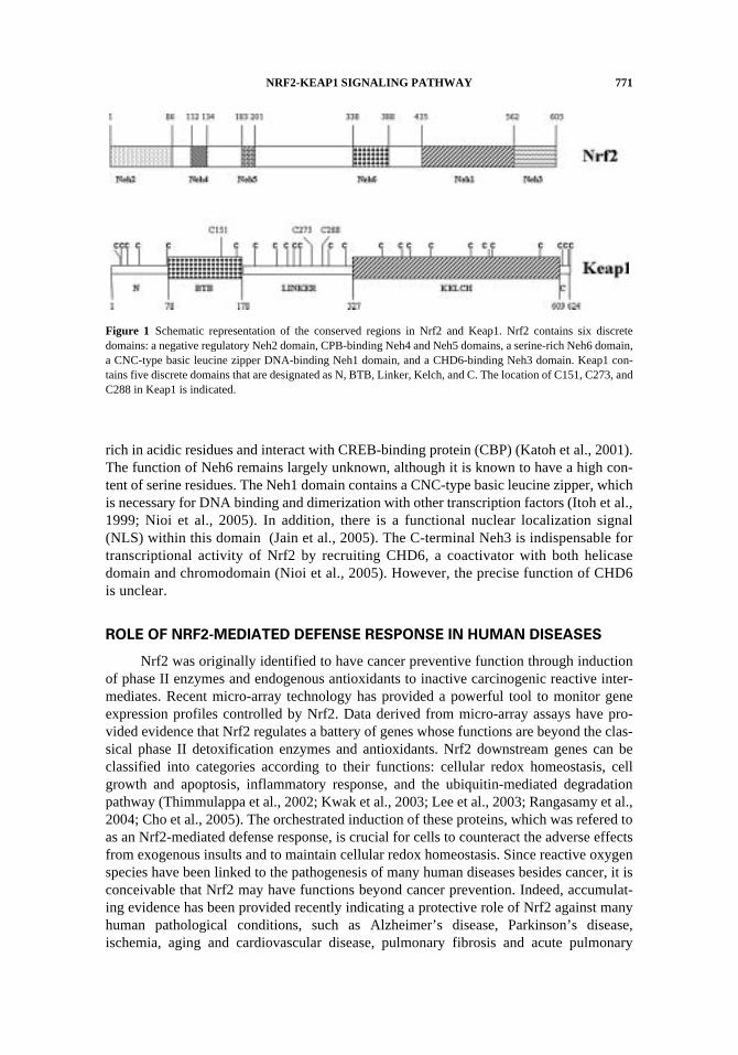
Several homologue domains were identified when different species of the Nrf2genes, such as human, mouse, and chicken, were aligned (Fig. 1). They are designated asNeh 1–6. The N-terminal Neh2 domain contains seven lysine residues for ubiquitin conju-gation, so it confers negative regulation of the Nrf2 activity through proteasome-mediateddegradation of Nrf2 (Zhang et al., 2004). It also binds to the Kelch domain of Keap1 (Itohet al., 1999). The Neh4 and Neh5 are two independent transactivation domains that are
NRF2-KEAP1 SIGNALING PATHWAY 771
rich in acidic residues and interact with CREB-binding protein (CBP) (Katoh et al., 2001).The function of Neh6 remains largely unknown, although it is known to have a high con-tent of serine residues. The Neh1 domain contains a CNC-type basic leucine zipper, whichis necessary for DNA binding and dimerization with other transcription factors (Itoh et al.,1999; Nioi et al., 2005). In addition, there is a functional nuclear localization signal(NLS) within this domain (Jain et al., 2005). The C-terminal Neh3 is indispensable fortranscriptional activity of Nrf2 by recruiting CHD6, a coactivator with both helicasedomain and chromodomain (Nioi et al., 2005). However, the precise function of CHD6is unclear.
ROLE OF NRF2-MEDIATED DEFENSE RESPONSE IN HUMAN DISEASES
Nrf2 was originally identified to have cancer preventive function through inductionof phase II enzymes and endogenous antioxidants to inactive carcinogenic reactive inter-mediates. Recent micro-array technology has provided a powerful tool to monitor geneexpression profiles controlled by Nrf2. Data derived from micro-array assays have pro-vided evidence that Nrf2 regulates a battery of genes whose functions are beyond the clas-sical phase II detoxification enzymes and antioxidants. Nrf2 downstream genes can beclassified into categories according to their functions: cellular redox homeostasis, cellgrowth and apoptosis, inflammatory response, and the ubiquitin-mediated degradationpathway (Thimmulappa et al., 2002; Kwak et al., 2003; Lee et al., 2003; Rangasamy et al.,2004; Cho et al., 2005). The orchestrated induction of these proteins, which was refered toas an Nrf2-mediated defense response, is crucial for cells to counteract the adverse effectsfrom exogenous insults and to maintain cellular redox homeostasis. Since reactive oxygenspecies have been linked to the pathogenesis of many human diseases besides cancer, it isconceivable that Nrf2 may have functions beyond cancer prevention. Indeed, accumulat-ing evidence has been provided recently indicating a protective role of Nrf2 against manyhuman pathological conditions, such as Alzheimer’s disease, Parkinson’s disease,ischemia, aging and cardiovascular disease, pulmonary fibrosis and acute pulmonary
Figure 1 Schematic representation of the conserved regions in Nrf2 and Keap1. Nrf2 contains six discretedomains: a negative regulatory Neh2 domain, CPB-binding Neh4 and Neh5 domains, a serine-rich Neh6 domain,a CNC-type basic leucine zipper DNA-binding Neh1 domain, and a CHD6-binding Neh3 domain. Keap1 con-tains five discrete domains that are designated as N, BTB, Linker, Kelch, and C. The location of C151, C273, andC288 in Keap1 is indicated.
772 D. D. ZHANG
injury, inflammation, emphysema, asthma, lupus-like autoimmune nephritis, and maculardegeneration (Aoki et al., 2001; Yoh et al., 2001; Braun et al., 2002; Cho et al., 2002; Choet al., 2004; Gao and Talalay, 2004; Suh et al., 2004; Ishii et al., 2005; Rangasamy et al.,2005; Shih et al., 2005a; Shih et al., 2005b).
KEAP1, A NEGATIVE REGULATOR OF NRF2
As discussed, Nrf2 is a critical factor regulating the cellular defense response whencells are under oxidative stress or are stimulated with chemopreventive compounds. Theactivity of Nrf2 is tightly regulated by a negative regulator named Keap1, which wascloned using the Neh2 domain of Nrf2 as bait in a yeast two-hybrid system by Yamamotoand coworkers (Itoh et al., 1999). Keap1 contains three major domains: an N-terminalBTB (broad complex, tramtrack, and bric-a-brac) domain, a linker region, and a C-terminal Kelch domain (Fig. 1). The N-terminal BTB domain was implicated inhomodimerization of the Keap1 protein (Zipper and Mulcahy, 2002). The linker region isa cysteine-rich domain that was proved to be indispensable for the activity of Keap1(Zhang and Hannink, 2003). The C-terminal Kelch domain contains six conserved Kelchrepeat sequences and binds to the Neh2 domain of Nrf2. Recently, the crystal structure ofthe Kelch domain has been solved and revealed a six β-propeller structure (Li et al., 2004;Padmanabhan et al., 2006).
Keap1 was initially described as a cytoplasmic factor that binds to actin cytoskele-ton and Nrf2 to retain Nrf2 in the cytoplasm. Upon exposure of cells to oxidative stress orchemopreventive compounds, Nrf2 dissociates from Keap1, translocates to the nucleus,forms a heterodimer with its obligatory partner Maf, and ultimately activates ARE-depen-dent gene expression. Recently, findings from our laboratory and others indicate thatKeap1 does not just passively sequester Nrf2 in the cytoplasm but plays an active role intargeting Nrf2 for ubiquitination and proteasomal degradation (Cullinan et al., 2004;Kobayashi et al., 2004; Zhang et al., 2004; Furukawa and Xiong, 2005). In addition, bothour in vivo data and the in vitro data from another group have challenged the Nrf2-Keap1dissociation model (Zhang et al., 2004; Eggler et al., 2005). Both ubiquitin-mediated deg-radation of Nrf2 and failure of dissociation of the Nrf2-Keap1 complex in response toNrf2-inducers will be discussed in the following sections.
SENSING MECHANISM OF KEAP1 TO OXIDATIVE STRESS OR
CHEMOPREVENTIVE COMPOUNDS
Identification of Keap1 as the key repressor for Nrf2 leads to the prevailing model thatone or more of the 27 cysteine residues in the human Keap1 protein are components of amolecular switch that is triggered by intracellular redox changes (Dinkova-Kostova et al.,2002). In support of this model, Nrf2-inducers with divergent structures share a commonability to react with protein thiols. Talalay and coworkers have demonstrated that four cys-teine residues in mouse Keap1, C257, C273, C288, C297, in the linker region of Keap1 reactwith a thiol-specific reagent, preferentially under in vitro conditions (Dinkova-Kostova et al.,2002). Consistent with this finding, Liebler and coworkers found strong correlation betweenthe ability of an electrophile to alkylate cysteine residues in the linker region of human Keap1and the potency of the electrophile to activate the Nrf2-Keap1 pathway in vitro (Hong et al.,2005). Interestingly, another in vitro study using human Keap1 and iodoacetamide identifiedC151, C288, and C297 as the most reactive cysteine residues (Eggler et al., 2005).
NRF2-KEAP1 SIGNALING PATHWAY 773
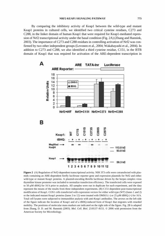
By comparing the inhibitory activity of Keap1 between the wild-type and mutantKeap1 proteins in cultured cells, we identified two critical cysteine residues, C273 andC288, in the linker domain of human Keap1 that were required for Keap1-mediated repres-sion of Nrf2 transcriptional activity under the basal condition (Fig. 2A) (Zhang and Hannink,2003). The importance of C273 and C288 residues in controlling activation of Nrf2 was con-firmed by two other independent groups (Levonen et al., 2004; Wakabayashi et al., 2004). Inaddition to C273 and C288, we also identified a third cysteine residue, C151, in the BTBdomain of Keap1 that was required for activation of the ARE-dependent transcription in
Figure 2 (A) Regulation of Nrf2-dependent transcriptional activity. NIH 3T3 cells were cotransfected with plas-mids containing an ARE-dependent firefly luciferase reporter gene and expression plasmids for Nrf2 and eitherwild-type or mutant Keap1 proteins. A plasmid-encoding Renilla luciferase driven by the herpes simplex virusthymidine kinase promoter was included to normalize transfection efficiency. The transfected cells were exposedto 50 μM tBHQ for 16 h prior to analysis. All samples were run in duplicate for each experiment, and the datarepresent the means of the results from three independent experiments. (B) C151-dependent post-transcriptionalmodification of Keap1. COS1 cells transfected with expression vectors for either wild-type (WT) (lanes 1 and 2)or the indicated mutant Keap1 proteins (lanes 3 to 12) were treated with DMSO (−) or 25 μM tBHQ (+) for 16 h.Total cell lysates were subjected to immunoblot analysis with anti-Keap1 antibodies. The arrows on the left sideof the figure indicate the location of Keap1 and of a tBHQ-induced form of Keap1 that migrates with retardedmobility. The positions of molecular mass markers are indicated on the right side of the figure. Fig. 2B is adaptedfrom Zhang, D. D. and M. Hannink (2003). Mol. Cell. Biol. 23:8137–8151. © 2006 with permission from theAmerican Society for Microbiology.
774 D. D. ZHANG
response to tBHQ (tert-butyhydroquinone) or sulforaphane treatment (Fig. 2A). Astonish-ingly, we also found that C151 in Keap1 is not only required for activation of Nrf2-depen-dent transcription, but also for a novel post-translational modification of Keap1 in cellsexposed to tBHQ (Zhang and Hannink, 2003) (Fig. 2B). As shown in Fig. 2B, the Keap1protein normally migrates on SDS-polyacrylamide gels with an apparent molecular size ofapproximately 60 kD, whereas the modified form of Keap1 has a size of approximately 130 kD.This modified form of Keap1 is stable to the reducing SDS-polyacrylamide gel electro-phoresis conditions, such as 10-min heating in the presence of 100 mM DTT. It is unlikelythat a disulfide bond is responsible for this modified form, although using a disulfide bond asa molecular switch is a very attractive model since disulfide linkage of Gpx3 and Yap1 hasbeen used in yeast as a redox sensor (Delaunay et al., 2002). In spite of the fact that theKeap1 protein contains a high number of cysteine residues (27 cysteine residues in humanKeap1), the modified form of Keap1 could not be an aggregation product because mutationof a single residue in Keap1, C151, completely abolished this modified form of Keap1 (Fig.2B). The modified form of Keap1 could be a covalently adducted protein from two differentproteins (an intermolecular adduct), an intramolecular adduct of Keap1, or two covalentlylinked Keap1 proteins (a dimeric form of Keap1). Formation of covalently adducted proteinsin response to chemical treatment has been reported previously. For example, quinol-thioet-her-derived protein adducts that are resistant to reducing conditions have been detected inthe kidneys of rats treated with hydroquinone or its derivatives (Kleiner et al., 1998). Thechemical nature of this modified form of Keap1 and the important of the modification inactivation of the Nrf2-Keap1 pathway are currently under investigation.
KEAP1: A SUBSTRATE ADAPTOR PROTEIN OF CUL3-CONTAINING E3
UBIQUTITIN LIGASE
Identification of the important roles of three cysteine mutants of Keap1 (C273, C288,and C151) in regulating the activity of Nrf2 has greatly speeded up our finding that Keap1facilitates the degradation of Nrf2. As shown in Table 1, ectopically expressed Nrf2 had ahalf-life of 2.7 h that was significantly reduced to 0.6 h when the wild-type Keap1 wascoexpressed. In contrast, the half-life of Nrf2 was 6.5 h in the presence of the mutantKeap1-C273S. Furthermore, sulforaphane treatment markedly increased the half-life ofNrf2 from 0.6 h to 2.5 h. Taken together, these data indicate that Keap1 speeds up the turn-over rate of the Nrf2 protein and sulforaphane is able to reverse the effect of Keap1 on Nrf2
Table 1 The regulation of Nrf2 half-life by Keap1 and sulforaphane.
Nrf2 Keap1 SF T1/2 (h)
+ – − 2.7+ – + 3.2+ WT − 0.6+ WT + 2.5+ C151S − 0.6+ C151S + 0.6+ C273S − 6.5
HA-Nrf2 was expressed in MDA-MB-231 cells in the absence or presence of Keap1 proteins. Cells wereeither left untreated or treated with 4 μM sulforaphane (SF) for 16 h prior to and during the pulse-chase experi-ment. The half-life of HA-Nrf2 was determined by pulse-chase labeling.
NRF2-KEAP1 SIGNALING PATHWAY 775
turnover. Immunoblot analysis was also performed to verify that the steady-state levels ofNrf2 were reduced in the presence of the wild-type Keap1 but increased if the Keap1-C273S was cotransfected. Sulforaphane or tBHQ treatment markedly enhanced the levelsof Nrf2. Collectively, these results led to our hypothesis that Keap1 is a novel E3 ubiquitinligase that is specifically inhibited by oxidative stress or electrophiles.
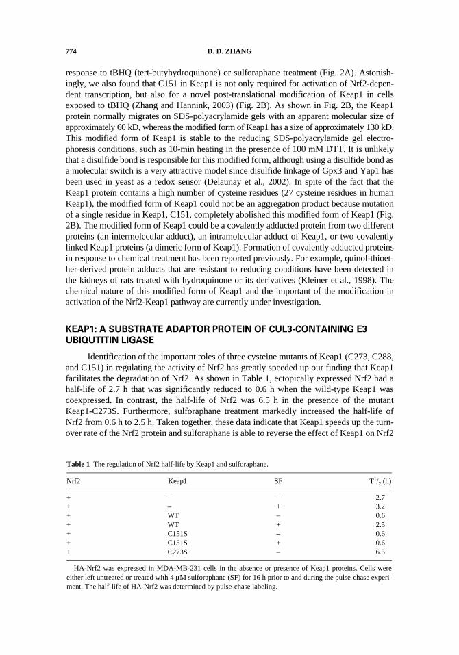
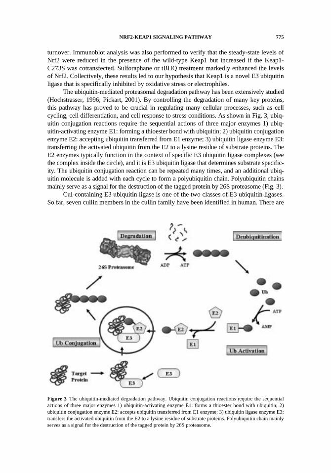
The ubiquitin-mediated proteasomal degradation pathway has been extensively studied(Hochstrasser, 1996; Pickart, 2001). By controlling the degradation of many key proteins,this pathway has proved to be crucial in regulating many cellular processes, such as cellcycling, cell differentiation, and cell response to stress conditions. As shown in Fig. 3, ubiq-uitin conjugation reactions require the sequential actions of three major enzymes 1) ubiq-uitin-activating enzyme E1: forming a thioester bond with ubiquitin; 2) ubiquitin conjugationenzyme E2: accepting ubiquitin transferred from E1 enzyme; 3) ubiquitin ligase enzyme E3:transferring the activated ubiquitin from the E2 to a lysine residue of substrate proteins. TheE2 enzymes typically function in the context of specific E3 ubiquitin ligase complexes (seethe complex inside the circle), and it is E3 ubiquitin ligase that determines substrate specific-ity. The ubiquitin conjugation reaction can be repeated many times, and an additional ubiq-uitin molecule is added with each cycle to form a polyubiquitin chain. Polyubiquitin chainsmainly serve as a signal for the destruction of the tagged protein by 26S proteasome (Fig. 3).
Cul-containing E3 ubiquitin ligase is one of the two classes of E3 ubiquitin ligases.So far, seven cullin members in the cullin family have been identified in human. There are
Figure 3 The ubiquitin-mediated degradation pathway. Ubiquitin conjugation reactions require the sequentialactions of three major enzymes 1) ubiquitin-activating enzyme E1: forms a thioester bond with ubiquitin; 2)ubiquitin conjugation enzyme E2: accepts ubiquitin transferred from E1 enzyme; 3) ubiquitin ligase enzyme E3:transfers the activated ubiquitin from the E2 to a lysine residue of substrate proteins. Polyubiquitin chain mainlyserves as a signal for the destruction of the tagged protein by 26S proteasome.
776 D. D. ZHANG
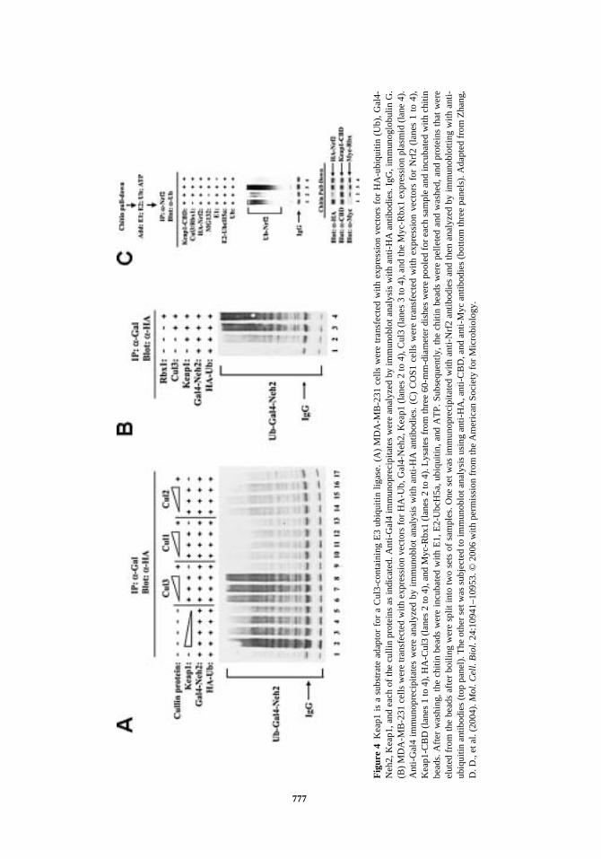
Cul1, Cul2, Cul3, Cul4A, Cul4B, Cul5, and Cul7. In the cullin-containing E3 ubiquitinligase, each of the cullin proteins serves as a binding platform for other subunits. Our firstapproach was to identify which cullin protein coexisted with Keap1 in an E3 ubiquitinligase. Using immunoprecipitation assay, we found that Keap1 associated strongly withCul3 and slightly with Cul2. Next, we performed an in vivo ubiquitination assay to test theability of Cul3 or Cul2 to facilitate ubiquitin addition onto the Neh2 domain of Nrf2, adomain with seven lysine residues that are the preferential targets for ubiquitin addition.Cul3, not Cul2, facilitated ubiquitination of Neh2 in a dose-dependent manner, indicatingthat Keap1 was in complex with Cul3, not Cul2 (Fig. 4A). We further verified the co-pres-ence of the Rbx1, Cul3, and Nrf2 in the complex immunoprecipitated with an anti-Keap1antibody. Furthermore, the functional importance of Rbx1, Cul3, and Keap1 in the facili-tation of the ubiquitination of Nrf2 was confirmed in both in vivo and in vitro ubiquitina-tion assays (Fig. 4B and 4C). These results provide clear evidence that Keap1 does not justpassively sequester Nrf2 in the cytoplasm to block Nrf2 nuclear translocation, as origi-nally proposed, but actively targets Nrf2 for ubiquitination and proteasomal degradationby functioning as a component of an E3 ubiquitin ligase (Zhang et al., 2004). The sameconclusion was reached by three other groups. All identified Keap1 as a substrate adaptorprotein for a Cul3-containing E3 ubiquitin ligase (Cullinan et al., 2004; Kobayashi et al.,2004; Furukawa and Xiong, 2005).
TBHQ OR SULFORAPHANE INHIBITS THE ACTIVITY OF KEAP1-CUL3 E3
UBIQUITIN LIGASE
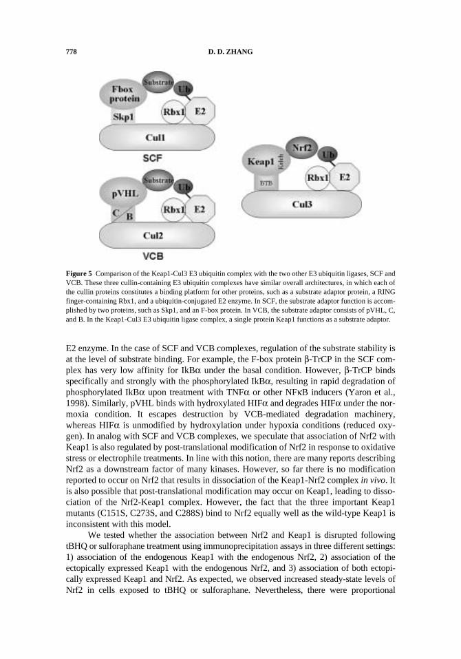
In addition to the finding that Keap1 is one of more than 100 E3 ubiquitin ligases,we went further to investigate how the Keap1-containing E3 ubiquitin ligase is regulated.First, we compared the Keap1-Cul3 E3 ubiquitin ligase with the two other well-studiedCul1- and Cul2-containing E3 ubiquitin ligases (Fig. 5). SCF (Skp1-Cullin-F-box protein)is the best studied class of E3 ubiquitin ligases (Zheng et al., 2002). In this complex, the F-box protein and Skp1 together serve as a substrate adaptor that brings in a specific sub-strate for ubiquitin conjugation. Notably, the cullin-containing E3 ubiquitin ligases aredynamically undergoing assembly and disassembly under the tight control of other twofamilies of proteins named CAND (also called Tip120A) and CSN (COP9 signalosome)(Wei and Deng, 2003). However, the common core complex, consisting of cullin andRbx1, remains assembled and is shared by different substrate adaptor proteins to facilitateubiquitination and degradation of different substrates. The advantage of using the samecore complex for a variety of substrate adaptor proteins is to allow rapid degradation ofdiverse factors, according to the changes of intracellular environment, without de novoassemble of the entire E3 ubiquitin ligase complex. The F-box proteins in the SCF com-plex represent a large family, and each member can have several specific substrates. SCFregulates many important factors, including cyclin-dependent kinase inhibitors, cyclins,IkBα, and β-catenin. VCB (von Hippel-Lindau protein–elonginC-elonginB) is anothercullin-containing E3 ubiquitin ligase that is responsible for degradation of HIFα (hypoxia-inducible transcription factor) under the normoxia (normal amount of oxygen) condition.In the VCB complex, the substrate adaptor function is accomplished by three proteins,pVHL, elonginC, and elonginB (Kamura et al., 1999) (Fig. 5). It is obvious that these threecullin-containing E3 ubiquitin complexes, SCF, VCB, and Keap1-Cul3, have similar overallarchitectures, in which the cullin protein constitutes a binding platform for other proteins, suchas a substrate adaptor protein, a RING finger-containing Rbx1, and a ubiquitin-conjugated
777
Fig
ure
4K
eap1
is
a su
bstr
ate
adap
tor
for
a C
ul3-
cont
aini
ng E
3 ub
iqui
tin l
igas
e. (
A)
MD
A-M
B-2
31 c
ells
wer
e tr
ansf
ecte
d w
ith e
xpre
ssio
n ve
ctor
s fo
r H
A-u
biqu
itin
(Ub)
, G
al4-
Neh
2, K
eap1
, and
eac
h of
the
culli
n pr
otei
ns a
s in
dica
ted.
Ant
i-G
al4
imm
unop
reci
pita
tes
wer
e an
alyz
ed b
y im
mun
oblo
t ana
lysi
s w
ith a
nti-
HA
ant
ibod
ies.
IgG
, im
mun
oglo
buli
n G
.(B
) M
DA
-MB
-231
cel
ls w
ere
tran
sfec
ted
with
exp
ress
ion
vect
ors
for
HA
-Ub,
Gal
4-N
eh2,
Kea
p1 (
lane
s 2
to 4
), C
ul3
(lan
es 3
to 4
), a
nd th
e M
yc-R
bx1
expr
essi
on p
lasm
id (
lane
4).
Ant
i-G
al4
imm
unop
reci
pita
tes
wer
e an
alyz
ed b
y im
mun
oblo
t an
alys
is w
ith a
nti-
HA
ant
ibod
ies.
(C
) C
OS1
cel
ls w
ere
tran
sfec
ted
with
exp
ress
ion
vect
ors
for
Nrf
2 (l
anes
1 t
o 4)
,K
eap1
-CB
D (
lane
s 1
to 4
), H
A-C
ul3
(lan
es 2
to 4
), a
nd M
yc-R
bx1
(lan
es 2
to 4
). L
ysat
es f
rom
thre
e 60
-mm
-dia
met
er d
ishe
s w
ere
pool
ed f
or e
ach
sam
ple
and
incu
bate
d w
ith c
hitin
bead
s. A
fter
was
hing
, the
chi
tin b
eads
wer
e in
cuba
ted
with
E1,
E2-
Ubc
H5a
, ubi
quiti
n, a
nd A
TP.
Sub
sequ
ently
, the
chi
tin b
eads
wer
e pe
llete
d an
d w
ashe
d, a
nd p
rote
ins
that
wer
eel
uted
fro
m th
e be
ads
afte
r bo
iling
wer
e sp
lit in
to tw
o se
ts o
f sa
mpl
es. O
ne s
et w
as im
mun
opre
cipi
tate
d w
ith a
nti-
Nrf
2 an
tibod
ies
and
then
ana
lyze
d by
imm
unob
lotti
ng w
ith a
nti-
ubiq
uitin
ant
ibod
ies
(top
pan
el).
The
oth
er s
et w
as s
ubje
cted
to im
mun
oblo
t ana
lysi
s us
ing
anti-
HA
, ant
i-C
BD
, and
ant
i-M
yc a
ntib
odie
s (b
otto
m th
ree
pane
ls).
Ada
pted
fro
m Z
hang
,D
. D.,
et a
l. (2
004)
. Mol
. Cel
l. B
iol.
24:1
0941
–109
53. ©
200
6 w
ith
perm
issi
on f
rom
the
Am
eric
an S
ocie
ty f
or M
icro
biol
ogy.
778 D. D. ZHANG
E2 enzyme. In the case of SCF and VCB complexes, regulation of the substrate stability isat the level of substrate binding. For example, the F-box protein β-TrCP in the SCF com-plex has very low affinity for IkBα under the basal condition. However, β-TrCP bindsspecifically and strongly with the phosphorylated IkBα, resulting in rapid degradation ofphosphorylated IkBα upon treatment with TNFα or other NFκB inducers (Yaron et al.,1998). Similarly, pVHL binds with hydroxylated HIFα and degrades HIFα under the nor-moxia condition. It escapes destruction by VCB-mediated degradation machinery,whereas HIFα is unmodified by hydroxylation under hypoxia conditions (reduced oxy-gen). In analog with SCF and VCB complexes, we speculate that association of Nrf2 withKeap1 is also regulated by post-translational modification of Nrf2 in response to oxidativestress or electrophile treatments. In line with this notion, there are many reports describingNrf2 as a downstream factor of many kinases. However, so far there is no modificationreported to occur on Nrf2 that results in dissociation of the Keap1-Nrf2 complex in vivo. Itis also possible that post-translational modification may occur on Keap1, leading to disso-ciation of the Nrf2-Keap1 complex. However, the fact that the three important Keap1mutants (C151S, C273S, and C288S) bind to Nrf2 equally well as the wild-type Keap1 isinconsistent with this model.
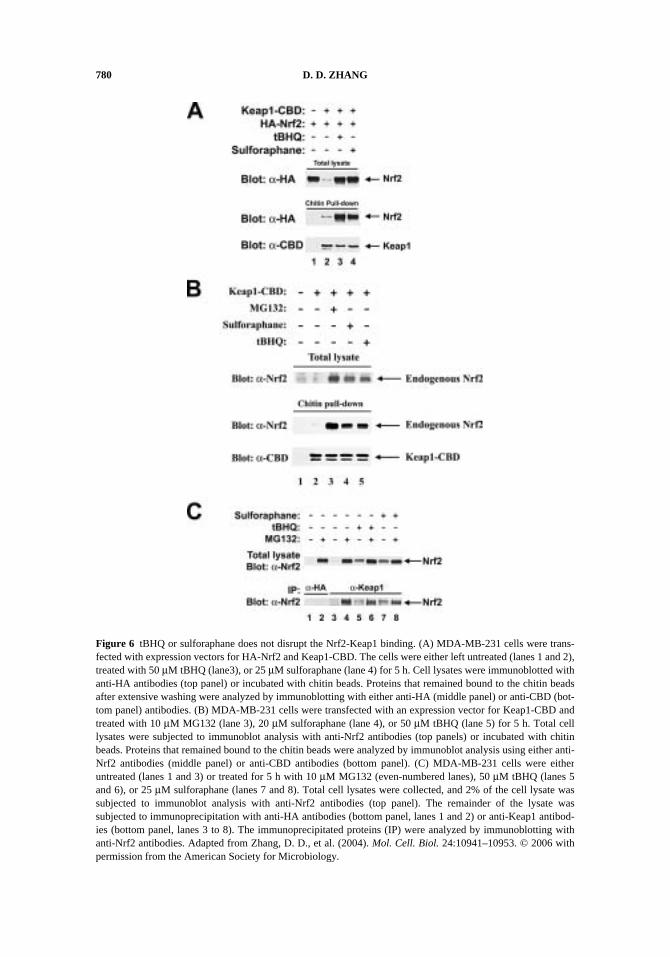
We tested whether the association between Nrf2 and Keap1 is disrupted followingtBHQ or sulforaphane treatment using immunoprecipitation assays in three different settings:1) association of the endogenous Keap1 with the endogenous Nrf2, 2) association of theectopically expressed Keap1 with the endogenous Nrf2, and 3) association of both ectopi-cally expressed Keap1 and Nrf2. As expected, we observed increased steady-state levels ofNrf2 in cells exposed to tBHQ or sulforaphane. Nevertheless, there were proportional
Figure 5 Comparison of the Keap1-Cul3 E3 ubiquitin complex with the two other E3 ubiquitin ligases, SCF andVCB. These three cullin-containing E3 ubiquitin complexes have similar overall architectures, in which each ofthe cullin proteins constitutes a binding platform for other proteins, such as a substrate adaptor protein, a RINGfinger-containing Rbx1, and a ubiquitin-conjugated E2 enzyme. In SCF, the substrate adaptor function is accom-plished by two proteins, such as Skp1, and an F-box protein. In VCB, the substrate adaptor consists of pVHL, C,and B. In the Keap1-Cul3 E3 ubiquitin ligase complex, a single protein Keap1 functions as a substrate adaptor.
NRF2-KEAP1 SIGNALING PATHWAY 779
increases in Nrf2 levels in Keap1 immunoprecipitates from tBHQ- or sulforaphane-exposedsamples, indicating that neither tBHQ nor sulforaphane is able to disrupt the Nrf2-Keap1complex (Fig. 6). The same conclusion was drawn from all three experimental settings. Ourfinding demonstrates that the original Nrf2-Keap1 dissociation model is likely incorrect. Inan in vitro system with purified Keap1 and Neh2 proteins, Mesecar’s group also demon-strated that Nrf2-inducers did not disrupt the Nrf2-Keap1 complex (Eggler et al., 2005).These results may imply that, unlike the SCF and VCB complexes, regulation of Nrf2 maynot be rendered at the level of substrate binding with the Cul3-containing E3 ubiquitin ligase.
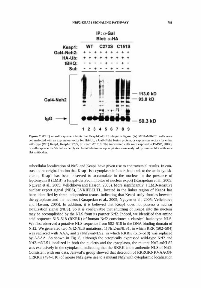
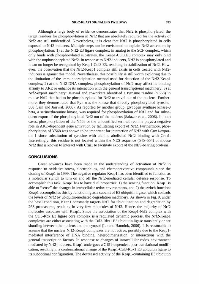
The important question then is how does tBHQ or sulforaphane block degradationof Nrf2? By performing in vivo ubiquitination assay in cells treated with tBHQ or sul-foraphane, we found that ubiquitination was significantly blocked by tBHQ or sul-foraphane treatment. Furthermore, co-transfection of Keap1-C273S or Keap1-C288S alsorepressed ubiquitination of Nrf2 (Fig. 7). Interestingly, tBHQ or sulforaphane was nolonger effective in blockage of Nrf2 ubiquitination in the presence of Keap1-C151S (Fig. 7).Taken together, these results unambiguously demonstrate that tBHQ or sulforaphane acti-vate ARE-dependent transcription primarily by inhibiting the activity of Keap1-Cul3ubiquitin ligase, resulting in decreased Nrf2 ubiquitination, enhanced Nrf2 stability, ele-vated Nrf2 levels, and, ultimately, activation of ARE-dependent gene transcription. Insupport of our notion that Nrf2 inducers target the Keap1-containing E3 ubiquitin ligaserather than the Nrf2-Keap1 complex, Yamamoto and coworkers recently reported thesame finding, i.e., tBHQ did not disrupt the Nrf2-Keap1 complex; rather, it blocked ubiq-uitination of Nrf2 (Kobayashi et al., 2006). However, the precise mechanism by whichtBHQ or sulforaphane inhibits the Keap1-Cul3 E3 ubiquitin ligase is still unclear.Although speculative at this time, it is conceivable that C-151-dependent post-trans-lational modification of Keap1 in response to tBHQ or sulforaphane treatment causes aconformational change of Keap1-containing E3 ubiquitin ligase, resulting in reducedactivity of the Keap1-Cul3-Rbx1 E3 ubiquitin ligase. The next challenge is to explain howNrf2 translocates into the nucleus if Nrf2-inducers do not dissociate the Nrf2-Keap1 com-plex. Yamamoto and coworkers have proposed that it is the newly synthesized Nrf2 pro-teins that translocate into the nucleus by showing the reduced nuclear Nrf2 levels in cellstreated with both tBHQ and the protein synthesis inhibitor cycloheximide (CHX), com-pared to the Nrf2 levels in cells treated with tBHQ alone (Kobayashi et al., 2006). How-ever, the caveat of this experiment is that CHX treatment should certainly reduce the totalamount of Nrf2 proteins, resulting in proportional reduction of Nrf2 in the nucleus. Fur-thermore, it is hard to explain how Keap1 can distinguish the “old” vs. the “new” Nrf2molecules. We would like to use a saturation model: Under the basal conditions, there aresignificant amounts of Keap1 and very few molecules of Nrf2, due to proteasomaldegradation of Nrf2. Upon treatment of tBHQ or sulforaphane, the levels of Nrf2 aremarkedly increased by blockage of proteasomal degradation of Nrf2. The amount of Nrf2overwhelms the binding capacity of Keap1, resulting in existence of free Nrf2 moleculesthat activate the transcription of ARE-dependent genes. Our model of Nrf2 activation willbe further discussed in the conclusion section.
MULTI-LEVELS OF NRF2 REGULATION
Although Keap1-mediated ubiquitination and degradation of Nrf2 are evidentlymajor controls of Nrf2 activity, Nrf2 is also subjected to additional modes of regulation,including subcellular localization and post-translational modification. Studies investigating
780 D. D. ZHANG
Figure 6 tBHQ or sulforaphane does not disrupt the Nrf2-Keap1 binding. (A) MDA-MB-231 cells were trans-fected with expression vectors for HA-Nrf2 and Keap1-CBD. The cells were either left untreated (lanes 1 and 2),treated with 50 μM tBHQ (lane3), or 25 μM sulforaphane (lane 4) for 5 h. Cell lysates were immunoblotted withanti-HA antibodies (top panel) or incubated with chitin beads. Proteins that remained bound to the chitin beadsafter extensive washing were analyzed by immunoblotting with either anti-HA (middle panel) or anti-CBD (bot-tom panel) antibodies. (B) MDA-MB-231 cells were transfected with an expression vector for Keap1-CBD andtreated with 10 μM MG132 (lane 3), 20 μM sulforaphane (lane 4), or 50 μM tBHQ (lane 5) for 5 h. Total celllysates were subjected to immunoblot analysis with anti-Nrf2 antibodies (top panels) or incubated with chitinbeads. Proteins that remained bound to the chitin beads were analyzed by immunoblot analysis using either anti-Nrf2 antibodies (middle panel) or anti-CBD antibodies (bottom panel). (C) MDA-MB-231 cells were eitheruntreated (lanes 1 and 3) or treated for 5 h with 10 μM MG132 (even-numbered lanes), 50 μM tBHQ (lanes 5and 6), or 25 μM sulforaphane (lanes 7 and 8). Total cell lysates were collected, and 2% of the cell lysate wassubjected to immunoblot analysis with anti-Nrf2 antibodies (top panel). The remainder of the lysate wassubjected to immunoprecipitation with anti-HA antibodies (bottom panel, lanes 1 and 2) or anti-Keap1 antibod-ies (bottom panel, lanes 3 to 8). The immunoprecipitated proteins (IP) were analyzed by immunoblotting withanti-Nrf2 antibodies. Adapted from Zhang, D. D., et al. (2004). Mol. Cell. Biol. 24:10941–10953. © 2006 withpermission from the American Society for Microbiology.
NRF2-KEAP1 SIGNALING PATHWAY 781
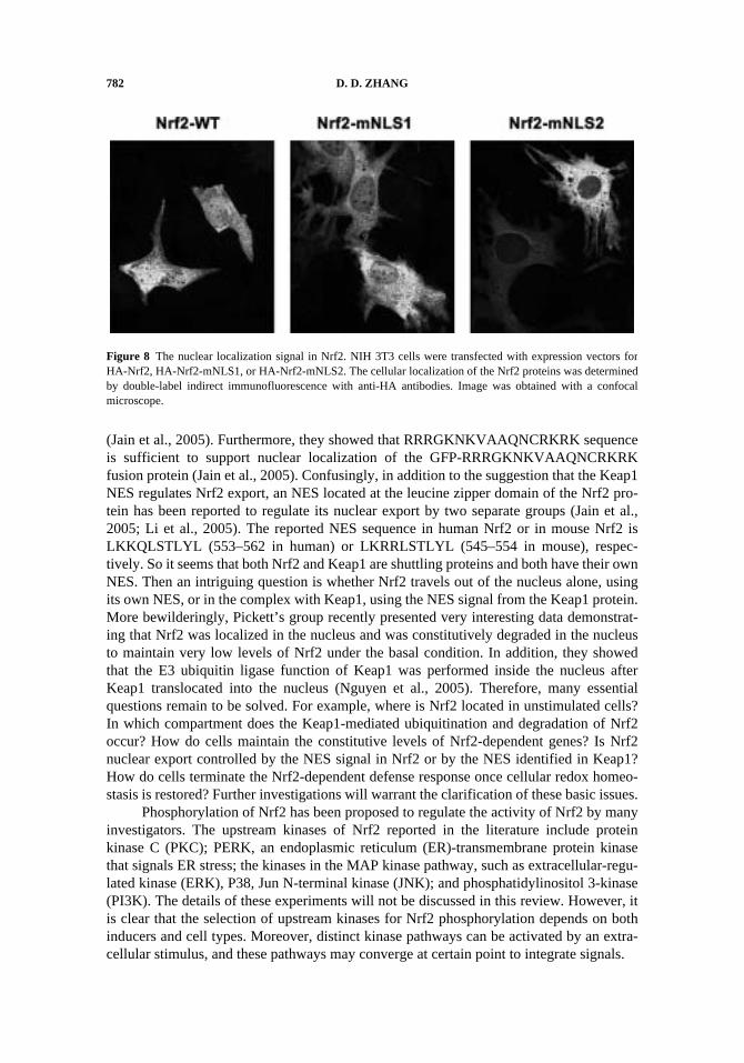
subcellular localization of Nrf2 and Keap1 have given rise to controversial results. In con-trast to the original notion that Keap1 is a cytoplasmic factor that binds to the actin cytosk-eleton, Keap1 has been observed to accumulate in the nucleus in the presence ofleptomycin B (LMB), a fungal-derived inhibitor of nuclear export (Karapetian et al., 2005;Nguyen et al., 2005; Velichkova and Hasson, 2005). More significantly, a LMB-sensitivenuclear export signal (NES), LVKIFEELTL, located in the linker region of Keap1 hasbeen identified by three independent teams, indicating that Keap1 truly shuttles betweenthe cytoplasm and the nucleus (Karapetian et al., 2005; Nguyen et al., 2005; Velichkovaand Hasson, 2005). In addition, it is believed that Keap1 does not possess a nuclearlocalization signal (NLS). So it is conceivable that shuttling of Keap1 into the nucleusmay be accomplished by the NLS from its partner Nrf2. Indeed, we identified that aminoacid sequence 515–518 (RKRK) of human Nrf2 constitutes a classical basic-type NLS.We first observed a putative NLS sequence from 502–518 in the DNA binding domain ofNrf2. We generated two Nrf2-NLS mutations: 1) Nrf2-mNLS1, in which RRR (502–504)was replaced with AAA, and 2) Nrf2-mNLS2, in which RKRK (515–518) was replacedby AAAA. As shown in Fig. 8, although the ectopically expressed wild-type Nrf2 andNrf2-mNLS1 localized in both the nucleus and the cytoplasm, the mutant Nrf2-mNLS2was exclusively in the cytoplasm, indicating that the RKRK is the authentic NLS of Nrf2.Consistent with our data, Jaiswal’s group showed that detection of RRRGKNKVAAQN-CRKRK (494–510) of mouse Nrf2 gave rise to a mutant Nrf2 with cytoplasmic localization
Figure 7 tBHQ or sulforaphane inhibits the Keap1-Cul3 E3 ubiquitin ligase. (A) MDA-MB-231 cells werecotransfected with an expression vector for HA-Ub, a Gal4-Neh2 fusion protein, or expression vectors for eitherwild-type (WT) Keap1, Keap1-C273S, or Keap1-C151S. The transfected cells were exposed to DMSO, tBHQ,or sulforaphane for 5 h before cell lysis. Anti-Gal4 immunoprecipitates were analyzed by immunoblot with anti-HA antibodies.
782 D. D. ZHANG
(Jain et al., 2005). Furthermore, they showed that RRRGKNKVAAQNCRKRK sequenceis sufficient to support nuclear localization of the GFP-RRRGKNKVAAQNCRKRKfusion protein (Jain et al., 2005). Confusingly, in addition to the suggestion that the Keap1NES regulates Nrf2 export, an NES located at the leucine zipper domain of the Nrf2 pro-tein has been reported to regulate its nuclear export by two separate groups (Jain et al.,2005; Li et al., 2005). The reported NES sequence in human Nrf2 or in mouse Nrf2 isLKKQLSTLYL (553–562 in human) or LKRRLSTLYL (545–554 in mouse), respec-tively. So it seems that both Nrf2 and Keap1 are shuttling proteins and both have their ownNES. Then an intriguing question is whether Nrf2 travels out of the nucleus alone, usingits own NES, or in the complex with Keap1, using the NES signal from the Keap1 protein.More bewilderingly, Pickett’s group recently presented very interesting data demonstrat-ing that Nrf2 was localized in the nucleus and was constitutively degraded in the nucleusto maintain very low levels of Nrf2 under the basal condition. In addition, they showedthat the E3 ubiquitin ligase function of Keap1 was performed inside the nucleus afterKeap1 translocated into the nucleus (Nguyen et al., 2005). Therefore, many essentialquestions remain to be solved. For example, where is Nrf2 located in unstimulated cells?In which compartment does the Keap1-mediated ubiquitination and degradation of Nrf2occur? How do cells maintain the constitutive levels of Nrf2-dependent genes? Is Nrf2nuclear export controlled by the NES signal in Nrf2 or by the NES identified in Keap1?How do cells terminate the Nrf2-dependent defense response once cellular redox homeo-stasis is restored? Further investigations will warrant the clarification of these basic issues.
Phosphorylation of Nrf2 has been proposed to regulate the activity of Nrf2 by manyinvestigators. The upstream kinases of Nrf2 reported in the literature include proteinkinase C (PKC); PERK, an endoplasmic reticulum (ER)-transmembrane protein kinasethat signals ER stress; the kinases in the MAP kinase pathway, such as extracellular-regu-lated kinase (ERK), P38, Jun N-terminal kinase (JNK); and phosphatidylinositol 3-kinase(PI3K). The details of these experiments will not be discussed in this review. However, itis clear that the selection of upstream kinases for Nrf2 phosphorylation depends on bothinducers and cell types. Moreover, distinct kinase pathways can be activated by an extra-cellular stimulus, and these pathways may converge at certain point to integrate signals.
Figure 8 The nuclear localization signal in Nrf2. NIH 3T3 cells were transfected with expression vectors forHA-Nrf2, HA-Nrf2-mNLS1, or HA-Nrf2-mNLS2. The cellular localization of the Nrf2 proteins was determinedby double-label indirect immunofluorescence with anti-HA antibodies. Image was obtained with a confocalmicroscope.
NRF2-KEAP1 SIGNALING PATHWAY 783
Although a large body of evidence demonstrates that Nrf2 is phosphorylated, thetarget residues for phosphorylation in Nrf2 that are absolutely required for the activity ofNrf2 are still unidentified. Nevertheless, it is clear that Nrf2 is phosphorylated in cellsexposed to Nrf2-inducers. Multiple steps can be envisioned to explain Nrf2 activation byphosphorylation: 1) at the Nrf2-E3 ligase complex: in analog to the SCF complex, whichonly binds with phosphorylated substrates, the Keap1-Cul3 E3 complex may only bindwith the unphosphorylated Nrf2. In response to Nrf2-inducers, Nrf2 is phosphorylated andit can no longer be recognized by Keap1-Cul3 E3, resulting in stabilization of Nrf2. How-ever, the observation that the Nrf2-Keap1 complex still exists in cells treated with Nrf2-inducers is against this model. Nevertheless, this possibility is still worth exploring due tothe limitation of the immunoprecipitation method used for detection of the Nrf2-Keap1complex; 2) at the Nrf2-DNA complex: phosphorylation of Nrf2 may affect its bindingaffinity to ARE or enhance its interaction with the general transcriptional machinery; 3) atNrf2-export machinery: Jaiswal and coworkers identified a tyrosine residue (Y568) inmouse Nrf2 that had to be phosphorylated for Nrf2 to travel out of the nucleus. Further-more, they demonstrated that Fyn was the kinase that directly phosphorylated tyrosine-568 (Jain and Jaiswal, 2006). As reported by another group, glycogen synthase kinase-3beta, a serine/threonine kinase, was required for phosphorylation of Nrf2 and for subse-quent export of the phosphorylated Nrf2 out of the nucleus (Salazar et al., 2006). In bothcases, phosphorylation of the Y568 or the unidentified serine/threonine plays a negativerole in ARE-dependent gene activation by facilitating export of Nrf2. Furthermore, phos-phorylation of Y568 was shown to be important for interaction of Nrf2 with Crm1/expor-tin 1 since substitution of tyrosine with alanine abolished Nrf2 binding with Crm1.Interestingly, this residue is not located within the NES sequence (545–554) of mouseNrf2 that is known to interact with Crm1 to facilitate export of the NES-bearing proteins.
CONCLUSIONS
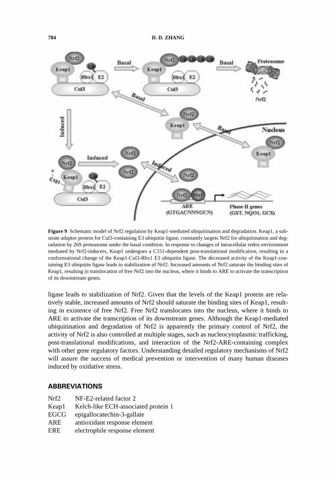
Great advances have been made in the understanding of activation of Nrf2 inresponse to oxidative stress, electrophiles, and chemopreventive compounds since thecloning of Keap1 in 1999. The negative regulator Keap1 has been identified to function asa molecular switch to turn on and off the Nrf2-mediated cellular defense response. Toaccomplish this task, Keap1 has to have dual properties: 1) the sensing function: Keap1 isable to “sense” the changes in intracellular redox environments, and 2) the switch function:Keap1 accomplishes this by functioning as a subunit of E3 ubiquitin ligase, which controlsthe levels of Nrf2 by ubiquitin-mediated degradation machinery. As shown in Fig. 9, underthe basal condition, Keap1 constantly targets Nrf2 for ubiquitination and degradation by26S proteasome, resulting in very few molecules of Nrf2. Hence, the majority of Nrf2molecules associate with Keap1. Since the association of the Keap1-Nrf2 complex withthe Cul3-Rbx E3 ligase core complex is a regulated dynamic process, the Nrf2-Keap1complexes are either associating with the Cul3-Rbx1 E3 ubiquitin ligase transiently or areshuttling between the nucleus and the cytosol (Lo and Hannink, 2006). It is reasonable toassume that the nuclear Nrf2-Keap1 complexes are not active, possibly due to the Keap1-mediated interference of DNA binding, heterodimerization, or interactions with thegeneral transcription factors. In response to changes of intracellular redox environmentmediated by Nrf2-inducers, Keap1 undergoes a C151-dependent post-translational modifi-cation, resulting in a conformational change of the Keap1-Cul3-Rbx1 E3 ubiquitin ligase toits suboptimal configuration. The decreased activity of the Keap1-containing E3 ubiquitin
784 D. D. ZHANG
ligase leads to stabilization of Nrf2. Given that the levels of the Keap1 protein are rela-tively stable, increased amounts of Nrf2 should saturate the binding sites of Keap1, result-ing in existence of free Nrf2. Free Nrf2 translocates into the nucleus, where it binds toARE to activate the transcription of its downstream genes. Although the Keap1-mediatedubiquitination and degradation of Nrf2 is apparently the primary control of Nrf2, theactivity of Nrf2 is also controlled at multiple stages, such as nucleocytoplasmic trafficking,post-translational modifications, and interaction of the Nrf2-ARE-containing complexwith other gene regulatory factors. Understanding detailed regulatory mechanisms of Nrf2will assure the success of medical prevention or intervention of many human diseasesinduced by oxidative stress.
ABBREVIATIONS
Nrf2 NF-E2-related factor 2Keap1 Kelch-like ECH-associated protein 1EGCG epigallocatechin-3-gallateARE antioxidant response elementERE electrophile response element
Figure 9 Schematic model of Nrf2 regulation by Keap1-mediated ubiquitination and degradation. Keap1, a sub-strate adaptor protein for Cul3-containing E3 ubiquitin ligase, constantly targets Nrf2 for ubiquitination and deg-radation by 26S proteasome under the basal condition. In response to changes of intracellular redox environmentmediated by Nrf2-inducers, Keap1 undergoes a C151-dependent post-translational modification, resulting in aconformational change of the Keap1-Cul3-Rbx1 E3 ubiquitin ligase. The decreased activity of the Keap1-con-taining E3 ubiquitin ligase leads to stabilization of Nrf2. Increased amounts of Nrf2 saturate the binding sites ofKeap1, resulting in translocation of free Nrf2 into the nucleus, where it binds to ARE to activate the transcriptionof its downstream genes.
NRF2-KEAP1 SIGNALING PATHWAY 785
GST glutathione S-transferaseNQO1 NAD(P)H quinone oxidoreductaseγGCS γ-glutamylcysteine synthetaseNF-E2 nuclear factor (erythroid-derived 2)cnc “cap ‘n’ collar”Neh Nrf2-Ech homologyCBP CREB-binding proteinBTB broad complex, tramtrack and bric-a-bracNLS nuclear localization signalNES nuclear export signaltBHQ tert-butylhydroquinoneCul cullinRbx1 ring-box protein 1SCF Skp1-Cullin-F-box proteinpVHL the von Hippel-Lindau tumor suppressor proteinVCB pVHL, elongins C and BCAND cullin-associated and neddylation-dissociatedCSN COP9 signalosomePKC protein kinase CERK extracellular-regulated kinaseJNK Jun N-terminal KinasePI3K Phosphatidylinositol 3-kinasse
ACKNOWLEDGMENTS
I thank Dr. Terrence J. Monks for critical review of this manuscript.
REFERENCES
Aoki, Y., Sato, H., Nishimura, N., Takahashi, S., Itoh, K., Yamamoto, M. (2001). Accelerated DNAadduct formation in the lung of the Nrf2 knockout mouse exposed to diesel exhaust. Toxicol.Appl. Pharmacol. 173:154–160.
Braun, S., Hanselmann, C., Gassmann, M. G., auf dem Keller, U., Born-Berclaz, C., Chan, K., Kan,Y. W., Werner, S. (2002). Nrf2 transcription factor, a novel target of keratinocyte growthfactor action which regulates gene expression and inflammation in the healing skin wound.Mol. Cell. Biol. 22:5492–5505.
Chan, J. Y., Kwong, M., Lu, R., Chang, J., Wang, B., Yen, T. S., Kan, Y. W. (1998). Targeted dis-ruption of the ubiquitous CNC-bZIP transcription factor, Nrf-1, results in anemia and embry-onic lethality in mice. Embo. J. 17:1779–1787.
Chan, K., Han, X. D., Kan, Y. W. (2001). An important function of Nrf2 in combating oxidativestress: detoxification of acetaminophen. Proc. Natl. Acad. Sci. USA 98:4611–4616.
Chan, K., Lu, R., Chang, J. C., Kan, Y. W. (1996). NRF2, a member of the NFE2 family of tran-scription factors, is not essential for murine erythropoiesis, growth, and development. Proc.Natl. Acad. Sci. USA 93:13943–13948.
Chanas, S. A., Jiang, Q., McMahon, M., McWalter, G. K., McLellan, L. I., Elcombe, C. R., Henderson,C. J., Wolf, C. R., Moffat, G. J., Itoh, K., Yamamoto, M., Hayes, J. D. (2002). Loss of theNrf2 transcription factor causes a marked reduction in constitutive and inducible expressionof the glutathione S-transferase Gsta1, Gsta2, Gstm1, Gstm2, Gstm3 and Gstm4 genes in thelivers of male and female mice. Biochem. J. 365:405–416.
786 D. D. ZHANG
Cho, H. Y., Jedlicka, A. E., Reddy, S. P., Kensler, T. W., Yamamoto, M., Zhang, L. Y., Kleeberger,S. R. (2002). Role of NRF2 in protection against hyperoxic lung injury in mice. Am. J. Respir.Cell. Mol. Biol. 26:175–182.
Cho, H. Y., Reddy, S. P., Debiase, A., Yamamoto, M., Kleeberger, S. R. (2005). Gene expressionprofiling of NRF2-mediated protection against oxidative injury. Free Radic. Biol. Med.38:325–343.
Cho, H. Y., Reddy, S. P., Yamamoto, M., Kleeberger, S. R. (2004). The transcription factor NRF2protects against pulmonary fibrosis. Faseb. J. 18:1258–1260.
Cullinan, S. B., Gordan, J. D., Jin, J., Harper, J. W., Diehl, J. A. (2004). The Keap1-BTB protein isan adaptor that bridges Nrf2 to a Cul3-based E3 ligase: oxidative stress sensing by a Cul3-Keap1 ligase. Mol. Cell. Biol. 24:8477–8486.
Delaunay, A., Pflieger, D., Barrault, M. B., Vinh, J., Toledano, M. B. (2002). A thiol peroxidase isan H2O2 receptor and redox-transducer in gene activation. Cell 111:471–481.
Derjuga, A. Gourley, T. S., Holm, T. M., Heng, H. H., Shivdasani, R. A., Ahmed, R., Andrews, N. C.,Blank, V. (2004). Complexity of CNC transcription factors as revealed by gene targeting ofthe Nrf3 locus. Mol. Cell. Biol. 24:3286–3294.
Dinkova-Kostova, A. T., Holtzclaw, W. D., Cole, R. N., Itoh, K., Wakabayashi, N., Katoh, Y.,Yamamoto, M., Talalay, P. (2002). Direct evidence that sulfhydryl groups of Keap1 are thesensors regulating induction of phase 2 enzymes that protect against carcinogens and oxi-dants. Proc. Natl. Acad. Sci. USA 99:11908–11913.
Dinkova-Kostova, A. T., Massiah, M. A., Bozak, R. E., Hicks, R. J., Talalay, P. (2001).Potency of Michael reaction acceptors as inducers of enzymes that protect against car-cinogenesis depends on their reactivity with sulfhydryl groups. Proc. Natl. Acad. Sci.USA 98:3404–3409.
Eggler, A. L., Liu, G., Pezzuto, J. M., van Breemen, R. B., Mesecar, A. D. (2005). Modifying spe-cific cysteines of the electrophile-sensing human Keap1 protein is insufficient to disrupt bind-ing to the Nrf2 domain Neh2. Proc. Natl. Acad. Sci. USA 102:10070–10075.
Enomoto, A., Itoh, K., Nagayoshi, E., Haruta, J., Kimura, T., O’Connor, T., Harada, T., Yamamoto,M. (2001). High sensitivity of Nrf2 knockout mice to acetaminophen hepatotoxicity associ-ated with decreased expression of ARE-regulated drug metabolizing enzymes and antioxidantgenes. Toxicol. Sci. 59:169–177.
Furukawa, M., Xiong, Y. (2005). BTB protein Keap1 targets antioxidant transcription factor Nrf2for ubiquitination by the Cullin 3-Roc1 ligase. Mol. Cell. Biol. 25:162–171.
Gao, X., Talalay, P. (2004). Induction of phase 2 genes by sulforaphane protects retinal pigment epi-thelial cells against photooxidative damage. Proc. Natl. Acad. Sci. USA 101:10446–10451.
Hochstrasser, M. (1996). Ubiquitin-dependent protein degradation. Annu. Rev. Genet. 30:405–439.Hong, F., Sekhar, K. R., Freeman, M. L., Liebler, D. C. (2005). Specific patterns of electrophile
adduction trigger Keap1 ubiquitination and Nrf2 activation. J. Biol. Chem. 280:31768–31775.Iida, K., Itoh, K., Kumagai, Y., Oyasu, R., Hattori, K., Kawai, K., Shimazui, T., Akaza, H.,
Yamamoto, M. (2004). Nrf2 is essential for the chemopreventive efficacy of oltipraz againsturinary bladder carcinogenesis. Cancer Res. 64:6424–6431.
Ishii, Y., Itoh, K., Morishima, Y., Kimura, T., Kiwamoto, T., Iizuka, T., Hegab, A. E., Hosoya, T.,Nomura, A., Sakamoto, T., Yamamoto, M., Sekizawa, K. (2005). Transcription factor Nrf2plays a pivotal role in protection against elastase-induced pulmonary inflammation andemphysema. J. Immunol. 175:6968–6975.
Itoh, K., Wakabayashi, N., Katoh, Y., Ishii, T., Igarashi, K., Engel, J. D., Yamamoto, M. (1999).Keap1 represses nuclear activation of antioxidant responsive elements by Nrf2 through bind-ing to the amino-terminal Neh2 domain. Genes Dev. 13:76–86.
Jain, A. K., Bloom, D. A., Jaiswal, A. K. (2005). Nuclear import and export signals in control ofNrf2. J. Biol. Chem. 280:29158–29168.
Jain, A. K., Jaiswal, A. K. (2006). Phosphorylation of tyrosine 568 controls nuclear export of Nrf2.J. Biol. Chem. 281:12132–12142.
NRF2-KEAP1 SIGNALING PATHWAY 787
Jeyapaul, J., Jaiswal, A. K. (2000). Nrf2 and c-Jun regulation of antioxidant response element(ARE)-mediated expression and induction of gamma-glutamylcysteine synthetase heavy sub-unit gene. Biochem. Pharmacol. 59:1433–1439.
Kamura, T., Koepp, D. M., Conrad, M. N., Skowyra, D., Moreland, R. J., Iliopoulos, O., Lane, W. S.,Kaelin, W. G., Jr., Elledge, S. J., Conaway, R. C., Harper, J. W., Conaway, J. W. (1999).Rbx1, a component of the VHL tumor suppressor complex and SCF ubiquitin ligase. Science284:657–661.
Karapetian, R. N., Evstafieva, A. G., Abaeva, I. S., Chichkova, N. V., Filonov, G. S., Rubtsov, Y. P.,Sukhacheva, E. A., Melnikov, S. V., Schneider, U., Wanker, E. E., Vartapetian, A. B. (2005).Nuclear oncoprotein prothymosin alpha is a partner of Keap1: implications for expression ofoxidative stress-protecting genes. Mol. Cell. Biol. 25:1089–1099.
Katoh, Y., Itoh, K., Yoshida, E., Miyagishi, M., Fukamizu, A., Yamamoto, M. (2001). Two domainsof Nrf2 cooperatively bind CBP, a CREB binding protein, and synergistically activate tran-scription. Genes Cells 6:857–868.
Kensler, T. W., Curphey, T. J., Maxiutenko, Y., Roebuck, B. D. (2000). Chemoprotection by orga-nosulfur inducers of phase 2 enzymes: dithiolethiones and dithiins. Drug Metabol. DrugInteract. 17:3–22.
Kleiner, H. E., Rivera, M. I., Pumford, N. R., Monks, T. J., Lau, S. S. (1998). Immunochemicaldetection of quinol--thioether-derived protein adducts. Chem. Res. Toxicol. 11:1283–1290.
Kobayashi, A., Kang, M. I., Okawa, H., Ohtsuji, M., Zenke, Y., Chiba, T., Igarashi, K., Yamamoto,M. (2004). Oxidative stress sensor Keap1 functions as an adaptor for Cul3-based E3 ligase toregulate proteasomal degradation of Nrf2. Mol. Cell. Biol. 24:7130–7139.
Kobayashi, A., Kang, M. I., Watai, Y., Tong, K. I., Shibata, T., Uchida, K., Yamamoto, M. (2006).Oxidative and electrophilic stresses activate Nrf2 through inhibition of ubiquitination activityof Keap1. Mol. Cell. Biol. 26:221–229.
Kwak, M. K., Itoh, K., Yamamoto, M., Sutter, T. R., Kensler, T. W. (2001). Role of transcriptionfactor Nrf2 in the induction of hepatic phase 2 and antioxidative enzymes in vivo by the can-cer chemoprotective agent, 3H-1, 2-dimethiole-3-thione. Mol. Med. 7:135–145.
Kwak, M. K., Wakabayashi, N., Itoh, K., Motohashi, H., Yamamoto, M., Kensler, T. W. (2003).Modulation of gene expression by cancer chemopreventive dithiolethiones through theKeap1-Nrf2 pathway. Identification of novel gene clusters for cell survival. J. Biol. Chem.278:8135–8145.
Lee, J. M., Calkins, M. J., Chan, K., Kan, Y. W., Johnson, J. A. (2003). Identification of the NF-E2-related factor-2-dependent genes conferring protection against oxidative stress in primary corti-cal astrocytes using oligonucleotide microarray analysis. J. Biol. Chem. 278:12029–12038.
Levonen, A. L., Landar, A., Ramachandran, A., Ceaser, E. K., Dickinson, D. A., Zanoni, G., Mor-row, J. D., Darley-Usmar, V. M. (2004). Cellular mechanisms of redox cell signalling: role ofcysteine modification in controlling antioxidant defences in response to electrophilic lipidoxidation products. Biochem. J .378:373–382.
Li, W., Jain, M. R., Chen, C., Yue, X., Hebbar, V., Zhou, R., Kong, A. N. (2005). Nrf2 Possesses aredox-insensitive nuclear export signal overlapping with the leucine zipper motif. J. Biol.Chem. 280:28430–28438.
Li, X., Zhang, D. D., Hannink, M., Beamer, L. J. (2004). Crystal structure of the Kelch domain ofhuman Keap1. J. Biol. Chem. 279:54750–54758.
Lo, S. C., Hannink, M. (2006). CAND1-mediated substrate adaptor recycling is required for effi-cient repression of Nrf2 by Keap1. Mol. Cell. Biol. 26:1235–1244.
Moi, P., Chan, K., Asunis, I., Cao, A., Kan, Y. W. (1994). Isolation of NF-E2-related factor 2(Nrf2), a NF-E2-like basic leucine zipper transcriptional activator that binds to the tandemNF-E2/AP1 repeat of the beta-globin locus control region. Proc. Natl. Acad. Sci. USA91:9926–9930.
Nguyen, T., Huang, H. C., Pickett, C. B. (2000). Transcriptional regulation of the antioxidant responseelement. Activation by Nrf2 and repression by MafK. J. Biol. Chem. 275: 15466–15473.
788 D. D. ZHANG
Nguyen, T., Sherratt, P. J., Nioi, P., Yang, C. S., Pickett, C. B. (2005). Nrf2 controls constitutive andinducible expression of ARE-driven genes through a dynamic pathway involving nucleocyto-plasmic shuttling by Keap1. J. Biol. Chem. 280:32485–32492.
Nioi, P., McMahon, M., Itoh, K., Yamamoto, M., Hayes, J. D. (2003). Identification of a novel Nrf2-regulated antioxidant response element (ARE) in the mouse NAD(P)H:quinone oxidoreduc-tase 1 gene: reassessment of the ARE consensus sequence. Biochem. J. 374:337–348.
Nioi, P., Nguyen, T., Sherratt, P. J., Pickett, C. B. (2005). The carboxy-terminal Neh3 domain ofNrf2 is required for transcriptional activation. Mol. Cell. Biol. 25:10895–10906.
Orsolic, N., Sver, L., Terzic, S., Basic, I. (2005). Peroral application of water-soluble derivative ofpropolis (WSDP) and its related polyphenolic compounds and their influence on immunolog-ical and antitumour activity. Vet. Res. Commun. 29:575–593.
Padmanabhan, B., Tong, K. I., Ohta, T., Nakamura, Y., Scharlock, M., Ohtsuji, M., Kang, M. I.,Kobayashi, A., Yokoyama, S., Yamamoto, M. (2006). Structural basis for defects of Keap1activity provoked by its point mutations in lung cancer. Mol. Cell 21:689–700.
Pickart, C. M. (2001). Mechanisms underlying ubiquitination. Annu. Rev. Biochem. 70:503–533.Ramos-Gomez, M., Kwak, M. K., Dolan, P. M., Itoh, K., Yamamoto, M., Talalay, P., Kensler T. W.
(2001). Sensitivity to carcinogenesis is increased and chemoprotective efficacy of enzymeinducers is lost in nrf2 transcription factor-deficient mice. Proc. Natl. Acad. Sci. USA98:3410–3415.
Rangasamy, T., Cho, C. Y., Thimmulappa, R. K., Zhen, L., Srisuma, S. S., Kensler, T. W., Yamamoto,M., Petrache, I., Tuder, R. M., Biswal, S. (2004). Genetic ablation of Nrf2 enhances susceptibil-ity to cigarette smoke-induced emphysema in mice. J. Clin. Invest. 114:1248–1259.
Rangasamy, T., Guo, J., Mitzner, W. A., Roman, J., Singh, A., Fryer, A. D., Yamamoto, M.,Kensler, T. W., Tuder, R. M., Georas, S. N., Biswal, S. (2005). Disruption of Nrf2 enhancessusceptibility to severe airway inflammation and asthma in mice. J. Exp. Med. 202:47–59.
Richardson, H. L., Cunningham, L. (1951). The inhibitory action of methylcholanthrene on rats fedthe azo dye 3’-methyl-4-dimethylaminobenzene. Cancer Res. 11:274.
Salazar, M., Rojo, A. I., Velasco, D., de Sagarra, R. M., Cuadrado, A. (2006). Glycogen synthasekinase-3beta inhibits the xenobiotic and antioxidant cell response by direct phosphorylationand nuclear exclusion of the transcription factor nrf2. J. Biol. Chem. 281:14841–14851.
Shen, G., Xu, C., Hu, R., Jain, M. R., Nair, S., Lin, W., Yang, C. S., Chan, J. Y., Kong, A. N. (2005).Comparison of (-)-epigallocatechin-3-gallate elicited liver and small intestine gene expressionprofiles between C57BL/6J mice and C57BL/6J/Nrf2 (-/-) mice. Pharm. Res. 22:1805–1820.
Shih, A. Y., Imbeault, S., Barakauskas, V., Erb, H., Jiang, L., Li, P., Murphy, T.H. (2005a). Induc-tion of the Nrf2-driven antioxidant response confers neuroprotection during mitochondrialstress in vivo. J. Biol. Chem. 280:22925–22936.
Shih, A. Y., Li, P., Murphy, T. H. (2005b). A small-molecule-inducible Nrf2-mediated antioxidantresponse provides effective prophylaxis against cerebral ischemia in vivo. J. Neurosci.25:10321–10335.
Shivdasani, R. A., Rosenblatt, M. F., Zucker-Franklin, D., Jackson, C. W., Hunt, P., Saris, C. J.,Orkin, S. H. (1995). Transcription factor NF-E2 is required for platelet formation independentof the actions of thrombopoietin/MGDF in megakaryocyte development. Cell 81:695–704.
Suh, J. H., Shenvi, S. V., Dixon, B. M., Liu, H., Jaiswal, A. K., Liu, R. M., Hagen, T. M. (2004).Decline in transcriptional activity of Nrf2 causes age-related loss of glutathione synthesis,which is reversible with lipoic acid. Proc. Natl. Acad. Sci. USA 101:3381–3386.
Talalay, P., Fahey, J. W. (2001.) Phytochemicals from cruciferous plants protect against cancer bymodulating carcinogen metabolism. J. Nutr. 131:3027S–3033S.
Thimmulappa, R. K., Mai, K. H., Srisuma, S., Kensler, T. W., Yamamoto, M., Biswal, S. (2002).Identification of Nrf2-regulated genes induced by the chemopreventive agent sulforaphane byoligonucleotide microarray. Cancer Res. 62:5196–5203.
Velichkova, M., Hasson, T. (2005). Keap1 regulates the oxidation-sensitive shuttling of Nrf2 into and outof the nucleus via a Crm1-dependent nuclear export mechanism. Mol. Cell. Biol. 25:4501–4513.
NRF2-KEAP1 SIGNALING PATHWAY 789
Venugopal, R., Jaiswal, A. K. (1996). Nrf1 and Nrf2 positively and c-Fos and Fra1 negatively regu-late the human antioxidant response element-mediated expression of NAD(P)H:quinoneoxidoreductase1 gene. Proc. Natl. Acad. Sci. USA 93:14960–14965.
Wakabayashi, N., Dinkova-Kostova, A. T., Holtzclaw, W. D., Kang, M. I., Kobayashi, A.,Yamamoto, M., Kensler, T. W., Talalay, P. (2004). Protection against electrophile andoxidant stress by induction of the phase 2 response: fate of cysteines of the Keap1 sensormodified by inducers. Proc. Natl. Acad. Sci. USA 101:2040–2045.
Wei, N., Deng, X. W. (2003). The COP9 signalosome. Annu. Rev. Cell. Dev. Biol .19:261–286.Wild, A. C., Moinova, H. R., Mulcahy, R. T. (1999). Regulation of gamma-glutamylcysteine synthetase
subunit gene expression by the transcription factor Nrf2. J. Biol. Chem. 274:33627–33636.Wolf, C. R. (2001). Chemoprevention: increased potential to bear fruit. Proc. Natl. Acad. Sci. USA
98:2941–2943.Yaron, A., Hatzubai, A., Davis, M., Lavon, I., Amit, S., Manning, A. M., Andersen, J. S., Mann, M.,
Mercurio, F., Ben-Neriah, Y. (1998). Identification of the receptor component of the Ikappa-Balpha-ubiquitin ligase. Nature 396:590–594.
Yoh, K., Itoh, K., Enomoto, A., Hirayama, A., Yamaguchi, N., Kobayashi, M., Morito, N., Koyama,A., Yamamoto, M., Takahashi, S. (2001). Nrf2-deficient female mice develop lupus-likeautoimmune nephritis. Kidney Int. 60:1343–1353.
Zhang, D. D., Hannink, M. (2003). Distinct cysteine residues in Keap1 are required for Keap1-dependent ubiquitination of Nrf2 and for stabilization of Nrf2 by chemopreventive agents andoxidative stress. Mol. Cell. Biol. 23:8137–8151.
Zhang, D. D., Lo, S. C., Cross, J. V., Templeton, D. J., Hannink, M. (2004). Keap1 is a redox-regu-lated substrate adaptor protein for a Cul3-dependent ubiquitin ligase complex. Mol. Cell. Biol.24:10941–10953.
Zhang, Y., Kensler, T. W., Cho, C. G., Posner, G. H., Talalay, P. (1994). Anticarcinogenic activitiesof sulforaphane and structurally related synthetic norbornyl isothiocyanates. Proc. Natl.Acad. Sci. USA 91:3147–3150.
Zheng, N., Schulman, B. A., Song, L., Miller, J. J., Jeffrey, P. D., Wang, P., Chu, C., Koepp, D. M.,Elledge, S. J., Pagano, M., Conaway, R. C., Conaway, J. W., Harper, J. W., Pavletich, N. P.(2002). Structure of the Cul1-Rbx1-Skp1-F-boxSkp2 SCF ubiquitin ligase complex. Nature416:703–709.
Zipper, L. M., Mulcahy, R. T. (2002). The Keap1 BTB/POZ dimerization function is required tosequester Nrf2 in cytoplasm. J. Biol. Chem. 277:36544–36552.