Embed Size (px)
Citation preview
Mechanistic Analysis of a Suicide Inactivator of Histone Demethylase LSD1†
Lawrence M. Szewczuk,‡,§ Jeffrey C. Culhane,‡,§ Maojun Yang,| Ananya Majumdar,⊥ Hongtao Yu,| andPhilip A. Cole*,‡
Department of Pharmacology and Molecular Sciences, Johns Hopkins UniVersity School of Medicine,Baltimore, Maryland 21205, Department of Pharmacology, The UniVersity of Texas Southwestern Medical Center,
6001 Forest Park Road, Dallas, Texas 75390, and Biomolecular NMR Center, Johns Hopkins UniVersity,Baltimore, Maryland 21210
ReceiVed March 1, 2007; ReVised Manuscript ReceiVed March 28, 2007
ABSTRACT: Lysine-specific demethylase 1 (LSD1) is a transcriptional repressor and a flavin-dependentamine oxidase that is responsible for the removal of methyl from lysine 4 of histone H3. In this study, wecharacterize the mechanism and scope of LSD1 inhibition by a propargylamine-derivatized histone H3substrate (1). Unlike aziridinyl and cyclopropylamine-derivatized histone H3 peptide substrate analogues,compound 1 appears to covalently modify and irreversibly inactivate LSD1 with high potency.Accompanying this inactivation is a spectroscopic change, which shifts the absorbance maximum to 392nm. Spectral changes associated with the1-LSD1 complex and reactivity to decreased pH and sodiumborohydride treatment were suggestive of a structure involving a flavin-linked inhibitor conjugate betweenN5 of the flavin and the terminal carbon of the inhibitor. Using a13C-labeled inhibitor, NMR analysis ofthe1-flavin conjugate was consistent with this structural assignment. Kinetic analysis of the spectroscopicshift induced by1 showed that the flavin adduct formed in a reaction with kinetic constants similar tothose of the LSD1 inactivation process. Taken together, these data support a mechanism of LSD1inactivation by1 involving amine oxidation followed by Michael addition to the propargylic imine. Wefurther examined the potential for a biotinylated analogue of1 (1-Btn) to be used as a tool in affinitypulldown experiments. Using1-Btn, it was feasible to selectively pull down spiked and endogenous LSD1from HeLa cell nuclear extracts, setting the stage for activity-based demethylase proteomics.
Chromatin remodeling has emerged as a major mechanismof epigenetic gene regulation (1-4). Within the frameworkof chromatin modulation, reversible, covalent modificationsof histone proteins play key roles in the accessibility of DNAto transcription, replication, and repair (1-4). Acetylation,phosphorylation, and methylation have long been known tosite-specifically modify histone residues, and the functionsof these modifications are beginning to become betterunderstood (1-4). Until relatively recently, histone methyl-ation on lysine was viewed as a static modification, but withthe discovery of demethylases including lysine-specificdemethylase 1 (LSD1)1 (5, 6) and the JmjC domain contain-ing demethylases (6-8), there is now recognition that lysinemethylation is a dynamic protein modification.
LSD1 belongs to the amine oxidase superfamily, membersof which are flavin enzymes that utilize oxygen and generatehydrogen peroxide (9). The LSD1-catalyzed reaction convertsmono- and dimethyllysine 4 of histone H3 to demethylatedproducts (5). In a complex with CoREST, LSD1 can effi-ciently demethylate histones in nucleosomes (10-12). LSD1serves as a transcriptional repressor since methylation of
histone H3 can activate gene expression. LSD1 is an∼100kDa protein which contains two domains, SWIRM and amineoxidase (5, 6). Recently published crystal structures of theamine oxidase domain reveal that it shares a fold with otheramine oxidases and suggest models for how substrateselectivity may be achieved (12-14). Inhibitors of LSD1may be useful biological tools and have therapeutic propertiesin the treatment of diseases involving abnormal epigeneticregulation, such as cancer (15, 16).
Previous approaches to the development of amine oxidaseinhibitors have exploited the potential that these enzymeshave for suicide inactivation (17). Suicide inactivators aretypically substrate analogues that can be processed by thetargeted enzyme to generate highly reactive species that thencovalently modify the enzyme and reduce its catalytic activity(18, 19). Because they are relatively inert until the targetedenzyme acts upon them, they have the potential for highspecificity. Furthermore, they typically exhibit irreversibleinhibition such that more enzyme has to be biosynthesizedbefore a catalytic pathway can recover.
† This work was supported by grants from the National Institutes ofHealth (P.A.C.), the W. M. Keck Foundation (H.Y.), the WelchFoundation (H.Y.), and the Leukemia and Lymphoma Society (H.Y.).
* To whom correspondence should be addressed. E-mail:[email protected]. Telephone: (410) 614-8849. Fax: (410) 614-7717.
‡ Johns Hopkins University School of Medicine.§ These authors contributed equally to this work.| The University of Texas Southwestern Medical Center.⊥ Johns Hopkins University.
1 Abbreviations: LSD1, lysine-specific demethylase 1; H3-21,histone H3 residues 1-21; K4, lysine 4;1, propargylK4H3-21;2,cyclopropylK4H3-21;1-Btn, propargylK4H3-21-biotin; DEAD, di-ethylazodicarboxylate; RP-HPLC, reverse-phase high-pressure liquidchromatography; TFA, trifluoroacetic acid; MALDI-TOF, matrix-assisted laser desorption ionization time-of-flight; diMe, dimethyl;IPTG, isopropyl â-D-1-thiogalactopyranoside; BSA, bovine serumalbumin; Gdn-HCl, guanidine hydrochloride; FAD, flavin adeninedinucleotide; HSQC, heteronuclear single-quantum correlation; COSY,correlation spectroscopy; ROESY, rotational nuclear Overhauser effectspectroscopy.
6892 Biochemistry2007,46, 6892-6902
10.1021/bi700414b CCC: $37.00 © 2007 American Chemical SocietyPublished on Web 05/19/2007
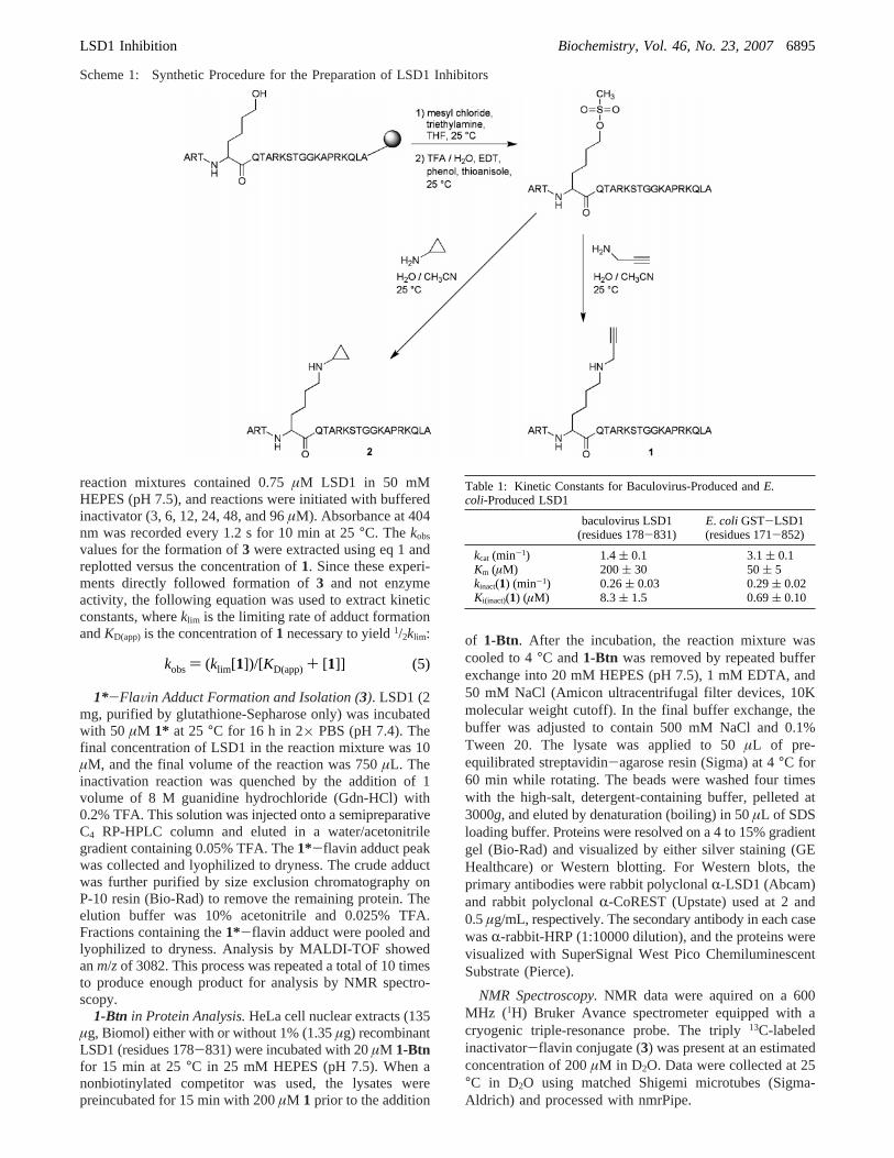
On the basis of the finding that pargyline is a suicideinactivator of monoamine oxidases (20-22), we previouslydesigned and synthesized a peptide substrate analogue inwhich the nitrogen atom of Lys4 was derivatized with apropargyl group1 (Scheme 1) (15). We showed that thiscompound exhibited time-dependent inactivation of LSD1and generated a covalent flavin adduct which was character-ized by mass spectrometric analysis (15). In contrast, apeptide aziridine inhibitor appeared to behave as a standardreversible inhibitor (15). In this study, we investigate thekinetic and mechanistic basis of inhibition by compound1in greater detail. We have shown that compound1 inducesa spectroscopic change in the flavin cofactor consistent withan N5 adduct. In contrast, a novel cyclopropylamine deriva-tive 2 (Scheme 1), which behaves as a competitive inhibitor,does not induce this spectroscopic change. Further analysisof the 1-flavin adduct using NMR is consistent with theproposed structure. We have measured the optical spectro-scopic change induced by1 as a function of time and foundthat it proceeds with kinetic constants similar to the rate ofinactivation. Finally, we show that a biotin-labeled analogueof compound1, 1-Btn, can be used to isolate endogenousLSD1 and CoREST from nuclear extracts, suggesting ap-plications in proteomics.
MATERIALS AND METHODS
[ 13C]Propargylamine Hydrochloride.Diethylazodicarbox-ylate (DEAD, 767µL, 4.87 mmol) was added dropwise over5 min to a solution of triply13C-labeled propargyl alcohol(Cambridge Isotope Lab) (250 mg, 4.23 mmol), triphen-ylphosphine (1.28 g, 4.87 mmol), andN-(tert-butoxycarbo-nyl)phosphoramidic acid diethyl ester (TCI) (1.07 g, 4.23mmol) in 20 mL of anhydrous THF at 0°C. After addition,the reaction mixture was allowed to warm to room temper-ature while being stirred for 4 h. The reaction mixture wasconcentratedin Vacuo to a yellow oil. Without furtherpurification, the oil was dissolved in 20 mL of anhydrousbenzene and saturated with dry hydrogen chloride for 2 hwith stirring. The solution was allowed to stand 12 h withoutstirring. The reaction mixture was concentratedin Vacuo,resuspended in dry diethyl ether, and allowed to stand at-80 °C for 4 h. The amine hydrochloride was pelleted bycentrifugation and washed three times with dry diethyl etherbefore being dissolved in H2O and lyophilized to a whitepowder, yielding 255 mg:1H NMR (CD3OD, 400 MHz)δ3.80 (dm,J ) 148 Hz, 2H), 3.13 (dm,J ) 306 Hz, 1H).
Fmoc-Hydroxynorleucine-OH.Fmoc-Lys-OH (5.2 g, 14.1mmol) was added to 300 mL of ddH2O and 50.0 mL ofglacial acetic acid (15). The solution was gently heated untilall solids dissolved. The solution was then cooled to 0°Cwith an ice/water bath. Sodium nitrite (2.92 g, 42.3 mmol)in 65 mL of H2O was added dropwise to the stirring Fmoc-Lys-OH solution over 180 min. The reaction mixture waswarmed to 25°C while being stirred over 20 h. The mixturewas concentratedin Vacuoto a yellow oil, dissolved in H2O,and acidified with glacial acetic acid. The aqueous solutionwas extracted with 3× 75 mL of ethyl acetate. The pooledorganics were washed with 1× 75 mL of brine. The organicphase was dried over MgSO4, filtered, and concentratedinVacuoto a yellow solid. Crude product was purified by pre-parative-scale RP-HPLC, yielding 1.3 g:1H NMR (CDCl3,400 MHz)δ 7.77 (d,J ) 7.2 Hz, 2H), 7.60 (d,J ) 7.2 Hz,
2H), 7.40 (t,J ) 7.2 Hz, 2H), 7.32 (t,J ) 7.2 Hz, 2H), 5.38(d, J ) 8.4 Hz, 1H), 4.43 (m, 3H), 4.23 (t,J ) 6.4 Hz, 1H),3.68 (t,J ) 6.4 Hz, 2H), 1.93 (m, 1H), 1.78 (m, 1H), 1.61(m, 2H), 1.49 (m, 2H); electospray ionization mass spec-trometry (PE Biosystems SCIEX API 150EX)m/z 369.
MesylK4H3-21.Standard Fmoc solid-phase peptide syn-thesis techniques were utilized to assemble the mesylK4H3-21 peptide. At the 4 position, Fmoc-hydroxynorleucine-OHwas coupled; subsequent protection of theε-alcohol by aceticanhydride yielded the acetic ester prior to the coupling ofThr3. The fully protected H3-21 peptide on Wang resin wastreated with 125 mM hydroxylamine at pH 10 in a 50:50H2O/dimethylformamide mixture for 12 h at room temper-ature. The primary alcohol, in position 4, was then treatedwith 20 equiv of mesyl chloride in the presence of 40 equivof triethylamine in tetrahydrofuran for 20 h at 25°C.Universal deprotection and cleavage from the resin wereaccomplished with a 95:5 trifluoroacetic acid (TFA)/H2Omixture in the presence of phenol, ethanedithiol, and thio-anisole for 5 h at 25°C. The stability of the mesylate tothese conditions is ascribed to its location on a primarycarbon and the low pH of the system (pHe1.0). Precipitationof the peptide with diethyl ether followed by lyophilizationyielded a crude peptide as an off-white solid that was purifiedby preparative-scale RP-HPLC. Analysis by MALDI-TOF,with a Voyager DE-STR MALDI-TOF mass spectrometer(Applied Biosystems), showed anm/z of 2335.
PropargylK4H3-21 (1). Lyophilized mesylK4H3-21 (5.0mg, 2.1µmol) was dissolved in 500µL of a 1:1 H2O/CH3-CN mixture. Freshly distilled propargylamine (44µL, 640µmol) in 500µL of a 1:1 H2O/CH3CN mixture was addedand the solution rotated for 65 h at 25°C. The crude reactionmixture was diluted to 3 mL with H2O, acidified to pH 2with TFA, and injected onto a preparative-scale column forRP-HPLC purification. Analysis by MALDI-TOF showedanm/z of 2294. The pure peptide was lyophilized to a whitesolid and stored at-80 °C.
[ 13C]PropargylK4H3-21 (1*). Lyophilized mesylK4H3-21 (5.0 mg, 2.1µmol) was dissolved in 1000µL of a 1:1H2O/CH3CN mixture. [13C]Propargylamine hydrochloride (57mg, 600µmol) in 100µL of a 1:1 H2O/CH3CN mixture wasadded to the solution followed by triethylamine (112µL,800µmol). The reaction mixture was rotated for 65 h at 25°C. The crude reaction mixture was diluted to 25 mL withH2O, acidified to pH 2 with TFA, and lyophilized to an oil.The oil was again diluted to 25 mL with H2O, acidified, andlyophilized to a solid/oil that was diluted to 3 mL with H2Oand injected onto a preparative-scale column for RP-HPLCpurification. Analysis by MALDI-TOF showed anm/z of2297. The pure peptide was lyophilized to a white solid andstored at-80 °C.
MesylK4H3-21-Biotin.This peptide was made in a manneranalogous to that of the nonbiotinylated mesyl peptide withthe following variations. Fmoc-Gly-Wang resin was used,and in position 22, Fmoc-Lys(biotin)-OH (Novabiochem)was coupled, resulting in a 23-amino acid peptide. Residues1-21 are histone H3, while residues 22 and 23 are abiotinylated lysine followed by a glycine, respectively.Analysis by MALDI-TOF showed anm/z of 2746.
PropargylK4H3-21-Biotin (1-Btn). This peptide was madein a manner analogous to that of the nonbiotinylated peptide.Analysis by MALDI-TOF showed anm/z of 2705.
LSD1 Inhibition Biochemistry, Vol. 46, No. 23, 20076893
CyclopropylK4H3-21 (2). Lyophilized mesylK4H3-21 (5.0mg, 2.1µmol) was dissolved in 500µL of a 1:1 H2O/CH3-CN mixture. Freshly distilled cyclopropylamine (Fluka) (55µL, 800µmol) in 500µL of a 1:1 H2O/CH3CN mixture wasadded and the solution rotated for 65 h at 25°C. The crudereaction mixture was diluted to 3 mL with H2O, acidified topH 2 with TFA, and injected onto a preparative-scale columnfor RP-HPLC purification. Analysis by MALDI-TOF showedan m/z of 2296.
diMeK4H3-21.The standard Fmoc solid-phase peptidesynthesis technique was utilized to assemble the diMeK4H3-21 peptide. The diMeK4 residue was coupled as com-mercially available Fmoc-diMeLys-OH (Novabiochem).Universal deprotection and cleavage of the peptide from theWang resin were accomplished with a 95:5 TFA/H2Omixture in the presence of phenol, ethanedithiol, and thio-anisole for 5 h at 25°C. Precipitation of the peptide withdiethyl ether followed by lyophilization yielded the crudepeptide as an off-white solid that was purified by preparative-scale RP-HPLC. Analysis by MALDI-TOF showed anm/zof 2284.
LSD1 Expression and Purification.LSD1 (residues 171-852) subcloned into the pGEX-6P-1 vector (12) (GE Health-care) was overexpressed inEscherichia coliBL21-CodonPlus-(DE3)-RIPL cells (Stratagene). Cells were grown to an OD600
of 1.8 in CircleGrow Media (Q-Biogene) at 37°C, inducedwith 1 mM IPTG (final concentration), and grown for 20 hat 16 °C. Cell pellets were harvested by centrifugation at5000g for 15 min and resuspended in ice-cold lysis buffer[280 mM NaCl, 5.4 mM KCl, 20 mM Na2HPO4, 3.6 mMKH2PO4, 1 mM EDTA, 10 mM DTT, and 10% glycerol (pH7.4)]. The cells were then lysed via double pass on a Frenchpress (16000-18000 psi), and the lysates were clarified bycentrifugation at 25000g for 30 min. The clarified lysate from1 L of culture was double loaded (0.5 mL/min) onto a 5 mLglutathione-Sepharose 4 fast flow column (GE Healthcare)that was pre-equilibrated with lysis buffer. The column wasthen washed with 75 mL of lysis buffer and eluted with 5×5 mL fractions of lysis buffer containing 50 mM reducedglutathione (Sigma). GST-LSD1-containing fractions werepooled and dialyzed against 3× 1 L changes of lysis buffercontaining 1 mMâ-mercaptoethanol instead of 10 mM DTT.The dialyzed protein was concentrated to 1-2 mL and furtherpurified by size exclusion chromatography using SephacrylS100 high-resolution media (GE Healthcare, 1.5 cm× 90cm column). The protein was eluted with lysis buffercontaining 1 mMâ-mercaptoethanol instead of 10 mM DTTat a flow rate of 0.25 mL/min. GST-LSD1-containingfractions were pooled, concentrated, aliquoted, and storedat -80 °C. The final protein concentration was determinedby a Bradford assay using BSA as the standard. Purificationof GST-LSD1 by this procedure was scalable to 18 L andyielded approximately 1 mg of protein (>90% pure) per literof culture.
Demethylase Assays.Initial velocity measurements wereperformed using a peroxidase-coupled assay, which monitorshydrogen peroxide production as previously described (23).The time courses of the reaction were measured underaerobic conditions using a Beckman Instruments DU series600 spectrophotometer equipped with thermostated cellholder (T ) 25 °C). The 150µL reactions were intitiated byadding 50µL of buffered substrate (diMeK4H3-21, final
concentration of 60µM) solution to reaction mixtures (100µL) consisting of 50 mM HEPES buffer (pH 7.5), 0.1 mM4-aminoantipyrine, 1 mM 3,5-dichloro-2-hydroxybenzene-sulfonic acid, 0.76µM horseradish peroxidase (WorthingtonBiochemical Corp.), and 120-360 nM LSD1. Reactionmixtures were equilibrated at 25°C for 2 min prior to activitymeasurement. Absorbance changes were monitored at 515nm, and an extinction coefficient of 26 000 M-1 cm-1 wasused to calculate product formation. Under these conditions,our GST-LSD1 displayed akcat of 3.1 ( 0.1 min-1 and aKm for diMeK4H3-21 of 50( 5 µM.
LSD1 inhibitors were tested by using the peroxidase-coupled assay described above. In these experiments, assayswere initiated by the simultaneous addition of bufferedsubstrate and inhibitor (either1 or 2). The final substrateconcentration was either 240µM (∼5Km when examining1) or 60 µM (∼Km when examining2). Progress curvesobtained in the presence of1 were fit to the following singleexponential for slow-binding inhibitors which assumes asteady-state velocity of zero (24):
Thekobsvalues were then analyzed by the method of Kitzand Wilson to yieldkinact andKi(inact). The following equationwas used to extract kinetic constants from the Kitz-Wilsonanalysis (25):
Ki(inact) was extrapolated to zero substrate by:
The t1/2 for inactivation at saturation was obtained fromeq 4:
Compound2 did not exhibit time-dependent inhibition.Initial velocities at increasing concentrations of2 wereobtained by linear regression to reaction progress curves.These velocities were used to determine theKi of 2 by Dixonanalysis (assuming a competitive mode of inhibition).
Absorbance Spectroscopy.LSD1 (10µM) was incubatedwith 1 (50µM), 2 (60µM), or no inhibitor in 50 mM HEPES(pH 7.5) at 25°C. After 1 h, the samples were clarified bycentrifugation at 14000g for 10 min (25°C), and the flavinabsorbance spectra were recorded versus a buffer blank(350-550 nm). The flavin adduct that formed upon incuba-tion with 1 was then acidified with HCl (300 mM final, pH<2) or reduced with NaBH4 (1 mM final concentration) at25 °C (10 min), and the flavin absorbance spectra wererecorded again. Difference spectra were generated by sub-tracting the native (untreated) LSD1 spectrum from theinhibitor-treated spectra.
Time Dependence of Formation of the1-FlaVin Adduct(3). Difference spectroscopy indicated a maximum at 404nm for LSD1 treated with1. The time- and concentration-dependent formation of this species could be monitored byabsorbance spectroscopy. LSD1 was clarified by centrifuga-tion at 14000g for 10 min (4 °C) prior to analysis. The
product) V0(1 - e-kt)/k + offset (1)
kobs) (kinact[I])/( Ki(inact)app+ [I]) (2)
Ki(inact)app) Ki(inact)(1 + [S]/Km) (3)
t1/2 ) (ln 2)/kinact (4)
6894 Biochemistry, Vol. 46, No. 23, 2007 Szewczuk et al.
reaction mixtures contained 0.75µM LSD1 in 50 mMHEPES (pH 7.5), and reactions were initiated with bufferedinactivator (3, 6, 12, 24, 48, and 96µM). Absorbance at 404nm was recorded every 1.2 s for 10 min at 25°C. Thekobs
values for the formation of3 were extracted using eq 1 andreplotted versus the concentration of1. Since these experi-ments directly followed formation of3 and not enzymeactivity, the following equation was used to extract kineticconstants, whereklim is the limiting rate of adduct formationandKD(app) is the concentration of1 necessary to yield1/2klim:
1*-FlaVin Adduct Formation and Isolation (3). LSD1 (2mg, purified by glutathione-Sepharose only) was incubatedwith 50 µM 1* at 25°C for 16 h in 2× PBS (pH 7.4). Thefinal concentration of LSD1 in the reaction mixture was 10µM, and the final volume of the reaction was 750µL. Theinactivation reaction was quenched by the addition of 1volume of 8 M guanidine hydrochloride (Gdn-HCl) with0.2% TFA. This solution was injected onto a semipreparativeC4 RP-HPLC column and eluted in a water/acetonitrilegradient containing 0.05% TFA. The1*-flavin adduct peakwas collected and lyophilized to dryness. The crude adductwas further purified by size exclusion chromatography onP-10 resin (Bio-Rad) to remove the remaining protein. Theelution buffer was 10% acetonitrile and 0.025% TFA.Fractions containing the1*-flavin adduct were pooled andlyophilized to dryness. Analysis by MALDI-TOF showedanm/zof 3082. This process was repeated a total of 10 timesto produce enough product for analysis by NMR spectro-scopy.
1-Btn in Protein Analysis.HeLa cell nuclear extracts (135µg, Biomol) either with or without 1% (1.35µg) recombinantLSD1 (residues 178-831) were incubated with 20µM 1-Btnfor 15 min at 25°C in 25 mM HEPES (pH 7.5). When anonbiotinylated competitor was used, the lysates werepreincubated for 15 min with 200µM 1 prior to the addition
of 1-Btn. After the incubation, the reaction mixture wascooled to 4°C and1-Btn was removed by repeated bufferexchange into 20 mM HEPES (pH 7.5), 1 mM EDTA, and50 mM NaCl (Amicon ultracentrifugal filter devices, 10Kmolecular weight cutoff). In the final buffer exchange, thebuffer was adjusted to contain 500 mM NaCl and 0.1%Tween 20. The lysate was applied to 50µL of pre-equilibrated streptavidin-agarose resin (Sigma) at 4°C for60 min while rotating. The beads were washed four timeswith the high-salt, detergent-containing buffer, pelleted at3000g, and eluted by denaturation (boiling) in 50µL of SDSloading buffer. Proteins were resolved on a 4 to 15%gradientgel (Bio-Rad) and visualized by either silver staining (GEHealthcare) or Western blotting. For Western blots, theprimary antibodies were rabbit polyclonalR-LSD1 (Abcam)and rabbit polyclonalR-CoREST (Upstate) used at 2 and0.5µg/mL, respectively. The secondary antibody in each casewasR-rabbit-HRP (1:10000 dilution), and the proteins werevisualized with SuperSignal West Pico ChemiluminescentSubstrate (Pierce).
NMR Spectroscopy.NMR data were aquired on a 600MHz (1H) Bruker Avance spectrometer equipped with acryogenic triple-resonance probe. The triply13C-labeledinactivator-flavin conjugate (3) was present at an estimatedconcentration of 200µM in D2O. Data were collected at 25°C in D2O using matched Shigemi microtubes (Sigma-Aldrich) and processed with nmrPipe.
Scheme 1: Synthetic Procedure for the Preparation of LSD1 Inhibitors
kobs) (klim[1])/[KD(app)+ [1]] (5)
Table 1: Kinetic Constants for Baculovirus-Produced andE.coli-Produced LSD1
baculovirus LSD1(residues 178-831)
E. coli GST-LSD1(residues 171-852)
kcat (min-1) 1.4( 0.1 3.1( 0.1Km (µM) 200 ( 30 50( 5kinact(1) (min-1) 0.26( 0.03 0.29( 0.02Ki(inact)(1) (µM) 8.3 ( 1.5 0.69( 0.10
LSD1 Inhibition Biochemistry, Vol. 46, No. 23, 20076895
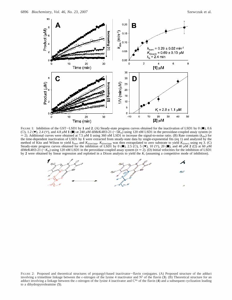
FIGURE 1: Inhibition of the GST-LSD1 by1 and2. (A) Steady-state progress curves obtained for the inactivation of LSD1 by 0 (b), 0.6(O), 1.2 (1), 2.4 (3), and 4.8µM 1 (9) at 240µM diMeK4H3-21 (∼5Km) using 120 nM LSD1 in the peroxidase-coupled assay system (n) 2). Additional curves were obtained at 7.5µM 1 using 360 nM LSD1 to increase the signal-to-noise ratio. (B) Rate constants (kobs) forthe time-dependent inactivation of LSD1 by1 were extracted from steady-state data by single-exponential fits (eq 1) and analyzed by themethod of Kitz and Wilson to yieldkinact andKi(inact)app. Ki(inact)app was then extrapolated to zero substrate to yieldKi(inact) using eq 3. (C)Steady-state progress curves obtained for the inhibition of LSD1 by 0 (b), 2.5 (O), 5 (1), 10 (3), 20 (9), and 40µM 2 (0) at 60 µMdiMeK4H3-21 (∼Km) using 120 nM LSD1 in the peroxidase-coupled assay system (n ) 2). (D) Initial velocities for the inhibition of LSD1by 2 were obtained by linear regression and replotted in a Dixon analysis to yield theKi (assuming a competitive mode of inhibition).
FIGURE 2: Proposed and theoretical structures of propargyl-based inactivator-flavin conjugates. (A) Proposed structure of the adductinvolving a trimethine linkage between theε-nitrogen of the lysine 4 inactivator and N5 of the flavin (3). (B) Theoretical structure for anadduct involving a linkage between theε-nitrogen of the lysine 4 inactivator and C4R of the flavin (4) and a subsequent cyclization leadingto a dihydropyrroleamine (5).
6896 Biochemistry, Vol. 46, No. 23, 2007 Szewczuk et al.
RESULTS
Cyclopropylamine Synthesis.Cyclopropylamine analoguescan be potent inhibitors for amine oxidases (26, 27) so weprepared the corresponding substrate analogue2 as a potentialLSD1 inhibitor. The synthetic approach to2 was similar tothat used previously for1 in that a hydroxy analogue of lysinewas used to generate a mesylated peptide (Scheme 1). Thepeptide mesylate was displaced with cyclopropylamine toproduce the desired analogue2 which could be purified byRP-HPLC and exhibited the correct molecular weight bymass spectrometry.
LSD1 Inhibition Studies.In prior studies (15), we usedbaculovirus-expressed recombinant LSD1, whereas a moreconvenientE. coli overproducing strain which generated theGST-LSD1 fusion protein was used here. Catalytic param-eters for the bacterially expressed LSD1 with histone H3-21 peptide substrate were modestly different from those ofthe insect cell-derived protein as shown in Table 1, withkcat/Km being∼9-fold higher for the former. The basis for theseobserved differences is uncertain but may be due to slightlydifferent construct sizes, post-translational modifications ininsect cells, and/or the presence of the GST tag on thebacterially produced LSD1. Compound1 proved to be asomewhat more potent time-dependent inactivator with thebacterially expressed LSD1 enzyme with akinact/Ki(inact)
increase of 13-fold over that of the insect cell-derived enzyme(Table 1 and Figure 1), consistent with the fact that thisenzyme also exhibited higherkcat and lowerKm values. Incontrast, the cyclopropylamine analogue2 did not exhibittime-dependent inactivation of LSD1 but instead appearedto show classical reversible inhibition (Figure 1). In addition,2 was able to compete with1 and decreasekobs forinactivation by∼30% when held in a 5-fold excess (datanot shown). Titration of2 versus LSD1 and Dixon analysisrevealed aKi of 2.8 ( 1.1 µM (assuming a standardcompetitive model of inhibition).
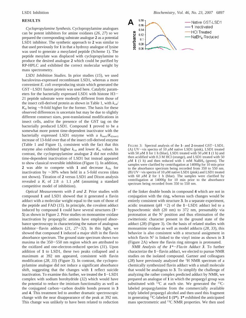
Optical Measurements with1 and 2. Prior studies withcompound1 and LSD1 showed that it generated a flavinadduct with a molecular weight equal to the sum of those ofthe peptide and FAD (15). In principle, the covalent adductinduced by compound1 could have several structures (3-5) as shown in Figure 2. Prior studies on monoamine oxidaseinactivation by propargylic amines have employed absor-bance spectroscopy in characterizing the nature of analogousinhibitor-flavin adducts (21, 27-32). In this light, weshowed that compound1 induced a major shift in the flavinabsorbance spectrum. The ground state spectrum shows twomaxima in the 350-550 nm region which are attributed tothe oxidized and one-electron-reduced species (31). Uponaddition of 1 to LSD1, these two peaks collapsed and amaximum at 392 nm appeared, consistent with flavinmodification (28, 33) (Figure 3). In contrast, the cyclopro-pylamine analogue did not induce a significant absorbanceshift, suggesting that the changes with1 reflect suicideinactivation. To examine this further, we treated the1-LSD1complex with sodium borohydride (32) which would havethe potential to reduce the iminium functionality as well asthe conjugated carbon-carbon double bonds present in3and4. This treatment led to another dramatic spectroscopicchange with the near disappearance of the peak at 392 nm.This change was unlikely to have been related to reduction
of the linker double bonds in compound4 which are not inconjugation with the ring, whereas such changes would beentirely consistent with structure3. In a separate experiment,acidic treatment (pH<2) of the 1-LSD1 adduct led to ahypsochromic shift (20 nm) to 372 nm, presumably viaprotonation at the N1 position and thus elimination of thezwitterionic character present in the ground state of theadduct (28) (Figure 3). On the basis of related studies withmonoamine oxidase as well as model adducts (28, 33), thisbehavior is also consistent with a structural assignment inwhich flavin N5 is linked to the vinyl imine as shown in3(Figure 2A) where the flavin ring nitrogen is protonated.
NMR Analysis of the1*-FlaVin Adduct 3. To furthercharacterize the1-flavin adduct, we elected to pursue NMRstudies on the isolated compound. Gartner and colleagues(28) have previously analyzed the1H NMR spectrum of achemically synthesized flavin adduct with a small moleculethat would be analogous to3. To simplify the challenge ofanalyzing the rather complex predicted adduct by NMR, weprepared an analogue of1 in which the propargyl group wassubstituted with13C at each site. We generated the13C-labeled propargylamine from the commercially availabletriply labeled propargyl alcohol and then used this fragmentin generating13C-labeled1 (1*). 1* exhibited the anticipatedmass spectrometric and13C NMR properties. We then used
FIGURE 3: Spectral analysis of the1- and2-treated GST-LSD1.(A) UV-vis spectra of 10µM native LSD1 (pink), LSD1 treatedwith 50 µM 1 for 1 h (blue), LSD1 treated with 50µM 1 (1 h) andthen acidified with 0.3 M HCl (orange), and LSD1 treated with 50µM 1 (1 h) and then reduced with 1 mM NaBH4 (green). Thesamples were clarified by centrifugation at 14000g for 10 min priorto the absorbance spectrum being recorded from 350 to 550 nm.(B) UV-vis spectra of 10µM native LSD1 (pink) and LSD1 treatedwith 60 µM 2 for 1 h (blue). The samples were clarified bycentrifugation at 14000g for 10 min prior to the absorbancespectrum being recorded from 350 to 550 nm.
LSD1 Inhibition Biochemistry, Vol. 46, No. 23, 20076897
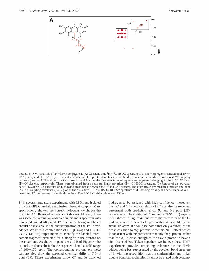
1* in several large-scale experiments with LSD1 and isolated3 by RP-HPLC and size exclusion chromatography. Massspectrometry showed the correct molecular weight for thepredicted1*-flavin adduct (data not shown). Although therewas some contamination observed in this mass spectrum withunreacted and dealkylated1*, the latter being unlabeledshould be invisible in the characterization of the1*-flavinadduct. We used a combination of HSQC (34) and HCCH-COSY (35, 36) experiments to identify the labeled three-carbon fragment predicted for3 along with the protons onthese carbons. As shown in panels A and B of Figure 4, theR- andγ-carbons cluster in the expected chemical shift rangeof 160-170 ppm. The corresponding protons on thesecarbons also show the expected chemical shifts of 7.5-8ppm (28). These experiments allow Câ and its attached
hydrogen to be assigned with high confidence; moreover,the 13C and1H chemical shifts of Cγ are also in excellentagreement with prediction at ca. 95 and 5.3 ppm (28),respectively. The additional13C-edited ROESY (37) experi-ment shown in Figure 4C indicates the proximity of the Cγ
hydrogen with a downfield proton that is very likely theflavin H6 atom. It should be noted that only a subset of thepeaks assigned toR/γ-protons show this NOE effect whichis consistent with the prediction that only theγ-proton (ratherthan theR) is close enough to the flavin proton to have asignificant effect. Taken together, we believe these NMRexperiments provide compelling evidence for the flavinadduct being best represented by the covalent bond structureof 3, with the recognition that the conformation and linkerdouble bond stereochemistry cannot be stated with certainty
FIGURE 4: NMR analysis of1*-flavin conjugate3. (A) Constant-time1H-13C HSQC spectrum of3, showing regions consisting of HR/γ-CR/γ (black) and Hâ-Câ (red) cross-peaks, which are of opposite phase because of the difference in the number of one-bond13C couplingpartners (one for CR/γ and two for Câ). Insetsa and b show the fine structures of representative peaks belonging to the HR/γ-CR/γ andHâ-Câ clusters, respectively. These were obtained from a separate, high-resolution1H-13C HSQC spectrum. (B) Region of an “out-and-back” HCCH-COSY spectrum of3, showing cross-peaks between the Câ and CR/γ clusters. The cross-peaks are mediated through one-bond13C-13C coupling constants. (C) Region of the13C-edited1H-13C HSQC-ROESY spectrum of3, showing cross-peaks between putative Hγ
peaks and H6 resonances of the flavin moiety. The ROESY mixing time was 250 ms.
6898 Biochemistry, Vol. 46, No. 23, 2007 Szewczuk et al.
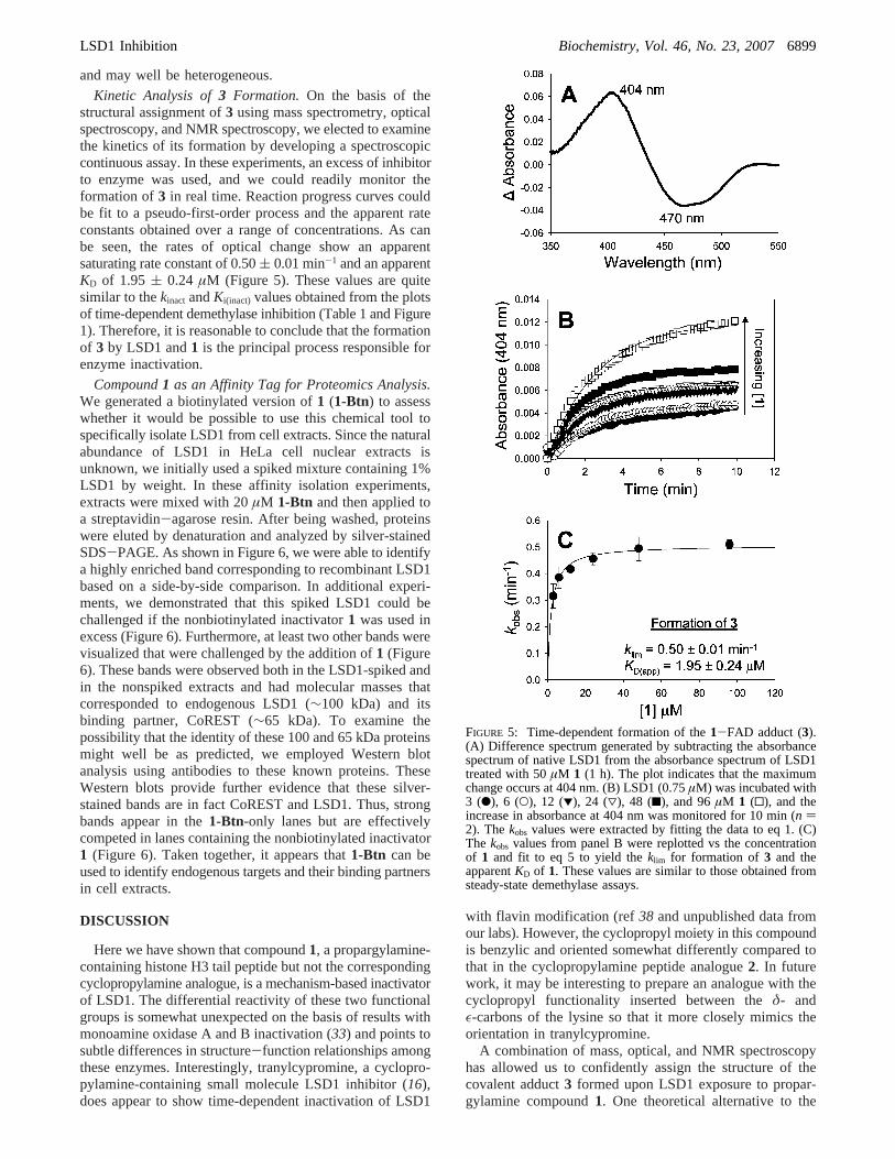
and may well be heterogeneous.Kinetic Analysis of3 Formation. On the basis of the
structural assignment of3 using mass spectrometry, opticalspectroscopy, and NMR spectroscopy, we elected to examinethe kinetics of its formation by developing a spectroscopiccontinuous assay. In these experiments, an excess of inhibitorto enzyme was used, and we could readily monitor theformation of3 in real time. Reaction progress curves couldbe fit to a pseudo-first-order process and the apparent rateconstants obtained over a range of concentrations. As canbe seen, the rates of optical change show an apparentsaturating rate constant of 0.50( 0.01 min-1 and an apparentKD of 1.95 ( 0.24 µM (Figure 5). These values are quitesimilar to thekinact andKi(inact) values obtained from the plotsof time-dependent demethylase inhibition (Table 1 and Figure1). Therefore, it is reasonable to conclude that the formationof 3 by LSD1 and1 is the principal process responsible forenzyme inactivation.
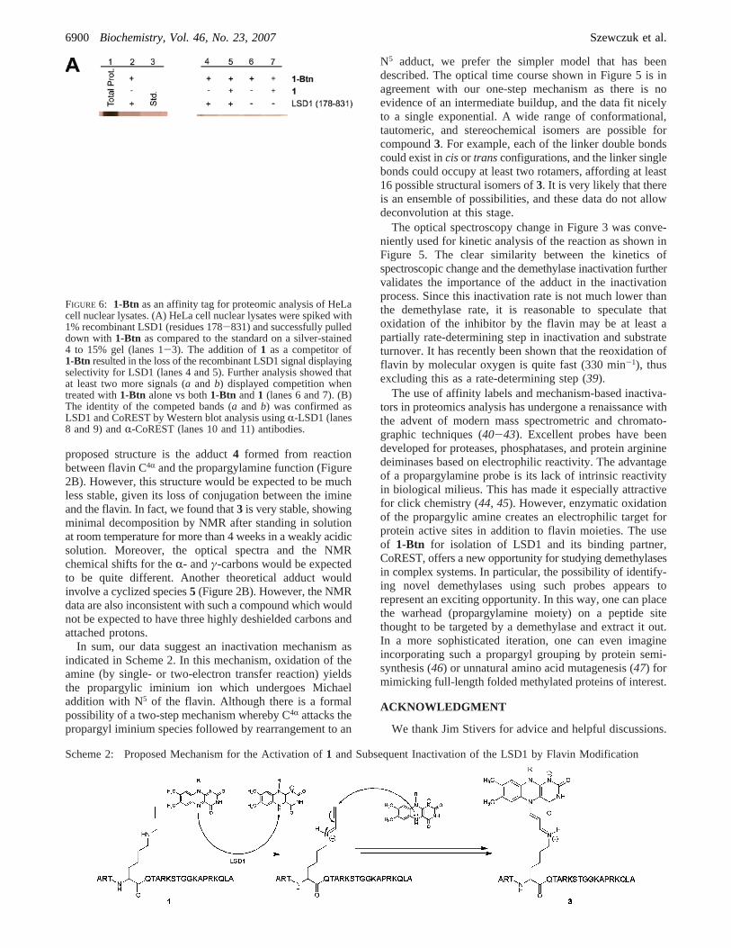
Compound1 as an Affinity Tag for Proteomics Analysis.We generated a biotinylated version of1 (1-Btn) to assesswhether it would be possible to use this chemical tool tospecifically isolate LSD1 from cell extracts. Since the naturalabundance of LSD1 in HeLa cell nuclear extracts isunknown, we initially used a spiked mixture containing 1%LSD1 by weight. In these affinity isolation experiments,extracts were mixed with 20µM 1-Btn and then applied toa streptavidin-agarose resin. After being washed, proteinswere eluted by denaturation and analyzed by silver-stainedSDS-PAGE. As shown in Figure 6, we were able to identifya highly enriched band corresponding to recombinant LSD1based on a side-by-side comparison. In additional experi-ments, we demonstrated that this spiked LSD1 could bechallenged if the nonbiotinylated inactivator1 was used inexcess (Figure 6). Furthermore, at least two other bands werevisualized that were challenged by the addition of1 (Figure6). These bands were observed both in the LSD1-spiked andin the nonspiked extracts and had molecular masses thatcorresponded to endogenous LSD1 (∼100 kDa) and itsbinding partner, CoREST (∼65 kDa). To examine thepossibility that the identity of these 100 and 65 kDa proteinsmight well be as predicted, we employed Western blotanalysis using antibodies to these known proteins. TheseWestern blots provide further evidence that these silver-stained bands are in fact CoREST and LSD1. Thus, strongbands appear in the1-Btn-only lanes but are effectivelycompeted in lanes containing the nonbiotinylated inactivator1 (Figure 6). Taken together, it appears that1-Btn can beused to identify endogenous targets and their binding partnersin cell extracts.
DISCUSSION
Here we have shown that compound1, a propargylamine-containing histone H3 tail peptide but not the correspondingcyclopropylamine analogue, is a mechanism-based inactivatorof LSD1. The differential reactivity of these two functionalgroups is somewhat unexpected on the basis of results withmonoamine oxidase A and B inactivation (33) and points tosubtle differences in structure-function relationships amongthese enzymes. Interestingly, tranylcypromine, a cyclopro-pylamine-containing small molecule LSD1 inhibitor (16),does appear to show time-dependent inactivation of LSD1
with flavin modification (ref38 and unpublished data fromour labs). However, the cyclopropyl moiety in this compoundis benzylic and oriented somewhat differently compared tothat in the cyclopropylamine peptide analogue2. In futurework, it may be interesting to prepare an analogue with thecyclopropyl functionality inserted between theδ- andε-carbons of the lysine so that it more closely mimics theorientation in tranylcypromine.
A combination of mass, optical, and NMR spectroscopyhas allowed us to confidently assign the structure of thecovalent adduct3 formed upon LSD1 exposure to propar-gylamine compound1. One theoretical alternative to the
FIGURE 5: Time-dependent formation of the1-FAD adduct (3).(A) Difference spectrum generated by subtracting the absorbancespectrum of native LSD1 from the absorbance spectrum of LSD1treated with 50µM 1 (1 h). The plot indicates that the maximumchange occurs at 404 nm. (B) LSD1 (0.75µM) was incubated with3 (b), 6 (O), 12 (1), 24 (3), 48 (9), and 96µM 1 (0), and theincrease in absorbance at 404 nm was monitored for 10 min (n )2). Thekobs values were extracted by fitting the data to eq 1. (C)The kobs values from panel B were replotted vs the concentrationof 1 and fit to eq 5 to yield theklim for formation of 3 and theapparentKD of 1. These values are similar to those obtained fromsteady-state demethylase assays.
LSD1 Inhibition Biochemistry, Vol. 46, No. 23, 20076899
proposed structure is the adduct4 formed from reactionbetween flavin C4R and the propargylamine function (Figure2B). However, this structure would be expected to be muchless stable, given its loss of conjugation between the imineand the flavin. In fact, we found that3 is very stable, showingminimal decomposition by NMR after standing in solutionat room temperature for more than 4 weeks in a weakly acidicsolution. Moreover, the optical spectra and the NMRchemical shifts for theR- andγ-carbons would be expectedto be quite different. Another theoretical adduct wouldinvolve a cyclized species5 (Figure 2B). However, the NMRdata are also inconsistent with such a compound which wouldnot be expected to have three highly deshielded carbons andattached protons.
In sum, our data suggest an inactivation mechanism asindicated in Scheme 2. In this mechanism, oxidation of theamine (by single- or two-electron transfer reaction) yieldsthe propargylic iminium ion which undergoes Michaeladdition with N5 of the flavin. Although there is a formalpossibility of a two-step mechanism whereby C4R attacks thepropargyl iminium species followed by rearrangement to an
N5 adduct, we prefer the simpler model that has beendescribed. The optical time course shown in Figure 5 is inagreement with our one-step mechanism as there is noevidence of an intermediate buildup, and the data fit nicelyto a single exponential. A wide range of conformational,tautomeric, and stereochemical isomers are possible forcompound3. For example, each of the linker double bondscould exist incisor transconfigurations, and the linker singlebonds could occupy at least two rotamers, affording at least16 possible structural isomers of3. It is very likely that thereis an ensemble of possibilities, and these data do not allowdeconvolution at this stage.
The optical spectroscopy change in Figure 3 was conve-niently used for kinetic analysis of the reaction as shown inFigure 5. The clear similarity between the kinetics ofspectroscopic change and the demethylase inactivation furthervalidates the importance of the adduct in the inactivationprocess. Since this inactivation rate is not much lower thanthe demethylase rate, it is reasonable to speculate thatoxidation of the inhibitor by the flavin may be at least apartially rate-determining step in inactivation and substrateturnover. It has recently been shown that the reoxidation offlavin by molecular oxygen is quite fast (330 min-1), thusexcluding this as a rate-determining step (39).
The use of affinity labels and mechanism-based inactiva-tors in proteomics analysis has undergone a renaissance withthe advent of modern mass spectrometric and chromato-graphic techniques (40-43). Excellent probes have beendeveloped for proteases, phosphatases, and protein argininedeiminases based on electrophilic reactivity. The advantageof a propargylamine probe is its lack of intrinsic reactivityin biological milieus. This has made it especially attractivefor click chemistry (44, 45). However, enzymatic oxidationof the propargylic amine creates an electrophilic target forprotein active sites in addition to flavin moieties. The useof 1-Btn for isolation of LSD1 and its binding partner,CoREST, offers a new opportunity for studying demethylasesin complex systems. In particular, the possibility of identify-ing novel demethylases using such probes appears torepresent an exciting opportunity. In this way, one can placethe warhead (propargylamine moiety) on a peptide sitethought to be targeted by a demethylase and extract it out.In a more sophisticated iteration, one can even imagineincorporating such a propargyl grouping by protein semi-synthesis (46) or unnatural amino acid mutagenesis (47) formimicking full-length folded methylated proteins of interest.
ACKNOWLEDGMENT
We thank Jim Stivers for advice and helpful discussions.
FIGURE 6: 1-Btn as an affinity tag for proteomic analysis of HeLacell nuclear lysates. (A) HeLa cell nuclear lysates were spiked with1% recombinant LSD1 (residues 178-831) and successfully pulleddown with 1-Btn as compared to the standard on a silver-stained4 to 15% gel (lanes 1-3). The addition of1 as a competitor of1-Btn resulted in the loss of the recombinant LSD1 signal displayingselectivity for LSD1 (lanes 4 and 5). Further analysis showed thatat least two more signals (a and b) displayed competition whentreated with1-Btn alone vs both1-Btn and1 (lanes 6 and 7). (B)The identity of the competed bands (a and b) was confirmed asLSD1 and CoREST by Western blot analysis usingR-LSD1 (lanes8 and 9) andR-CoREST (lanes 10 and 11) antibodies.
Scheme 2: Proposed Mechanism for the Activation of1 and Subsequent Inactivation of the LSD1 by Flavin Modification
6900 Biochemistry, Vol. 46, No. 23, 2007 Szewczuk et al.
REFERENCES
1. Schreiber, S. L., and Bernstein, B. E. (2002) Signaling networkmodel of chromatin,Cell 111, 771-778.
2. Jenuwein, T., and Allis, C. D. (2001) Translating the histone code,Science 293, 1074-1080.
3. Olins, D. E., and Olins, A. L. (2003) Chromatin history: Our viewfrom the bridge,Nat. ReV. Mol. Cell Biol. 4, 809-814.
4. Martin, C., and Zhang, Y. (2005) The diverse functions of histonelysine modification,Nat. ReV. Mol. Cell Biol. 6, 838-849.
5. Shi, Y., Lan, F., Matson, C., Mulligan, P., Whetstine, J. R., Cole,P. A., Casero, R. A., and Shi, Y. (2004) Histone demethylationmediated by the nuclear amine oxidase homolog LSD1,Cell 119,941-953.
6. Shi, Y., and Whetstine, J. R. (2007) Dynamic regulation of histonelysine methylation by demethylases,Mol. Cell 25, 1-14.
7. Tsukada, T., Fang, J., Erdjument-Bromage, H., Warren, M. E.,Borchers, C. H., Tempst, P., and Zhang, Y. (2006) Histonedemethylation by a family of JmjC domain-containing proteins,Nature 439, 811-816.
8. Whetstine, J. R., Nottke, A., Lan, F., Huarte, M., Smolikov, S.,Chen, Z., Spooner, E., Li, E., Zhang, G., Colaiacovo, M., andShi, Y. (2006) Reversal of histone lysine trimethylation by theJMJD2 family of histone demethylases,Cell 125, 467-481.
9. Binda, C., Mattevi, A., and Edmonson, D. E. (2002) Structure-function relationships in flavoenzyme-dependent amine oxida-tions: A comparison of polyamine oxidase and monoamineoxidase,J. Biol. Chem. 277, 23973-23976.
10. Lee, M. G., Wynder, C., Cooch, N., and Shiekhattar, R. (2005)An essential role for CoREST in nucleosomal histone 3 lysine 4demethylation,Nature 437, 432-435.
11. Shi, Y. J., Matson, C., Lan, F., Iwase, S., Baba, T., and Shi, Y.(2005) Regulation of LSD1 histone demethylase activity by itsassociated factors,Mol. Cell 19, 857-864.
12. Yang, M., Gocke, C. B., Luo, X., Borek, D., Tomchick, D. R.,Machius, M., Otwinowski, Z., and Yu, H. (2006) Structural basisfor CoREST-dependent demethylation of nucleosomes by thehuman LSD1 histone demethylase,Mol. Cell 23, 377-387.
13. Chen, Y., Yang, Y., Wang, F., Wan, K., Yamane, K., Zhang, Y.,and Lei, M. (2006) Crystal structure of human histone lysine-specific demethylase 1 (LSD1),Proc. Natl. Acad. Sci. U.S.A. 103,13956-13961.
14. Stavropoulos, P., Blobel, G., and Hoelz, A. (2006) Crystal structureand mechanism of human lysine-specific demethylase-1,Nat.Struct. Mol. Biol. 13, 626-632.
15. Culhane, J. C., Szewczuk, L. M., Liu, X., Da, G., Marmorstein,R., and Cole, P. A. (2006) A mechanism-based inactivator forhisone demethylase LSD1,J. Am. Chem. Soc. 128, 4536-4537.
16. Lee, M. G., Wynder, C., Scmidt, D. M., McCafferty, D. G., andShiekhattar, R. (2006) Histone H3 lysine 4 demethylation is atarget of nonselective antidepressive medications,Chem. Biol. 13,563-567.
17. Edmondson, D. E., Mattevi, A., Binda, C., Li, M., and Huba´lek,F. (2004) Structure and mechanism of monoamine oxidase,Curr.Med. Chem. 11, 1983-1993.
18. Walsh, C. T. (1984) Suicide substrates, mechanism-based enzymeinactivators: Recent developments,Annu. ReV. Biochem. 53, 493-535.
19. Silverman, R. B. (1995) Mechanism-based enzyme inactivators,Methods Enzymol. 249, 240-283.
20. Hellerman, L., and Erwin, V. G. (1968) Mitochondrial monoamineoxidase. II. Action of various inhibitors for the bovine kidneyenzyme. Catalytic mechanism,J. Biol. Chem. 243, 5234-5243.
21. Maycock, A. L., Abeles, R. H., Salach, J. I., and Singer, T. P.(1976) The structure of the covalent adduct formed by theinteraction of 3-dimethylamino-1-propyne and the flavine ofmitochondrial amine oxidase,Biochemistry 15, 114-125.
22. Binda, C., Hubalek, F., Li, M., Herzig, Y., Sterling, J., Edmondson,D. E., and Mattevi, A. (2005) Binding of rasagiline-relatedinhibitors to human monoamine oxidases: A kinetic and crystal-lographic analysis,J. Med. Chem. 48, 8148-8154.
23. Forneris, F., Binda, C., Vanoni, M. A., Battaglioli, E., and Mattevi,A. (2005) Human histone demethylase LSD1 reads the histonecode,J. Biol. Chem. 280, 41360-41365.
24. Copeland, R. A. (2000)Enzymes: A Practical Introduction toStructure, Mechanism, and Data Analysis, 2nd ed., Wiley-VCH,New York.
25. Kitz, R., and Wilson, I. B. (1962) Esters of methanesulfonic acidas irreversible inhibitors of acetylcholinesterase,J. Biol. Chem.237, 3245-3249.
26. McEwen, C. M., Sasaki, G., and Jones, D. C. (1969) Human livermitochondrial monoamine oxidase. 3. Kinetic studies concerningtime-dependent inhibitions,Biochemistry 8, 3963-3972.
27. Paech, C., Salach, J. I., and Singer, T. P. (1980) Suicideinactivation of monoamine oxidase by trans-phenylcyclopropyl-amine,J. Biol. Chem. 255, 2700-2704.
28. Gartner, B., Hemmerich, P., and Zeller, E. A. (1976) Structure offlavin adducts with acetylenic substrates. Chemistry of monoamineoxidase and lactate oxidase inhibition,Eur. J. Biochem. 63, 211-221.
29. Hubalek, F., Binda, C., Li, M., Herzig, Y., Sterling, J., Youdim,M. B. H., Mattevi, A., and Edmondson, D. E. (2004) Inactivationof purified human recombinant monoamine oxidases A and B byrasagiline and its analogues,J. Med. Chem. 47, 1760-1766.
30. Mitchell, D. J., Nikolic, D., Rivera, E., Sablin, S. O., Choi, S.,van Breeman, R. B., Singer, T. P., and Silverman, R. B. (2001)Spectrometric evidence for the flavin-1-phenylcyclopropylamineinactivator adduct with monoamine oxidase N,Biochemistry 40,5447-5456.
31. Woo, J. C. G., and Silverman, R. B. (1994) Observation of twodifferent chromophores in the resting state of monoamine oxidaseB by fluorescence spectroscopy,Biochem. Biophys. Res. Commun.202, 1574-1578.
32. Ghisla, S., Ogata, H., Massey, V., Schonbrunn, A., Abeles, R.H., and Walsh, C. T. (1976) Kinetic studies on the inactivationof L-lactate oxidase by [the acetylenic suicide substrate] 2-hydroxy-3-butynoate,Biochemistry 15, 1791-1797.
33. Sablin, S. O., Yankovskaya, V., Bernard, S., Cronin, C. N., andSinger, T. P. (1998) Isolation and characterization of an evolution-ary precursor of human monoamine oxidases A and B,Eur. J.Biochem. 253, 270-279.
34. Vuister, G. W., and Bax, A. (1992) Resolution enhancement andspectral editing of uniformly C-13-enriched proteins by homo-nuclear broad-band C-13 decoupling,J. Magn. Reson. 98, 428-435.
35. Eggenberger, U., Karimi-Nejad, Y., Thuering, H., Ru¨terjans, H.,and Griesinger, C. (1992) Determination of H-R, H-â and H-â,C′ coupling-constants in C-13 labeled proteins,J. Biomol. NMR2, 583-590.
36. Karimi-Nejad, Y., Schmidt, J. M., Ru¨terjans, H., Schwalbe, H.,and Griesinger, C. (1994) Conformation of the valine side chainsin ribonuclease T-1 determined by NMR studies of homonuclearand heteronuclear3J coupling-constants,Biochemistry 33, 5481-5492.
37. Bax, A., and Davis, D. G. (1985) Practical aspects of two-dimensional transverse NOE spectroscopy,J. Magn. Reson. 63,207-213.
38. Schmidt, D. M. Z., and McCafferty, D. G. (2007) trans-2-Phenylcyclopropylamine is a mechanism-based inactivator of thehistone demethylase LSD1,Biochemistry 46, 4408-4416.
39. Forneris, F., Binda, C., Dall’Aglio, A., Fraaije, M. W., Battaglioli,E., and Mattevi, A. (2006) A highly specific mechanism of histoneH3-K4 recognition by histone demethylase LSD1,J. Biol. Chem.281, 35289-35295.
40. Sieber, S. A., Niessen, S., Hoover, H. S., and Cravatt, B. F. (2006)Proteomic profiling of metalloprotease activities with cocktailsof active-site probes,Nat. Chem. Biol. 2, 274-281.
41. Blum, G., Mullins, S. R., Keren, K., Fonovic, M., Jedeszko, C.,Rice, M. J., Sloane, B. F., and Bogyo, M. (2005) Dynamic imagingof protease activity with fluorescently quenched activity-basedprobes,Nat. Chem. Biol. 1, 203-209.
42. Kumar, S., Zhou, B., Liang, F., Wang, W. Q., Huang, Z., andZhang, Z. Y. (2004) Activity-based probes for protein tyrosinephosphatases,Proc. Natl. Acad. Sci. U.S.A. 101, 7943-7948.
43. Luo, Y., Knuckley, B., Bhatia, M., Pellechia, P. J., and Thompson,P. R. (2006) Activity-based protein profiling reagents for proteinarginine deiminase 4 (PAD4): Synthesis and in vitro evaluationof a fluorescently labeled probe,J. Am. Chem. Soc. 128, 14468-14469.
44. Kolb, H. C., Finn, M. G., and Sharpless, K. B. (2001) ClickChemistry: Diverse Chemical Function from a Few GoodReactions,Angew. Chem., Int. Ed. 40, 2004-2021.
45. Lewis, W. G., Green, L. G., Grynszpan, F., Radic, Z., Carlier, P.R., Taylor, P., Finn, M. G., and Sharpless, K. B. (2002) Clickchemistry in situ: Acetylcholinesterase as a reaction vessel for
LSD1 Inhibition Biochemistry, Vol. 46, No. 23, 20076901

the selective assembly of a femtomolar inhibitor from an array ofbuilding blocks,Angew. Chem., Int. Ed. 41, 1053-1057.
46. Muir, T. W., Sondhi, D., and Cole, P. A. (1998) Expressed proteinligation: A general method for protein engineering,Proc. Natl.Acad. Sci. U.S.A. 95, 6705-6710.
47. Wang, L., Brock, A., Herberich, B., and Schultz, P. G. (2001)Expanding the genetic code ofEscherichia coli, Science 292,498-500.
BI700414B
6902 Biochemistry, Vol. 46, No. 23, 2007 Szewczuk et al.