Embed Size (px)
Citation preview
Mechanisms of Genetic Transmission
Genetic information is combined and transmitted by gametes, the reproductive cells of a child’s parent
In the father the gametes are produced in the testicles (each is called a sperm cell)
In the mother they are developed in the ovaries (each is called an ovum or egg cell)
Mechanisms of Genetic Transmission (continued)
The sperm and egg cells contain genetic information molecular structure called genes
The genes form threads called chromosomes
Genetic material that the child will inherit from the parent is contained in the chromosomes
Mechanisms of Genetic Transmission (continued)
Each human sperm or egg cell contains 23 chromosomes
All other cells of the body contain 46 chromosomes and about 100,000 genes
A single chromosome may contain as many as 20,000 genes
DNA
The genes are made up of DNA (deoxyribonucleic acid)
DNA have a spiral chemical structure that allows them to divide easily and duplicate DNA molecules
DNA contains the genetic information that directs the form and function of each body cell as it develops
DNA (continued)
DNA is shared at conception when a sperm (father) penetrates the egg from the mother
Within a few hours the walls of the sperm cell and the nucleus of the egg cell both begin to disintegrate releasing its chromosomes which join to form a new cell
The new cell formed is called a zygote
Meiosis and MitosisAll cells of the body develop from this
original zygote through a process called cell division
The sperm and ova contains only 23 chromosomes making them unique from other body cells
This ensures that when an egg and sperm join to form a new zygote the zygote will contain the complete set of 46 chromosomes (23 pair)
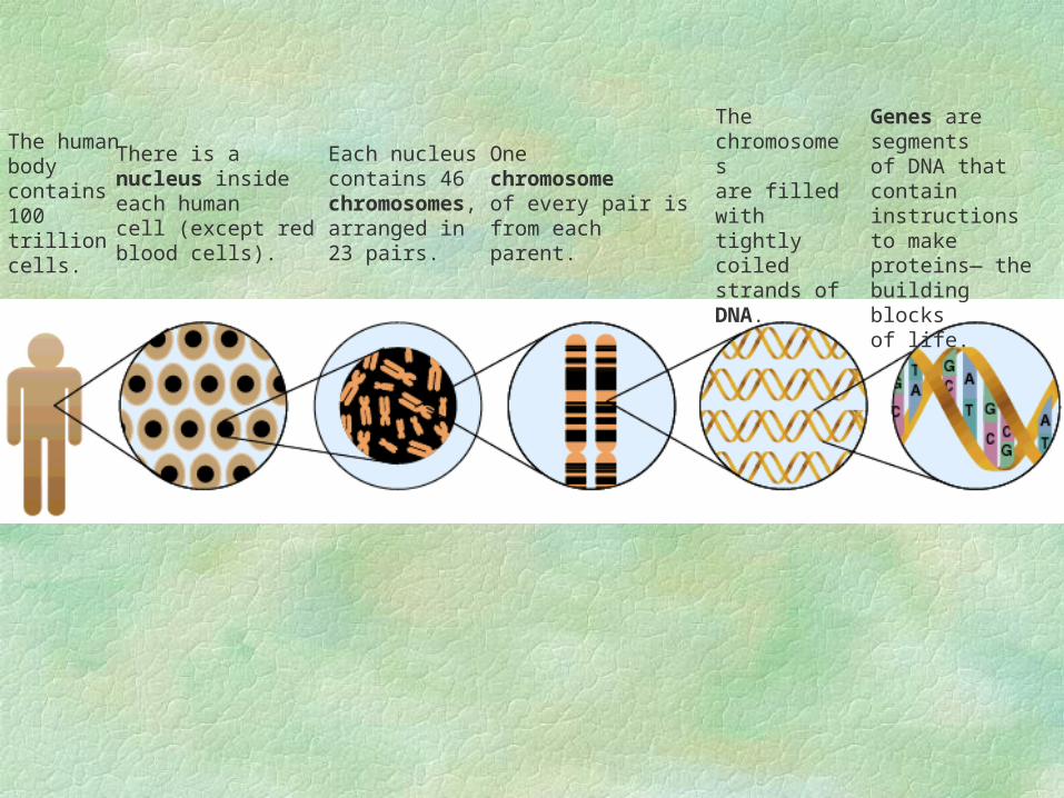
The humanbody contains100 trillioncells.
There is anucleus insideeach humancell (except redblood cells).
Each nucleuscontains 46chromosomes,arranged in 23 pairs.
Onechromosomeof every pair isfrom eachparent.
Thechromosomesare filled withtightly coiledstrands ofDNA.
Genes are segmentsof DNA that containinstructions to makeproteins— thebuilding blocksof life.
Meiosis and Mitosis (continued)The zygote contain the genetic materials
from which all of an individual’s cells are formed
The reproductive cells (gametes) divide by a process called meiosis and recombine into a zygote at conception
All other cells will develop from the zygote through a simpler division called mitosis
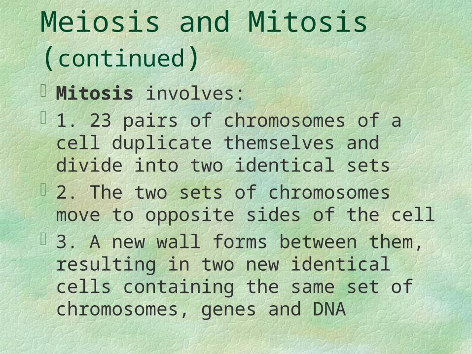
Meiosis and Mitosis (continued)Mitosis involves:1. 23 pairs of chromosomes of a cell
duplicate themselves and divide into two identical sets
2. The two sets of chromosomes move to opposite sides of the cell
3. A new wall forms between them, resulting in two new identical cells containing the same set of chromosomes, genes and DNA
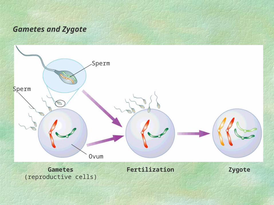
Gametes and Zygote
Sperm
Ovum
Gametes(reproductive cells)
Fertilization Zygote
Sperm
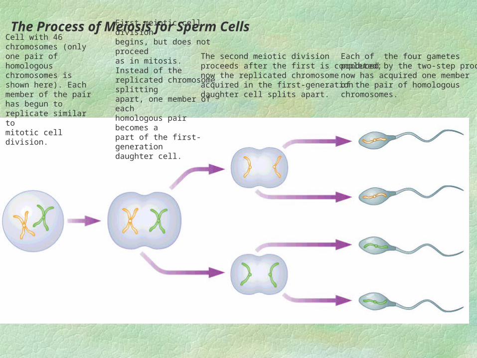
Cell with 46 chromosomes (only one pair of homologouschromosomes is shown here). Each member of the pair has begun to replicate similar tomitotic cell division.
First meiotic cell divisionbegins, but does not proceedas in mitosis. Instead of thereplicated chromosome splitting apart, one member of eachhomologous pair becomes a part of the first-generation daughter cell.
The second meiotic division proceeds after the first is completed;now the replicated chromosomeacquired in the first-generationdaughter cell splits apart.
Each of the four gametesproduced by the two-step processnow has acquired one memberof the pair of homologouschromosomes.
The Process of Meiosis for Sperm Cells
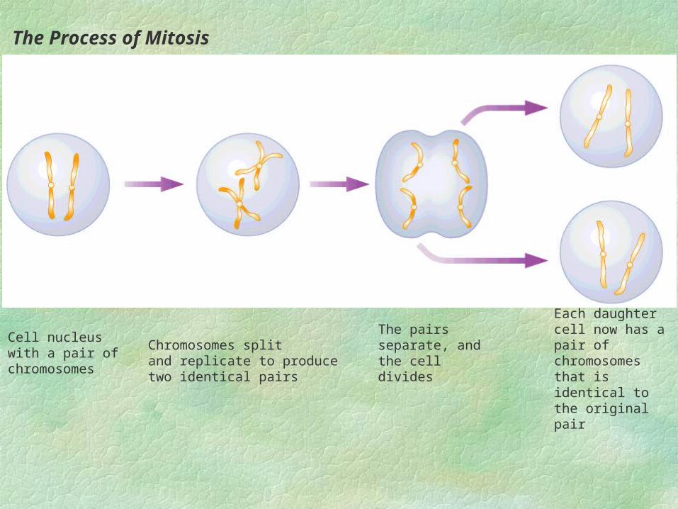
Cell nucleus with a pair of chromosomes
Chromosomes split and replicate to producetwo identical pairs
The pairs separate, and the cell divides
Each daughter cell now has a pair of chromosomes that is identical to the original pair
The Process of Mitosis
GENOTYPE AND PHENOTYPE
GENOTYPE: Set of genetic traits a person inherits; a person’s inborn capacity or potential
PHENOTYPE: Set of traits a person actually displays, resulting from a combination of the person’s genotype (potential) and life experiences that modify that potential
Individual Genetic Expression (continued)
Examples of phenotype may include a certain height, intelligence score, shyness
Phenotype results from all the interactions of the person’s genotype with the environment from conception onward
Genes (dominant and recessive)Genes are inherited in pairs (one from each
parent)Dominant gene--in a paired set of genes,
the gene with the greater influence in determining physical characteristics
Recessive gene--in a paired set of genes, the gene that influences or determining physical characteristics only when no dominant gene is present
Genes (dominant and recessive) continuedA dominant gene will influence a child’s
phenotype even if it is paired with a recessive gene
A recessive gene must be paired with another recessive gene to be able to influence the phenotype
If paired with a dominant gene its influence is blocked
Genes (dominant and recessive) continuedEye color--blue eyes (recessive trait), brown
eyes (dominant trait)a child’s eyes will remain blue only if they
have received the appropriate blue-producing gene from both parents
If they received it from only one parent or from neither they will end up with brown eyes (dominant)
Genes (dominant and recessive) continuedGenes may take on two or more alternatives
forms called allelesa person who inherits two identical alleles
for a particular trait is homozygous for that trait
if they inherit two different alleles for the trait it is heterozygous
Polygenic
Blood type and eye color have a limited number of distinct ways of transmission
height, weight, skin color, personality, behavioral traits are polygenic (involve many genes)
Environment influence them in important ways (can diet and change weight)
Determination of Sex
One pair of chromosomes among the 23 is largely responsible for determining whether a child is male or female
In women the pair of chromosomes is XXIn men the pair is XYall eggs cells contain a single X chromosomea sperm cell may contain either an X or Y
Determination of Sex (continued)
If a Y bearing sperm fertilizes the egg, a male (XY) zygote develops
If a X bearing sperm fertilizes the egg a female (XX) zygote develops
Genetic Abnormalities
Sometime too many or too few chromosomes transfer (or transfer a defective gene) to newly forming zygote
This may affect a child mentally, physically or both
Inheriting too many or too few chromosomes is usually fatal
Genetic Abnormalities (continued)
In a few cases children with a missing or an extra chromosome survive
Down syndrome--caused by extra 21st chromosome or transfer or part of the 21st on to another chromosome
Genetic Abnormalities (continued)
Characteristics of Down’s Syndromealmond shaped eyes, round headstubby hands and feetmay have abnormal heart and intestinal tractfacial deformitiesvulnerable to diseases such as leukemiaas they age fall behind developmentally
Genetic Abnormalities (continued)
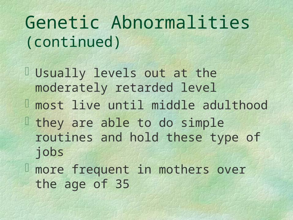
Usually levels out at the moderately retarded level
most live until middle adulthoodthey are able to do simple routines and hold
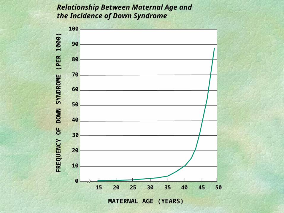
these type of jobsmore frequent in mothers over the age of 35
FR
EQ
UE
NC
Y O
F D
OW
N S
YN
DR
OM
E (
PE
R 1
000)
MATERNAL AGE (YEARS)
15 20 25 30 35 40 45 50
100
90
80
70
60
50
40
30
20
10
0
Relationship Between Maternal Age and the Incidence of Down Syndrome
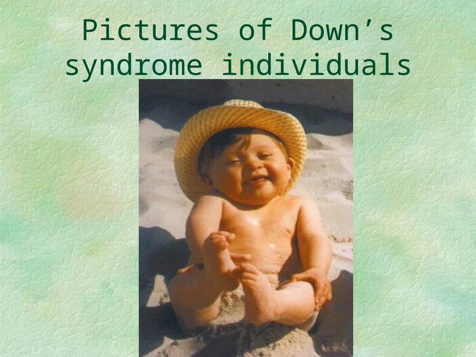
Pictures of Down’s syndrome individuals
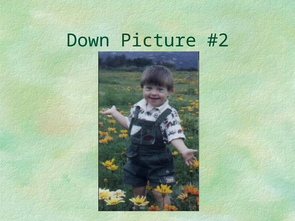
Down Picture #2
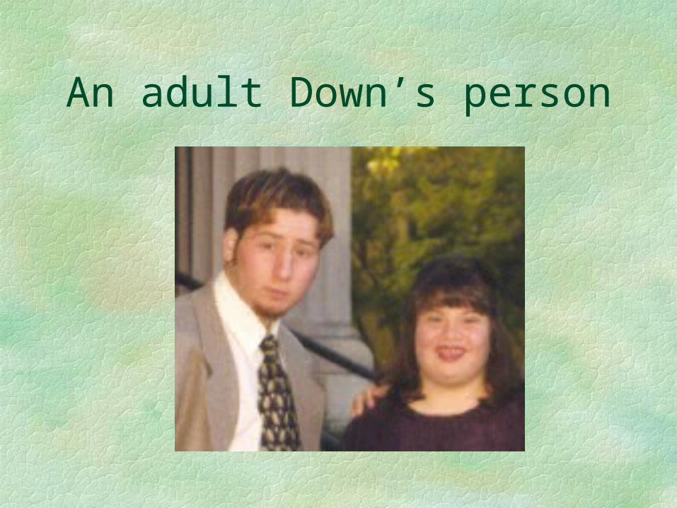
An adult Down’s person
Genetic Abnormalities (continued)
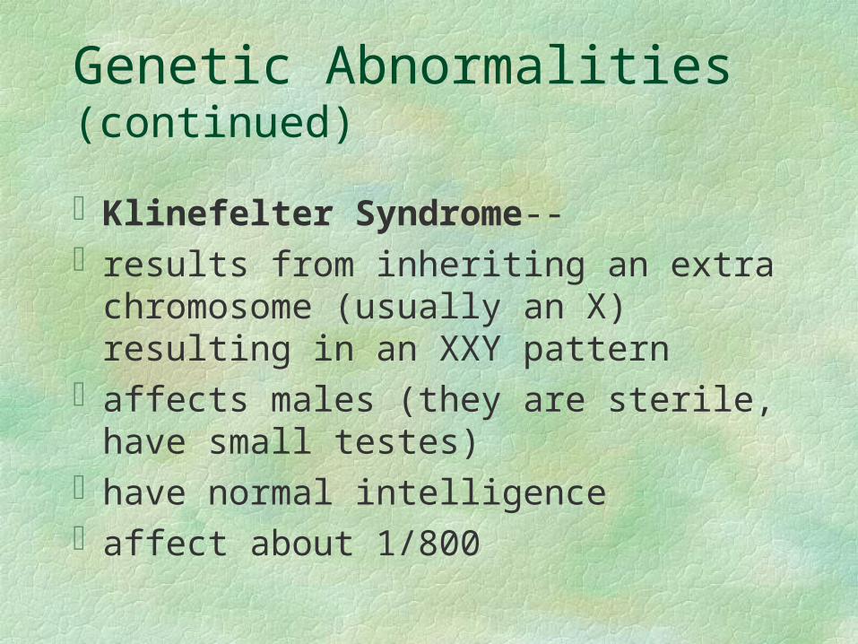
Klinefelter Syndrome--results from inheriting an extra chromosome
(usually an X) resulting in an XXY patternaffects males (they are sterile, have small
testes)have normal intelligenceaffect about 1/800
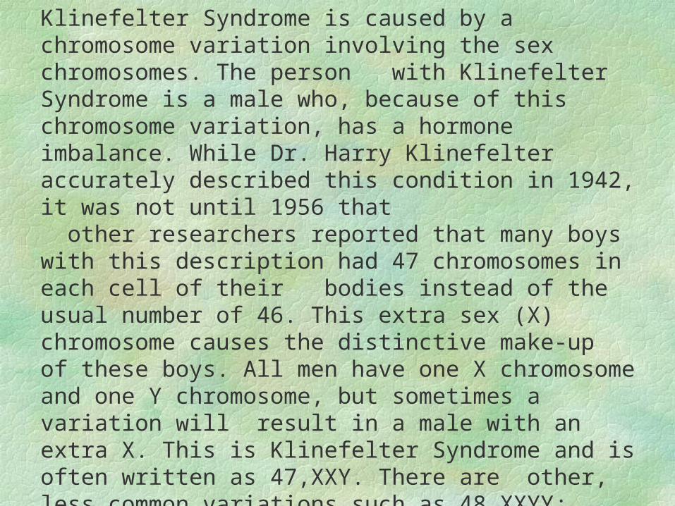
Klinefelter Syndrome is caused by a chromosome variation involving the sex chromosomes. The person with Klinefelter Syndrome is a male who, because of this chromosome variation, has a hormone imbalance. While Dr. Harry Klinefelter accurately described this condition in 1942, it was not until 1956 that other researchers reported that many boys with this description had 47 chromosomes in each cell of their bodies instead of the usual number of 46. This extra sex (X) chromosome causes the distinctive make-up of these boys. All men have one X chromosome and one Y chromosome, but sometimes a variation will result in a male with an extra X. This is Klinefelter Syndrome and is often written as 47,XXY. There are other, less common variations such as 48,XXYY; 48,XXXY; 49,XXXXY ; and XY/XXY mosaic. All of these are considered Klinefelter Syndrome variants.
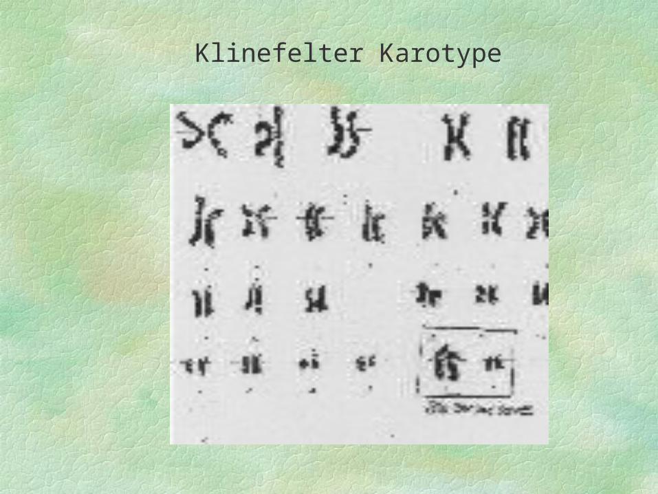
Klinefelter Karotype
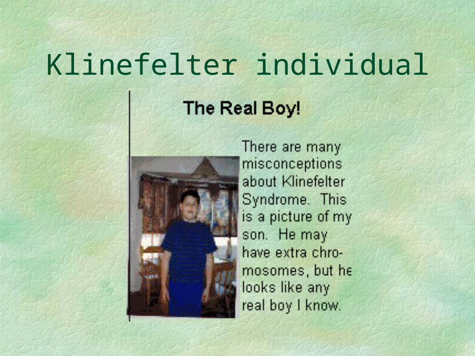
Klinefelter individual
Genetic Abnormalities (continued)
Turner syndrome--results from having only one sex
chromosome (X0) affecting femaleswill develop learning disabilitiesnot fully sexually differentiated are very short as adults (4 and 5 ft.)have webbed necksears are set lower than usual
Adults with Turner syndrome are short, averaging around four feet, eight inches in height. But girls with Turner syndrome don't start life as very short individuals - they become short over time, growing more slowly than their sisters and friends with each passing year. Studies have shown that a medicine called recombinant human growth hormone, or GH, can improve the height of girls with Turner syndrome. However, these studies have tended to start GH treatment around age 9 or later, after years of deteriorating growth. So, even with treatment, many girls remain shorter than would be expected based on the heights of their parents. The purpose of this new study is to determine if GH started at a young age can prevent the growth failure typical of girls with Turner syndrome. In addition, the study will monitor all participants for development of ear infections and hearing loss, problems that trouble many girls with Turner syndrome.
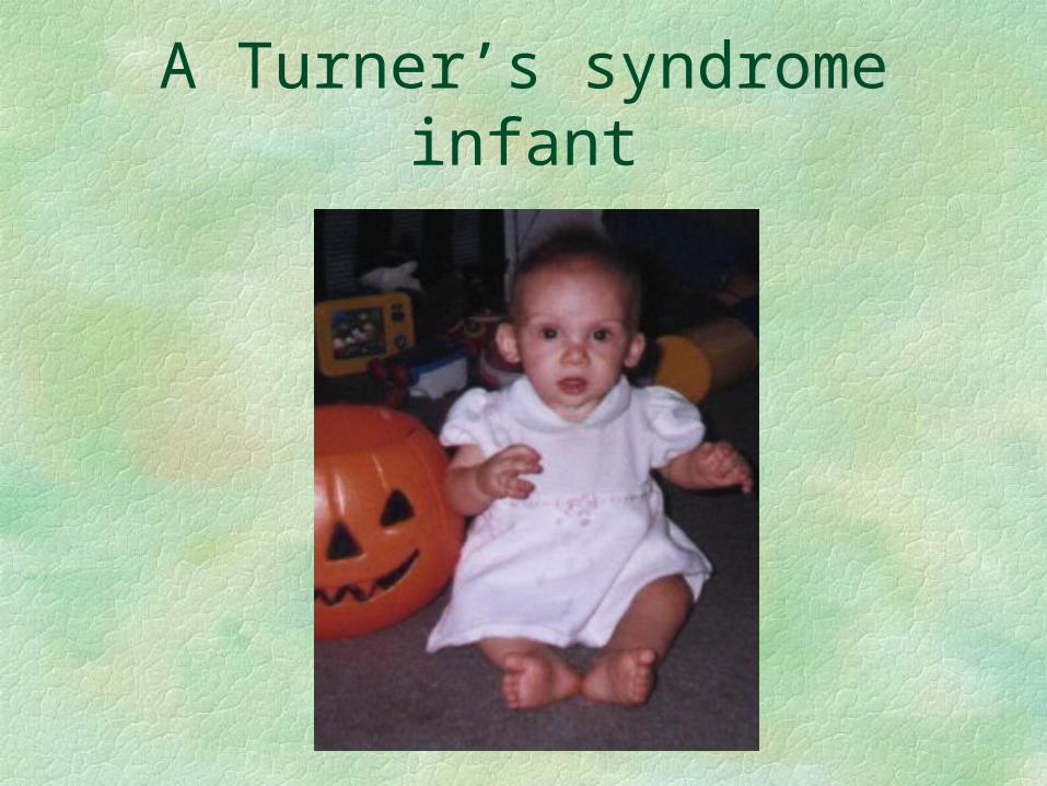
A Turner’s syndrome infant
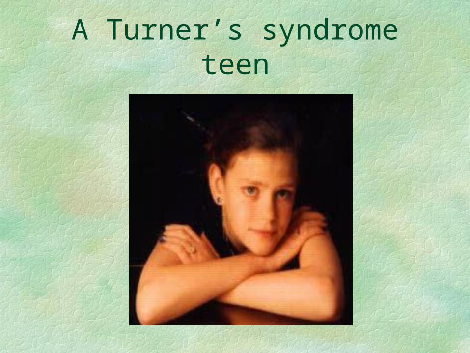
A Turner’s syndrome teen
Disorders from Abnormal Genes
Three types of genetic disorders:1. dominant gene disorder2. recessive gene disorder3. multifactorial gene disorderDominant gene disorder require only one
abnormal gene from either parent to affect a child
Disorders from Abnormal Genes (continued)
Huntington disease (dominant gene disorder)
gradual deterioration of the central nervous system, causing mental retardation and uncontrollable movements
appear when person is in their 30’s or 40’salways fatal
Disorders from Abnormal Genes (continued)
Recessive gene disorder--can occur when the fetus inherits a pair of
recessive genes, one from each parentCystic Fibrosis--involves production of
thick mucus, clogging the lungscauses delayed growth and sexual
maturation, shortened life and vulnerable to infections
Disorders from Abnormal Genes (continued)
Sickle-Cell Disease--red blood cells take on a sickle shape instead of a round shape
These cells get caught in the blood vessels cutting off circulation, reducing oxygen supply causing pain
have bacterial infectionsdegeneration of organs (due to lack of
oxygen)few live past 40 years of age
Disorders from Abnormal Genes (continued)
Tay-Sachs Disease--disease of the nervous system (chemical imbalance)
about age 6 months will show poor tolerance for sudden loud noises, seem weak and slow to develop
lead to seizures, deafness and blindnessmost die by age 3
Disorders from Abnormal Genes (continued)
Phenylketonuria (PKU)--occurs in 5-10/1000, inability to utilize an amino acid called phenylalanine (found in milk, meat) and needed for normal growth
increases in levels of phenylalanine in the blood and spinal fluid affects the brain causing deterioration of cognitive functioning and mental retardation
Disorders from Abnormal Genes (continued)
Multifactorial disorders--result from a combination of genetic and
environmental factorsNeural tube defects--result when the tube
enclosing the spinal cord fails to close completely or normally
Sometimes the upper part of the brain is absent or underdeveloped
Disorders from Abnormal Genes (continued)
Causes include heredity and environmental aspects such as pollutants, poor nutrition, diseases (diabetes)
geographical background and racial/ethic background)
British Isles has the highestU.S. highest is in the the Appalachian region
Disorders from Abnormal Genes (continued)
Cleft palate/lip--when the upper lip and/or roof of the mouth fail to grow resulting in a split or “cleft”
If severe may lead to difficulties breathing, speaking, hearing and eating
surgery can usually repair the problem
Disorders from Abnormal Genes (continued)
Congenital heart disease--structural and/or electrical abnormalities in the formation of the heart
medication and surgery can usual correct
Prenatal Diagnostic Techniques
Diagnostic techniques after conception but before birth
Ultrasound--high frequency sound waves are projected through the mother’s womb
Waves bounce off the fetus creating a television image of the size, shape, and position of the fetus
Prenatal Diagnostic Techniques (continued)
Ultrasound can be used to:determine age of fetusmultiple pregnanciesphysical defects in internal/external organsdetermine Down’s Syndrome (extra fold of
skin on the neck)
Prenatal Diagnostic TechniquesAmniocentesis--performed at weeks 14-18Using, ultrasound a slender needle is inserted
through the mother’s abdomen into the uterus and the amniotic sac and a sample of amniotic fluid is withdrawn
Fluid contain the cells which hold the genetic makeup of the fetus
can be used to determine if they have abnormal chromosomes
Prenatal Diagnostic Techniques (continued)
AmniocentesisIt can also determine disorders such as
Down’s Syndrome, neural tube defect and sex of the baby
Prenatal Diagnostic Techniques (continued)Chorionic Villus Sampling (CVS)tests for most of the same disorders as
amniocentesis doesperformed between the 8th & 10th week of
pregnancy involves collecting and analyzing tissue by
inserting a catheter through the vagina into the uterus between the uterine lining and the chorion (surrounds the embryo, becomes the placenta later)
Prenatal Diagnostic Techniques (continued)This technique lets parents know very early
whether the fetus has inherited any serious defects
primary risk of procedure is miscarriageFetoscopy--inserting a fetoscope (telesopic
type fiber optic lens through the abdomen into the uterus to observe the amniotic fluid, placenta, and fetus
performed 15th-18th week
Prenatal Diagnostic Techniques (continued)Fetoscopy is used to observe already
identified problems or to confirm other prenatal tests
Maternal Serum Alpha Fetoprotein test measuring the amount of alpha-
fetoprotein (AFP) in the mother’s bloodperformed 15th-18th weekfetoprotein is produced by the fetus and
passes from the amniotic fluid through the placenta into the mother’s bloodstream
Prenatal Diagnostic Techniques (continued)High level of AFP are associated with
various problems as anencephaly (lacking a brain), spina bifida, and Down’s Syndrome
Percutaneous Umbilical Blood Sampling (PUBS)
experimental method of sampling fetal blood by guiding a needle through the mother’s abdomen and uterus and umbilical vein
performed between the 18th and 36th week
Prenatal Diagnostic Techniques (continued)Used to diagnosis numerous conditions, such
as Down Syndrome, neural tube defects, cystic fibrosis)
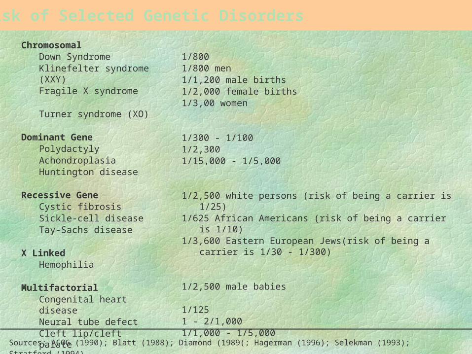
Risk of Selected Genetic Disorders
ChromosomalDown SyndromeKlinefelter syndrome (XXY)Fragile X syndrome
Turner syndrome (XO)
Dominant GenePolydactylyAchondroplasiaHuntington disease
Recessive GeneCystic fibrosisSickle-cell diseaseTay-Sachs disease
X LinkedHemophilia
MultifactorialCongenital heart diseaseNeural tube defectCleft lip/cleft palate
Sources: ACOG (1990); Blatt (1988); Diamond (1989(; Hagerman (1996); Selekman (1993); Stratford (1994).
1/8001/800 men1/1,200 male births1/2,000 female births1/3,00 women
1/300 - 1/1001/2,3001/15,000 - 1/5,000
1/2,500 white persons (risk of being a carrier is 1/25)1/625 African Americans (risk of being a carrier is 1/10)1/3,600 Eastern European Jews(risk of being a carrier is 1/30 -
1/300)
1/2,500 male babies
1/1251 - 2/1,0001/1,000 - 1/5,000
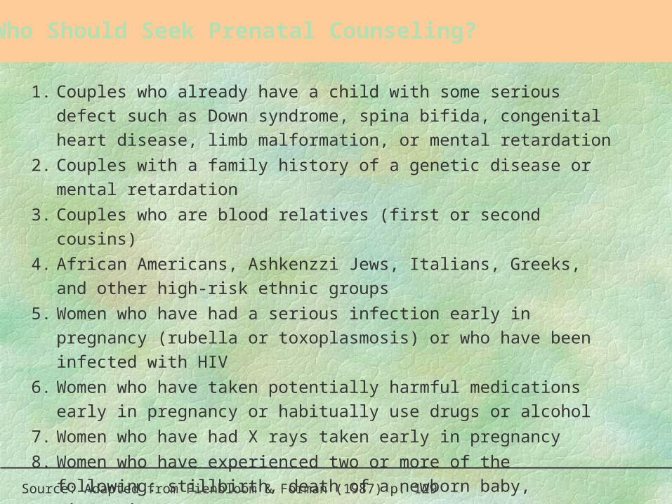
Who Should Seek Prenatal Counseling?
1. Couples who already have a child with some serious defect such as Down
syndrome, spina bifida, congenital heart disease, limb malformation, or
mental retardation
2. Couples with a family history of a genetic disease or mental retardation
3. Couples who are blood relatives (first or second cousins)
4. African Americans, Ashkenzzi Jews, Italians, Greeks, and other high-risk
ethnic groups
5. Women who have had a serious infection early in pregnancy (rubella or
toxoplasmosis) or who have been infected with HIV
6. Women who have taken potentially harmful medications early in
pregnancy or habitually use drugs or alcohol
7. Women who have had X rays taken early in pregnancy
8. Women who have experienced two or more of the following: stillbirth,
death of a newborn baby, miscarriage
9. Any woman thirty-five years or older
Source: Adapted from Fienbloom & Forman (1987) p. 129
Behavior Genetics
Behavior genetics--is the study of how genetic inheritance (genotype) and environmental experience jointly influence physical and behavioral development (phenotype)
Behavior Genetics (continued)
Four concepts of behavior geneticsrange of reaction-the range of possible
phenotypes that an individual with a particular genotype might exhibit in response to the specific sequence of environmental influences they experience
influences as neighborhood, child’s family, school and community etc.
Behavior Genetics (continued)
Canalization--tendency of genes to narrowly restrict the development of certain phenotypic characteristics to a single or relatively limited number of outcomes in spite of environmental pressures toward other outcomes
early perceptual-motor (crawling, sitting up are canalized)
Behavior Genetics (continued)
Others include personality, temperament, intellectual functioning
Gene-Environment Relationship-patterns of interaction between a newborn
infant and his caregiving environment and the extent to which that pattern supports the expression and development of the child’s inborn traits
Behavior Genetics (continued)
Examples include:shynessathletic abilityparents may display their own genetically
inherited traits that support the development of similar traits in their children
Behavior Genetics (continued)
A passive gene-environment relationship-if support for the development of traits come
from others (parents and family members)Evocative gene-environment relationship-displayed by child of certain traits evokes
support from others
Behavior Genetics (continued)
Active gene-environment relationship--when a child with a particular trait actively
seeks out support from others with similar traits (sports, etc.)