Embed Size (px)
Citation preview
PRINCIPLES OF ASTHMA
Mechanisms of asthmaChris Corrigan
AbstractAsthma is a syndrome of variable airflow obstruction. It is characterized
pathologically by bronchial inflammation and remodelling changes, phys-
iologically by bronchial hyperresponsiveness, and clinically by cough,
chest tightness and wheeze. Cytokines secreted by CD4þ Th2 type
T cells play a major role in coordinating asthmatic bronchial inflammation,
while other effector cells, such as myofibroblasts, epithelial cells, smooth
muscle cells and endothelial cells (activated partly by Th2 type cytokines
and partly by other mediators), play an intermediary role in airways
damage and remodelling. Although the pathological changes in the
airways in association with asthma are now well described, there is
a gap in our understanding of precisely how these changes cause clinical
symptoms. A key aetiological trigger factor for asthma is exposure to
environmental antigens, in particular inhaled allergens, including occupa-
tional allergens and infectious agents, which are probably a major drive to
T cell activation in asthma. Genetic factors governing the production of
T cell cytokines and their actions on target cells, as well as variability in
the structure and development of the mesenchymal elements of the bron-
chial mucosa influence the risk of developing asthma. Many other envi-
ronmental agents exacerbate asthma, but the evidence that they cause
disease is much less clear.
Keywords Asthma; atopy; cytokine; eosinophil; pathogenesis; remod-
elling; T cell
Clinical pathology
The diagnosis of asthma is made on the basis of typical symp-
toms and abnormalities in lung function. The key clinical
features of asthma include:
Variable airways obstruction: airways obstruction in asthma, as
measured by spirometry, may vary spontaneously from none to
severe in the course of hours to minutes, and improves after
suitable therapy. Obstruction, particularly of the smaller airways,
in asthmatics causes shortness of breath, impaired exercise
tolerance, tightness in the chest that may be perceived as
Chris Corrigan MA MSc PhD FRCP is Professor of Asthma, Allergy and
Respiratory Science at King’s College London School of Medicine and the
MRC and Asthma UK Centre for Allergic Mechanisms of Asthma, and
Consultant Physician at Guy’s and St Thomas’ NHS Foundation Trust,
London, UK. Competing interests: Dr Corrigan has received funding to
attend meetings and conferences from Schering-Plough, Allergy
Therapeutics, Meda AB, UCB Pharma, Novartis, Teva Pharmaceuticals,
undertaken research collaborations with GlaxoSmithKline, Novartis,
ALK-Abello, Allergy Therapeutics, Leti and acted as a consultant for Meda
AB, GlaxoSmithKline, Novartis, Merck Sharpe Dohme and Allergopharma
Joachim Ganzer AB. None of these interests is of any relevance to this
article.
MEDICINE 40:5 223
wheeze, and chest hyperinflation (small airways obstruction
prevents complete emptying of the alveoli, causing gas trapping).
Non-specific bronchial hyperreactivity: this refers to the
tendency of asthmatic airways to constrict in response to a host
of non-specific (that is, non-immunological) stimuli (including,
for example, strong smells, cold air, fog, smoke, exercise, aerosol
sprays and dust) that would not cause clinically significant
bronchoconstriction in non-asthmatics. Bronchial hyperreac-
tivity causes excessive cough and contributes to bronchospasm,
which may have different triggers in different patients.
Histopathology
Asthma is characterized by inflammatory changes throughout
the airways, but not the alveoli or lung parenchyma. The
inflammation is characterized by the activation of CD4þ helper
T lymphocytes,1 as well as accumulation of leukocytes in the
bronchial mucosa (Figures 1 and 2),2 although these changes do
not allow a definitive diagnosis on histopathological grounds.
Typically, there is an abundance of eosinophils but some
chronic, severe asthmatics show a more prominent neutrophil
leukocyte infiltrate.3 Mast cells are present in the bronchial
mucosa but they are not particularly abundant. It has so far not
been possible to associate aetiological subdivisions of asthma
with reproducible and discernible variability in histopathology,
although there is intense interest in this possibility and its
possible implications for tailored therapy.3
Asthma is also associated with structural changes in the
airways, collectively termed ‘remodelling’ (Figure 3).4 These
include hypertrophy and hyperplasia of airways smooth muscle
cells, increased numbers of mucous goblet cells in the airways
epithelium, laying down of fibrous proteins (including collagen,
fibronectin and tenascin) beneath the epithelial basement
membrane and in the submucosa, and neovascularization
(proliferation of vascular capillary beds within the submucosa).
Pathophysiology
Asthmatic inflammation appears to be coordinated principally by
activated CD4þ T lymphocytes of the Th2 type phenotype,
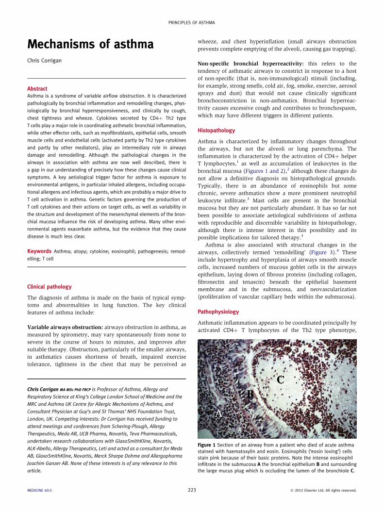
Figure 1 Section of an airway from a patient who died of acute asthma
stained with haematoxylin and eosin. Eosinophils (‘eosin loving’) cells
stain pink because of their basic proteins. Note the intense eosinophil
infiltrate in the submucosa A the bronchial epithelium B and surrounding
the large mucus plug which is occluding the lumen of the bronchiole C.
� 2012 Elsevier Ltd. All rights reserved.

Histamine
Mast cell
IL-4 IL-1 3
IL-4 IL-13, TGF-β
Th2 T cell Eosinophil
B cell
Epi
Fib AHR
Remodelling
Narrowing
Acute bronchospasm
ASM
IL-4IL-13 TGF- β
IL-5
Major basic proteins
Leukotrienes
IgE
Allergen
Other antigens?
ACUTE
CHRONIC
Prostaglandin D2
Leukotrienes
DC
B
CD4+
An overview of asthma pathogenesis
Th2-type, CD4+ T cells are activated by allergens and perhaps other antigens following presentation of specific epitopes by dendritic cells (DC). The T cells produce
the cytokines interleukin-4 (IL-4), IL-5 and IL-13. IL-5 promotes eosinophil recruitment to, and survival in, the airways mucosa. IL-4 and IL-13 contribute and also act on epithelial cells (Epi), fibroblasts (Fib) and airways smooth muscle cells (ASM) to cause remodelling changes. Eosinophils damage the mucosa through the release of basic proteins and exacerbate bronchospasm, vascular leakage and oedema through the release of leukotrienes. They also contribute to remodelling through release of pro-fibrotic mediators, such as transforming growth factor (TGF)-β. IL-4 and IL-13 cause IgE switching in B cells, promoting their secretion of allergen-specific IgE. Cross-linking of this IgE by allergens on the surface of mast cells in atopic subjects causes degranulation, with release of mast cell mediators (histamine, prostaglandins and leukotrienes). This is a mechanism for acute exacerbation of symptoms superimposed on the chronic inflammatory background created by T cells and their cytokines. AHR, airways
hyperresponsiveness.
Figure 2
PRINCIPLES OF ASTHMA
characterized particularly by the production of the cytokines
interleukin (IL)-4, IL-13 and IL-5 (see Figure 2).1 These cytokines
are implicated in causing eosinophil accumulation, since IL-4 and
IL-13 up-regulate adhesion molecules in the capillary endothe-
lium of the bronchial mucosa, resulting in increased adhesion
and diapedesis of eosinophils. Once in the tissues, IL-5 prolongs
the survival of eosinophils and primes them for increased release
of their basic granular proteins and production of other media-
tors, in particular cysteinyl leukotrienes, which promote vascular
leakage and thus oedema of the lining of the airways, and are
also the most potent natural bronchoconstrictors so far
described.5 All of these effects probably contribute to airways
obstruction.6
The cytokines IL-4 and IL-136 appear to be vital for remodelling.
They act directly on various structural cells of the airways (such as
epithelial cells) to induce mucous hyperplasia, and indirectly on
others (such as endothelial cells) to induce production of further
remodellingmediators, such as vascular endothelial growth factor
(VEGF), which in turn promotes angiogenesis. Also of emerging
importance is the Th2-type cytokine IL-25 which, in addition to
promoting memory Th2 T cell survival, has a number of direct
effects on remodelling such as angiogenesis.7 Activated eosino-
phils produce a growth factor (transforming growth factor
(TGF)-b) that acts on fibroblasts, causing them to proliferate and
transform into myofibroblasts, an intermediary between fibro-
blasts and smooth muscle cells, which are responsible for the
laying down of fibrous proteins in the bronchial mucosa. TGF-
b and other growth-regulating cytokines also cause increased
MEDICINE 40:5 224
proliferation of airways smooth muscle cells. Injured epithelial
cells also release TGF-b, forming the so-called ‘epithelial mesen-
chymal trophic unit’, which may also contribute to remodelling
changes in asthmatic airways.8 Thus, cytokines derived largely
from T cells are thought to drive most of the inflammatory and
airways structural changes characteristic of asthma, regardless of
its aetiology (Figure 4). Airways structural cells, as well as other
infiltrating leukocytes, including eosinophils and mast cells, are
likely also to contribute to the production of cytokines and growth
factors causing airways remodelling. In addition, there is evidence
that bronchial smooth muscle itself behaves abnormally in
asthma9 and that the stress of repeated bronchoconstriction may
actually cause some of the remodelling changes.10
Finally, IL-4 and IL-13 are the only human cytokines that
induce B lymphocytes to switch to IgE synthesis following acti-
vation by interaction with antigen-specific T cells (see Figure 2).
These cytokines are therefore implicated in the pathogenesis of
atopy (a propensity for inappropriate production of IgE anti-
bodies against antigens or ‘allergens’, encountered at mucosal
surfaces and detected by skin prick or laboratory tests).
Inflammation and asthma therapy
Corticosteroids strongly inhibit T cell pro-inflammatory cytokine
production1; in the bronchial mucosa of asthmatics, they reduce
infiltration of inflammatory granulocytes such as eosinophils. In
contrast, corticosteroids have few direct inhibitory effects on gran-
ulocytes, andmaynot reverse remodelling changes. Other actions of
� 2012 Elsevier Ltd. All rights reserved.

Features of airways remodelling in asthma: laying down of
fibrous protein beneath the epithelial basement membrane
causing apparent thickening (top), hypertrophy and hyperplasia
of airways smooth muscle (middle), and hypertrophy and
hyperplasia of epithelial mucous glands (bottom).
Figure 3
Activated, CD4+ Th2 cells produce many cytokinesimplicated in asthma pathogenesis
All of these cytokines are potential molecular targets in asthma but their redundancy of function, coupled with lack of knowledge as toprecisely how the pathological changes they induce cause clinical symptoms, has hindered therapeutic advances. AHR, airways hyperresponsiveness; IL, interleukin; TNF, tumour necrosis factor.
eosinophil and basophil activation
IL-9, IL-13
IL-4, IL-9, IL-13 mucushypersection
IL-4, IL-5, IL-9, IL-13
IL-4, IL-13 IgE switching
IL-4, IL-9, IL-13 mast cellactivation
TNF-α smooth muscle activation, remodelling
CD4+
Inflammatory Th2
T cell
AHR
Figure 4
PRINCIPLES OF ASTHMA
corticosteroids, such as reduction of vascular leakage, may also be
relevant to reducing mucosal oedema, thereby improving airways
obstruction and bronchial hyperresponsiveness. Whereas cortico-
steroids reduce production of pro-inflammatory cytokines, they
actually increase T cell production of the anti-inflammatory cyto-
kine, IL-10, a key product of a T cell subset called T regulatory cells,
which are now thought to play a role in immune homoeostasis.11
Cysteinyl leukotrienes are important pro-inflammatory prod-
ucts of cells implicated in asthma, particularly eosinophils.5 They
havemany effects thatmay be relevant to asthmapathophysiology,
such as capillary leakage, bronchoconstriction and further eosin-
ophil recruitment. Cysteinyl leukotriene receptor blockers have
a proven role in the treatment of some, but not all asthmatics.
b2-adrenoreceptor agonists relax bronchial smooth muscle and
are useful as ‘reliever’ therapy for acute symptoms of asthma. They
have not been shown convincingly to exert an anti-inflammatory
effect in asthma, although there is some evidence that long-
acting b-agonists may enhance some of the anti-inflammatory
effects of corticosteroids.
A recent therapeutic innovation for asthma is omalizumab,
a humanized monoclonal antibody that binds to the constant
MEDICINE 40:5 225
region of the IgE molecule, holding it in the circulation and
preventing its binding to high-affinity receptors on mast cells and
basophils and low-affinity IgE receptors on B cells and antigen-
presenting cells.12 Omalizumab, administered by intermittent
subcutaneous injection, reduces the frequency of severe exacer-
bations of asthma and sometimes spares corticosteroid therapy.
This confirms a mechanistic role for IgE in asthma, although it is
not clear if omalizumab acts primarily by reducing IgE-induced
mast cell degranulation, IgE-mediated capture of antigens by
B cells and other antigen-presenting cells, or by as yet uniden-
tified mechanisms.
Aetiology of asthma
Genetic susceptibility
The risk of developing asthma tends to run in families. Twin
studies estimate the proportion of asthma risk to be 50e60%
genetically determined, although it is likely that there are
extensive and complex interactions of gene effects with envi-
ronmental influences.13 Genome screens have linked inheritance
of various chromosomal regions with an increased risk of
asthma. Some of these regions contain genes encoding cytokines,
such as IL-4 or IL-13, or their receptors, implicated in asthma
pathogenesis, suggesting that genetic variability in the expression
or actions of these mediators may contribute to asthma risk.
More recent positional cloning strategies have identified new
genes, such as ADAM33, the products of which do not play a role
in known asthma pathways but are involved in the normal
development of the airways during embryogenesis; this raises the
fascinating possibility that variability, as yet uncharacterized, in
� 2012 Elsevier Ltd. All rights reserved.

PRINCIPLES OF ASTHMA
the structure or development of the airways predisposes some
individuals to asthma.14
Role of allergen exposure
When atopic patients inhale allergens to which they are clinically
sensitized through the production of allergen-specific IgE, the
result is cross-linkage of surface-bound IgE on mast cells within
the respiratory mucosa, causing them to degranulate acutely and
release inflammatory mediators, in particular histamine, prosta-
glandin D2 and leukotrienes. These mediators make asthma
acutely worse, since they all cause bronchial smooth muscle
contraction, vascular leakage and inflammatory cell recruitment.
Thus, allergen exposure is an important immunologically specific
trigger in some, but not all, asthmatics, acting on the background
of mucosal inflammation instigated by T cells. These patients
may have other diseases associated with atopy (eczema and
allergic rhinitis).
Early life sensitization to allergens, particularly indoor aller-
gens such as house dust mite, is a major risk factor for the
development of asthma in children, but only in the genetically
predisposed, raising doubts as to whether allergic sensitization is
actually causative of asthma or reflects parallel susceptibility
pathways arising from common genetic origins.15 Many atopic
patients do not develop asthma. Furthermore, asthma developing
later in life is less likely to be associated with atopy, suggesting
that IgE sensitization to allergens is neither necessary nor suffi-
cient for the development of asthma, although further appraisal
of the response, if any, of non-atopic asthmatics to omalizumab
(see above) may reveal that IgE plays a role in asthma even in
patients who are not atopic.
Occupational asthma
Some individuals develop asthma for the first time after exposure
to occupational agents, which may be broadly divided into
reactive chemicals (such as isocyanates) and proteins (such as
wheat flour). Proteins activate T cells directly in the manner of
an inhaled allergen and, like allergens, often induce an associ-
ated IgE response. Reactive chemicals react with native body
proteins such as albumin, creating new antigens that are recog-
nized as ‘foreign’ by T cells. The pathophysiology of occupa-
tional asthma, once developed, appears to be indistinguishable
from that seen in other clinical phenotypes of asthma. Again, it is
not clear what governs individual susceptibility.
Other influences on the development of asthma
Asthma and atopy are, geographically, diseases of developed
countries. This has resulted in speculation about the role of
‘Westernized’ society in their causation.
Pollution and smoking: many pollutants exacerbate asthma but
there is limited evidence that they predispose to its development.15
Air pollution has declined in many Western industrialized coun-
tries while asthma prevalence has increased. Epidemiological and
experimental evidence suggests that diesel exhaust particulates
and ozone promote sensitization to allergens. Exposure of chil-
dren to tobacco smoke in utero or early childhood increases their
risk of developing wheeze. Smoking in adults increases the risk of
developing asthma, including occupational asthma.16 The patho-
physiological mechanisms are unknown.
MEDICINE 40:5 226
Diet: the growth of the fast food diet and escalating obesity have
led to speculation about lack of nutrients (antioxidant vitamins,
omega-3 fatty acids, selenium, magnesium, sodium and zinc)
and obesity playing a role in asthma causation, but there is no
conclusive evidence.17 Obesity itself may be associated with
accelerated decline in lung function so the epidemiological
problems are complex.18 Breast feeding reduces the risk of
eczema in atopic ‘at risk’ children, but the evidence relating to
asthma is inconclusive.
Infection: the pattern of microbial exposure in infancy and
childhood has changed as a result of a cleaner environment and
widespread use of antibiotics and vaccination. The ‘hygiene
hypothesis’ invokes this as a risk factor for asthma and atopy.19
The precise mechanisms are unknown but the hypothesis has
focused research on the effects of alteration of the intestinal flora
early in life, exposure to airborne bacterial products (such as
endotoxins on farms) and the influence of specific (viral,
helminthic) infections on the risk of developing asthma.
Research is ongoing. A
REFERENCES
1 Corrigan C. T cells and cytokines in asthma and allergic inflammation.
In: Kay AB, Kaplan AP, Bousquet J, Holt PG, eds. Allergy and allergic
diseases. 2nd edn. Oxford: Wiley-Blackwell, 2008.
2 Lemanske RF, Busse WW. Asthma: clinical expression and molecular
mechanisms. J Allergy Clin Immunol 2010; 125: S95e102.
3 Haldar P, Pavord ID. Noneosinophilic asthma: a distinct clinical and
pathologic phenotype. J Allergy Clin Immunol 2007; 119: 1043e52.
4 James AL, Wenzel S. Clinical relevance of airway remodelling in airway
diseases. Eur Respir J 2007; 30: 134e55.
5 Ogawa Y, Calhoun WJ. The role of leukotrienes in airway inflamma-
tion. J Allergy Clin Immunol 2007; 118: 789e98.
6 Kraft M. Asthma phenotypes and interleukin-13: moving closer to
personalised medicine. New Engl J Med 2011; 365: 1141e4.
7 Corrigan CJ, Wang W, Meng Q, et al. T-helper cell type 2 (Th2) memory
T cell-potentiating cytokine IL-25 has the potential to promote
angiogenesis in asthma. Proc Natl Acad Sci USA 2011; 108:
1579e84.
8 Holgate ST, Holloway J, Wilson S, Bucchieri F, Puddicombe S,
Davies DE. Epithelial-mesenchymal communication in the pathogen-
esis of chronic asthma. Proc Am Thorac Soc 2004; 1: 93e8.
9 Mahn K, Hirst SJ, Ying S, et al. Diminished sarco/endoplasmic retic-
ulum Ca2þ ATPase (SERCA) expression contributes to airway
remodelling in bronchial asthma. Proc Natl Acad Sci USA 2009;
106: 10775e80.
10 Grainge CL, Lau LCK, Ward JA, et al. Effect of bronchoconstriction on
airway remodelling in asthma. N Engl J Med 2011; 364: 2006e15.
11 Ryanna K, Staryigou V, Safinia N, Hawrylowicz C. Regulatory T cells in
bronchial asthma. Allergy 2009; 64: 335e47.
12 Avila PC. Does anti-IgE therapy help in asthma? Efficacy and
controversies. Annu Rev Med 2007; 58: 185e203.
13 Holgate ST, Davies DE, Powell RM, Howarth PH, Haitchi HM,
Holloway JW. Local genetic and environmental factors in asthma
disease pathogenesis: chronicity and persistence mechanisms. Eur
Respir J 2007; 29: 793e803.
14 Wills-Karp M, Ewart SL. Time to draw breath: asthma susceptibility
genes are identified. Nat Rev Genet 2004; 5: 376e87.
� 2012 Elsevier Ltd. All rights reserved.
PRINCIPLES OF ASTHMA
15 Arruda LK, Sole D, Baena-Cagnani CE, Naspitz CK. Risk factors
for asthma and atopy. Curr Opin Allergy Clin Immunol 2005; 5:
153e9.
16 Thomson NC. The role of environmental tobacco smoke in the
origins and progression of asthma. Curr Allergy Asthma Rep 2007;
7: 303e9.
MEDICINE 40:5 227
17 Ford ES. The epidemiology of obesity and asthma. J Allergy Clin
Immunol 2005; 115: 897e909.
18 Tantisira KG. In asthma, the apple falls faster than the pear (Editorial).
J Allergy Clin Immunol 2009; 123: 1075e6.
19 Ramsay C, Celedon J. The hygiene hypothesis and asthma. Curr Opin
Pulm Med 2005; 11: 14e20.
� 2012 Elsevier Ltd. All rights reserved.