Embed Size (px)
Citation preview

Mechanism of urine forming

The Nephron Is the Functional Unit of the Kidney
• Each kidney in the human contains about 1 million nephrons, each capable of forming urine. The kidney cannot regenerate new nephrons. Therefore, with renal injury, disease, or normal aging, there is a gradual decrease in nephron number.

• Each nephron contains (1) a tuft of glomerular capillaries called the glomerulus, through which large amounts of fluid are filtered from the blood, and (2) a long tubule in which the filtered fluid is converted into urine on its way to the pelvis of the kidney.

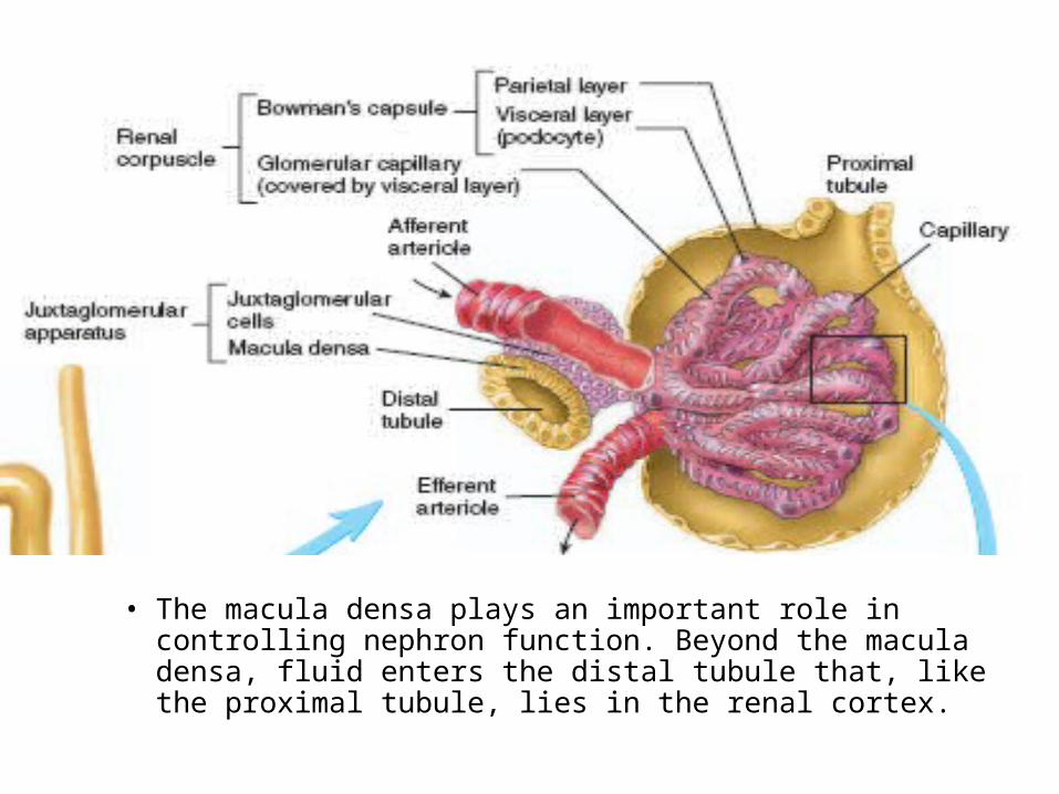
• The macula densa plays an important role in controlling nephron function. Beyond the macula densa, fluid enters the distal tubule that, like the proximal tubule, lies in the renal cortex.

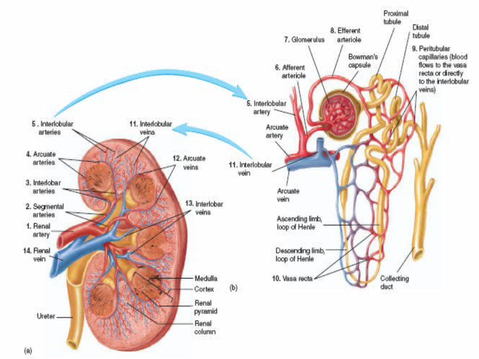
Renal Blood Supply
• Blood flow to the two kidneys is normally about 22 per cent of the cardiac output, or 1100 ml/min.
• The renal artery enters the kidney through the hilum and then branches progressively to form the interlobar arteries, arcuate arteries, interlobular arteries (also called radial arteries), and afferent arterioles, which lead to the glomerular capillaries, where large amounts of fluid and solutes (except the plasma proteins) are filtered to begin urine formation.
• The distal ends of the capillaries of each glomerulus coalesce to form the efferent arteriole, which leads to a second capillary network. the peritubular capillaries, that surrounds the renal tubules.
• Video
PHYSIOLOGIC CONTROL OF GLOMERULAR FILTRATION AND RENAL BLOOD FLOW
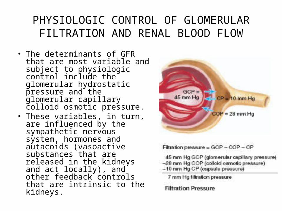
• The determinants of GFR that are most variable and subject to physiologic control include the glomerular hydrostatic pressure and the glomerular capillary colloid osmotic pressure.
• These variables, in turn, are influenced by the sympathetic nervous system, hormones and autacoids (vasoactive substances that are released in the kidneys and act locally), and other feedback controls that are intrinsic to the kidneys.

Sympathetic Nervous System Activation Decreases GFR
• Strong activation of the renal sympathetic nerves can constrict the renal arterioles and decrease renal blood flow and GFR.
• Moderate or mild sympathetic stimulation has little influence on renal blood flow and GFR. For example, reflex activation of the sympathetic nervous system resulting from moderate decreases in pressure at the carotid sinus baroreceptors or cardiopulmonary receptors has little influence on renal blood flow or GFR. Moreover, because the baroreceptors adapt within minutes or hours to sustained changes in arterial pressure, il is unlikely that these reflex mechanisms have an important role in longterm control of renal blood flow and GFR.
• The renal sympathetic nerves seem to be most important in reducing GFR during severe, acute disturbances, lasting for a few minutes to a few hours, such as those elicited by the defense reaction, brain ischemia, or severe hemorrhage. In the healthy resting person, there appears to be little sympathetic tone to the kidneys.

Hormonal and Autacoid Control of Renal Circulation
• Norepinephrine, Epinephrine, and Endothelin Constrict Renal Blood Vessels and Decrease GFR. Hormones that constrict afferent and efferent arterioles, causing reductions in GFR and renal blood flow, include norepinephrine and epinephrine released from the adrenal medulla.
• The endothelin may contribute to hemostasis (minimizing blood loss) when a blood vessel is severed, which damages the endothelium and releases this powerful vasoconstrictor. Plasma endothelin levels also are increased in certain disease states associated with vascular injury, such as toxemia of pregnancy, acute renal failure, and chronic uremia.

Angiotensin II Constricts Efferent Arterioles
• A powerful renal vasoconstrictor, angiotensin II, can be considered as a circulating hormone as well as a locally produced autacoid because it is formed in the kidneys as well as in the systemic circulation. Because angiotensin II preferentially constricts efferent arterioles, increased angiotensin II levels raise glomerular hydrostatic pressure while reducing renal blood flow.
• It should be kept in mind that increased angiotensin II formation usually occurs in circumstances associated with decreased arterial pressure or volume depletion, which tend to decrease GFR
• Increased angiotensin II levels that occur with a low-sodium diet or volume depletion help to preserve GFR and to maintain a normal excretion of metabolic waste products, such as urea and creatinine, that depend on glomerular filtration for their excretion.

Endothelial-Derived Nitric Oxide Decreases Renal Vascular Resistance and Increases GFR
• A basal level of nitric oxide production appears to be important for preventing excessive vasoconstriction of the kidneys and allowing them to excrete normal amounts of sodium and water.
• Administration of drugs that inhibit the formation of nitric oxide increases renal vascular resistance and decreases GFR and urinary sodium excretion, eventually causing high blood pressure.
• In some hypertensive patients, impaired nitric oxide production may contribute to renal vasoconstriction and increased blood pressure.

Prostaglandins and Bradykinin Tend to Increase GFR
• Hormones and autacoids that cause vasodilation and increased renal blood flow and GFR include the prostaglandins (PGE 2 and PG12) and bradykinin.
• By opposing vasoconstriction of afferent arterioles, the prostaglandins may help to prevent excessive reductions in GFR and renal blood flow.
• Under stressful conditions, such as volume depletion or after surgery, the administration of nonsteroidal anti-inflammatory agents, such as aspirin, that inhibit prostaglandin synthesis may cause significant reductions in GFR.

Function of nephrone Video

AUTOREGULATION OF GFR AND RENAL BLOOD FLOW
• Feedback mechanisms intrinsic to the kidneys normally keep the renal blood flow and GFR relatively constant, despite marked changes in arterial blood pressure. These mechanisms still function in blood-perfused kidneys thal have been removed from the body, independent of systemic influences. This relative constancy of GFR and renal blood flow is referred to as autoregulation.
• The primary function of blood flow autoregulation in most other tissues besides the kidneys is to maintain delivery of oxygen and nutrients to the tissues at a normal level and to remove the waste products of metabolism, despite changes in the arterial pressure. In the kidneys, the normal blood flow is much higher than required for these functions. The major function of autoregulation in the kidneys is to maintain a relatively constant GFR and to allow precise control of renal excretion of water and solutes. The GFR normally remains autoregulated (that is, remains relatively constant), despite considerable arterial pressure fluctuations that occur during a person's usual activities. In general, renal blood flow is autoregulated in parallel with GFR, but GFR is more efficiently autoregulated under certain conditions.

Myogenic Autoregulation of Renal Blood Flow and GFR
• A second mechanism that contributes to the maintenance of a relatively constant renal blood flow and GFR is the ability of individual blood vessels to resist stretching during increased arterial pressure, a phenomenon referred to as the myogenic mechanism.
• Stretch of the vascular wall allows increased movement of calcium ions from the extracellular fluid into the cells, causing them to contract through the mechanisms. This contraction prevents overdistention of the vessel and at the same time, by raising vascular resistance, helps to prevent excessive increases in renal blood flow and GFR when arterial pressure increases.

URINE FORMATION• The rates at which different substances are excreted in
the urine represent the sum of three renal processes, (1) glomerular filtration, (2) reabsorption of substances from the renal tubules into the blood, and (3) secretion of substances from the blood into the renal tubules.
• Expressed mathematically, • Urinary excretion rate = Filtration rate • - Reabsorption rate + Secretion rate

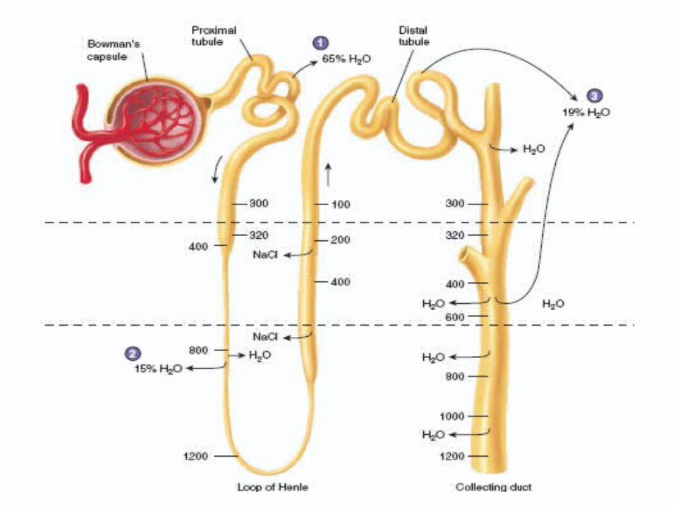
• Urine formation begins with filtration from the glomerular capillaries into Bowman's capsule of a large amount of fluid that is virtually free of protein.
• Most substances in the plasma, except for proteins, are freely filtered so that their concentrations in the glomerular filtrate in Bowman's capsule are almost the same as in the plasma.

Why Are Large Amounts of Solutes Filtered and Then Reabsorbed by the Kidneys?
• One advantage of a high GFR is that it allows the kidneys to rapidly remove waste products from the body that depend primarily on glomerular filtration for their excretion. Most waste products are poorly reabsorbed by the tubules and, therefore, depend on a high GFR for effective removal from the body.
• A second advantage of a high GFR is that it allows all the body fluids to be filtered and processed by the kidney many times each day. Because the entire plasma volume is only about 3 liters, whereas the GFR is about 180 L/day, the entire plasma can be filtered and processed about 60 times each day. This high GFR allows the kidneys to precisely and rapidly control the volume and composition of the body fluids.

Glomerular Capillary Membrane
• The glomerular capillary membrane is similar to that of other capillaries, except that it has three (instead of the usual two) major layers:
• (1) the endothelium of the capillary, • (2) a basement membrane, and • (3) a layer of epithelial cells (podocytes)
surrounding the outer surface of the capillary basement membrane.
• Together, these layers make up the filtration barrier that, despite the three layers, filters several hundred times as much water and solutes as the usual capillary membrane.
Glomerular Capillary Membrane
• Although the fenestrations are relatively large, endothelial cells are richly endowed with fixed negative charges that hinder the passage of plasma proteins.
• The basement membrane effectively prevents filtration of plasma proteins.

Podocytes• The final part of
the glomerular membrane is a layer of epithelial cells (podocytes) that encircle the outer surface of the capillaries.
• The foot processes are separated by gaps called slit pores through which the glomemlar filtrate moves. The epithelial cells, which also have negative charges, provide additional restriction to filtration of plasma proteins.

Three basic renal processes
• The substance is freely filtered but is also partly reabsorbed from the tubules back into the blood.
• For each substance in the plasma, a particular combination of filtration, reabsorption, and secretion occurs. The rate at which the substance is excreted in the urine depends on the relative rates of these three basic renal processes.

Filtration, Reabsorption, and Secretion of Different Substances
• In general, tubular, reabsorption is quantitatively more important than tubular secretion in the formation of urine, but secretion plays an important role in determining the amounts of potassium and hydrogen ions and a few other substances that are excreted in the urine.
• Most substances that must be cleared from the blood, especially the end products of metabolism such as urea, creatinine, uric acid, and urates, are poorly reabsorbed and are, therefore, excreted in large amounts in the urine.
• Certain foreign substances and drugs are also poorly reabsorbed but, in addition, are secreted from the blood into the tubules, so that their excretion rates are high.

Filtration, Reabsorption, and Secretion of Different Substances
• Nutritional substances, such as amino acids and glucose, are completely reabsorbed from the tubules and do not appear in the urine even though large amounts are filtered by the glomerular capillaries. Each of the processes - glomerular filtration, tubular reabsorption, and tubular secretion - is regulated according to the needs of the body.

Tubular reabsorption

Tubular secretion

Countercurrent mecanism and concentration of urine