Embed Size (px)
Citation preview
R e s e a r c h A r t i c l e
The Rockefeller University Press $30.00J. Gen. Physiol. 2017 Vol. 149 No. 4 511–522https://doi.org/10.1085/jgp.201611747
511
I n t R o d u c t I o n
The Fluc family of fluoride channels counteracts F− tox-icity in microorganisms by undermining weak acid ac-cumulation of environmental F− ion (Baker et al., 2012; Stockbridge et al., 2013; Ji et al., 2014). Recent crystal structures of two different Fluc homologues reveal an antiparallel homodimer with twofold symmetry about the plane of the membrane and two antiparallel pores, each with two resolved F− ions (Stockbridge et al., 2015). The channel is capped by monobody crystalliza-tion chaperones, one on each side of the membrane, which wedge loops into a deep cleft between the sub-units of the dimer. These monobodies, small synthetic proteins based on a human fibronectin III domain scaf-fold, were selected from combinatorial libraries and ex-hibit tight, specific binding to Fluc channels (Koide et al., 2012). Electrophysiological experiments show that several of the monobodies selected to bind Fluc pro-teins also inhibit F− current when applied to conduct-ing channels in planar lipid bilayers (Stockbridge et al., 2014, 2015). Because they play dual roles as inhibitors and crystallization chaperones, understanding the
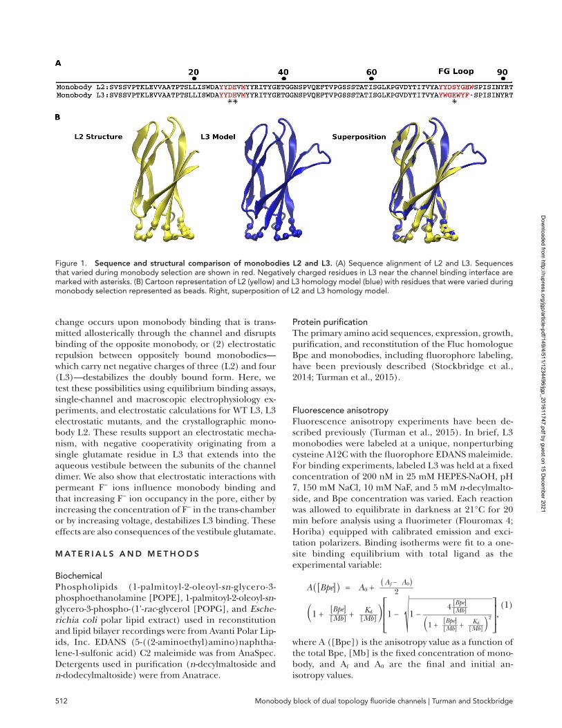
mechanism by which they inhibit provides critical con-text for interpreting crystal structures of Fluc channels. In this paper, we discuss two closely related monobod-ies: L3, which has been extensively characterized using electrophysiology, and L2, which was used to crystallize the Fluc homologue from Bordetella pertussis, Bpe (Stockbridge et al., 2015). The L2 and L3 sequences dif-fer only in an eight-residue loop (Fig. 1).
When applied to either side of the bilayer, monobody L3 blocks Bpe with identical kinetics regardless of the side to which is it is added (Stockbridge et al., 2014). These results appeared to be in harmony with the dou-bly L2-bound channel observed in the crystal structure. However, recent single-channel experiments show that when L3 is applied to both sides of the channel simulta-neously, binding of the second L3 molecule is ∼10-fold weaker than binding of the first, a difference in affinity of ∼1.4 kcal/mol (Turman et al., 2015). The origin of this negative cooperativity is unknown, but two possible mechanisms were suggested: (1) a conformational
The Fluc family of proteins comprises small, electrodiffusive fluoride channels, which prevent accumulation of toxic F− ions in microorganisms. Recent crystal structures have confirmed their unusual architecture, in which a pair of antiparallel subunits convenes to form a dimer with a twofold symmetry axis parallel to the plane of the membrane. These structures have also revealed the interactions between Fluc channels and several different fi-bronectin domain monobodies that inhibit Fluc-mediated F− currents; in all structures, each channel binds to two monobodies symmetrically, one on either side of the membrane. However, these structures do not reveal the mechanism of monobody inhibition. Moreover, the results appear to diverge from a recent electrophysiological study indicating that monobody binding is negatively cooperative; that is, a bound monobody on one side of a Fluc channel decreases the affinity of an oppositely bound monobody by ∼10-fold. In this study, we reconcile these observations by probing the mechanism of monobody binding and its negative cooperativity using elec-trophysiological experiments in planar lipid bilayers. Our results indicate that monobody inhibition occurs via a pore-blocking mechanism and that negative cooperativity arises from electrostatic repulsion between the oppo-sitely bound monobodies. A single glutamate residue, on a loop of the monobody that extends into the channel interior, is responsible for negatively cooperative binding. This glutamate side chain also confers voltage depen-dence and sensitivity to the concentration of trans-F− ion to monobody binding. Neutralization by mutation to glutamine abolishes these electrostatic effects. Monobodies that are amenable to cocrystallization with Fluc channels lack an analogous negatively charged side chain and bind independently to opposite sides of the chan-nel. Thus, this work reveals the source of voltage dependence and negative cooperativity of monobody binding to Fluc channels along with the pore-blocking mechanism.
Mechanism of single- and double-sided inhibition of dual topology fluoride channels by synthetic monobodies
Daniel L. Turman1 and Randy B. Stockbridge2,3
1Department of Biochemistry and Howard Hughes Medical Institute, Brandeis University, Waltham, MA 024542Program in Biophysics and 3Department of Molecular, Cellular, and Developmental Biology, University of Michigan, Ann Arbor, MI 48109
The
Jo
urn
al o
f G
ene
ral
Phy
sio
log
y
© 2017 Turman and Stockbridge This article is distributed under the terms of an Attribution–Noncommercial–Share Alike–No Mirror Sites license for the first six months after the publication date (see http ://www .rupress .org /terms /). After six months it is available under a Creative Commons License (Attribution–Noncommercial–Share Alike 4.0 International license, as described at https ://creativecommons .org /licenses /by -nc -sa /4 .0 /).
Correspondence to Randy B. Stockbridge: [email protected] used: APBS, adaptive Poisson-Boltzmann solver.
Dow
nloaded from http://rupress.org/jgp/article-pdf/149/4/511/1234496/jgp_201611747.pdf by guest on 15 D
ecember 2021
Monobody block of dual topology fluoride channels | Turman and Stockbridge512
change occurs upon monobody binding that is trans-mitted allosterically through the channel and disrupts binding of the opposite monobody, or (2) electrostatic repulsion between oppositely bound monobodies—which carry net negative charges of three (L2) and four (L3)—destabilizes the doubly bound form. Here, we test these possibilities using equilibrium binding assays, single-channel and macroscopic electrophysiology ex-periments, and electrostatic calculations for WT L3, L3 electrostatic mutants, and the crystallographic mono-body L2. These results support an electrostatic mecha-nism, with negative cooperativity originating from a single glutamate residue in L3 that extends into the aqueous vestibule between the subunits of the channel dimer. We also show that electrostatic interactions with permeant F− ions influence monobody binding and that increasing F− ion occupancy in the pore, either by increasing the concentration of F− in the trans-chamber or by increasing voltage, destabilizes L3 binding. These effects are also consequences of the vestibule glutamate.
M At e R I A l s A n d M e t h o d s
BiochemicalPhospholipids (1-palmitoyl-2-oleoyl-sn-glycero-3- phosphoethanolamine [POPE], 1-palmitoyl-2-oleoyl-sn-glycero-3-phospho-(1'-rac-glycerol [POPG], and Esche-richia coli polar lipid extract) used in reconstitution and lipid bilayer recordings were from Avanti Polar Lip-ids, Inc. EDA NS (5-((2-aminoethyl)amino)naphtha-lene-1-sulfonic acid) C2 maleimide was from AnaSpec. Detergents used in purification (n-decylmaltoside and n-dodecylmaltoside) were from Anatrace.
Protein purificationThe primary amino acid sequences, expression, growth, purification, and reconstitution of the Fluc homologue Bpe and monobodies, including fluorophore labeling, have been previously described (Stockbridge et al., 2014; Turman et al., 2015).
Fluorescence anisotropyFluorescence anisotropy experiments have been de-scribed previously (Turman et al., 2015). In brief, L3 monobodies were labeled at a unique, nonperturbing cysteine A12C with the fluorophore EDA NS maleimide. For binding experiments, labeled L3 was held at a fixed concentration of 200 nM in 25 mM HEP ES-NaOH, pH 7, 150 mM NaCl, 10 mM NaF, and 5 mM n-decylmalto-side, and Bpe concentration was varied. Each reaction was allowed to equilibrate in darkness at 21°C for 20 min before analysis using a fluorimeter (Flouromax 4; Horiba) equipped with calibrated emission and exci-tation polarizers. Binding isotherms were fit to a one-site binding equilibrium with total ligand as the experimental variable:
A ( [ Bpe ] ) = A 0 + ( A f − A 0 )
_______ 2
(1 +
[ Bpe ] _____ [ Mb ] + K d ____ [ Mb ] ) ⎡ ⎢
⎣1 − √
________________
1 − 4
[ Bpe ] _____ [ Mb ] ______________ (1 +
[ Bpe ] _____ [ Mb ] + K d ____ [ Mb ] ) 2
⎤ ⎥
⎦ , (1)
where A ([Bpe]) is the anisotropy value as a function of the total Bpe, [Mb] is the fixed concentration of mono-body, and Af and A0 are the final and initial an-isotropy values.
Figure 1. sequence and structural comparison of monobodies l2 and l3. (A) Sequence alignment of L2 and L3. Sequences that varied during monobody selection are shown in red. Negatively charged residues in L3 near the channel binding interface are marked with asterisks. (B) Cartoon representation of L2 (yellow) and L3 homology model (blue) with residues that were varied during monobody selection represented as beads. Right, superposition of L2 and L3 homology model.
Dow
nloaded from http://rupress.org/jgp/article-pdf/149/4/511/1234496/jgp_201611747.pdf by guest on 15 D
ecember 2021
513JGP Vol. 149, No. 4
Single-channel and macroscopic bilayer recordingBpe proteoliposomes were prepared at 0.05 µg of pro-tein/mg E. coli polar lipid extract for single-channel recordings and 10 µg/mg for macroscopic recordings and fused with a synthetic planar lipid bilayer (70% POPE/30% POPG). In all recordings, electrical ground is defined as the trans-chamber. Temperature was main-tained between 21 and 23°C. Single and macroscopic block recording data were acquired as described previ-ously (Stockbridge et al., 2014). For single-side block, voltage dependence, double-side block, and macro-scopic recordings, the cis- and trans-chambers con-tained 15 mM MOPS, pH 7, 300 mM NaF, and, to prevent nonspecific monobody adhesion to the record-ing chamber, 50 µg/ml bovine serum albumin. In some experiments, the trans-F− concentration was varied as described in the Results section (Fig. 9). Single-channel recordings acquired at voltages higher than ±100 were electronically filtered at 500 Hz to 1 kHz during acquisi-tion and digitally filtered to 100 Hz for analysis. Record-ings acquired at lower holding voltages required additional digital filtering to as low as 10 Hz for analysis. Control experiments were performed to ensure that block events were not missed as a result of filtering as described previously (Turman et al., 2015). Dwell time kinetics was calculated from single or double exponen-tial fits to cumulative distribution histograms as de-scribed in the Results section (Fig. 5). For L3 WT and L3 E29Q, histograms contained 25–480 events per sin-gle-channel run, and for L3 E79Q, histograms con-tained 800–1,500 events. All data points represent the mean and SEM of at least three independent sin-gle-channel recordings. Macroscopic recordings of hundreds or thousands of channels were acquired at 1-kHz electronic filtering. Liposome fusion was allowed to proceed until a steady current level (∼200 pA to 1 nA) was reached, and solution was exchanged to pre-vent additional fusion events. After monobody addition, current was allowed to reach steady-state levels (∼10 min), and current was recorded as a mean value over a 1-min recording time.
L3 homology model and electrostatic calculationsThe L3 homology model (L3–Bpe) was calculated using Modeller 9.15 software with L2–Bpe (PDB accession no. 5FXB) as the template (Webb and Sali, 2016). The auto-model function was used with no additional spatial re-straints. The structure with the lowest discrete protein optimized energy score was selected for further study. L3–Bpe was then explicitly centered within an 80 × 80–Å POPE membrane slab and explicitly solvated with VMD 1.9.3 solvate (Humphrey et al., 1996). Electroneu-tral conditions were established by placing NaCl above and below the membrane slab at 0.150 M. The simula-tion system comprising a channel dimer, two monobod-ies, POPE membrane, water, and ions consisted of
50,260 atoms with simulation dimensions of 100, 100, and 140 Å. This system was minimized and equilibrated stepwise over a total of 10 ns at 100, 200, and 300 K using NAMD 2.11, initially restraining all atoms except lipid tails, and then harmonic restraints were imposed on the protein only (Phillips et al., 2005). The final step was completed with no restraints over 2 ns. Alignment of L3–Bpe before and after minimization resulted in a root mean square (RMS) of 1.07 Å. Protein charge and radius parameters for L3–Bpe were produced with the adaptive Poisson-Boltzmann solver (APBS) using CHA RMM force field parameters. APBS and APBSmem were used to implicitly model L3–Bpe in a membrane envi-ronment (Baker et al., 2001; Callenberg et al., 2010). APBS was then used to solve the nonlinear Poisson Boltzmann equation with dielectric charge and ionic accessibility maps from APBSmem. Grid discretization was optimized to convergence for this system with grid dimensions of 161 × 161 × 161 Å and a grid length of 120 × 120 × 120 Å for grid spacing of 0.75 Å. Boundary conditions were determined by focusing from a coarse grid spacing of 1.23 Å. Per-atom electrostatic energies at E79 oxygen atoms OE1 and OE2 were used to calcu-late ΔESF values.
R e s u lt s
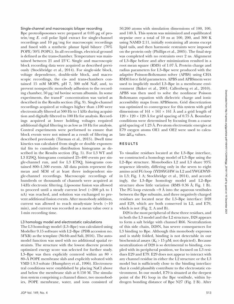
To visualize residues located at the L3–Bpe interface, we constructed a homology model of L3–Bpe using the L2–Bpe structure. Monobodies L2 and L3 share 93% sequence identity, differing only in the seven to eight amino acid FG loop (YYD SYG HW in L2 and YWG EWYF- in L3; Fig. 1 A; Stockbridge et al., 2014), and accord-ingly, the L3–Bpe homology model and L2–Bpe structure show little variation (RMS 0.36 Å; Fig. 1 B). The FG loop extends ∼9 Å into the aqueous vestibules between the Bpe subunits, and three negatively charged residues are located near the L3–Bpe interface: D28 and E29, which are both conserved in L2, and E79, which is not (Fig. 2, A and B).
D28 is the most peripheral of these three residues, and in both the L3 model and the L2 structure, D28 appears to form a salt bridge with channel R66. Neutralization of this side chain, D28N, has severe consequences for L3 binding to Bpe. Although this monobody expresses and is stably folded, binding is not detectable in our biochemical assays (Kd > 15 µM; not depicted). Because neutralization of D28 is so detrimental to binding, cou-pled with its peripheral position, we focused on L3 resi-dues E29 and E79. E29 does not appear to interact with any channel residue in either the L2 structure or the L3 model but is sufficiently close to the binding interface that it could plausibly contribute to the electrostatic en-vironment. In our model, E79 is situated at the deepest point of the FG loop in the Bpe vestibule, within hy-drogen bonding distance of Bpe N27 (Fig. 2 B). After
Dow
nloaded from http://rupress.org/jgp/article-pdf/149/4/511/1234496/jgp_201611747.pdf by guest on 15 D
ecember 2021
Monobody block of dual topology fluoride channels | Turman and Stockbridge514
mutation of E79 and E29 individually to glutamine, the binding affinities of the mutant monobodies to deter-gent-solubilized Bpe were measured using fluorescence anisotropy as a function of increasing channel concen-tration (Fig. 2 C). These experiments do not provide an indication of binding stoichiometry but allow us to estimate the equilibrium binding affinities of ∼150 nM for E29Q and 3.3 µM for E79Q compared with 96 nM for L3 WT (Turman et al., 2015). Because of the rela-tively weak interaction between E79Q and channel, stoi-chiometry experiments using anisotropy measurements are not experimentally feasible, and we instead focused on an electrophysiological test of binding cooperativity (Turman et al., 2015).
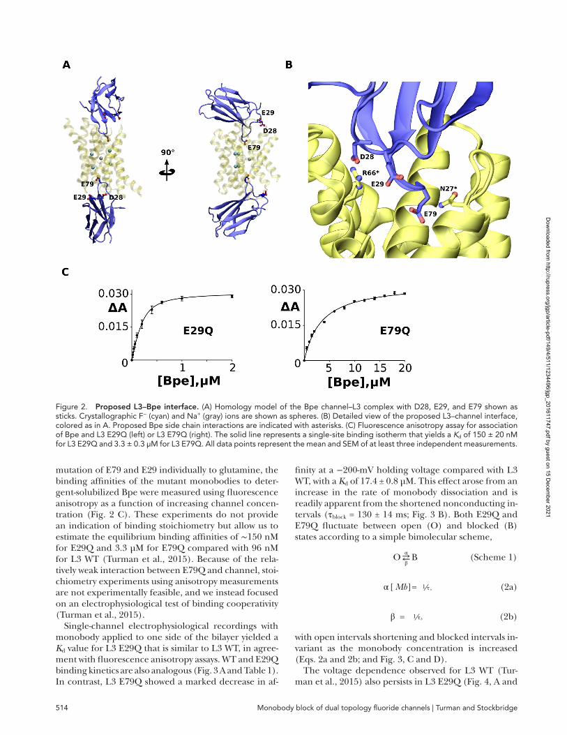
Single-channel electrophysiological recordings with monobody applied to one side of the bilayer yielded a Kd value for L3 E29Q that is similar to L3 WT, in agree-ment with fluorescence anisotropy assays. WT and E29Q binding kinetics are also analogous (Fig. 3 A and Table 1). In contrast, L3 E79Q showed a marked decrease in af-
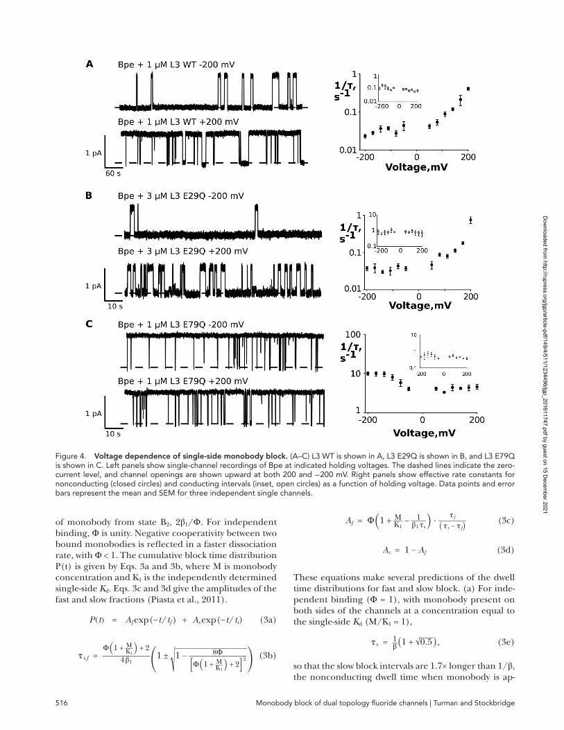
finity at a −200-mV holding voltage compared with L3 WT, with a Kd of 17.4 ± 0.8 µM. This effect arose from an increase in the rate of monobody dissociation and is readily apparent from the shortened nonconducting in-tervals (τblock = 130 ± 14 ms; Fig. 3 B). Both E29Q and E79Q fluctuate between open (O) and blocked (B) states according to a simple bimolecular scheme,
O ⇄ β α B (Scheme 1)
α [ Mb ] = 1 ⁄ τ o (2a)
β = 1 ⁄ τ b (2b)
with open intervals shortening and blocked intervals in-variant as the monobody concentration is increased (Eqs. 2a and 2b; and Fig. 3, C and D).
The voltage dependence observed for L3 WT (Tur-man et al., 2015) also persists in L3 E29Q (Fig. 4, A and
Figure 2. Proposed l3–Bpe interface. (A) Homology model of the Bpe channel–L3 complex with D28, E29, and E79 shown as sticks. Crystallographic F− (cyan) and Na+ (gray) ions are shown as spheres. (B) Detailed view of the proposed L3–channel interface, colored as in A. Proposed Bpe side chain interactions are indicated with asterisks. (C) Fluorescence anisotropy assay for association of Bpe and L3 E29Q (left) or L3 E79Q (right). The solid line represents a single-site binding isotherm that yields a Kd of 150 ± 20 nM for L3 E29Q and 3.3 ± 0.3 µM for L3 E79Q. All data points represent the mean and SEM of at least three independent measurements.
Dow
nloaded from http://rupress.org/jgp/article-pdf/149/4/511/1234496/jgp_201611747.pdf by guest on 15 D
ecember 2021
515JGP Vol. 149, No. 4
B). For both L3 WT and E29Q, the mean block times decrease between −200 and 200 mV, from 45 to 2.4 s for WT and from 28 to 1.5 s for E29Q. The bulk of this in-crease in the rate of monobody dissociation occurs at positive voltages. Again, the behavior of L3 E79Q differs from WT and E29Q. The dissociation rate, β, is insensi-tive to voltage at positive voltages (Fig. 4 C) and in-creases by a smaller margin, approximately twofold, at negative voltages between −200 and −110 mV. These results inform the experimental design of double-sided block experiments: to ensure that block kinetics on both sides (cis and trans) are identical, both L3 E29Q and L3 E79Q must be recorded at low voltages, −25 and −100 mV, respectively. Under these conditions, the measured values of τblock for both L3 E29Q and E79Q are identical within error: 22 ± 5 s (cis) and 24 ± 8 s (trans) for E29Q and 0.16 ± 0.03 s (cis) and 0.19 ± 0.05 s (trans) for E79Q.
To evaluate the contribution of the E29 and E79 side chains to the negative cooperativity observed for two-sided monobody binding, we revisited the two-sided block experiments performed previously with WT L3 (Turman et al., 2015). In these experiments, symmetri-cal monobody addition follows a two-state block scheme:
O ⇄ β 1
2 α 1 B 1 ⇄
2 β 1 /Φ
α 1 B 2 (Scheme 2)
The first monobody can bind at either of two open-channel (O) epitopes with rate 2α1 and dissociates at rate β1. State B1 is a composite of singly blocked states, with monobody bound to either the cis or trans side. The presence of the double-bound state, B2, is revealed by the emergence of longer blocked intervals that in-crease with increasing L3 concentration, a feature in-compatible with a bimolecular reaction. Cooperativity between binding events is reflected in the dissociation
Figure 3. single-side block by monobody. (A and B) Single-channel recordings of Bpe with L3 E29Q (A) and L3 E79Q (B) at in-dicated concentrations. Dashed lines signify zero current with channel openings shown upward. Right panels show representative cumulative distributions of block and open time intervals, with single exponential fits shown as solid smooth curves. (C and D) Bimolecular kinetics of single-side block by L3 E29Q (C) and L3 E79Q (D). Rate constants for conducting (closed circles) and noncon-ducting intervals (open circles) are plotted against monobody concentration. Data points represent the mean and SEM of three to five single channels, and solid lines are linear fits to data, with parameters reported in Table 1.
Table 1. single-side monobody block
Monobody τblock τopen [MB] koff kon Kd
s s s−1 s−1M−1
L3 WT 41 ± 3 77 ± 9 30 nM 0.025 4.3 × 10⁵ 58 ± 8 nML3 E29Q 40 ± 7 19 ± 8 100 nM 0.025 5.4 × 10⁵ 25 ± 9 nML3E79Q 0.13 ± 0.01 0.19 ± 0.03 12 μM 7.7 4.4 × 10⁵ 17.4 ± 0.8 μM
Dow
nloaded from http://rupress.org/jgp/article-pdf/149/4/511/1234496/jgp_201611747.pdf by guest on 15 D
ecember 2021
Monobody block of dual topology fluoride channels | Turman and Stockbridge516
of monobody from state B2, 2β1/Φ. For independent binding, Φ is unity. Negative cooperativity between two bound monobodies is reflected in a faster dissociation rate, with Φ < 1. The cumulative block time distribution P(t) is given by Eqs. 3a and 3b, where M is monobody concentration and K1 is the independently determined single-side Kd. Eqs. 3c and 3d give the amplitudes of the fast and slow fractions (Piasta et al., 2011).
P ( t ) = A f exp (− t / t f ) + A s exp (− t / t s ) (3a)
τ s,f = Φ (1 + M __ K 1
) + 2 ________ 4 β 1
(
1 ± √ _____________
1 − 8Φ ___________ [Φ (1 + M __ K 1
) + 2] 2 )
(3b)
A f = Φ (1 + M __ K 1 − 1 ___ β 1 τ s
) ⋅ τ f ______ ( τ s − τ f )
(3c)
A s = 1 − A f (3d)
These equations make several predictions of the dwell time distributions for fast and slow block. (a) For inde-pendent binding (Φ = 1), with monobody present on both sides of the channels at a concentration equal to the single-side Kd (M/K1 = 1),
τ s = 1 __ β ( 1 + √ _
0.5 ) , (3e)
so that the slow block intervals are 1.7× longer than 1/β, the nonconducting dwell time when monobody is ap-
Figure 4. Voltage dependence of single-side monobody block. (A–C) L3 WT is shown in A, L3 E29Q is shown in B, and L3 E79Q is shown in C. Left panels show single-channel recordings of Bpe at indicated holding voltages. The dashed lines indicate the zero- current level, and channel openings are shown upward at both 200 and −200 mV. Right panels show effective rate constants for nonconducting (closed circles) and conducting intervals (inset, open circles) as a function of holding voltage. Data points and error bars represent the mean and SEM for three independent single channels.
Dow
nloaded from http://rupress.org/jgp/article-pdf/149/4/511/1234496/jgp_201611747.pdf by guest on 15 D
ecember 2021
517JGP Vol. 149, No. 4
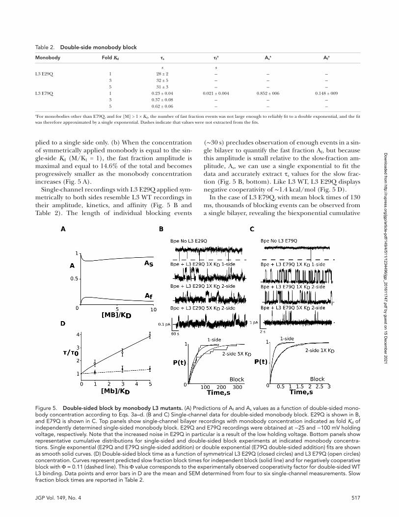
plied to a single side only. (b) When the concentration of symmetrically applied monobody is equal to the sin-gle-side Kd (M/K1 = 1), the fast fraction amplitude is maximal and equal to 14.6% of the total and becomes progressively smaller as the monobody concentration increases (Fig. 5 A).
Single-channel recordings with L3 E29Q applied sym-metrically to both sides resemble L3 WT recordings in their amplitude, kinetics, and affinity (Fig. 5 B and Table 2). The length of individual blocking events
(∼30 s) precludes observation of enough events in a sin-gle bilayer to quantify the fast fraction Af, but because this amplitude is small relative to the slow-fraction am-plitude, As, we can use a single exponential to fit the data and accurately extract τs values for the slow frac-tion (Fig. 5 B, bottom). Like L3 WT, L3 E29Q displays negative cooperativity of ∼1.4 kcal/mol (Fig. 5 D).
In the case of L3 E79Q, with mean block times of 130 ms, thousands of blocking events can be observed from a single bilayer, revealing the biexponential cumulative
Figure 5. double-sided block by monobody l3 mutants. (A) Predictions of Af and As values as a function of double-sided mono-body concentration according to Eqs. 3a–d. (B and C) Single-channel data for double-sided monobody block. E29Q is shown in B, and E79Q is shown in C. Top panels show single-channel bilayer recordings with monobody concentration indicated as fold Kd of independently determined single-sided monobody block. E29Q and E79Q recordings were obtained at −25 and −100 mV holding voltage, respectively. Note that the increased noise in E29Q in particular is a result of the low holding voltage. Bottom panels show representative cumulative distributions for single-sided and double-sided block experiments at indicated monobody concentra-tions. Single exponential (E29Q and E79Q single-sided addition) or double exponential (E79Q double-sided addition) fits are shown as smooth solid curves. (D) Double-sided block time as a function of symmetrical L3 E29Q (closed circles) and L3 E79Q (open circles) concentration. Curves represent predicted slow fraction block times for independent block (solid line) and for negatively cooperative block with Φ = 0.11 (dashed line). This Φ value corresponds to the experimentally observed cooperativity factor for double-sided WT L3 binding. Data points and error bars in D are the mean and SEM determined from four to six single-channel measurements. Slow fraction block times are reported in Table 2.
Table 2. double-side monobody block
Monobody Fold Kd τs τfa As
a Afa
s sL3 E29Q 1 28 ± 2 − − −
3 32 ± 5 − − −5 31 ± 3 − − −
L3 E79Q 1 0.23 ± 0.04 0.021 ± 0.004 0.852 ± 006 0.148 ± 0093 0.37 ± 0.08 − − −5 0.62 ± 0.06 − − −
aFor monobodies other than E79Q, and for [M] > 1 × Kd, the number of fast fraction events was not large enough to reliably fit to a double exponential, and the fit was therefore approximated by a single exponential. Dashes indicate that values were not extracted from the fits.
Dow
nloaded from http://rupress.org/jgp/article-pdf/149/4/511/1234496/jgp_201611747.pdf by guest on 15 D
ecember 2021
Monobody block of dual topology fluoride channels | Turman and Stockbridge518
block time distribution predicted by Eq. 3a. With 17 µM symmetric monobody (equal to the single-side Kd), we observe a fast fraction amplitude of 14.8% and slow fraction amplitude of 85.2%, in precise agreement with the relative amplitudes predicted by Eq. 3a. At 5× Kd, the longer blocking events are predicted to dominate, and the cumulative distribution is well approximated by a single exponential (Fig. 5 C, bottom).
Analysis of these recordings reveals that two-sided binding of L3 E79Q is not negatively cooperative (Fig. 5 D). The time constants of the long block inter-vals exceed the single-side block times by a factor of 1.8, which is essentially in quantitative agreement with the prediction of Eq. 3a for independent block (Table 2). At monobody concentrations equal to 3× and 5× the sin-gle-side Kd, the block times increase along the predicted isotherm for an independent double-side blocked sys-tem. Thus, neutralization of a single negative charge on the monobody, E79, is sufficient to abolish both the voltage dependence and the negative cooperativity ob-served for double-sided L3 block.
The rapid kinetics of E79Q, coupled with the clear resolution of the fast and slow fractions when [Mb] = Kd, also permits a kinetic comparison of the first and second monobody-binding events. Although the previ-ous experiments demonstrated that the equilibrium constants are identical for the first and second binding event (Φ = 1), they do not exclude the possibility that the kinetics are substantially different for consecutive binding events, covarying according to kinetic offset δ such that the binding constant is unchanged:
K d = δβ __ δα . (4a)
The double-sided blocking experiment would then be described by Scheme 3:
O ⇄ β
2αM β 1 ⇄
2βδ αMδ β 2 (Scheme 3)
For experiments performed at low monobody con-centration, with [M] = Kd, Eqs. 3b–d can be rearranged according to this scheme, so that
τ s,f = 1 __ β * 1 + 3δ ____ 4δ (1 ± √ _________
1 − 8δ ______ ( 3δ + 1 ) 2
) (5a)
and
A f = ( ( β + δβ ) − 1 __ τ s ) ⋅ τ f __________ ( 2βδ ) ( τ s − τ f )
. (5b)
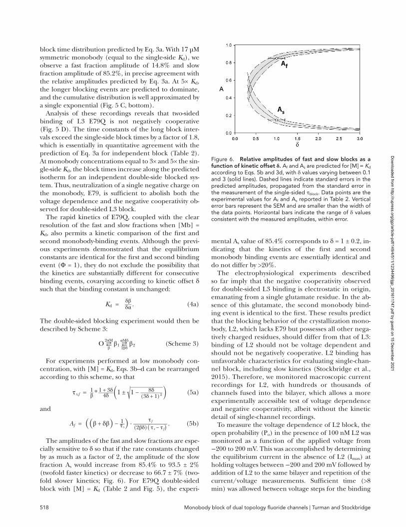
The amplitudes of the fast and slow fractions are espe-cially sensitive to δ so that if the rate constants changed by as much as a factor of 2, the amplitude of the slow fraction As would increase from 85.4% to 93.5 ± 2% (twofold faster kinetics) or decrease to 66.7 ± 7% (two-fold slower kinetics; Fig. 6). For E79Q double-sided block with [M] = Kd (Table 2 and Fig. 5), the experi-
mental As value of 85.4% corresponds to δ = 1 ± 0.2, in-dicating that the kinetics of the first and second monobody binding events are essentially identical and do not differ by >20%.
The electrophysiological experiments described so far imply that the negative cooperativity observed for double-sided L3 binding is electrostatic in origin, emanating from a single glutamate residue. In the ab-sence of this glutamate, the second monobody bind-ing event is identical to the first. These results predict that the blocking behavior of the crystallization mono-body, L2, which lacks E79 but possesses all other nega-tively charged residues, should differ from that of L3: binding of L2 should not be voltage dependent and should not be negatively cooperative. L2 binding has unfavorable characteristics for evaluating single-chan-nel block, including slow kinetics (Stockbridge et al., 2015). Therefore, we monitored macroscopic current recordings for L2, with hundreds or thousands of channels fused into the bilayer, which allows a more experimentally accessible test of voltage dependence and negative cooperativity, albeit without the kinetic detail of single-channel recordings.
To measure the voltage dependence of L2 block, the open probability (Po) in the presence of 100 nM L2 was monitored as a function of the applied voltage from −200 to 200 mV. This was accomplished by determining the equilibrium current in the absence of L2 (Imax) at holding voltages between −200 and 200 mV followed by addition of L2 to the same bilayer and repetition of the current/voltage measurements. Sufficient time (>8 min) was allowed between voltage steps for the binding
Figure 6. Relative amplitudes of fast and slow blocks as a function of kinetic offset δ. Af and As are predicted for [M] = Kd according to Eqs. 5b and 3d, with δ values varying between 0.1 and 3 (solid lines). Dashed lines indicate standard errors in the predicted amplitudes, propagated from the standard error in the measurement of the single-sided τblock. Data points are the experimental values for Af and As reported in Table 2. Vertical error bars represent the SEM and are smaller than the width of the data points. Horizontal bars indicate the range of δ values consistent with the measured amplitudes, within error.
Dow
nloaded from http://rupress.org/jgp/article-pdf/149/4/511/1234496/jgp_201611747.pdf by guest on 15 D
ecember 2021
519JGP Vol. 149, No. 4
reaction to reach equilibrium. Because the individual Bpe channels are constitutively open in the absence of monobody (Po = 1),
P o ( L2 ) = I L2 / I max . (6)
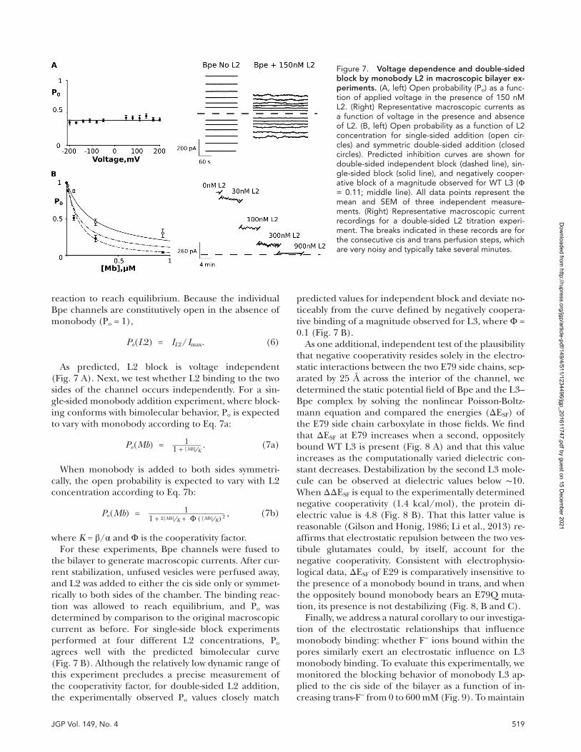
As predicted, L2 block is voltage independent (Fig. 7 A). Next, we test whether L2 binding to the two sides of the channel occurs independently. For a sin-gle-sided monobody addition experiment, where block-ing conforms with bimolecular behavior, Po is expected to vary with monobody according to Eq. 7a:
P o ( Mb ) = 1 ___ 1 + [Mb] ⁄ K . (7a)
When monobody is added to both sides symmetri-cally, the open probability is expected to vary with L2 concentration according to Eq. 7b:
P o ( Mb ) = 1 _________ 1 + 2 [ Mb ] ⁄ K + Φ ( [ Mb ] ⁄ K ) 2
, (7b)
where K = β/α and Φ is the cooperativity factor.For these experiments, Bpe channels were fused to
the bilayer to generate macroscopic currents. After cur-rent stabilization, unfused vesicles were perfused away, and L2 was added to either the cis side only or symmet-rically to both sides of the chamber. The binding reac-tion was allowed to reach equilibrium, and Po was determined by comparison to the original macroscopic current as before. For single-side block experiments performed at four different L2 concentrations, Po agrees well with the predicted bimolecular curve (Fig. 7 B). Although the relatively low dynamic range of this experiment precludes a precise measurement of the cooperativity factor, for double-sided L2 addition, the experimentally observed Po values closely match
predicted values for independent block and deviate no-ticeably from the curve defined by negatively coopera-tive binding of a magnitude observed for L3, where Φ = 0.1 (Fig. 7 B).
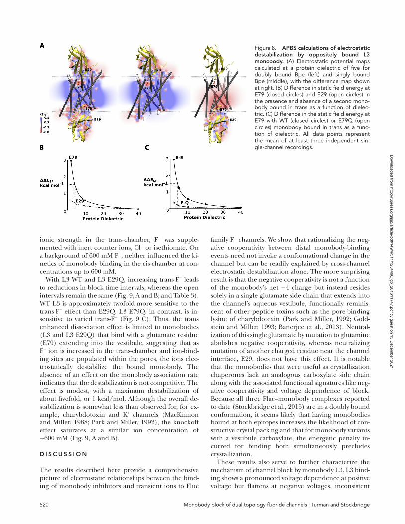
As one additional, independent test of the plausibility that negative cooperativity resides solely in the electro-static interactions between the two E79 side chains, sep-arated by 25 Å across the interior of the channel, we determined the static potential field of Bpe and the L3–Bpe complex by solving the nonlinear Poisson-Boltz-mann equation and compared the energies (ΔESF) of the E79 side chain carboxylate in those fields. We find that ΔESF at E79 increases when a second, oppositely bound WT L3 is present (Fig. 8 A) and that this value increases as the computationally varied dielectric con-stant decreases. Destabilization by the second L3 mole-cule can be observed at dielectric values below ∼10. When ΔΔESF is equal to the experimentally determined negative cooperativity (1.4 kcal/mol), the protein di-electric value is 4.8 (Fig. 8 B). That this latter value is reasonable (Gilson and Honig, 1986; Li et al., 2013) re-affirms that electrostatic repulsion between the two ves-tibule glutamates could, by itself, account for the negative cooperativity. Consistent with electrophysio-logical data, ΔESF of E29 is comparatively insensitive to the presence of a monobody bound in trans, and when the oppositely bound monobody bears an E79Q muta-tion, its presence is not destabilizing (Fig. 8, B and C).
Finally, we address a natural corollary to our investiga-tion of the electrostatic relationships that influence monobody binding: whether F− ions bound within the pores similarly exert an electrostatic influence on L3 monobody binding. To evaluate this experimentally, we monitored the blocking behavior of monobody L3 ap-plied to the cis side of the bilayer as a function of in-creasing trans-F− from 0 to 600 mM (Fig. 9). To maintain
Figure 7. Voltage dependence and double-sided block by monobody l2 in macroscopic bilayer ex-periments. (A, left) Open probability (Po) as a func-tion of applied voltage in the presence of 150 nM L2. (Right) Representative macroscopic currents as a function of voltage in the presence and absence of L2. (B, left) Open probability as a function of L2 concentration for single-sided addition (open cir-cles) and symmetric double-sided addition (closed circles). Predicted inhibition curves are shown for double-sided independent block (dashed line), sin-gle-sided block (solid line), and negatively cooper-ative block of a magnitude observed for WT L3 (Φ = 0.11; middle line). All data points represent the mean and SEM of three independent measure-ments. (Right) Representative macroscopic current recordings for a double-sided L2 titration experi-ment. The breaks indicated in these records are for the consecutive cis and trans perfusion steps, which are very noisy and typically take several minutes.
Dow
nloaded from http://rupress.org/jgp/article-pdf/149/4/511/1234496/jgp_201611747.pdf by guest on 15 D
ecember 2021
Monobody block of dual topology fluoride channels | Turman and Stockbridge520
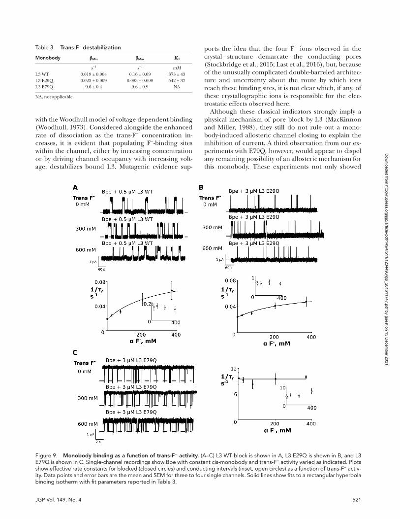
ionic strength in the trans-chamber, F− was supple-mented with inert counter ions, Cl− or isethionate. On a background of 600 mM F−, neither influenced the ki-netics of monobody binding in the cis-chamber at con-centrations up to 600 mM.
With L3 WT and L3 E29Q, increasing trans-F− leads to reductions in block time intervals, whereas the open intervals remain the same (Fig. 9, A and B; and Table 3). WT L3 is approximately twofold more sensitive to the trans-F− effect than E29Q. L3 E79Q, in contrast, is in-sensitive to varied trans-F− (Fig. 9 C). Thus, the trans enhanced dissociation effect is limited to monobodies (L3 and L3 E29Q) that bind with a glutamate residue (E79) extending into the vestibule, suggesting that as F− ion is increased in the trans-chamber and ion-bind-ing sites are populated within the pores, the ions elec-trostatically destabilize the bound monobody. The absence of an effect on the monobody association rate indicates that the destabilization is not competitive. The effect is modest, with a maximum destabilization of about fivefold, or 1 kcal/mol. Although the overall de-stabilization is somewhat less than observed for, for ex-ample, charybdotoxin and K+ channels (MacKinnon and Miller, 1988; Park and Miller, 1992), the knockoff effect saturates at a similar ion concentration of ∼600 mM (Fig. 9, A and B).
d I s c u s s I o n
The results described here provide a comprehensive picture of electrostatic relationships between the bind-ing of monobody inhibitors and transient ions to Fluc
family F− channels. We show that rationalizing the neg-ative cooperativity between distal monobody-binding events need not invoke a conformational change in the channel but can be readily explained by cross-channel electrostatic destabilization alone. The more surprising result is that the negative cooperativity is not a function of the monobody’s net −4 charge but instead resides solely in a single glutamate side chain that extends into the channel’s aqueous vestibule, functionally reminis-cent of other peptide toxins such as the pore-binding lysine of charybdotoxin (Park and Miller, 1992; Gold-stein and Miller, 1993; Banerjee et al., 2013). Neutral-ization of this single glutamate by mutation to glutamine abolishes negative cooperativity, whereas neutralizing mutation of another charged residue near the channel interface, E29, does not have this effect. It is notable that the monobodies that were useful as crystallization chaperones lack an analogous carboxylate side chain along with the associated functional signatures like neg-ative cooperativity and voltage dependence of block. Because all three Fluc–monobody complexes reported to date (Stockbridge et al., 2015) are in a doubly bound conformation, it seems likely that having monobodies bound at both epitopes increases the likelihood of con-structive crystal packing and that for monobody variants with a vestibule carboxylate, the energetic penalty in-curred for binding both simultaneously precludes crystallization.
These results also serve to further characterize the mechanism of channel block by monobody L3. L3 bind-ing shows a pronounced voltage dependence at positive voltage but flattens at negative voltages, inconsistent
Figure 8. APBs calculations of electrostatic destabilization by oppositely bound l3 monobody. (A) Electrostatic potential maps calculated at a protein dielectric of five for doubly bound Bpe (left) and singly bound Bpe (middle), with the difference map shown at right. (B) Difference in static field energy at E79 (closed circles) and E29 (open circles) in the presence and absence of a second mono-body bound in trans as a function of dielec-tric. (C) Difference in the static field energy at E79 with WT (closed circles) or E79Q (open circles) monobody bound in trans as a func-tion of dielectric. All data points represent the mean of at least three independent sin-gle-channel recordings.
Dow
nloaded from http://rupress.org/jgp/article-pdf/149/4/511/1234496/jgp_201611747.pdf by guest on 15 D
ecember 2021
521JGP Vol. 149, No. 4
with the Woodhull model of voltage-dependent binding (Woodhull, 1973). Considered alongside the enhanced rate of dissociation as the trans-F− concentration in-creases, it is evident that populating F−-binding sites within the channel, either by increasing concentration or by driving channel occupancy with increasing volt-age, destabilizes bound L3. Mutagenic evidence sup-
ports the idea that the four F− ions observed in the crystal structure demarcate the conducting pores (Stockbridge et al., 2015; Last et al., 2016), but, because of the unusually complicated double-barreled architec-ture and uncertainty about the route by which ions reach these binding sites, it is not clear which, if any, of these crystallographic ions is responsible for the elec-trostatic effects observed here.
Although these classical indicators strongly imply a physical mechanism of pore block by L3 (MacKinnon and Miller, 1988), they still do not rule out a mono-body-induced allosteric channel closing to explain the inhibition of current. A third observation from our ex-periments with E79Q, however, would appear to dispel any remaining possibility of an allosteric mechanism for this monobody. These experiments not only showed
Figure 9. Monobody binding as a function of trans-F− activity. (A–C) L3 WT block is shown in A, L3 E29Q is shown in B, and L3 E79Q is shown in C. Single-channel recordings show Bpe with constant cis-monobody and trans-F− activity varied as indicated. Plots show effective rate constants for blocked (closed circles) and conducting intervals (inset, open circles) as a function of trans-F− activ-ity. Data points and error bars are the mean and SEM for three to four single channels. Solid lines show fits to a rectangular hyperbola binding isotherm with fit parameters reported in Table 3.
Table 3. trans-F− destabilization
Monobody βMin βMax Kd
s−1 s−1 mML3 WT 0.019 ± 0.004 0.16 ± 0.09 373 ± 43L3 E29Q 0.023 ± 0.009 0.083 ± 0.008 542 ± 37L3 E79Q 9.6 ± 0.4 9.6 ± 0.9 NA
NA, not applicable.
Dow
nloaded from http://rupress.org/jgp/article-pdf/149/4/511/1234496/jgp_201611747.pdf by guest on 15 D
ecember 2021
Monobody block of dual topology fluoride channels | Turman and Stockbridge522
that negative cooperativity is abolished in this mutant—that is, that the equilibrium constants for the first and second monobody binding event are identical—but also that the kinetics are identical within the error of the measurement. This provides a persuasive argument that the conformation of the channel is unchanged upon the first monobody binding event, an observation that is incompatible with allosteric inhibition.
A c k n o w l e d g M e n t s
We thank Christopher Miller (Brandeis University) for his sup-port of this project and for helpful discussions and unrelenting criticism. We also thank Janice Robertson (University of Iowa) for helpful discussions and critical reading of the manuscript.
This work was supported, in part, by National Institutes of Health grant R00-GM111767 to R.B. Stockbridge. D.L. Turman is supported by the Howard Hughes Medical Institute in the labora-tory of Christopher Miller.
The authors declare no competing financial interests.Author contributions: R.B. Stockbridge and D.L. Turman
designed and performed experiments, analyzed the data, and wrote the paper.
Richard W. Aldrich served as editor.
Submitted: 20 December 2016Accepted: 3 February 2017
R e F e R e n c e sBaker, J.L., N. Sudarsan, Z. Weinberg, A. Roth, R.B. Stockbridge,
and R.R. Breaker. 2012. Widespread genetic switches and toxicity resistance proteins for fluoride. Science. 335:233–235. http ://dx .doi .org /10 .1126 /science .1215063
Baker, N.A., D. Sept, S. Joseph, M.J. Holst, and J.A. McCammon. 2001. Electrostatics of nanosystems: application to microtubules and the ribosome. Proc. Natl. Acad. Sci. USA. 98:10037–10041. http ://dx .doi .org /10 .1073 /pnas .181342398
Banerjee, A., A. Lee, E. Campbell, and R. Mackinnon. 2013. Structure of a pore-blocking toxin in complex with a eukaryotic voltage-dependent K+ channel. eLife. 2:e00594. http ://dx .doi .org /10 .7554 /eLife .00594
Callenberg, K.M., O.P. Choudhary, G.L. de Forest, D.W. Gohara, N.A. Baker, and M. Grabe. 2010. APBSmem: a graphical interface for electrostatic calculations at the membrane. PLoS One. 5:e12722. http ://dx .doi .org /10 .1371 /journal .pone .0012722
Gilson, M.K., and B.H. Honig. 1986. The dielectric constant of a folded protein. Biopolymers. 25:2097–2119. http ://dx .doi .org /10 .1002 /bip .360251106
Goldstein, S.A., and C. Miller. 1993. Mechanism of charybdotoxin block of a voltage-gated K+ channel. Biophys. J. 65:1613–1619. http ://dx .doi .org /10 .1016 /S0006 -3495(93)81200 -1
Humphrey, W., A. Dalke, and K. Schulten. 1996. VMD: visual molecular dynamics. J. Mol. Graph. 14:33–38: 27–28. http ://dx .doi .org /10 .1016 /0263 -7855(96)00018 -5
Ji, C., R.B. Stockbridge, and C. Miller. 2014. Bacterial fluoride resistance, Fluc channels, and the weak acid accumulation effect. J. Gen. Physiol. 144:257–261. http ://dx .doi .org /10 .1085 /jgp .201411243
Koide, A., J. Wojcik, R.N. Gilbreth, R.J. Hoey, and S. Koide. 2012. Teaching an old scaffold new tricks: monobodies constructed using alternative surfaces of the FN3 scaffold. J. Mol. Biol. 415:393–405. http ://dx .doi .org /10 .1016 /j .jmb .2011 .12 .019
Last, N.B., L. Kolmakova-Partensky, T. Shane, and C. Miller. 2016. Mechanistic signs of double-barreled structure in a fluoride ion channel. eLife. 5:e18767. http ://dx .doi .org /10 .7554 /eLife .18767
Li, L., C. Li, Z. Zhang, and E. Alexov. 2013. On the dielectric “constant” of proteins: smooth dielectric function for macromolecular modeling and its implementation in DelPhi. J. Chem. Theory Comput. 9:2126–2136. http ://dx .doi .org /10 .1021 /ct400065j
MacKinnon, R., and C. Miller. 1988. Mechanism of charybdotoxin block of the high-conductance, Ca2+-activated K+ channel. J. Gen. Physiol. 91:335–349. http ://dx .doi .org /10 .1085 /jgp .91 .3 .335
Park, C.S., and C. Miller. 1992. Interaction of charybdotoxin with permeant ions inside the pore of a K+ channel. Neuron. 9:307–313. http ://dx .doi .org /10 .1016 /0896 -6273(92)90169 -E
Phillips, J.C., R. Braun, W. Wang, J. Gumbart, E. Tajkhorshid, E. Villa, C. Chipot, R.D. Skeel, L. Kalé, and K. Schulten. 2005. Scalable molecular dynamics with NAMD. J. Comput. Chem. 26:1781–1802. http ://dx .doi .org /10 .1002 /jcc .20289
Piasta, K.N., D.L. Theobald, and C. Miller. 2011. Potassium-selective block of barium permeation through single KcsA channels. J. Gen. Physiol. 138:421–436. http ://dx .doi .org /10 .1085 /jgp .201110684
Stockbridge, R.B., J.L. Robertson, L. Kolmakova-Partensky, and C. Miller. 2013. A family of fluoride-specific ion channels with du-al-topology architecture. eLife. 2:e01084.
Stockbridge, R.B., A. Koide, C. Miller, and S. Koide. 2014. Proof of dual-topology architecture of Fluc F- channels with monobody blockers. Nat. Commun. 5:5120. http ://dx .doi .org /10 .1038 /ncomms6120
Stockbridge, R.B., L. Kolmakova-Partensky, T. Shane, A. Koide, S. Koide, C. Miller, and S. Newstead. 2015. Crystal structures of a double-barrelled fluoride ion channel. Nature. 525:548–551. http ://dx .doi .org /10 .1038 /nature14981
Turman, D.L., J.T. Nathanson, R.B. Stockbridge, T.O. Street, and C. Miller. 2015. Two-sided block of a dual-topology F- channel. Proc. Natl. Acad. Sci. USA. 112:5697–5701. http ://dx .doi .org /10 .1073 /pnas .1505301112
Webb, B., and A. Sali. 2016. Comparative protein structure modeling using MOD ELL ER. Curr. Protoc. Protein Sci. 86:2.9.1–2.9.37. http ://dx .doi .org /10 .1002 /cpps .20
Woodhull, A.M. 1973. Ionic blockage of sodium channels in nerve. J. Gen. Physiol. 61:687–708. http ://dx .doi .org /10 .1085 /jgp .61 .6 .687
Dow
nloaded from http://rupress.org/jgp/article-pdf/149/4/511/1234496/jgp_201611747.pdf by guest on 15 D
ecember 2021


![Mechanism of Reaction, Tissue Distribution, and Inhibition of … · [CANCER RESEARCH 34, 1503-1515, June 1974] Mechanism of Reaction, Tissue Distribution, and Inhibition of Arylhydroxamic](https://img.pdfslide.us/doc/110x75/5e58a5ee3cd8ad16721400a4/mechanism-of-reaction-tissue-distribution-and-inhibition-of-cancer-research-34.jpg)