Embed Size (px)
Citation preview
Mechanical Stresses in Vocal Fold Tissue during Voice Production
A thesis presented by
Heather Elspeth Gunter
to
The Division of Engineering & Applied Sciences
in partial fulfillment of the requirements
for the degree of
Doctor of Philosophy
in the subject of
Engineering Sciences
Harvard University
Cambridge, Massachusetts
May, 2003

© 2003 Heather Elspeth Gunter
All rights reserved
iii
ABSTRACT Mechanical Stresses in Vocal Fold Tissue During Voice Production
Advisor: Robert D. Howe Author: Heather Elspeth Gunter
Theoretical and experimental techniques are used to study the tissue mechanics
governing vocal fold closure and collision during phonation in order to evaluate the
development of stresses that may be risk factors for pathology development. An original
three-dimensional finite element model of vocal fold tissue predicts these quantities with
high spatial resolution. Models predict that compressive stress in three directions and
vertical shear stress are increased during collision in the typical location of lesions (i.e.
the center of the superior medial edge of the vocal fold in the middle of the vibrating and
contact region). This supports the hypothesis that stress is a cause of vocal fold
pathology, and suggests modes of tissue injury. Predictions of increased stress due to
increased voicing intensity are consistent with the hypothesis and clinical observations
that loud voicing is a risk factor for benign vocal fold pathology development.
Additional finite element models include a representation of the superficial lamina
propria (SLP), which is a soft tissue layer near the surface of the vocal folds that
iv
contributes to voice quality and vocal fold injury. Increases in SLP stiffness are
associated with increases in compressive and shear stresses in both the epithelium and
SLP during collision. Increases in SLP stiffness are also associated with decreases in
longitudinal tensile stress in the epithelium prior to collision. These results support the
proposed role of SLP stiffness in determining mechanical stress and injury risk, and guide
design and selection of SLP replacements used in vocal fold augmentation surgery. In
vivo vocal fold collision forces in humans are measured using a new low profile force
sensor that minimizes measurement artifacts and maintains voice quality. Impact force
correlates more strongly with voice intensity than pitch. The finite element models
translate increased force magnitudes into increases in compressive stress, vertical shear
stress and Von Mises stress magnitudes. Avoidance of the conditions of increased
collision force may prevent development of lesions.
v
TABLE OF CONTENTS
Abstract .............................................................................................................................. iii
Table of contents................................................................................................................. v
List of figures and tables.................................................................................................. viii
Acknowledgements........................................................................................................... xii
Chapter 1: Introduction ....................................................................................................... 1
Mechanics of vocal fold tissue........................................................................................ 6
Research objectives and thesis organization................................................................... 9
Chapter 2: A mechanical model of vocal fold collision with high spatial and temporal resolution,.......................................................................................................................... 11
Introduction................................................................................................................... 11
Development................................................................................................................. 14
Assumptions.............................................................................................................. 14
Implementation ......................................................................................................... 16
Validation...................................................................................................................... 21
Mechanical stress during collision................................................................................ 26
vi
Discussion..................................................................................................................... 27
Chapter 3: Modeling mechanical stresses as a factor in the etiology of benign vocal fold lesions,............................................................................................................................... 32
Introduction................................................................................................................... 32
Methods ........................................................................................................................ 33
Lumped mass model ................................................................................................. 33
Finite element model................................................................................................. 38
Results........................................................................................................................... 41
Discussion..................................................................................................................... 47
Chapter 4: A theoretical investigation of the role of superficial lamina propria stiffness during voice production .................................................................................................... 51
Introduction................................................................................................................... 51
Methods ........................................................................................................................ 53
Results........................................................................................................................... 58
Discussion..................................................................................................................... 66
Chapter 5: Measurements of vocal fold collision forces during phonation ...................... 71
Introduction................................................................................................................... 71
Methods ........................................................................................................................ 73
Collision force sensor design, construction and characterization ............................. 73
Experimental setup.................................................................................................... 79
Experimental protocol............................................................................................... 80
Data processing and analysis .................................................................................... 81
Results........................................................................................................................... 82
Discussion..................................................................................................................... 87
Chapter 6: Contributions and proposed work ................................................................... 94
vii
Method for theoretical study of vocal fold collision..................................................... 94
Method for in vivo study of vocal fold collision .......................................................... 96
Support for mechanical injury of vocal fold tissue hypothesis..................................... 97
Measurement and interpretation of vocal fold collision forces .................................... 99
Future directions ......................................................................................................... 100
Bibliography ................................................................................................................... 101
viii
LIST OF FIGURES AND TABLES
Figure 1.1: Vocal fold and air flow oscillation during voice production............................ 2
Figure 1.2: Anatomy of human voice production............................................................... 3
Figure 1.3: Geometry of the vocal folds prepared for voice production. ........................... 8
Figure 2.1: Calculation of initial conditions for glottal closure using a finite element model of the vocal fold. ............................................................................................ 17
Figure 2.2: Finite element model of the membranous portion of a vocal fold during glottal closure. ...................................................................................................................... 20
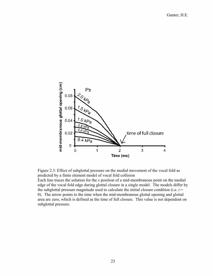
Figure 2.3: Effect of subglottal pressure on the medial movement of the vocal fold as predicted by a finite element model of vocal fold collision...................................... 23
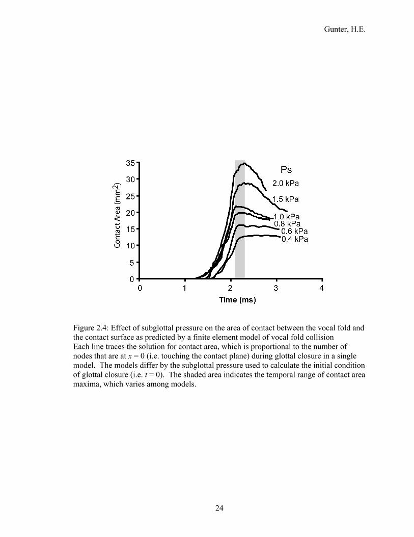
Figure 2.4: Effect of subglottal pressure on the area of contact between the vocal fold and the contact surface as predicted by a finite element model of vocal fold collision .. 24
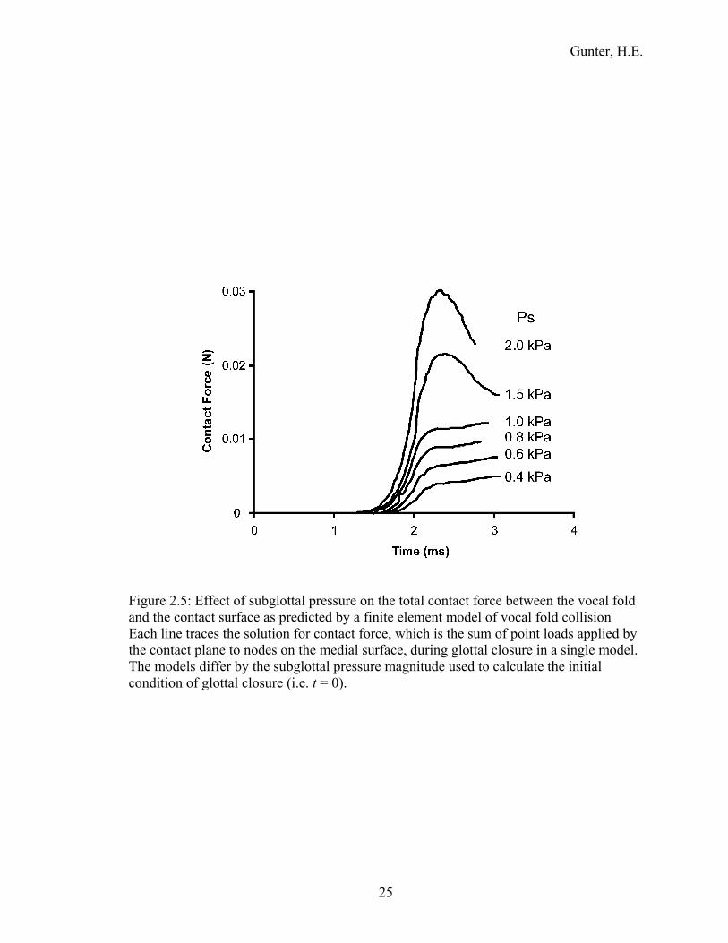
Figure 2.5: Effect of subglottal pressure on the total contact force between the vocal fold and the contact surface as predicted by a finite element model of vocal fold collision................................................................................................................................... 25
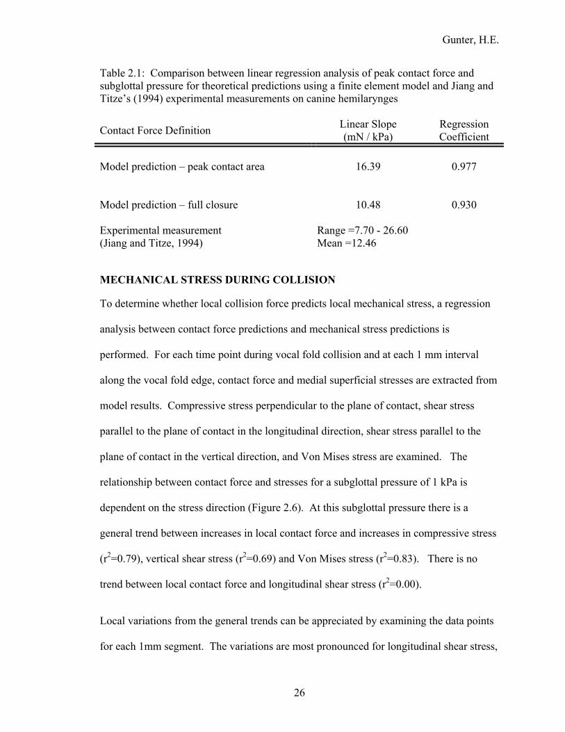
Table 2.1: Comparison between linear regression analysis of peak contact force and subglottal pressure for theoretical predictions using a finite element model and Jiang and Titze’s (1994) experimental measurements on canine hemilarynges ................ 26
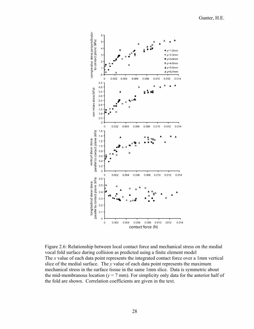
Figure 2.6: Relationship between local contact force and mechanical stress on the medial vocal fold surface during collision as predicted using a finite element model ......... 28
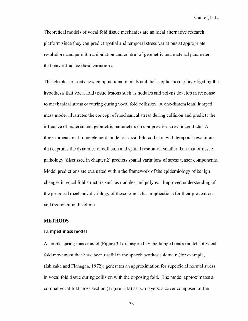
Figure 3.1: Development of a lumped mass model of vocal fold collision. ..................... 34
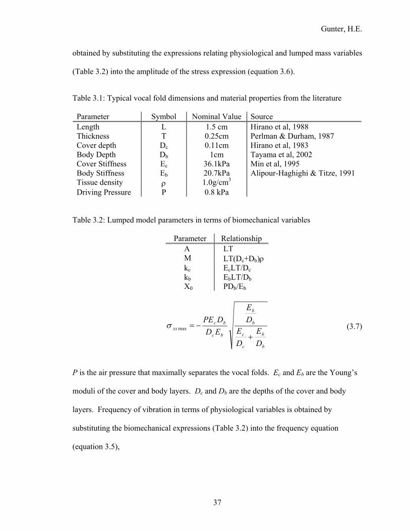
Table 3.1: Typical vocal fold dimensions and material properties from the literature..... 37
ix
Table 3.2: Lumped model parameters in terms of biomechanical variables .................... 37
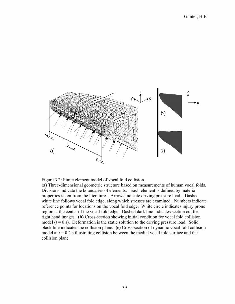
Figure 3.2: Finite element model of vocal fold collision.................................................. 39
Table 3.3: Effect of physiological parameter manipulation on compressive stress magnitude as predicted by a lumped mass model of vocal fold collision ................ 42
Figure 3.3: Dependence of superficial stress in vocal fold tissue during collision and vocal fold vibration frequency (i.e. pitch) on geometric and material parameters as predicted by a lumped mass model of vocal fold collision....................................... 43
Figure 3.4: Effect of vocal fold collision on components of the stress tensor at the injury prone region for a driving pressure of 0.8 kPa as predicted with a finite element model......................................................................................................................... 44
Figure 3.5: Spatial variation of stress tensor components in superficial tissue along the vocal fold edge for a driving pressure of 0.8 kPa as predicted by a finite element model......................................................................................................................... 45
Figure 3.6: Dependence of stress tensor components in the injury prone region during collision on driving pressure as predicted by a finite element model....................... 46
Figure 4.1: Finite element model of vocal fold closure and collision with four distinct tissue layers. .............................................................................................................. 54
Table 4.1: Material properties of vocal fold tissues used in a finite element model of vocal fold collision.................................................................................................... 56
Figure 4.2: Effect of SLP stiffness on vocal fold deformation in the midmembrous region at the start of glottal closure as predicted by a layered finite element model ........... 60
Figure 4.3: Effects of SLP stiffness on the falling portion of the aerodynamic waveform as estimated by glottal area squared and glottal area as predicted by a layered finite element model ........................................................................................................... 61
Figure 4.4: Effect of SLP stiffness on maximum contact force between the vocal folds and glottal area as predicted by a layered finite element model ............................... 62
Figure 4.5: Effect of SLP stiffness on maximum normal compression in the midmembranous region during vocal fold collision as predicted by a layered finite element model ........................................................................................................... 63
Figure 4.6: Effect of SLP stiffness on maximum vertical shear stress on the plane parallel with the plane of collision (zx) in the midmembranous region during vocal fold collision as predicted by a layered finite element model .......................................... 64
x
Figure 4.7: Effect of SLP stiffness on maximum longitudinal (yy) tension in the midmembranous region prior to vocal fold collision as predicted by a layered finite element model ........................................................................................................... 65
Figure 5.1: Mechanical housing of sensor for measuring in vivo vocal fold collision forces and placement of the sensor between the vocal folds .................................... 74
Figure 5.2: Construction of the active area of a low profile force sensor for measuring vocal fold collision forces in vivo............................................................................. 75
Figure 5.3: Analog processing of signal from pressure sensitive piezoelectric film to produce voltage output that is proportional to force input........................................ 77
Figure 5.4: Voltage output of a low profile force sensor placed between human vocal folds during production voice ................................................................................... 78
Figure 5.5: Production of voice by a subject while a low profile sensor is rested against his one rigid fold ....................................................................................................... 84
Figure 5.6: Segment of simultaneously acquired intraglottal force, EGG and audio data during voice production. ........................................................................................... 85
Table 5.1: Regression analysis of vocal fold collision force as a function of voice frequency and voice intensity (subject 1) ................................................................. 86
Table 5.2: Regression analysis of vocal fold collision force as a function of voice frequency and voice intensity (subject 2) ................................................................. 87
Figure 5.7: Scatter plots of vocal fold collision force, voice intensity and voice frequency data points measured in two male subjects ............................................................... 88
xi
This thesis is dedicated to Professor Thomas A. McMahon (1943-1998), who opened my
eyes to the intriguing relationship between solid mechanics and voice production
xii
ACKNOWLEDGEMENTS Thank you to all who helped to make this thesis a reality. I am appreciative of a
graduate fellowship from the Whitaker foundation that has supported me during my final
five years of graduate study and of scholarships from the Alberta Heritage Scholarship
Fund, the Canadian Federation for University Women and the Harvard Division of
Engineering and Applied Sciences that contributed to my initial year of graduate study.
I am indebted to Professors Robert D. Howe (Harvard Division of Engineering &
Applied Sciences), Robert E. Hillman (Massachusetts Eye and Ear Infirmary) and
Kenneth N. Stevens (Massachusetts Institute of Technology Department of Electrical
Engineering & Computer Science) who have helped to transform me from a fledgling
graduate student into a young researcher. These faculty members along with Professor
John Hutchinson (Harvard Division of Engineering & Applied Sciences) helped to sculpt
this scientific work from its early overly ambitious state into its current form. The
insights of informal advisors have also left their mark on this document. Markus Hess
(Hamburg University) shared fascinating thoughts about vocal tissue injury and his
xiii
experiences measuring impact forces with me. Jim Kobler (Massachusetts Eye and Ear
Infirmary) lent his creativity to experimental design and proof reading skills to
manuscript drafts on numerous occasions. Dr. Steven Zeitels (Massachusetts Eye and
Ear Infirmary) welcomed me into the operating room so that I could learn about surgical
aspects of voice treatment and lent his manual dexterity to the placement of the vocal fold
impact force sensor. The generous and enthusiastic participation of five individuals in
my vocal fold collision experiments inspired me to try to make contributions to the fields
of speech pathology and laryngology.
Many others also contributed to the work contained in the pages that follow. Jim
MacArthur (Harvard Division of Engineering & Applied Sciences) and Stan Coutreau
(Harvard Department of Physics) offered invaluable advice and assistance with the
design and assembly of the sensor. Cami Lau, Jaime Lee and Philippe Bouzaglou took
time out of their undergraduate studies to lend a hand prototyping sensors, extracting
finite element data and constructing finite element models.
On a personal note, I am grateful to my friends and family who have kept me sane
throughout the past six years. Mom & Dad – you have always been a phone call or e-
mail away in times of frustration. Thank you for gently pushing me on the track when I
became distracted by other pursuits. Josh – you’ve been my sounding board, therapist,
and companion through the highs and lows. Thank you for being supportive, keeping me
grounded and making me smile.
Gunter, H.E.
1
CHAPTER 1: INTRODUCTION The human vocal apparatus and its control capabilities are unique in the animal kingdom
and enable our exceptional abilities to speak and sing. In addition to the importance of
daily communication in our personal lives, an estimated 23% of people in the workforce
have occupations in which voice use is essential (Titze et al., 1997). Pathologies of the
speech system may increase the effort needed to speak, decrease the quality of sound
produced, cause pain, or eliminate the ability to speak.
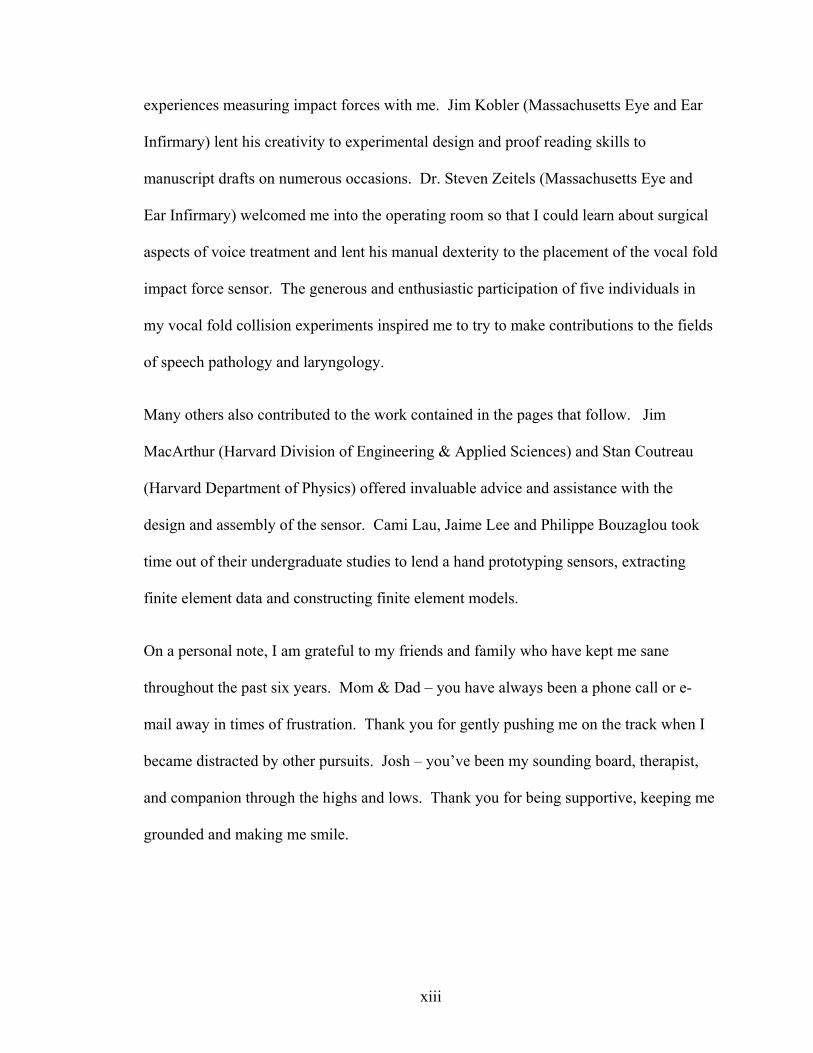
Voice is produced through a coupled oscillation between folds of tissue called the vocal
folds and the airflow between them (Figure 1.1). When the folds are brought together to
the midline and there is a sufficient, but not excessive, pressure buildup in the lungs, the
coupled oscillation ensues. The tissue oscillation can be observed using endoscopic
techniques. The aerodynamic oscillation is a sound wave that is filtered by the rest of the
vocal tract, including the pharynx, oral, and nasal cavities, to generate the sound heard by
an observer (Figure 1.2). This method of sound production is used in the production of
vowels and some consonants, and is the foundation of pitch contours.
Gunter, H.E.
2
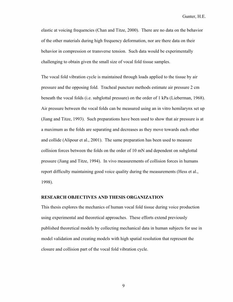
Figure 1.1: Vocal fold and air flow oscillation during voice production. (a) Coronal sections through the larynx showing the medial profile of the vocal folds during one cycle of vibration. The double lines are the medial contours of the two vocal folds. The spaces between the double lines are continuous with the airway. Arrows indicate airflow between the vocal folds. Opening is caused by air pressure below the vocal folds. Closing is caused by decreased air pressure between the vocal folds and elastic recoil of the tissue. (b) Plot of volume airflow between the vocal folds during one cycle of vibration. Air pressure beneath the vocal folds and vocal fold configuration govern the airflow between the vocal folds. The parameters maximum flow declination rate and minimum flow are associated with voice quality.

Gunter, H.E.
3
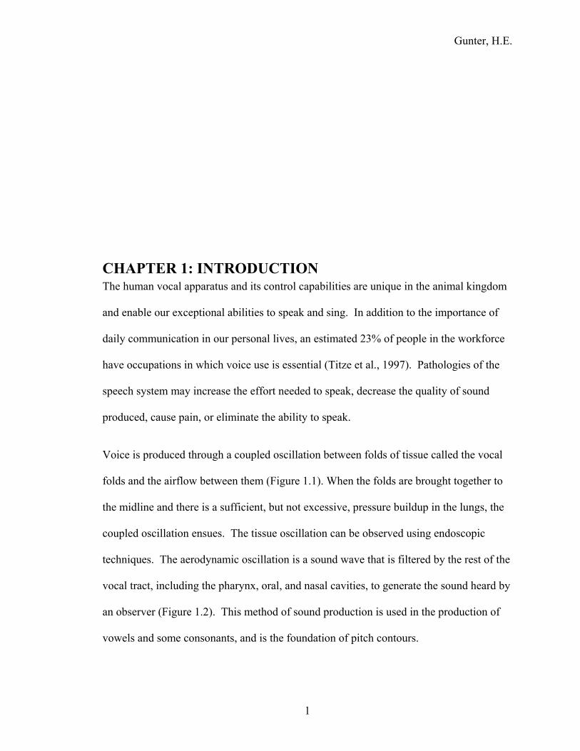
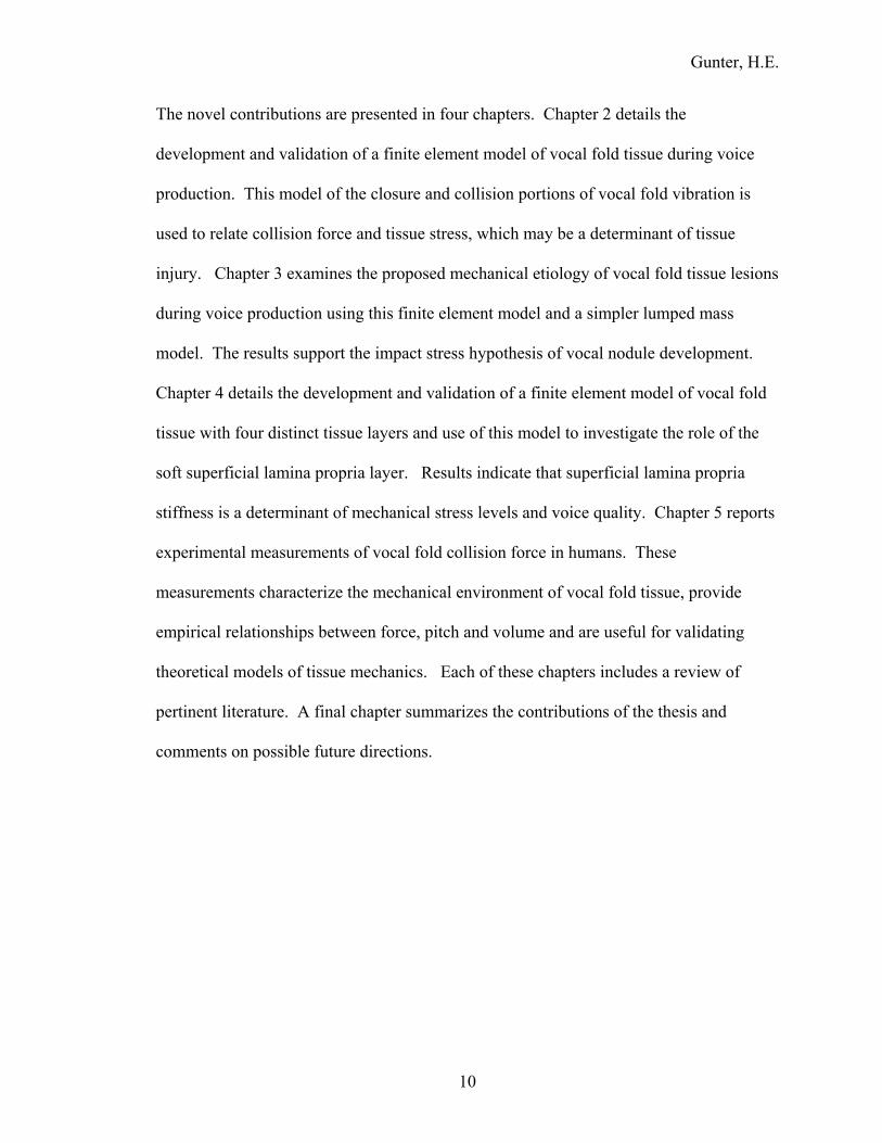
Figure 1.2: Anatomy of human voice production. The vocal folds are sketched in gray and are located in the larynx. During voice production air moves from the lungs, between the vocal folds, through the oral and nasopharynx, and out of the mouth and nostrils as shown by the arrows.
Gunter, H.E.
4
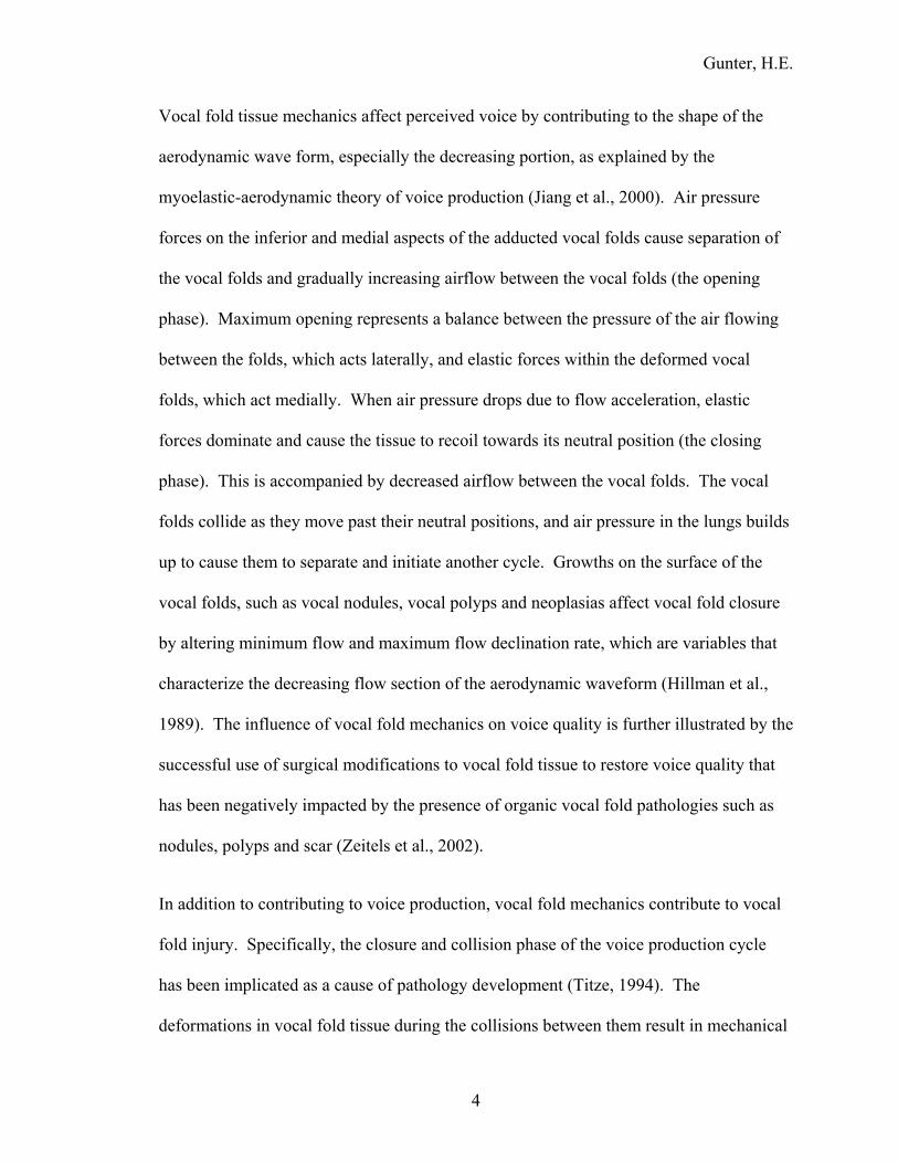
Vocal fold tissue mechanics affect perceived voice by contributing to the shape of the
aerodynamic wave form, especially the decreasing portion, as explained by the
myoelastic-aerodynamic theory of voice production (Jiang et al., 2000). Air pressure
forces on the inferior and medial aspects of the adducted vocal folds cause separation of
the vocal folds and gradually increasing airflow between the vocal folds (the opening
phase). Maximum opening represents a balance between the pressure of the air flowing
between the folds, which acts laterally, and elastic forces within the deformed vocal
folds, which act medially. When air pressure drops due to flow acceleration, elastic
forces dominate and cause the tissue to recoil towards its neutral position (the closing
phase). This is accompanied by decreased airflow between the vocal folds. The vocal
folds collide as they move past their neutral positions, and air pressure in the lungs builds
up to cause them to separate and initiate another cycle. Growths on the surface of the
vocal folds, such as vocal nodules, vocal polyps and neoplasias affect vocal fold closure
by altering minimum flow and maximum flow declination rate, which are variables that
characterize the decreasing flow section of the aerodynamic waveform (Hillman et al.,
1989). The influence of vocal fold mechanics on voice quality is further illustrated by the
successful use of surgical modifications to vocal fold tissue to restore voice quality that
has been negatively impacted by the presence of organic vocal fold pathologies such as
nodules, polyps and scar (Zeitels et al., 2002).
In addition to contributing to voice production, vocal fold mechanics contribute to vocal
fold injury. Specifically, the closure and collision phase of the voice production cycle
has been implicated as a cause of pathology development (Titze, 1994). The
deformations in vocal fold tissue during the collisions between them result in mechanical
Gunter, H.E.
5
stresses that are proposed to cause mechanical and functional tissue injury that manifests
as benign vocal fold pathologies such as vocal nodules. The pathologies observed are 1-
2mm in size and involve the thickness of the lamina propria (Dikkers, 1994). Therefore
it is likely that mechanical stress changes occur on a similar or smaller scale. The
stresses that are likely to be associated with collision and cause tissue damage include
compressive stress perpendicular to the plane of contact that may cause cellular rupture,
and shear stress parallel to the plane of contact that may cause separation of tissue
elements. Damage may also be caused by alterations in cellular function in response to
environmental stresses. However, it is not clear which components of stress govern
cellular function. Von Mises stress may be a quantity that is suitable for evaluating
cellular deformation. It is a scalar quantity that represents distortion energy and whose
magnitude is a predictor of failure in engineering materials (Chandrupatla and
Belegundu, 1997).
Simple and complex theoretical models have been used to quantify mechanical stress in
vocal fold tissue. Titze (1994) uses impact/momentum principles and empirical
relationships between vocal fold structure and function to generate an order of magnitude
estimate of collision-induced compressive stress. The inputs do not relate directly to
tissue structure, and the output is limited to one spatial invariant component of the stress
tensor. Jiang et al. (1998) use finite element models to examine the spatial variation of
tissue stress levels. They restrict their analysis to the case of natural vibration, examine
one stress invariant and do not examine stress dependence on geometric and material
parameters. Other models of voice production have not been used to predict tissue stress.
Lumped mass models of voice production that were developed for speech synthesis
Gunter, H.E.
6
applications and subsequently used for quantitative study of voice physiology (Story and
Titze, 1995;Wong et al., 1991) have not interpreted spring deformation as an indicator of
stress. Finite element models of voice production (Alipour et al., 2000) have neither
sufficient spatial resolution nor an appropriate three-dimensional mesh to examine stress
variations on the submillimeter scale of vocal fold pathology. Jiang and Titze (1994) and
Hess et al. (1998) cite the impact stress hypothesis as their motivation for identifying
situations that increase intra-fold collision forces and implying corresponding increases in
mechanical stresses. The relationship between measured total collision forces and
distributed mechanical stresses remains to be defined. The work presented in this thesis
builds on these efforts by developing new theoretical models of vocal fold mechanics
with high spatial resolution, developing a new method of measuring vocal fold collision
forces during voice production in humans, and applying these methods to the study of
mechanical stress levels in vocal fold tissue.
MECHANICS OF VOCAL FOLD TISSUE
The mechanical behavior of vocal fold tissue has implications for voice production and
vocal fold pathology development. In order to apply principles of solid mechanics to
gain insight to these topics it is necessary to understand the mechanical details of vocal
fold tissue including its geometric dimensions, material properties, boundary conditions
and applied loads.
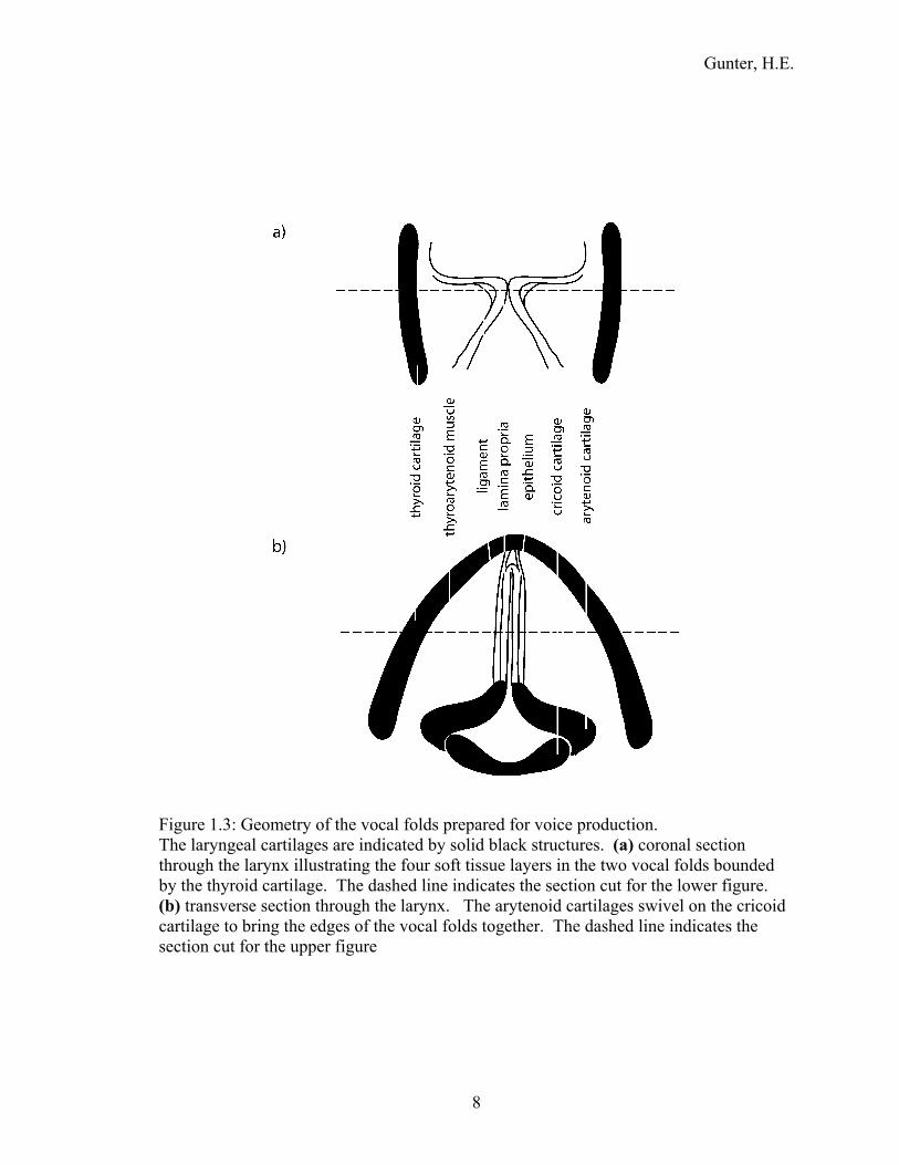
A resting vocal fold is a three dimensional structure with outer dimensions of 1- 2 cm in
length, 0.5 – 1 cm in width and 0.5 – 1 cm in height (Titze and Talkin, 1979). It
protrudes into the middle of the airway such that the longitudinal dimension spans the
diameter of the larynx. The superior, inferior and medial faces are unconstrained. The
Gunter, H.E.
7
anterior, posterior and lateral faces are fixed to laryngeal cartilages (Figure 1.3). It is
composed of tissue layers. The epithelium is the outermost layer on the three exposed
faces and is 0.1 mm thick. In longitudinal tension it is strain stiffening, and characterized
by a Young’s modulus of 41.9 kPa for strains of less than 15% (Alipour-Haghighi and
Titze, 1991). Beneath the epithelium lies a 0.5 mm thick layer called the lamina propria
(Hirano et al., 1983). It is a soft, gel like substance that is viscoelastic and shear thinning
with a shear modulus on the order of 100 Pa and a dynamic viscosity on the order of 1
Pa-s at 1 Hz (Chan and Titze, 1999a). Beneath the lamina propria, forming the bulk of
the vocal fold interior is the thyroarytenoid muscle, which is characterized in longitudinal
tension by a Young’s modulus of 20.7 kPa for strains of less the 15% and higher moduli
for higher strains (Alipour-Haghighi and Titze, 1991). At their medial aspects, the
muscle and the lamina propria are separated by the vocal ligament. This string like
structure is approximately 0.3 mm thick (Hirano et al., 1983) and demonstrates strain
stiffening behavior in longitudinal tension that can be approximated by a Young’s
modulus of 36.1 kPa (Min et al., 1995).
During voicing the vocal folds undergo static and dynamic deformations. The vocal folds
are statically stretched 10 % in the process of being moved into a voice producing
configuration (Hirano et al., 1988). Additional static strains are imposed through the
actions of extrinsic laryngeal muscles in order to manipulate pitch and vocal register.
During voice production the center of the medial surface of the vocal folds deforms
approximately 1 mm laterally during each vibration cycle, which occurs approximately
150 times per second to produce voice at 150 Hz (Zemlin, 1997). Theoretical extensions
of frequency dependent behavior of the lamina propria suggest that it is almost purely
Gunter, H.E.
8
Figure 1.3: Geometry of the vocal folds prepared for voice production. The laryngeal cartilages are indicated by solid black structures. (a) coronal section through the larynx illustrating the four soft tissue layers in the two vocal folds bounded by the thyroid cartilage. The dashed line indicates the section cut for the lower figure. (b) transverse section through the larynx. The arytenoid cartilages swivel on the cricoid cartilage to bring the edges of the vocal folds together. The dashed line indicates the section cut for the upper figure
Gunter, H.E.
9
elastic at voicing frequencies (Chan and Titze, 2000). There are no data on the behavior
of the other materials during high frequency deformation, nor are there data on their
behavior in compression or transverse tension. Such data would be experimentally
challenging to obtain given the small size of vocal fold tissue samples.
The vocal fold vibration cycle is maintained through loads applied to the tissue by air
pressure and the opposing fold. Tracheal puncture methods estimate air pressure 2 cm
beneath the vocal folds (i.e. subglottal pressure) on the order of 1 kPa (Lieberman, 1968).
Air pressure between the vocal folds can be measured using an in vitro hemilarynx set up
(Jiang and Titze, 1993). Such preparations have been used to show that air pressure is at
a maximum as the folds are separating and decreases as they move towards each other
and collide (Alipour et al., 2001). The same preparation has been used to measure
collision forces between the folds on the order of 10 mN and dependent on subglottal
pressure (Jiang and Titze, 1994). In vivo measurements of collision forces in humans
report difficulty maintaining good voice quality during the measurements (Hess et al.,
1998).
RESEARCH OBJECTIVES AND THESIS ORGANIZATION
This thesis explores the mechanics of human vocal fold tissue during voice production
using experimental and theoretical approaches. These efforts extend previously
published theoretical models by collecting mechanical data in human subjects for use in
model validation and creating models with high spatial resolution that represent the
closure and collision part of the vocal fold vibration cycle.
Gunter, H.E.
10
The novel contributions are presented in four chapters. Chapter 2 details the
development and validation of a finite element model of vocal fold tissue during voice
production. This model of the closure and collision portions of vocal fold vibration is
used to relate collision force and tissue stress, which may be a determinant of tissue
injury. Chapter 3 examines the proposed mechanical etiology of vocal fold tissue lesions
during voice production using this finite element model and a simpler lumped mass
model. The results support the impact stress hypothesis of vocal nodule development.
Chapter 4 details the development and validation of a finite element model of vocal fold
tissue with four distinct tissue layers and use of this model to investigate the role of the
soft superficial lamina propria layer. Results indicate that superficial lamina propria
stiffness is a determinant of mechanical stress levels and voice quality. Chapter 5 reports
experimental measurements of vocal fold collision force in humans. These
measurements characterize the mechanical environment of vocal fold tissue, provide
empirical relationships between force, pitch and volume and are useful for validating
theoretical models of tissue mechanics. Each of these chapters includes a review of
pertinent literature. A final chapter summarizes the contributions of the thesis and
comments on possible future directions.
Gunter, H.E.
11
CHAPTER 2: A MECHANICAL MODEL OF VOCAL FOLD COLLISION WITH HIGH SPATIAL AND TEMPORAL RESOLUTION1,2
INTRODUCTION
A detailed theoretical model of vocal fold tissue mechanics during collision that connects
tissue damaging variables with clinical or experimental variables and vice versa is an
excellent tool with which to clarify the impact stress hypothesis (Chapter 1). A model of
vocal fold collision that captures the dynamics of vocal fold closure also has the potential
to be applied to prediction of the surgical outcomes by predicting the tissue correlates of
the aerodynamic variables minimum flow and maximum flow declination rate, which
relate to voice quality perceptions (Holmberg et al., 1988).
The idea to represent vocal fold tissue mechanics mathematically is not a new one. The
simplest models represent the vocal folds using the basic mechanical elements (i.e.
masses, springs and dampers) and were initially developed for speech synthesis
1 Portions of this work were presented in “Analysis of Factors Affecting Vocal Fold Impact Stress Using a Mechanical Model” 142nd meeting of the Acoustical Society of America, Ft. Lauderdale, FL, December 2001 2 This work is published as Gunter, H. E. (2003), "A mechanical model of vocal fold collision with high spatial and temporal resolution," Journal of the Acoustical Society of America. 113, 994-1000.
Gunter, H.E.
12
applications. The classic example uses two masses to represent a single vocal fold
(Ishizaka and Flanagan, 1972). Variations by Story and Titze (1995), Wong et al. (1991)
and Titze (1973;1974) incorporate additional masses to provide resolution in the depth
and longitudinal dimensions. Story and Titze predict intraglottal collision pressures
based on the deformation of the collision spring element that agree qualitatively with
Jiang and Titze’s (1994) experimental measurements. However, their peak pressure
prediction is approximately five-fold less than the comparable experimental measurement
(Jiang and Titze, 1994). The computational simplicity of these tissue mechanics models
makes them an excellent foundation for the exploration of complex theories of voice
aerodynamics (de Vries et al., 2002;Pelorson et al., 1994). Analysis using nonlinear
dynamic techniques (Jiang et al., 2001b;Wong et al., 1991) provides some insight into
origins of pathological voice. However, the spatial resolution of the lumped mass models
is insufficient to reflect the scale of pathology and surgery, and their empirically assigned
spring, damper, and mass values have few direct implications for vocal fold tissue
physiology. This limits their applicability to the study of vocal fold tissue mechanics.
Recently Titze & Story (2002) developed rules relating the lumped mass parameters to
muscle activation levels. While this improves the physiological relevance, the spatial
resolution remains low and therefore this model is not suited to the study of vocal fold
tissue mechanics.
Continuum approaches represent the vocal folds more realistically at the expense of
computational efficiency. To maintain computational time within a reasonable range
these models require simplification of either the solid or fluid mechanics representation.
Use of simplified two dimensional (Ikeda et al., 2001) or three dimensional beam (Berry
Gunter, H.E.
13
and Titze, 1996;Titze and Strong, 1975) geometries in analytical models does not capture
the subtleties of vocal fold tissue anatomy. Numerical approaches permit the use of
realistic three dimensional geometries (Titze and Talkin, 1979)). The numerical
technique of finite elements has been used to create models that overcome the barriers of
low spatial resolution and restricted geometries (Alipour et al., 2000;Jiang et al.,
1998;Lobo and O'Malley, 1996). These techniques involve spatially and temporally
discretizing a continuum mechanics problem into solid elements and time increments
followed by numerical solution. In order to maintain computation at reasonable levels
these have used spatial resolution that is inadequate for the study of tissue mechanics.
Alipour et al.’s (2000) self-oscillating model requires increased spatial resolution,
calculation of mechanical stress distributions and extension to true three-dimensionality
in order to represent vocal fold pathologies and examine mechanical stress distributions.
Jiang et al.’s (1998) and Lobo and O’Malley’s (1996) finite element models of vocal fold
tissue have sufficient spatial resolution to represent pathology, but limit analysis to
resonant and forced vibration. They require an explicit representation of collision forces
between the vocal folds in order to be used in investigations of contact related pathology
etiologies and voice quality. Variations on these finite element models have the potential
to be excellent tools with which to investigate vocal fold tissue mechanics.
A finite element model of vocal fold collision that is not self oscillating is presented
below. It has spatial resolution that is capable of representing the submillimeter scale of
vocal fold injury and repair, temporal resolution that captures the submillisecond time
scale of vocal fold collision and an explicit representation of vocal fold collision. Model
predictions of collision force are validated against experimental data and model potential
Gunter, H.E.
14
is illustrated in an examination of the relationships between collision force and
mechanical stress levels.
DEVELOPMENT
Assumptions
The model of vocal fold collision presented below has three unique features. The spatial
resolution of 250 µm is finer than that incorporated in other vocal fold models. A single
isotropic, linear elastic material characterizes the entire vocal fold structure. Glottal
closure during phonation is represented using a non-oscillating model consisting of
appropriate initial conditions and a high fidelity model of vocal fold solid mechanics.
Justifications for these features are outlined below.
The necessary spatial resolution is dictated by the size of geometric variations in the
model, the desired resolution of model outputs, and desired computational speed. The
model is intended for use in studying the effects of vocal fold pathologies and their repair
on tissue movement and for investigating the distribution of mechanical stresses in vocal
fold tissue as an indication of pathology development risk. Benign vocal fold pathologies
such as nodules and polyps have dimensions of 1-2 mm on the vocal fold surface
(Dikkers, 1994). Spanning the pathology with a minimum of four elements requires
element dimensions of 250 µm and allows for some sculpting of the geometry. Surgical
repair of the vocal fold can involve manipulation of only the superficial lamina propria
(Zeitels, 1998), which, at approximately 500 µm thick (Hirano et al., 1983), can be
represented by two elements. Mechanical stresses that are important in tissue injury are
on the same scale as or smaller scale than the pathologies that they are proposed to cause.
Gunter, H.E.
15
Therefore a resolution that is sufficient to represent pathologies provides appropriate
information on stress distributions. Further resolution would increase geometric fidelity
and output detail, but would also increase computational load significantly.
A simple material definition is used in the model. The assumption of linear elasticity is
consistent with Min et al.’s (1995) observation of linear stress-strain behavior of human
vocal ligaments for strains of less than 15%. Strains in the model do not exceed this 15%
upper bound. The primary mode of deformation of the vocal fold during vocal fold
closure is compressive in the transverse (i.e. medial-lateral and inferior-superior)
directions. However, there is a lack of direct data on the elastic properties of vocal fold
tissue when undergoing this kind of deformation. Therefore parameters based on tissue
behavior during longitudinal tension are used to characterize the material properties. The
range of elastic moduli derived by Min et al. (1995) using human vocal ligament samples
(21.2 kPa-42.2 kPa) spans the moduli derived by Alipour-Haghighi and Titze (1991) for
unstimulated canine thyroarytenoid muscle (20.7 kPa) and canine vocal fold cover (41.1
kPa) and are the same order of magnitude as moduli derived by Kakita et al. (1981) using
canine vocal fold muscle and cover. The assumption of homogeneous properties is
within the spread of the data in the literature.
Some studies suggest that air pressure in the glottis is low during glottal closure. The
myoelastic-aerodynamic theory of voice production cites three reasons for glottal closure:
elastic recoil of the deformed tissue, decreased pressure on the glottal walls due to the
high velocity of the air stream through the opening and decreased subglottal pressure
(Jiang et al., 2000). Experimental measurements performed on canine hemilarynges
illustrate that, in an almost fully adducted larynx, air pressure in the glottis remains below
Gunter, H.E.
16
10% of subglottal pressure during glottal closure and does not rise until full closure has
occurred (Alipour and Scherer, 2000). Measurements performed on excised human
hemilarynges also demonstrate reduced glottal air pressures during closure (Alipour et
al., 2001). It is consistent with observations of minimum air pressures and maximum
vocal fold deformation when the glottis is fully open to postulate that elastic forces
dominate during glottal closure, and to focus on these forces in a model of glottal closure.
Implementation
The model geometry represents the membranous portion of a fully adducted single vocal
fold (Figure 2.1). The model is defined in Cartesian coordinates. The origin is in the
midposterior glottis, inferior to the bulge of the vocal fold. The y axis defines the midline
of the glottis, x dimensions indicate lateral distance from the glottal midline, and z
translations represent movement in the vertical direction. The vocal fold dimensions are
1.4 cm long (y direction), 0.5 cm wide anteriorly, 1.0 cm wide posteriorly (x direction),
and 1 cm high posteriorly (z direction). The anterior, posterior and lateral surfaces are
fixed to represent their attachment to the laryngeal cartilages. This structure is based on
Titze and Talkin (1979) and uses their nominal parameters (i.e. shaping factor (s) equal to
0.05 radians, glottal angle (w) equal to 0 radians and inferior surface angle (q) equal to
0.7 radians). The Titze and Talkin geometry is modified by applying a fillet with a radius
of 0.05 cm to the superior medial curve in order to create a smooth contour in the coronal
plane. The glottal width (g) between the inferior aspect of the fillet and the superior
aspect of the inferior surface is derived from an expression given by Titze and Talkin:
( ) ( )25.025.09.145.0)5.005.0( −+−−=≤≤ hhhg (2.1)
Gunter, H.E.
17
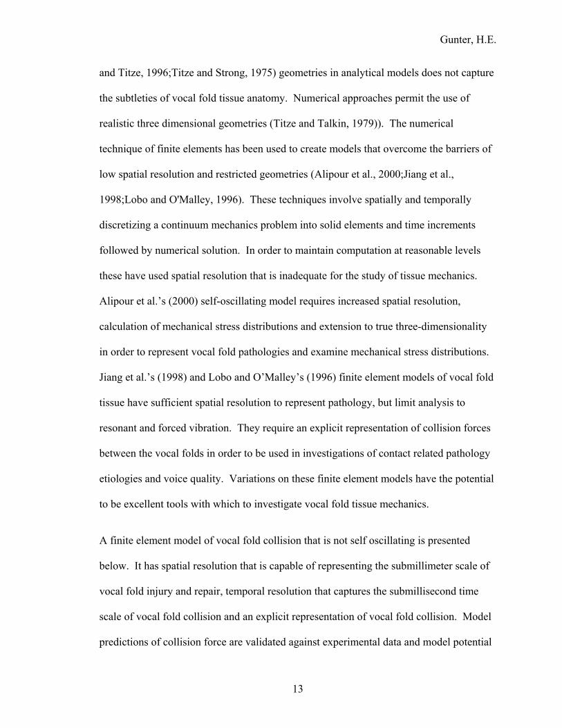
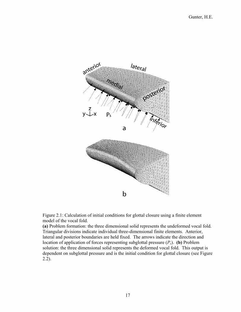
Figure 2.1: Calculation of initial conditions for glottal closure using a finite element model of the vocal fold. (a) Problem formation: the three dimensional solid represents the undeformed vocal fold. Triangular divisions indicate individual three-dimensional finite elements. Anterior, lateral and posterior boundaries are held fixed. The arrows indicate the direction and location of application of forces representing subglottal pressure (Ps). (b) Problem solution: the three dimensional solid represents the deformed vocal fold. This output is dependent on subglottal pressure and is the initial condition for glottal closure (see Figure 2.2).
Gunter, H.E.
18
h is the vertical distance below the superior surface. The minimum glottal width of 0.0
cm that occurs at the inferior end of the fillet (h equal to 0.05 cm) indicates that the
undeformed vocal fold is tangent to the glottal midline. A similar geometry has been
used by Alipour et al. (2000) with good results.
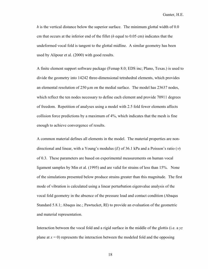
A finite element support software package (Femap 8.0; EDS inc; Plano, Texas.) is used to
divide the geometry into 14242 three-dimensional tetrahedral elements, which provides
an elemental resolution of 250 µm on the medial surface. The model has 23637 nodes,
which reflect the ten nodes necessary to define each element and provide 70911 degrees
of freedom. Repetition of analyses using a model with 2.5 fold fewer elements affects
collision force predictions by a maximum of 4%, which indicates that the mesh is fine
enough to achieve convergence of results.
A common material defines all elements in the model. The material properties are non-
directional and linear, with a Young’s modulus (E) of 36.1 kPa and a Poisson’s ratio (ν)
of 0.3. These parameters are based on experimental measurements on human vocal
ligament samples by Min et al. (1995) and are valid for strains of less than 15%. None
of the simulations presented below produce strains greater than this magnitude. The first
mode of vibration is calculated using a linear perturbation eigenvalue analysis of the
vocal fold geometry in the absence of the pressure load and contact condition (Abaqus
Standard 5.8.1; Abaqus inc.; Pawtucket, RI) to provide an evaluation of the geometric
and material representation.
Interaction between the vocal fold and a rigid surface in the middle of the glottis (i.e. a yz
plane at x = 0) represents the interaction between the modeled fold and the opposing
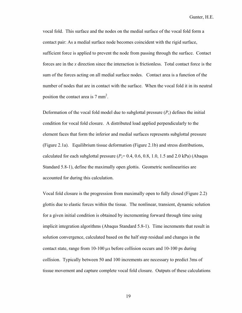
Gunter, H.E.
19
vocal fold. This surface and the nodes on the medial surface of the vocal fold form a
contact pair: As a medial surface node becomes coincident with the rigid surface,
sufficient force is applied to prevent the node from passing through the surface. Contact
forces are in the x direction since the interaction is frictionless. Total contact force is the
sum of the forces acting on all medial surface nodes. Contact area is a function of the
number of nodes that are in contact with the surface. When the vocal fold it in its neutral
position the contact area is 7 mm2.
Deformation of the vocal fold model due to subglottal pressure (Ps) defines the initial
condition for vocal fold closure. A distributed load applied perpendicularly to the
element faces that form the inferior and medial surfaces represents subglottal pressure
(Figure 2.1a). Equilibrium tissue deformation (Figure 2.1b) and stress distributions,
calculated for each subglottal pressure (Ps= 0.4, 0.6, 0.8, 1.0, 1.5 and 2.0 kPa) (Abaqus
Standard 5.8-1), define the maximally open glottis. Geometric nonlinearities are
accounted for during this calculation.
Vocal fold closure is the progression from maximally open to fully closed (Figure 2.2)
glottis due to elastic forces within the tissue. The nonlinear, transient, dynamic solution
for a given initial condition is obtained by incrementing forward through time using
implicit integration algorithms (Abaqus Standard 5.8-1). Time increments that result in
solution convergence, calculated based on the half step residual and changes in the
contact state, range from 10-100 µs before collision occurs and 10-100 ps during
collision. Typically between 50 and 100 increments are necessary to predict 3ms of
tissue movement and capture complete vocal fold closure. Outputs of these calculations
Gunter, H.E.
20
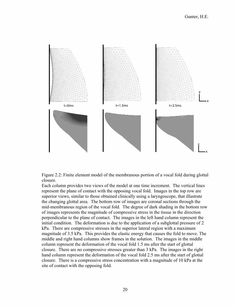
Figure 2.2: Finite element model of the membranous portion of a vocal fold during glottal closure. Each column provides two views of the model at one time increment. The vertical lines represent the plane of contact with the opposing vocal fold. Images in the top row are superior views, similar to those obtained clinically using a laryngoscope, that illustrate the changing glottal area. The bottom row of images are coronal sections through the mid-membranous region of the vocal fold. The degree of dark shading in the bottom row of images represents the magnitude of compressive stress in the tissue in the direction perpendicular to the plane of contact. The images in the left hand column represent the initial condition. The deformation is due to the application of a subglottal pressure of 2 kPa. There are compressive stresses in the superior lateral region with a maximum magnitude of 3.5 kPa. This provides the elastic energy that causes the fold to move. The middle and right hand columns show frames in the solution. The images in the middle column represent the deformation of the vocal fold 1.5 ms after the start of glottal closure. There are no compressive stresses greater than 3 kPa. The images in the right hand column represent the deformation of the vocal fold 2.5 ms after the start of glottal closure. There is a compressive stress concentration with a magnitude of 10 kPa at the site of contact with the opposing fold.
Gunter, H.E.
21
include movement of each node, mechanical stresses in each element, and interactions
between nodes and the rigid midline surface.
The goals with this model are to validate predictions of closure kinematics and collision
surface forces against published experimental measurements and to examine the
relationship between collision surface forces and mechanical stress levels in the tissue
during collision. Model predictions that reflect these goals are discussed below.
VALIDATION
The natural frequency is 127 Hz, which is consistent with experimentally measured
resonant frequencies (Kaneko et al., 1987) and theoretically predicted resonant
frequencies for adult male voices (Alipour et al., 2000). This agreement corroborates the
geometric and material definitions.
Glottal closure dynamics are an important output of the model, and model behavior
agrees with observations of vocal fold vibration. The qualitative movement of the fold
includes movement towards the midline and collision with the opposing fold (Figure 2.2).
Closure begins at the anterior and posterior ends and progresses towards the mid-
membranous region (Figure 2.2, top row), which is consistent with in vivo stroboscopic
observations of fully adducted phonation. The initial mid-membranous glottal
displacement (i.e. the maximum lateral displacement of the medial vocal fold edge) is an
approximately linear function of subglottal pressure and ranges between 0.15 mm and 0.8
mm (Figure 2.3). These predictions are consistent with previous visual observations of
normal vocal fold vibration (Zemlin, 1997). The inferior portion of the vocal fold edge is
the first point of collision with the opposing fold. As collision proceeds, contact includes
Gunter, H.E.
22
the superior vocal fold edge (Figure 2.2, bottom row). This progression is similar to
phase differences that are observed clinically. Contact areas, which are calculated based
on the number of nodes touching the midline surface in the vocal fold closure and
collision solutions, are also functions of Ps and time (Figure 2.4). The contact areas
range from 0 to 35 mm2, which lies within the area of vocal fold contact imaged by Jiang
and Titze (1994).
Collision dynamics during closure are another important output of the model, and model
behavior agrees with published experimental results. The total collision force increases
with time and reaches either a peak or a plateau depending on the initial Ps (Figure 2.5).
The rise time of collision in a 3 mm mid-membranous region for Ps=1.5 kPa and 2.0 kPa
is approximately 0.5 ms, which is equal to the experimentally measured rise time for
impact with a 3 mm diameter central sensor as measured in canine hemilarynges by Jiang
and Titze (1994). Due to the collision force plateau, there are no clear force peaks for Ps
less than 1.5 kPa. Collision force peaks are defined either as the force magnitude at the
time of full closure, which is independent of Ps (Figure 2.3), or as the force magnitude at
the time of maximum contact area, which is dependent on Ps (Figure 2.4). These peaks
are used to derive linear relationships between subglottal pressure and peak collision
force. Table 2.1 compares the slopes and regression coefficients for lines fit to model
predictions with those fit to in vitro experimental data collected from canine
hemilarynges by Jiang and Titze (1994). Both definitions of theoretical peak collision
force fall within the span of the experimental results.
Gunter, H.E.
23
Figure 2.3: Effect of subglottal pressure on the medial movement of the vocal fold as predicted by a finite element model of vocal fold collision Each line traces the solution for the x position of a mid-membranous point on the medial edge of the vocal fold edge during glottal closure in a single model. The models differ by the subglottal pressure magnitude used to calculate the initial closure condition (i.e. t = 0). The arrow points to the time when the mid-membranous glottal opening and glottal area are zero, which is defined as the time of full closure. This value is not dependent on subglottal pressure.
Gunter, H.E.
24
Figure 2.4: Effect of subglottal pressure on the area of contact between the vocal fold and the contact surface as predicted by a finite element model of vocal fold collision Each line traces the solution for contact area, which is proportional to the number of nodes that are at x = 0 (i.e. touching the contact plane) during glottal closure in a single model. The models differ by the subglottal pressure used to calculate the initial condition of glottal closure (i.e. t = 0). The shaded area indicates the temporal range of contact area maxima, which varies among models.
Gunter, H.E.
25
Figure 2.5: Effect of subglottal pressure on the total contact force between the vocal fold and the contact surface as predicted by a finite element model of vocal fold collision Each line traces the solution for contact force, which is the sum of point loads applied by the contact plane to nodes on the medial surface, during glottal closure in a single model. The models differ by the subglottal pressure magnitude used to calculate the initial condition of glottal closure (i.e. t = 0).
Gunter, H.E.
26
Table 2.1: Comparison between linear regression analysis of peak contact force and subglottal pressure for theoretical predictions using a finite element model and Jiang and Titze’s (1994) experimental measurements on canine hemilarynges
Contact Force Definition Linear Slope (mN / kPa)
Regression Coefficient
Model prediction – peak contact area
16.39 0.977
Model prediction – full closure
10.48 0.930
Experimental measurement (Jiang and Titze, 1994)
Range =7.70 - 26.60 Mean =12.46
MECHANICAL STRESS DURING COLLISION
To determine whether local collision force predicts local mechanical stress, a regression
analysis between contact force predictions and mechanical stress predictions is
performed. For each time point during vocal fold collision and at each 1 mm interval
along the vocal fold edge, contact force and medial superficial stresses are extracted from
model results. Compressive stress perpendicular to the plane of contact, shear stress
parallel to the plane of contact in the longitudinal direction, shear stress parallel to the
plane of contact in the vertical direction, and Von Mises stress are examined. The
relationship between contact force and stresses for a subglottal pressure of 1 kPa is
dependent on the stress direction (Figure 2.6). At this subglottal pressure there is a
general trend between increases in local contact force and increases in compressive stress
(r2=0.79), vertical shear stress (r2=0.69) and Von Mises stress (r2=0.83). There is no
trend between local contact force and longitudinal shear stress (r2=0.00).
Local variations from the general trends can be appreciated by examining the data points
for each 1mm segment. The variations are most pronounced for longitudinal shear stress,
Gunter, H.E.
27
where there are local inverse trends between contact force and stress despite the lack of a
general trend.
DISCUSSION
This model examines the time and spatial course of vocal fold closure, but not separation,
during phonation and makes two contributions. The first contribution is reasonably
accurate prediction of closure kinematics and collision surface forces, despite the absence
of an aerodynamic representation and an assumption of homogeneous material properties.
This provides insight as to the importance of elastic forces in glottal closure and supports
future application to the study of the effects of vocal fold tissue elasticity and structure on
the dynamics of glottal closure, corresponding aerodynamic variables and, by extension,
voice quality.
The second contribution is an illustration of the potential of this model of glottal closure
to provide a window to mechanical conditions in the vocal fold interior that may increase
injury risk. Regressions between surface contact force and relative predictions of
mechanical stress in the tissue indicate that the implications of experimental impact force
measurements are position dependent and identify compressive stress perpendicular to
the contact plane, shear stress parallel to the contact plane in the longitudinal direction
and Von Mises stress as candidate mechanical stresses that may cause tissue damage as a
result of high collision forces.
The ability of this model to predict closure kinematics and collision surface forces despite
exclusion of aerodynamic forces from the model provides insight into the mechanics of
voice production. Agreements between model predictions and published data suggest
Gunter, H.E.
28
Figure 2.6: Relationship between local contact force and mechanical stress on the medial vocal fold surface during collision as predicted using a finite element model The x value of each data point represents the integrated contact force over a 1mm vertical slice of the medial surface. The y value of each data point represents the maximum mechanical stress in the surface tissue in the same 1mm slice. Data is symmetric about the mid-membranous location (y = 7 mm). For simplicity only data for the anterior half of the fold are shown. Correlation coefficients are given in the text.
Gunter, H.E.
29
that elastic forces within the tissue dominate the mechanics of vocal fold closure and
collision, and are therefore a major determinant of aerodynamic variables that are
associated with closure, such as minimum flow and maximum flow declination rate
(MFDR). The dependence of voice quality on MFDR and minimum flow allows
extension of this hypothesis to propose that solid mechanics of vocal fold tissue are a
dominant factor in voice quality. In vivo associations between structural changes that
affect vocal fold closure, such as vocal nodules, increased minimum flow, decreased
MFDR, and altered voice quality perceptions (Kuo, 1998) support this hypothesis.
The suggestion that tissue mechanics dominate vocal fold closure kinematics, important
aerodynamic variables, and voice quality reinforces the proposed use of a vocal fold
collision model to predict surgical outcomes. Comparison of the closure dynamics
between models of vocal folds with organic pathologies and models of the same folds
following surgical repair will allow prediction of the effects of surgery on voice quality.
The prognostic value of this application may be further refined by incorporation of a
layered geometrical structure and more complex material property definitions; these will
facilitate the creation of higher fidelity models of pathology and surgical changes. This
idea is explored in Chapter 4.
The lack of a defined contact force peak in models with low subglottal pressures and the
lack of a reopening phase in all models does not agree with clinical observations of
glottal opening, but was not an intended focus of this model. These results indicate that
bouncing of the vocal folds following collision is not sufficient to cause glottal opening
for the subglottal pressures and configurations studied and reinforces the important role
played by air pressure in vocal fold separation. Comparison of the current results with
Gunter, H.E.
30
those from a new model with a sophisticated aerodynamic representation will test the
hypothesis that tissue elasticity forces dominate during glottal closure and that
aerodynamic forces dominate during vocal fold separation.
Agreement between model glottal closure kinematics and collision surface forces and
experimental measurements does not necessarily imply that model predictions of
mechanical stress are accurate. It is the case in any complex model with multiple input
variables, that there may not be one unique combination that produces the desired output.
In the case of this model, prediction of appropriate contact surface forces and closure
kinematics only implies that one appropriate set of input parameters was identified. A
different combination of input parameters may have provided the same surface contact
force predictions, but different absolute mechanical stress predictions. However, it is
likely that the relative nature of mechanical stress predictions would remain the same.
Therefore it is reasonable to compare them against each other.
The relationships between contact forces on the vocal fold surface and superficial tissue
mechanical stress in the medial edge of the vocal fold, derived using a model of vocal
fold collision, guide the interpretation of experimental contact force measurements, such
as those presented in Chapter 5. Local collision force measurements are indicative of
local compressive stress, vertical shear stress, and Von Mises stress. This observation
suggests that pathologies associated with maneuvers that have increased local impact
forces (e.g. phonation with high subglottal pressures) may be due to a compressive mode
of tissue failure, a shear mode of tissue failure or alterations in cellular behavior.
Unfortunately, without knowledge of what stress magnitudes cause tissue failure, it is
impossible to comment on which stress levels are most damaging to the tissue.
Gunter, H.E.
31
Correlations between stress predictions and epidemiology of lesions that reflect tissue
injury, such as vocal nodules, will test the impact stress hypothesis of pathology
development. This is explored in Chapter 3.
Additional experimental measurements will improve definition and validation of the
vocal fold collision model and other models of vocal fold tissue mechanics. Three
dimensional material property data based on human vocal fold tissue samples undergoing
compression would improve the fidelity of model inputs. In vivo human results of
surface collision forces would be a preferred source against which to validate model
predictions because agreement will enhance the applicability of the model to human
voice. Such measurements are presented in Chapter 5. Other experimental
measurements that will be useful in validation include electoglottographic measurements
of contact area and photoglottographic measurements of glottal area.
Gunter, H.E.
32
CHAPTER 3: MODELING MECHANICAL STRESSES AS A FACTOR IN THE ETIOLOGY OF BENIGN VOCAL FOLD LESIONS1,2
INTRODUCTION
Vocal nodules and polyps are convex lesions on the midline of the medial surface of the
vocal folds associated with excessive voice use, loud voicing, inadequate hydration, and
voice misuse (Buckmire and Rosen, 2001). Mechanical stress during vocal fold collision
is implicated as a cause of benign vocal lesions, such as vocal nodules and vocal polyps,
based on observations of high velocity impact between vocal folds during speech (Titze,
1994) and of structural disruptions of the basement membrane in these lesions (Gray and
Titze, 1988). Experimental investigation of this hypothesis is impeded by the difficulty
of quantifying spatial and temporal stress variations in the small tissue volume (1-2 cm3)
that vibrates, on average, between 100 and 200 Hz during voice production and the
challenge of measuring and manipulating independent variables such as tissue stiffness.
1 Portions of this work were presented in “Mechanical stress levels in vocal folds as predictors of tissue injury” International Conference on Voice Physiology and Biomechanics, Denver, CO, September 17-19, 2002. 2 This work has been submitted for publication as Gunter, H.E. (2003) “Modeling Mechanical Stresses as a Factor in the Etiology of Benign Vocal Fold Lesions” to the Journal of Biomechanics (JB2002-187)
Gunter, H.E.
33
Theoretical models of vocal fold tissue mechanics are an ideal alternative research
platform since they can predict spatial and temporal stress variations at appropriate
resolutions and permit manipulation and control of geometric and material parameters
that may influence these variations.
This chapter presents new computational models and their application to investigating the
hypothesis that vocal fold tissue lesions such as nodules and polyps develop in response
to mechanical stress occurring during vocal fold collision. A one-dimensional lumped
mass model illustrates the concept of mechanical stress during collision and predicts the
influence of material and geometric parameters on compressive stress magnitude. A
three-dimensional finite element model of vocal fold collision with temporal resolution
that captures the dynamics of collision and spatial resolution smaller than that of tissue
pathology (discussed in chapter 2) predicts spatial variations of stress tensor components.
Model predictions are evaluated within the framework of the epidemiology of benign
changes in vocal fold structure such as nodules and polyps. Improved understanding of
the proposed mechanical etiology of these lesions has implications for their prevention
and treatment in the clinic.
METHODS
Lumped mass model
A simple spring mass model (Figure 3.1c), inspired by the lumped mass models of vocal
fold movement that have been useful in the speech synthesis domain (for example,
(Ishizaka and Flanagan, 1972)) generates an approximation for superficial normal stress
in vocal fold tissue during collision with the opposing fold. The model approximates a
coronal vocal fold cross section (Figure 3.1a) as two layers: a cover composed of the
Gunter, H.E.
34
Figure 3.1: Development of a lumped mass model of vocal fold collision. The dashed line indicates the plane of collision between opposing vocal folds in the center of the larynx. The gray line on the right represents the fixed boundary formed by the laryngeal cartilages. (a) Sketch of a histological cross section illustrating the layered structure of the vocal fold - LP: lamina propria, VL: vocal ligament, TM: thyroarytenoid muscle. The arrows indicate the direction that air pressure acts during voice production (b) Rectangular prism representation of a vocal fold with two layers that serves as a conceptual intermediate between the anatomical structure in (a) and the lumped mass model in (c). Cover represents the mucosa and ligament. Body represents the muscle. Each layer is defined by geometric properties of depth (D), thickness (T) and length (L) and material properties density (ρ) and stiffness (E). Nominal values of all parameters are given in Table 1. (c) One-dimensional lumped mass representation of a vocal fold defined by 3 parameters (m, kc, kb) that are defined by rectangular prism model parameters.
Gunter, H.E.
35
mucosa and underlying connective tissue, and a body composed of the thyroarytenoid
muscle. Springs (kc, kb) in series define the two layers and a mass (m) at the junction of
the springs represents the center of gravity. Interaction with the opposing fold occurs
through the cover spring (kc): When the folds are separated (x > 0), this spring is not
deformed. When the folds are in contact (x < 0), the cover spring is constrained by the
opposing fold at the midline plane, and deforms accordingly.
Closure of the glottis begins when the vocal folds are maximally separated. In this
configuration the cover spring is not constrained and the system is described by a second
order differential equation, which is valid until the folds are in contact.
00 ≥=+ xforvalidxkxm b&& (3.1)
x is the lateral displacement of the vocal fold away from its neutral position. For a
maximum opening equal to xo and an initial velocity of zero, the pre-collision solution for
movement of the vocal fold is valid from the time of release until x = 0.
b
b
kmtfort
mk
xx2
0cos0π
≤≤
= (3.2)
When the center of gravity crosses its neutral position the cover spring becomes
constrained by contact with the opposing fold at the midline plane and begins to deform.
The system is now described by another second order differential equation, which is valid
until x > 0, when the folds cease to be in contact.
( ) 00 ≤=++ xforvalidxkkxm bc&& (3.3)
Gunter, H.E.
36
Using initial conditions for position and velocity from the boundary of the precollision
solution (equation 3.2), the postcollision solution is obtained.
++≤
−
++
−=bcbbb
bc
bc
b
kkm
kmt
kmfor
kmt
mkk
kkk
xx 2222
sin0πππ (3.4)
The frequency of vibration (F0) is the inverse of the average of the pre- and post-collision
periods.
1
10
−
−
+
+=
bbc km
kkmF π (3.5)
Stress in superficial tissue is the product of the spring constant (kc) and the displacement
of the mass (x) divided by a cross sectional area (A). For noncollision phases (x > 0)
stress is equal to zero since the cover spring is not deformed. For collision phases stress
is a function of the location of the mass (equation 3.4).
−
++
−=b
bc
bc
bcxx k
mtm
kkkk
kAxk
2sin0 πσ (3.6)
The lumped mass parameters are expressed in terms of physiological variables by
comparing them to a rectangular prism approximation of a vocal fold (Figure 3.1b) that is
defined by geometric and material parameters from the literature (Table 3.1).
Consideration of longitudinal compression of the beam generates expressions for lumped
mass parameters in terms of material and geometric properties of the beam (Table 3.2).
The maximum compressive stress during collision in terms of physiological variables is
Gunter, H.E.
37
obtained by substituting the expressions relating physiological and lumped mass variables
(Table 3.2) into the amplitude of the stress expression (equation 3.6).
Table 3.1: Typical vocal fold dimensions and material properties from the literature
Parameter Symbol Nominal Value Source Length L 1.5 cm Hirano et al, 1988 Thickness T 0.25cm Perlman & Durham, 1987 Cover depth Dc 0.11cm Hirano et al, 1983 Body Depth Db 1cm Tayama et al, 2002 Cover Stiffness Ec 36.1kPa Min et al, 1995 Body Stiffness Eb 20.7kPa Alipour-Haghighi & Titze, 1991 Tissue density ρ 1.0g/cm3 Driving Pressure P 0.8 kPa
Table 3.2: Lumped model parameters in terms of biomechanical variables
Parameter Relationship A LT M LT(Dc+Db)ρ kc EcLT/Dc kb EbLT/Db X0 PDb/Eb
b
b
c
c
b
b
bc
bcxx
DE
DE
DE
EDDPE
+−=maxσ (3.7)
P is the air pressure that maximally separates the vocal folds. Ec and Eb are the Young’s
moduli of the cover and body layers. Dc and Db are the depths of the cover and body
layers. Frequency of vibration in terms of physiological variables is obtained by
substituting the biomechanical expressions (Table 3.2) into the frequency equation
(equation 3.5),
Gunter, H.E.
38
( )
1
21
0
−
−
++
+=
b
b
b
b
c
c
bc
ED
DE
DE
DDF
ρπ (3.8)
where ρ is the density of the tissue. Predictions made using these equations provide
insights into the variables contributing to superficial stress in vocal fold tissue during
vocal fold collision and are directly comparable with predictions made using a more
complex model.
Finite element model
A three-dimensional model of vocal fold closure and collision, implemented using
commercial finite element software (Abaqus Standard 5.8-1, Abaqus Inc., Pawtucket,
RI), generates spatial and temporal predictions of the stress tensor. The model consists of
14,242 ten noded tetrahedral linear elastic elements that define a single vocal fold (Figure
3.2a). This number of elements provides a resolution of 250 µm, which is necessary to
examine the formation of 500 µm pathologies such as vocal nodules. The geometric
arrangement of the elements is based on human male vocal fold dimensions used by Titze
and Talkin (1979). Their material properties (E=36.1 kPa, ν=0.3) are based on human
vocal fold ligament measurements made by Min, et al. (1995) that have been used in
previous finite element models of vocal fold tissue (Jiang et al., 1998). The assumption
of isotropy is made due to lack of quantitative data to guide statements of anisotropy.
The assumption of elasticity is based on evidence that viscous properties of vocal fold
tissue make negligible contributions to deformations at voicing frequencies (i.e. damping
ratio of 0.01 at 100 Hz) (Chan and Titze, 2000).
Gunter, H.E.
39
Figure 3.2: Finite element model of vocal fold collision (a) Three-dimensional geometric structure based on measurements of human vocal folds. Divisions indicate the boundaries of elements. Each element is defined by material properties taken from the literature. Arrows indicate driving pressure load. Dashed white line follows vocal fold edge, along which stresses are examined. Numbers indicate reference points for locations on the vocal fold edge. White circle indicates injury prone region at the center of the vocal fold edge. Dashed dark line indicates section cut for right hand images. (b) Cross-section showing initial condition for vocal fold collision model (t = 0 s). Deformation is the static solution to the driving pressure load. Solid black line indicates the collision plane. (c) Cross-section of dynamic vocal fold collision model at t = 0.2 s illustrating collision between the medial vocal fold surface and the collision plane.
Gunter, H.E.
40
The model predicts tissue deformation of and forces in vocal fold tissue during a single
vocal fold closure. It does not include a representation of the airflow between the vocal
folds. This simplification is suggested by data indicating that air pressures between the
vocal folds are low during glottal closure and vocal fold collision (Alipour et al., 2001)
and is supported by the ability of this model to generate reasonably accurate predictions
of vocal fold closure dynamics and collision kinematics (Chapter 2). The initial
condition for vocal fold closure is the static solution for deformation by a medial driving
pressure (P) (Figure 3.2b). The solution for vocal fold closure is the transient dynamic
recoil of the unloaded vocal fold from this initial configuration. Collision with the
opposing fold is defined by a collision pairing between a rigid midline plane and nodes
on the medial surface of the vocal fold. This reflects an assumption of left-right
symmetry in the larynx. The solution includes movement of the tissue towards its neutral
position and collision with a midline plane (Figure 3.2c) and is calculated using an
implicit integration algorithm with automatic time incrementation and consideration of
geometric nonlinearities. Details of model development, calculation of a natural
frequency of 128 Hz, and validation against experimental measurements of collision
forces in excised larynges are discussed elsewhere (Chapter 2). The magnitude of the
driving pressure in the finite element model is manipulated in a range that is typical of
human voice production (0.4 kPa to 1.4 kPa). For each pressure the three normal stresses
(σxx, σyy, σzz) and the three shear stresses (σxy, σyz, σzx) are examined spatially and
temporally.
Values that characterize regional stress magnitudes are used to compare post-collision
stresses. Regions are defined by a node along the vocal fold edge and seven elements
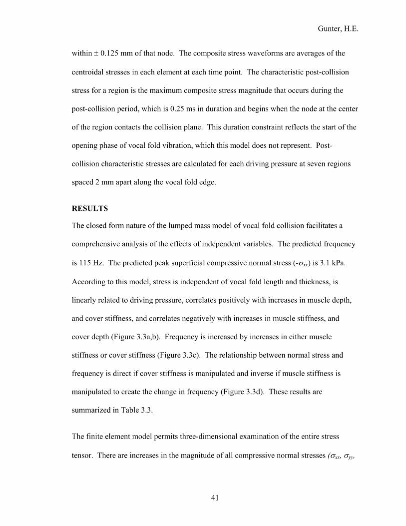
Gunter, H.E.
41
within ± 0.125 mm of that node. The composite stress waveforms are averages of the
centroidal stresses in each element at each time point. The characteristic post-collision
stress for a region is the maximum composite stress magnitude that occurs during the
post-collision period, which is 0.25 ms in duration and begins when the node at the center
of the region contacts the collision plane. This duration constraint reflects the start of the
opening phase of vocal fold vibration, which this model does not represent. Post-
collision characteristic stresses are calculated for each driving pressure at seven regions
spaced 2 mm apart along the vocal fold edge.
RESULTS
The closed form nature of the lumped mass model of vocal fold collision facilitates a
comprehensive analysis of the effects of independent variables. The predicted frequency
is 115 Hz. The predicted peak superficial compressive normal stress (-σxx) is 3.1 kPa.
According to this model, stress is independent of vocal fold length and thickness, is
linearly related to driving pressure, correlates positively with increases in muscle depth,
and cover stiffness, and correlates negatively with increases in muscle stiffness, and
cover depth (Figure 3.3a,b). Frequency is increased by increases in either muscle
stiffness or cover stiffness (Figure 3.3c). The relationship between normal stress and
frequency is direct if cover stiffness is manipulated and inverse if muscle stiffness is
manipulated to create the change in frequency (Figure 3.3d). These results are
summarized in Table 3.3.
The finite element model permits three-dimensional examination of the entire stress
tensor. There are increases in the magnitude of all compressive normal stresses (σxx, σyy,
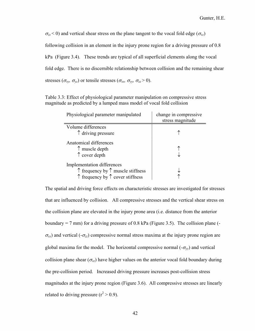
Gunter, H.E.
42
σzz < 0) and vertical shear stress on the plane tangent to the vocal fold edge (σxz)
following collision in an element in the injury prone region for a driving pressure of 0.8
kPa (Figure 3.4). These trends are typical of all superficial elements along the vocal
fold edge. There is no discernible relationship between collision and the remaining shear
stresses (σxy, σyz) or tensile stresses (σxx, σyy, σzz > 0).
Table 3.3: Effect of physiological parameter manipulation on compressive stress magnitude as predicted by a lumped mass model of vocal fold collision
Physiological parameter manipulated change in compressive stress magnitude
Volume differences ↑ driving pressure ↑ Anatomical differences ↑ muscle depth ↑ ↑ cover depth ↓ Implementation differences ↑ frequency by ↑ muscle stiffness ↓ ↑ frequency by ↑ cover stiffness ↑
The spatial and driving force effects on characteristic stresses are investigated for stresses
that are influenced by collision. All compressive stresses and the vertical shear stress on
the collision plane are elevated in the injury prone area (i.e. distance from the anterior
boundary = 7 mm) for a driving pressure of 0.8 kPa (Figure 3.5). The collision plane (-
σxx) and vertical (-σzz) compressive normal stress maxima at the injury prone region are
global maxima for the model. The horizontal compressive normal (-σyy) and vertical
collision plane shear (σxz) have higher values on the anterior vocal fold boundary during
the pre-collision period. Increased driving pressure increases post-collision stress
magnitudes at the injury prone region (Figure 3.6). All compressive stresses are linearly
related to driving pressure (r2 > 0.9).
Gunter, H.E.
43
Figure 3.3: Dependence of superficial stress in vocal fold tissue during collision and vocal fold vibration frequency (i.e. pitch) on geometric and material parameters as predicted by a lumped mass model of vocal fold collision. Each curve is generated by manipulating a single parameter while maintaining all others at their nominal value. Negative stresses are compressive. Stress axes are reversed to illustrate compressive stresses. All manipulated parameters are normalized to the nominal values given in Table 3.1.

Gunter, H.E.
44
Figure 3.4: Effect of vocal fold collision on components of the stress tensor at the injury prone region for a driving pressure of 0.8 kPa as predicted with a finite element model. Time = 0 is the beginning of collision. Shaded area indicates post collision period (a) Normal stresses during vocal fold closure and collision. Tensile stresses are greater than zero. Compressive stresses are less than zero. Stress axis is reversed to illustrate compressive stress magnitudes. (b) Shear stresses during vocal fold closure and collision.
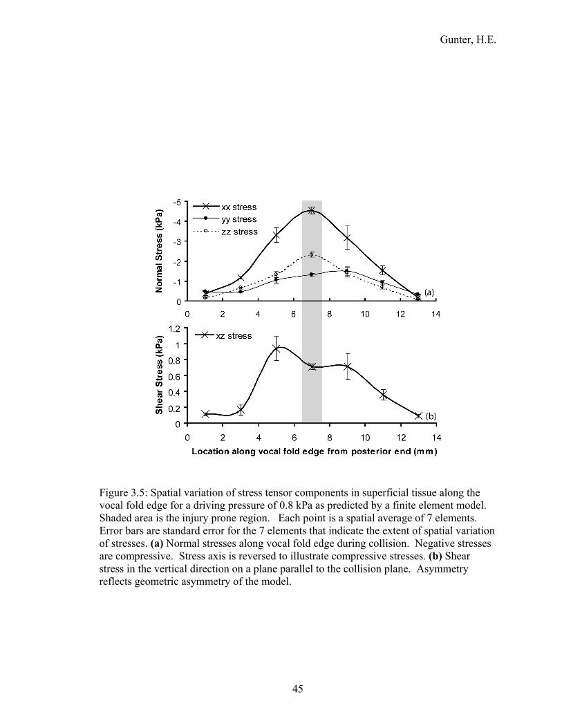
Gunter, H.E.
45
Figure 3.5: Spatial variation of stress tensor components in superficial tissue along the vocal fold edge for a driving pressure of 0.8 kPa as predicted by a finite element model. Shaded area is the injury prone region. Each point is a spatial average of 7 elements. Error bars are standard error for the 7 elements that indicate the extent of spatial variation of stresses. (a) Normal stresses along vocal fold edge during collision. Negative stresses are compressive. Stress axis is reversed to illustrate compressive stresses. (b) Shear stress in the vertical direction on a plane parallel to the collision plane. Asymmetry reflects geometric asymmetry of the model.
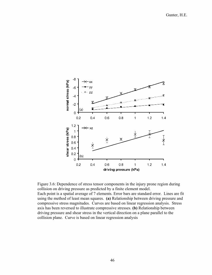
Gunter, H.E.
46
Figure 3.6: Dependence of stress tensor components in the injury prone region during collision on driving pressure as predicted by a finite element model. Each point is a spatial average of 7 elements. Error bars are standard error. Lines are fit using the method of least mean squares. (a) Relationship between driving pressure and compressive stress magnitudes. Curves are based on linear regression analysis. Stress axis has been reversed to illustrate compressive stresses. (b) Relationship between driving pressure and shear stress in the vertical direction on a plane parallel to the collision plane. Curve is based on linear regression analysis
Gunter, H.E.
47
DISCUSSION
Two computational models, one simple and one complex, provide estimations of
mechanical stress in vocal fold tissue during vocal fold collision in order to investigate
the role of stresses as a cause of tissue injuries that lead to the developments of lesions
such as nodules and polyps. A one-dimensional lumped-mass model (Figure 3.1c)
parameterizes vocal fold tissue mechanics to generate closed form solutions. Predictions
are limited to superficial compressive normal stress perpendicular to the plane of
collision (-σxx) during vocal fold collision. A three-dimensional finite element model
(Figure 3.2) assumes isotropic linear elasticity, material homogeneity and simplified
aerodynamics to generate numerical solutions of the stress tensor. Predictions of stress
tensor components are limited to the closing phase of vocal fold vibration including
collision between the vocal folds as represented by collision between a single vocal fold
and a rigid plane.
The predictions of the finite element model and lumped mass model agree with published
theoretical and experimental data. The frequencies predicted by both models fall within
the normal range of human male voice (Zemlin, 1997). The suggestion made by the
lumped mass model that increases in cover and body stiffness increase frequency (Figure
3.3c) is consistent with in vivo EMG measurements by Shipp and McGlone (1971) and
stiffness measurements by Berke and Gerratt (1993), which indicate that activity in the
thyroarytenoid muscle that composes the body increases vocal fold stiffness and pitch
and that activity in the cricothyroid muscle, which longitudinally lengthens the
hyperelastic vocal fold cover, also increases stiffness and pitch. However, independent
manipulation of body and cover stiffnesses is not consistent with voice physiology since
Gunter, H.E.
48
activity in the thyroarytenoid muscle also longitudinally shortens the hyperelastic vocal
fold cover and therefore causes a decrease in effective cover stiffness in addition to an
increase in body stiffness. The linear relation between driving pressure and collision
stress, which is proportional to collision force, in the lumped-mass model is consistent
with the experimental observations of Jiang and Titze (1994) and earlier predictions made
using the finite element model (Chapter 2). Predictions of compressive stress during
collision by the lumped mass model are the same order of magnitude as those made with
the finite element model (Chapter 2).
Predictions made by the finite element model support and expand the hypothesis that
benign vocal fold pathologies such as nodules and polyps are a reaction to mechanical
stress. The magnitudes of multiple components of the stress tensor increase during
collision in the region where pathologies often develop (i.e. center of the vocal fold
edge)(Figure 3.5). This is also the region that attains the highest velocity immediately
prior to contact. Therefore the stresses reflect the development of elastic energy as
contact forces decelerate the tissue. The nature of the increased components suggests
possible mechanisms of tissue damage that lead to lesions, such as nodules and polyps,
which are characterized by basement membrane disruption (Gray and Titze, 1988) and
expanded extracellular matrix (Mossallam et al., 1986). The collision-associated
compressive stress maxima in two coordinate directions and elevated compressive stress
in the third at the injury prone region (Figure 3.5a) are of similar magnitude to those that
increase protein synthesis and secretion by tracheal epithelial cells in culture as shown by
Swartz et al. (2001) and are therefore likely to lead to an expansion of the extra-cellular
matrix. Vertical shear stress on the plane parallel to collision is elevated at the injury
Gunter, H.E.
49
prone location (Figure 3.5b) and is of the appropriate orientation to cause the disrupted
adhesions between epithelial cells and the basement membrane. The lack of tensile stress
in the injury prone region during collision (Figure 3.4a) suggests that tension is not the
cause of tissue failure that leads to vocal nodule and polyp formation. In the absence of
data pertaining to the dose response of vocal fold tissue elements to the components of
the stress tensor it is difficult to say which magnitudes and directions of stress pose the
greatest injury risks. Carefully controlled studies using vocal fold tissue culture models
would further guide interpretation of these results. Such models would also permit study
of the role played by frequency of deformation in tissue injury.
The models provide insights into mechanical bases for risk factors associated with
development of vocal fold pathologies. Sound intensity varies with driving pressure
(Isshiki, 1964). Therefore, the linear relationship between driving pressure and
compressive stress magnitude in the region that is prone to injury (Figure 3.6a) provides
an explanation for the presumed association between high volume voicing and formation
of pathologies. The inverse contribution of cover depth to compressive stress magnitude
(Figure 3.3a) offers an explanation for proposed protective nature of hydration and warm
up against vocal fold injury since both are likely to increase fluid volume in, and depth
of, the cover. The prediction by the lumped mass model that mechanical stress is
dependent on the method of frequency manipulation (Figure 3.3d) addresses the role
played by voicing practice in determining mechanical stress levels and risk of tissue
injury. According to these predictions, it is conceivable that two individuals with similar
anatomy may produce sounds of similar pitch and volume using different production
strategies (i.e. different muscle activation patterns) and therefore have different
Gunter, H.E.
50
mechanical stress levels and injury risk. This lends credibility to anecdotal evidence that
lack of training or inappropriate training leads to voice “misuse” and increased risk of
lesion formation. It also suggests that information on how voice is generated may prove
useful for clinical diagnosis and treatment of these benign lesions.
The results of the models discussed in this paper provide inspiration for additional
models of vocal fold tissue. Each model was used for its relative strengths; the lumped
mass model facilitated parameter sensitivity studies, and the finite element model
facilitated physiological fidelity. Additional simple models that address stresses other
than compressive stress in the direction perpendicular to the plane of collision would
permit further parameter variation studies. Finite element models that incorporate a
broader spectrum of input variables with increased complexity (for example, isotropic
nonlinear viscoelasticity and material inhomogeneity) would provide further validation of
the simple models. Models of both types would be useful for addressing sources of vocal
fold tissue stress other than collision, for example those induced by changes in laryngeal
configurations. Inhomogeneous finite element models are presented in Chapter 4.
Gunter, H.E.
51
CHAPTER 4: A THEORETICAL INVESTIGATION OF THE ROLE OF SUPERFICIAL LAMINA PROPRIA STIFFNESS DURING VOICE PRODUCTION1
INTRODUCTION
The superficial lamina propria (SLP) is a soft connective tissue matrix containing elastin
and collagen fibers that lies beneath the epithelia of the vocal folds and of other surfaces
in the human body. It is implicated as an important contributor to the mechanics of voice
production (Gray et al., 2000). Pathological changes to SLP structure such as scar are
associated with deteriorating voice quality (Cotron and Casper, 1996) and surgical
augmentation is reported to restore voice quality (Zeitels et al., 2002). Its viscous
properties are linked with phonation threshold pressure (Titze et al., 1995) and its
deformation contributes to a mucosal wave on the superior vocal fold surface (Berke and
Gerratt, 1993), qualitative assessment of which is part of a clinical voice evaluation.
Quantitative understanding of the relationship between SLP stiffness and movement of
1 This work has been submitted for publication as Gunter, H.E. (2003) “Theoretical investigation of the role of superficial lamina propria stiffness during voice production” to the Journal of the Acoustical Society of America
Gunter, H.E.
52
the vocal folds has applications in surgical planning and tissue engineering of
replacement SLP.
The contributions of the soft elastic properties of the SLP to voice mechanics have
received little quantitative attention. Rheometric data confirm anecdotal reports that the
superficial lamina propria has important viscous properties at low deformation
frequencies (ξ = 0.1 – 1) and is over 100 fold less stiff for incremental strains from rest
length than other vocal fold tissues (Chan and Titze, 1999a). Some theoretical models of
vocal fold tissue purport to represent the SLP by including it in a vocal fold cover layer
that is defined using data collected from samples including both epithelium and SLP in
parallel undergoing longitudinal tension (Alipour et al., 2000;Jiang et al., 1998). This is
not a good representation of the SLP behavior since, at low strain rates, properties of the
relatively nonviscous and stiffer epithelium dominate such data (Alipour-Haghighi and
Titze, 1991;Kakita et al., 1981). More accurate quantification of the SLP’s material
properties is challenging due to its frequency dependent behavior. The behavior of
epithelium and SLP samples in series undergoing dynamic transverse shear deformations
provide an indication of the viscoelastic properties of the SLP in the frequency range that
the rheometer operates in (Chan and Titze, 1999a). Theoretical extrapolations of this
data to deformation frequencies characteristic of voice predict that the SLP behaves in a
predominantly elastic fashion (ξ < 0.01 for a human male sample) during voice
production and remains much less stiff than other vocal fold tissues (Chan and Titze,
2000).
Gunter, H.E.
53
This chapter presents an investigation of the contributions of superficial lamina propria
elasticity to voice mechanics including effects on collision force, tissue stress and glottal
area dynamics. A finite element model that builds upon a previous model of vocal fold
closure and collision (Chapter 2) by incorporating four tissue layers including distinct
epithelium and superficial lamina propria layers is used.
METHODS
A finite element model of the vocal fold that represents four layers of tissue has been
developed based on a single layer model of vocal fold closure and collision (Chapter 2).
There are three differences between the current model and the previous model. The
lateral contour of the vocal fold geometry is represented linearly rather than curvilinearly
in the transverse plane. This geometric simplification has been implemented in other
models with good results (Alipour et al., 2000). The geometry represents one half of a
single vocal fold rather than an entire vocal fold. This reflects recognition of anterior-
posterior symmetry that is created though the use of a linear lateral boundary and reduces
computational time by a factor of two. The vocal fold geometry is subdivided into four
layers with distinct material definitions rather than being defined homogeneously using
one material definition.
The external and internal dimensions of the hemi-fold model are based on histological
section measurements. The midmembranous to end length is 7 mm and the vocal fold
depth and height are both 10 mm (Figure 4.1). These external dimensions have been
used in previous models (Alipour et al., 2000;Gunter, 2003a, b;Titze and Talkin, 1979).
The dimensions of the four layers that define the vocal fold cross section are based on
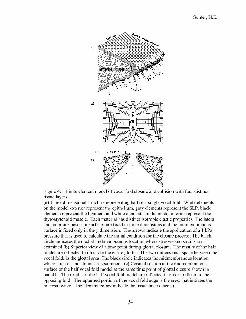
Gunter, H.E.
54
Figure 4.1: Finite element model of vocal fold closure and collision with four distinct tissue layers. (a) Three dimensional structure representing half of a single vocal fold. White elements on the model exterior represent the epithelium, gray elements represent the SLP, black elements represent the ligament and white elements on the model interior represent the thyroarytenoid muscle. Each material has distinct isotropic elastic properties. The lateral and anterior / posterior surfaces are fixed in three dimensions and the midmembranous surface is fixed only in the y dimension. The arrows indicate the application of a 1 kPa pressure that is used to calculate the initial condition for the closure process. The black circle indicates the medial midmembranous location where stresses and strains are examined.(b) Superior view of a time point during glottal closure. The results of the half model are reflected to illustrate the entire glottis. The two dimensional space between the vocal folds is the glottal area. The black circle indicates the midmembranous location where stresses and strains are examined. (c) Coronal section at the midmembranous surface of the half vocal fold model at the same time point of glottal closure shown in panel b. The results of the half vocal fold model are reflected in order to illustrate the opposing fold. The upturned portion of the vocal fold edge is the crest that initiates the mucosal wave. The element colors indicate the tissue layers (see a).
Gunter, H.E.
55
data from human vocal folds (Hirano et al., 1983). The 0.1 mm thick epithelium and 0.6
mm thick SLP form the medial and superior surfaces of the vocal fold. The 0.2 mm thick
ligament is located in the vocal fold apex, beneath the SLP. The muscle defines the
interior of the vocal fold between the lateral boundary and the SLP and ligament (Figure
4.1). The tissues are defined with 9036 hexahedral (i.e. brick shaped) finite elements
each defined with 20 nodes with three degrees of freedom. The lateral and end
boundaries are fixed (i.e. are not permitted to translate) and the midmembranous
boundary is fixed to its plane (i.e. it is permitted to slide along the plane). This latter
boundary accounts for anterior/posterior symmetry of the vocal fold. The nodes on the
medial boundary are free if their location is in the positive x domain, but are not
permitted to translate into the negative x domain. This boundary definition effectively
defines a contact plane at x = 0, which defines the midglottis.
Isotropic linear elastic material properties are assigned to each tissue layer (Table 4.1).
The values defining the ligament and muscle are approximations (Min et al., 1995) based
on the behavior of human ligament (Min et al., 1995) and canine muscle (Alipour-
Haghighi and Titze, 1991) under longitudinal tension. The value defining the behavior of
the epithelium is an approximation (Min et al., 1995) based on the behavior of canine
mucosa (i.e. epithelium and superficial lamina propria) under longitudinal tension at low
strain rates (Min et al., 1995). Since the epithelium and lamina propria are in parallel for
longitudinal tension, the viscous lamina propria likely has minimal effect on the
resistance of the sample to strain. Therefore this value is representative of the behavior
of the epithelium. These linear approximations are valid for strains of 15% (Min et al.,
1995), a limit that is not exceeded during model calculations. The values defining the
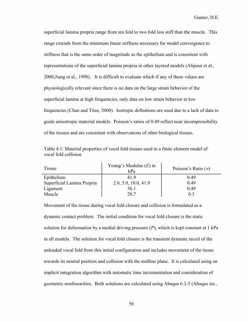
Gunter, H.E.
56
superficial lamina propria range from ten fold to two fold less stiff than the muscle. This
range extends from the minimum linear stiffness necessary for model convergence to
stiffness that is the same order of magnitude as the epithelium and is consistent with
representations of the superficial lamina propria in other layered models (Alipour et al.,
2000;Jiang et al., 1998). It is difficult to evaluate which if any of these values are
physiologically relevant since there is no data on the large strain behavior of the
superficial lamina at high frequencies, only data on low strain behavior at low
frequencies (Chan and Titze, 2000). Isotropic definitions are used due to a lack of data to
guide anisotropic material models. Poisson’s ratios of 0.49 reflect near incompressibility
of the tissues and are consistent with observations of other biological tissues.
Table 4.1: Material properties of vocal fold tissues used in a finite element model of vocal fold collision
Tissue Young’s Modulus (E) in kPa Poisson’s Ratio (ν)
Epithelium 41.9 0.49 Superficial Lamina Propria 2.0, 5.0, 10.0, 41.9 0.49 Ligament 36.1 0.49 Muscle 20.7 0.3 Movement of the tissue during vocal fold closure and collision is formulated as a
dynamic contact problem. The initial condition for vocal fold closure is the static
solution for deformation by a medial driving pressure (P), which is kept constant at 1 kPa
in all models. The solution for vocal fold closure is the transient dynamic recoil of the
unloaded vocal fold from this initial configuration and includes movement of the tissue
towards its neutral position and collision with the midline plane. It is calculated using an
implicit integration algorithm with automatic time incrementation and consideration of
geometric nonlinearities. Both solutions are calculated using Abaqus 6.2-5 (Abaqus inc.,
Gunter, H.E.
57
Pawtucket, RI). Validation of this method against experimental and clinical data is
described elsewhere (Chapter 2).
The results of four models that differ in the stiffness of the SLP are compared. The
midmembranous contour of the vocal fold in a coronal plane at the initial condition for
glottal closure provides a snapshot of the crest that becomes the mucosal wave (Figure
4.1c). Height of the mucosal wave crest is the vertical difference between the most
superior point on the deformed vocal fold surface and linear extrapolation of the contour
of the lateral half of the coronal section to the same point. Glottal area (i.e. the area
between the vocal folds, denoted by A) is calculated by summing the areas of trapezoids
formed by adjacent nodes on the vocal fold edge and the midline (Figure 4.1b). This
quantity is used as an indicator of glottal air flow based on experimental data that
illustrate a direct relationship between glottal area and flow if the pressure drop across the
glottis (∆P) is constant (Stevens, 1998):.
2
2
AUPα∆ (4.1)
The square of glottal area is an alternative indicator of airflow based on a model of
laminar flow through a cylinder (Poiseuille flow) if the pressure drop across the glottis,
viscosity (η ) and glottal length (L) are constant:
2
28 AP
AL
PU ∆∆
= α
πη (4.2)
Gunter, H.E.
58
The minimum of the first derivative of these two indicators of airflow with respect to
time provides a comparative estimate of maximum flow declination rate. In order to
obtain smooth derivatives, they are taken analytically from between third and sixth order
polynomials that approximate the glottal area and glottal area squared curves with
correlation coefficients greater than 0.99. Contact force between the vocal folds is
calculated as the sum of point forces acting on medial nodes by the contact plane in the
glottal midline. Mechanical stresses in the different vocal fold tissues are examined at
the medial midmembranous region where vocal fold pathologies often manifest.
Particular attention is paid to normal stress in the three coordinate directions and vertical
shear stress on the plane parallel to the collision plane during collision since these have
been identified as candidate stresses for vocal fold tissue damage (Chapter 3).
RESULTS
Model results show substantial variation with changes in SLP stiffness. Increases in SLP
stiffness decrease the maximum glottal opening and the height of the mucosal wave at the
onset of vocal fold closure (Figure 4.2). This decrease in glottal opening corresponds
with decreased maximum airflow, an indicator of voice intensity, as estimated using the
square of glottal area (Figure 4.3a) or glottal area (Figure 4.3b). Increases in SLP
stiffness increase the maximum normalized flow declination rate, a voice quality
parameter, as estimated by the first derivative of normalized glottal area squared or
normalized glottal area (Figure 4.3c).
The finite element model also predicts the mechanical forces that load the vocal fold
during closure and collision. Increases in SLP stiffness increase the maximum contact
force between the vocal folds, as estimated using the sum of point contact forces between
Gunter, H.E.
59
the medial vocal fold surface and the contact plane. Increases in maximum contact force
are paralleled by decreases in maximum volume as estimated by glottal area squared
(Figure 4.4).
Alterations in SLP stiffness alter the nature and magnitude of mechanical stresses.
Collision between the vocal folds is associated with compression in all three normal
directions in the medial epithelial and SLP layers in the midmembrous region. The
magnitudes of these compressive stresses are greater in the epithelium than the SLP and
are proportional to SLP stiffness (Figure 4.5a). Collision between the vocal folds is
associated with an increase in shear stress on the plane of collision in the vertical
direction in the medial epithelium and SLP adjacent to the midmembrous region, the
magnitude of which varies directly with SLP stiffness (Figure 4.6a). The maximum open
position of the vocal fold is associated with high epithelial tension along the length of the
vocal fold (σyy) in models with low SLP stiffness. The σyy stress in the medial SLP in
the maximally open position of the vocal fold varies in the opposite manner (i.e. it
increases as SLP stiffness increases) (Figure 4.7a). All stresses in the epithelium are
related to strain by the same elastic constant in all models. Therefore epithelial stress
trends indicate epithelial strain trends. Stresses in the SLP are related to strain by model
specific elastic moduli. The magnitudes of all deformations examined in the
midmembranous, medial SLP (normal compression during collision, shear deformation
during collision, longitudinal tension prior to collision) decrease as SLP stiffness
increases (Figures 4.5b, 4.6b, 4.7b).
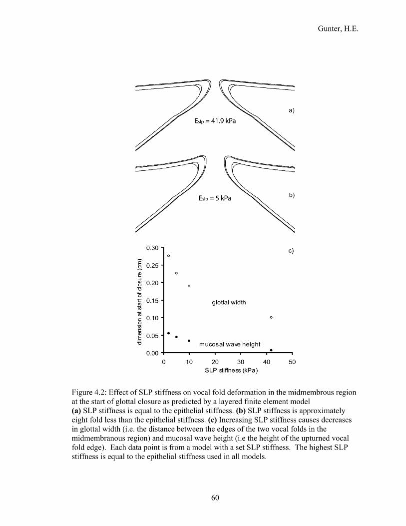
Gunter, H.E.
60
Figure 4.2: Effect of SLP stiffness on vocal fold deformation in the midmembrous region at the start of glottal closure as predicted by a layered finite element model (a) SLP stiffness is equal to the epithelial stiffness. (b) SLP stiffness is approximately eight fold less than the epithelial stiffness. (c) Increasing SLP stiffness causes decreases in glottal width (i.e. the distance between the edges of the two vocal folds in the midmembranous region) and mucosal wave height (i.e the height of the upturned vocal fold edge). Each data point is from a model with a set SLP stiffness. The highest SLP stiffness is equal to the epithelial stiffness used in all models.
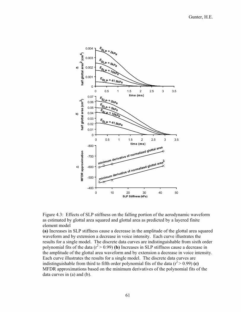
Gunter, H.E.
61
Figure 4.3: Effects of SLP stiffness on the falling portion of the aerodynamic waveform as estimated by glottal area squared and glottal area as predicted by a layered finite element model (a) Increases in SLP stiffness cause a decrease in the amplitude of the glottal area squared waveform and by extension a decrease in voice intensity. Each curve illustrates the results for a single model. The discrete data curves are indistinguishable from sixth order polynomial fits of the data (r2 > 0.99) (b) Increases in SLP stiffness cause a decrease in the amplitude of the glottal area waveform and by extension a decrease in voice intensity. Each curve illustrates the results for a single model. The discrete data curves are indistinguishable from third to fifth order polynomial fits of the data (r2 > 0.99) (c) MFDR approximations based on the minimum derivatives of the polynomial fits of the data curves in (a) and (b).
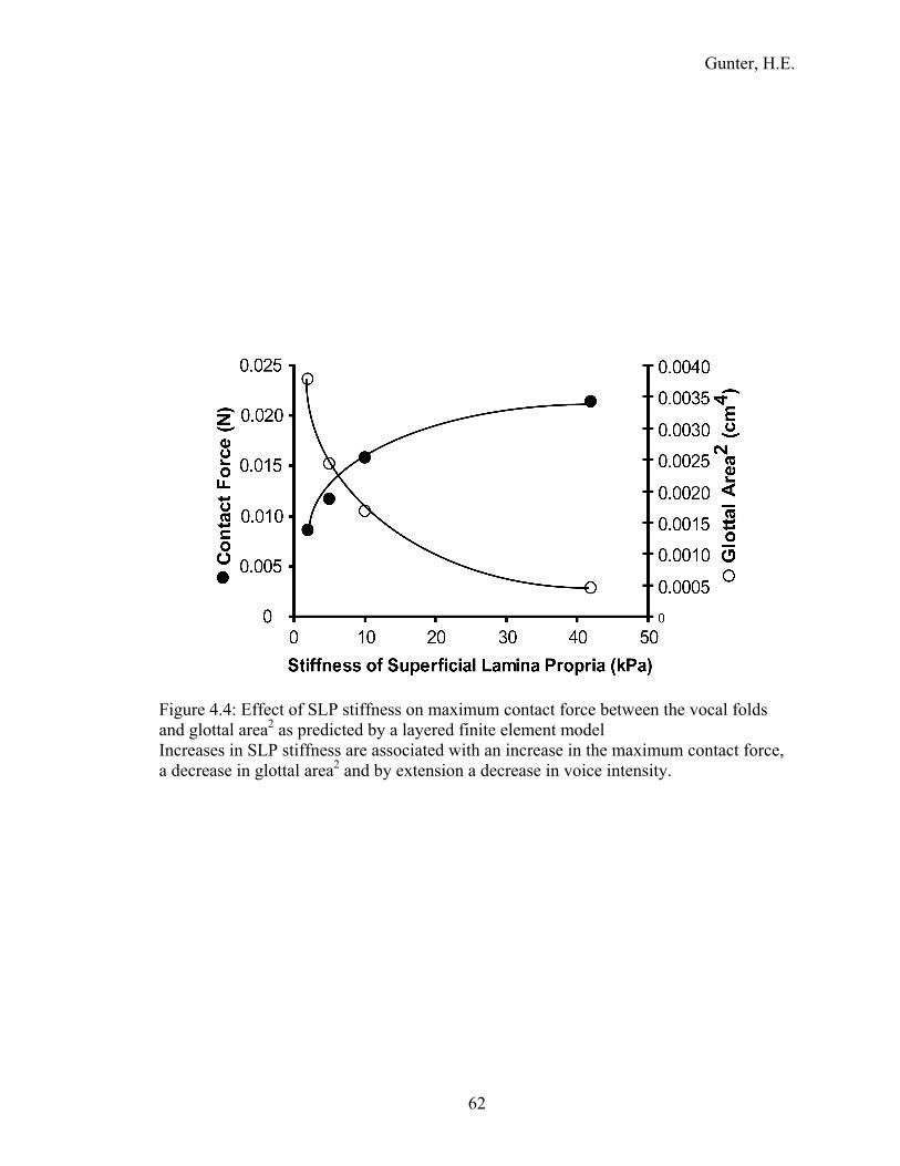
Gunter, H.E.
62
Figure 4.4: Effect of SLP stiffness on maximum contact force between the vocal folds and glottal area2 as predicted by a layered finite element model Increases in SLP stiffness are associated with an increase in the maximum contact force, a decrease in glottal area2 and by extension a decrease in voice intensity.
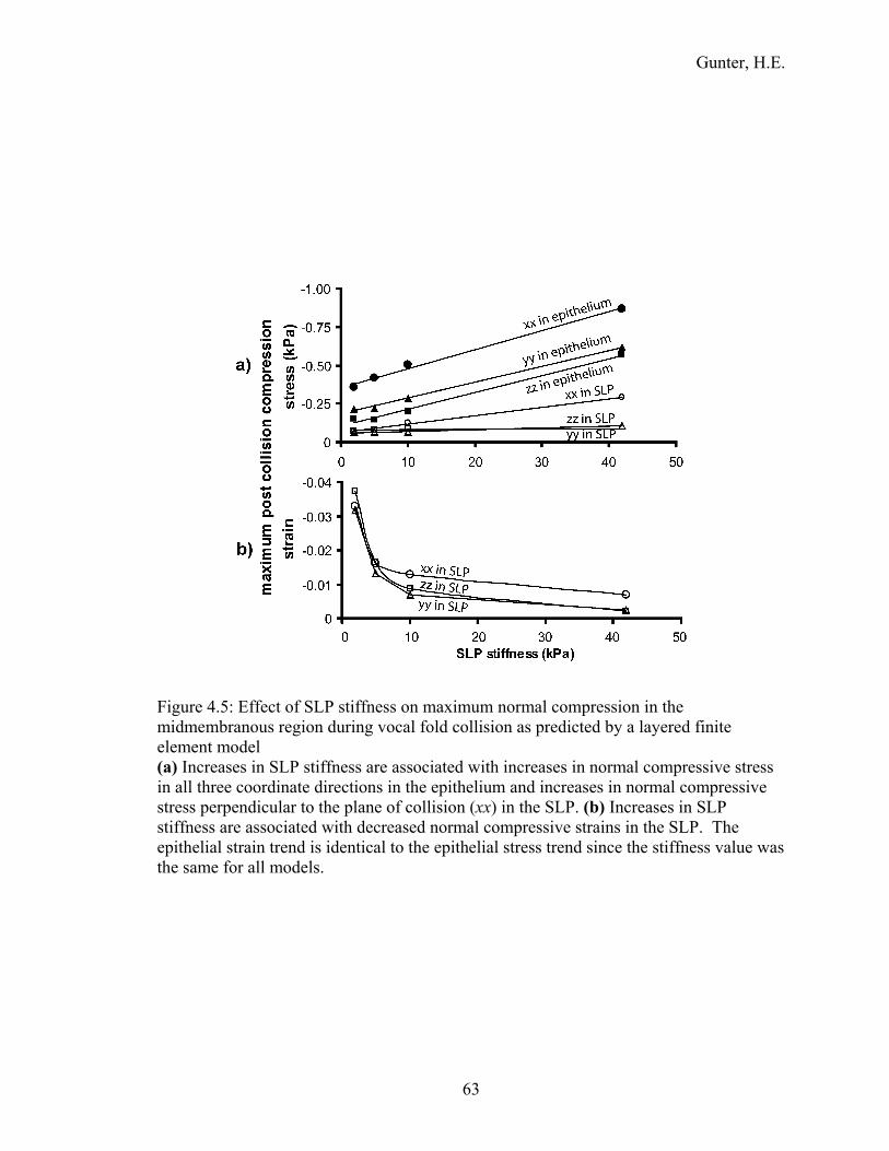
Gunter, H.E.
63
Figure 4.5: Effect of SLP stiffness on maximum normal compression in the midmembranous region during vocal fold collision as predicted by a layered finite element model (a) Increases in SLP stiffness are associated with increases in normal compressive stress in all three coordinate directions in the epithelium and increases in normal compressive stress perpendicular to the plane of collision (xx) in the SLP. (b) Increases in SLP stiffness are associated with decreased normal compressive strains in the SLP. The epithelial strain trend is identical to the epithelial stress trend since the stiffness value was the same for all models.
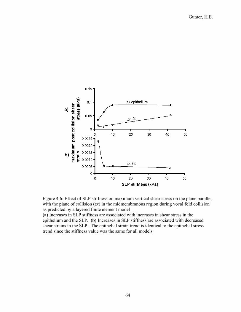
Gunter, H.E.
64
Figure 4.6: Effect of SLP stiffness on maximum vertical shear stress on the plane parallel with the plane of collision (zx) in the midmembranous region during vocal fold collision as predicted by a layered finite element model (a) Increases in SLP stiffness are associated with increases in shear stress in the epithelium and the SLP. (b) Increases in SLP stiffness are associated with decreased shear strains in the SLP. The epithelial strain trend is identical to the epithelial stress trend since the stiffness value was the same for all models.
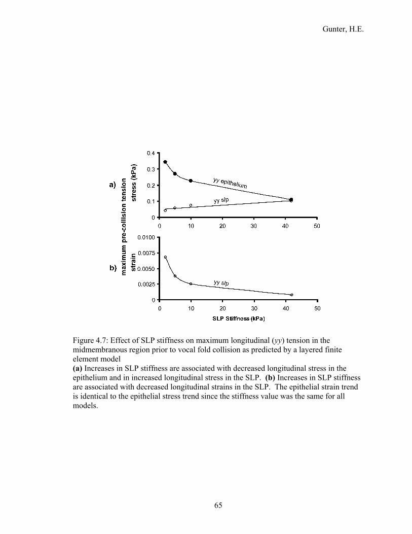
Gunter, H.E.
65
Figure 4.7: Effect of SLP stiffness on maximum longitudinal (yy) tension in the midmembranous region prior to vocal fold collision as predicted by a layered finite element model (a) Increases in SLP stiffness are associated with decreased longitudinal stress in the epithelium and in increased longitudinal stress in the SLP. (b) Increases in SLP stiffness are associated with decreased longitudinal strains in the SLP. The epithelial strain trend is identical to the epithelial stress trend since the stiffness value was the same for all models.
Gunter, H.E.
66
DISCUSSION
The model predicts the behavior of four layers of tissue, each of which is defined using
isotropic linear elastic material parameters, during vocal fold closure and collision.
Model predictions agree with published experimental and theoretical data. Vibration
amplitudes on the scale of millimeters for a subglottal pressure of 1 kPa (Figure 4.2c) are
in accordance with those observed using high speed imaging of canine larynges (Berry et
al., 2001a). Contact force magnitudes on the order of 10-20 mN for a subglottal pressure
of 1 kPa (Figure 4.4) compare closely with experimental measurements of 12 mN (Jiang
and Titze, 1994) and theoretical predictions of 10-16 mN (Chapter 2), both for a 1 kPa
subglottal pressure. Predicted normal stresses during collision for a model with a stiff
SLP are of the same pattern and order of magnitude but less than those predicted using a
homogeneous finite element model (Chapter 2). This may reflect the use of layer distinct
material properties in the current model. The predicted direct relationship between SLP
stiffness and epithelial normal stress follows the prediction of a simple lumped mass
model (Chapter 3).
Models of vocal fold tissue mechanics provide a means of interpreting indirect data that
is obtained during clinical examination of vocal fold health, such as stroboscopic images
and acoustic recordings. Model prediction of an inverse relationship between SLP
stiffness and mucosal wave height (Figure 4.2c) supports a common interpretation of
absent mucosal waves in stroboscopic images as SLP pathology, including regional
stiffening caused by scar. Predictions of vocal fold margin deformation during closure
and collision indicate that increases in SLP stiffness contribute to a decreased maximum
glottal area squared (Figure 4.4a), an indicator of voice intensity, for a given subglottal
Gunter, H.E.
67
pressure, and suggest that increased vocal efficiency (the ratio between oral sound power
and the product of subglottal pressure and flow) is an indicator of decreased SLP
stiffness. This could be used in clinical assessment since noninvasive pressure sensors in
the oral cavity can approximate both oral and subglottal pressures. Indirect assessment of
SLP stiffness is useful since the SLP is a target for surgical manipulation (Zeitels et al.,
2002). However, the current pool of data is insufficient since variables such as geometry
that may contribute to the mucosal wave and vocal efficiency have not been explored. A
complete exploration of the vocal fold tissue parameter space has the potential to create a
library that connects clinical measurements with tissue properties in order to augment
clinical diagnosis.
In addition to assisting with the interpretation of indirect clinical data, understanding the
link between vocal fold tissue parameters and voice can guide surgical planning.
Evaluation of glottal area dynamics using a model of vocal fold tissue mechanics predicts
changes in voice intensity and maximum flow declination rate due to changes in SLP
stiffness (Figure 4.4). These parameters could be used as inputs to a physiologically
based speech synthesis program in order to predict voice waveforms specific to vocal
fold tissue alterations. The combination of a detailed tissue model and voice synthesis
would form a model capable of predicting surgical outcomes that could be used to test
proposed manipulations. Quantification of the relationship between glottal closure
dynamics and the falling arm of the aerodynamic waveform, for example using in vivo
photoglottographic studies or by including aerodynamics in a theoretical model, is a
necessary step towards achieving this goal. Optimizing the computation of the tissue
Gunter, H.E.
68
mechanics is also necessary so that the effect of multiple manipulations could be
predicted in a reasonable time scale.
Relationships between SLP stiffness and mechanical stress indicate the role of SLP
stiffness in determining tissue mechanical injury risk. Increases in compressive stress
(Figure 4.5a) and vertical shear stress on the plane parallel to the plane of collision
(Figure 4.6a) in the medial midmembranous epithelium during collision that are caused
by SLP stiffness increases suggest that increasing SLP stiffness increases the risk of
epithelial failure in compressive or shear modes. The former may result in changes in
epithelial gene expression (Ressler et al., 2000) that lead to changes in protein synthesis
and secretion in nearby fibroblasts (Swartz et al., 2001). The latter may manifest as
basement membrane damage (Gray and Titze, 1988). Injury risk increases as a result of
SLP stiffness increases are associated with decreases in voice intensity as indicated by
maximum glottal area squared or glottal area (Figure 4.3). These contrast with stress
associated risk increases due to increases in subglottal pressure (Chapter 3), which are
associated with increases in voice intensity. Thus voice intensity alone does not indicate
injury risk. However, injury risk as a result of SLP stiffness increases are associated with
increases in collision force between the vocal folds (Figure 4.4), as are those due to
increases in subglottal pressure. This supports the use of interfold collision forces to
assess injury risk.
Low SLP stiffness results in a pre-collision tensile stress in the epithelium (Figure 4.7a)
that has not been observed in models that combine the epithelium and SLP into a single
layer. This tensile stress occurs along the medial edge of the vocal fold when it is in the
fully open position, much like tension in a vibrating string, and acts to separate adjacent
Gunter, H.E.
69
epithelial cells and impair wound healing (Waters et al., 2001). If the SLP is the same
stiffness as the epithelium it provides support for the deformation by effectively
increasing the cross sectional area of the vibrating string and therefore minimizes this
tensile stress. Thus, increased SLP stiffness decreases injury risk due to epithelial
tension.
Increases in SLP stiffness cause increases in post collision stresses in the SLP to a lesser
degree than in the epithelium (Figures 4.5a, 4.6a). However, lower stress magnitudes in
the SLP than the epithelium do not translate into lower injury risks since the two tissues
are distinct and likely respond to stress differently. Therefore increases in SLP stiffness
are associated with an increased risk of compressive and shear failure in the SLP due to
stress. As in the case of the epithelium these stress increases are accompanied by
increases in collision force. Another possible indicator for injury risk is strain. Increases
in SLP stiffness are associated with decreases in compressive and shear mechanical strain
in midmembranous SLP, and therefore decreased risk of injury due to strain induced
failure, a trend that is opposite to the stress trend. Which of stress or strain is most
concerning is not clear. If the damage is physical failure of proteins then stress in the
SLP proteins that support forces is likely the most important quantity. If the damage is
functional then strain of the fibroblasts and other cellular components that deform with
the matrix but likely support little force is the most important quantity. Clever
experiments involving synthesized SLP are necessary to determine the relevant modes of
failure.
A finite element of vocal fold tissue model predicts that superficial lamina propria
stiffness plays a role in determining voice output, its own injury risk and the injury risk of
Gunter, H.E.
70
adjacent tissues. In the design and selection of SLP implant materials consideration
needs to be given to minimizing SLP strain and epithelial longitudinal tension by
increasing SLP stiffness and minimizing collision induced epithelial and SLP
compressive and shear stresses and maximizing vocal efficiency by decreasing SLP
stiffness. Other considerations that have not been addressed here include the viscous SLP
properties (Chan and Titze, 1999b) that contribute to phonation threshold pressure (Titze
et al., 1995), regional variations in SLP stiffness and SLP geometry. Precise
recommendations will require detailed characterization of the nonlinearity of SLP
material properties at voicing frequency and identification of relevant modes of tissue
injury.
Gunter, H.E.
71
CHAPTER 5: MEASUREMENTS OF VOCAL FOLD COLLISION FORCES DURING PHONATION1
INTRODUCTION
Collisions between the vocal folds during phonation have implications for vocal fold
injury and voice quality. In the former instance, the application of high forces to the
medial surfaces of the vocal folds may cause tissue injury that manifests as organic
pathologies (Gunter, 2003a;Titze, 1994). In the latter instance glottal closure is the tissue
correlate of maximum airflow declination rate, which relates to voice quality perceptions
(Holmberg et al., 1988). Experimental measurements of the forces transmitted between
human vocal folds during phonation have the potential to improve our understanding of
vocal fold collision mechanics.
In vivo measurements of intra vocal fold collision forces during human phonation are
challenging to make. Placing a sensor between the vocal folds in vivo can be achieved
by topically anesthetizing the subject’s pharynx and larynx, mounting the sensor on a
1 This chapter will be submitted as Gunter, H.E., Howe, R.D., Kobler, J., Zeitels, S.M., Hillman, R.E. (2003) “Measurements of vocal fold collision forces during phonation: Methods and preliminary data” to the Journal of Speech, Language and Hearing Research
Gunter, H.E.
72
curved canulla and visualizing placement between the vocal folds using transoral
endoscopic images (Hess et al., 1998;Yamana and Kitajima, 2000). These studies report
membranous vocal fold collision pressures between 1 and 4 kPa and examine the effects
of qualitative changes in register, pitch and intensity. However, they also report that the
presence of a sensor between the vocal folds tends to disrupt in vivo phonation, thereby
eliminating the parameter that is being measured. It is likely that even when phonation
is maintained that the deformation of the tissue around the sensor introduces
measurement artifacts. A similar artifact concern applies to in vitro measurements in
whole canine larynges using sensors between 0.4 mm and 1.8 mm thick with 2 mm
diameters (Berry et al., 2001b;Jiang et al., 2001a). The hemilarynx preparation used by
Jiang and Titze (1994) addresses this concern. In this preparation a single vocal fold
vibrates against an instrumented plexiglass surface. The active sensor surface is flush
with the medial surface of the plexiglass and therefore the collision plane remains flat.
Hemilarynx phonation is a reasonable approximation of that seen in whole larynges
(Jiang and Titze, 1993). However, impact force data collected from excised canine
larynges are not necessarily equivalent to human impact force magnitudes due to
differences in anatomy, physiology and tissue viability.
We report a novel method for obtaining measurements of in vivo vocal fold collision
forces during human phonation. This method uses subjects with hemilarynx-like
anatomy (i.e. one rigid, medialized vocal fold). A custom designed low profile force
sensor covers the surface of the rigid fold. This reduces measurement artifacts by
maintaining flat collision plane topography. The quantitative force measurements
characterize the in vivo mechanical environment of human vocal fold tissue, provide data
Gunter, H.E.
73
with which to validate models of vocal fold mechanics and have implications for
investigating mechanical injury of vocal fold tissue.
METHODS
Collision force sensor design, construction and characterization
We designed a force sensor consisting of a transducing element, analog electronics, and
mechanical components for transoral endolaryngeal placement (Figure 5.1). The
transducing element is a 0.29 mm thick flat rectangle with dimensions of approximately
10 mm by 15 mm (Figure 5.2). The active part of the transducer is composed of two
stacked pieces of 28 µm thick metalized piezo-electric PVDF film (Measurement
Specialties Inc.; Fairfield, NJ), with leads attached to the outsides of the stack and
between the two pieces of film. In response to force across the stack, the piezo-electric
film deforms and generates an electric charge difference across the film that is amplified
by subsequent electronics. Kapton polyimide film with pressure sensitive silicone
adhesive (Kapton Tape Inc.; Torrence, CA), applied to both sides of the piezo-electric
film stack, provides electrical insulation and protection from the in vivo saline
environment. An outer coating of FDA approved silicone elastomer (MED10-6605,
Nusil Technology, Carpinteria, CA), applied by dipping, provides additional insulation.
The charge difference across the stack of piezo-electric film is converted to a voltage
signal using simple analog electronics (Figure 5.3a). The three leads connected to the
piezo-electric film stack are connected to insulated and shielded twisted three conductor
cable that is 2 mm in diameter (CW 2516 Rev 1, Cooner Wire; Chatsworth, CA). The
signals from the outside of the stacks are tied to ground, and the signal from the middle of
the stack is fed through a charge amplifier, a high pass filter with a 30 Hz corner
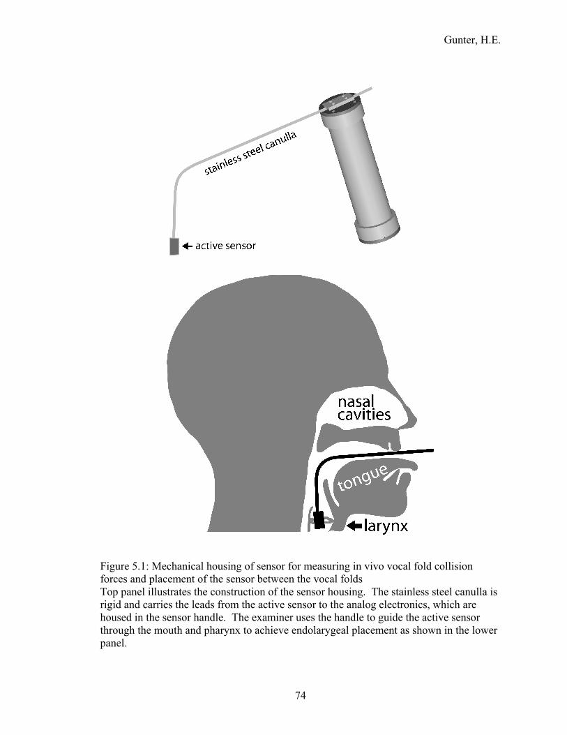
Gunter, H.E.
74
Figure 5.1: Mechanical housing of sensor for measuring in vivo vocal fold collision forces and placement of the sensor between the vocal folds Top panel illustrates the construction of the sensor housing. The stainless steel canulla is rigid and carries the leads from the active sensor to the analog electronics, which are housed in the sensor handle. The examiner uses the handle to guide the active sensor through the mouth and pharynx to achieve endolarygeal placement as shown in the lower panel.
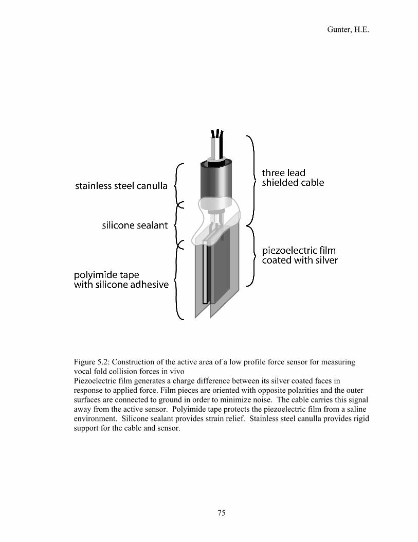
Gunter, H.E.
75
Figure 5.2: Construction of the active area of a low profile force sensor for measuring vocal fold collision forces in vivo Piezoelectric film generates a charge difference between its silver coated faces in response to applied force. Film pieces are oriented with opposite polarities and the outer surfaces are connected to ground in order to minimize noise. The cable carries this signal away from the active sensor. Polyimide tape protects the piezoelectric film from a saline environment. Silicone sealant provides strain relief. Stainless steel canulla provides rigid support for the cable and sensor.
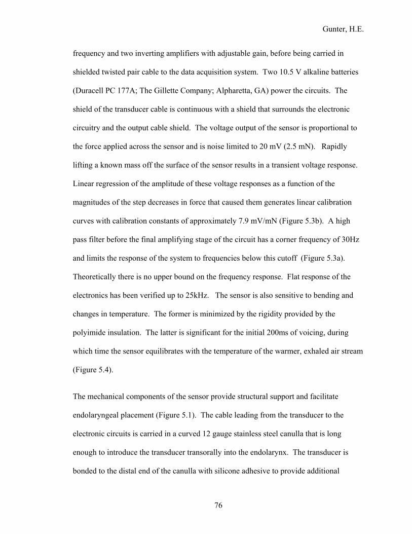
Gunter, H.E.
76
frequency and two inverting amplifiers with adjustable gain, before being carried in
shielded twisted pair cable to the data acquisition system. Two 10.5 V alkaline batteries
(Duracell PC 177A; The Gillette Company; Alpharetta, GA) power the circuits. The
shield of the transducer cable is continuous with a shield that surrounds the electronic
circuitry and the output cable shield. The voltage output of the sensor is proportional to
the force applied across the sensor and is noise limited to 20 mV (2.5 mN). Rapidly
lifting a known mass off the surface of the sensor results in a transient voltage response.
Linear regression of the amplitude of these voltage responses as a function of the
magnitudes of the step decreases in force that caused them generates linear calibration
curves with calibration constants of approximately 7.9 mV/mN (Figure 5.3b). A high
pass filter before the final amplifying stage of the circuit has a corner frequency of 30Hz
and limits the response of the system to frequencies below this cutoff (Figure 5.3a).
Theoretically there is no upper bound on the frequency response. Flat response of the
electronics has been verified up to 25kHz. The sensor is also sensitive to bending and
changes in temperature. The former is minimized by the rigidity provided by the
polyimide insulation. The latter is significant for the initial 200ms of voicing, during
which time the sensor equilibrates with the temperature of the warmer, exhaled air stream
(Figure 5.4).
The mechanical components of the sensor provide structural support and facilitate
endolaryngeal placement (Figure 5.1). The cable leading from the transducer to the
electronic circuits is carried in a curved 12 gauge stainless steel canulla that is long
enough to introduce the transducer transorally into the endolarynx. The transducer is
bonded to the distal end of the canulla with silicone adhesive to provide additional
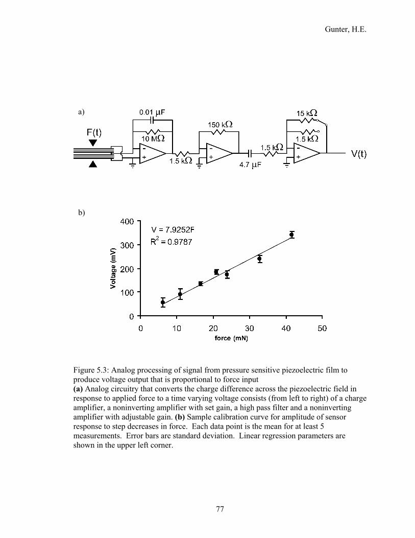
Gunter, H.E.
77
Figure 5.3: Analog processing of signal from pressure sensitive piezoelectric film to produce voltage output that is proportional to force input (a) Analog circuitry that converts the charge difference across the piezoelectric field in response to applied force to a time varying voltage consists (from left to right) of a charge amplifier, a noninverting amplifier with set gain, a high pass filter and a noninverting amplifier with adjustable gain. (b) Sample calibration curve for amplitude of sensor response to step decreases in force. Each data point is the mean for at least 5 measurements. Error bars are standard deviation. Linear regression parameters are shown in the upper left corner.
a)
b)
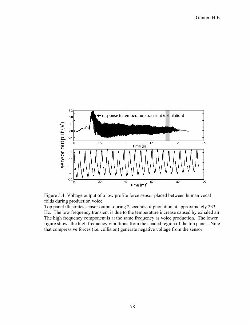
Gunter, H.E.
78
Figure 5.4: Voltage output of a low profile force sensor placed between human vocal folds during production voice Top panel illustrates sensor output during 2 seconds of phonation at approximately 233 Hz. The low frequency transient is due to the temperature increase caused by exhaled air. The high frequency component is at the same frequency as voice production. The lower figure shows the high frequency vibrations from the shaded region of the top panel. Note that compressive forces (i.e. collision) generate negative voltage from the sensor.
Gunter, H.E.
79
insulation and strain relief. The proximal end of the canulla is attached to one end of an
aluminum cylinder. This cylinder serves as a handle for manipulating the sensor and as
housing for the electronic circuits. Two switches on the bottom of the handle control
power to the circuits and gain of the second amplifying stage.
Experimental setup
During experiments to measure vocal fold collision forces, endoscopic and acoustic data
were collected in addition to the intraglottal force data. Videolaryngoscopic images were
obtained using a rigid transoral endoscope (Rhino-Laryngeal Stroboscope model 1900;
Kay Elemetrics; Lincoln Park, NJ) and a system that includes halogen (constant) and
xenon (strobe) light sources, a CCD camera with endoscope coupler, and a super VHS
tape deck. These images were used during the experiment to monitor sensor placement
and voice quality. Acoustic data was collected using a microphone (ECM50PSW, Sony)
placed approximately 30 cm from the subject’s mouth. Microphone output was
calibrated using a sound pressure level meter for three different sound intensities
produced using a 150 Hz buzzer. A calibration equation for each subject was obtained by
performing linear regression on the voltage-SPL data points. Electroglottographic
(EGG) waveforms were collected for an initial subject’s recordings in order to assess the
timing of the force waveform, but this practice was not continued due to concerns
regarding interference between the EGG system and the force sensor.
Force data was collected using the force sensor described above when it was positioned
between each subject’s vocal folds. Calibration data were collected either prior to or
following data collection from each subject. Calibration coefficients valid for a single
experiment were obtained as described in the previous section. These coefficients varied
Gunter, H.E.
80
by up to 10 % between experiments. Audio and force sensor data were digitized at
50kHz using a 12 bit analog to digital converter (Digidata 1200A, Axon instruments,
Union City, CA).
Experimental protocol
The experimental protocol, including use of the collision force sensor, was approved by
the Massachusetts Eye and Ear Infirmary Human Subjects Committee (Protocol number
01-09-050), and by the Harvard University Committee on the Use of Human Subjects in
Research. Prior to data collection, each subject read and signed an approved consent
form that described the nature of the project, the measures to be made, and the potential
risks and benefits of their participation. Subjects were instructed not to eat or drink for at
least 1 hour following their participation in the study.
Three males and one female who had undergone laryngeal reconstruction surgery
subsequent to surgical excision of laryngeal cancer participated in the study. These
subjects produce voice using one vibrating and nonvibrating, reconstructed vocal fold.
They were selected based on good conversational voice quality, minimal vibration of the
reconstructed fold during phonation and minimal constriction of the supraglottal
structures. One male subject with normal laryngeal anatomy was also a study
participant.
Topical anesthesia minimized subject discomfort. The pharynx was sprayed with 14%
benzocaine prior to endoscope insertion. 4% lidocaine (xylocaine) was swabbed and
dripped on the endo-larynx walls under endoscopic guidance until there was no response
to instrument palpation of the vocal fold surface. Evidence that anesthesia was
Gunter, H.E.
81
insufficient or wearing off, for example gagging, was grounds for terminating data
collection.
Following anesthesia, data collection was initiated. Subjects held their tongues with a
piece of gauze to maximize laryngeal access. The examiner inserted a camera-coupled
rigid laryngoscope transorally to visualize the larynx. Using the endoscopic images on
the video monitor for guidance, the examiner inserted the collision force transducer
between the vocal folds such that it rested against one vocal fold. Throughout the
experiment sensor placement was refined based on auditory perception of voice quality,
and phonation quality as visualized with stroboscopy.
The subjects phonated in response to instructions from the examiner while the force
sensor was in place. The phonation tokens requested of the subject include pitch/volume
pairs (normal/normal, high/normal, low/normal, normal/high, normal/low), pitch glides at
constant volume and volume glides at constant pitch. In all cases tokens were repeated
until good voice quality, as judged by the experimental team, was achieved. Following
each 1-2 minute segment of data recording all instrumentation was removed and the
subject was given a chance to rest. Collection of data was terminated when the subject
was no longer able to tolerate sensor and scope placement due to anesthesia wearing off
or when sufficient data was collected.
Data processing and analysis
A primary goal was to restrict analysis to data segments with both good quality voice and
good sensor placement. Two members of the experimental team rated the quality of each
phonation segment as poor, acceptable or good during review of the video and audio
Gunter, H.E.
82
experimental recordings while blinded to the sensor and audio waveforms. Thus, voice
quality judgments were based on a combined assessment of both audio and stroboscopy
recordings. Judgments of sensor placement accuracy were also based on stroboscopic
images.
For recorded samples judged to have good voice quality and good sensor placement the
data waveforms were examined and a consistent segment with at least 5 consecutive
cycles of vibration was selected. A script executed in Matlab was used to estimate
frequency, audio amplitude and force amplitude from these segments. Frequency
estimation was based on a fourier transform of the audio signal. Audio and force
amplitudes were based on averaging the peak and valley magnitudes for all cycles in the
segment. Audio and force amplitudes in volts were converted to dB and mN respectively
by applying the appropriate derived calibration factors. In order to do this it was assumed
that the lowest force was equal to 0 N, which is consistent with experimental
measurements of impact force using in vitro canine hemilarynges.
RESULTS
Data collection was attempted from four subjects (3 males and 1 female) who had
undergone vocal fold reconstruction surgery and one male subject with no history of
laryngeal pathology or surgery. The sole female subject was unable to tolerate passage of
the sensor through her pharynx. In the three male subjects who had undergone laryngeal
surgeries, the examiner was able to consistently place the sensor in the glottis and rest it
against the immobile fold, such that the sensor covered 50-75% of the membranous vocal
fold (i.e. anterior to the vocal process), while the subject produced voice (Figure 5.5). In
one male subject who had undergone laryngeal reconstruction surgery supraglottal
Gunter, H.E.
83
constriction impaired visualization of his vocal folds such that voice quality and sensor
placement could not be reliably evaluated. The examiner was able to successfully place
the sensor between the vocal folds of the male subject with no history of laryngeal
surgery. For the normal subject, two strategies resulted in good quality phonation. One
involved stabilizing the sensor in the center of the glottis by placing the posterior half
between the adducted arytenoid cartilages. The other involved halting the vibration of
one of the vocal folds by applying lateral force to it using the sensor.
Data was collected in approximately 100-second intervals between which the sensor and
layngoscope were removed from the subject’s larynx and pharynx. This limit was
determined by mucous collection, fogging of scope and subject tolerance. Each of the
male subjects participated in 3 or 4 intervals of data collection. Sensor placement and
voice quality were good during voice segments that were produced in response to relative
instructions (i.e. lower pitch, louder volume etc.) and crescendo requests (i.e. same pitch,
increasing volume). Sensor placement during glides (i.e. changing pitch) was poor due to
difficulties adjusting the sensor to account for vertical movement of the larynx. A sample
segment of the variety selected for analysis with a frequency of 137 Hz is shown in
Figure 5.6. EGG data was initially collected simultaneously to help guide the
interpretation of the force sensor waveform but was not collected for all subsequent
participants.
In the normal subject one voice segment was identified with good sensor placement and
good voice quality. In this segment the collision force amplitude was 95 mN. Other
segments with good voice quality were judged unsuitable for analysis due to the
interarytenoid location of part of the sensor.
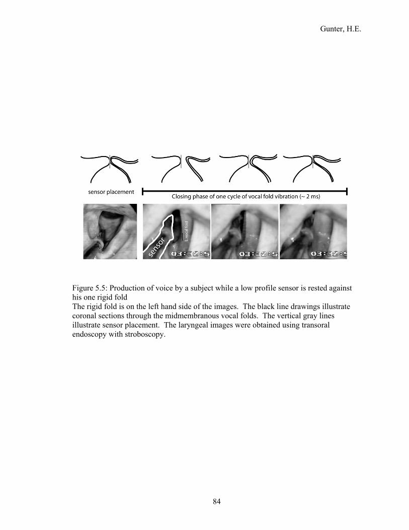
Gunter, H.E.
84
Figure 5.5: Production of voice by a subject while a low profile sensor is rested against his one rigid fold The rigid fold is on the left hand side of the images. The black line drawings illustrate coronal sections through the midmembranous vocal folds. The vertical gray lines illustrate sensor placement. The laryngeal images were obtained using transoral endoscopy with stroboscopy.
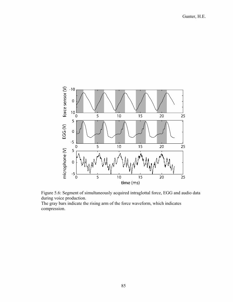
Gunter, H.E.
85
Figure 5.6: Segment of simultaneously acquired intraglottal force, EGG and audio data during voice production. The gray bars indicate the rising arm of the force waveform, which indicates compression.
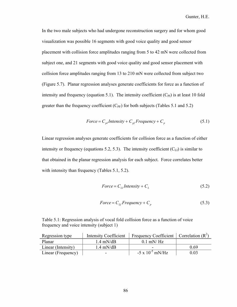
Gunter, H.E.
86
In the two male subjects who had undergone reconstruction surgery and for whom good
visualization was possible 16 segments with good voice quality and good sensor
placement with collision force amplitudes ranging from 5 to 42 mN were collected from
subject one, and 21 segments with good voice quality and good sensor placement with
collision force amplitudes ranging from 13 to 210 mN were collected from subject two
(Figure 5.7). Planar regression analyses generate coefficients for force as a function of
intensity and frequency (equation 5.1). The intensity coefficient (CPI) is at least 10 fold
greater than the frequency coefficient (CPF) for both subjects (Tables 5.1 and 5.2)
ppfpI CFrequencyCIntensityCForce ++= (5.1)
Linear regression analyses generate coefficients for collision force as a function of either
intensity or frequency (equations 5.2, 5.3). The intensity coefficient (CLI) is similar to
that obtained in the planar regression analysis for each subject. Force correlates better
with intensity than frequency (Tables 5.1, 5.2).
LLI CIntensityCForce += (5.2)
pLf CFrequencyCForce += (5.3)
Table 5.1: Regression analysis of vocal fold collision force as a function of voice frequency and voice intensity (subject 1) Regression type Intensity Coefficient Frequency Coefficient Correlation (R2) Planar 1.4 mN/dB 0.1 mN/ Hz Linear (Intensity) 1.4 mN/dB - 0.69 Linear (Frequency) - -5 x 10⁻⁵ mN/Hz 0.03
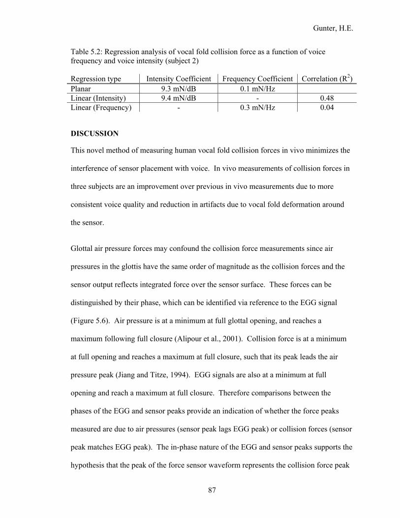
Gunter, H.E.
87
Table 5.2: Regression analysis of vocal fold collision force as a function of voice frequency and voice intensity (subject 2) Regression type Intensity Coefficient Frequency Coefficient Correlation (R2) Planar 9.3 mN/dB 0.1 mN/Hz Linear (Intensity) 9.4 mN/dB - 0.48 Linear (Frequency) - 0.3 mN/Hz 0.04
DISCUSSION
This novel method of measuring human vocal fold collision forces in vivo minimizes the
interference of sensor placement with voice. In vivo measurements of collision forces in
three subjects are an improvement over previous in vivo measurements due to more
consistent voice quality and reduction in artifacts due to vocal fold deformation around
the sensor.
Glottal air pressure forces may confound the collision force measurements since air
pressures in the glottis have the same order of magnitude as the collision forces and the
sensor output reflects integrated force over the sensor surface. These forces can be
distinguished by their phase, which can be identified via reference to the EGG signal
(Figure 5.6). Air pressure is at a minimum at full glottal opening, and reaches a
maximum following full closure (Alipour et al., 2001). Collision force is at a minimum
at full opening and reaches a maximum at full closure, such that its peak leads the air
pressure peak (Jiang and Titze, 1994). EGG signals are also at a minimum at full
opening and reach a maximum at full closure. Therefore comparisons between the
phases of the EGG and sensor peaks provide an indication of whether the force peaks
measured are due to air pressures (sensor peak lags EGG peak) or collision forces (sensor
peak matches EGG peak). The in-phase nature of the EGG and sensor peaks supports the
hypothesis that the peak of the force sensor waveform represents the collision force peak
Gunter, H.E.
88
Figure 5.7: Scatter plots of vocal fold collision force, voice intensity and voice frequency data points measured in two male subjects (a) Subject 1 (b) Subject 2
Gunter, H.E.
89
rather than an air pressure peak (Figure 5.6). This conclusion rests upon an assumption
that the EGG and intraglottal force waveforms are synchronized, which is likely but
unconfirmed. High-speed laryngoscopy could provide a reference against which to
evaluate time delays between the EGG and force signals in order to confirm this
assumption. The collision force peak may include a contribution from air pressure acting
on the portion of the sensor that is not in contact with the vocal folds. It is likely that
previously published measurements of vocal fold collision forces also include air
pressure contributions due to use of sensors that extend beyond the collision area.
Simultaneous subglottal pressure measurements would assist in the evaluation of the
magnitude of air pressure contributions to the intraglottal force signal.
Theoretical studies also guide interpretation of the force sensor waveform since they
distinguish contributions of collision force and air pressure to the intraglottal force
waveform in their predictions. Three dimensional finite element models predict a period
of noncollision during the initial moments of the closing phase followed by an increase in
collision force that begins when the anterior and posterior aspects of the vocal folds come
into contact, between 0.5 ms and 0.75 ms prior to complete closure (for a 127Hz
vibration) (Chapter 2). Thus, these models predict a collision force rise time occupying
approximately 10% of the vibration cycle. These theoretical results include all forces
between the midmembranous folds. Since the sensor does not record forces at the
extreme anterior and posterior aspects of the membranous folds, the time prior to the
peak in the intraglottal waveform that is due to collision force is less than that predicted
theoretically. A combination of elastic recoil (i.e. bouncing) of the tissue and forced
separation by air pressure causes the collision force to decrease after full closure occurs.
Gunter, H.E.
90
In the experimental measurements, the force rise time occupies almost 50% of the
vibration cycle (Figure 5.6). Comparison with theoretical results suggests that the
nonzero intraglottal forces during the initial stages of the rising force waveform are due
to air pressure forces, while those during the latter stage of closure are due to collision
forces. Intraglottal force measurements at higher spatial resolution or simultaneous
subglottal pressure measurements would guide further interpretation of the force sensor
waveform. Visual data such as high speed laryngoscopy or kymography would assist in
the alignment of force data with vocal fold movement.
The collision forces measured in both male subjects who had undergone laryngeal
reconstruction surgery, and for whom visualization was good, span those obtained using a
smaller sensor in a canine hemilarynx model (Jiang and Titze, 1994). These comparisons
are valid despite the difference in sensor size since collision force between the vocal folds
is concentrated in the midmembranous region, which is within the active area of both
sensors (Jiang and Titze, 1994). The 5 fold difference between the maximum collision
forces of each subject is consistent with the spread between different canine
hemilarynges. The 10 fold differences between minimum and maximum collision forces
in a single subject are consistent with changes achieved through manipulations of vocal
fold length, adduction and subglottal pressure in the canine hemilarynx model. The
collision forces measured at low intensities in these in vivo experiments are of the same
order of magnitude of those obtained in other in vivo human experiments, for which there
are doubts regarding measurement artifacts due to tissue deformation around thicker
sensors and in which difficulties were encountered maintaining good voice quality
(Verdolini and Hess, 1999). The collision force measurement obtained in the subject
Gunter, H.E.
91
with no history of laryngeal pathology is of the same order of magnitude as those
obtained in the subjects who had undergone laryngeal reconstruction. This validates the
use of the subjects with unique anatomy in whom measurements were technically easier
to make as well as the use of the in vitro canine hemilarynx preparation, in which many
more independent variables can be manipulated and controlled.
Acoustic intensity is a better predictor of collision force than frequency (Figure 5.7,
Tables 5.1 and 5.2). Theoretical models demonstrate that collision force predicts certain
compressive and shear mechanical stresses in the tissue that may disrupt tissue and
manifest as pathology (Chapter 2). Therefore, the increase of force measurements with
intensity suggests that mechanical stresses also increase leading to an increased risk of
vocal fold injury. This complements theoretical evidence that demonstrates a dependence
of mechanical stress magnitudes on subglottal pressure, which contributes to increasing
intensity (Chapter 3) in explaining associations between vocal fold pathologies and loud
voicing (Buckmire and Rosen, 2001).
The lack of trend between voice frequency and collision force may reflect complex
strategies that are used to manipulate frequency rather than a lack of dependence of
collision force on vocal fold length and stiffness. Theoretical results also fail to identify a
direct relationship between pitch and mechanical stress and provide insight by
demonstrating that the mechanical stresses that result from generation of a pitch depend
on the muscle activation patterns used (Chapter 3). Lack of data on muscle activation
patterns in our subjects prevents comparison with these predictions. The current analysis
is restricted to correlations between dependent variables, since the true independent
determinants of voice frequency, intensity and vocal fold collision force, such as
Gunter, H.E.
92
subglottal pressure, anatomy and muscle activities were not measured. More complex
experiments involving tracheal puncture subglottal pressure measurements, three
dimensional anatomical images, and EMG measurements of muscle activity, while
experimentally challenging, have the potential to provide such data and improve our
understanding of the determinants of collision force and by extension mechanical stress
levels and injury risk. Such data would evaluate whether the 5 fold difference in the
maximum collision forces between the two subjects reflects differences in vocal fold
anatomy or voice technique, illuminate the intraindividual determinants of vocal fold
collision forces and by extension injury risk and have implications for voice pathology
prevention.
The method used to obtain estimates of vocal fold collision forces serves as a paradigm
for characterizing the intraglottal mechanical environment since the low profile sensor is
able to collect data during periods of normal phonation. We are not aware of published
results using another method that has achieved good voice quality while taking
intraglottal measurements in vivo. Enhanced sensors will expand the range of data that
can be collected. One possibility is to decrease the active area of the current sensor
without decreasing the size of the sensor in order to make point force measurements.
This would provide a point estimate of collision force while maintaining the ease of
placement of the current sensor and minimizing measurement artifacts due to vocal fold
deformation around a smaller sensor. Other possibilities include an array of sensors in a
plane such as those developed by Pawluk et al. (1998) that could measure spatial
variations in contact force and estimate contact area.
Gunter, H.E.
93
The method used in this study was most successful in subjects with one rigid, medialized
vocal fold that acted as a support for the sensor. Interarytenoid placement of the posterior
end of the sensor offers an alternative method of stabilization in the subject with two
vibrating vocal folds. However, measurements obtained using this method with the
current sensor were difficult to interpret due to lack of understanding of the nature of
interarytenoid forces during voice production and therefore inability to subtract them
from the force sensor waveform in order to isolate the vocal fold collision force
waveform. The addition of an inactive posterior extension to the sensor would provide
an inactive mechanical structure that could be used for stabilization without contributing
to the force sensor signal. This would facilitate making intraglottal measurements during
phonation in normal subjects.
Gunter, H.E.
94
CHAPTER 6: CONTRIBUTIONS AND PROPOSED WORK This thesis presents studies regarding the biomechanics of vocal fold tissue during voice
production and provides insights into the role of solid mechanics in voice production and
voice pathology development. Both the methods developed and the results presented in
this thesis represent novel contributions to the fields of biomechanics and voice science.
The results provide answers to some of the original research questions, and also provoke
new queries.
METHOD FOR THEORETICAL STUDY OF VOCAL FOLD COLLISION
Theoretical models of vocal fold tissue mechanics are useful for exploring the
interactions between many variables in voice production. However, maximization of
geometric, material and aerodynamic detail is prohibitively computationally intensive. A
new approach to theoretical models of vocal fold tissue has been developed. It does not
include a representation of aerodynamics and therefore can accommodate increased detail
in the vocal fold tissue representation. These models are valid when air pressure in the
larynx is low, i.e. during closing and collision of the vocal folds during voice production.
Three dimensional models with a spatial resolution of 250 µm, which is suitable for
Gunter, H.E.
95
examining stress distributions on the scale of pathology development, have been created
using finite element techniques. One model uses a homogeneous material definition
(Chapter 2,3), and another uses four distinct material definitions to represent the tissue
layers that compose the vocal fold (Chapter 4). These models’ predictions of collision
force magnitudes are comparable to those measured in vitro in canine larynges and
during in vivo human voice production (Chapter 5) and their deformations are consistent
with qualitative clinical observations. A lumped mass model that is computationally
efficient and has physiologically meaningful inputs, but has low spatial resolution has
also been developed (Chapter 3). Its predictions of collision force magnitudes are
comparable to those predicted by the three dimensional models.
These models rely on published data for geometric and material definition and on
published collision force data for validation. There is a need for additional data that fully
parameterizes vocal fold mechanics by representing behavior of the component materials
in multiple deformation modes, directions and rates, and illustrating the deformation of
the individual vocal fold layers. The former could be obtained using careful and creative
studies of excised vocal fold tissue and would enhance the fidelity of the input
parameters. The latter could be obtained using high spatial and temporal resolution
imaging studies and would provide additional validation data. Comparisons with models
that include aerodynamic representations would further test the model assumptions and
improve accuracy of predictions.
Extensions of these models have the potential to be clinically useful surgical planning
tools. Such extensions might predict aerodynamic waveforms using the glottal
deformation predictions, and then apply speech synthesis techniques to predict acoustic
Gunter, H.E.
96
waveforms. This would provide a quantitative connection between surgical anatomy and
sound. If well implemented, then it could be used to compare proposed alternative
approaches to voice surgery.
METHOD FOR IN VIVO STUDY OF VOCAL FOLD COLLISION
In vivo measurements of vocal fold mechanics are necessary to quantify the mechanical
environment of the vocal fold, but are difficult to make due to the small size of the
larynx, its precarious location within the airway and the delicacy of tissue movement
during voice production. A novel method of measuring mechanical quantities during
vocal fold collision has been developed that does not interrupt the process of interest (i.e.
voice production) (Chapter 5). It involves stabilizing thin, flat sensors between the vocal
folds. In individuals with one medialized, nonvibrating vocal fold the sensor is rested
against the nonvibrating fold. Vibration of the remaining fold occurs with a smooth
mucosal wave and voice quality is maintained in a reasonable range. In individuals with
two vibrating vocal folds either the entire sensor is placed against one fold and used to
stop its vibration, or the posterior aspect of the sensor is clamped between the arytenoid
cartilages. Using the latter technique, both vocal folds vibrate well and voice quality
remains good.
Design challenges during the development of a thin, flat force sensor included sealing the
sensor from the in vivo environment, minimizing noise and calibrating voltage output
with force input. Collision force magnitudes measured in humans are of the same order
of magnitude as those measured using an in vitro canine preparation and demonstrate a
similar relationship between voice intensity and force magnitude. The collision forces
measured in a human with no history of laryngeal pathology or surgery are of the same
Gunter, H.E.
97
order of magnitude as those measured in subjects with one rigid, non-vibrating vocal
fold. These results provide validation for all three preparations: in vitro canine
hemilarynx, in vivo human with one rigid vocal fold, and in vivo human with two soft
folds. The latter has the highest fidelity with normal voice production, but is the most
technically difficult and experimentally restrictive. The other preparations offer
reasonable alternatives for the experimental study of voice mechanics.
Other thin, flat sensors have the potential to provide additional information regarding the
mechanics of vocal fold collision. For example, multiple force sensors on a single panel
would provide spatial resolution to force measurements and an indication of contact area.
This information could be used to interpret less invasive measurements of vocal fold
mechanics such as those obtained using electroglottography.
SUPPORT FOR MECHANICAL INJURY OF VOCAL FOLD TISSUE HYPOTHESIS
The theoretical models of vocal fold collision described above were used to investigate
the validity of the hypothesis that mechanical stresses induced by collision between the
vocal folds are responsible for structural and functional changes in vocal fold tissue that
manifest as vocal nodules (Chapter 3,4). The finite element model of vocal fold tissue
revealed elevation of compressive stresses in all three coordinate directions, and vertical
shear stress on the plane parallel to the plane of collision during collision (Chapter 3).
These stress elevations were maximal in the midmembranous location where vocal
nodules are often found and were correlated with increases in driving force, an indicator
of volume, which is a known risk factor for vocal nodule development. Vocal nodule
histology is consistent with injury by these types of stresses.
Gunter, H.E.
98
The lumped mass model of vocal fold collision revealed a dependence of compressive
stress perpendicular to the plane of collision on volume, vocal fold tissue layer
thicknesses and stiffnesses (Chapter 3). The latter two observations suggest an
explanation for varying risk for vocal fold injury between individuals since they are
dependent on vocal fold anatomy as determined by genetics and environment, and vocal
fold configuration as determined by voice production strategies (for example,
manipulation of stiffness and tension to control pitch). Systematic variation of material
properties and geometry in the finite element model would produce predictions of the
spatial variation of multiple stresses and provide a clearer assessment of risk factors than
the lumped element model does, but would be computationally expensive.
The predictions of the finite element model of vocal fold collision that incorporates a
layered structure explore the role played by the stiffness of the superficial lamina propria
(SLP) in vocal fold injury (Chapter 4). Decreasing SLP stiffness decreased normal and
shear stresses in the SLP and epithelium, as well as normal and shear strains in the
epithelium and therefore reduces epithelial injury risk. However, decreasing SLP
stiffness increased normal and shear strains in the SLP. The effects of stress and strain in
tissues with different properties are not understood. Therefore it is not clear how SLP
stiffness affects SLP injury risk.
Experimental support of the proposed tissue injury modes could be provided by vocal
fold tissue culture studies. Such studies would help to interpret the magnitudes of the
stress tensor components in terms of injury risk. They could also be used to investigate
the roles of stress vs. strain in causing functional changes in SLP cell lines in
extracellular matrices of varying composition.
Gunter, H.E.
99
MEASUREMENT AND INTERPRETATION OF VOCAL FOLD COLLISION FORCES
The novel method of measuring in vivo vocal fold collision quantities described above
has been used to obtain in vivo estimates of vocal fold collision forces, which are loads
that may contribute to vocal fold injury (Chapter 5). The finite element model of vocal
fold tissue during collision guides the interpretation of the force data in terms of tissue
stress levels (Chapter 2).
The magnitude of the experimentally measured collision forces in the two subjects with
one rigid, nonvibrating vocal fold correlate better with voice intensity than frequency.
The magnitude of the theoretically predicted contact forces correlates with theoretical
predictions of normal stress perpendicular to the plane of collision, vertical shear stress
on the plane parallel to the plan of collision and Von Mises stress. These stresses are
therefore candidates for vocal fold tissue injury that is associated with high volumes.
There is no clear case for the dependence of collision force and by extension mechanical
stress and voice injury on voice pitch.
A systematic examination of collision forces for different laryngeal anatomies (i.e. child,
male, female) using different modes of voice production (i.e. pressed, chest, breathy)
would shed additional light on contributions to vocal fold collision forces and by
extension injury risk. Collection of muscle activity, subglottal pressure and anatomical
information during these experiments would help to tease out the risks associated with
different pitch manipulation mechanisms.
Gunter, H.E.
100
FUTURE DIRECTIONS
The theoretical and experimental studies presented in this thesis explore vocal fold tissue
mechanics during the closure and collision phase of voice production and provide insight
into mechanical injury of vocal fold tissue. The logical extensions of this work are along
the lines of continuing exploration of the role of mechanical stress in vocal fold injury
and further development of the finite element models of vocal fold tissue mechanics.
Further exploration of the contributions of geometric and material properties of tissue to
mechanical stress levels, combined with experimental studies of vocal fold tissue
response to stress will refine the impact stress hypothesis and have the potential to
generate concrete recommendations for voice injury prevention. Further development of
the theoretical models might include comparison with models with full aerodynamics or
combination with speech synthesis algorithms. These extensions have the potential to
generate robust tools that would be useful in future voice science investigations and
clinical practice.

Gunter, H.E.
101
BIBLIOGRAPHY Alipour, F., Berry, D. A. and Titze, I. R. (2000), "A finite-element model of vocal-fold vibration," Journal of the Acoustical Society of America. 108, 3003-12.
Alipour, F., Montequin, D. and Tayama, N. (2001), "Aerodynamic profiles of a hemilarynx with a vocal tract," Annals of Otology, Rhinology & Laryngology. 110, 550-5.
Alipour, F. and Scherer, R. C. (2000), "Dynamic glottal pressures in an excised hemilarynx model," Journal of Voice. 14,
Alipour-Haghighi, F. and Titze, I. R. (1991), "Elastic models of vocal fold tissues," Journal of the Acoustical Society of America. 90, 1326-31.
Berke, G. S. and Gerratt, B. R. (1993), "Laryngeal biomechanics: An overview of mucosal wave mechanics," Journal of Voice. 7, 123-8.
Berry, D. A., MOntequin, D. W. and Tayama, N. (2001a), "High-speed digital imaging of the medial surface of the vocal folds," Journal of the Acoustical Society of America. 110, 2539-47.
Berry, D. A. and Titze, I. R. (1996), "Normal modes in a continuum model of vocal fold tissues," Journal of the Acoustical Society of America. 100, 3345-54.
Berry, D. A., Verdolini, K., Montequin, D. W., Hess, M. M., Chan, R. W. and Titze, I. R. (2001b), "A quantitative output-cost ratio in voice production," Journal of Speech, Language, & Hearing Research. 44, 29-37.
Buckmire, R. A. and Rosen, C. A. (2001), "Vocal polyp and nodules," eMedicine Journal. 2, 1-11.
Gunter, H.E.
102
Chan, R. and Titze, I. (2000), "Theoretical modeling of viscoelastic shear properties of human vocal fold mucosa: Theoretical characterization based on constitutive modeling," Journal of the Acoustical Society of America. 107, 565-80.
Chan, R., W. and Titze, I. R. (1999a), "Viscoelastic shear properties of human vocal fold mucosa: Measurement methodology and empirical results," Journal of the Acoustical Society of America. 106, 2008-21.
Chan, R. W. and Titze, I. R. (1999b), "Hyaluronic acid (with fibronectin) as a bioimplant for the vocal fold mucosa," Laryngoscope. 109, 1142-9.
Chandrupatla, T. R. and Belegundu, A. D. (1997), Introduction to finite elements in engineering, 2 Ed.(Prentice-Hall, New Jersey)
Cotron, R. and Casper, J. K. (1996), Understanding voice problems: A physiological perspective for diagnosis and treatment, 2 Ed.(Williams & Wilkins; a Waverly Company, Baltimore) 418
de Vries, M. P., Schutte, H. K., Veldman, A. E. P. and Verkerke, G. J. (2002), "Glottal flow through a two-mass model: Comparison of navier-stokes solutions with simplified models," Journal of the Acoustical Society of America. 111, 1847-53.
Dikkers, F. G. (1994), Benign lesions of the vocal folds: Clinical and histopathological aspects, Ed.(Drukkerij Van Denderen B.V., Groningen, Netherlands)
Gray, S. and Titze, I. (1988), "Histologic investigation of hyperphonated canine vocal cords," Annals of Otology, Rhinology & Laryngology. 97, 381-8.
Gray, S. D., Titze, I. R., Alipour, F. and Hammond, T. H. (2000), "Biomechanical and histologic observations of vocal fold fibrous proteins," Annals of Otology, Rhinology & Laryngology. 109, 77-85.
Gunter, H. E. (2003a), "A mechanical model of vocal fold collision with high spatial and temporal resolution," Journal of the Acoustical Society of America. 113, 994-1000.
Gunter, H. E. (2003b), "Modeling mechanical stresses as a factor in the etiology of benign vocal fold lesions," submitted for review.
Hess, M. M., Verdolini, K., Bierhals, W., Mansmann, U. and Gross, M. (1998), "Endolaryngeal contact pressures," Journal of Voice. 12, 50-67.
Hillman, R. E., Holmberg, E. B., Perkell, J. S., Walsh, M. and Vaughan, C. (1989), "Objective assessment of vocal hyperfunction: An experimental framework and initial results," Journal of Speech & Hearing Research. 32, 373-92.
Hirano, M., Kiyokawa, K. and Kurita, S. (1988), "Laryngeal muscles and glottic shaping," in Vocal physiology: Voice production mechanisms and functions, edited by O. Fujimara (Raven Press Ltd., New York) 49-65
Gunter, H.E.
103
Hirano, M., Kurita, S. and Nakashima, T. (1983), "Growth, development and aging of human vocal folds," in Vocal fold physiology: Contempory research and clinical issues, edited by D. M. Bless and J. H. Abbs (College-Hill Press, San Diego) 22-43
Holmberg, E. B., Hillman, R. E. and Perkell, J. S. (1988), "Glottal air-flow and transglottal air-pressure measurements for male and female speakers in soft, normal and loud voice," Journal of the Acoustical Society of America. 84, 511-29.
Ikeda, T., Matsuzaki, Y. and Aomatsu, T. (2001), "A numerical analysis of phonation using a two-dimensional flexible channel model of the vocal folds," Journal of Biomechanical Engineering. 123, 571-9.
Ishizaka, K. and Flanagan, J. L. (1972), "Synthesis of voiced sounds from a two-mass model of the vocal cords," The Bell System Technical Journal. 51, 1233-68.
Isshiki, N. (1964), "Regulatory mechanism of voice intensity variation," Journal of Speech, & Hearing Research. 7, 17-29.
Jiang, J., Lin, E. and Hanson, D. G. (2000), "Voice disorders and phonosurgery i: Vocal fold physiology," otolaryngologic clinics of north america. 33, 699-718.
Jiang, J. J., Diaz, C. E. and Hanson, D. G. (1998), "Finite element modeling of vocal fold vibration in normal phonation and hyperfunctional dysphonia: Implications for the pathogenesis of vocal nodules," Annals of Otology, Rhinology & Laryngology. 107, 603-10.
Jiang, J. J., Shah, A. G., Hess, M. M., Verdolini, K., Banzali, F. M. J. and Hanson, D. G. (2001a), "Vocal fold impact stress analysis," Journal of Voice. 15, 4-14.
Jiang, J. J. and Titze, I. R. (1993), "A methodological study of hemilaryngeal phonation," Laryngoscope. 103, 872-82.
Jiang, J. J. and Titze, I. R. (1994), "Measurement of vocal fold intraglottal pressure and impact stress," Journal of Voice. 8, 132-44.
Jiang, J. J., Zhang, Y. and Stern, J. (2001b), "Modeling chaotic vibrations in symmetric vocal folds," Journal of the Acoustical Society of America. 110, 2120-8.
Kakita, Y., Hirano, M. and Ohmaru, K. (1981), "Physical properties of vocal tissue: Measurements on excised larynges," in Vocal fold physiology, edited by M. Hirano and K. Stevens (University of Tokyo Press, Tokyo) 377-98
Kaneko, T., Masuda, T., Shimada, A., Suzuki, H., Hayasaki, K. and Komatsu, K. (1987), "Resonance characteristics of the human vocal fold in vivo and in vitro by an impulse excitation," in Laryngeal function in phonation and respiration, edited by T. Baer, C. Sasaki and K. S. Harris (Little, Brown and Company (Inc.), Boston) 349-65
Gunter, H.E.
104
Lieberman, P. (1968), "Direct comparison of subglottal and esophageal pressure during speech," Journal of the Acoustical Society of America. 43, 1157-64.
Min, Y. B., Titze, I. R. and Alipour-Haghighi, F. (1995), "Stress-strain response of the human vocal ligament," Annals of Otology, Rhinology & Laryngology. 104, 563-9.
Mossallam, I., Kotby, M. N., Ghaly, A. F., Nassar, A. M. and Barakah, M. A. (1986), "Histopathological aspects of benign vocal fold lesions associated with dysphonia," in Vocal fold histopathology; a symposium, edited by J. A. Kirchner (College Hill Press, San Diego) 65-80
Pawluk, D., Son, J., Wellman, P., Peine, W. and Howe, R. (1998), "A distributed pressure sensor for biomechanical measurements.," Journal of Biomechanical Engineering. 120, 302-5.
Pelorson, X., Hirschberg, A. and van Hassel, R. R., Wijnands, A.P.J. (1994), "Theoretical and experimental study of quasisteady-flow separation within the glottis during phonation. Application to a modified two-mass model," Journal of the Acoustical Society of America. 96, 3416-31.
Ressler, B., Lee, R. T., Randell, S. H., Drazen, J. M. and Kamm, R. D. (2000), "Molecular responses of rat tracheal epithelial cells to transmembrane pressure," American Journal of Physiology Lung Cell and Molecular Physiology. 278, L1264-72.
Shipp, T. and McGlone, R. E. (1971), "Laryngeal dynamics associated with voice frequency change," Journal of Speech & Hearing Research. 14, 761-8.
Stevens, K. N. (1998), Acoustic phonetics.(The MIT Press, Cambridge)
Story, B. H. and Titze, I. R. (1995), "Voice simulation with a body-cover model of the vocal folds," Journal of the Acoustical Society of America. 97, 1249-60.
Swartz, M. A., Tschumperlin, D. J., Kamm, R. D. and Drazen, J. M. (2001), "Mechanical stress is communicated between different cell types to elicit matrix remodeling," Proceedings of the National Academy of Sciences of the United States of America. 98, 6180-5.
Titze, I. R. (1973), "The human vocal cords: A mathematical model. I," Phonetica. 28, 129-70.
Titze, I. R. (1974), "The human vocal cords: A mathematical model. Ii," Phonetica. 29, 1-21.
Titze, I. R. (1994), "Mechanical stress in phonation," Journal of Voice. 8, 99-105.
Titze, I. R., Lemke, J. and Montequin, D. (1997), "Populations in the u.S. Workforce who rely on voice as a primary tool of trade: A preliminary report," Journal of Voice. 11, 254-9.
Gunter, H.E.
105
Titze, I. R., Schmidt, S. S. and Titze, M. R. (1995), "Phonation threshold pressure in a physical model of the vocal fold mucosa," Journal of the Acoustical Society of America. 97, 3080-4.
Titze, I. R. and Story, B. H. (2002), "Rules for controlling low-dimensional vocal fold models with muscle activation," Journal of the Acoustical Society of America. 112, 1064-76.
Titze, I. R. and Strong, W. J. (1975), "Normal modes in vocal cord tissues," Journal of the Acoustical Society of America. 57, 736-49.
Titze, I. R. and Talkin, D. T. (1979), "A theoretical study of the effects of various laryngeal configurations on the acoustics of phonation," Journal of the Acoustical Society of America. 66, 60-74.
Waters, C. M., Glucksberg, M. R., Lautenschlager, E. P., Lee, C. W., Van Matre, R. M., Warp, R. J., Salva, U., Healy, K. E., Moran, B., Castner, D. G. and bearinger, J. P. (2001), "A system to impose prescribed homogeneous strains on cultured cells," Journal of Applied Physiology. 91, 1600-10.
Wong, D., Ito, M. R., Cox, N. B. and Titze, I. R. (1991), "Observation of perturbations in a lumped-element model of the vocal folds with application to some pathological cases," Journal of the Acoustical Society of America. 89, 383-94.
Yamana, T. and Kitajima, K. (2000), "Laryngeal closure pressure during phonation in humans," Journal of Voice. 14, 1-7.
Zeitels, S. M. (1998), "Phonosurgery - past, present & future," Operative Techniques in Otolaryngology - Head and Neck Surgery. 9, 179.
Zeitels, S. M., Hillman, R. E., Desloge, R., Mauri, M. and PB, D. (2002), "Phonomicrosurgery in singers and performing artists: Treatment outcomes, management theories, and future directions.," Annals of Otology, Rhinology, & Laryngology. 190, 21-40.
Zemlin, W. R. (1997), Speech and hearing science; anatomy & physiology, 4 Ed.(Allyn & Bacon, Massachusetts) 610