Embed Size (px)
Citation preview

PhD Yearbook | 2017
M E C H A N I CA L E N G I N E E R I N G I P H Y S I C S I PRESERVATION OF THE ARCHITECTURAL HERITAGE I SPATIAL PLANNING AND URBAN DEVELOPMENT I STRUCTURAL SEISMIC AND GEOTECHNICAL ENGINEERING I TECHNOLOGY AND DESIGN FOR ENVIRONMENT AND BUILDING I TERRITORIAL DESIGN AND GOVERNMENT I URBAN PLANNING, DESIGN AND POLICY I AEROSPACE ENGINEERING I A R C H I T E C T U R A L A N D U R B A N D E S I G N I ARCHITECTURAL COMPOSITION I ARCHITECTURE, BUILT ENVIRONMENT AND CONSTRUCTION ENGINEERING I ARCHITECTURE, URBAN DESIGN, CONSERVATION OF HOUSING AND LANDSCAPE I
BIOENGINEERING I DESIGN I ELECTRICAL ENGINEERING I ENERGY AND NUCLEAR SCIENCE A N D T E C H N O L O G Y I E N V I R O N M E N TA L A N D I N F R A S T R U C T U R E E N G I N E E R I N G I I N D U S T R I A L C H E M I S T R Y A N D C H E M I C A L ENGINEERING I INFORMATION TECHNOLOGY I I N T E R I O R A R C H I T E CT U R E A N D D E S I G N I MANAGEMENT ENGINEERING I MATERIALS E N G I N E E R I N G I M AT H E M AT I C A L M O D E L S A N D M E T H O D S I N E N G I N E E R I N G

115
PhD
Year
book
I 20
17B
IOE
NG
INE
ER
ING
Chair:
Prof. Andrea Aliverti
114
DOCTORAL PROGRAM
IN BIOENGINEERING
The main objective of the PhD Programme in Bioengineering is to prepare the PhD candidates to develop high level engineering problem-solving abilities in biomedical, healthcare and life sciences, inside research groups or in private/public industrial contexts, through a strong interdisciplinary training bridging engineering and medical/biological knowledge.During the PhD, the candidates develop a scientifi c research project dealing with a complex problem which can be at diff erent scales, from the molecular and the cellular levels to living organisms up to biomedical systems. They investigate original methods, devices, and systems with diff erent purposes: increasing knowledge, proposing innovative methods for diagnosis and therapy as well as improving healthcare and daily life structures and services. At the end of the PhD programme, the candidate are expected to be able to carry out innovative projects and research and development in the fi eld of Bioengineering, by proposing new methodological and technological solutions and properly evaluating the technology impact in healthcare, life science and biomedical industry. During the three years of the program, PhD candidates perform their research through theoretical and experimental activities in four major areas: biomimetic engineering and micro-nano technologies; rehabilitation engineering and technology; technologies for therapy; physiological modelling and non-invasive diagnostics.More specifi c areas include but are not limited to: molecular and cellular engineering, biomaterials, tissue engineering, bio-artifi cial interfaces and devices, neuro-prostheses, movement analysis, cardiovascular and respiratory system bioengineering, central nervous system signal and image processing for rehabilitation, biomechanics, computational fl uid-dynamics, computer assisted surgery and radiotherapy, robotics, artifi cial organs, implantable devices, biomedical signal and image processing, E-Health, bioinformatics, functional genomics and molecular medicine. The PhD Program in Bioengineering is organized with an inter-departmental structure. Faculty members of the PhD Advisory Board belong to two Departments of the Politecnico di Milano, namely DEIB (Department of Electronics, Information and Bioengineering) and CMIC (Department of Chemistry, Materials and Chemical Engineering “G. Natta”). PhD candidates (who are, in average, 20 per year) develop their PhD research programs within experimental laboratories located at the
Politecnico di Milano or outside it, typically biomedical research centers, hospitals or industries. When the research is performed within the Politecnico, PhD candidates are usually assigned to one of the following laboratories belonging to the DEIB and CMIC: Laboratory of Biological Structure Mechanics (LaBS, CMIC), Laboratory of movement analysis “Luigi Divieti” (DEIB), Medical Informatics laboratory (DEIB), Neuroengineering and medical robotics Laboratory (NearLab, DEIB), Biosignals, Bioimaging and Bioinformatics Lab (B3 lab, DEIB), Biomaterials laboratory (CMIC), Biomedical Technology Lab (TBMLab, DEIB), Experimental Micro and Biofl uid dynamics (μBS Lab, DEIB), Computational Biomechanics Lab (DEIB), Biocompatibility and Cell culture Lab (BioCell, CMIC), Bioreactors Laboratory (CMIC). The Istituto di Elettronica, Ingegneria dell’Informazione e delle Telecomunicazioni (IEIIT) of the Consiglio Nazionale delle Ricerche (CNR), which is located at DEIB, represents another possible option.Stage periods in distinguished research institutes in Italy and abroad are an essential feature of the PhD candidate training. The candidates are encouraged to carry out part of their research activities in contact with other research groups, preferably abroad through periods of at least three months spent in laboratories where the candidate can acquire further skills to develop his/her research work and thesis. Collaborations that may involve the PhD students are presently active with several national and international
COMPOSITION OF THE PHD BOARD
Aliverti Andrea (Coordinator) DEIB Dubini Gabriele Angelo CMIC Pedrocchi Alessandra DEIB
Baselli Giuseppe DEIB Farè Silvia CMIC Pennati Giancarlo CMIC
Bianchi Anna Maria DEIB Frigo Carlo Albino DEIB Pozzi Giuseppe DEIB
Caiani Enrico DEIB Galli Manuela DEIB Raimondi Manuela Teresa CMIC
Candiani Gabriele CMIC Guazzoni Chiara DEIB Ravazzani Paolo DEIB
Cerveri Pietro DEIB Mainardi Luca DEIB Redaelli Alberto DEIB
Chiesa Roberto CMIC Mantero Sara CMIC Signorini Maria Gabriella DEIB
Costantino Maria Laura CMIC Migliavacca Francesco CMIC Soncini Monica DEIB
research and academic Institutions. Very often, the involvement of companies and clinical partners facilitates the technological transfer of applied research into industry and clinical applications.The educational off er includes ad hoc advanced courses specifi cally designed for the PhD in Bioengineering. The off er includes also the school of the National Bioengineering Group, which is held yearly for one week in Bressanone (Bz). Every year, the School is focused on diff erent topics. As examples, the themes of the last few years have been: Neuro-informatics (2011), Biomedical devices from research to market (2012), Regenerative medicine (2013), From functional recovery to artifi cial organs (2014), Experimental models for development methods for 3R (2015), Bioengineering for Active ageing (2016).The PhD Board of professors (‘PhD Board’) is composed by highly qualifi ed and active researchers in Bioengineering, belonging to DEIB and CMIC. The PhD Board is responsible of all the candidate’s activities. The competencies of Faculty members cover a wide spectrum of research fi elds. This allows a continuous updating of the PhD program and ensures that the PhD candidates are involved in innovative work.The PhD Programme in Bioengineering relies also on an Advisory Board Member, formed by distinguished experts coming from R&D industries, research and clinical centers, in order to ensure that that the goals of the PhD Program are in line also with the needs of non-academic world.

117
PhD
Year
book
I 20
17B
IOE
NG
INE
ER
ING
116
Introduction: The aim of this study was to implement and test objective methods for quantifying the relationship between gray matter (GM) and white matter (WM) damage, applicable in regards to neurodegenerative diseases, with a particular focus on multiple sclerosis (MS). MS is an auto-immune mediated infl ammatory disorder causing widespread damage throughout the central nervous system (CNS). Although magnetic resonance imaging (MRI) has allowed for the monitoring of the eff ects of the disease within the CNS in vivo, conventional imaging techniques and measures are generally lacking in their specifi city. For example, the classic WM lesions, easily identifi ed on a T2-weighted scan, correspond to a wide range of pathological substrates and have a relatively poor relationship with clinical outcomes. This discrepancy, often referred to as the clinico-radiological paradox, has motivated the investigation and development of other imaging measures in an aim to better characterize the disease. Moreover, conventional measures may not be sensitive enough to detect changes within the so-called normal appearing WM (NAWM). Moreover, it is now widely recognized that MS damage is much more widespread
than just focal WM lesions. A number of advanced acquisition and post-processing techniques have been developed for both a more precise description and a better localization of the eff ects of the disease. However, the eff ects of MS-related focal damage often require careful evaluation and tuning of the algorithms commonly in use within the neuroimaging community. Nonetheless, advanced techniques off er the possibility to interrogate the various pathological processes involved in MS. Examples include morphological reconstruction of the cortex and subcortical GM structures for a better characterization of tissue atrophy, diff usion imaging and tractographic reconstruction for quantitative assessment of WM characteristics (including the so-called normal appearing WM (NAWM)) and susceptibility-weighted imaging for assessing iron deposition. Furthermore, the integration of the aforementioned techniques can provide information on the associations and interactions between tissue damage in the WM and GM compartments in a way which is not possible when utilizing a single imaging modality.Protocol and Results: (i) Methodological Developments:
The impact of WM lesions on cortical reconstructions was qualitatively assessed in a sample of relapsing remitting MS patients. As WM lesions were found to negatively aff ect automated cortical reconstructions, we developed and implemented an optimized process for lesion fi lling as pre-processing step. We combined cortical reconstruction and tractography techniques to assess the relationship between WM injury and cortical thinning in a functionally and anatomically connected region. As WM pathology was found to interfere with tractography of the WM fi ber tracts (both for deterministic and probabilistic approaches), we implemented a method for generating probabilistic atlases based on successful reconstructions in healthy controls (HC). The eff ect of lesions on automated deep GM (DGM) segmentations was also quantifi ed. Finally, we implemented a novel, optimized and unbiased processing pipeline for assessing the relationship between putative iron deposition in the DGM and injury within the connected WM tracts.(ii) Applications: The optimized lesion fi lling process was used in all four studies that make up the current work. Next, a multi-modal imaging approach was used to
study the relationship between damage in the WM and that GM in a sample MS patients. The FreeSurfer software package was used for cortical morphological reconstruction whereas diff usion-weighted imaging was used for the assessment of WM structural integrity. Although MS causes widespread damage throughout the central nervous system, we hypothesized that damage within the WM would be more closely linked to an anatomically and functionally connected GM area with respect to an area which is not. To this aim, we investigated the relationship between WM injury in the corticospinal tract (CST) and cortical thickness of the primary motor cortex, whereas the primary auditory cortex was used as a control region for testing our hypothesis. We found that axial diff usivity within the normal appearing WM of the CST was the best imaging predictor of primary motor cortex thickness in RRMS patients whereas no relationships were seen with respect to the primary auditory cortex. Although the study was cross-sectional in nature, the results suggest a direct association between WM and GM injury in MS. This appears to be specifi c to MS as no such relation was found in the HC group. In a group of 152 MS patients, we demonstrated a clear eff ect of WM lesions on automated thalamic and caudate segmentations. This motivated the use of lesion fi lled images in our third study which investigated the relationships between DGM atrophy and cognitive status in a sample of 64 MS patients.
ADVANCED MRI TECHNIQUES IN MULTIPLE SCLEROSIS: MULTIMODAL ASSESSMENT OF WM AND GM DAMAGE MECHANISMS
Bergsland Niels Peter – Advisor: Prof. Giuseppe Baselli
We highlighted the advantage of surface-based methods for a more precise localization of the eff ects of atrophy and cognitive defi cits in MS. Cross-sectionally, associations were found between atrophy of the thalamus, putamen and caudate versus cognitive defi cits. Moreover, longitudinally, the study revealed that focal, anterior atrophy of the left thalamus is associated with decreased cognitive processing speed over three years of follow-up. Of particular note is that imaging techniques widely used in the literature (region of interest and voxel-based morphometry) were not suffi cient to detect this relationship. Rather, the correlation between localized thalamic atrophy and cognitive decline was only seen when using vertex-wise based analysis of the thalamic surface. Finally, a multimodal imaging method was implemented for better characterizing the relationship between white matter injury and increased iron deposition in MS patients. An unbiased, voxelwise approach was used to compare MS and HC groups for the identifi cation of areas indicative of increased iron concentrations in the deep GM. These regions were subsequently used as seeds for probabilistic tractography for subsequent WM integrity assessments in the anatomically connected tracts. The proposed approach of combining an iron-sensitive MRI technique with one that is able to quantify tissue microstructure damage may help shed further light on the pathogenetic mechanisms involved in MS.
Conclusion: The aim of this study was to implement and test objective methods for quantifying the relationships between GM and WM damage in multiple sclerosis. We consistently demonstrated the importance of paying close attention, in the preprocessing phase, to the confounding eff ects of WM lesions in terms of obtaining reliable results. In two separate studies, we found clear evidence of an association between WM injury and GM pathology. For the former, we used WM tractographic methodologies while for the latter we utilized both cortical morphological reconstruction and iron-sensitive acquisition/post-processing techniques. We found that both cortical thinning and a putative marker of increased iron deposition were related to increased NAWM injury in the connected tracts. We also showed for the fi rst time that focal atrophy of the thalamus is associated with a decline in cognitive processing speed over three years of follow-up. Taken together, the results obtained from the investigations performed as part of this thesis appear to be promising in leading to a better characterization of the association between structure-specifi c GM, both deep and cortical, and WM injury in multiple sclerosis. These results, moreover, foster new insights yielded by multimodal imaging approaches for studying the multifaceted aspects of the disease.
119
PhD
Year
book
I 20
17B
IOE
NG
INE
ER
ING
118
Casaroli Gloria – Supervisors: Prof. Tomaso Villa, Dr. Fabio Galbusera
MECHANICAL FAILURE OF THE INTERVERTEBRAL
DISC: EXPERIMENTAL TESTING
AND NUMERICAL MODELLING
Low Back Pain (LBP) is one of the most disabling pathologies in modern society with high economic and social related costs. One of the main causes of LBP is lumbar intervertebral disc (IVD) herniation, which is defi ned as the displacement of part of the annulus fi brosus, of the nucleus pulposus or of the endplate farther the margins of the vertebrae due to the structural failure of the disc. Despite many groups have investigated the mechanical behavior of the IVD, the mechanisms of failure are still not completely understood. The aim of this project was to investigate the mechanical causes of disc herniation and to generate a numerical model able to predict the risk of failure in complex loading conditions. In the fi rst part of the thesis, an introduction about the social impact of the LBP and disc herniation shows how much important it is to study disc pathologies and the causes of herniation. From this point of view, the knowledge of the mechanical causes of disc failure could contribute to prevent the generation of herniation. Moreover, the main biomechanical studies and results are shown and commented. Despite their relevance, there is still a lack of knowledge in the literature due
to the use of diff erent set-ups and species of specimens, and there is no published study in which experimental testing and numerical investigation have been combined. Furthermore, there is not a mechanical criterion that defi nes when the mechanical failure of the disc can occur. In the second part of the thesis, the use of the ovine lumbar IVD as a model of the human one was supported by the literature. To investigate the mechanical behavior of the disc, a novel fi nite element model of the ovine lumbar IVD was developed. The geometry was generated starting from magnetic resonance images of a L3-4 ovine lumbar segment. The annulus fi brosus was characterized by combining experimental tests and numerical investigation. An anisotropic hyperelastic formulation was used for the annulus fi brosus. The annulus resulted stiff est in the anterior region, followed by the lateral and the posterior ones. For the sake of validation, the predicted fl exibility of the IVD was compared with the literature, showing a good agreement with in vitro data. Since in this project the mechanical behavior of the disc was investigated under complex loading scenarios, it has been assessed that the ovine disc can be used as a model
of the human one also in such conditions. In the third part of the thesis, the mechanical response of the disc was investigated under combined loads and compared with the human model in terms of intradiscal pressure, maximum strains and maximum shear strains in the layer between the annulus and the endplate. In general, the ovine and the human models had similar behavior, especially when fl exion – extension and axial rotation were applied, and the maximum fi bers strains were in both models located in the posterior and postero - lateral regions. In the fourth part of the thesis, the failure of the disc has been experimentally investigated. An in vitro test on thirty ovine lumbar specimens has been performed and fi ve diff erent complex loading scenarios were applied. A multiple linear regression analysis was conducted to investigate which condition was responsible for disc failure. Thirteen endplate failures and fi fteen annulus prolapses were obtained in the posterior and postero – lateral regions. The combination of fl exion, lateral bending, axial rotation and axial compression generated the highest number of failures, but fl exion and lateral bending resulted as the main responsible. Finally,
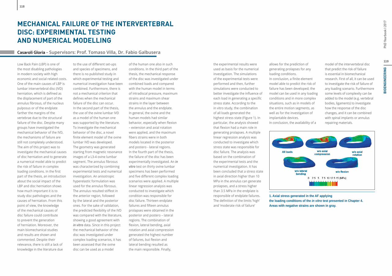
the experimental results were used as basis for the numerical investigation. The simulations of the experimental tests were performed and then, further simulations were conducted to better investigate the infl uence of each load in generating a specifi c stress state. According to the in vitro study, the combination of all loads generated the highest stress state (Figure 1). In particular, the analysis showed that fl exion had a main role in generating prolapses. A multiple linear regression analysis was conducted to investigate which stress state was responsible for disc failure. The analysis was based on the combination of the experimental tests and the numerical investigation. It has been concluded that a stress state in axial direction higher than 10 MPa in the annulus can generate prolapses, and a stress higher than 3.5 MPa in the endplate is responsible of endplate failures. The defi nition of the limits ’high’ and ’moderate risk of failure’
allows for the prediction of generating prolapses for any loading conditions. In conclusion, a fi nite element model able to predict the risk of failure has been developed; the model can be used in any loading conditions and in more complex situations, such as in models of the entire motion segments, as well as for the investigation of implantable devices.In conclusion, the availability of a
1. Axial stress generated in the AF applying the loading conditions of the in vitro test presented in Chapter 4. Areas with negative strains are shown in gray.
model of the intervertebral disc that predict the risk of failure is essential in biomechanical research. First of all, it can be used to investigate the risk of failure of any loading scenario. Furthermore some levels of complexity can be added to the model (e.g. vertebral bodies, ligaments) to investigate how the response of the disc changes, and it can be combined with spinal implants or annulus repairing materials.

121
PhD
Year
book
I 20
17B
IOE
NG
INE
ER
ING
120
Chía Bejarano Noelia - Supervisors: Prof. Alessandra Pedrocchi
Prof. Simona Ferrante
Stroke is the main cause of adult disability worldwide. Its most common impairment is hemiparesis, producing weakness or loss of motor function on the contralesional side. These defi cits undermine the ability of stroke patients to walk, leaving their locomotion characterized by abnormal motor activation patterns and reduced muscle force generation. Novel rehabilitation techniques for stroke patients have exploited the brain remaining ability to compensate for impairments and to adapt to new behaviour. Neuromuscular Electrical Stimulation has provided very positive results at enhancing the plasticity of the Central Nervous System (CNS) if supporting repetitive, close-to-normal, goal-oriented and active movements that require new skill acquisition. Neuroprostheses, i.e. devices that deliver Functional Electrical stimulation, have greatly advanced and now incorporate the activation of several muscles into complex locomotion patterns. Nevertheless, the progress in control strategies that synchronize the stimulation to the patient’s gait has been somewhat limited, using the information of only one or two gait sub-phases to trigger the delivery of the
stimulation. Regarding the stimulation patterns, rectangular or trapezoidal patterns have started to be substituted by biomimetic strategies, in which the stimulation is based on physiological EMG activations. However, their translation into walking recovery has not happened yet.The aim of this Doctoral Thesis was the development of a biomimetic lower-limb neuroprosthesis for stroke patients, designed to facilitate motor relearning during overground gait training. The neuroprosthesis had to tackle the patient-specifi c impairment, inducing a biomimetic muscle stimulation based on the physiological activations synchronized to the current gait cycle of the patient, and require the active participation of the patient. Additionally, the neurorehabilitation treatment had to be easy to apply, in order to allow its use by non-technical operators and its eventual exportability to clinical environments. The general control scheme of the developed neuroprosthesis is shown in the fi gure. Its design has been based on two main pillars. The fi rst element is a real-time, gait segmentation algorithm
that fully synchronized the stimulation strategy to the gait of the patient. This algorithm, used the information from two inertial sensors placed on the shanks to detect three gait events per leg: initial contact, end contact, and mid-swing. The algorithm was validated with 22 healthy subjects and 10 stroke patients with heterogeneous levels of impairment. The knowledge in real time of the duration of six gait sub-phases of the patient was used to fully remap the stimulation strategy, by stretching or expanding homogeneously the stimulation profi le delivered during each gait sub-phase. The second concept used in the neuroprosthesis design is the use of the muscle-synergies paradigm to identify the patients’ impaired biomechanical functions and create a personalized stimulation strategy that targets their specifi c needs. Muscle synergies are a theory of motor control that hypothesizes that the CNS recruits the muscles in modules to reduce the complexity of the highly-dimensional musculo-skeletal system. Muscle synergies provide non-invasive information on the structure and variability of the patient’s motor coordination. This knowledge was used to identify the patient’s impaired
muscle synergies and focus the rehabilitation training on those that lacked a correct recruitment or composition. The neuroprosthesis was integrated into a software that provided a user-friendly interface and additional functionalities. The Italian Ministry of Health approved the use for research purposes of the system, called FESGait, and a pilot RCT study that assessed the therapeutic eff ect of a neuroprosthesis-based training on twenty stroke patients. The data from four chronic patients was analysed in this PhD Thesis, who underwent a four-week treatment based on treadmill training supported by the neuroprosthesis. The patients were divided into control and experimental groups, who were only diff erentiated by the added support of the neuroprosthesis for the experimental group. The training was comprised of 12 sessions of 30 minutes, and it was kept intensive and challenging. Full assessments were performed
before and after the treatment, in terms of clinical scales, kinematics, and muscle coordination during rectilinear and curvilinear locomotion, analysed through muscle synergies.The results showed that the patients from the experimental group reached a higher increment in walking speed during treatment, obtaining mean diff erences of 0.36 m/s, which is higher than the minimal detectable change, 0.3 m/s. On the other hand, the control patients incremented 0.16 m/s on average. By the end of the intervention, one of the patients from the experimental group had increased his MiniBest test score by fi ve points, which is higher than the minimal clinically important change. He also perceived this positive eff ect and improved in terms of muscle coordination. The second patient included in the experimental group, who started with a less severe impairment, showed a mild recovery also in both muscle coordination and clinical scales. Regarding the two
control patients, one did not show signifi cant changes in any area, whereas the other increased her MiniBest test score by 9 points. In summary, the results showed that the patients who were more compromised were those who obtained the greatest treatment benefi t. Nevertheless, the heterogeneity of the pathology and the diverse treatment courses that were created to personalize the treatment to the patients’ needs, implies that we will have to wait until the full pilot RCT with 20 patients has been completed before any trend might emerge from the data with any statistical power. These patients will also undergo a third assessment session, four weeks after the end of the treatment to stablish if any eventual changes are also maintained through time. The multichannel neuroprosthesis here introduced presented novel aspects with respect to other studies published in the literature. More specifi cally, it provided biomimetic multichannel stimulation during walking, completely synchronized to the duration of six gait sub-phases of the patient. Additionally, muscle synergies were used to assess the treatment eff ects on neural control and to defi ne the initial impairment of the diff erent biomechanical functions, which allowed the neuroprosthesis to deliver a stimulation strategy completely personalized to the patient’s needs. The pilot RCT on chronic stroke patients is currently ongoing, to test whether this neuroprosthesis-based treatment provides a better outcome than traditional treadmill training.
1. Neuroprosthesis general scheme.
A PERSONALIZED GAIT NEUROPROSTHESIS
FOR STROKE PATIENTS.
MUSCLE SYNERGIES FOR MOTOR RELEARNING

123
PhD
Year
book
I 20
17B
IOE
NG
INE
ER
ING
122
Dipasquale Ottavia - Advisors: Prof. Giuseppe Baselli
Prof. Mara Cercignani, Dr. Francesca Baglio
Background and aim: Resting state functional magnetic resonance imaging (rfMRI) has been widely used as non-invasive tool for understanding the complex functional mechanisms of the brain. However, it has no or poor translation into clinical practice due to the presence of open methodological issues, poor standardization, and lack of clinical validation. The aim of this thesis is to explore the major issues and compare novel and existing approaches on proper datasets. We focused on data de-noising comparing a wealth of data-driven approaches with a novel one (FIX) based on independent component analysis (ICA) and, in a further study, them all with one relying on scan data improved by multiple-echo sequence (ME-ICA). Further, we investigated various defi nitions of nodes interacting through the investigated functional connectivity (FC), either data-driven or based on prior model of anatomo-functional parcellation. While enlightening their pros and cons in clinical application, we proposed a novel parcellation of ICA components into anatomically localized clusters (CL-ICA).Methods: a) Data de-noising – FIX was compared to the most used single-echo data-driven de-noising
methods in terms of temporal signal to noise ratio (SNR), BOLD signal fl uctuation reductions with respect to the uncleaned data and ability to detect FC alterations in two clinical datasets: 21 Alzheimer’s disease patients versus 20 healthy control (HC) subjects and 11 Multiple Sclerosis patients versus 10 HC. Then, we compared ME-ICA to FIX and other single-echo techniques, using multi-echo rfMRI data of 30 low-motion young HC and 30 Attention Defi cit and Hyperactivity Disorder patients displaying a high level of movement artifacts and verifi ed the artifact removal eff ectiveness and the signal preservation by quantifying the ability to uncouple FC and motion, reduce distance-dependent connectivity biases, and preserve the default mode network (DMN) FC strength. b) FC analysis methods – We applied diff erent analysis methods in three studies. 1) High dimensional ICA (70 components) was compared to the typical low dimensional approach (25 components), which identifi es the standard resting state networks (RSNs) by the signal components; validation was performed on 21 AD patients and 20 HCs. A labelling criterion based on spatial and temporal
DATA DE-NOISING, BRAIN PARCELLATION AND NETWORK ANALYSIS METHODS FOR RESTING STATE FUNCTIONAL MRI: COMPARISON OF NOVEL AND EXISTING APPROACHES IN CLINICAL DATASETS
comparisons between the high dimensional components as sub-network of a specifi c RSN was also proposed for an automatic and robust classifi cation of the sub-networks. 2) We also proposed CL-ICA, a combination of the low dimensional ICA and a local clustering algorithm, which splits each RSN into anatomically distinct areas, and compared it to a-priori localization (model-based approach) of anatomo-functional ROIs, using 51 HC and addressing the localized sub-elements of the DMN. 3) We used a model based approach (i.e., a priori anatomical parcellation of the cortex) and graph theory for the investigation of the acute eff ects of systemic infl ammation on the whole brain FC architecture and its relationship to interferon-alpha (IFN-α)-induced mood changes on 22 patients aff ected by Hepatitis C acquired before and after the specifi c treatment with IFN-α. Results: a) Data de-noising – In the fi rst study FIX was more eff ective in removing multiple sources of artifacts and allowing the detection of pathological FC alterations in AD. However, FIX partially reduced BOLD signal fl uctuation of MS patients in the grey matter and was not able to detect the typical FC alterations in MS. Conversely, ME-ICA showed
better performances compared to FIX and the other single-echo methods, it well reduced the coupling between FC and motion and preserved the FC in the network of interest. b) FC analysis methods – 1) Analyses of the spatial maps and time series obtained with the high dimensional ICA were performed. Spatial analyses better localized the functional damage both in the posterior and anterior parts of the DMN and highlighted a FC loss in the sensory-motor network. Temporal analysis showed a widespread within-network damage in both networks. Figure 1: Between-group diff erences in resting state networks (RSNs) spatial maps. Group level ICA spatial maps of the RSNs (red-yellow) at low and high dimensionality are overlaid with clusters showing signifi cantly lower (blue) functional connectivity in patients with Alzheimer’s disease (AD) relative to healthy controls (HC). PCC = posterior cingulate cortex; PCC1 = sub-network 1 of the PCC;
mPFC2 = sub-network 2 of the medial prefrontal cortex; SMN2 = sub-network 2 of the sensory motor network.2) By comparing CL-ICA to the traditional model-based method, we found that our novel approach showed stronger within-network FC in the DMN. 3) Graph analysis showed that peripheral IFN-α distorts whole brain functional network architecture, in parallel with the observed mood and cognitive changes: the global capacity for parallel information transfer was impaired and effi ciency of a sub-network reduced.Figure 2: Graphical representation of the discrete cortical-subcortical sub-network showing a signifi cant reduction in functional connectivity 4-hours after IFN-α. Thickness of edges (lines) is proportional to the magnitude of IFN-α induced reductions in functional connectivity.Conclusion: The present work has demonstrated and validated both the optimization of
1. 2.
known methods and also novel approaches in two directions: a) an eff ective cleaning of rfMRI data for reliable FC analyses; b) a more detailed parcellation of the brain and the whole brain analysis towards the investigation of the functional connectome. The presented overview of methods and the discussed results off ers an overall review of methodologies and results in the perspective of a translation of rfMRI and FC studies into the clinical practice.
125
PhD
Year
book
I 20
17B
IOE
NG
INE
ER
ING
124
Lunardini Francesca - Supervisors: Prof. Alessandra Pedrocchi
and Prof. Terence D. Sanger
FUNCTIONAL ASSESSMENT METHODS
AND EMG-BASED INTERVENTIONS
FOR CHILDREN WITH DYSTONIA
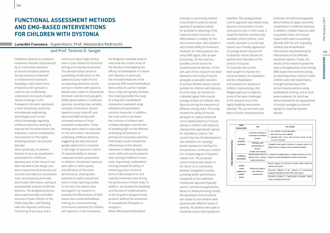
Childhood dystonia is a pediatric movement disorder characterized by an involuntary alteration of muscle activation patterns during voluntary movement or maintenance of posture. Nowadays, early-onset forms of dystonia still represent a common yet insuffi ciently understood and poorly studied clinical challenge. In this framework, this work represents a multi-disciplinary study that encompasses engineering technologies and current clinical knowledge regarding childhood dystonia, seeking to improve the characterization, the evaluation, and the rehabilitation interventions for this highly-disabling pediatric neuromotor disorder. More specifi cally, to address the lack of accurate quantitative assessment for childhood dystonia, part of the research has been devoted to the design and test of experimental protocols and accurate and objective assessment tools, encompassing kinematic and muscle information, aiming at quantitatively evaluate childhood dystonia. The designed protocols were experimentally controlled versions of tasks relevant to the child’s daily life: a self-feeding task that required continuous monitoring of accuracy, and a
continuous fi gure-eight writing task in close relation to functional writing and drawing movements. The devised indices aimed at quantifying modifi cations in the speed-accuracy trade-off and at evaluating abnormal muscle activity in children with dystonia. Results were useful to characterize kinematic and electromyographic (EMG) abnormalities in childhood dystonia, revealing slow, variable, and jerky movements, altered speed-accuracy trade-off , and abnormal EMG activity with increased amount of noisy unwanted components. These fi ndings were useful to speculate on the still unclear mechanisms underlying childhood dystonia, suggesting the idea that basal ganglia dysfunction is involved in the origin of dystonia in terms of impaired ability to remove unwanted motion components. In addition, the devised measures were able to refl ect quality and effi ciency of the motor performance, showing their potential as useful assessment tools in motor learning studies. To this aim, the indices were leveraged in my research to evaluate the eff ectiveness of EMG-based vibro-tactile biofeedback training as a novel promising noninvasive treatment for children with dystonia. In this framework,
we designed a detailed study to overcome the current limits of the literature investigating the effi cacy of biofeedback in children with dystonia. In particular, the innovative features of the proposed EMG-based biofeedback device allow its use for multiple hours a day during daily activities, thus making possible the study of a long-term biofeedback intervention evaluated using validated and quantitative outcome measures. In addition, the multi-center trial allows the inclusion of children with multiple etiologies, with the aim of shedding light on the diff erent underlying mechanisms of primary and secondary dystonia. Preliminary results showed the eff ectiveness of the devised measures in detecting improved motor skills and muscle patterns after training of diff erent motor tasks. Importantly, biofeedback training showed the ability of enhancing motor control in terms of decreased error and reduced movement time during the performance of both tasks. In addition, we showed the feasibility and the ease of implementation of the long-term designed study protocol, without the assistance of occupational therapists or caregivers.While EMG-based biofeedback
training is a promising method to be tested on patients whose severity of symptoms allows for possible (re-)learning of the impaired motor functions, its eff ectiveness is unlikely for the most severe cases, who present a very limited ability of movement. However, for these patients, the noisy EMG signal, after proper processing, can be used as a possible control source for assistive external devices. To this aim, part of this research has been devoted to the study of muscle synergies as possible solutions to achieve fl exible robotic control suitable for children with dystonia. As a fi rst step, we carried out a detailed upper limb muscle synergy analysis on children with dystonia during the execution of diff erent writing tasks. This study showed the ability of muscle synergies to capture fi xed and uncorrupted patterns of muscle activity in children with dystonia, making them appropriate signals for myoelectric control. The second step was the development and validation of a synergy-based myoelectric interface for simultaneous, continuous control of a multiple degree of freedom robotic arm. The proposed control scheme was shown to be robust to co-contraction between antagonist muscles, providing better performance compared to the traditional muscle-pair approach typically used in commercial applications. Based on these promising results, the developed control scheme was tested on fi ve children with dystonia with diff erent levels of severity. All patients were able to intuitively control the myoelectric
interface. The synergy-based control approach was tested using dynamic and isometric muscle contractions and, in both cases, it outperformed the commercially available control scheme. Such results represent a crucial step toward user-friendly application of synergy-based myocontrol of assistive robotic devices for patients with disorders of the control of muscles. To conclude, the current work sought to improve the characterization, the evaluation, and the rehabilitation interventions for dystonia in children, representing a full-fl edged approach to address some of the open challenges in the research line of this highly disabling neuromotor disorder. The current work was able to further characterize the
kinematic and electromyographic abnormalities of upper-extremity movements in childhood dystonia. In addition, reliable measures able to quantify motor and muscle impairments were successfully devised, with the aim of guiding medical and rehabilitation intervention and evaluating the eff ectiveness of the diff erent treatment options. Finally, the results of this research paved the way to the application of promising EMG-based interventions aiming at improving motor control in both children with mild impairments, through the learning of the correct muscle patterns using biofeedback training, and in most severe patients, for which we demonstrated the strong potential of muscle synergies as control signals for external assistive devices.

127
PhD
Year
book
I 20
17B
IOE
NG
INE
ER
ING
126
Marchi Andrea – Supervisors: Prof. Sergio Cerutti, Prof. Alberto Porta
IntroductionThere are multiple subconscious nervous control mechanisms that operate all the time to maintain the arterial pressure at or near normal levels. Barorefl ex (BR) is probably the most important one given its rapidity of intervetion. BR is a neurally mediated negative feedback mechanism that contributes importantly to blood pressure homeostasis. BR control is commonly evaluated by estimating the magnitude of the heart period variation to changes in systolic arterial pressure. However, the cardiac BR pathway is not the sole portion of the BR deserving an evaluation. Indeed, modifi cations of diastolic arterial pressure (DAP) leads to variations of sympathetic activity as estimated from integrated muscle sympathetic nerve activity (MSNA). Despite the relevance of evaluating simultaneously sympathetic and cardiac arms of the BR, few studies so far assessed contemporaneously the two diff erent aspects of the same control refl ex because of the diffi culties in defi ning a common framework to assure a homogenous characterization of both BR arms.
AimsThe aim of this thesis is to assess and compare two of the most
relevant components of the BR: cardiac BR and sympathetic BR arms. The contemporaneous characterization of both cardiac BR and sympathetic BR is hypothesized to be valuable to better understand BR physiology. The issue of setting a common framework of the contemporaneous assessment of cardiac BR and sympathetic BR is tackled to provide a uniform methodological description of their functioning. Particularly, the thesis pursues the following specifi c aims: i) to propose a new approach for obtaining a beat-to-beat MSNA variability from the integrated MSNA signal, referred to as calibrated MSNA variability; ii) to propose a sympathetic BR sequence method for characterization of the sympathetic BR and optimize the parameters of the method by using a graded orthostatic challenge and compare sympathetic BR indexes with those derived from cardiac BR sequence analysis; iii) to monitor cardiac BR and sympathetic BR indexes during several experimental conditions in humans; iv) to compute the correlation between indexes derived from cardiac BR and sympathetic BR arms to assess their degree of independence in controlling arterial pressure.
Experimental protocolsThree experimental protocols are exploited:1. graded head-up tilt protocol: twelve healthy subjects were enrolled (age from 20 to 36 years, median = 22.5 years; BMI: from 18.6 to 28.4 kg·m-2, median = 24.2 kg·m-2; 9 females). After instrumentation, subjects were allowed to rest for at least 30 min. Subjects were then sequentially tilted to 0°, 20°, 30°, 40°, and 60° for 10 min at each angle. The head-up tilt test started from the horizontal position and occurred incrementally with respect to the previous tilt table inclination, thus allowing us to maintain the positioning of the microelectrodes in the same subject and to preserve the invariable quality of the MSNA rec ordings over the entire experimental session;2. bilateral MSNA protocol: ten normotensive right-handed volunteers (5 females; age = 33 ± 9 years; BMI = 26.8 ± 2.7 kg·m-2) without evidence of organic disease were enrolled. MSNA was recorded from the peroneal nerve of the right and left leg simultaneously. Thirty minutes after instrumentation, supine data acquisition during spontaneous breathing was initiated and lasted for 15 min;3. head-down bed rest protocol: as part of the European Space
Agency study, eight healthy male volunteers (age = 33 ± 1 years, BMI = 23.5 ± 0.2 kg·m−2) were studied. Each experiment session consisted of 10 min of baseline recording at rest in supine position followed by 10 min of recording at 80 degrees head-up tilt. The pre-syncope condition was evoked by the application of stepwise lower body negative pressure for 3 min. The protocol was repeated before and after 21 days -6 degrees head-down bed rest confi nement.
Results and conclusionsFirstly, this thesis proposed a new way to obtain MSNA variability. It preserves the dimensionality of a neural discharge (i.e., bursts per second). The defi ned calibrated MSNA variability limits the eff ect of factors that directly aff ect the quality of the MSNA recordings by altering amplitude and area of the bursts. Due its physical dimensionality this study proposes the exploitation of the calibrated MSNA series instead of the more traditional uncalibrated MSNA series in studies that model cardiovascular variability interactions and integrate direct measurements of sympathetic activity in suitable descriptions of cardiovascular control.Secondly, by exploiting the defi nition of calibrated MSNA variability, a causal method for the characterization of the sympathetic BR from spontaneous beat-to-beat variability of MSNA burst rate and DAP based on the detection and extraction of MSNA and DAP sequences of sympathetic BR origin was implemented. Since
FROM BAROREFLEX TO BAROREFLEXESthis proposed method follows the same logic as the cardiac BR sequence technique, the contemporaneous exploitation of both techniques allows one to set an analysis framework assuring a homogenous characterization of both cardiac BR and sympathetic BR, thus favoring studies aiming at understanding their degree of independence. This framework might be particularly helpful in pathological patients, e.g. in heart failure patients. In addition, the proposed method allows one to overcome some limitations of more traditional estimates of sympathetic BR sensitivity such as the possible exploration of DAP ranges characterized by nonlinearities in the MSNA-DAP relation that might occur, for example. When a pharmacological approach is exploited and the inherent need of long recordings in case of non-pharmacological method.Thirdly, sympathetic BR sensitivity and cardiac BR sensitivity are calculated in three diff erent experimental protocols. In graded head-up tilt protocol, inducing a BR unloading in healthy volunteers, both cardiac BR sensitivity and sympathetic BR sensitivity are reduced in proportion to the magnitude of the challenge, while the percentage of the cardiac BR and sympathetic BR sequences increased. In bilateral MSNA protocol, where left and right MSNA were acquired simultaneously from the same subject, no signifi cant diff erences were detected between indexes derived from calibrated MSNA variability
and sympathetic BR sequence analysis, thus suggesting that the extracted parameters are robust and reliable and represent a fi ngerprint of the sympathetic control. In head-down bed rest protocol it was observed that the period just preceding syncope is characterized by an altered cardiac BR and sympathetic BR controls and the cardiovascular deconditioning imposed by 21 days of head-down bed rest reduced the eff ectiveness of sympathetic BR and its degree of involvement. Findings suggesting that sympathetic BR impairment may play a key role in the pathogenesis of neural-mediated syncope and that the inactivity in cardiovascular deconditioning may contribute to orthostatic intolerance. Finally, this thesis evaluated the complementary information carried out by the characterization of cardiac BR and sympathetic BR during experimental conditions challenging BR control and evoking its breakdown. The results can be taken as a supporting evidence of a certain degree of independence of sympathetic BR and cardiac BR controls and stress the relevance of the contemporaneous monitoring of cardiac BR and sympathetic BR to achieve a more complete and insightful characterization of the human BR regulation. The joint assessment of the BR control of heart rate and sympathetic activity might be particularly helpful in individuals with impaired BR function such as in chronic orthostatic intolerance patients.

129
PhD
Year
book
I 20
17B
IOE
NG
INE
ER
ING
128
Mazzuca Enrico – Advisor: Prof. Andrea Aliverti
NUMERICAL AND EXPERIMENTAL
MODELS OF PULMONARY
INTERSTITIAL EDEMA DEVELOPMENT
Pulmonary edema can be generated by several physiological and pathological concurrent mechanisms. Understanding the possible interactions among the variables involved in the fl uid accumulation in pulmonary interstitium allows not only to obtain a deeper insight within the lung adaptive response to perturbative stimuli and within the pathophysiology of lung edema, but also to derive useful indications about possible treatments and preventive interventions for subjects prone to the development of this critical condition. In the present thesis, these mechanisms have been investigated in control and edemagenic conditions in animal models.A complete evaluation of the functional adaptive response of capillary compartment to edemagenic condition involves the use of diff erent imaging technique to image the in-vivo adaptive response to perturbative stimuli, such as hypoxia administration or saline injection. The aim of this Thesis is to provide a theoretical framework to analyze the interaction between lung capillaries and interstitium during the onset of edema, in order to systematically review and integrate all the mechanisms involved in fl uid balance control.
For this purpose, a computational model of a morphologically-based alveolar capillary unit (ACU) in the rabbit was developed to relate lung fl uid balance to mechanical forces between capillary surface and interstitium during development of interstitial edema; the model was validated against available data in literature about capillary perfusion. The modelling results relative to capillary recruitment were verifi ed with data present in literature for the same species, interpolated and inserted within a model of extra-
vascular lung water. The model was applied to two edemagenic conditions, namely hypoxia administration and collagenase injection, a treatment generating a destruction of the capillary barrier, thus increasing remarkably fi ltration rate. For hypoxia exposure, fi tting data of interstitial liquid pressure required a linear increase in hydraulic conductivity and capillary pressure , that fulfi lls the need of increase in oxygen delivery. For severe fragmentation of capillary endothelial barrier (collagenase injection), fi tting
required a rapid increase in both hydraulic and protein permeability, causing ACU de-recruitment, followed by an increase in as a late response to restore blood fl ow. In conclusion, the model allowed to describe the lung adaptive response to edemagenic perturbations; the increase in , related to the low interstitial compliance, provides an effi cient control of extra-vascular water, by limiting micro-vascular fi ltration. In order to assess the possibility of electing in-vivo microscopy (IVM) as the standard technique for the study of lung edema at the alveolar level, subpleural alveolar mechanics was analyzed in a rabbit model of healthy lung during maneuvers of infl ation and defl ation and evaluated alveolar specifi c and absolute compliance, estimated from a statistical method applied to alveolar area distributions. Results showed that absolute alveolar compliance is proportional to the baseline alveolar area. Furthermore, no evidence of topological dependence of alveolar area was found and scaling up from single alveoli to randomly selected alveolar regions, a relatively homogenous mechanical behavior with minimal hysteresis of overall alveolar expansion was observed. This suggests that the considerable heterogeneity of alveolar size and of the corresponding alveolar mechanical behavior are homogenously distributed, resulting in a substantially homogenous mechanical behavior of lung units and whole organ. Furthermore, IVM proved to be a proper technique for studying pulmonary microstructures.
For this reason, IVM was applied to study the vasoactive response of subpleural micro-vascular compartment to hypoxic administration. Two main temporal phases were identifi ed. The fi rst is characterized by a strong arteriolar vasoconstriction, a reduction of the caliber of corner vessels feeding septal network, greater at end-inspiration compared to end-expiration. In the second phase, a substantial re-perfusion of distribution vessels and of corner capillaries was observed. A signifi cant alteration of alveolar mechanics characterized the whole experiment. In order to estimate the functional mechanical variables acting at the capillary micro-level for the control of alveolar perfusion, the ACU model was applied to the experimental images. A comparison between the vascular pressure estimated from the model and the changes of caliber of corner capillaries confi rmed the hypothesis of arteriolar vasoconstriction but suggested also an important role for venular vasoconstriction. Thus, the model allows to estimate hemodynamic parameters from morphological measurements and to address several questions about the physiological mechanisms underlying adaptive response to hypoxia.Finally, an MRI study on in-vivo mouse lung model was conducted to evaluate the regional distribution of fl uid accumulation due to two diff erent treatments providing subclinical interstitial edema, namely hypoxia administration and saline injection. Results show a correspondence between wet-to-dry ratio and
1 A. model of lung alveolar sac, surrounded by a capillary network fed by terminal arterioles (TA) and drained by collecting venules (CV). B: top view of an alveolar-capillary unit (ACU), made of 20 contiguous alveoli. C: enlargement of a sub-region of ACU. D: 3D enlargement of the geometrical model of capillaries and alveolar interstitial space.
the proton density estimated by MRI. Furthermore, a gradient of edema distribution in both the caudo-cranial and the gravity-dependent antero-posterior direction was found, with both the apical and the dependent regions showing a greater increase of proton density. This result was interpreted in terms of ACU model by comparing the behaviour of two ACUs diff ering by the capillary density. Compared with the high density ACU, representing the bottom lung, low density ACU, typical of the upper lung, displays a greater increase of capillary recruitment while capillary pressure raises, especially in condition of elevated interstitial pressure. A greater increase of surface area for fi ltration for low density ACU may be the cause for increased fi ltration and thus for the observed distribution of lung edema. Theoretical modelling, corroborated by further experimental data from MRI sequence about lung perfusion and interstitial thickness (obtained from 129Xe imaging) may be used for exploring the overall lung response to perturbations of fl uid balance, both in terms of regional and whole organ functional changes.

131
PhD
Year
book
I 20
17B
IOE
NG
INE
ER
ING
1. Comparison of MAVEN and mono/bi-parametric methods (m/bPS) with manual segmentation as reference in axial (upper row) and sagittal (lower row) brain images at 3 T. From left to right, fusion of SWI-mIP with: manual segmentation, mPS-MIP, bPS-MIP and MAVEN-MIP. Image projections cover 20 mm in the axial slabs and 10 mm in the sagittal slabs. MAVEN better matches manual segmentation and has a higher sensitivity, correctly classifying as veins the tubular dark structures that are barely detected by mPS and bPS (false negatives, indicated by green arrows). Moreover, MAVEN shows higher specifi city in recognizing as false positives the structures that are wrongly classifi ed as veins by mPS and bPS (yellow arrows). Structures and voxels corresponding to false negatives and false positives in m/bPS are indicated with blue arrows in MAVEN and manual segmentation.
130
Monti Serena – Supervisors: Prof. Maria Gabriella Signorini; Giuseppe Palma, PhD;
Marco Aiello, PhD; Tutor: Prof. Paolo Ravazzani
Cerebral vein analysis provides a chance to study, from an unusual viewpoint, an entire class of brain diseases, including neurodegenerative disorders and traumatic brain injuries. Venous tree can be profi ciently visualized by Susceptibility Weighted Imaging (SWI), which allows for detection of vascular abnormalities in diff erent cerebral pathological conditions. Manual segmentation approaches can be used to assess vascular anatomy, but they are a complex, time-consuming and observer-dependent task; therefore, automated approaches are desiderable, as they also improve reproducibility. The aim of this PhD thesis is to obtain a fully automated algorithm to segment the entire brain venous system from MR images. The study starts from the optimization of the MR acquisition protocol, in order to obtain optimal input images for the algorithm to be developed. Then, starting from statistical segmentation methods described in literature, the designed improvements are described up to the development of the fi nal algorithm for Multi-parametric Automated segmentation of brain VEiNs (MAVEN), which is based on a combined investigation of
ADVANCED METHODS FOR VISUALIZATION
AND SEGMENTATION OF BRAIN VEINS
IN MRI ACQUISITIONS
multi-parametric information (structural – SWI, morphological – Vesselness maps – and relaxometric –R2*map). MAVEN is an iterative segmentation algorithm that refi nes the vein mask at each step by adding newly detected vessel voxels that satisfy local thresholds computed excluding voxels previously marked as veins. The aim is to obtain, at each iteration, adaptive thresholds that allow to add voxels belonging to increasingly small and tortuous vessels which typically have higher SWI and lower Vesselness and R2* than large vessels due to partial volume eff ect. MAVEN incorporates a preliminary regularization step on the Vesselness functions: this solution modifi es the Vesselness values, making the algorithm less sensitive in the regions where severe non local fi eld inhomogeneities (SNLFI) induce susceptibility artifacts not related to tissue properties. Moreover, regarding the detection of large vessels that may elude the local moment criteria used in the iterations of the algorithm, this problem is bypassed by the MAVEN initial condition, thanks to a combination of morphological information extracted from SWI and QSM.
The method was tested on brain datasets, composed of gradient echo acquisitions, at 1.5 T, 3 T and 7 T. It was compared to previous methods (described in literature or developed during the PhD course) against manual segmentation as gold standard (see Fig.1), by means of quantitative scores (Dice Index, Cohen’s coeffi cient and Modifi ed Housdorff Distance) and the qualitative vascular tree depiction score. The vessel density and its dependence on B0 was estimated by computing the length of the segmented vascular trees and the inter-scan reproducibility was assessed by measuring the overlap between two coregistered segmentations obtained from two acquisitions of the same subject after head repositioning.MAVEN better matched the actual gold standard: the achieved accuracy and reproducibility were good, outperforming previous methods at both quantitative and qualitative analyses. The proposed method was usable at all the fi eld strengths explored, showing comparable accuracy scores, with no need of algorithm parameter adjustments. However, the increasing lengths of the segmented vascular trees with the B0 (see Fig.2), proved that, due to diff erent resolution and high
variability of the susceptibility contrast, the density of visible veins increased with B0, and this was refl ected in the segmentation results.In conclusion, the result of this thesis represents a step toward the development of a comprehensive method, including optimized acquisition and accurate quantifi cation algorithm, that could facilitate or
2. Measured length of the segmented vascular trees as function of the magnetic fi eld B0.
the Institute of Biostructure and Bioimaging of the CNR, the Department of Advanced Biomedical Sciences of University Federico II of Naples and the Department of Medical Physics in Radiology of the German Cancer Research Center of Heidelberg.
improve a wide range of clinically relevant quantitative analyses for detection of alterations in diff erent clinical conditions, ranging from traumatic brain injuries to neurodegenerative or neurovascular disorders.
AcknowledgmentThis PhD thesis was supported by IRCCS SDN of Naples and developed in collaboration with
133
PhD
Year
book
I 20
17B
IOE
NG
INE
ER
ING
132
Piazza Caterina – Adv isors: Prof. Anna Maria Bianchi, Ing. Gianluigi Reni
The electroencephalography (EEG) and in particular the event related potentials (ERPs) represent one of the most eff ective current ways to look at infant brain function. This is due to both ethical and practical concerns regarding infant assessment using other imaging technologies such as functional magnetic resonance, magnetoencephalography and positron emission tomography. Nonetheless, EEG/ERP studies with infant populations pose several challenges including data acquisition, data analysis and interpretation of the results. Auditory sensory processing is among the topics that have received more attention in infant ERP research, particularly because lower-level auditory skills (e.g. the ability to discriminate between auditory stimuli presented in rapid succession, called rapid auditory processing, RAP) have been shown to play a crucial role in language development and suggested to be a risk marker for the development of language and learning disorders. Nevertheless, the debate around physiological and functional meaning of diff erent infant ERP components is still open and more clarity about the underlying neural mechanisms is therefore called for. Advanced EEG/ERP signal processing can address this issue, even though
the applicability of these methods is often problematic with infant data. The present PhD dissertation is within this framework aiming at the in-depth electrophysiological study of early auditory sensory processing and at the investigation of its implication in later language development. In particular, the main goals were: (1) to test the effi cacy of the traditional ERP technique as a tool to investigate RAP, identifying marker of risk for language-learning impairment (LLI) in Italian infants; (2) to use and develop advanced analysis methods for a better understanding of the neural mechanisms that underlie RAP in infancy; (3) to correlate the diff erent electrophysiological parameters identifi ed with the physiological phenomena under investigation. RAP abilities have been studied for the fi rst time in Italian infants, testing fi ne-grained auditory processing of two acoustic features (sound frequency and duration), which are critical for language acquisition. RAP was assed at 6 months of age by means of an electrophysiological oddball task. Linguistic outcome measures were performed using the Language Development Survey (LDS) at 20 and 24 months of age.First, the traditional approach
ADVANCED EEG/ERP SIGNAL ANALYSIS
FOR THE INVESTIGATION OF EARLY SENSORY
PROCESSING AND COGNITIVE DEVELOPMENT
for ERP analysis was performed. The results showed that both acoustic features resulted to be already detectable at 6 months of age. Moreover, the hypothesis that early RAP skills are impaired in Italian infants at familial risk (FH+) for LLI was supported by the evidence that FH+ infants showed slower processing and a compromised discrimination of changes in both fi ne-grained information (sound frequency) and slowly-varying envelope (sound duration).Next, methods of analysis, that have never been applied to infant data, have been used to better investigate the neural electrophysiological substrates of infant auditory sensory processing. Specifi cally, a time-frequency approach, based on the adaptive autoregressive (AAR) modeling, was performed to investigate the neuronal oscillatory mechanisms undergoing RAP. The results showed that synchronization mechanisms in the delta/theta band encodes auditory stimuli processing in 6 month-old infants. An approach based on independent component analysis (ICA) was used to explore source-resolved ERPs, identifying their primary cortical source areas. The source localization was made possible thanks to the creation of a realistic template head model
for 6-month old infants. The results showed the involvement of auditory cortex and multiple extra-auditory cortical areas in RAP with the contribution of diff erent cortical sources to the processing of diff erent acoustic features. Moreover, a great contribution of the cingulate cortex was identifi ed, which refers to attentional and memory functions engagement in auditory information processing at 6 months. In order to simplify the source modeling of the EEG independent components (ICs) derived with ICA, a new algorithm for the automatic identifi cation of
bilaterally symmetric IC scalp maps was developed and tested, as well.Finally, the predictive value for later linguistic development of the diff erent electrophysiological correlates of RAP analyzed (i.e. scalp-ERP, time-frequency and source-resolved ERP measures) was examined, thus concretely evaluating the relevance and the potentiality of the parameters extracted. The results supported RAP involvement in language development, since all the considered electrophysiological measures were in some extent related to the linguistic outcome.
Moreover, prediction analysis suggested that source resolved parameters might be more sensitive for the identifi cation of neurophysiological biomarker.In conclusion the methods of analysis applied resulted to facilitate a deeper functional understanding of infant sensory processing. The potential of the techniques used and the feasibility of their application to infant data has been demonstrated. Thus, providing to researchers involved in studies with infant populations useful references for a deep analysis of their data.

135
PhD
Year
book
I 20
17B
IOE
NG
INE
ER
ING
134
Piazzese Concetta – Advisors: Prof. Enrico G. Caiani, Prof. Rolf Krause
(Università della Svizzera Italiana)
Cardiac magnetic resonance (CMR) imaging is considered the reference modality for quantifi cation of ventricular volume and function. Important clinical parameters, such as stroke volume, ejection fraction, left ventricular (LV) mass or wall thickness are derived by accurate delineation of LV endocardial and epicardial contours. Even if manual tracing on CMR images remains the gold standard, diff erent automated or semi-automated segmentation techniques have been proposed to improve reproducibility and preserve accuracy. To this respect, statistical shape models (SSM) have become a powerful tool to segment medical images. In this model-based technique a predefi ned geometric shape, trained on a set of samples to encode morphology and statistical variability of the structure of interest, is deformed on the base of extracted features in new unseen images. In literature, several SSM approaches have been proposed to segment the LV and other cardiac structures using CMR images. However, one critical point of such approaches is the limited number of subject (i.e., samples) collected and used as training set. Another potential limitation is related to the fact that the 3D shapes used to train the 3D model are usually derived by interpolation along the longitudinal dimension of
manually traced contours from 2D CMR short-axis (SAX) images with anisotropic off -plane resolution. Also, due to the fact that CMR is a gated 2D imaging technique, slice misalignment could aff ect the generation of the SSM with inaccuracies or morphological artefacts, such as stair-case aliasing. Moreover, accurate description of the ventricles in the basal and apical regions from CMR SAX images is problematic, due to slice thickness and in- and out-of-plane motion. For these reasons, detailed anatomical information of these levels is usually not included in the SSM.The goal of this PhD thesis was to develop and optimize an inter-modality SSM approach and adapt it to segment diff erent cardiac structures with minimal user interaction. The proposed approach was fi rst investigated and optimized to segment the LV endocardium in CMR images. The method was then adapted with minor adjustments for LV epicardial segmentation and the RV endocardium. Also, a preliminary application to non–cardiac structures, such as the left kidney, was tested to show the potentials and the versatility of the method and its feasibility in this particularly diffi cult task.The choice of using intrinsically and not interpolated 3D surfaces,
AN INTER-MODALITY STATISTICAL SHAPE MODELLING APPROACH FOR THE 3D SEGMENTATION OF CARDIAC STRUCTURES FROM MAGNETIC RESONANCE IMAGES
semi-automatically extracted from 3DE images, allowed to obtain a 3D SSM consistent with the ventricles morphology, in particular at the level of the LV apex and base. Also, it was possible to obtain a large number of surfaces (>7000 for the LV and >4000 for the RV) over one cardiac cycle thanks to the high temporal resolution of 3DE imaging and thus a better cardiac phase selection for inclusion in the training set. The number of cardiac frames included in the database and the type of registration employed for training shapes alignment were studied so to investigate their possible impact on the LV segmentation accuracy in CMR SAX and LAX images of 45 healthy and pathological patients. Before segmentation, each CMR dataset was pre-processed to compensate for potential spatial misalignments. Also, the LAX images were manually initialized to estimate the scaling factor and the initial pose of the model and the SAX planes in the CMR image stack to be used for the LV detection. During the segmentation process, the scaled SSM was iteratively deformed according to features (i.e., sparse boundary contour points) extracted from the CMR images, until a stable condition was reached. More specifi cally, at each
iteration, the intersections between the SSM and each CMR plane were computed along with the lines connecting each intersection point to the geometric centre of all intersections in a circular region of interest. By converting all line profi les to polar coordinates, a radial image containing grey level intensity information was obtained and subsequently segmented with a k-means clustering algorithm. The detected blood-endocardium edge was transformed back to Cartesian coordinates and further processed to include papillary muscle. The SSM was then repositioned and deformed recursively to match simultaneously the position of all LV endocardial candidate points in all SAX and LAX planes until the convergence was reached.The statistical quality of the diff erent SSMs, generated by varying the temporal information included in the database and the type of registration used during the training samples alignment, was evaluated in terms of compactness, generalization ability and specifi city. Also, the possible integration of independent component analysis and principal component analysis was investigated so to model the samples of the training database with a global-to-local approach.Hypothesizing that the morphology of the LV epicardium refl ects the morphology of the LV endocardium, an epicardium SSM was trained using a database of 3D LV scaled-up endocardial surfaces and applied to segment the LV myocardium in multi-view CMR images. Mainly this choice was driven by the limitation of not being able to obtain a database of 3D epicardial surfaces by segmenting
3DE images because the epicardium is often not as visible as the endocardium and it extends outside the echo imaging pyramid once the LV is dilated. Since the segmentation of the RV is not an easy task due to diff erent issues (highly variable shape from apical to basal level, thin and often indistinguishable myocardial walls and presence of trabeculations), a 3D CMR nearly automated RV endocardial segmentation procedure based on the inter-modality SSM approach described before was also proposed and validated. A new SSM was created from a database composed by 3D RV endocardial surfaces and iteratively deformed to segment the RV cavity in CMR SAX images of 30 patients. Due to the semilunar shape of the RV, at each iteration the space surrounding the computed model-image intersections was explored with two diff erent strategies: by considering the lines connecting each intersection with the geometric centre of all intersections or by considering the lines normal to the model-image intersections contour. Also, due to the tripartite
structure of the RV, an additional pre-processing step was performed for basal planes to distinguish between multiple intersected contours. The fl exibility of the developed inter-modality SSM approach was tested by applying it to segment left kidneys in patients aff ected by autosomal dominant polycystic kidney disease. In this case, the SSM was trained on 3D surfaces derived from 2D contours manually traced on computed tomography (CT) images of 15 pathological patients, thus facing the point-to-point correspondence problem among shapes. The model was then used to segment the left kidney in both CT and MR images from a clinical population of 17 patients.In conclusion, in this work a fl exible and reliable SSM approach to segment diff erent cardiac structures was presented and then extended to non-cardiac (i.e., left kidney) organs. The algorithm is simple and fast and it allows to obtain a 3D mesh representation consistent with the morphology of the structure of interest with minimal user interaction.
Endocardial LV, epicardial LV and endocardial RV meshes (orange, yellow, green, respectively) obtained with the proposed inter-modality SSM approach.

137
PhD
Year
book
I 20
17B
IOE
NG
INE
ER
ING
136
Rigoldi Federica – Advisors: Prof. Simone Vesentini, Prof. Alfonso Gautieri
Introduction: The progressive accumulation of Advanced Glycation End-products (AGEs) in the human body leads to several deleterious consequences, which include tissue stiff ening and pathologies such as arteriosclerosis, nephropathy, retinopathy and Alzheimer’s disease. Nowadays, there are no eff ective therapies against AGEs build-up. A promising strategy is believed to consist in the development of pharmacological tools based on the use of specifi c deglycating enzymes (FAOXs) that are known to catalyze the de-glycosylation of fructosyl amino acids. However, in their wild type form these enzymes are completely inactive towards entire proteins owing to the hindered accessibility to their buried active site. In addition to their use as therapeutic tools for protein deglycation, enzymatic assays based on FAOXs can potentially serve as rapid and economical diagnostic tools to measure glycated hemoglobin (HbA1c), which is a validated marker for the insurgence and the development of diabetes mellitus. Finally, a possible further application of engineered FAOXs is related to food preservation as exogenous AGEs precursors (the so-called Amadori products) are readily formed in heat-processed food.
The negative eff ects of dietary AGEs on human health are related to their ability to induce cellular oxidative stress and tissue infl ammation. Moreover, AGEs formation is detrimental to the nutritional quality of dairy products. Therefore, the development of effi cient strategies for preventing the formation of glycation products in heat-processed food is warranted. In this context, FAOX enzymes represent a potential tool for limiting Amadori product formation in dairy and meat products.To date, an engineering approach to rendering naturally occurring FAOXs active toward entire proteins or allowing them to specifi cally recognize fructosyl-valine (the glycated N-terminal residue of HbA1c) has been hindered by the limited knowledge that is available on the molecular mechanisms that regulate substrate specifi city in this class of enzymes as well as by the almost complete lack of information on their tridimensional structures.
Aim: The goal of the work that I did for my thesis is to develop a rational in-silico design strategy for engineering novel FAOX mutants with a wider active site to allow for the deglycation of intact
COMPUTATIONAL DESIGN OF A NOVEL ENZYME
FOR THE PREVENTION OF ADVANCED GLYCATION
END-PRODUCTS PROTEIN CROSSLINKS
proteins [Figure 1 ]. To this end, a comparison between diff erent FAOX family members is necessary in order to shed light on the molecular bases of their substrate specifi city.
Phase I: Crystal structure and modeling of wild-type Amadoriase I. We obtained the high resolution complete crystal structures of the free and the substrate-bound form of Amadoriase I using high-energy synchrotron radiation. Based on these data and using molecular dynamics simulations, we then provided a detailed description of the tunnel conformation that leads to the active site of the enzyme as well as the free energy profi le experienced by the ligand in going from bulk water to the catalytic cavity. We demonstrated the presence of four gating helices/loops, followed by an “L-shaped” narrow cavity. In summary, we believe that the tridimensional architecture of Amadoriase I that we determined provides a reference structural framework for the design of novel enzymes for protein deglycation. We also provided further enzyme characterization by a number of in vitro experiments, including thermal denaturation, determination of monomeric state, binding assay and rapid
kinetic experiments in normal and anaerobic conditions.
Phase II: Computational characterization of diff erent FAOX enzymes We used molecular dynamics simulations and advanced modeling techniques to investigate the most relevant wild-type FAOX enzymes (Amadoriase I, Amadoriase II, PnFPOX, FPOX-E and N1-1-FAOD) in order to elucidate the molecular mechanisms that drive their specifi city towards polar and nonpolar substrates. Specifi cally, we compared these fi ve diff erent FAOX in terms of overall folding, ligand entry tunnel, ligand binding residues and ligand binding energies. This part of work provides the foundation for the the next step, the rational design of an engineered FAOX enzyme.
Phase III: Computational design and experimental production of an engineered FAOX enzyme with a potentially expanded substrate recognition spectrum.Using an iterative approach, we built a library of FAOX mutants based on Amadoriase I and similar wild type structures available in Protein Data Bank (PDB). In our engineering approach, we focused on the redesign of two regions that are not directly involved in the catalytic activity of the enzyme. The templates that we used as scaff old structures for
our computational work have a similar, albeit in selected regions slightly more open, global fold when compared to Amadoriase I. We then modelled the primary sequence of these specifi c regions using the Rosetta software for protein engineering. Finally, library screening and mutant optimization was conducted using extensive molecular dynamics simulations in order to identify a mutant structure that showed (a) a stable and proper fold and (b) high affi nity for f-lys.After selecting the most suitable candidate among our panel of designed enzyme mutants, we experimentally tested its expression and solubility using standard molecular biology and protein purifi cation techniques. In particular, we have already optimized the expression conditions in bacteria and we are currently focusing on improving the solubility of the protein
Conclusions: We provided the fi rst complete high resolution crystal structure of a FAOX enzyme in its free and ligand-bound states. Using these information, we accurately characterized this enzyme in term of active site architecture, catalytic interactions, tunnel conformation, binding pathway and energies. Furthermore, using homology modelling techniques we compared the features of Amadoriase I with the other four most relevant members of the FAOX family. Finally, we computationally designed an engineered FAOX enzyme that may represent a step forward towards an effi cient protein deglycation strategy. Furthermore, this work provides a general computational framework that can be exploited in the design of virtually any engineered proteins in a wide context of diff erent applications.
1. Amadoriase I structural modifi cations in order to enlarge its accessibility to the active pocket.In particular, red and blue regions, which are identifi ed as gating structures, are shorten without affecting catalytic and fl avin arrangement.

139
PhD
Year
book
I 20
17B
IOE
NG
INE
ER
ING
138
Tresoldi Claudia – Advisor: Prof. Sara Mantero
The absence of successful solutions in treatments of small-caliber vessel (inner diameter < 5 mm) diseases has led to the Tissue Engineering application in vascular fi eld in order to develop functional non-immunogenic tissue engineered blood vessels (TEBVs). For this, in vitro reproduction and maintenance of physiological biochemical microenvironment and biomechanical stimulations are one of the key factors. In particular, the biochemical environment requires the presence of some vascular growth factors (GF), such as vascular endothelial GF (VEGF) or platelet-derived GF (PDGF), as well as the ability to provide suitable nourishment and oxygen to the three-dimensional (3D) constructs. Concerning the biomechanical stimulations caused by heart beats, vascular smooth muscle cells (VSMCs) are physiologically subjected to circumferential deformations (εcirc) about 10%. Endothelial cells (ECs) and their progenitors (EPCs) are subjected to wall shear stress (WSS) due to the blood fl ow in two diff erent ranges: ECs are exposed to 5-20 dyne/cm2 and EPCs to 0.1-2.5 dyne/cm2 of WSS. This mechanical conditioning ensures the maintenance of the contractile phenotype by VSMCs
and the quiescent phenotype by ECs. Also the scaff old architecture and structure play a pivotal role in obtaining successful outcomes in the fi eld and in ensuring both the chemical-physics conditions, especially in presence of a layered structure similar to the natural blood vessels, and in presence of some proteins of extracellular matrix (ECM) and adhesive proteins to promote scaff old-cells interactions. In this context, the present research project focused on the development of new strategies and materials to improve the in vitro maturation of tubular scaff olds, permitting a better cell retention towards endothelial stability. We apply a coupled
VASCULAR TISSUE ENGINEERING: COUPLED APPROACH
BETWEEN A COMPUTATIONAL MODEL AND EXPERIMENTAL
STRATEGIES FOR IN VITRO PULSATILE PERFUSION CULTURES
ON THREE-DIMENSIONAL TUBULAR SCAFFOLDS
approach between the classical Tissue Engineering approach and a computational model for a priori estimation of physiological mechanical conditioning of the construct to be tissue-engineered. The adopted strategy allows to: (1) enhance mass exchange through the 3D scaff old to improve the biochemical microenvironment, (2) biomechanically stimulate the graft with a pulsatile perfusion predicted by a computational model, and (3) emulate the layered structure of blood vessels with a polymeric gel-coated scaff old (Figure 1).In this regard, the alternating exposure to culture medium and air, coupled with a convective fl ow induced by the longitudinal
rotation permits effi ciently to improve mass exchange through the thickness of the 3D porous tubular scaff old (Figure 1A). The resulted optimization of mass transport determines advantages for 3D cell cultures in terms of cellular metabolism and colonization of the construct, thanks to nutrients and oxygen reaching also deeper structures of the scaff old. The effi ciency of this phenomenon, demonstrated on a novel polyurethane foam, is ensured when pore size is at least 68 μm and in presence of a high ratio between pore and endothelial cell size.Concerning the mechanical stimulations, a scaff old-specifi c fl uid-structure interaction (FSI) model is developed as a useful tool for the evaluation of εcirc
and WSS acting on VSMCs and ECs, respectively. The FSI model described each scaff old as a viscoelastic material, which properties (Young’s modulus and relaxation modulus) are experimental measured through uniaxial stretch tests and relaxation tests. For the scaff old we tested (decellularized swine artery), pressure values (80/120 mmHg) and circumferential deformations (about 10%) are comparable with physiological values and with experimental ones. In relation to these results,
WSS values fall into the range of WSS for EPCs diff erentiation towards an EC phenotype. According to these mechanical outcomes and starting from the mechanical properties of the scaff old, the model allows to a priori determine optimal physiological mechanical stimulation acting on the tubular scaff old seeded with ECs and VSMCs, avoiding over- and under-loading of the scaff old-cells complex. This permits to suitably set and tune fl ow rates, especially in the case of viscoelastic scaff olds (Figure 1B). To investigate cellular behavior in response to the FSI-defi ned mechanical stimulation pattern, pulsatile perfusion cultures are performed on decellularized human humbilical veins seeded with ECs. Our fi ndings well support the ability of the FSI model to estimate working pressures and εcirc of the scaff old; on the contrary, cellular behavior under FSI-model-estimated WSS is still on going, requiring a deeper investigation.To perfuse tubular porous scaff olds, a gel coating made of 8% w/v alginate and 6% w/v gelatin, functionalized with fi bronectin, is suitable to prevent transmural leakage through scaff old pores thanks to the crosslinking properties of alginate, occluding the pores. The
cell-adhesive ability of gelatin, further improved by the presence of fi bronectin functionalization, allowed to ECs to adhere and integrate within the gel, with advantages for cell proliferation. The superfi cial coating that covers just luminal pores without penetrating into the whole thickness of porous scaff olds permits also to create a double-layered 3D structure, reproducing the organization of healthy blood vessels (Figure 1C). The obtained scaff old is promising to keep anatomically separated the luminal surface from the media tunica but maintained functionally linked ECs with VSMCs. Thus, in the eff ort towards the development of not-immunogenic and functional TEBVs, this research project laid the basis to improve the in vitro development of small-caliber constructs in relation to: (1) the a priori optimization and control of biochemical environment and biomechanical conditioning, and (2) the production of tubular double-layered scaff olds with high potential to promote EC adhesion on the luminal side and VSMC colonization on the external surface, maintaining cells within two separated structures.
1. Working phases toward the development of functional and not immunogenic TEBVs. (A) Optimization of mass transport; (B) Implementation of the scaffold-specifi c FSI model, and (C) Leakproofi ng of porous tubular scaffolds. Rounded squares highlight the main outcomes of each phase.

141
PhD
Year
book
I 20
17B
IOE
NG
INE
ER
ING
140
Trujillo Diaz Paula – Advisor: Prof. Luca Mainardi
Background: Parkinson’s disease (PD) is the most common neurodegenerative movement disorder resulting from the death of dopaminergic neurons in the substantia nigra (SN). In PD, symptoms appear when a signifi cant number of neurons in the SN are already destroyed. Severe loss of the pigmented neurons of the SN is considered a hallmark of PD, and therefore, detecting changes in the SN is key to understanding the earliest manifestation of PD. Neuromelanin (NM), a dark, complex pigment particularly concentrated in the SN, accumulates normally with age, but it is relatively diminished in patients with PD, suggesting a role of NM in the neurodegenerative processes of PD. A method allowing in-vivo quantitative measurement of NM may off er a biomarker for the evolution of the disease. Recently, an MRI technique dubbed “neuromelanin-sensitive MRI” (NM-MRI) has been found to provide notable contrast between the SN and surrounding brain tissues with potential applications as biomarker of PD. Nonetheless, the mechanism by which the presence of NM might give rise to signal hyperintensities on MRI, and the nature of the relationship between a loss of hyperintensity with advancing
PD and NM status of the tissues are still unclear. Neuromelanin can function as a paramagnetic agent when combined with metals such as iron or copper, and prior in-vitro experiments have revealed concentration-dependent T1-shortening eff ects of the melanin pigment. However, recent studies suggest that the contrast observed with NM-MRI sequences may actually result from magnetization transfer (MT) eff ects. MT contrast in MRI is the result of the interaction between mobile water protons and the protons contained in macromolecules of tissues. The MT eff ect can be characterized via the MT ratio from an MT-prepared and non-prepared image pair, and more recently via quantitative MT (qMT), which typically requires images to be acquired at multiple off sets to generate the Z-spectrum. The resulting Z-spectrum can be fi tted to a model to estimate quantitative indices. While qMT may off er indices of tissue physiology, translation of this methodology into the SN within a clinically feasible scan time is challenging. A new method to perform a qMT analysis using only a single MT acquisition and a reference measurement has been recently proposed. Using this model, improved resolution or
DEVELOPMENT OF NOVEL METHODS
OF SATURATION TRANSFER MRI
FOR CLINICAL APPLICATION
reduced sensitivity to motion can potentially be realized in clinically relevant scan times, making it a viable approach to study SN changes in patients with PD. Aims: The aims of the thesis were: 1) to investigate the mechanisms associated with NM-contrast; 2) to characterize the impact of MT on NM-MR; 3) to determine the feasibility of performing qMT in the SN and to translate the single-point approach to perform high resolution, rapid qMT imaging of the SN; and 4) to apply the novel methods of saturation transfer MRI to study some of the brain structures associated with PD.Methods: First, we investigated the roles played by NM and iron in determining relaxations times and the exchange of saturation between the pools by examining the Z-spectra of phantoms containing synthetic melanins and iron at diff erent concentrations (similar to those observed in human SN tissue). The samples were prepared by the Institute of Biomedical Technologies, of the National Research Council of Italy. The phantom was subjected to qMT measurements and NM-MRI scan, as well as T1, T2, B0 and B1 mapping. Simulations of the Bloch equations of the two pool system were performed to assess the eff ect of the system parameters on the Z-spectra.
Then, we moved towards the in-vivo application of the techniques, by performing two pilot study on healthy subjects, to characterize the impact of MT on NM-MRI in-vivo, to determine the feasibility of performing qMT imaging of the SN, and to translate the ‘single-point’ approach to performing high resolution, rapid qMT imaging of the SN a step closer to clinical application in patients with PD. The fi nal stage of this project was to apply the novel methods of saturation transfer MRI to study some of the nervous system structures of the human brain associated with PD, particularly the SN. We performed two studies with patients. In the fi rst study, we examined healthy controls and patients with PD, with the purpose of detecting NM and iron changes in the SN. The MRI results of the patients were compared with the striatal dopaminergic innervation measured as density of dopamine transporter (DAT) by means of SPECT with DaTSCAN. The second study consisted of a qMT and NM-MRI study of the SN in healthy controls and patients with PD, with the purpose of evaluating the clinical utility of performing qMT imaging of the SN. This study was performed in collaboration between the UOC of Neuroradiology of Fondazione IRCCS Ca’ Granda – Ospedale Maggiore Policlinico and the Vanderbilt University Institute of Imaging Science (VUIIS). Results: The results of the phantom study showed that presence of the complex melanin-iron shortens relaxation times and decreases the MT eff ect,
suggesting that a combination of T1-weighted imaging and MT-preparation may provide increased NM-contrast compared with standard methods. Our pilot studies with healthy controls also showed that in NM-MRI the contrast between the SN and surrounding tissues is enhanced by adding an MT prepulse. We showed that qMT can be consistently performed in healthy volunteers to estimate quantitative indices, such as the macromolecular-to-free pool size ratio (PSR), and that the single-point approach can be used to obtain high-resolution quantitative PSR maps of the SN in clinically relevant scan times, making it a viable approach to study SN changes in patients with PD. The fi rst study with patients showed that NM-MRI based measures of the SN (volume and contrat-to-noise-ratio (CNR)) correlated signifi cantly with dopaminergic striatal innervation loss as measured by SPECT with DaTSCAN. Our results support the ability of NM-MRI to diff erentiate PD patients from healthy subjects as indicated in previous reports, and in agreement with prior NM-MRI studies, PD patients showed signifi cant reductions in SN contrast and volume. The second study on patients also showed reductions in SN contrast in patients with PD compared with healthy controls. The PSR map obtained with the qMT methods showed a clear distinction white matter and grey matter, and high contrast between SN and surrounding brain tissues.Conclusions: In NM-MRI, the
contrast between the SN and adjacent tissues can be enhanced by adding MT preparation, and it can be used to diff erentiate patients with PD from healthy subjects. The correlation between NM-MRI and DaTSCAN SPECT is a novel result supporting the idea that SN neuron loss and dopaminergic striatal innervation loss in PD are associated processes. Our in-vitro results suggest that the presence of NM does not aff ect the PSR, but it creates an unusual relationship between the T1 and T2 values of the tissue. We demonstrated the feasibility of performing high resolution qMT imaging in human SN. The ability to generate PSR maps from only a single qMT measurement may increase the clinical applicability of SN qMT imaging because of the opportunity to obtain either high-resolution quantitative mapping, or rapid estimation of the PSR at lower resolution. Finally, the non-invasive MRI markers obtained with NM-MRI and qMT may be useful to quantify NM content and detect alterations of the SN, thus off ering unique opportunities to link pathophysiologic and clinical progression in PD.

143
PhD
Year
book
I 20
17B
IOE
NG
INE
ER
ING
142
Ugolini Giovanni Stefano – Advisors: Prof. Monica Soncini, Ing. Marco Rasponi
The latest advancements in microfabrication techniques have led to the development of advanced cell culture platforms aimed at not only reducing the amounts of cells and reagents used but also at an in vitro mimicking of in vivo cellular-scale physico-chemical stimuli and conditions. The present PhD thesis was aimed at designing, developing and employing multi-layer, micron-scale devices for advanced cell culture applications. By employing standard techniques for polydimethylsiloxane (PDMS) microstructuring, microdevices were developed and employed aimed at i) applying fi nely controlled cyclic strain to human cardiac cells and investigating mechanisms of cardiac fi brosis disease; ii) characterizing an oxygen-control strategy to enable multi-stimulus experiments on cardiac fi broblasts subject to both mechanical strain and oxygen dynamics; iii) modeling the blood-brain barrier, with electric resistance monitoring of
an endothelial layer and capability of transport studies. wThe present PhD thesis achieved both technical and biological novelties. With regards to technological advancements, the microdevices here described present improvements in terms of ease of use, throughput and combination of multiple stimuli. By employing these newly developed microdevices, biological insights were provided describing novel
MULTI-LAYER MICRODEVICES
FOR ADVANCED CELL CULTURE APPLICATIONS
cellular responses under the application of one or multiple physiologically relevant stimuli. The main achievements of the present PhD work include both technical solutions to improve ease of use and applications of multi-layer microdevices and biological insights into specifi c cellular mechanisms, such as cardiac fi brosis, regulating major organ functions in physiology and pathology.
1. Left: picture of a fabricated multi-chamber microdevice for application of cyclic stretch to cell monolayers. Color dyes highlight the different microfl uidic circuits: blue represent culture chambers, red represents actuation circuit and green represents conditioning channel. Center: 3D sketch of a chamber of the microdevice. Same color code as left panel. Right: Multi-layer microdevice for transport studies and electrical monitoring of blood-btain barrier endothelial cells monolayers.