Embed Size (px)
Citation preview
Rozemeijer et al. Crit Care (2021) 25:310 https://doi.org/10.1186/s13054-021-03670-x
REVIEW
Measuring vitamin C in critically ill patients: clinical importance and practical difficulties— Is it time for a surrogate marker?Sander Rozemeijer1*, Frans A. L. van der Horst2 and Angélique M. E. de Man1
Abstract
This article is one of ten reviews selected from the Annual Update in Intensive Care and Emergency Medicine 2021. Other selected articles can be found online at https:// www. biome dcent ral. com/ colle ctions/ annua lupda te2021. Further information about the Annual Update in Intensive Care and Emergency Medicine is available from https:// link. sprin ger. com/ books eries/ 8901.
© Rozemeijer et al. 2021.
IntroductionInterest in intravenous vitamin C administration has rap-idly increased in the field of critical care medicine over recent years. The first studies investigating the effect of intravenous vitamin C in septic (shock) patients showed a decrease in organ dysfunction, vasopressor dependency, and even a reduction in mortality [1–3]. Within a short period of time, multiple trials in septic patients were con-ducted to confirm these promising findings, but results were not uniform [4–12]. The inconsistencies in effects on outcome may partially be explained by differences in study design [8], in particular the dosing regimens (tim-ing, duration and dose) and choice of co-medication. For example, vitamin C administration has been investigated alone, or in combination with thiamine and/or hydro-cortisone, sometimes with uncontrolled use of hydro-cortisone in the control group. There is also considerable variety among septic patients as sepsis is a heterogeneous syndrome. Therefore, some subgroups of patients might benefit more than others from intravenous vitamin C
therapy. A recently published meta-analysis on mortal-ity performed subgroup analyses and found a beneficial effect of vitamin C on short-term mortality (< 30 days). Additionally, survival was improved by a treatment dura-tion of 3–4 days [13]. The results of vitamin C alone ver-sus combination therapy were not different. A particular subgroup of interest is patients with vitamin C deficiency. None of the studies performed subgroup analyses on vitamin C deficient patients. This is unfortunate, but understandable, since the measurement of plasma vita-min C concentration is difficult.
In this chapter, we discuss the practical problems and pitfalls of measuring vitamin C and describe a novel potential surrogate marker that can estimate vitamin C status.
Rationale of vitamin C administrationVitamin C has pleiotropic functions in the human body, including anti-oxidative, anti-inflammatory and immune-supporting effects. It serves as a cofactor in the biosyn-thesis of norepinephrine and vasopressin, increases catecholamine sensitivity, protects the microcirculation, and improves wound healing [14]. Therefore, low plasma concentrations may have untoward effects in the ICU population.
Decreased plasma vitamin C concentrations are common in critically ill patients with sepsis, trauma,
Open Access
*Correspondence: [email protected] Department of Intensive Care Medicine, Research VUmc Intensive Care (REVIVE), Amsterdam Cardiovascular Science (ACS), Amsterdam Infection and Immunity Institute (AI&II), Amsterdam Medical Data Science (AMDS), Amsterdam UMC, Location VUmc, Vrije Universiteit Amsterdam, Amsterdam, The NetherlandsFull list of author information is available at the end of the article
Page 2 of 7Rozemeijer et al. Crit Care (2021) 25:310
hemorrhage, post-cardiac arrest and burns [15–20]. In septic shock patients, hypovitaminosis C (< 23 μmol/l) and vitamin C deficiency (< 11 μmol/l) rates are as high as 88% and 38%, respectively [15]. Causes of deficiency are decreased intake and absorption, and, most impor-tantly, increased metabolic consumption and reduced recycling due to overwhelming oxidative stress [14]. This increase in oxidative stress plays a key role in the pathophysiology of systemic inflammation and ischemia/reperfusion injury [17–20]. Vitamin C protects against oxidative injury to lipids, proteins and DNA by donat-ing its electrons. When the amount of oxidative stress is overwhelming, vitamin C is readily consumed, recycling becomes insufficient and deficiency develops. As a result, the body’s protection against oxidative injury becomes inadequate.
This effect creates a strong rationale for vitamin C sup-plementation in vitamin C deficient patients with over-whelming oxidative stress [21]. Available literature has already addressed the importance of selecting vitamin C deficient patients by measuring baseline plasma vita-min C concentrations to create a clear difference in tissue and plasma vitamin C concentrations between control and treatment groups after supplementation [22]. In addition, measuring achieved plasma vitamin C concen-trations could help to estimate optimal plasma concen-trations. The direct radical scavenging effect of vitamin C increases with higher, supraphysiological concentrations [23]. Therefore, measuring vitamin C will provide more insight into the dose-concentration-clinical outcome relationship [22].
Plasma vitamin C measurementThe determination of plasma vitamin C, or ascorbic acid, necessitiates considerable logistical and analytical effort.
Drawing bloodTo assess the in vivo vitamin C status, it is crucial to avoid ex vivo artefacts, i.e., the oxidation of vitamin C (ascor-bic acid) to dehydroascorbic acid (DHA) and subsequent
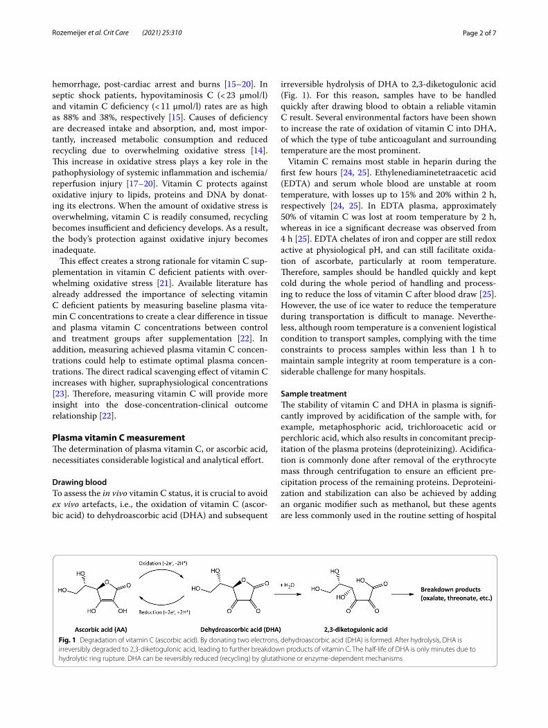
irreversible hydrolysis of DHA to 2,3-diketogulonic acid (Fig. 1). For this reason, samples have to be handled quickly after drawing blood to obtain a reliable vitamin C result. Several environmental factors have been shown to increase the rate of oxidation of vitamin C into DHA, of which the type of tube anticoagulant and surrounding temperature are the most prominent.
Vitamin C remains most stable in heparin during the first few hours [24, 25]. Ethylenediaminetetraacetic acid (EDTA) and serum whole blood are unstable at room temperature, with losses up to 15% and 20% within 2 h, respectively [24, 25]. In EDTA plasma, approximately 50% of vitamin C was lost at room temperature by 2 h, whereas in ice a significant decrease was observed from 4 h [25]. EDTA chelates of iron and copper are still redox active at physiological pH, and can still facilitate oxida-tion of ascorbate, particularly at room temperature. Therefore, samples should be handled quickly and kept cold during the whole period of handling and process-ing to reduce the loss of vitamin C after blood draw [25]. However, the use of ice water to reduce the temperature during transportation is difficult to manage. Neverthe-less, although room temperature is a convenient logistical condition to transport samples, complying with the time constraints to process samples within less than 1 h to maintain sample integrity at room temperature is a con-siderable challenge for many hospitals.
Sample treatmentThe stability of vitamin C and DHA in plasma is signifi-cantly improved by acidification of the sample with, for example, metaphosphoric acid, trichloroacetic acid or perchloric acid, which also results in concomitant precip-itation of the plasma proteins (deproteinizing). Acidifica-tion is commonly done after removal of the erythrocyte mass through centrifugation to ensure an efficient pre-cipitation process of the remaining proteins. Deproteini-zation and stabilization can also be achieved by adding an organic modifier such as methanol, but these agents are less commonly used in the routine setting of hospital
Fig. 1 Degradation of vitamin C (ascorbic acid). By donating two electrons, dehydroascorbic acid (DHA) is formed. After hydrolysis, DHA is irreversibly degraded to 2,3-diketogulonic acid, leading to further breakdown products of vitamin C. The half-life of DHA is only minutes due to hydrolytic ring rupture. DHA can be reversibly reduced (recycling) by glutathione or enzyme-dependent mechanisms

Page 3 of 7Rozemeijer et al. Crit Care (2021) 25:310
laboratories. The metal chelator EDTA or diethylene-tri-aminepentaacetic acid (DTPA) can be added at this point to further prevent ex vivo oxidation of vitamin C [25–27]. After acidification of the sample, this should be stored at low temperature, preferably at − 80 °C. Stability has been demonstrated for both vitamin C and DHA at − 80 °C for at least 5 years [28].
There is ongoing controversy in the literature about the generation of DHA in clinical samples [25]. Physiologi-cally, the amount of DHA in plasma in vivo is < 2% of that of total vitamin C (ascorbic acid + DHA) [22]. Higher amounts of DHA can be expected in critically ill patients because of overwhelming oxidative stress with reduced recycling of DHA [14, 29], and in patients receiving vita-min C at pharmacological doses [22]. However, one study showed that the amount of DHA was low to negligible in clinical samples. Only samples that contained hemolysis had appreciable amounts of DHA, which was explained by the ex vivo release of iron from hemoglobin during the acidification step in the sample pretreatment [25]. An in vitro study showed that an increase in DHA due to fer-ric ions only occurred in plasma acidified with trichloro-acetic acid or perchloric acid, not with metaphosphoric acid. Nevertheless, plasma hemoglobin catalyzed the oxi-dation of vitamin C in all acidic solutions [30]. Therefore, hemolysis should be avoided. Metaphosphoric acid is the best stabilizing agent of choice, as ferric ions, which are more easily released from transferrin in acidic solutions, did not accelerate the oxidation of vitamin C into DHA when using metaphosphoric acid [30]. One study showed a higher proportion of total vitamin C as DHA in met-aphosphoric acid-acidified clinical samples compared to controls [31], implying that in vivo generation of DHA may also occur.
In a pharmacokinetic study we performed in criti-cally ill patients, 10 patients received 2 g vitamin C and
10 patients received 10 g vitamin C intravenously, as a twice daily bolus or continuously over 48 h [32]. In the entire population, the median total vitamin C concen-tration at baseline was 22.7 [interquartile range (IQR) 14.7–39.5] μmol/l, and the median plasma DHA was 2.5 [0.9–5.1] μmol/l which was 10% (95% confidence interval (CI) 6.1–14.0%) of the total vitamin C (unpublished data). Patients receiving a bolus dosing regimen achieved peak plasma concentrations 1 and 2 h after infusion compared to the continuous dosing regimen in which peak plasma concentrations were achieved 24 and 48 h after infusion. In Table 1, total vitamin C (ascorbic acid + DHA), DHA, and DHA as a percentage of total vitamin C are shown at these peak moments. The absolute amount of DHA increased in patients receiving intravenous vitamin C therapy, but the percentage DHA of the total vitamin C remained comparable to baseline. Thus, DHA amounts greater than 2% may be caused by increased oxidative stress with reduced recycling due to critical illness, by supraphysiological plasma concentrations, and by the ex vivo oxidation of vitamin C despite adequate sample han-dling and processing [32].
AnalysisThere are two distinct approaches to quantitative deter-mination of vitamin C in plasma samples: enzymatic and chromatographic.
Enzymatic vitamin C assaysThere are several commercially available kits based on enzymatic conversion of vitamin C resulting in a sig-nal that can be detected photospectrometrically. Nor-mally, the enzyme ascorbate oxidase is used in this type of assay. The common method for these assays is the enzyme-linked immunosorbent assay (ELISA), which is well suited for batchwise processing of samples, but less
Table 1 Total vitamin C and DHA in plasma samples of critically ill patients receiving vitamin C therapy. Data from [32]
Data are presented as median [IQR]
Bolus dosing regimen T = 1 T = 2
2 g (n = 5) 10 g (n = 5) 2 g (n = 5) 10 g (n = 5)
Total vitamin C, μmol/l 174 [163.8–254.8] 1101 [1067.5–1287.6] 136.1 [97.2–203.3] 733.1 [703.2–879.6]
DHA, μmol/l 12.7 [7.5–25.6] 100.3 [61.9–113.2] 8.7 [3.3–24.8] 63.6 [31.2–69.5]
DHA, % of Tot (95% CI) 7.7 (2.8–12.7) 7.7 (4.1–11.2) 8.8 (0.8–16.8) 6.8 (3.6–10.0)
Continuous dosing regimen T = 24 T = 48
2 g (n = 5) 10 g (n = 5) 2 g (n = 5) 10 g (n = 5)
Total vitamin C, μmol/l 101.4 [15.8–134.9] 428.3 [352.8–612] 108.2 [42.0–174.1] 453.5 [318.9–1230.8]
DHA, μmol/l 11.7 [1.5–18.3] 31.9 [15.2–48.7] 8.4 [4.2–18.9] 18.6 [9.1–50.3]
DHA, % of Tot (95% CI) 13.7 (5.3–22.0) 6.5 (3.1–9.9) 10.2 (2.5–17.9) 4.2 (0.1–8.3)
Page 4 of 7Rozemeijer et al. Crit Care (2021) 25:310
convenient for immediate determination of values in a few samples.
There have been several attempts to adapt these enzyme-based assays to auto-mated analyzers, but based on the methods reported in a European External Qual-ity Assessment Scheme (Instand EQAS) enzyme-based assays are not routinely used within hospitals. If the clinical demand for immediate vitamin C determination increases, these enzyme-based assays could be used in point-of-care or centralized platforms because of their straightforward technical nature [33].
Chromatographic vitamin C assaysQuantitative ascorbic acid and DHA measurements are currently performed by high-performance liquid chromatography (HPLC) methods. HPLC methods are superior if multiple compounds with similar properties have to be analyzed or if there are many substances that might interfere with the quantification of a compound of interest.
After injection of the acidified sample into the HPLC instrument, the compounds are separated by passing through a column that differentially retains compounds based on their physical properties. As a result, at the end of the separation column, ascorbic acid and DHA can be detected selectively without interference of other compounds. Currently, there are two methods to detect ascorbic acid and DHA after passing through the col-umn [34]. First, electrochemical detection, which uses the redox-properties of ascorbic acid and DHA, and sec-ond, ultraviolet (UV) detection, which is based on the UV absorption of these compounds. Despite the fact that both detection methods give identical results, UV detec-tion is more widely used in the routine setting because of its relative technical simplicity [34]. Other detection techniques have been used, such as fluorescence detec-tion after pre-column chemical modification of ascorbic acid and DHA into a fluorescent compound, but are less common. Colorimetric/fluorometric methods may gen-erate higher DHA concentrations due to the lack of spec-ificity of the method [25].
Chromatographical assessment of ascorbic acid, DHA and total vitamin CTo assess the total vitamin C status, both ascorbic acid and DHA have to be determined. Although in principle these compounds can be quantified simultaneously in a single HPLC run, this approach is not often used because there is no traditional need for the separate quantifica-tion of DHA to assess vitamin C deficiency in patients. Another reason is that it is technically much easier to determine only ascorbic acid instead of both ascor-bic acid and DHA simultaneously. Therefore, medical
laboratories have optimized their HPLC assays to opti-mally detect ascorbic acid and not the combination of ascorbic acid and DHA. A convenient way to solve this is to convert DHA into ascorbic acid in the sample prior to HPLC analysis. Any generated DHA that has not yet been hydrolyzed to the irreversible 2,3-diketogulonic acid can be reduced to ascorbic acid by adding a reducing agent, such as tris(2-carboxyethyl) phosphine hydrochlo-ride (TCEP) or dithiothreitol (DTT). It is even possible to use pre-modified heparinized tubes with added DTT to immediately reduce any formed DHA [35]. In this manner, the total vitamin C concentration (ascorbic acid + DHA) in the sample can be measured. It has been shown that compared to its instability at room tempera-ture, DHA is stable for several hours at 4 °C, for at least a year when stored non-acidified at − 80 °C [25], and for at least 5 years when stored acidified at − 80 °C [28]. Thus, if samples are appropriately handled and processed, it should be possible to recover any oxidized ascorbic acid with agents such as TCEP and DTT.
With respect to clinical utilization of the total vitamin C determination, response times have to be considered. If sample pre-treatment is limited to centrifugation and deproteination prior to analysis, this will take approxi-mately 45 min, whereas the analytical procedure itself, including calculations and verification, will take an addi-tional 30 min. So, under optimized and dedicated condi-tions, it will take over 1 h to obtain quantitative results.
Because of the analytical complexity, many clinical chemistry laboratories outsource the analysis of vitamin C to reference laboratories, making vitamin C determi-nation unavailable for routine care. If they do supply this diagnostic service themselves, it is unlikely this it is avail-able for immediate determinations. Moreover, the sam-ple pre-treatment to safeguard correct determination of vitamin C is rather cumbersome and not easily accom-modated into routine hospital logistics, especially not in intensive care and emergency departments. A point-of-care vitamin C measurement could therefore be useful, but such a measure is not available yet. A potential sur-rogate marker is the static oxidation-reduction potential (sORP), which can be measured quickly at the bedside.
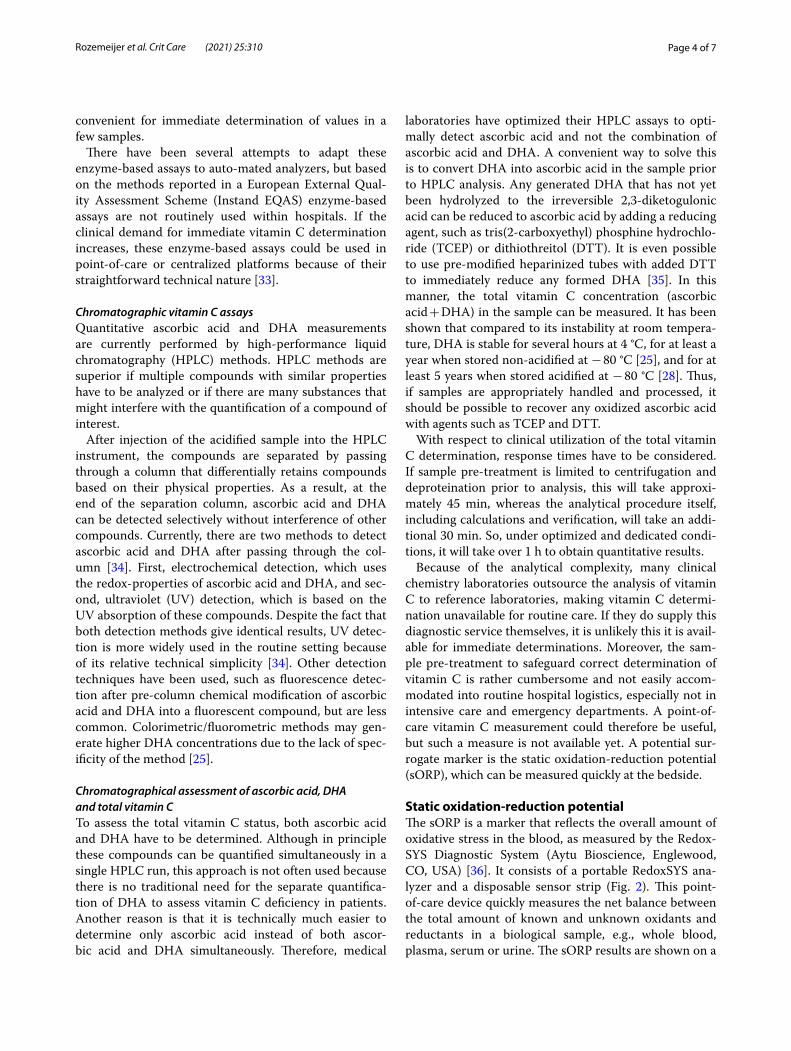
Static oxidation‑reduction potentialThe sORP is a marker that reflects the overall amount of oxidative stress in the blood, as measured by the Redox-SYS Diagnostic System (Aytu Bioscience, Englewood, CO, USA) [36]. It consists of a portable RedoxSYS ana-lyzer and a disposable sensor strip (Fig. 2). This point-of-care device quickly measures the net balance between the total amount of known and unknown oxidants and reductants in a biological sample, e.g., whole blood, plasma, serum or urine. The sORP results are shown on a
Page 5 of 7Rozemeijer et al. Crit Care (2021) 25:310
small display screen within 4 min after applying approxi-mately 30 μl of the sample to the sensor. The total time between obtaining blood and getting a sORP test result is less than 20 min. More detailed technical information about the test is available elsewhere [36, 37]. One of the advantages of measuring sORP is that it does not rely on a single biomarker of oxidative stress, such as lipid peroxidation. It provides a complete picture of the total amount of oxidative stress in the sample.
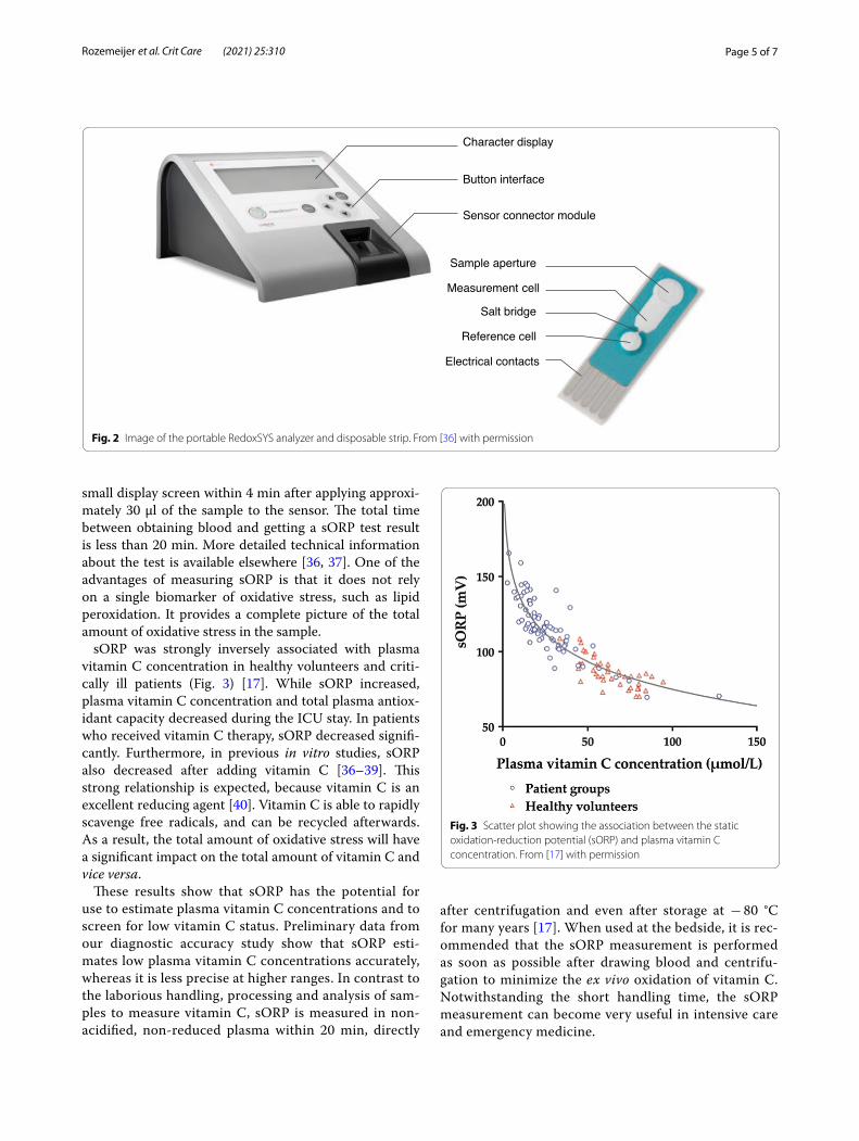
sORP was strongly inversely associated with plasma vitamin C concentration in healthy volunteers and criti-cally ill patients (Fig. 3) [17]. While sORP increased, plasma vitamin C concentration and total plasma antiox-idant capacity decreased during the ICU stay. In patients who received vitamin C therapy, sORP decreased signifi-cantly. Furthermore, in previous in vitro studies, sORP also decreased after adding vitamin C [36–39]. This strong relationship is expected, because vitamin C is an excellent reducing agent [40]. Vitamin C is able to rapidly scavenge free radicals, and can be recycled afterwards. As a result, the total amount of oxidative stress will have a significant impact on the total amount of vitamin C and vice versa.
These results show that sORP has the potential for use to estimate plasma vitamin C concentrations and to screen for low vitamin C status. Preliminary data from our diagnostic accuracy study show that sORP esti-mates low plasma vitamin C concentrations accurately, whereas it is less precise at higher ranges. In contrast to the laborious handling, processing and analysis of sam-ples to measure vitamin C, sORP is measured in non-acidified, non-reduced plasma within 20 min, directly
after centrifugation and even after storage at − 80 °C for many years [17]. When used at the bedside, it is rec-ommended that the sORP measurement is performed as soon as possible after drawing blood and centrifu-gation to minimize the ex vivo oxidation of vitamin C. Notwithstanding the short handling time, the sORP measurement can become very useful in intensive care and emergency medicine.
Character display
Button interface
Sensor connector module
Sample aperture
Measurement cell
Salt bridge
Reference cell
Electrical contacts
Fig. 2 Image of the portable RedoxSYS analyzer and disposable strip. From [36] with permission
Fig. 3 Scatter plot showing the association between the static oxidation-reduction potential (sORP) and plasma vitamin C concentration. From [17] with permission
Page 6 of 7Rozemeijer et al. Crit Care (2021) 25:310
ConclusionThis chapter underlines the potential clinical relevance of measuring plasma vitamin C concentrations in criti-cally ill patients and the practical difficulties that go along with the currently available measurement. Multiple clini-cal studies have investigated the effects of intravenous vitamin C in critically ill patients, but results are not uni-form. A possible explanation is the heterogeneity in study designs and included patients. Patients with vitamin C deficiency might benefit more from vitamin C therapy compared to non-deficient patients. Rapid plasma vita-min C measurement could identify this subgroup, but the ready oxidation of vitamin C ex vivo leads to several practical difficulties. Vitamin C measurement is there-fore cumbersome, time consuming and not available for routine care. Proper blood handling, processing and analysis to estimate plasma vitamin C concentrations are crucial to obtain reliable results. It is recommended to use heparin-anticoagulated tubes, to process the samples within less than 1 h at low temperature, and to stabilize the sample through acidification and deproteinization with metaphosphoric acid. Oxidized vitamin C (DHA) can be recovered using a reducing agent such as DTT. The sORP can estimate vitamin C status at the bedside within 20 min using fresh unprocessed plasma samples. As this measurement is much more practical, especially for emergency medicine, sORP can serve as a surrogate marker for vitamin C allowing evaluation of the effec-tiveness of vitamin C therapy in the subgroup of patients with low vitamin C status.
AcknowledgementsNot applicable.
Authors’ contributionsSR, FALvdH and AMEdM all contributed to the conception and drafting of the work. All authors have read and approved the final manuscript.
FundingPublication costs were funded by the department of Intensive Care Medicine, Amsterdam UMC, Location VUmc, De Boelelaan 1117, 1081 HV Amsterdam, The Netherlands.
Availability of data and materialsThe data analysed during the current study are available from the correspond-ing author on reasonable request.
Declarations
Ethics approval and consent to participateAll included participants in the pharmacokinetic study, or their legal representative, provided written informed consent prior to inclusion. The pharmacokinetic study was approved by the medical ethics committee of VU University Medical Center (registration NL50578.029.15, decision 2014.539).
Consent for publicationNot applicable.
Competing interestsThe authors declare that they have no competing interests.
Author details1 Department of Intensive Care Medicine, Research VUmc Intensive Care (REVIVE), Amsterdam Cardiovascular Science (ACS), Amsterdam Infection and Immunity Institute (AI&II), Amsterdam Medical Data Science (AMDS), Amsterdam UMC, Location VUmc, Vrije Universiteit Amsterdam, Amsterdam, The Netherlands. 2 Department of Clinical Chemistry, Reinier Medical Diagnos-tic Center, Delft, The Netherlands.
References 1. Fowler AA III, Syed AA, Knowlson S, et al. Phase I safety trial of intravenous
ascorbic acid in patients with severe sepsis. J Transl Med. 2014;12:32. 2. Zabet M, Mohammadi M, Ramezani M, Khalili H. Effect of high-dose
Ascorbic acid on vasopressor′s requirement in septic shock. J Res Pharm Pract. 2016;5:94–100.
3. Marik PE, Khangoora V, Rivera R, Hooper MH, Catravas J. Hydrocortisone, vitamin C, and thiamine for the treatment of severe sepsis and septic shock: a retrospective before-after study. Chest. 2017;151:1229–38.
4. Moskowitz A, Huang DT, Hou PC, et al. Effect of ascorbic acid, corticoster-oids, and thiamine on organ injury in septic shock: the ACTS randomized clinical trial. JAMA. 2020;324:642–50.
5. Fowler AA 3rd, Truwit JD, Hite RD, et al. Effect of vitamin C infusion on organ failure and biomarkers of inflammation and vascular injury in patients with sepsis and severe acute respi-ratory failure: The CITRIS-ALI randomized clinical trial. JAMA. 2019;322:1261–70.
6. Iglesias J, Vassallo AV, Patel VV, Sullivan JB, Cavanaugh J, Elbaga Y. Outcomes of metabolic resuscitation using ascorbic acid, thiamine, and glucocorticoids in the early treatment of sepsis: the ORANGES trial. Chest. 2020;158:164–73.
7. Chang P, Liao Y, Guan J, et al. Combined treatment with hydrocortisone, vitamin C, and thiamine for sepsis and septic shock: a randomized con-trolled trial. Chest. 2020;158:174–82.
8. Fujii T, Udy AA. Additional trials of vitamin C in septic shock: a bag of mixed fruit. Chest. 2020;158:13–4.
9. Mohamed ZU, Prasannan P, Moni M, et al. Vitamin C therapy for routine care in septic shock (ViCTOR) trial: effect of intravenous vitamin C, thia-mine, and hydrocortisone administration on inpatient mortality among patients with septic shock. Indian J Crit Care Med. 2020;24:653–61.
10. Wani SJ, Mufti SA, Jan RA, et al. Combination of vitamin C, thiamine and hydrocortisone added to standard treatment in the management of sepsis: results from an open label randomised controlled clinical trial and a review of the literature. Infect Dis. 2020;52:271–8.
11. Hwang SY, Ryoo SM, Park JE, et al. Combination therapy of vitamin C and thiamine for septic shock: a multi-centre, double-blinded randomized, controlled study. Intensive Care Med. 2020;46:2015–25.
12. Fujii T, Luethi N, Young PJ, et al. Effect of vitamin C, hydrocortisone, and thiamine vs hydro-cortisone alone on time alive and free of vasopressor support among patients with septic shock: the VITAMINS randomized clinical trial. JAMA. 2020;323:423–31.
13. Scholz SS, Borgstedt R, Ebeling N, Menzel LC, Jansen G, Rehberg S. Mor-tality in septic patients treated with vitamin C: a systematic meta-analysis. Crit Care. 2021;25:17.
14. Spoelstra-de Man AME, Elbers PWG, Oudemans-Van Straaten HM. Vitamin C: should we supplement? Curr Opin Crit Care. 2018;24:248–55.
15. Carr AC, Rosengrave PC, Bayer S, Chambers S, Mehrtens J, Shaw GM. Hypovitaminosis C and vitamin C deficiency in critically ill patients despite recommended enteral and parenteral intakes. Crit Care. 2017;21:300.
16. Gardner R, Liu X, Wang Y, et al. Vitamin C levels amongst initial survivors of out of hospital cardiac arrest. Resuscitation. 2020;156:190–3.
17. Rozemeijer S, Spoelstra-de Man AME, Coenen S, et al. Estimating vitamin C status in critically ill patients with a novel point-of-care oxidation-reduction potential measurement. Nutrients. 2019;11:1031.
18. Bar-Or D, Bar-Or R, Rael LT, Brody EN. Oxidative stress in severe acute ill-ness. Redox Biol. 2015;4:340–5.
19. Anand T, Skinner R. Vitamin C in burns, sepsis, and trauma. J Trauma Acute Care Surg. 2018;85:782–7.

Page 7 of 7Rozemeijer et al. Crit Care (2021) 25:310
20. Horton JW. Free radicals and lipid peroxidation mediated injury in burn trauma: the role of antioxidant therapy. Toxicology. 2003;189:75–88.
21. Oudemans-van Straaten HM, Spoelstra-de Man AM, de Waard MC. Vita-min C revisited. Crit Care. 2014;18:460.
22. Padayatty SJ, Levine M. Vitamin C: the known and the unknown and Goldilocks. Oral Dis. 2016;22:463–93.
23. Jackson TS, Xu A, Vita JA, Keaney JF Jr. Ascorbate prevents the interaction of superoxide and nitric oxide only at very high physiological concentra-tions. Circ Res. 1998;83:916–22.
24. Karlsen A, Blomhoff R, Gundersen TE. Stability of whole blood and plasma ascorbic acid. Eur J Clin Nutr. 2007;61:1233–6.
25. Pullar JM, Bayer S, Carr AC. Appropriate handling, processing and analysis of blood samples is essential to avoid oxidation of vitamin C to dehy-droascorbic acid. Antioxidants (Basel). 2018;7:29.
26. Washko PW, Welch RW, Dhariwal KR, Wang Y, Levine M. Ascorbic acid and dehydroascorbic acid analyses in biological samples. Anal Biochem. 1992;204:1–14.
27. Collie JTB, Greaves RF, Jones OAH, Eastwood G, Bellomo R. Vitamin C measurement in critical illness: challenges, methodologies and quality improvements. Clin Chem Lab Med. 2020;58:460–70.
28. Lykkesfeldt J. Ascorbate and dehydroascorbic acid as reliable biomarkers of oxidative stress: analytical reproducibility and long-term stability of plasma samples subjected to acidic deproteinization. Cancer Epidemiol Biomark Prev. 2007;16:2513–6.
29. Lykkesfeldt J, Tveden-Nyborg P. The pharmacokinetics of vitamin C. Nutri-ents. 2019;2412:11.
30. Koshiishi I, Mamura Y, Liu J, Imanari T. Evaluation of an acidic deproteiniza-tion for the measurement of ascorbate and dehydroascorbate in plasma samples. Clin Chem. 1998;44:863–8.
31. Schorah CJ, Downing C, Piripitsi A, et al. Total vitamin C, ascorbic acid, and dehydroascorbic acid concentrations in plasma of critically ill patients. Am J Clin Nutr. 1996;63:760–5.
32. de Grooth HJ, Manubulu-Choo WP, Zandvliet AS, et al. Vitamin C pharma-cokinetics in critically ill patients: a randomized trial of four IV regimens. Chest. 2018;153:1368–77.
33. Benzie IFF. An automated, specific, spectrophotometric method for meas-uring ascorbic acid in plasma (EFTSA). Clin Biochem. 1996;29:111–6.
34. Robitaille L, Hoffer LJ. A simple method for plasma total vitamin C analysis suitable for routine clinical laboratory use. Nutr J. 2016;15:40.
35. Bernasconi L, Saxer C, Neyer P, Huber AR, Steuer C. Suitable preanalytical conditions for vitamin C measurement in clinical routine. SDRP J Food Sci Technol. 2018;3:1–8.
36. Rael LT. RedoxSYSTM ORP scientific data synopsis. Greenwood Village, CO: Luoxis Diagnostics, Inc; 2014.
37. Rael LT, Bar-Or R, Kelly MT, Carrick MM, Bar-Or D. Assessment of oxidative stress in patients with an isolated traumatic brain injury using disposable electrochemical test strips. Electroanalysis. 2015;27:2567–73.
38. Polson D, Villalba N, Freeman K. Optimization of a diagnostic platform for oxidation-reduction potential (ORP) measurement in human plasma. Redox Rep. 2018;23:125–9.
39. Bobe G, Cobb TJ, Leonard SW, et al. Increased static and decreased capac-ity oxidation-reduction potentials in plasma are predictive of metabolic syndrome. Redox Biol. 2017;12:121–8.
40. Buettner GR, Jurkiewicz BA. Catalytic metals, ascorbate and free radicals: combinations to avoid. Radiat Res. 1996;145:532–41.
Publisher’s NoteSpringer Nature remains neutral with regard to jurisdictional claims in pub-lished maps and institutional affiliations.


![Renal Failure Critically Ill[1]](https://img.pdfslide.us/doc/110x75/577d26df1a28ab4e1ea26f52/renal-failure-critically-ill1.jpg)