Embed Size (px)
Citation preview

RESEARCH PAPER
Measuring the pressure applied to the skinsurrounding pressure ulcers while patients arenursed in the 30° positionMayumi Okuwa', Junko Sugama2
, Hiromi Sanada3, Chizuko Konya4
, Atsuko Kitagawa5
1 Assistant Profess01; School of Health Sciences, Faculty of Medicine, Kanazawa University, Japan2 Professor, Department of Gerontological Nursing, Division of Health Sciences and Nursing, GraduateSchool of Medicine, University of Tokyo, Japan3 Associate Professor, School of Health Sciences, Faculty of Medicine, Kanazawa University, Japan4 Assistant Professor, School of Health Sciences, Faculty of Medicine, Kanazawa Unive~sity, Japan5 Assistant Profess01; Department of Gerontological Nursing, Division of Health Sciences and Nursing,Graduate School of Medicine, University of Tolryo, Japan
The 30° laterally inclined and 30° head elevatedpositions (hereafter referred as the 'rule of 30'unless otherwise specified) are widely used as ameans of both primary and secondary prevention ofpressure ulcers as a result of reductions inlocalised pressures over bony prominences.However, the authors observed that some someparts of the wound margin were thickened. Thesethickened edges may be caused by use of the ruleof 30 positioning and may also be responsible for adelay in the healing process. This study includedfive bedbound elderly patients with pressure ulcerslocated at the sacrum and coccyx. The localpressure was measured at the thickened edgesand normal edges of the subjects' wounds by anewly developed sensor while the subjects werepositioned according to the rule of 30. The resultsshowed the maximum pressure as well as theaverage pressure of the thickened edges to besignificantly greater than that of the normal edges.Thus, it is suggested that higher pressure ondifferent areas of the wound margin may beresponsible for the thickened edges phenomenon,which may consequently delay the healing process.Clinical use of the rule of 30 for patients withpressure ulcers in the sacrum and coccyx regionsshould be reconsidered.
Key words: 30° laterally inclined position, 30° headelevated position, pressure ulcer, 'thickenededges', local pressure.
ressure ulcers are areas of localized tissuedestruction caused by the compression of soft tissueover a bony prominence J. Such compression, when
continuously applied for long periods of time, is a concernfor bedbound patients. In a clinical setting, since pressure
© Tissue Viability Society
JOURNAL OF TISSUE VIABILITY VOL 15 NO.1 JANUARY 2005
reduction is a key priority, it is important to use the mosteffective pressure-reduction techniques and select the mostappropriate support surfaces. Positioning patients in the 30°laterally inclined position2 and 30° head elevated position),known as the 'rule of 30', has been widely used as a meansof prevention to minimise the pressures exerted at bonyprominences. The rule of 30 is used to facilitate woundhealing by avoiding further compression of sacral pressureulcers and to prevent a pressure ulcer from developing inthe trochanter region.
However, after implementing this pressure reduction,some areas of the periwound or wound margin of a pressureulcer may become thickened and this may potentially leadto delayed healing. Such thickening and sclerosis of theperiwound has often been observed in patients with chronicpressure ulcers at the ischium who sit in a chair forextended periods of time4. Since conventional pressuresensors are often too big, may be sensitive to water anddifficult to fix to a wound, they are unsuitable formeasuring the pressure distribution at the wound margin.
Thus the authors developed a new sensor device tomeasure the local pressure on the periwound of a pressureulcer and evaluated its reliability, validity and safety5 Usingthis new device, the pressure on the normal edges and onthickened edges of the periwound was measured while thesubjects were positioned according to the rule of 30.
Subjects and methods
SubjectsEnrollment: The subjects of this study were patients whohad pressure ulcers and routinely used the rule of 30positioning. Pressure ulcers were visually inspected forthickened edges, which the examiner assessed as a pressuresore status tool (PSST)6,7 'edges' score of 4.Ethical consideration: In selecting the subjects, the opinionsof the nursing staff at the facility where the study wasconducted was taken into account. All candidates wereinformed about the aims of the study, and only thosepatients who gave informed consent participated in the
3

RESEARCH PAPER
Sensors
I/Fbox
y = 0.046x2 + 2.7076x + 21.32
of information: Before commencing,was collected about each subject's daily
400
350
~ 300E
250.s+-' 200~
B- 150~
a100
50
00
ProcedureCollectioninformation
Figure 2. Measurement system. I/F =Interface.
20 40 60 80 100
Applied pressure (mmHg)
MOUS~
Personal computer
The device's sensor simultaneously measures pressuresat three points as independent voltage levels. While thesensors are attached to the skin, any changes in pressure canbe observed on a real-time basis on the computer display.Reliability (hysteresis): For a measured range of0-350 mmHg and a temperature range of 0-40°C,hysteresis when loading and unloading was approximatelylinear (±10% variation)(Figure 3). A major cause ofhysteresis was a delayed response of the sensor film toloading.Concurrent validity: When tested in a laboratory, thecoefficient of correlation with an air-pad type interfacepressure-measuring device (Teikoku Hormone Mfg Co. Ltd, .Tokyo, Japan) was 0.985 for the sensor plate (hard surface)and 0.959 for the human body (soft surface) (Figw-e 4). Thedevice's clinical validity for measuring periwound pressurewas also confirmed5
Sensor
Pressure
B"'fU~ ~aq~Ag-C electrode:
spacer~
Resistance objec OFF ON b
Pad material: 3 Polyethylene naphthalate sensor pads
Sensor (diameter 5 mm) Lead (300 mm)
~ II
I I
IWidth (0.3 mm)
study. In cases where a patient was incapable of making thisdecision, informed assent was received from a familymember or guardian. When exposing the pressure ulcer (i.e.buttocks) of the subjects, adequate care was taken toprotect their privacy and maintain an acceptableenvironment. Informed consent was also given before anyphotographs were taken.
MethodsMethod of measurement: Local pressure was measured onthe normal and thickened edges of the periwound andcompared between subjects in the 30° laterally inclinedposition and those in the 30° head elevated position. Allsubjects were tested in both positions.The device: The new device's (developed by the authors incollaboration with DENSO Co Ltd, Kariya, Japan)reliability and validity was tested. The device is composedof a sensor unit (5 mm in diameter and 0.3 mm inthickness) and a lead (300 mm in length). The electrode ofthe sensor unit is covered with a waterproof base film toprevent exudate or other liquid from causing damage(Figure 1).
The sensor unit, which is disposable for hygienepurposes, is membranous and pressure-sensitive, perceives aload on the base film and detects any changes in resistancebetween silver electrodes on the basis of the area of contactbetween the upper electrode and the pressure-sensitiveresistant body. The sensor operates by measuring changes inresistance that occur as pressure is applied to a pressuresensitive film mounted within the sensor.Measuring system: The voltage levels are recorded by thesensor every 0.125 seconds, digitalized and transmitted to apersonal computer (using Windows Excel®, operating onWindows 95 or higher) (Figure 2).
Figure 1. Sensor structure and pressure-detecting principle. Figure 3. Hysteresis exhibited by pressure sensor.
4 JOURNAL OF TISSUE VIABILITY VOL 15 NO.1 JANUARY 2005

posItioning. The general status of each patient was alsochecked based on their nursing records, medical records andcharts.
II
"
II II
Setting for measurement: To prevent the device from beingdamaged by wound exudate, a medical film-forrning agent(Skin-PrepTM, Smith and Nephew Ltd, London, UK) wasapplied to the sensor's surface. During a test run of thesystem, sensor calibration was checked.
The sensors were directly attached to a normal edge anda thickened edge of a pressure ulcer. The sensors weresecurely attached (Figure 5), using adhesive tape (Yu-KiBan®, Nitto Medical Co., Osaka, Japan) and polyurethanefilm (Tegaderm™, 3M Health Care, St Paul, Minnesota,USA).Body position during measurement: The measurementswere performed in the supine positi,on, with the subjects ineitl1er the 30° laterally inclined position or tl1e 30° headelevated position.
The posture of each patient was changed by two nurses.The pillows, bedding and bedclothes were all the same.Duration of measurement: Measurement in each positionlasted for 30 minutes. Measurement was not performedwhile repositioning the body.
y =0.933x + 12.65
IIII
x.y
i•
50 100 150 200 250 300
TEIZO (gjcm 2 )
Comparison of new sensor and existing technology(TEllO) with sensors attached to solid object
Correlation coefficient r = 0.985
300
250
NE
200u'-~
0 150IIIcQ)III
~ 100Q)
z50
00
250
200NEu
~ 150
0IIIC
~ 100
~Q)
z50
00
y = 0.9388x + ·42.4831
50 100 150 200 250 300
TEIZO (gjcm 2)
As above but with sensors attached to soft tissueCorrelation coefficient r = 0.959
~ U', ,..... Iii}"" i·".t..·'Il"....-~·- ." '" .• ,,,., .I, - . "~ • __.. ll'"'.: ,. 't.I
lcm (, ,·C:.:'·.. .,..,'~'<'...,«./'/ '.' .' ./ ". .:
....... .: '/
t...... . .';:.0/.:.;
.' .'. A ,. . , ·~- . . ,,' ,",. ;,.~;., ... '
,~',B
Figure 4. Concurrent validity quantified through comparison of the newsensor and an existing pressure measuring technology.
Figure 5, Attachment of sensor pads. A =normal edge area; B =thickened edge area; c =adhesive tape.
..... .Mi':t'·:· !.'o.
~-.-_" " ~. ~ , 1 . . ."".... '." '. ,'" ... '. '. ',~,...:....j.; ~ . c-':'-. ' ." . - •
•• . '{'<', ',;... J~'''~!if'''':~••i('·; .,.{ . "l'.' ",:,.....•,.~.t ~ 1 . r, ., .... '., ,.," " '" ", '..
PSST
Wound sizeNo Grade Location perpendicular (cm2 ) Edges Undermining Total score
1 4 Sacrum 23.97 4 1 40
2 3 Coccyx 3.96 4 1 37
3 4 Coccyx 0.75 4 3 37
4 4 Sacrum 4.56 4 2 37
5 4 Coccyx 11.25 4 3 41
PSST = pressure sore status tool
JOURNAL OF TISSUE VIABILITY VOL 15 NO.1 JANUARY 2005 5

RESEARCH PAPER
Supine 30· lateral position 30· head elevated position
No Normal edge Normal edge Thickened edge Normal edge Thickened edge
1 4.6 6.5 8.7 27.2 64.4
2 38.5 0.0 61.7 40.4 81.7
3 13.1 10.3 148.0 56.7 139.0
4 5.9 10.1 66.6 28.7 49.3
5 7.0 10.3 87.6 5.6 66.0
Data analysisThe data for all 30-second intervals were extracted fromthe 30-minute data collected for each body position.
The actual recorded values of each individual subjectwere converted into relative values and compared with thepressure values of the normal edges measured in the supineposition. Conversion into relative values was performed toallow a comparison without being influenced by thesupport surface or specific individual characteristics of asubject. This was based on the previous finding that in thesupine position the local pressure did not differ
significantly between the normal edges and thickenededgesS,8 The maximum as well as the average pressurevalues of the thickened edges and normal edges werecompared using the Wilcoxon test. A value of p<0.05 wasused to indicate a statistical significance.
Results
SubjectsFive patients consented to the study. There were two malesand three females, with ages ranging from 72 to 84 years
(mean: 78.6±7.6 years). They had cerebrovascular disease,and were suffering from paralysis or contracture. TheirBraden scale9 scores ranged from 8 to 13. Their body massindex (BMI) ranged from 17.1 to 20.5 kg/m2 (normal BMIis between 18.5 and 24.9 kg/m2). All of these patients werebedbound, and being nursed on support surfaces. None ofthe patients showed a change in their general conditionfollowing the study.Pressure ulcers of subjects: All five of the subjects hadthickened edges on the periwound of their pressure ulcers(Table 1). In addition, three of th~ subjects hadundermining. One subject had a grade 3 pressure ulcerand the remaining four subjects had grade 4 pressureulcers (according to the National Pressure Ulcer AdvisoryPanel (NPUAP) classification10). The perpendi~ular
method, which was used to measure the pressure ulcersizell , gave ulcer sizes from 0.75 to 23.97 cm2. The PSSTscore ranged from 37 to 41. The sites of the pressureulcers were as follows: two sacral region and three coccyxregion pressure ulcers. The measurements wereperformed safely, without causing any additional pain orinjury.
Supine 30· lateral position 30· head elevated position
No Normal edge Normal edge Thickened edge Normal edge Thickened edge
1 4.6 4.4 ± 2.2 5.4 ± 3.9 18.2 ± 8.5 51.4 ± 13.8
2 38.5 0.0 ± 0.0 54.9 ± 9.5 24.6 ± 15.4 42.6 ± 31.4
3 13.1 6.4 ± 2.2 124.0 ± 15.1 32.3 ± 13.5 90.3 ± 46.8
4 5.9 1.0 ± 3.1 40.9 ± 15.7 16.4 ± 6.6 36.7 ± 9.8
5 7.0 6.7 ± 2.6 34.6 ± 23.6 2.8 ± 2.1 45.2 ± 12.7
6 JOURNAL OF TISSUE VIABILITY VOL 15 NO.1 JANUARY 2005

o I ! I !! I I! I
30° lateral position 30° head elevated position
30° lateral position 30° head elevated position
11=5 Wilcoxon signed rank test
Relationship between thickened edges and local pressureon the wound marginWhen the pressure was simultaneously measured on thethickened edges and the normal edges of the periwound ofthe subjects while assuming the rule of 30 positioning, thelocal pressure was higher on the thickened edges than onthe normal edges. These results indicate that the thickenededges may adversely affect the healing of pressure ulcerspotentially as a result of restricted epithelial cellmigration16.
The reason that higher local pressure occurred on thethickened edges in both the 30° laterally inclined positionand the 30° head elevated position may be attributable totwo factors. One factor may be atrophy of the buttockmuscle and skin loosening, which is often seen inbedbound elderly patients. It is possible that changingbody positions (e.g. 30° laterally inclined position, headelevated position) causes the loose skin to form folds, andthat the resultant protrusion at the edge of the periwoundis more likely to be compressed. It is also possible that incases of thickened pressure ulcers involving the formationof undermining (pocket), there is a space where thepocket lining has not adhered to the wound bed so whenthe patient assumes the rule of 30 positioning, the shapeof the periwound becomes distorted causing deformationat the opening of the pressure ulcer, leading tocompression of the skin in the pressure ulcer perimeter
Discussion
The Agency for Health Care Policy and Research(AHCPR) prepared several guidelines on clinical practicewith its guidance upon pressure ulcer prevention issued in1992. The guideline explicitly indicates the necessity ofavoiding direct compression on tl1e trochanter when thepatient assumes a lateral position3, and recommends the30° laterally inclined position to avoid compression at thetrochanter and the sacrum regions. The effectiveness of thisrecommendation has been shown in studies comparing the30° laterally inclined position with the 90° lateralposition12 and studies evaluating blood flow through thetrochanter and sacrum in the 30° laterally inclinedposition13,14. The guideline also recommends that when ahead elevated position is taken, the angle should be 30° orless to avoid compression at the sacrum and to preventfriction3.
More recently, the work of Defloor1S supported thisrecommendation. However, these studies were performedby healthy volunteers or pressure ulcer-free patients withspinal cord injury, and none of them involved patients withpressure ulcers. Thus, no previous study has attempted tochallenge the effectiveness of the rule of 30 policy byexamining this rule in patients with pressure ulcers. Thisstudy examined the probable effectiveness of the rule of 30on patients suffering from pressure ulcers in the sacrumand coccyx regions by measuring the pressure on thewound margin.
Thickened edge
Thickened edge
*1'=0.05
Normal edge
Normal edge
*1'=0.01
*1'=0.03
·~O" " 17 Teo
~'"
2
10
8<l)::J'iii
6><l)>.~
4Q)n::
2
0
10
8<l)::J'iii 6><l)
~ 4coQ)n::
Local periwound pressureMaximum pressure values: Table 2 shows the maximumpressure values for all five periwound measurements in thesupine, 30° laterally inclined position and the 30° headelevated position. The pressure values measured on thenormal edges were compared with the pressure valuesmeasured on the thickened edges (Figure 6). The maximumpressure values of the thickened edges were significantlygreater than those of the normal edges for both the 30°laterally inclined position and the 30° head elevatedposition.Average pressure values: Table 3 shows the averagepressure values for all five periwound measurements in thesupine, 30° laterally inclined position, and 30° headelevated position. The pressure values measured on thenormal edges were compared with the pressure valuesmeasured on the thickened edges (Figure 7). The averagepressure values of the thickened edges were significantlygreater than those of the normal edges for both the 30°laterally inclined position and the 30° head elevatedposition.
Figure 7. Average pressure values (normal edge vs thickened edge).These actual measurements were converted into relative values,compared with the pressure of the normal edges measured In thesupine position. Both positions show that the maximum pressure valuesof the thickened edges were significantly greater than those at thenormal edges.
Figure 6. Maximum pressure values (normal edge vs thickened edge).These actual measurements were converted into relative values,compared with the pressure of the normal edges measured in thesupine position. Both positions show that the maximum pressure valuesof the thickened edges were significantly greater than those at thenormal edges.
JOURNAL OF TISSUE VIABILITY VOL 15 NO.1 JANUARY 2005 7
RESEARCH PAPER
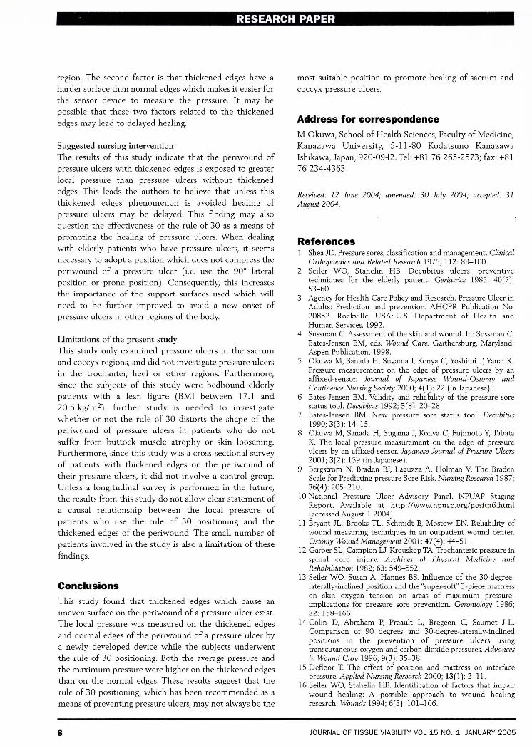
region. The second factor is that thickened edges have a
harder surface than normal edges which makes it easier for
the sensor device to measure the pressure. It may be
possible that these two factors related to the thickened
edges may lead to delayed healing.
Suggested nursing intervention
The results of this study indicate that the periwound of
pressure ulcers with thickened edges is exposed to greater
local pressure than pressure ulcers without thickened
edges. This leads the authors to believe that unless this
thickened edges phenomenon is avoided healing of
pressure ulcers may be delayed. This finding may also
question the effectiveness of the rule of 30 as a means of
promoting the healing of pressure ulcers. When dealing
with elderly patients who have pressure ulcers, it seems
necessary to adopt a position which does not compress the
periwound of a pressure ulcer (i.e. use the 90° lateral
position or prone position). Consequently, this increases
the importance of the support surfaces used which will
need to be further improved to avoid a new onset of
pressure ulcers in other regions of the body.
Limitations of the present study
This study only examined pressure ulcers in the sacrum
and coccyx regions, and did not investigate pressure ulcers
in the trochanter, heel or other regions. Furthermore,
since the subjects of this study were bedbound elderly
patients with a lean figure (BMI between 17.1 and
20.5 kg/m2), further study is needed to investigate
whether or not the rule of 30 distorts the shape of the
periwound of pressure ulcers in patients who do not
suffer from buttock muscle atrophy or skin loosening.
Furthermore, since this study was a cross-sectional survey
of patients with thickened edges on the periwound of
their pressure ulcers, it did not involve a control group.
Unless a longitudinal survey is performed in the future,
the results from this study do not allow clear statement of
a causal relationship between the local pressure of
patients who use the rule of 30 positioning and the
thickened edges of the periwound. The small number of
patients involved in the study is also a limitation of these
findings.
ConclusionsThis study found that thickened edges which cause an
uneven surface on the periwound of a pressure ulcer exist.
The local pressure was measured on the thickened edges
and normal edges of the periwound of a pressure ulcer by
a newly developed device while the subjects underwent
the rule of 30 positioning. Both the average pressure and
the maximum pressure were higher on the thickened edges
than on the normal edges. These results suggest that the
rule of 30 positioning, which has been recommended as a
means of preventing pressure ulcers, may not always be the
8
most suitable position to promote healing of sacrum and
coccyx pressure ulcers.
Address for correspondenceM Okuwa, School of Health Sciences, Faculty of Medicine,
Kanazawa University, 5-11-80 Kodatsuno Kanazawa
Ishikawa, Japan, 920-0942. Tel: +81 76265-2573; fax: +81
76234-4363
Received: 12 June 2004; amended: 30 July 2004; accepted: 31August 2004.
References1 Shea JD. Pressure sores, classification and management. Clinical
Orthopaedics and Related Research 1975; 112: 89-100.2 Seiler WO, Stahelin HB. Decubitus ulcers: preventive
techniques for the elderly patient. Geriatrics 1985; 40(7):53-60.
3 Agency for Health Care Policy and Research. Pressure Ulcer inAdults: Prediction and prevention. AHCPR Publication No.20852. Rockville, USA: U.S. Department of Health andHuman Services, 1992.
4 Sussman C. Assessment of the skin and wound. In: Sussman C,Bates-Jensen BM, eds. Wound Care. Gaithersburg, Maryland:Aspen Publication, 1998.
5 Okuwa M, Sanada H, Sugama J, Konya C, Yoshimi T, Yanai K.Pressure measurement on the edge of pressure ulcers by anaffixed-sensor. Journal of Japanese Wound-Ostomy andContinence Nursing Society 2000; 4(1): 22 (in Japanese).
6 Bates-Jensen BM. Validity and reliability of the pressure sorestatus tool. Decubitus 1992; 5(8): 20-28.
7 Bates-Jensen BM. New pressure sore status tool. Decubitus1990; 3(3): 14-15.
8 Okuwa M, Sanada H, Sugama J, Konya C, Fujimoto Y, TabataK. The local pressure measurement on the edge of pressureulcers by an affi.xed-sensor. Japanese Journal of Pressure Ulcers2001; 3(2): 159 (in Japanese).
9 Bergstrom N, Braden BJ, Laguzza A, Holman V. The BradenScale for Predicting pressure Sore Risk. Nursing Research 1987;36(4): 205-210.
10 National Pressure Ulcer Advisory Panel. NPUAP StagingReport. Available at http://www.npuap.org/positn6.html(accessed August 1 2004)
11 Bryant JL, Brooks TL, Schmidt B, Mostow EN. Reliability ofwound measuring techniques in an outpatient wound center.Ostomy Wound Management 2001; 47(4): 44-5l.
12 Garber SL, Campion U, Krouskop TA. Trochanteric pressure inspinal cord injury. Archives of Physical Medicine andRehabilitation 1982; 63: 549-552.
13 Seiler WO, Susan A, Hannes BS. Influence of the 30-degreelaterally-inclined position and the "super-soft" 3-piece mattresson skin oxygen tension on areas of maximum pressureimplications for pressure sore prevention. Gerontology 1986;32: 158-166.
14 Colin D, Abraham P, Preault L, Bregeon C, Saumet J-L.Comparison of 90 degrees and 30-degree-Iaterally-inclinedpositions in the prevention of pressure ulcers usingtranscutaneous oxygen and carbon dioxide pressures. Advancesin Wound Care 1996; 9(3): 35-38.
15 Defloor T. The effect of position and mattress on interfacepressure. Applied Nursing Research 2000; 13(1): 2-1l.
16 Seiler WO, Stahelin HE. Identification of factors that impairwound healing: A possible approach to wound healingresearch. Wounds 1994; 6(3): 101-106.
JOURNAL OF TISSUE VIABILITY VOL 15 NO.1 JANUARY 2005